Search results
757 results found for “Myosin Light Chain”
Name
Description
Product #
Price
Quantity
Shipping Method
- View Data Sheet
Name :
HOMER3 HumanDescription:
Homer Homolog-3 Human Recombinant
Homer homolog 3 (Drosophila)1 2 , HOMER-31 2 , homer protein homolog 32 , Homer neuronal immediate early gene 32, Homer-33, VESL32.
Product # :
PRO-1108Price :
Quantity :
Shipping Method :
Shipped with Ice Packs
More Info
- description
- source
- formulation
- purity
- More Info
- Citations
Description
HOMER3 Human Recombinant produced in E. coli is a single polypeptide chain containing 386 amino acids (1-361) and having a molecular mass of 42.5kDa.HOMER3 is fused to a 24 amino acid His-tag at N-terminus & purified by proprietary chromatographic techniques.
Source
E.coli.
Formulation
The HOMER3 solution (1mg/1ml) contains 20mM Tris-HCl buffer (pH 8.0), 150mM NaCl, 1mM DTT and 30% glycerol.
Purity
Greater than 90% as determined by SDS-PAGE.
Citations
More Info
-
Introduction
HOMER3 belongs to the HOMER family of postsynaptic density scaffolding proteins which have an analogous domain structure consisting of an N-terminal Enabled/vasodilator-stimulated phosphoprotein homology 1 domain that facilitates protein-protein relations, and two leucine zipper and a carboxy-terminal coiled-coil domain motifs which take part in self-oligomerization.
-
Synonyms
Homer homolog 3 (Drosophila)1 2 , HOMER-31 2 , homer protein homolog 32 , Homer neuronal immediate early gene 32, Homer-33, VESL32.
-
Physical Appearance
Sterile Filtered colorless solution.
-
Stability
Store at 4°C if entire vial will be used within 2-4 weeks. Store, frozen at -20°C for longer periods of time. For long term storage it is recommended to add a carrier protein (0.1% HSA or BSA).Avoid multiple freeze-thaw cycles.
-
Amino Acid Sequence
MGSSHHHHHH SSGLVPRGSH MGSHMMSTAR EQPIFSTRAH VFQIDPATKR NWIPAGKHAL TVSYFYDATR NVYRIISIGG AKAIINSTVT PNMTFTKTSQ KFGQWADSRA NTVYGLGFAS EQHLTQFAEK FQEVKEAARL AREKSQDGGE LTSPALGLAS HQVPPSPLVS ANGPGEEKLF RSQSADAPGP TERERLKKML SEGSVGEVQW EAEFFALQDS NNKLAGALRE ANAAAAQWRQ QLEAQRAEAE RLRQRVAELE AQAASEVTPT GEKEGLGQGQ SLEQLEALVQ TKDQEIQTLK SQTGGPREAL EAAEREETQQ KVQDLETRNA ELEHQLRAME RSLEEARAER ERARAEVGRA AQLLDVSLFE LSELREGLAR LAEAAP
ProSpec's products are furnished for LABORATORY RESEARCH USE ONLY. The product may not be used as drugs, agricultural or pesticidal products, food additives or household chemicals.
- View Data Sheet
Name :
SURF2 HumanDescription:
Surfeit-2 Human Recombinant
Surfeit 2, SURF-2, SURF2, Surfeit locus protein 2, Surfeit-2.
Product # :
PRO-1470Price :
Quantity :
Shipping Method :
Shipped with Ice Packs
More Info
- description
- source
- formulation
- purity
- More Info
- Citations
Description
SURF2 Human Recombinant produced in E.coli is a single, non-glycosylated polypeptide chain containing 279 amino acids (1-256) and having a molecular mass of 32 kDa. SURF2 is fused to a 23 amino acid His-tag at N-terminus & purified by proprietary chromatographic techniques.
Source
Escherichia Coli.
Formulation
The SURF2 solution (0.5mg/ml) contains 20mM Tris-HCl buffer (pH 8.0), 0.2M NaCl, 50% glycerol and 2mM DTT.
Purity
Greater than 90.0% as determined by SDS-PAGE.
Citations
More Info
-
Introduction
Surfeit-2 (SURF2) is a member of the SURF2 family. SURF2 interacts with beta-1, 4-Gal-T3, uPAR and WDR20. SURF2 is located in the surfeit gene cluster, a group of extremely tightly linked genes that don't share sequence similarity. The SURF2 gene maps to human chromosome 9q34.2 and shares a bidirectional promoter with SURF1 that is found on the opposite strand. The intergenic region between the SURF1 and SURF2 genes is expected to have bidirectional promoter activity, as is found in mice.
-
Synonyms
Surfeit 2, SURF-2, SURF2, Surfeit locus protein 2, Surfeit-2.
-
Physical Appearance
Sterile Filtered colorless solution.
-
Stability
Store at 4°C if entire vial will be used within 2-4 weeks. Store, frozen at -20°C for longer periods of time. For long term storage it is recommended to add a carrier protein (0.1% HSA or BSA).Avoid multiple freeze-thaw cycles.
-
Amino Acid Sequence
MGSSHHHHHH SSGLVPRGSH MGSMSELPGD VRAFLREHPS LRLQTDARKV RCILTGHELP CRLPELQVYT RGKKYQRLVR ASPAFDYAEF EPHIVPSTKN PHQLFCKLTL RHINKCPEHV LRHTQGRRYQ RALCKYEECQ KQGVEYVPAC LVHRRRRRED QMDGDGPRPR EAFWEPTSSD EGGAASDDSM TDLYPPELFT RKDLGSTEDG DGTDDFLTDK EDEKAKPPRE KATDEGRRET TVYRGLVQKR GKKQLGSLKK KFKSHHRKPK SFSSCKQPG.
ProSpec's products are furnished for LABORATORY RESEARCH USE ONLY. The product may not be used as drugs, agricultural or pesticidal products, food additives or household chemicals.
- View Data Sheet
Name :
TNFR HumanDescription:
Tumor Necrosis Factor Receptor Human Recombinant
Tumor necrosis factor receptor superfamily member 1A, Tumor necrosis factor receptor 1, Tumor necrosis factor receptor type I, TNF-R1, TNF-RI, TNFR-I, p60, p55, CD120a, TNFRSF1A, TNFAR, TNFR1, FPF, TBP1, TNF-R, p55-R, TNFR55, TNFR60, TNF-R-I, TNF-R55, MGC19588.
Product # :
CYT-707Price :
Quantity :
Shipping Method :
Shipped at Room temp
More Info
- description
- source
- formulation
- purity
- More Info
- Citations
Description
TNFR Human Recombinant produced in E.Coli is a single, non-glycosylated, polypeptide chain containing 162 amino acids and having a total molecular mass of 18.2 kDa. TNFR Human Recombinant is purified by proprietary chromatographic techniques.
Source
Escherichia Coli.
Formulation
The TNFR protein was lyophilized from 10mM sodium phosphate buffer pH-7.5.
Purity
Greater than 97.0% as determined by:
(a) Analysis by RP-HPLC.
(b) Analysis by SDS-PAGE.Citations
More Info
-
Introduction
TNFR1 belongs to the TNF-receptor superfamily. TNFR1 is a receptor for TNFSF2/TNF-alpha and homotrimeric TNFSF1/lymphotoxin-alpha.
There are 2 types of soluble TNF receptors: sTNFR-I and sTNFR-II, which act to neutralize the biological activities of TNF alpha and TNF beta. The levels of these soluble receptors seem to increase as a result of shedding of the extracellular domains of the membrane bound receptors. TNF-a, TNFR1 and TNFR2 have roles in cellular differentiation. TNFR1 and TNFR2 function in cell type-specific renal injury.
TNFR1 is capable of signaling both cell survival and apoptosis. TNFR1-induced apoptosis requires 2 sequential signaling complexes. TNFR1 is capable of activating NF-kappaB, mediate apoptosis, and function as a regulator of inflammation. Oxidative stress promotes TNFR1 and TNFR2 self-interaction, ligand-independent and enhanced ligand-dependent TNF signaling. TNFR1 contributes to the induction of non-cytocidal TNF effects including anti-viral state and activation of the acid sphingomyelinase. Human TNFR1 has a major region which controls cell surface expression. High levels of soluble TNF receptors are found in the amniotic fluid of pregnant women.
Germline mutations of the extracellular domains of TNFR1 are linked to the autosomal dominant periodic fever syndrome. The impaired receptor clearance is believed to be a mechanism of the disease. Familial hibernian fever (FHF) is caused by defects in TNFRSF1A gene. -
Synonyms
Tumor necrosis factor receptor superfamily member 1A, Tumor necrosis factor receptor 1, Tumor necrosis factor receptor type I, TNF-R1, TNF-RI, TNFR-I, p60, p55, CD120a, TNFRSF1A, TNFAR, TNFR1, FPF, TBP1, TNF-R, p55-R, TNFR55, TNFR60, TNF-R-I, TNF-R55, MGC19588.
-
Physical Appearance
Sterile Filtered White lyophilized (freeze-dried) powder.
-
Stability
Lyophilized TNFR although stable at room temperature for 3 weeks, should be stored desiccated below -18C. Upon reconstitution TNFR should be stored at 4C between 2-7 days and for future use below -18C. For long term storage it is recommended to add a carrier protein (0.1% HSA or BSA). Please prevent freeze-thaw cycles.
-
Solubility
It is recommended to reconstitute the lyophilized TNFR in sterile 18M-cm H2O not less than 100µg/ml, which can then be further diluted to other aqueous solutions.
-
Amino Acid Sequence
MDSVCPQGKY IHPQNNSICC TKCHKGTYLY NDCPGPGQDT DCRECESSGSF TASENHLRHC LSCSKCRKEM GQVEKSSCTV DRDTVCGCRK NQYRHYWSEN LFQCFNCSLC LNGTVHLSCQ EKQNTVCTCH AGFFLRENEC VSCSNCKKSL ECTKLCLPQI EN.
ProSpec's products are furnished for LABORATORY RESEARCH USE ONLY. The product may not be used as drugs, agricultural or pesticidal products, food additives or household chemicals.
- View Data Sheet
Name :
TNFR2 HumanDescription:
Tumor Necrosis Factor Receptor Type 2 Human Recombinant
Tumor necrosis factor receptor superfamily member 1B, Tumor necrosis factor receptor 2, TNF-R2, Tumor necrosis factor receptor type II, TNF-RII, TNFR-II, p75, p80 TNF-alpha receptor, TNFRSF1B, TNFBR, TNFR2.
Product # :
CYT-769Price :
Quantity :
Shipping Method :
Shipped at Room temp
More Info
- description
- source
- formulation
- purity
- biological activity
- More Info
- Citations
Description
TNFR2 Human produced in E.Coli is a single, non-glycosylated polypeptide chain containing 184 amino acids and having a molecular mass of 20kDa. The TNFR2 is purified by proprietary chromatographic techniques.
Source
Escherichia Coli.
Formulation
TNFR2 was Lyophilized from a 0.2µm filtered concentrated solution in PBS, pH7.4.
Purity
Greater than 97.0% as determined by SDS-PAGE.
Biological Activity
The ED50 as determined by its ability to inhibit the TNF-a mediated cytotoxicity in the L-929 cells is less than 0.2µg/ml, corresponding to a specific activity of > 5000IU/mg in the presence of 0.25ng/mL of rHuTNF-a.Citations
More Info
-
Introduction
TNFR2 belongs to the TNF-receptor superfamily. TNFR2 is receptor with high affinity for TNFSF2/TNF-alpha and approximately 5-fold lower affinity for homotrimeric TNFSF1/lymphotoxin-alpha. TNFR2 mediates the majority of the metabolic effects of TNF-alpha. In addition, knockout studies in mice propose a role for TNFR2 in protecting neurons from apoptosis by stimulating antioxidative pathways. TNFR2 expression might have a significant role in the angiogenesis, tumor cell proliferation and metastasis of Invasive micropapillary carcinoma of the breast.
There are 2 types of soluble TNF receptors: sTNFR-I and sTNFR-II, which act to neutralize the biological activities of TNF alpha and TNF beta. The levels of these soluble receptors seem to increase as a result of shedding of the extracellular domains of the membrane bound receptors. High levels of soluble TNF receptors are found in the amniotic fluid of pregnant women. TNFR2 and TNFR1 form a heterocomplex which mediates the recruitment of 2 anti-apoptotic proteins, c-IAP1 and c-IAP2, which possess E3 ubiquitin ligase activity. IAPs’ function in TNF-receptor signaling is unknown; nevertheless, c-IAP1 is believed to potentiate TNF-induced apoptosis by the ubiquitination and degradation of TNF-receptor-associated factor 2, which mediates anti-apoptotic signals. Oxidative stress promotes TNFR1 and TNFR2 self-interaction, ligand-independent and enhanced ligand-dependent TNF signaling. TNF-a, TNFR1 and TNFR2 have roles in cellular differentiation. TNFR1 and TNFR2 function in cell type-specific renal injury. -
Synonyms
Tumor necrosis factor receptor superfamily member 1B, Tumor necrosis factor receptor 2, TNF-R2, Tumor necrosis factor receptor type II, TNF-RII, TNFR-II, p75, p80 TNF-alpha receptor, TNFRSF1B, TNFBR, TNFR2.
-
Physical Appearance
Sterile Filtered White lyophilized (freeze-dried) powder.
-
Stability
Lyophilized TNFR2 although stable at room temperature for 3 weeks, should be stored desiccated below -18C. Upon reconstitution TNFR2 should be stored at 4C between 2-7 days and for future use below -18C. For long term storage it is recommended to add a carrier protein (0.1% HSA or BSA). Please prevent freeze-thaw cycles.
-
Solubility
It is recommended to reconstitute the lyophilized TNFR2 in sterile 18M-cm H2O not less than 100µg/ml, which can then be further diluted to other aqueous solutions.
-
Amino Acid Sequence
MPAQVAFTPY APEPGSTCRL REYYDQTAQM CCSKCSPGQH AKVFCTKTSD TVCDSCEDST YTQLWNWVPE CLSCGSRCSS DQVETQACTR EQNRICTCRP GWYCALSKQE GCRLCAPLRK CRPGFGVARP GTETSDVVCK PCAPGTFSNT TSSTDICRPH QICNVVAIPG NASMDAVCTS TSPT.
ProSpec's products are furnished for LABORATORY RESEARCH USE ONLY. The product may not be used as drugs, agricultural or pesticidal products, food additives or household chemicals.
- View Data Sheet
Name :
UGT8 HumanDescription:
UDP Glycosyltransferase 8 Human Recombinant
UDP Glycosyltransferase 8, CGT, UDP-Galactose Ceramide Galactosyltransferase, 2-Hydroxyacylsphingosine 1-Beta-Galactosyltransferase, Ceramide UDP-Galactosyltransferase, Cerebroside Synthase, UGT4, UDP-Galactose-Ceramide Galactosyltransferase, Uridine Diphosphate Glycosyltransferase 8, EC 2.4.1.45, EC 2.4.1.
Product # :
ENZ-754Price :
Quantity :
Shipping Method :
Shipped with Ice Packs
More Info
- description
- source
- formulation
- purity
- More Info
- Citations
Description
UGT8 Human Recombinant produced in E.Coli is a single, non-glycosylated polypeptide chain containing 544 amino acids (21-541 a.a) and having a molecular mass of 61.6kDa.UGT8 is fused to a 23 amino acid His-tag at N-terminus.
Source
Escherichia Coli.
Formulation
UGT8 protein solution (1mg/ml) containing 20mM Tris-HCl buffer (pH 8.0), 0.4M urea and 10% glycerol.
Purity
Greater than 85% as determined by SDS-PAGE.
Citations
More Info
-
Introduction
UDP Glycosyltransferase 8 (UGT8) is a member of the UDP-glycosyltransferase family. UGT8 catalyzes the transfer of galactose to ceramide, a crucial enzymatic step in the biosynthesis of galactocerebrosides, which are abundant sphingolipids of the myelin membrane of the central nervous system and peripheral nervous system.
-
Synonyms
UDP Glycosyltransferase 8, CGT, UDP-Galactose Ceramide Galactosyltransferase, 2-Hydroxyacylsphingosine 1-Beta-Galactosyltransferase, Ceramide UDP-Galactosyltransferase, Cerebroside Synthase, UGT4, UDP-Galactose-Ceramide Galactosyltransferase, Uridine Diphosphate Glycosyltransferase 8, EC 2.4.1.45, EC 2.4.1.
-
Physical Appearance
Sterile Filtered clear solution.
-
Stability
Store at 4°C if entire vial will be used within 2-4 weeks. Store, frozen at -20°C for longer periods of time.For long term storage it is recommended to add a carrier protein (0.1% HSA or BSA).Please avoid freeze thaw cycles.
-
Amino Acid Sequence
MGSSHHHHHH SSGLVPRGSH MGSAKIIIVP PIMFESHMYI FKTLASALHE RGHHTVFLLS EGRDIAPSNH YSLQRYPGIF NSTTSDAFLQ SKMRNIFSGR LTAIELFDIL DHYTKNCDLM VGNHALIQGL KKEKFDLLLV DPNDMCGFVI AHLLGVKYAV FSTGLWYPAE VGAPAPLAYV PEFNSLLTDR MNLLQRMKNT GVYLISRLGV SFLVLPKYER IMQKYNLLPE KSMYDLVHGS SLWMLCTDVA LEFPRPTLPN VVYVGGILTK PASPLPEDLQ RWVNGANEHG FVLVSFGAGV KYLSEDIANK LAGALGRLPQ KVIWRFSGPK PKNLGNNTKL IEWLPQNDLL GHSKIKAFLS HGGLNSIFET MYHGVPVVGI PLFGDHYDTM TRVQAKGMGI LLEWKTVTEK ELYEALVKVI NNPSYRQRAQ KLSEIHKDQP GHPVNRTIYW IDYIIRHNGA HHLRAAVHQI SFCQYFLLDI AFVLLLGAAL LYFLLSWVTK FIYRKIKSLW SRNKHSTVNG HYHNGILNGK YKRNGHIKHE KKVK.
ProSpec's products are furnished for LABORATORY RESEARCH USE ONLY. The product may not be used as drugs, agricultural or pesticidal products, food additives or household chemicals.
- View Data Sheet
Name :
VEGF Human, HisDescription:
Vascular Endothelial Growth Factor Human Recombinant, His
Vascular endothelial growth factor A, VEGF-A, Vascular permeability factor, VPF, VEGF, MGC70609.
Product # :
CYT-496Price :
Quantity :
Shipping Method :
Shipped with Ice Packs
More Info
- description
- source
- formulation
- purity
- More Info
- Citations
Description
Vascular Endothelial Growth Factor Human Recombinant produced in E.Coli is a non-glycosylated, polypeptide chain (aa 207-371) containing a total of 185 amino acids and having a molecular mass of 21.3 kDa (corresponding to Isoform L-VEGF165 UniProt acc#P15692-11). The VEGF is fused to a 20 a.a His-tag at N-terminus and is purified by proprietary chromatographic techniques.
Source
Escherichia Coli.
Formulation
VEGF His (0.5mg/ml) is supplied in 20mM Tris-HCl pH-8.5, 50% glycerol, 5mM DTT, 200mM NaCl & 2mM EDTA.
Purity
Greater than 90% as determined by SDS-PAGE.
Citations
More Info
-
Introduction
Vascular endothelial growth factor is an important signaling protein involved in both vasculogenesis and angiogenesis. As its name implies, VEGF activity has been mostly studied on cells of the vascular endothelium, although it does have effects on a number of other cell types (e.g. stimulation monocyte/ macrophagemigration, neurons, cancer cells, kidney epithelial cells ).VEGF mediates increased vascular permeability, induces angiogenesis, vasculogenesis and endothelial cell growth, promotes cell migration, and inhibits apoptosis. In vitro, VEGF has been shown to stimulate endothelial cell mitogenesisand cell migration. VEGF is also a vasodilator and increases microvascular permeability and was originally referred to as vascular permeability factor.
Elevated levels of this protein are linked to POEMS syndrome, also known as Crow-Fukase syndrome. Mutations in this gene have been associated with proliferative and nonproliferative diabetic retinopathy. -
Synonyms
Vascular endothelial growth factor A, VEGF-A, Vascular permeability factor, VPF, VEGF, MGC70609.
-
Physical Appearance
Sterile Filtered clear solution.
-
Stability
Store at 4°C if entire vial will be used within 2-4 weeks.Store, frozen at -20°C for longer periods of time. For long term storage it is recommended to add a carrier protein (0.1% HSA or BSA).Avoid multiple freeze-thaw cycles.
-
Amino Acid Sequence
MGSSHHHHHH SSGLVPRGSH APMAEGGGQN HHEVVKFMDV YQRSYCHPIE TLVDIFQEYP DEIEYIFKPS CVPLMRCGGC CNDEGLECVP TEESNITMQI MRIKPHQGQH IGEMSFLQHN KCECRPKKDR ARQENPCGPC SERRKHLFVQ DPQTCKCSCK NTDSRCKARQ LELNERTCRC DKPRR.
ProSpec's products are furnished for LABORATORY RESEARCH USE ONLY. The product may not be used as drugs, agricultural or pesticidal products, food additives or household chemicals.
- View Data Sheet
Name :
Shiga Like Toxin 2B AntibodyDescription:
Mouse Anti Shiga Like Toxin 2B
Product # :
ANT-661Price :
Quantity :
Shipping Method :
Shipped with Ice Packs
More Info
- description
- formulation
- purity
- More Info
- Citations
Description
Shiga like toxin 2B monoclonal antibody IgG & IgG2B derived from mouse, immunized with recombinant Shiga-like toxin 2 subunit B ( E. coli O157:H7). The subunit B has nontoxic action and is a functional region which binds to the receptor. Shiga-like toxin 2 subunit B contains full amino acid sequence and forms pentamer binding to the host cell receptor.
Formulation
1x PBS and 0.05% sodium nitrate.
Purity
Greater than 95%.
Citations
More Info
-
Introduction
Shiga-like toxin (verotoxin) is a toxin produced by some strains of Escherichia coli. Shiga-like toxin is named for its similarity to the AB5-type Shiga toxin produced by the bacteria Shigella dysenteriae. There are two known types-SLT1 and SLT2. The Shiga-like toxin is linked with hemolytic-uremic syndrome. Shiga-like toxin requires highly specific receptors on the cells'' surface in order to attach and enter the cell. Species such as cattle, swine, and deer which do not carry these receptors may harbor toxigenic bacteria without any ill effect, dropping them in their feces, from where they may be distributed to humans. Shiga Like Toxin-1 Subunit B has nontoxic action, it is the functional region which binds to the receptor. The Shiga Like Toxin-1 Subunit B can be useful in vaccine study, antibody test and other functional research.
-
Physical Appearance
Sterile Filtered clear colorless solution.
-
Stability
Shiga like toxin 2B monoclonal antibody although stable at 4°C for 1 week, should be stored below -18°C. For long term storage it is recommended to add a carrier protein (0.1% HSA or BSA). Please prevent freeze-thaw cycles.
-
Type
Mouse antibody Monoclonal.
-
Purification Method
Purified monoclonal IgG by protein A chromatography.
ProSpec's products are furnished for LABORATORY RESEARCH USE ONLY. The product may not be used as drugs, agricultural or pesticidal products, food additives or household chemicals.
- View Data Sheet
Name :
Ebola Zaire VP40Description:
Ebola Zaire VP40 Recombinant
Product # :
EVD-076Price :
Quantity :
Shipping Method :
Shipped with Ice Packs
More Info
- description
- source
- formulation
- purity
- More Info
- Citations
Description
Recombinant Ebola Zaire VP40 is a full length of Zaire Ebola VP40 containing 325 amino acids produced in E.coli, having an Mw of 35kDa, and fused to a 6xHis tag at its C-terminus.Ebola Zaire VP40 is purified by a proprietary chromatographic technique.
Source
Escherichia Coli.
Formulation
Ebola Zaire VP40 protein solution is supplied in 25mM Tris Base, K2CO3 and 0.02% sodium azide.
Purity
Greater than 95% as determined by 12% PAGE (Coomassie staining).
Citations
More Info
-
Introduction
Ebolavirus (EVD) belongs to the Filoviridae family of proteins which is comprised of a single-strand, non-infectious RNA genome. EVD genome is about 19,000 base pairs long and covers 7 genes in the order 3'-UTR-NP-VP35-VP40-GP-VP30-VP24-L-5'-UTR. There are 4 different ebolaviruses such as: Zaire (EBO-Z), Sudan (EBO-S), Cote d’Ivoire (EBO-CI) and Reston (EBO-R) that differ in amino acid sequence and location of where the gene overlaps. Except for the nucleocapsid and glycoprotein, VP40 is another Ebola structural protein. VP40 is a matrix protein which is essential for virion assembly as well as budding from infected cell. It has been shown that VP40 is one of target recognized by specific Ebola antibodies from Ebola virus infected patients.
-
Stability
Store at 4°C if entire vial will be used within 2-4 weeks. Store, frozen at -20°C for longer periods of time. For long term storage it is recommended to add a carrier protein (0.1% HSA or BSA).Avoid multiple freeze-thaw cycles.
-
Applications
Immunoassay.
ProSpec's products are furnished for LABORATORY RESEARCH USE ONLY. The product may not be used as drugs, agricultural or pesticidal products, food additives or household chemicals.
- View Data Sheet
Name :
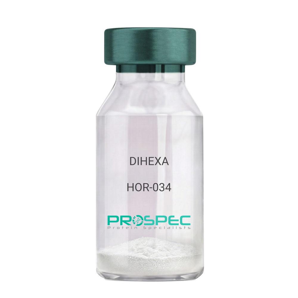
DIHEXADescription:
DIHEXA
Product # :
HOR-034Price :
Quantity :
Shipping Method :
Shipped at Room temp
More Info
- description
- formulation
- purity
- More Info
- Citations
Description
DIHEXA Synthetic is a single, non-glycosylated polypeptide chain containing 3 amino acids, having a molecular mass of 504.28 Dalton and a Molecular formula of C27H44N4O5.
Formulation
The protein was lyophilized with no additives.
Purity
Greater than 97.0% as determined by analysis by RP-HPLC.
Citations
More Info
-
Physical Appearance
Sterile Filtered White lyophilized (freeze-dried) powder.
-
Stability
Lyophilized DIHEXA although stable at room temperature for 3 weeks, should be stored desiccated below -18°C. Upon reconstitution DIHEXA should be stored at 4°C between 2-7 days and for future use below -18°C. For long term storage it is recommended to add a carrier protein (0.1% HSA or BSA). Please prevent freeze-thaw cycles.
-
Solubility
It is recommended to reconstitute the lyophilized DIHEXA in sterile 18MΩ-cm H2O not less than 100 µg/ml, which can then be further diluted to other aqueous solutions.
-
Amino Acid Sequence
Hexanoyl-Tyr-Ile-Ahx-NH2.
-
Background
Dihexa, also known as N-hexanoic-Tyr-Ile-(6) aminohexanoic amide, is a potent and orally active small peptide that has been the focus of significant research due to its potential neurogenic and neuroprotective effects. This compound, derived from angiotensin IV, has been shown to possess a wide range of biological activities, including enhancing cognitive function, promoting neurogenesis, and potentially mitigating the effects of neurodegenerative diseases.
Dihexa's primary mechanism of action involves its interaction with hepatocyte growth factor (HGF) and its receptor, c-Met. By mimicking the effects of HGF, Dihexa can stimulate the c-Met receptor, leading to a cascade of events that promote neurogenesis and synaptic plasticity. Studies by Benoist et al. (2014) have demonstrated that Dihexa can enhance cognitive function in rats, suggesting potential applications in cognitive enhancement and the treatment of cognitive disorders.
In addition to its neurogenic effects, Dihexa has been shown to possess neuroprotective properties. Research by Kawas et al. (2017) found that Dihexa could protect neurons from apoptosis, suggesting potential applications in the treatment of neurodegenerative diseases such as Alzheimer's and Parkinson's disease.
Given its neurogenic and neuroprotective effects, Dihexa has been proposed as a potential therapeutic agent for a variety of conditions, including cognitive disorders, neurodegenerative diseases, and stroke. For instance, a study by Harding et al. (2018) found that Dihexa could improve outcomes in animal models of stroke, indicating its potential as a therapeutic agent in stroke recovery.
While research on Dihexa is promising, it is important to note that most studies have been conducted in animals or in vitro. More research is needed to fully understand the potential effects and applications of Dihexa in humans. However, the existing body of research suggests that Dihexa could be a promising tool in the treatment of cognitive disorders and neurodegenerative diseases.
ProSpec's products are furnished for LABORATORY RESEARCH USE ONLY. The product may not be used as drugs, agricultural or pesticidal products, food additives or household chemicals.
- View Data Sheet
Name :
Zika NS1Description:
Zika NS1 Protein Recombinant
Product # :
ZKV-002Price :
Quantity :
Shipping Method :
Shipped with Ice Packs
More Info
- description
- source
- formulation
- purity
- More Info
- Citations
Description
The E.Coli derived Recombinant Zika NS1 protein (Strain: ZikaSPH2015) having an Mw of 45kDa is derived from the full length Zika NS1 protein. The Zika NS1 protein is fused to a 6xHis tag at C-terminus and purified by proprietary chromatographic technique.
Source
Escherichia Coli.
Formulation
Zika NS1 protein solution contains PBS and 100mM arginine.
Purity
Zika NS1 protein is >95% pure as determined by SDS-PAGE.
Citations
More Info
-
Introduction
Zika virus (ZIKV) belongs to the family Flaviviridae and the genus Flavivirus, it is transmitted by daytime-active Aedes mosquitoes, such as A. aegypti and A. albopictus. The Zika virus is related to the dengue, yellow fever, Japanese encephalitis, and West Nile viruses. Much like the other flaviviruses, Zika virus is enveloped and icosahedral and has a nonsegmented, single-stranded, positive-sense RNA genome. Zika fever is an infection, which often causes no symptoms or only mild ones, like a mild form of dengue fever, and it is treated by rest. As of February 2016, there has been mounting evidence that Zika fever in pregnant women can cause abnormal brain development in their fetuses by mother-to-child transmission, which may result in miscarriage or microcephaly, however it is not yet known whether Zika virus causes microcephaly. Furthermore, a connection has been established with neurologic conditions in infected adults, including Guillain–Barre syndrome. Since the 1950s, Zika virus has been detected only within a narrow equatorial belt from Africa to Asia. Between the years 2013 and 2014, Zika virus has spread eastward across the Pacific Ocean to French Polynesia, New Caledonia, the Cook Islands, and Easter Island, and in 2015 to Mexico, Central America, the Caribbean, and South America, where the Zika outbreak has reached pandemic levels.
-
Physical Appearance
Sterile Filtered solution.
-
Stability
Zika NS1 protein although stable at 4°C for 1 week, should be stored below -18°C. Please prevent freeze thaw cycles.
-
Applications
Each laboratory should determine an optimum working titer for use in its particular application.
ProSpec's products are furnished for LABORATORY RESEARCH USE ONLY. The product may not be used as drugs, agricultural or pesticidal products, food additives or household chemicals.
- View Data Sheet
Name :
NRG1 A HumanDescription:
Neuregulin-1/Heregulin Alpha (EGF Domain) Human Recombinant
Neuregulin-1, Heregulin Alpha, NRG1-A, NRG1 A.
Product # :
CYT-736Price :
Quantity :
Shipping Method :
Shipped at Room temp
More Info
- description
- source
- formulation
- purity
- biological activity
- More Info
- Citations
Description
Recombinant Human Neuregulin-1/Heregulin Alpha (EGF Domain) produced in E.Coli is a single, non-glycosylated, polypeptide chain containing 65 amino acids and having a total molecular mass of 7.4kDa. NRG1-A is purified by proprietary chromatographic techniques.
Source
Escherichia Coli.
Formulation
Lyophilized from a 0.2µm filtered solution in 20mM PB, pH 6.0 and 150mM NaCl.
Purity
Greater than 97.0% as determined by:
(a) Analysis by RP-HPLC.
(b) Analysis by SDS-PAGE.Biological Activity
The ED50 was determined by the dose-dependent stimulation of the proliferation of human MCF-7 cells is less than 40 ng/ml, corresponding to a specific activity of > 2.5×10⁴ units/mg.Citations
More Info
-
Introduction
Neuregulin/Heregulin is a family of structurally related polypeptide growth factors which are stemmed from alternatively spliced genes (NRG1, NRG2, NRG3 and NRG4). Thus far, there are more than 14 soluble and transmembrane proteins derived from the NRG1 gene. Proteolytic processing of the extracellular domain of the transmembrane NRG1 isoforms release soluble growth factors. These isoforms contain the heregulins (HRGs), glial growth factors (GGFs) and sensory and motor neuron-derived factor (SMDF). All these factors have the Ig and EGF-like domain, and are able to bind to ErbB3 and ErbB4 receptor tyrosin kinases. This binding stimulates erb3 and erb4 heterodimerization with erb2, promoting intrinsic kinase activity, which results in tyrosine phosphorylation. NRG1 isoforms act to induce the growth and differentiation of epithelial, neuronal, glial, and other types of cells.
-
Synonyms
Neuregulin-1, Heregulin Alpha, NRG1-A, NRG1 A.
-
Physical Appearance
Sterile Filtered White lyophilized (freeze-dried) powder.
-
Stability
Lyophilized NRG1-A although stable at room temperature for 3 weeks, should be stored desiccated below -18°C. Upon reconstitution NRG1-A should be stored at 4°C between 2-7 days and for future use below -18°C. Please prevent freeze-thaw cycles.
-
Solubility
It is recommended to reconstitute the lyophilized NRG1-A in sterile 18M-cm H2O not less than 100µg/ml, which can then be further diluted to other aqueous solutions.
-
Amino Acid Sequence
SHLVKCAEKE KTFCVNGGEC FMVKDLSNPS RYLCKCQPGF TGARCTENVP MKVQNQEKAE ELYQK.
ProSpec's products are furnished for LABORATORY RESEARCH USE ONLY. The product may not be used as drugs, agricultural or pesticidal products, food additives or household chemicals.
- View Data Sheet
Name :
TATDN1 HumanDescription:
TatD DNase Domain Containing 1 Human Recombinant
TatD DNase Domain Containing 1, Hepatocarcinoma High Expression Protein, Putative Deoxyribonuclease TATDN1, EC 3.1.21, CDA11, TATDN1.
Product # :
PRO-2160Price :
Quantity :
Shipping Method :
Shipped with Ice Packs
More Info
- description
- source
- formulation
- purity
- More Info
- Citations
Description
TATDN1 Human Recombinant produced in E.Coli is a single, non-glycosylated polypeptide chain containing 320 amino acids (1-297 a.a) and having a molecular mass of 36kDa. TATDN1 is fused to a 23 amino acid His-tag at N-terminus & purified by proprietary chromatographic techniques.
Source
Escherichia Coli.
Formulation
TATDN1 protein solution (1mg/ml) containing Phosphate buffered saline (pH7.4), 10% glycerol and 1mM DTT.
Purity
Greater than 95.0% as determined by SDS-PAGE.
Citations
More Info
-
Introduction
TatD DNase Domain Containing 1, also known as TATDN1, is a protein coding gene which is a part of the TatD DNase family. TATDN1 which is a deoxyribonuclease binds two divalent metal cations per subunit.
-
Synonyms
TatD DNase Domain Containing 1, Hepatocarcinoma High Expression Protein, Putative Deoxyribonuclease TATDN1, EC 3.1.21, CDA11, TATDN1.
-
Physical Appearance
Sterile Filtered clear solution.
-
Stability
Store at 4°C if entire vial will be used within 2-4 weeks. Store, frozen at -20°C for longer periods of time. For long term storage it is recommended to add a carrier protein (0.1% HSA or BSA).Avoid multiple freeze-thaw cycles.
-
Amino Acid Sequence
MGSSHHHHHH SSGLVPRGSH MGSMSRFKFI DIGINLTDPM FRGIYRGVQK HQDDLQDVIG RAVEIGVKKF MITGGNLQDS KDALHLAQTN GMFFSTVGCH PTRCGEFEKN NPDLYLKELL NLAENNKGKV VAIGECGLDF DRLQFCPKDT QLKYFEKQFE LSEQTKLPMF LHCRNSHAEF LDIMKRNRDR CVGGVVHSFD GTKEAAAALI DLDLYIGFNG CSLKTEANLE VLKSIPSEKL MIETDAPWCG VKSTHAGSKY IRTAFPTKKK WESGHCLKDR NEPCHIIQIL EIMSAVRDED PLELANTLYN NTIKVFFPGI.
ProSpec's products are furnished for LABORATORY RESEARCH USE ONLY. The product may not be used as drugs, agricultural or pesticidal products, food additives or household chemicals.
- View Data Sheet
Name :
GH MouseDescription:
GH Mouse Recombinant
GH1, GH, GHN, GH-N, hGH-N, Pituitary GH, GH-1.
Product # :
CYT-540Price :
Quantity :
Shipping Method :
Shipped at Room temp
More Info
- description
- source
- formulation
- purity
- More Info
- Citations
Description
Growth Hormone Mouse Recombinant produced in E.Coli is a single, non-glycosylated, polypeptide chain containing 190 amino acids and having a molecular mass of 22 kDa. Mouse GH is purified by proprietary chromatographic techniques.
Source
Escherichia Coli.
Formulation
Mouse Growth Hormone Recombinant Lyophilized from a 0.2 μm filtered solution of 20mM HEPES, 4% Sucrose, 4% Mannitol, 0.02% Tween 80, pH 8.0.
Purity
Greater than 95.0% as determined by SDS-PAGE.
Citations
More Info
-
Introduction
GH is a member of the prolactin family of hormones which play an important role in growth control. The gene, along with four other related genes, is located at the GH locus on chromosome 17 where they are interspersed in the same transcriptional orientation; an arrangement which is thought to have evolved by a series of gene duplications. The five genes share a remarkably high degree of sequence identity. Alternative splicing generates additional isoforms of each of the five GHs, leading to further diversity and potential for specialization. This particular family member is expressed in the pituitary but not in placental tissue as is the case for the other four genes in the GH locus. Mutations in or deletions of the gene lead to GH deficiency and short stature.
-
Synonyms
GH1, GH, GHN, GH-N, hGH-N, Pituitary GH, GH-1.
-
Physical Appearance
Sterile Filtered White lyophilized (freeze-dried) powder.
-
Stability
Lyophilized GH mouse although stable at room temperature for 3 weeks, should be stored desiccated below -18°C. Upon reconstitution GH mouse should be stored at 4°C between 2-7 days and for future use below -18°C.For long term storage it is recommended to add a carrier protein (0.1% HSA or BSA).Please prevent freeze-thaw cycles.
-
Solubility
It is recommended to reconstitute the lyophilized Growth Hormone Murine in sterile 18MΩ-cm H2O not less than 100µg/ml, which can then be further diluted to other aqueous solutions.
ProSpec's products are furnished for LABORATORY RESEARCH USE ONLY. The product may not be used as drugs, agricultural or pesticidal products, food additives or household chemicals.
- View Data Sheet
Name :
BCL2 AntibodyDescription:
B-Cell Leukemia/Lymphoma 2, Mouse Anti Human
Apoptosis regulator Bcl-2, BCL2, B-cell CLL/lymphoma 2, Bcl-2.
Product # :
ANT-022Price :
Quantity :
Shipping Method :
Shipped with Ice Packs
More Info
- formulation
- More Info
- Citations
Formulation
1mg/ml containing PBS, pH-7.4, & 0.1% Sodium Azide.
Citations
More Info
-
Introduction
BCL2 gene encodes an integral outer mitochondrial membrane protein that blocks the apoptotic death of some cells such as lymphocytes. Constitutive expression of BCL2, such as in the case of translocation of BCL2 to Ig heavy chain locus, is thought to be the cause of follicular lymphoma. Two transcript variants, produced by alternate splicing, differ in their C-terminal ends.
-
Synonyms
Apoptosis regulator Bcl-2, BCL2, B-cell CLL/lymphoma 2, Bcl-2.
-
Immunogen
Anti-human BCL2 mAb, is derived from hybridization of mouse FO myeloma cells with spleen cells from BALB/c mice immunized with recombinant human BCL2 amino acids 1-211 purified from E. coli.
-
Ig Subclass
Mouse IgG2b heavy chain and k light chain.
-
Clone
PAT1B5AT.
-
Applications
BCL2 antibody has been tested by ELISA, Western blot, and Immunofluorescence analyses to assure specificity and reactivity. Since applications vary, however, each investigation should be titrated by a reagent to obtain optimal results. Recommended dilution range for Western blot analysis and Immunofluorescence is 1:250 ~ 500.
Recommended starting dilution is 1:500. -
Type
Mouse Anti Human Monoclonal.
-
Storage Procedures
For periods up to 1 month store at 4°C, for longer periods of time, store at -20°C. Prevent freeze thaw cycles.
-
Purification Method
BCL2 antibody was purified from mouse ascitic fluids by protein-G affinity chromatography.
ProSpec's products are furnished for LABORATORY RESEARCH USE ONLY. The product may not be used as drugs, agricultural or pesticidal products, food additives or household chemicals.
- View Data Sheet
Name :
HCV NS4 Mosaic Genotype-3Description:
Hepatitis C Virus NS4 Mosaic Genotype-3 Recombinant
Product # :
HCV-259Price :
Quantity :
Shipping Method :
Shipped with Ice Packs
More Info
- description
- formulation
- purity
- More Info
- Citations
Description
The E.coli derived recombinant protein contains the HCV NS4 immunodominant regions, amino acids 1691-1710, 1712-1733 & 1921-1940, fused with a GST tag at N-terminus.
Formulation
1.5M urea, 25mM Tris-HCl, pH 8.0, 0.2% Triton-X & 50% glycerol.
Purity
Protein is >95% pure as determined by 10% PAGE (coomassie staining).
Citations
More Info
-
Introduction
HCV is a small 50nm, enveloped, single-stranded, positive sense RNAvirus in the family Flaviviridae.
HCV has a high rate of replication with approximately one trillion particles produced each day in an infected individual. Due to lack of proofreading by the HCV RNA polymerase, the HCV has an exceptionally high mutation rate, a factor that may help it elude the host's immune response. Hepatitis C virus is classified into six genotypes(1-6) with several subtypes within each genotype. The preponderance and distribution of HCV genotypes varies globally. Genotype is clinically important in determining potential response to interferon-based therapy and the required duration of such therapy. Genotypes 1 and 4 are less responsive to interferon-based treatment than are the other genotypes (2, 3, 5 and 6). -
Stability
HCV NS4 Mosaic Genotype-3 although stable at 4°C for 1 week, should be stored below -18°C. Please prevent freeze thaw cycles.
-
Applications
Antigen in ELISA and Western blots, excellent antigen for detection of HCV with minimal specificity problems.
-
Specificity
Immunoreactive with sera of HCV-infected individuals.
-
Purification Method
Purified by proprietary chromatographic technique.
ProSpec's products are furnished for LABORATORY RESEARCH USE ONLY. The product may not be used as drugs, agricultural or pesticidal products, food additives or household chemicals.
- View Data Sheet
Name :
HIV-1 p31 IntegraseDescription:
HIV-1 p31 Integrase Recombinant
Product # :
HIV-145Price :
Quantity :
Shipping Method :
Shipped with Ice Packs
More Info
- description
- source
- formulation
- purity
- More Info
- Citations
Description
The E.coli derived recombinant protein is a non-glycosylated polypeptide chain, containing the HIV-1 immunodominant regions from the p31 protein (integrase) 9-289 amino acids, fused with six histidines at N-terminus.
Source
Escherichia Coli.
Formulation
1.5M urea, 25mM Tris-HCl pH 8.0, 0.2% Triton-X & 50% Glycerol.
Purity
Greater than 95.0% as determined by HPLC analysis and SDS-PAGE.
Citations
More Info
-
Introduction
Integrase is an enzyme produced by the HIV which enables its genetic material to be integrated into the DNA of the infected cell and is a key component in the pre-integration complex. HIV integrase contains 3 domains, an N-terminal HH-CC zinc fingerdomainwhich is partially responsible for multimerization, a central catalytic domain and a C-terminal domain. Both Central catalytic domain and C-terminal domains have been shown to bind both viral and cellular DNA. No crystal structure data exists with Integrase bound to its DNA substrates. HIV-1 integrase functions as a dimeror a tetramer. Additionally, several host cellular proteins interact with integrase and may facilitate the integration process.
-
Physical Appearance
Sterile filtered colorless clear solution.
-
Stability
HIV-1 Integrase p31 although stable at 4°C for 1 week, should be stored below -18°C. Please prevent freeze thaw cycles.
-
Applications
HIV-1 Integrase p31 antigen is suitable for ELISA and Western blots, excellent antigen for early detection of HIV seroconvertors with minimal specificity problems.
-
Specificity
Immunoreactive with all sera of HIV-1 infected individuals.
ProSpec's products are furnished for LABORATORY RESEARCH USE ONLY. The product may not be used as drugs, agricultural or pesticidal products, food additives or household chemicals.
- View Data Sheet
Name :
Dengue Envelope-1 & 4Description:
Dengue Virus Subtype 1 & 4 fused Envelope 55kDa Recombinant
Product # :
DEN-025Price :
Quantity :
Shipping Method :
Shipped with Ice Packs
More Info
- description
- source
- formulation
- purity
- More Info
- Citations
Description
The E.coli derived recombinant 55kDa protein is genetically engineered peptide which is derived from Dengue Type-1 and 4 to be expressed as a fused envelope, each part in this fusion contains 170 a.a (positions 46-217), it is used in ELISA assay. This fusion protein is connected to a 6xHis Tag.Dengue Type-1 and 4 is purified by proprietary chromatographic technique.
Source
Escherichia Coli.
Formulation
Phosphate buffered saline, pH-7.4.
Purity
Protein is >95% pure as determined by 12% PAGE (coomassie staining).
Citations
More Info
-
Introduction
Caused by one of four closely related virus serotypes of the genus Flavivirus, family Flaviviridae, each serotype is sufficiently different that there is no cross-protection and epidemics caused by multiple serotypes (hyperendemicity) can occur. In cell culture experiments and mice Morpholino antisense oligos have shown specific activity against Dengue virus.
-
Physical Appearance
Sterile filtered colorless solution.
-
Stability
Dengue Envelope-1 & 4 Recombinant although stable at 4°C for 1 week, should be stored below -18°C. Please prevent freeze thaw cycles.
ProSpec's products are furnished for LABORATORY RESEARCH USE ONLY. The product may not be used as drugs, agricultural or pesticidal products, food additives or household chemicals.
- View Data Sheet
Name :
NFATC2 HumanDescription:
Nuclear Factor Of Activated T Cells 2 Human Recombinant
Nuclear Factor Of Activated T Cells 2, Nuclear Factor Of Activated T-Cells, Cytoplasmic, Calcineurin-Dependent 2, Nuclear Factor Of Activated T-Cells 2, NFAT Pre-Existing Subunit, NF-ATc2, NFAT1, NFATP, Nuclear Factor Of Activated T-Cells, Preexisting Component , Nuclear Factor Of Activated T-Cells, Cytoplasmic 2, NFAT Transcription Complex, Preexisting Component, Preexisting Nuclear Factor Of Activated T-Cells 2, T Cell Transcription Factor NFAT1, T-Cell Transcription Factor NFAT1, NF-ATp, NFATc2.
Product # :
PRO-2535Price :
Quantity :
Shipping Method :
Shipped with Ice Packs
More Info
- description
- source
- formulation
- purity
- More Info
- Citations
Description
NFATC2 produced in Sf9 Baculovirus cells is a single, glycosylated polypeptide chain containing 290 amino acids (396-678a.a.) and having a molecular mass of 33.1kDa. (Molecular size on SDS-PAGE will appear at approximately 28-40kDa). NFATC2 is expressed with a 7 amino acid His tag at C-Terminus and purified by proprietary chromatographic techniques.
Source
Sf9, Baculovirus cells.
Formulation
NFATC2 protein solution (0.25mg/ml) contains Phosphate Buffered Saline (pH 7.4) 40% glycerol and 1mM DTT.
Purity
Greater than 90.0% as determined by SDS-PAGE.
Citations
More Info
-
Introduction
NFATC2, also known as nuclear factor of activated T-cells 2, belongs to the nuclear factor of activated T cells family. NFATC2 takes a significant part in the course of T helper cell differentiation, activation, and effector function. Even though KO of an individual NFAT isoform in T cells directs to rather minor effects, T cells lacking for NFATC1 and 2 totally fail to produce T helper cell effector cytokines, for instance the interleukins IL-4 and IL-2. Moreover, NFATC2 is highly phosphorylated and kept in the cytoplasm. Next T cell receptor stimulation, dephosphorylation via calcium-activated calcineurin induces a conformational modification of NFATC2 which reveals a few nuclear localization sequences.
-
Synonyms
Nuclear Factor Of Activated T Cells 2, Nuclear Factor Of Activated T-Cells, Cytoplasmic, Calcineurin-Dependent 2, Nuclear Factor Of Activated T-Cells 2, NFAT Pre-Existing Subunit, NF-ATc2, NFAT1, NFATP, Nuclear Factor Of Activated T-Cells, Preexisting Component , Nuclear Factor Of Activated T-Cells, Cytoplasmic 2, NFAT Transcription Complex, Preexisting Component, Preexisting Nuclear Factor Of Activated T-Cells 2, T Cell Transcription Factor NFAT1, T-Cell Transcription Factor NFAT1, NF-ATp, NFATc2.
-
Physical Appearance
Sterile Filtered colorless solution.
-
Stability
Store at 4°C if entire vial will be used within 2-4 weeks. Store, frozen at -20°C for longer periods of time. For long term storage it is recommended to add a carrier protein (0.1% HSA or BSA). Avoid multiple freeze-thaw cycles.
-
Amino Acid Sequence
MPLEWPLSSQ SGSYELRIEV QPKPHHRAHY ETEGSRGAVK APTGGHPVVQ LHGYMENKPL GLQIFIGTAD ERILKPHAFY QVHRITGKTV TTTSYEKIVG NTKVLEIPLE PKNNMRATID CAGILKLRNA DIELRKGETD IGRKNTRVRL VFRVHIPESS GRIVSLQTAS NPIECSQRSA HELPMVERQD TDSCLVYGGQ QMILTGQNFT SESKVVFTEK TTDGQQIWEM EATVDKDKSQ PNMLFVEIPE YRNKHIRTPV KVNFYVINGK RKRSQPQHFT YHPVHHHHHH
ProSpec's products are furnished for LABORATORY RESEARCH USE ONLY. The product may not be used as drugs, agricultural or pesticidal products, food additives or household chemicals.
- View Data Sheet
Name :
IL17RA HumanDescription:
Interleukin-17 Receptor Alpha Human Recombinant
Interleukin 17 Receptor A, IL-17 Receptor A, IL-17RA, CDw217, IL17R, Interleukin-17 Receptor A, Interleukin 17 Receptor, CD217 Antigen, HIL-17R, CANDF5, CD217, IMD51.
Product # :
CYT-1023Price :
Quantity :
Shipping Method :
Shipped with Ice Packs
More Info
- description
- source
- formulation
- purity
- More Info
- Citations
Description
IL17RA Human Recombinant produced in Sf9 Baculovirus cells is a single, glycosylated polypeptide chain containing 527 amino acids (33-320) and having a molecular mass of 60.4kDa (Molecular size on SDS-PAGE will appear at approximately 70-100kDa). IL17RA is fused to a 239 amino acids hIgG-His-Tag at C-terminus and purified by proprietary chromatographic techniques.
Source
Sf9, Baculovirus cells.
Formulation
IL17RA protein solution (0.5mg/ml) containing Phosphate Buffered Saline (pH 7.4) and 10% glycerol.
Purity
Greater than 90% as determined by SDS-PAGE.
Citations
More Info
-
Introduction
Interleukin-17 receptor A isoform 1 (IL17RA) is a cytokine receptor which binds interleukin 17. IL17RA is a potent inducer of the maturation of CD34-positive hematopoietic precursors into neutrophils. IL17RA is a type 1 membrane glycoprotein which binds with low affinity to interleukin 17A. The IL17RA protein and Interleukin 17A has a pathogenic role in many inflammatory and autoimmune diseases such as rheumatoid arthritis.
-
Synonyms
Interleukin 17 Receptor A, IL-17 Receptor A, IL-17RA, CDw217, IL17R, Interleukin-17 Receptor A, Interleukin 17 Receptor, CD217 Antigen, HIL-17R, CANDF5, CD217, IMD51.
-
Physical Appearance
Sterile Filtered colorless solution.
-
Stability
Store at 4°C if entire vial will be used within 2-4 weeks. Store, frozen at -20°C for longer periods of time. For long term storage it is recommended to add a carrier protein (0.1% HSA or BSA).Avoid multiple freeze-thaw cycles.
-
Amino Acid Sequence
LRLLDHRALV CSQPGLNCTV KNSTCLDDSW IHPRNLTPSS PKDLQIQLHF AHTQQGDLFP VAHIEWTLQT DASILYLEGA ELSVLQLNTN ERLCVRFEFL SKLRHHHRRW RFTFSHFVVD PDQEYEVTVH HLPKPIPDGD PNHQSKNFLV PDCEHARMKV TTPCMSSGSL WDPNITVETL EAHQLRVSFT LWNESTHYQI LLTSFPHMEN HSCFEHMHHI PAPRPEEFHQ RSNVTLTLRN LKGCCRHQVQ IQPFFSSCLN DCLRHSATVS CPEMPDTPEP IPDYMPLWVE PKSCDKTHTC PPCPAPELLG GPSVFLFPPK PKDTLMISRT PEVTCVVVDV SHEDPEVKFN WYVDGVEVHN AKTKPREEQY NSTYRVVSVL TVLHQDWLNG KEYKCKVSNK ALPAPIEKTI SKAKGQPREP QVYTLPPSRD ELTKNQVSLT CLVKGFYPSD IAVEWESNGQ PENNYKTTPP VLDSDGSFFL YSKLTVDKSR WQQGNVFSCS VMHEALHNHY TQKSLSLSPG KHHHHHH.
ProSpec's products are furnished for LABORATORY RESEARCH USE ONLY. The product may not be used as drugs, agricultural or pesticidal products, food additives or household chemicals.
- View Data Sheet
Name :
PIH1D2 HumanDescription:
PIH1 Domain Containing 2 Human Recombinant
PIH1 Domain Containing 2, PIH1D2.
Product # :
PRO-2118Price :
Quantity :
Shipping Method :
Shipped with Ice Packs
More Info
- description
- source
- formulation
- purity
- More Info
- Citations
Description
PIH1D2 Human Recombinant produced in E.Coli is a single, non-glycosylated polypeptide chain containing 338 amino acids (1-315 a.a) and having a molecular mass of 38.3kDa.PIH1D2 is fused to a 23 amino acid His-tag at N-terminus & purified by proprietary chromatographic techniques.
Source
Escherichia Coli.
Formulation
PIH1D2 protein solution (0.25mg/ml) containing Phosphate buffered saline (pH7.4), 20% glycerol and 1mM DTT.
Purity
Greater than 85.0% as determined by SDS-PAGE.
Citations
More Info
-
Introduction
PIH1 Domain Containing 2, also known as PIH1D2 is a member of the PIH1 family.PIH1D2 was present in the common ancestor of chordates. There are 45 Species with no ortholog for PIH1D2. In addition, no disorders were found for PIH1D2 Gene.
-
Synonyms
PIH1 Domain Containing 2, PIH1D2.
-
Physical Appearance
Sterile Filtered colorless solution.
-
Stability
Store at 4°C if entire vial will be used within 2-4 weeks. Store, frozen at -20°C for longer periods of time. For long term storage it is recommended to add a carrier protein (0.1% HSA or BSA).Avoid multiple freeze-thaw cycles.
-
Amino Acid Sequence
MGSSHHHHHH SSGLVPRGSH MGSMETSSKG LLTQVTQFWN LLDDLAQSDP EGYEKFIQQQ LKEGKQLCAA PEPQLCLQTR ILKPKEKILF INLCQWTRIP APQSTTHPVP LTVGKPEDTT EISDAYTVID VAYNPDVLHA AEKDQVKKNQ LIQMAMKCIE EKFQFTLSHS YHITKFRIKG SIQRMKQNLM GIQTDSIDLR EKMRRELTLG QIRSSTMSNP DHFPQLLLPK DQVSGKAVCL IEEISSTEIQ VEMKMPAYEL KIVHDHSEKP LKIELKVELP GINSVSLCDL SVSEDDLLIE VSEKYRLHLN LPKLIDTEMT TAKFIKEKST LIITMPLV.
ProSpec's products are furnished for LABORATORY RESEARCH USE ONLY. The product may not be used as drugs, agricultural or pesticidal products, food additives or household chemicals.
- View Data Sheet
Name :
TNFRSF10B Human, Sf9Description:
TNF Ligand Receptor Superfamily Member 10B Human Recombinant, Sf9
TNFRSF10B, CD262, DR5, KILLER, KILLER/DR5, TRAIL-R2, TRAILR2, TRICK2, TRICK2A, TRICK2B, TRICKB, ZTNFR9, Death receptor 5, TNF-related apoptosis-inducing ligand receptor 2.
Product # :
CYT-1055Price :
Quantity :
Shipping Method :
Shipped with Ice Packs
More Info
- description
- source
- formulation
- purity
- biological activity
- More Info
- Citations
Description
TNFRSF10B produced in Sf9 Baculovirus cells is a single, glycosylated polypeptide chain containing 394 amino acids (56-210a.a.) and having a molecular mass of 43.9kDa. (Molecular size on SDS-PAGE will appear at approximately 40-57kDa).TNFRSF10B is expressed with a 239 amino acid hIgG-His tag at C-Terminus and purified by proprietary chromatographic techniques.
Source
Sf9, Insect cells.
Formulation
TNFRSF10B protein solution (1mg/ml) contains Phosphate Buffered Saline (pH 7.4) and 10% glycerol.
Purity
Greater than 90% as determined by SDS-PAGE.
Biological Activity
Measured by its ability to inhibit cytotoxicity assay using Jurkat human T lymphocyte. The ED50 for this effect ≤ to 5ng/ml with TRAIL
.
Citations
More Info
-
Introduction
TRAIL Receptor-1 (DR4) and TRAIL Receptor-2(DR5) are members of the TNFR superfamily of transmembrane proteins and contain a cytoplasmic "death domain", which is capable of activating the cell's apoptotic machinery. These receptors are activated by binding to either membrane anchored or soluble TRAIL/Apo2L.
-
Synonyms
TNFRSF10B, CD262, DR5, KILLER, KILLER/DR5, TRAIL-R2, TRAILR2, TRICK2, TRICK2A, TRICK2B, TRICKB, ZTNFR9, Death receptor 5, TNF-related apoptosis-inducing ligand receptor 2.
-
Physical Appearance
Sterile Filtered colorless solution.
-
Stability
Store at 4°C if entire vial will be used within 2-4 weeks. Store, frozen at -20°C for longer periods of time. For long term storage it is recommended to add a carrier protein (0.1% HSA or BSA).Avoid multiple freeze-thaw cycles.
-
Amino Acid Sequence
ITQQDLAPQQ RAAPQQKRSS PSEGLCPPGH HISEDGRDCI SCKYGQDYST HWNDLLFCLR CTRCDSGEVE LSPCTTTRNT VCQCEEGTFR EEDSPEMCRK CRTGCPRGMV KVGDCTPWSD IECVHKESGT KHSGEVPAVE ETVTSSPGTP ASPCSLEPKS CDKTHTCPPC PAPELLGGPS
VFLFPPKPKD TLMISRTPEV TCVVVDVSHE DPEVKFNWYV DGVEVHNAKT KPREEQYNST YRVVSVLTVL HQDWLNGKEY KCKVSNKALP APIEKTISKA KGQPREPQVY TLPPSRDELT KNQVSLTCLV KGFYPSDIAV EWESNGQPEN NYKTTPPVLD SDGSFFLYSK LTVDKSRWQQ
GNVFSCSVMH EALHNHYTQK SLSLSPGKHH HHHH.
ProSpec's products are furnished for LABORATORY RESEARCH USE ONLY. The product may not be used as drugs, agricultural or pesticidal products, food additives or household chemicals.
- View Data Sheet
Name :
LRG1 CanineDescription:
Leucine-Rich Alpha-2-Glycoprotein 1 Canine
Leucine-rich alpha-2-glycoprotein, LRG, LRG1, FLJ45787, HMFT1766.
Product # :
PRO-1592Price :
Quantity :
Shipping Method :
Shipped at Room temp
More Info
- description
- source
- formulation
- purity
- More Info
- Citations
Description
The Canine LRG1 produced from pooled Canine Serum has a molecular mass of 38.129kDa (calculated without glycosylation) containing 347 amino acid residues.
Source
Canine Serum.
Formulation
LRG1 protein filtered (0.4µm) and lyophilized in 0.5mg/ml in 20mM TRIS and 50mM NaCl, pH 8.0.
Purity
Purity greater than 90% as determined by SDS-PAGE.
Citations
More Info
-
Introduction
LRG1 belongs to the leucine-rich repeat (LRR) family of proteins, which have been shown to be involved in protein-protein interaction, signal transduction, and cell adhesion and development. The LRG1 is expressed during granulocyte differentiation.
-
Synonyms
Leucine-rich alpha-2-glycoprotein, LRG, LRG1, FLJ45787, HMFT1766.
-
Physical Appearance
Filtered White lyophilized (freeze-dried) powder.
-
Stability
Store lyophilized protein at -20°C. Aliquot the product after reconstitution to avoid repeated freezing/thawing cycles. Reconstituted protein can be stored at 4°C for a limited period of time; it does not show any change after two weeks at 4°C.
-
Solubility
It is recommended to add deionized water to prepare a working stock solution of approximately 0.5 mg/ml and let the lyophilized pellet dissolve completely. Product is not sterile! Please filter the product by an appropriate sterile filter before using it in the cell culture.
-
Amino Acid Sequence
MSPWSPERSQ SPGGLSSHLS KTLLLLLFSV ASAQGVTPDR DTCLVFPSSN GSSVSCHPPA KLPHHFPSDT VHLVVEFFNL TQLRPDTLQG VSNLQELHLS TNQLESLSPK LLLPVPLLKV LDLTRNALTQ LPPGLFRVSA ALHTLVLKEN RLEVLEPSWL CGLKALGHLD LSGNHLQTLP PGLLANVTAL RILDLSNNQL KTLPLDLLRG PLQLERLHLE GNRLQVLEEG LLAPQPGLRY LFLSDNKLAA VAAGAFRGLQ QLDMLDLSNN SLTSVPKGLW TSLGQPTRDM EDGFDISGNP WICDHNLDDL YRWLVANKDK MFSRNDTRCA RPEALKGQTL LEAAESH.
ProSpec's products are furnished for LABORATORY RESEARCH USE ONLY. The product may not be used as drugs, agricultural or pesticidal products, food additives or household chemicals.
- View Data Sheet
Name :
Leptin Super Antagonist RatDescription:
Leptin Super Antagonist Rat Recombinant
Product # :
CYT-1240Price :
Quantity :
Shipping Method :
Shipped at Room temp
More Info
- description
- source
- formulation
- purity
- biological activity
- More Info
- Citations
Description
Super Leptin Antagonist Rat Recombinant is a single polypeptide chain containing 146 amino acids. Super Rat Leptin Antagonist was mutated, resulting in D23L/L39A/D40A/F41A super Rat leptin antagonist that was purified by proprietary chromatographic techniques.
Source
Escherichia coli.
Formulation
The protein was lyophilized from a concentrated (1mg/ml) solution with 0.003mM NaHCO3.
Purity
Greater than 98.0% as determined by:
(a) Gel filtration analysis.
(b) Analysis by SDS-PAGE.
Biological Activity
ProSpec’s super Rat leptin antagonist is capable of inhibiting leptin-induced proliferation of BAF/3 cells stably transfected with the long form of human leptin receptor. Super Rat leptin antagonist also inhibits various leptin effects in several in vitro bioassays.
Citations
More Info
-
Physical Appearance
White lyophilized (freeze-dried) powder.
-
Stability
Lyophilized Super Leptin Antagonist although stable at room temperature for several weeks, should be stored desiccated below -18°C. Upon reconstitution of super Rat leptin antagonist at > 0.1 mg/ml and up to 2 mg/ml and filter sterilization super Rat leptin antagonist can be stored at 4C or even room temperature for several weeks making it suitable for long term infusion studies using osmotic pumps. At lower concentration addition of a carrier protein (0.1% HSA or BSA) is suggested. Please prevent freeze-thaw cycles.
-
Solubility
It is recommended to reconstitute the lyophilized Super Leptin Antagonist in sterile 0.4% NaHCO3 adjusted to pH 8-9, not less than 100µg/ml, which can then be further diluted to other aqueous solutions.
-
Amino Acid Sequence
The sequence of the first five N-terminal amino acids was determined and was found to be Ala-Val-Pro-Ile-His.
-
Background
Leptin is a hormone which takes part in regulating body weight, metabolism and reproductive function and is encoded by the obese gene. Leptin is expressed predominantly by adipocytes, which fits with the idea that body weight is sensed as the total mass of fat in the body. Smaller amounts of leptin are also secreted by cellsin the epithelium of the stomach and in the placenta. Leptin receptors are expressed mainly in areas of the hypothalamus which regulates body weight, as well as in T lymphocytes and vascular endothelial cells.
ProSpec's products are furnished for LABORATORY RESEARCH USE ONLY. The product may not be used as drugs, agricultural or pesticidal products, food additives or household chemicals.
- View Data Sheet
Name :
M CSF Human, HisDescription:
Macrophage Colony Stimulating Factor Human Recombinant, His Tag
CSF-1, Lanimostim, MCSF, MGC31930, M-CSF.
Product # :
CYT-695Price :
Quantity :
Shipping Method :
Shipped with Ice Packs
More Info
- description
- source
- formulation
- purity
- More Info
- Citations
Description
Macrophage Colony Stimulating Factor Human Recombinant produced in E.coli is a single, non-glycosylated, polypeptide chain containing 179 amino acids (33-190 a.a.) and having a total molecular mass of 20.7 kDa.MCSF is fused to a 20 amino acid His Tag at N-terminus and purified by proprietary chromatographic techniques.
Source
Escherichia Coli.
Formulation
The MCSF protein solution contains 20mM Tris-HCl, pH-8, 2mM DTT & 10% Glycerol.
Purity
Greater than 90.0% as determined by SDS-PAGE.
Citations
More Info
-
Introduction
Granulocyte/Macrophage Colony-Stimulating Factors are cytokines that act in hematopoiesis by controlling the production, differentiation, and function of 2 related white cell populations of the blood, the granulocytes and the monocytes-macrophages. CSF-1 induces cells of the monocyte/macrophage lineage. It plays a role in immunological defenses, bone metabolism, lipoproteins clearance, fertility and pregnancy.
-
Synonyms
CSF-1, Lanimostim, MCSF, MGC31930, M-CSF.
-
Physical Appearance
Sterile filtered colorless solution.
-
Stability
Store at 4°C if entire vial will be used within 2-4 weeks. Store, frozen at -20°C for longer periods of time. For long term storage it is recommended to add a carrier protein (0.1% HSA or BSA).Avoid multiple freeze-thaw cycles.
-
Amino Acid Sequence
MGSSHHHHHH SSGLVPRGSH MEEVSEYCSH MIGSGHLQSL QRLIDSQMET SCQITFEFVD QEQLKDPVCY LKKAFLLVQD IMEDTMRFRD NTPNAIAIVQ LQELSLRLKS CFTKDYEEHD KACVRTFYET PLQLLEKVKN VFNETKNLLD KDWNIFSKNC NNSFAECSSQ DVVTKPDCN.
ProSpec's products are furnished for LABORATORY RESEARCH USE ONLY. The product may not be used as drugs, agricultural or pesticidal products, food additives or household chemicals.