Search results
1000 results found for “dysbindin”
Name
Description
Product #
Price
Quantity
Shipping Method
- View Data Sheet
Name :
GHBP RabbitDescription:
Growth Hormone Binding Protein Rabbit Recombinant
GHR, GHBP, GH receptor, Somatotropin receptor.
Product # :
CYT-593Price :
Quantity :
Shipping Method :
Shipped at Room temp
More Info
- description
- source
- formulation
- purity
- biological activity
- More Info
Description
Growth Hormone Binding Protein Rabbit Extracellular Domain Recombinant produced in E.Coli is a single, non-glycosylated, polypeptide chain containing 249 amino acids and having a molecular mass of 28 kDa. GHBP Rabbit is purified by proprietary chromatographic techniques.
Source
Escherichia Coli.
Formulation
The Growth Hormone Binding Protein Rabbit was lyophilized from a concentrated (1mg/ml) solution with 0.0045mM NaHCO3.
Purity
Greater than 98.0% as determined bySDS-PAGE.
Biological Activity
Evidenced by its ability of forming 2:1 complex with non-primate Growth Hormones.More Info
-
Introduction
GHBP is a transmembrane receptor for growth hormone. Binding of growth hormone to the receptor leads to receptor dimerization and the activation of an intra- and intercellular signal transduction pathway leading to growth. A common alternate allele of this gene, called GHRd3, lacks exon three and has been well-characterized. Mutations in this gene have been associated with Laron syndrome, also known as the growth hormone insensitivity syndrome (GHIS), a disorder characterized by short stature. Other splice variants, including one encoding a soluble form of the protein (GHRtr), have been observed but have not been thoroughly characterized.
-
Synonyms
GHR, GHBP, GH receptor, Somatotropin receptor.
-
Physical Appearance
Sterile Filtered White lyophilized (freeze-dried) powder.
-
Stability
Lyophilized Growth Hormone Binding Protein Rabbit although stable at room temperature for 3 weeks, should be stored desiccated below -18°C. Upon reconstitution GHBP Rabbit should be stored at 4°C between 2-7 days and for future use below -18°C.For long term storage it is recommended to add a carrier protein (0.1% HSA or BSA).Please prevent freeze-thaw cycles.
-
Solubility
It is recommended to reconstitute the lyophilized GHBP Rabbit in sterile 0.4% NaHCO3 pH 10, not less than 100µg/ml, which can then be further diluted to other aqueous solutions.
-
Amino Acid Sequence
AFSGSEATPATLGRASESVQRVHPGLGTNSSGKPKFTKCRSPELETFSCHWTDGVHHGLK
SPGSVQLFYIRRNTQEWTQEWKECPDYVSAGENSCYFNSSYTSIWIPYCIKLTNNGGMVD
QKCFSVEEIVQPDPPIGLNWTLLNVSLTGIHADIQVRWEPPPNADVQKGWIVLEYELQYK
EVNETQWKMMDPVLSTSVPVYSLRLDKEYEVRVRSRQRSSEKYGEFSEVLYVTLPQMSPFTCEEDFRFP.
ProSpec's products are furnished for LABORATORY RESEARCH USE ONLY. The product may not be used as drugs, agricultural or pesticidal products, food additives or household chemicals.
- View Data Sheet
Name :
PRRT2 HumanDescription:
Proline-Rich Transmembrane Protein 2 Human Recombinant
Proline-Rich Transmembrane Protein 2, Infantile Convulsions And Paroxysmal Choreoathetosis, Dispanin Subfamily B Member 3, DSPB3, BFIC2, BFIS2, DYT10, EKD1, PKC, FICCA, IFITMD1, ICCA.
Product # :
PRO-1852Price :
Quantity :
Shipping Method :
Shipped with Ice Packs
More Info
- description
- source
- formulation
- purity
- More Info
Description
PRRT2 Human Recombinant produced in E.coli is a single, non-glycosylated polypeptide chain containing 291 amino acids (1-268) and having a molecular mass of 29.7 kDa. PRRT2 is fused to a 23 amino acid His-tag at N-terminus.
Source
Escherichia Coli.
Formulation
The PRRT2 solution (0.25mg/ml) contains 20mM Tris-HCl buffer (pH 8.0), 0.15M NaCl, 1mM DTT and 10% glycerol.
Purity
Greater than 85% as determined by SDS-PAGE.
More Info
-
Introduction
Transmembrane protein PRRT2 holds a proline-rich domain in its N-terminal half. PRRT2 is known to be primarily expressed in brain and spinal cord in embryonic and postnatal stages. Alterations in this gene are the cause for paroxysmal kinesigenic dyskinesia. PRRT2 mutations were found in nearly 30% of sporadic PKC patients.
-
Synonyms
Proline-Rich Transmembrane Protein 2, Infantile Convulsions And Paroxysmal Choreoathetosis, Dispanin Subfamily B Member 3, DSPB3, BFIC2, BFIS2, DYT10, EKD1, PKC, FICCA, IFITMD1, ICCA.
-
Physical Appearance
Sterile Filtered clear solution.
-
Stability
Store at 4°C if entire vial will be used within 2-4 weeks. Store, frozen at -20°C for longer periods of time. For long term storage it is recommended to add a carrier protein (0.1% HSA or BSA).Avoid multiple freeze-thaw cycles.
-
Amino Acid Sequence
MGSSHHHHHH SSGLVPRGSH MGSMAASSSE ISEMKGVEES PKVPGEGPGH SEAETGPPQV LAGVPDQPEA PQPGPNTTAA PVDSGPKAGL APETTETPAG ASETAQATDL SLSPGGESKA NCSPEDPCQE TVSKPEVSKE ATADQGSRLE SAAPPEPAPE PAPQPDPRPD SQPTPKPALQ PELPTQEDPT PEILSESVGE KQENGAVVPL QAGDGEEGPA PEPHSPPSKK SPPANGAPPR VLQQLVEEDR MRRAHSGHPG SPRGSLSRHP SSQLAGPGVE GGEGTQKPRD Y.
ProSpec's products are furnished for LABORATORY RESEARCH USE ONLY. The product may not be used as drugs, agricultural or pesticidal products, food additives or household chemicals.
- View Data Sheet
Name :
Activin A HumanDescription:
Activin A Human Recombinant
Activin Beta-A chain, Erythroid differentiation protein, EDF, FRP, Activin-A, Inhibin-B, Inihibin-Beta A chain.
Product # :
CYT-569Price :
Quantity :
Shipping Method :
Shipped with Ice Packs
More Info
- description
- source
- formulation
- purity
- More Info
- sds-page
Description
Activin-A Human Recombinant produced in E.coli is a single, non-glycosylated polypeptide chain containing 137 amino acids (311-426) and having a molecular mass of 15.2kDa.Activin-A is fused to a 21 amino acid His-tag at N-terminus & purified by proprietary chromatographic techniques.
Source
Escherichia Coli.
Formulation
Activin-A protein solution (1mg/ml) contains 20mM Tris pH 8.0 and 10% glycerol.
Purity
Greater than 90.0% as determined by SDS-PAGE.
sds-page
More Info
-
Introduction
Inhibins are dimeric peptide hormones produced by female ovarian granulose cells and male Sertoli cells as well as a variety of other tissues. Inhibins have two isoforms, A and B, with the same alpha subunit but different beta subunits. Inhibin A is a dimer of alpha and beta A subunits, inhibin B is a dimer of alpha and beta B subunits.
Inhibins are thought to inhibit the production of follicle-stimulating hormone (FSH) by the pituitary gland. In addition, Inhibins are also thought to play a role in the control of gametogenesis, and embryonic and fetal development. -
Synonyms
Activin Beta-A chain, Erythroid differentiation protein, EDF, FRP, Activin-A, Inhibin-B, Inihibin-Beta A chain.
-
Physical Appearance
Sterile Filtered clear solution.
-
Stability
Store at 4°C if entire vial will be used within 2-4 weeks. Store, frozen at -20°C for longer periods of time. For long term storage it is recommended to add a carrier protein (0.1% HSA or BSA).Avoid multiple freeze-thaw cycles.
-
Amino Acid Sequence
MGSSHHHHHH SSGLVPRGSH MGLECDGKVN ICCKKQFFVS FKDIGWNDWI IAPSGYHANY CEGECPSHIA GTSGSSLSFH STVINHYRMR GHSPFANLKS CCVPTKLRPM SMLYYDDGQN IIKKDIQNMI VEECGCS.
-
Background
What is the molecular weight / Mw of Activin A Protein?
Activin A Protein has a total Mw of 15.2 kDa.What is the source or expression system of Activin A Protein?
E.ColiWhat is the Purity of Activin A Protein?
Activin A Protein is >90% pure as determined by SDS-PAGE.
What is the Biological Activity of Activin A Protein?
The biological functionality of Activin-A Protein will be determined in the future.What is the endotoxin level for Activin A Protein?
The endotoxin level is minimal, ACTIVIN A Protein was purified using conventional chromatography techniques.What is the amino acid sequence of ACTIVIN A Protein?MGSSHHHHHH SSGLVPRGSH MGLECDGKVN ICCKKQFFVS FKDIGWNDWI IAPSGYHANY CEGECPSHIA GTSGSSLSFH STVINHYRMR GHSPFANLKS CCVPTKLRPM SMLYYDDGQN IIKKDIQNMI VEECGCS
What applications can ACTIVIN-A Protein be used in?
ACTIVIN A Protein can probably be used in western blot, ELISA and Lateral Flow.
ProSpec's products are furnished for LABORATORY RESEARCH USE ONLY. The product may not be used as drugs, agricultural or pesticidal products, food additives or household chemicals.
- View Data Sheet
Name :
STXBP6 HumanDescription:
Syntaxin Binding Protein 6 Human Recombinant
Syntaxin-binding protein 6, Amisyn, STXBP6, HSPC156, Syntaxin Binding Protein 6.
Product # :
PRO-2056Price :
Quantity :
Shipping Method :
Shipped with Ice Packs
More Info
- description
- source
- formulation
- purity
- More Info
Description
STXBP6 Human Recombinant produced in E.Coli is a single, non-glycosylated polypeptide chain containing 233 amino acids (1-210 a.a.) and having a molecular mass of 25.9kDa.STXBP6 is fused to a 23 amino acid His-tag at N-terminus & purified by proprietary chromatographic techniques.
Source
Escherichia Coli.
Formulation
STXBP6 protein solution (0.5mg/ml) containing 20mM Tris-HCl buffer (pH8.0), 10% glycerol, 0.15M NaCl and 1mM DTT.
Purity
Greater than 90% as determined by SDS-PAGE.
More Info
-
Introduction
Syntaxin-binding protein 6 (STXBP6) binds components of the SNARE complex and takes part in regulating SNARE complex configuration. STXBP6 forms non-fusogenic complexes with SNAP25 and STX1A and modulates the formation of SNARE complexes and exocytosis.
-
Synonyms
Syntaxin-binding protein 6, Amisyn, STXBP6, HSPC156, Syntaxin Binding Protein 6.
-
Physical Appearance
Sterile Filtered clear solution.
-
Stability
Store at 4°C if entire vial will be used within 2-4 weeks. Store, frozen at -20°C for longer periods of time.For long term storage it is recommended to add a carrier protein (0.1% HSA or BSA).Please avoid freeze thaw cycles.
-
Amino Acid Sequence
MGSSHHHHHH SSGLVPRGSH MGSMSAKSAI SKEIFAPLDE RMLGAVQVKR RTKKKIPFLA TGGQGEYLTY ICLSVTNKKP TQASITKVKQ FEGSTSFVRR SQWMLEQLRQ VNGIDPNGDS AEFDLLFENA FDQWVASTAS EKCTFFQILH HTCQRYLTDR KPEFINCQSK IMGGNSILHS AADSVTSAVQ KASQALNERG ERLGRAEEKT EDLKNSAQQF AETAHKLAMK HKC.
ProSpec's products are furnished for LABORATORY RESEARCH USE ONLY. The product may not be used as drugs, agricultural or pesticidal products, food additives or household chemicals.
- View Data Sheet
Name :
DRG1 HumanDescription:
Developmentally Regulated GTP Binding Protein 1 Human Recombinant
Developmentally-regulated GTP-binding protein 1, DRG-1, Neural precursor cell expressed developmentally down-regulated protein 3, NEDD-3, DRG1, NEDD3.
Product # :
PRO-885Price :
Quantity :
Shipping Method :
Shipped with Ice Packs
More Info
- description
- source
- formulation
- purity
- More Info
Description
DRG1 Human Recombinant produced in E.Coli is a single, non-glycosylated polypeptide chain containing 387 amino acids (1-367 a.a.) and having a molecular mass of 42.7kDa.DRG1 is fused to a 20 amino acid His-tag at N-terminus & purified by proprietary chromatographic techniques.
Source
Escherichia Coli.
Formulation
DRG1 protein solution (0.5mg/ml) containing 20mM Tris-HCl buffer (pH 8.0), 1mM DTT, 30% glycerol and 1mM EDTA.
Purity
Greater than 85.0% as determined by SDS-PAGE.
More Info
-
Introduction
Developmentally-regulated GTP-binding protein 1 (DRG1) is a member of the GTP1/OBG family. DRG1 has a role in cell proliferation and differentiation, as well as in apoptosis, proposing a role in tumor formation and metastasis. Expression of the DRG1 was considerably reduced in breast tumor cells, particularly in patients with lymph node or bone metastasis as compared to those with localized breast cancer. The DRG1 protein is expressed at high levels in the heart, kidney and skeletal muscle and at lower levels in the brain, liver, placenta, lung, colon and spleen.
-
Synonyms
Developmentally-regulated GTP-binding protein 1, DRG-1, Neural precursor cell expressed developmentally down-regulated protein 3, NEDD-3, DRG1, NEDD3.
-
Physical Appearance
Sterile Filtered clear solution.
-
Stability
Store at 4°C if entire vial will be used within 2-4 weeks. Store, frozen at -20°C for longer periods of time. For long term storage it is recommended to add a carrier protein (0.1% HSA or BSA).Avoid multiple freeze-thaw cycles.
-
Amino Acid Sequence
MGSSHHHHHH SSGLVPRGSH MSSTLAKIAE IEAEMARTQK NKATAHHLGL LKARLAKLRR ELITPKGGGG GGPGEGFDVA KTGDARIGFV GFPSVGKSTL LSNLAGVYSE VAAYEFTTLT TVPGVIRYKG AKIQLLDLPG IIEGAKDGKG RGRQVIAVAR TCNLILIVLD VLKPLGHKKI IENELEGFGI RLNSKPPNIG FKKKDKGGIN LTATCPQSEL DAETVKSILA EYKIHNADVT LRSDATADDL IDVVEGNRVY IPCIYVLNKI DQISIEELDI IYKVPHCVPI SAHHRWNFDD LLEKIWDYLK LVRIYTKPKG QLPDYTSPVV LPYSRTTVED FCMKIHKNLI KEFKYALVWG LSVKHNPQKV GKDHTLEDED VIQIVKK.
ProSpec's products are furnished for LABORATORY RESEARCH USE ONLY. The product may not be used as drugs, agricultural or pesticidal products, food additives or household chemicals.
- View Data Sheet
Name :
DsbE E.ColiDescription:
Thiol Disulfide Interchange Protein E.Coli Recombinant DsbE
Thiol:disulfide interchange protein dsbE, Cytochrome c biogenesis protein ccmG, dsbE, ccmG, yejQ, b2195, JW2183.
Product # :
HSP-025Price :
Quantity :
Shipping Method :
Shipped with Ice Packs
More Info
- description
- source
- formulation
- purity
- More Info
Description
Recombinant DsbE produced in E.Coli is a single, non-glycosylated polypeptide chain containing 161 amino acids and having a molecular mass of 18.1 kDa. DsbE is purified by conventional chromatography techniques.
Source
Escherichia Coli.
Formulation
The DsbE protein solution contains 20mM Tris-HCl, pH-7.5, 2mM EDTA and 10% Glycerol.
Purity
Greater than 95.0% as determined by SDS-PAGE.
More Info
-
Introduction
DsbE is a reducing Dsb protein involved in electron transfer for cytochrome c maturation in the periplasm of Escherichia coli. DsbE is one of 12 proteins required for their assembly in the periplasm. DsbE functions is to decrease disulphide bonds formed among correctly paired cysteine residues in the cytochrome c apoproteins prior to haem attachment by CcmF and CcmH.
-
Synonyms
Thiol:disulfide interchange protein dsbE, Cytochrome c biogenesis protein ccmG, dsbE, ccmG, yejQ, b2195, JW2183.
-
Physical Appearance
Sterile filtered colorless solution.
-
Stability
Store at 4°C if entire vial will be used within 2-4 weeks. Store, frozen at -20°C for longer periods of time. For long term storage it is recommended to add a carrier protein (0.1% HSA or BSA).Avoid multiple freeze-thaw cycles.
-
Amino Acid Sequence
MRNAEGDDPT NLESALIGKP VPKFRLESLD NPGQFYQADV LTQGKPVLLN VWATWCPTCR AEHQYLNQLS AQGIRVVGMN YKDDRQKAIS WLKELGNPYA LSLFDGDGML GLDLGVYGAP ETFLIDGNGI IRYRHAGDLN PRVWEEEIKP LWEKYSKEAA Q.
ProSpec's products are furnished for LABORATORY RESEARCH USE ONLY. The product may not be used as drugs, agricultural or pesticidal products, food additives or household chemicals.
- View Data Sheet
Name :
DKK1 MouseDescription:
Dickkopf-Related Protein 1 Mouse Recombinant
Dkk-1, mDkk-1, Dkk1, Dickkopf-related protein 1, Dickkopf-1.
Product # :
PRO-2756Price :
Quantity :
Shipping Method :
Shipped with Ice Packs
More Info
- description
- source
- formulation
- purity
- More Info
Description
DKK1 Mouse Recombinant produced in HEK293 cells is a single, glycosylated polypeptide chain (32-272a.a) containing 247 amino acids and having a molecular mass of 26.9kDa.DKK1 is fused to a 6 amino acid His-tag at C-terminus,and is purified by proprietary chromatographic techniques.
Source
HEK293 Cells.
Formulation
The DKK1 solution (0.25mg/ml) contains 50mM MES buffer (pH 6.5) and 30% glycerol.
Purity
Greater than 95% as determined by SDS-PAGE.
More Info
-
Introduction
Dickkopf-related protein 1 (DKK1) antagonizes canonical Wnt signaling by inhibiting LRP5/6 interaction with Wnt and by creating a ternary complex with the transmembrane protein KREMEN which promotes internalization of LRP5/6. DKKs have a significant role in vertebrate development, where they locally inhibit Wnt controlled processes for instance antero-posterior axial patterning, limb development, somitogenesis and eye formation. Furthermore, Dkks are involved in bone formation and bone disease, cancer and Alzheimer disease in Adults.
-
Synonyms
Dkk-1, mDkk-1, Dkk1, Dickkopf-related protein 1, Dickkopf-1.
-
Physical Appearance
Sterile Filtered clear solution.
-
Stability
Store at 4°C if entire vial will be used within 2-4 weeks. Store, frozen at -20°C for longer periods of time. For long term storage it is recommended to add a carrier protein (0.1% HSA or BSA).Avoid multiple freeze-thaw cycles.
-
Amino Acid Sequence
TLNSVLINSN AIKNLPPPLG GAGGQPGSAV SVAPGVLYEG GNKYQTLDNY QPYPCAEDEE CGSDEYCSSP SRGAAGVGGV QICLACRKRR KRCMRHAMCC PGNYCKNGIC MPSDHSHFPR GEIEESIIEN LGNDHNAAAG DGYPRRTTLT SKIYHTKGQE GSVCLRSSDC AAGLCCARHF WSKICKPVLK EGQVCTKHKR KGSHGLEIFQ RCYCGEGLAC RIQKDHHQAS NSSRLHTCQR
HHHHHHH
ProSpec's products are furnished for LABORATORY RESEARCH USE ONLY. The product may not be used as drugs, agricultural or pesticidal products, food additives or household chemicals.
- View Data Sheet
Name :
CACYBP HumanDescription:
Calcyclin Binding Protein Human Recombinant
S100A6BP, S100A6 Binding Protein, GIG5, PNAS-107, RP1-102G20.6, SIP, CACYBP, hCacyBP, Siah-interacting protein, MGC87971.
Product # :
PRO-831Price :
Quantity :
Shipping Method :
Shipped with Ice Packs
More Info
- description
- source
- formulation
- purity
- More Info
Description
CACYBP Human Recombinant produced in E.Coli is a single, non-glycosylated, polypeptide chain containing 205 amino acids (1-185a.a.) and having a molecular mass of 23.4 kDa. CACYBP protein is fused to a 20 amino acid His tag at N-terminus and is purified by standard chromatography.
Source
Escherichia Coli.
Formulation
CACYBP Human solution containing 20mM Tris HCL pH-8, 1mM DTT, 0.1M NaCl & 10% glycerol.
Purity
Greater than 85% as determined by SDS-PAGE.
More Info
-
Introduction
CACYBP (calcyclin binding protein) participates in calcium-dependent ubiquitination and subsequent proteosomal degradation of target proteins. CACYBP acts as a molecular bridge in ubiquitin E3 complexes. CACYBP is involved in the ubiquitin-mediated degradation of beta-catenin.
-
Synonyms
S100A6BP, S100A6 Binding Protein, GIG5, PNAS-107, RP1-102G20.6, SIP, CACYBP, hCacyBP, Siah-interacting protein, MGC87971.
-
Physical Appearance
Sterile filtered colorless solution.
-
Stability
Store at 4°C if entire vial will be used within 2-4 weeks. Store, frozen at -20°C for longer periods of time. For long term storage it is recommended to add a carrier protein (0.1% HSA or BSA).Avoid multiple freeze-thaw cycles.
-
Amino Acid Sequence
MGSSHHHHHH SSGLVPRGSH MQQKSQKKAE LLDNEKPAAV VAPITTGYTV KISNYGWDQS DKFVKIYITL TGVHQVPTEN VQVHFTERSF DLLVKNLNGK SYSMIVNNLL KPISVEGSSK KVKTDTVLIL CRKKVENTRW DYLTQVEKEC KEKEKPSYDT ETDPSEGLMN VLKKIYEDGD DDMKRTINKA WVESREKQAK GDTEF.
ProSpec's products are furnished for LABORATORY RESEARCH USE ONLY. The product may not be used as drugs, agricultural or pesticidal products, food additives or household chemicals.
- View Data Sheet
Name :
PMSGDescription:
Pregnant Mare Serum Gonadotropin
Product # :
HOR-272Price :
Quantity :
Shipping Method :
Shipped at Room temp
More Info
- description
- source
- formulation
- More Info
Description
PMSG is a complex glycoprotein obtained from the serum of pregnant mares. This 43-63 kda protein is capable of supplementing and being substituted for the follicle stimulating and interstitial cell-stimulating hormone of the anterior pituitary gland in both the male and female. Thus PMSG-Intervet stimulates development of the ovarian follicle in the female.
Source
Serum of pregnant mares.
Formulation
The PMSG was lyophilized with no additives.
More Info
-
Introduction
PMSG Hormone is a well know used hormone together with progestogen to increase ovulation just before to artificial insemination. PMSG hormone is a placental glycoprotein produced from the serum of pregnant mares. PMSG comprises of an alfa subunit and a beta subunit. PMSG hormone is secreted from endometrial cups within the pregnant mare uterus aging from 40 to 130 days into their maturation, and once extracted, it can been used to promote artificially estrus in female animals. These assemblies produce PMSG hormone to induce mare's ovarian and repsouctive structures. PMSG can induce the growth of follicles by ovaries and results in ovulatation. PMSG hormone has an about a 4 day half-life of bioactivity in species other than horses. The extended biological activity can cause ovarian stimulation and ovulation. However, PMSG use alone often causes cystic ovarian disease because of the unrestrained ovarian stimulation and due to the sugar molecules which decrease clearance of the hormone. PMSG is more likely to be used than other pituitary hormones due to the extended circulatory half-life. PMSG solely exhibits luteinizing hormone like activity, however in other animal classes it has FSH & LH like activity.
-
Physical Appearance
Sterile Filtered White lyophilized (freeze-dried) powder.
-
Stability
Lyophilized PMSG although stable at room temperature for 3 weeks, should be stored between 2-8°C.
-
Solubility
It is recommended to reconstitute the lyophilized PMSG in sterile 18M-cm H2O at a concentration of 1000 IU/ml, which can then be further diluted to other aqueous solutions.
ProSpec's products are furnished for LABORATORY RESEARCH USE ONLY. The product may not be used as drugs, agricultural or pesticidal products, food additives or household chemicals.
- View Data Sheet
Name :
PSMD10 AntibodyDescription:
Gankyrin, Mouse Anti Human
26S proteasome non-ATPase regulatory subunit 10, 26S proteasome regulatory subunit p28, Gankyrin, PSMD10, p28, dJ889N15.2.
Product # :
ANT-609Price :
Quantity :
Shipping Method :
Shipped with Ice Packs
More Info
- formulation
- More Info
Formulation
1mg/ml containing PBS, pH-7.4, 10% Glycerol and 0.02% Sodium Azide.
More Info
-
Introduction
Gankyrin (proteasome 26S subunit) is a multicatalytic proteinase oncoprotein commonly overexpressed in most hepatocellular carcinomas. Proteasomes are found throughout eukaryotic cells at a high concentrations and cleave peptides in an ATP/ubiquitin-dependent process in a non-lysosomal pathway. Gankyrin interacts with S6 ATPase of the 19S regulatory particle of the 26S proteasome. Gankyrin is involved in theregulation of the phosphorylation of the retinoblastoma protein by CDK4, and to enhance the ubiquitinylation of p53 by MDM2. Gankyrin consists of 7 ankyrin repeats and is structurally similar to I kappa Bs. Gankyrin acts as a regulatory subunit of the 26s proteasome which is involved in the atp-dependent degradation of ubiquitinated proteins. Gankyrin is involved in progression of esophageal squamous cell carcinoma. gankyrin plays an oncogenic role especially in early stages of human epatocarcinogenesis. Gankyrin binds to NF-kappaB and suppresses its activity at the transcription level by modulating acetylation through SIRT1. Structural comparison between Gankyrin & p16(INK4A) identified numerous residues of gankyrin that are potentially important for CDK4 binding.
-
Synonyms
26S proteasome non-ATPase regulatory subunit 10, 26S proteasome regulatory subunit p28, Gankyrin, PSMD10, p28, dJ889N15.2.
-
Physical Appearance
Sterile filtered colorless solution.
-
Immunogen
Anti-human PSMD10 mAb, is derived from hybridization of mouse F0 myeloma cells with spleen cells from BALB/c mice immunized with a recombinant human PSMD10 protein 1-226 amino acids purified from E.coli.
-
Ig Subclass
Mouse IgG1 heavy chain and k light chain.
-
Clone
PAT1F4AT.
-
Applications
PSMD10 antibody has been tested by ELISA, Western blot analysis, Flow cytometry and ICC/IF to assure specificity and reactivity. Since application varies, however, each investigation should be titrated by the reagent to obtain optimal results.
-
Type
Mouse Anti Human Monoclonal.
-
Storage Procedures
For periods up to 1 month store at 4°C, for longer periods of time, store at -20°C. Prevent freeze thaw cycles.
-
Purification Method
PSMD10 antibody was purified from mouse ascitic fluids by protein-A affinity chromatography.
ProSpec's products are furnished for LABORATORY RESEARCH USE ONLY. The product may not be used as drugs, agricultural or pesticidal products, food additives or household chemicals.
- View Data Sheet
Name :
Thymosin beta 4Description:
Thymosin β4
Thymosin beta-4. TB500, TB-500
Product # :
HOR-275Price :
Quantity :
Shipping Method :
Shipped at Room temp
More Info
- description
- formulation
- purity
- More Info
Description
Thymosin b4 is a 43 amino acid peptide which is regarded as the main intracellular G-actin sequestering peptide. It has a molecular weight of 4963.55 Da, and its molecular formula is: C212H350N56O78S1. Extracellular Thymosin b4 may contribute to physiological processes such as angiogenesis, wound healing, and regulation of inflammation.
Formulation
The protein (1mg/ml) was lyophilized with no additives.
Purity
Greater than 98.0% as determined by RP-HPLC.
More Info
-
Introduction
Thymosin is a hormone secreted from the thymus. Its primary function is to stimulate the production of T cells, which are an important part of the immune system. Thymosin also assists in the development of B cells to plasma cells to produce antibodies. The predominant form of thymosin, thymosin b4, is a member of a highly conserved family of actin monomer-sequestering proteins. b-thymosins are the primary regulators of unpolymerized actin, and are essential for maintaining the small cytoplasmic pool of free G-actin monomers required for rapid filament elongation and allowing for the flux of monomers between the thymosin-bound pool and F-actin.
-
Synonyms
Thymosin beta-4. TB500, TB-500
-
Physical Appearance
Sterile Filtered White lyophilized (freeze-dried) powder.
-
Stability
Lyophilized Thymosin b4 although stable at room temperature for 3 weeks, should be stored desiccated below -18°C. Upon reconstitution T beta 4 should be stored at 4°C between 2-7 days and for future use below -18°C.For long term storage it is recommended to add a carrier protein (0.1% HSA or BSA).Please prevent freeze-thaw cycles.
-
Solubility
It is recommended to reconstitute the lyophilized Thymosin beta-4 in sterile 18MΩ-cm H2O not less than 100 µg/ml, which can then be further diluted to other aqueous solutions.
-
Amino Acid Sequence
Thymosin b4 has an a.a. sequence of Ac-Ser-Asp-Lys-Pro-Asp-Met-Ala-Glu-Ile-Glu-Lys-Phe-Asp-Lys-Ser-Lys-Leu-Lys-Lys-Thr-Glu-Thr-Gln-Glu-Lys-Asn-Pro-Leu-Pro-Ser-Lys-Glu-Thr-Ile-Glu-Gln-Glu-Lys-Gln-Ala-Gly-Glu-Ser-OH.
ProSpec's products are furnished for LABORATORY RESEARCH USE ONLY. The product may not be used as drugs, agricultural or pesticidal products, food additives or household chemicals.
- View Data Sheet
Name :
sRANKL HumanDescription:
RANK Ligand Soluble Human Recombinant
Soluble Receptor Activator of NFkB Ligand, TNFSF11, TRANCE, TNF-related activation-induced cytokine, OPGL, ODF, Osteoclast differentiation factor, Tumor necrosis factor ligand superfamily member 11, Receptor activator of nuclear factor kappa B ligand, RANKL, Osteoprotegerin ligand, CD254 antigen, sRANKL, sOdf, hRANKL2.
Product # :
CYT-334Price :
Quantity :
Shipping Method :
Shipped at Room temp
More Info
- description
- source
- formulation
- purity
- biological activity
- More Info
- HPLC, SDS-PAGE
Description
sRANKL Human Recombinant produced in E.Coli is a single, non-glycosylated polypeptide chain containing 175 amino acids and having a molecular mass of 19.7kDa.
Source
Escherichia Coli.
Formulation
The protein was lyophilized from a concentrated (1mg/ml) solution containing 10mM Sodium phosphate, pH-7.5.
Purity
Greater than 95.0% as determined by analysis by SDS-PAGE.
Biological Activity
The activity of RAW-Blue was measured to be 46.96 ng/ml, corresponding to a specific activity of 2.1x10⁴ units/mg.
HPLC, SDS-PAGE
More Info
-
Introduction
RANKL binds to tnfrsf11b/opg and to tnfrsf11a/rank. Osteoclast differentiation and activation factor. Augments the ability of dendritic cells to stimulate naive t-cell proliferation. May be an important regulator of interactions between t-cells and dendritic cells and may play a role in the regulation of the t-cell-dependent immune response. sRANKL may also play an important role in enhanced bone-resorption in humoral hypercalcemia of malignancy.
-
Synonyms
Soluble Receptor Activator of NFkB Ligand, TNFSF11, TRANCE, TNF-related activation-induced cytokine, OPGL, ODF, Osteoclast differentiation factor, Tumor necrosis factor ligand superfamily member 11, Receptor activator of nuclear factor kappa B ligand, RANKL, Osteoprotegerin ligand, CD254 antigen, sRANKL, sOdf, hRANKL2.
-
Physical Appearance
Sterile Filtered White lyophilized (freeze-dried) powder.
-
Stability
Lyophilized TNFSF11 although stable at room temperature for 3 weeks, should be stored desiccated below -18°C. Upon reconstitution sRANKL should be stored at 4°C between 2-7 days and for future use below -18°C.For long term storage it is recommended to add a carrier protein (0.1% HSA or BSA).Please prevent freeze-thaw cycles.
-
Solubility
It is recommended to reconstitute the lyophilized sRANKL in sterile 18MΩ-cm H2O at a concentration of 100µg/ml, which can then be further diluted to other aqueous solutions.
-
Amino Acid Sequence
EKAMVDGSW LDLAKRSKLE AQPFAHLTIN ATDIPSGSHK VSLSSWYHDR GWAKISNMTF SNGKLIVNQD GFYYLYANIC FRHHETSGDL ATEYLQLMVY VTKTSIKIPS SHTLMKGGST KYWSGNSEFH FYSINVGGFF KLRSGEEISI EVSNPSLLDP DQDATYFGAF KVRDID.
ProSpec's products are furnished for LABORATORY RESEARCH USE ONLY. The product may not be used as drugs, agricultural or pesticidal products, food additives or household chemicals.
- View Data Sheet
Name :
DYNLT3 HumanDescription:
Dynein, Light Chain, Tctex-Type 3 Human Recombinant
Dynein light chain Tctex-type 3, t-complex-associated-testis-expressed 1-like, TCTE1XL, Protein 91/23, TCTEX1L, TCTE1L, RP3.
Product # :
PRO-1197Price :
Quantity :
Shipping Method :
Shipped with Ice Packs
More Info
- description
- source
- formulation
- purity
- More Info
Description
DYNLT3 Human Recombinant produced in E. coli is a single polypeptide chain containing 139 amino acids (1-116) and having a molecular mass of 15.5 kDa.DYNLT3 is fused to a 23 amino acid His-tag at N-terminus & purified by proprietary chromatographic techniques.
Source
E.coli.
Formulation
The DYNLT3 solution (0.25mg/1ml) contains 20mM Tris-HCl buffer (pH 8.0), 0.15M NaCl and 10% glycerol.
Purity
Greater than 80% as determined by SDS-PAGE.
More Info
-
Introduction
DYNLT3 belongs to a subclass of dynein light chains. The DYNLT3 protein homodimerizes and forms the light chain component of the cytoplasmic dynein motor protein complex. DYNLT3 functions as one of several non-catalytic accessory components of the cytoplasmic dynein 1 complex which are believed to be involved in linking dynein to cargos and to adapter proteins that regulate dynein function. DYNLT3 may also work independently of dynein as a transcriptional modulator. DYNLT3 is required for the effective progression through mitosis.
-
Synonyms
Dynein light chain Tctex-type 3, t-complex-associated-testis-expressed 1-like, TCTE1XL, Protein 91/23, TCTEX1L, TCTE1L, RP3.
-
Physical Appearance
Sterile Filtered colorless solution.
-
Stability
Store at 4°C if entire vial will be used within 2-4 weeks. Store, frozen at -20°C for longer periods of time. For long term storage it is recommended to add a carrier protein (0.1% HSA or BSA).Avoid multiple freeze-thaw cycles.
-
Amino Acid Sequence
MGSSHHHHHH SSGLVPRGSH MGSMEEYHRH CDEVGFNAEE AHNIVKECVD GVLGGEDYNH NNINQWTASI VEQSLTHLVK LGKAYKYIVT CAVVQKSAYG FHTASSCFWD TTSDGTCTVR WENRTMNCIV NVFAIAIVL
ProSpec's products are furnished for LABORATORY RESEARCH USE ONLY. The product may not be used as drugs, agricultural or pesticidal products, food additives or household chemicals.
- View Data Sheet
Name :
MBP E.Coli, HisDescription:
Maltose Binding Protein E.coli Recombinant, His Tag
Maltose-binding periplasmic protein, MBP, MMBP, Maltodextrin-binding protein, malE, b4034, JW3994.
Product # :
PRO-2322Price :
Quantity :
Shipping Method :
Shipped with Ice Packs
More Info
- description
- source
- formulation
- purity
- More Info
Description
Recombinant E.Coli MBP produced in E.Coli is a single, non-glycosylated polypeptide chain containing 410 amino acids (27-392 a.a) and having a molecular mass of 44.9kDa. MBP is fused to a 24 amino acid His-tag at N-terminus & purified by proprietary chromatographic techniques.
Source
Escherichia Coli.
Formulation
MBP protein solution (0.5mg/ml) containing 20mM Tris-HCl buffer (pH 8.0), 0.1M NaCl and 10% glycerol.
Purity
Greater than 95.0% as determined by SDS-PAGE.
More Info
-
Introduction
Maltose Binding Protein is a member of the maltose/maltodextrin system of E.Coli, which is accountable for the uptake and efficient catabolism of maltodextrins. The maltose/maltodextrin is a complex regulatory and transport system involving many proteins and protein complexes.
MBP elevates the yield of its fusion partner in many cases and is often able to promote the solubility of polypeptides to which it is fused. -
Synonyms
Maltose-binding periplasmic protein, MBP, MMBP, Maltodextrin-binding protein, malE, b4034, JW3994.
-
Physical Appearance
Sterile Filtered colorless solution.
-
Stability
Store at 4°C if entire vial will be used within 2-4 weeks. Store, frozen at -20°C for longer periods of time. For long term storage it is recommended to add a carrier protein (0.1% HSA or BSA).Avoid multiple freeze-thaw cycles.
-
Amino Acid Sequence
MGSSHHHHHH SSGLVPRGSH MGSMKIEEGK LVIWINGDKG YNGLAEVGKK FEKDTGIKVT VEHPDKLEEK FPQVAATGDG PDIIFWAHDR FGGYAQSGLL AEITPDKAFQ DKLYPFTWDA VRYNGKLIAY PIAVEALSLI YNKDLLPNPP KTWEEIPALD KELKAKGKSA LMFNLQEPYF TWPLIAADGG YAFKYENGKY DIKDVGVDNA GAKAGLTFLV DLIKNKHMNA DTDYSIAEAA FNKGETAMTI NGPWAWSNID TSKVNYGVTV LPTFKGQPSK PFVGVLSAGI NAASPNKELA KEFLENYLLT DEGLEAVNKD KPLGAVALKS YEEELAKDPR IAATMENAQK GEIMPNIPQM SAFWYAVRTA VINAASGRQT VDEALKDAQT NSSSNNNNNN NNNNLGIEGR.
ProSpec's products are furnished for LABORATORY RESEARCH USE ONLY. The product may not be used as drugs, agricultural or pesticidal products, food additives or household chemicals.
- View Data Sheet
Name :
GlycininDescription:
Allergen Ara h 3.0101 Recombinant
Glycinin, Arah3.
Product # :
ALR-008Price :
Quantity :
Shipping Method :
Shipped with Ice Packs
More Info
- description
- source
- formulation
- purity
- More Info
Description
Recombinant Glycinin produced in E. coli is a non- glycosylated, polypeptide chain having a calculated molecular mass of 63 kDa. Glycinin is expressed with a 10xHis tag at N-terminus and purified by proprietary chromatographic techniques.
Source
Escherichia Coli.
Formulation
Glycinin is supplied in 20mM HEPES buffer pH-8, 6M Urea and 0.25M NaCl.
Purity
Greater than 95% as determined by SDS-PAGE.
More Info
-
Introduction
Glycinin Ara h 3 is a seed storage protein, 11 S globulin and trypsin inhibitor from peanut. Each subunit of the hexamer is composed of an acidic and a basic chain derived from a single precursor and linked by a disulfide bond. Ara h 3 and Ara h 4 are isoforms. Glycinin is the source of sulfur-containing amino acids in seed meals and it exists in the seeds of many leguminous and non-leguminous plants.
-
Synonyms
Glycinin, Arah3.
-
Physical Appearance
Sterile Filtered clear solution.
-
Stability
Store at 4°C if entire vial will be used within 2-4 weeks. Store, frozen at -20°C for longer periods of time. Avoid multiple freeze-thaw cycles.
-
Immunological Functions
1. Binds IgE type human antibodies. 2. Immunodot test with positive/negative sera panels.
ProSpec's products are furnished for LABORATORY RESEARCH USE ONLY. The product may not be used as drugs, agricultural or pesticidal products, food additives or household chemicals.
- View Data Sheet
Name :
RANBP3 HumanDescription:
RAN Binding Protein 3 Human Recombinant
RAN binding protein 3, RanBP3.
Product # :
PRO-1986Price :
Quantity :
Shipping Method :
Shipped with Ice Packs
More Info
- description
- source
- formulation
- purity
- More Info
Description
RANBP3 Human Recombinant produced in E.Coli is a single, non-glycosylated polypeptide chain containing 234 amino acids (235-445 a.a) and having a molecular mass of 25.5kDa. RANBP3 is fused to a 23 amino acid His-tag at N-terminus & purified by proprietary chromatographic techniques.
Source
Escherichia Coli.
Formulation
RANBP3 protein solution (0.5mg/ml) containing 20mM Tris-HCl buffer (pH 8.0), 0.15M NaCl and 10% glycerol.
Purity
Greater than 95.0% as determined by SDS-PAGE.
More Info
-
Introduction
RAN Binding Protein 3, also known as RANBP3, is found in the nucleus and cytoplasm. RANBP3 which owns RanBD1 domain participates in nuclear export as part of a heteromeric complex. RANBP3 is also negatively regulates TGF-beta signaling by interacting with the R-SMAD proteins, SMAD2 and SMAD3.
-
Synonyms
RAN binding protein 3, RanBP3.
-
Physical Appearance
Sterile Filtered colorless solution.
-
Stability
Store at 4°C if entire vial will be used within 2-4 weeks. Store, frozen at -20°C for longer periods of time. For long term storage it is recommended to add a carrier protein (0.1% HSA or BSA).Avoid multiple freeze-thaw cycles.
-
Amino Acid Sequence
MGSSHHHHHH SSGLVPRGSH MGSISSSLEN STNSADASSN KFVFGQNMSE RVLSPPKLNE VSSDANRENA AAESGSESSS QEATPEKESL AESAAAYTKA TARKCLLEKV EVITGEEAES NVLQMQCKLF VFDKTSQSWV ERGRGLLRLN DMASTDDGTL QSRLVMRTQG SLRLILNTKL WAQMQIDKAS EKSIRITAMD TEDQGVKVFL ISASSKDTGQ LYAALHHRIL ALRS.
ProSpec's products are furnished for LABORATORY RESEARCH USE ONLY. The product may not be used as drugs, agricultural or pesticidal products, food additives or household chemicals.
- View Data Sheet
Name :
GDF6 HumanDescription:
Bone Morphogenetic protein-13 Human Recombinant
Growth Differentiation Factor 6, Growth/Differentiation Factor 16, Bone Morphogenetic Protein 13, BMP-13, BMP13, GDF-6, Klippel-Feil Malformation, Segmentation Syndrome 1, Klip-Feil Malformation, Klippel-Feil Syndrome, MCOPCB6, SCDO4, CDMP2, LCA17, MCOP4, GDF16, KFS1, KFSL, SGM1, KFM, KFS, GDF6.
Product # :
CYT-938Price :
Quantity :
Shipping Method :
Shipped at Room temp
More Info
- description
- source
- formulation
- purity
- biological activity
- More Info
Description
BMP13 Human Recombinant produced in E.coli is a non-glycosylated disulfide linked homodimer containing 2 chains of 120 amino acids and having a molecular mass of 27.1kDa.The BMP-13 is purified by proprietary chromatographic techniques.
Source
Escherichia Coli.
Formulation
BMP-13 protein was lyophilized from a 0.2µm filtered concentrated solution in 30% Acetonitrile and 0.1% TFA.
Purity
Greater than 95.0% as determined by: (a) Analysis by HPLC. (b) Analysis by SDS-PAGE.
Biological Activity
The ED50 as determined by inducing alkaline phosphatase production of murine ATDC5 cells is less than 2.0µg/ml, corresponding to a specific activity of > 500IU/mg.More Info
-
Introduction
Growth/differentiation factors (GDF1-GDF15) belong to the BMP family of TGF-beta superfamily proteins. These factors are produced as inactive preproproteins which are subsequently cleaved and assembled into active secreted homodimers. BMP13 is a growth factor which controls proliferation and cellular differentiation in the retina and bone formation. BMP13 has a central role in regulating apoptosis during retinal development. GDF proteins are vital during embryonic development, particularly in the skeletal, nervous, and muscular systems. BMP13 gene mutations result in colobomata, which are congenital abnormalities in ocular development, and in Klippel-Feil syndrome (KFS), which is a congenital disorder of spinal segmentation.
-
Synonyms
Growth Differentiation Factor 6, Growth/Differentiation Factor 16, Bone Morphogenetic Protein 13, BMP-13, BMP13, GDF-6, Klippel-Feil Malformation, Segmentation Syndrome 1, Klip-Feil Malformation, Klippel-Feil Syndrome, MCOPCB6, SCDO4, CDMP2, LCA17, MCOP4, GDF16, KFS1, KFSL, SGM1, KFM, KFS, GDF6.
-
Physical Appearance
Sterile Filtered White lyophilized (freeze-dried) powder.
-
Stability
Lyophilized BMP13 although stable at room temperature for 3 weeks, should be stored desiccated below -18°C. Upon reconstitution BMP-13 should be stored at 4°C between 2-7 days and for future use below -18°C. For long term storage it is recommended to add a carrier protein (0.1% HSA or BSA). Please prevent freeze-thaw cycles.
-
Solubility
It is recommended to reconstitute the lyophilized BMP13 in sterile 18M-cm H2O not less than 100µg/ml, which can then be further diluted to other aqueous solutions.
-
Amino Acid Sequence
TAFASRHGKR HGKKSRLRCS KKPLHVNFKE LGWDDWIIAP LEYEAYHCEG VCDFPLRSHL EPTNHAIIQT LMNSMDPGST PPSCCVPTKL TPISILYIDA GNNVVYKQYE DMVVESCGCR.
-
Background
Bone Morphogenetic Protein-13 Human Recombinant: Unraveling its Potential in Tissue Engineering and Regenerative Medicine
Abstract:
Bone Morphogenetic Protein-13 (BMP-13) human recombinant is a pivotal member of the bone morphogenetic protein family, known for its crucial role in tissue development, regeneration, and repair. This research paper aims to provide a comprehensive analysis of BMP-13, including its characteristics, signaling pathways, and potential therapeutic applications. Furthermore, innovative methodologies for the production and optimization of BMP-13 human recombinant are proposed, shedding light on its future implications in the field of tissue engineering and regenerative medicine.
Introduction:
Tissue engineering and regenerative medicine hold great promise in addressing tissue repair and regeneration challenges. BMP-13, a prominent member of the BMP family, plays a vital role in orchestrating cellular responses during tissue development and healing. This paper explores the distinctive features of BMP-13 and presents novel approaches for the production and optimization of BMP-13 human recombinant, aiming to unlock its therapeutic potential in various regenerative contexts.
Characteristics and Signaling Pathways:
BMP-13 is a secreted growth factor belonging to the transforming growth factor-beta (TGF-β) superfamily. It exerts its biological effects by binding to specific cell surface receptors, initiating intricate intracellular signaling cascades. BMP-13 signaling pathways, including Smad-dependent and Smad-independent pathways, regulate critical processes such as cell differentiation, proliferation, and extracellular matrix synthesis, influencing tissue development and repair.
Production of BMP-13 Human Recombinant:
Efficient production methodologies are crucial for harnessing the therapeutic potential of BMP-13 human recombinant. Various recombinant protein expression systems, such as mammalian cells or baculovirus-insect cell systems, have been utilized for the production of functional BMP-13. Optimization strategies, including codon optimization, signal peptide engineering, and protein folding enhancement, have been employed to improve the yield and bioactivity of BMP-13 recombinant protein.
Potential Therapeutic Applications:
BMP-13 human recombinant holds immense promise in the field of tissue engineering and regenerative medicine. Its involvement in cartilage formation, osteogenesis, and tissue repair makes it a potential candidate for the treatment of musculoskeletal disorders, joint injuries, and cartilage defects. Furthermore, the ability of BMP-13 to modulate cell behavior and tissue remodeling indicates its wider therapeutic applications in diverse regenerative processes.
Conclusion:
BMP-13 human recombinant emerges as a crucial regulator in tissue engineering and regenerative medicine, offering significant potential for tissue repair and regeneration. Optimizing production methodologies and further unraveling its signaling mechanisms will undoubtedly enhance its therapeutic applications. Given its involvement in cartilage and bone formation, as well as tissue repair, BMP-13 human recombinant represents a valuable tool for promoting tissue regeneration and addressing the unmet clinical needs in regenerative medicine.
What is the molecular weight/Mw of GDF6 Protein?
GDF6 Protein has a total Mw of 27.1kDa.
What is the source or expression system of GDF6 Protein?
Escherichia Coli.
What is the Purity of GDF6 Protein?
GDF6 Protein is >95% pure as determined by SDS-PAGE.
What is the Biological Activity of GDF6 Protein?
The ED50 as determined by inducing alkaline phosphatase production of murine ATDC5 cells is less than 2.0µg/ml, corresponding to a specific activity of > 500IU/mg.
What is the amino acid sequence of GDF6 Protein?
TAFASRHGKR HGKKSRLRCS KKPLHVNFKE LGWDDWIIAP LEYEAYHCEG VCDFPLRSHL EPTNHAIIQT LMNSMDPGST PPSCCVPTKL TPISILYIDA GNNVVYKQYE DMVVESCGCR.
What applications can GDF6 Protein be used in?
GDF6 Protein can probably be used in western blot, ELISA and Lateral Flow.
What is the endotoxin level for GDF6 Protein?
The endotoxin level is minimal, GDF6 Protein was purified using conventional chromatography techniques.
ProSpec's products are furnished for LABORATORY RESEARCH USE ONLY. The product may not be used as drugs, agricultural or pesticidal products, food additives or household chemicals.
- View Data Sheet
Name :
Inhibin a HumanDescription:
Inhibin Alpha Human Recombinant
Product # :
HOR-303Price :
Quantity :
Shipping Method :
Shipped with Ice Packs
More Info
- description
- source
- formulation
- purity
- More Info
Description
Inhibin-Alpha Human Recombinant produced in E.Coli is a non-glycosylated, polypeptide chain containing 264 amino acids comprising of both A and B chains, having a molecular mass of 33.5 kDa.The Inhibin-Alpha is fused with an amino-terminal hexahistidine tag. The Inhibin-Alpha is purified by standard chromatographic techniques.
Source
Escherichia Coli.
Formulation
Inhibin-A alpha chain is supplied in 20mM Tris and 50% glycerol.
Purity
Greater than 95.0% as determined by SDS-PAGE analysis.
More Info
-
Introduction
Inhibins are dimeric peptide hormones produced by female ovarian granulose cells and male Sertoli cells as well as a variety of other tissues. Inhibins have two isoforms, A and B, with the same alpha subunit but different beta subunits. Inhibin A is a dimer of alpha and beta A subunits, inhibin B is a dimer of alpha and beta B subunits.
Inhibins are thought to inhibit the production of follicle-stimulating hormone (FSH) by the pituitary gland. In addition, Inhibins are also thought to play a role in the control of gametogenesis, and embryonic and fetal development. -
Physical Appearance
Sterile Filtered clear solution.
-
Stability
Store at 4°C if entire vial will be used within 2-4 weeks. Store, frozen at -20°C for longer periods of time.Please avoid freeze thaw cycles.
-
Amino Acid Sequence
Alpha chain:
STPLMSWPWSPSALRLLQRPPEEPAAHANCHRVALNISFQELGWERWIVYPPSFIFHYCHGGCGLHIP PNLSLPVPGAPPTPAQPYSLLPGAQPCCAALPGTMRPLHVRTTSDGGYSFKYETVPNLLTQHCACI.
Beta Chain:
GLECDGKVNICCKKQFFVSFKDIGWNDWIIAPSGYHANYCEGECPSHIAGTSGSSLSFHSTVINHYRMR
GHSPFANLKSCCVPTKLRPMSMLYYDDGQNIIKKDIQNMIVEECGCS.
ProSpec's products are furnished for LABORATORY RESEARCH USE ONLY. The product may not be used as drugs, agricultural or pesticidal products, food additives or household chemicals.
- View Data Sheet
Name :
Borrelia Spielmanii DbpADescription:
Borrelia Spielmanii Decorin Binding Protein A Recombinant
Product # :
BOR-026Price :
Quantity :
Shipping Method :
Shipped with Ice Packs
More Info
- description
- source
- formulation
- purity
- More Info
Description
Recombinant Borrelia Spielmanii Decorin Binding Protein A produced in E.coli is a non-glycosylated, polypeptide chain having a calculated molecular mass of 17kDa. Borrelia Spielmanii DbpA is expressed with a -10x His tag at N-terminus and purified by proprietary chromatographic techniques.
Source
Escherichia Coli.
Formulation
Borrelia Spielmanii DbpA is supplied in 20mM HEPES buffer pH-7.6, 250mM NaCl and 20% glycerol.
Purity
Greater than 80.0% as determined by SDS-PAGE.
More Info
-
Introduction
Borrelia is a part of the genus of bacteria of the spirochete phylum. Borrelia causes borreliosis, which is a zoonotic, vector-borne disease transmitted mostly by ticks and some by lice, depending on the species. Of the 36 known species of Borrelia, 12 are distinguished to cause Lyme disease or borreliosis and are transmitted by ticks. The Gram-negative bacterium Borreliella spielmanii is one of the pathogens of the Borreliella burgdorferi sensu lato complex causing Lyme disease. DbpA, also known as p17 or Osp17, is a heterogeneous protein among the human pathogenic B. burgdorferi sensu lato species.
-
Physical Appearance
Sterile Filtered clear solution.
-
Stability
Store at 4°C if entire vial will be used within 2-4 weeks. Store, frozen at -20°C for longer periods of time. Avoid multiple freeze-thaw cycles.
-
Immunological Functions
1. Binds IgG- and IgM-type human antibodies.2. Immunodot test with Lyme disease positive/negative plasma.
-
Applications
Western blot with Lyme positive plasma.
ProSpec's products are furnished for LABORATORY RESEARCH USE ONLY. The product may not be used as drugs, agricultural or pesticidal products, food additives or household chemicals.
- View Data Sheet
Name :
LA/SS-B Human, BiotinDescription:
LA / SS-B Human Recombinant, Biotinylated
Lupus La protein, Sjoegren syndrome type B antigen, SS-B, La ribonucleoprotein, La autoantigen, SSB, La, LARP3, LA/SS-B, La(SS-B).
Product # :
PRO-2562Price :
Quantity :
Shipping Method :
Shipped with Ice Packs
More Info
- description
- source
- formulation
- purity
- More Info
Description
LA/SS-B Human Recombinant produced in SF9 is a single, glycosylated, polypeptide chain having a calculated molecular mass of 48 kDa. The LA/SS-B is expressed with a -6x His tag at N-terminus and purified by proprietary chromatographic techniques.
Source
Sf9 insect cells.
Formulation
The protein solution contains 20mM HEPES, pH 7.5, 400mM NaCl, 20% Glycerol.
Purity
Greater than 80.0% as determined by SDS-PAGE.
More Info
-
Introduction
The La protein is a 47 kDa polypeptide that frequently acts as an autoantigen in systemic lupus erythematosus and Sjogren's syndrome patients. La is involved in various aspects of RNA metabolism, including binding and protecting 3-prime UUU(OH) elements of newly RNA polymerase III - transcribed RNA, processing 5-prime and 3-prime ends of pre-tRNA precursors, acting as an RNA chaperone, and binding viral RNAs linked to hepatitis C virus. It occurs in both the nucleus and the cytoplasm, where it assumes different roles. In the nucleus, La protein facilitates the production of tRNAs, acting as an RNA polymerase III (RNAP III) transcription factor by attaching to the U-rich 3'UTR of nascent transcripts, aiding in their folding and maturation. In the cytoplasm, La protein facilitates the translation of specific mRNAs, acting as a translation factor. As an RNA binding protein (RBP), La protein associates with subsets of mRNAs which contain a 5'-terminal oligopyrimidine (5'TOP) motif known to direct protein synthesis. The binding of La protein to particular classes of RNA molecules regulates their downstream processing, guards them from endonuclease digestion, and organizes their export from the nucleus. La/SS-B appears to be readily disposed to proteolysis, which results in many smaller (42kD, 320, and 270) nevertheless still immunoreactive polypeptides. La/SS-B antigen is strongly conserved across species. Anti-La/SS-B autoantibodies were originally found as precipitating autoantibodies in sera of Sjogren's Syndrome patients and referred to as SjT. Anti-La/SS-B precipitins are most frequently found in Sjogren's Syndrome, Systemic Lupus Erythematosus (SLE) and Subacute Cutaneous Lupus. Also, there seems to be a correlation between anti-La/SS-B and the absence of nephritis in SLE patients.
-
Synonyms
Lupus La protein, Sjoegren syndrome type B antigen, SS-B, La ribonucleoprotein, La autoantigen, SSB, La, LARP3, LA/SS-B, La(SS-B).
-
Physical Appearance
Sterile Filtered clear solution.
-
Stability
Store at 4°C if entire vial will be used within 2-4 weeks. Store, frozen at -20°C for longer periods of time. Avoid multiple freeze-thaw cycles.
-
Immunological Functions
1. Binds IgG type human auto antibodies.2. Functional Streptavidin based ELISA test (analysis of positive/negative samples.)
ProSpec's products are furnished for LABORATORY RESEARCH USE ONLY. The product may not be used as drugs, agricultural or pesticidal products, food additives or household chemicals.
- View Data Sheet
Name :
BD 1 HumanDescription:
Beta Defensin-1 Human Recombinant
Beta-defensin 1, BD-1, Defensin beta 1, hBD-1, HBD1, HBP1, DEFB1, HBD-1, HBP-1, DEFB101, DEFB-1, MGC51822.
Product # :
CYT-564Price :
Quantity :
Shipping Method :
Shipped at Room temp
More Info
- description
- source
- formulation
- purity
- biological activity
- More Info
Description
Beta Defensin-1 Human Recombinant produced in E.Coli is a single, non-glycosylated polypeptide chain containing 47 amino acids and having a molecular mass of 5 kDa.The BD-1 is purified by proprietary chromatographic techniques.
Source
Escherichia Coli.
Formulation
The Human BD-1 was lyophilized from a concentrated (1mg/ml) solution containing 20mM PBS pH-7.4 and 130mM sodium chloride.
Purity
Greater than 98.0% as determined by(a) Analysis by RP-HPLC.
(b) Analysis by SDS-PAGE.Biological Activity
Determined by its ability to chemoattract CD34+ dendritic cells using a concentration range of 100-1000ng/ml corresponding to a specific activity of 1,000-10,000IU/mg.More Info
-
Synonyms
Beta-defensin 1, BD-1, Defensin beta 1, hBD-1, HBD1, HBP1, DEFB1, HBD-1, HBP-1, DEFB101, DEFB-1, MGC51822.
-
Physical Appearance
Sterile Filtered White lyophilized (freeze-dried) powder.
-
Stability
Lyophilized Beta Defensin-1 Recombinant although stable at room temperature for 3 weeks, should be stored desiccated below -18°C. Upon reconstitution BD-1 should be stored at 4°C between 2-7 days and for future use below -18°C.Please prevent freeze-thaw cycles.
-
Solubility
It is recommended to reconstitute the lyophilized Beta Defensin-1 in sterile 18MΩ-cm H2O not less than 100µg/ml, which can then be further diluted to other aqueous solutions.
-
Amino Acid Sequence
GNFLTGLGHR SDHYNCVSSG GQCLYSACPI FTKIQGTCYR GKAKCCK.
-
Background
Beta Defensin-1 Human Recombinant: Unveiling its Potential in Innate Immunity and Therapeutic Applications
Abstract:
Beta Defensin-1 (BD-1), a member of the defensin family, plays a crucial role in innate immunity and host defense. This research paper provides an overview of BD-1 human recombinant, exploring its molecular characteristics, antimicrobial properties, and therapeutic applications. Understanding the multifaceted role of BD-1 offers new avenues for developing innovative immunotherapies. This article offers a concise analysis of BD-1, highlighting its impact on innate immunity and its therapeutic potential.Introduction:
Innate immunity serves as the first line of defense against invading pathogens. BD-1, a key peptide within the defensin family, exhibits broad-spectrum antimicrobial activity and plays a pivotal role in host defense mechanisms. This paper provides an overview of BD-1, shedding light on its structure, function, and therapeutic potential.BD-1 Structure and Function:
BD-1 is a cationic peptide with a conserved cysteine motif that confers its antimicrobial properties. It acts by disrupting the integrity of microbial cell membranes, leading to microbial death. Additionally, BD-1 exhibits immunomodulatory effects by stimulating immune cell recruitment and cytokine production.Antimicrobial Properties and Therapeutic Applications:
BD-1 demonstrates antimicrobial activity against a wide range of pathogens, including bacteria, fungi, and viruses. Its ability to combat multidrug-resistant strains makes it an attractive candidate for the development of novel antimicrobial therapies. Furthermore, BD-1's immunomodulatory effects contribute to its potential in treating inflammatory and infectious diseases.Therapeutic Potential of BD-1 Human Recombinant:
BD-1 human recombinant holds significant promise in the field of immunotherapy. Strategies aimed at enhancing BD-1 expression or delivering exogenous BD-1 may help boost innate immune responses in patients with compromised immune systems or chronic infections. Furthermore, BD-1-based therapeutics could be developed to combat antibiotic-resistant infections and prevent biofilm formation.Challenges and Future Directions:
While BD-1 shows immense therapeutic potential, challenges must be addressed. Further research is necessary to optimize the delivery methods of BD-1 and evaluate its long-term safety and efficacy. Additionally, understanding the interplay between BD-1 and other immune factors will aid in developing combinatorial approaches for enhanced therapeutic outcomes.Conclusion:
BD-1 human recombinant represents a promising avenue for developing novel immunotherapies and combating antimicrobial resistance. Understanding the molecular mechanisms and functional implications of BD-1 in innate immunity opens new horizons for innovative treatments. Continued research in this field has the potential to revolutionize the field of immunotherapy and improve patient outcomes.What is the molecular weight/Mw of BD1 Protein?
BD1 Protein has a total Mw of 5kDa.
What is the source or expression system of BD1 Protein?
Escherichia Coli.
What is the Purity of BD1 Protein?
BD1 Protein is >98% pure as determined by SDS-PAGE.
What is the Biological Activity of BD1 Protein?
Determined by its ability to chemoattract CD34+ dendritic cells using a concentration range of 100-1000ng/ml corresponding to a specific activity of 1,000-10,000IU/mg.
What is the amino acid sequence of BD1 Protein?
GNFLTGLGHR SDHYNCVSSG GQCLYSACPI FTKIQGTCYR GKAKCCK.
What applications can BD1 Protein be used in?
BD1 Protein can probably be used in western blot, ELISA and Lateral Flow.
What is the endotoxin level for BD1 Protein?
The endotoxin level is minimal, BD1 Protein was purified using conventional chromatography techniques.
ProSpec's products are furnished for LABORATORY RESEARCH USE ONLY. The product may not be used as drugs, agricultural or pesticidal products, food additives or household chemicals.
- View Data Sheet
Name :
DSTN HumanDescription:
Destrin Human Recombinant
Destrin (actin depolymerizing factor), ACTDP, ADF, bA462D18.2 (destrin (actin depolymerizing factor ADF) (ACTDP)), destrin, DSN.
Product # :
PRO-1137Price :
Quantity :
Shipping Method :
Shipped with Ice Packs
More Info
- description
- source
- formulation
- purity
- More Info
Description
DSTN Human Recombinant produced in E. coli is a single polypeptide chain containing 173 amino acids (1-165) and having a molecular mass of 19.5 kDa.DSTN is fused to an 8 amino acid His-tag at C-terminus & purified by proprietary chromatographic techniques.
Source
E.coli.
Formulation
The DSTN solution (0.5mg/ml) contains 20mM Tris-HCl buffer (pH 8.0), 0.1M NaCl, 1mM DTT and 10% glycerol.
Purity
Greater than 95% as determined by SDS-PAGE.
More Info
-
Introduction
Actin depolymerizing factor (Destrin/DSTN) belongs to the ADF/Cofilin/destrin superfamily which has the ability to swiftly depolymerize F-Actin in a stoichiometric mode. The ADF family of proteins is responsible for enhancing the turnover rate of actin in vivo. Destrin is a small phosphoinositide-sensitive actin-binding protein capable of depolymerizing actin-filaments in vitro. DSTN functions in a pH-independent manner. DSTN is found in a variety of epithelial and endothelial cells, however it is virtually nonexistent in adult mouse heart and skeletal muscle cells. Destrin shares a 71% sequence homology with Cofilin, however the 2 proteins vary in their interaction with Actin.
-
Synonyms
Destrin (actin depolymerizing factor), ACTDP, ADF, bA462D18.2 (destrin (actin depolymerizing factor ADF) (ACTDP)), destrin, DSN.
-
Physical Appearance
Sterile Filtered colorless solution.
-
Stability
Store at 4°C if entire vial will be used within 2-4 weeks. Store, frozen at -20°C for longer periods of time. For long term storage it is recommended to add a carrier protein (0.1% HSA or BSA).Avoid multiple freeze-thaw cycles.
-
Amino Acid Sequence
MASGVQVADE VCRIFYDMKV RKCSTPEEIK KRKKAVIFCL SADKKCIIVE EGKEILVGDV GVTITDPFKH FVGMLPEKDC RYALYDASFE TKESRKEELM FFLWAPELAP LKSKMIYASS KDAIKKKFQG IKHECQANGP EDLNRACIAE KLGGSLIVAF EGCPVLEHHH HHH
ProSpec's products are furnished for LABORATORY RESEARCH USE ONLY. The product may not be used as drugs, agricultural or pesticidal products, food additives or household chemicals.
- View Data Sheet
Name :
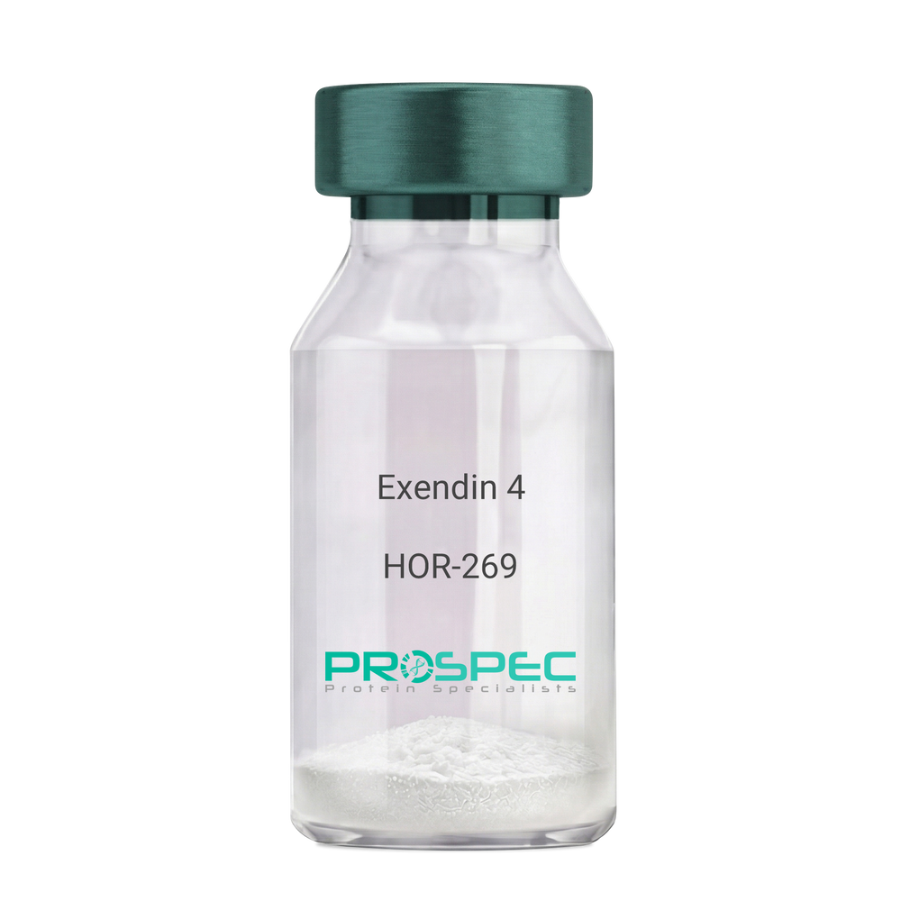
Exendin 4Description:
Exendin-4 Recombinant
Product # :
HOR-269Price :
Quantity :
Shipping Method :
Shipped at Room temp
More Info
- description
- source
- formulation
- purity
- biological activity
- More Info
Description
Exendin-4 Recombinant produced in E.Coli is a non-glycosylated polypeptide chain containing 39 amino acids and having a molecular mass of approximately 4.2kDa. Exendin-4 is purified by proprietary chromatographic techniques.
Source
Escherichia Coli.
Formulation
Lyophilized from a 0.2μm filtered concentrated solution in PBS, pH 7.4.
Purity
Greater than 96.0% as determined by:
(a) Analysis by RP-HPLC.
(b) Analysis by SDS-PAGE.Biological Activity
1. Regulates Glucose levels rapidly
2. Reduces Insulin resistance
3. Reduces Glucagon4. Stimulates beta cell growth production.
More Info
-
Introduction
Exedin-4 Recombinant is a like peptide-1 receptor agonist. The native hormone is produced in the gut of Gila monster Heloderma suspectrum (a type of reptile found in the desert) that stimulates production without causing threateningly low blood sugar, which can occur after using some anti-diabetes products. Recently, researchers used extracted saliva from gila monsters to create an unprecedented breakthrough in Diabetes Type 2 treatment. Unlike other products taken for type 2 diabetes, Exedin-4 has not been linked with weight gain and actually resulted in weight loss, according to the researchers. Exedin-4 enhances glucose-dependant secretion, suppresses inappropriately elevated secretion and slows gastric emptying in vivo. It also promotes B-cell proliferation and neogenesis in vitro and in animal models. Exedin-4 stimulates an increase in acinar cAMP, without stimulating the release of amylase.
-
Physical Appearance
Sterile Filtered White lyophilized (freeze-dried) powder.
-
Stability
Lyophilized Exendin Recombinant although stable at room temperature for 3 weeks, should be stored desiccated below -18°C. Upon reconstitution Exendin-4 should be stored at 4°C between 2-7 days and for future use below -18°C. For long term storage it is recommended to add a carrier protein (0.1% HSA or BSA).Please prevent freeze-thaw cycles.
-
Solubility
It is recommended to reconstitute the lyophilized Exendin Recombinant in sterile 18MΩ-cm H2O not less than 100 µg/ml, which can then be further diluted to other aqueous solutions.
-
Amino Acid Sequence
HGEGTFTSDL SKQMEEEAVR LFIEWLKNGG PSSGAPPPS.
ProSpec's products are furnished for LABORATORY RESEARCH USE ONLY. The product may not be used as drugs, agricultural or pesticidal products, food additives or household chemicals.
- View Data Sheet
Name :
Activin-A RatDescription:
Activin-A Rat Recombinant
Inhba, Inhibin beta A, FSH releasing protein.
Product # :
CYT-147Price :
Quantity :
Shipping Method :
Shipped at Room temp
More Info
- description
- source
- formulation
- purity
- biological activity
- More Info
Description
Active form Activin-A Rat Recombinant produced in e.coli is a homodimeric, non-glycosylated, polypeptide chain containing 2 x 117 amino acids and having a molecular weight of 26.2kDa.The Active form Activin-A is purified by standard chromatographic techniques.
Source
E.Coli.
Formulation
Rat Activin-A was lyophilized from a concentrated 1mg/ml protein solution containing 0.02% TFA.
Purity
Greater than 95% as obsereved by SDS-PAGE.
Biological Activity
Biological activity is assessed by the ability to induce cytoxicity of MPC-11 cells and was found to be 8.8ng/mlMore Info
-
Introduction
Activins are homodimers or heterodimers of the different β subunit isoforms, part of the TGFβ family. Mature Activin A has two 116 amino acids residues βA subunits (βA-βA). Activin displays an extensive variety of biological activities, including mesoderm induction, neural cell differentiation, bone remodelling, haematopoiesis, and reproductive physiology. Activins takes part in the production and regulation of hormones such as FSH, LH, GnRH and ACTH. Cells that are identified to express Activin A include fibroblasts, endothelial cells, hepatocytes, vascular smooth muscle cells, macrophages, keratinocytes, osteoclasts, bone marrow monocytes, prostatic epithelium, neurons, chondrocytes, osteoblasts, Leydig cells, Sertoli cells, and ovarian granulosa cells.
-
Synonyms
Inhba, Inhibin beta A, FSH releasing protein.
-
Physical Appearance
Lyophilized freeze dried powder.
-
Stability
Lyophilized Activin-A although stable at room temperature for 3 weeks, should be stored desiccated below -18°C. Upon reconstitution Activin-A should be stored at 4°C between 2-7 days and for future use below -18°C.For long term storage it is recommended to add a carrier protein (0.1% HSA or BSA).Please prevent freeze-thaw cycles.
-
Solubility
Rat INHBA protein should be reconstituted in distilled pyrogen free water to a concentration of 100ug /ml which can then be further diluted to other aqueous solutions.
-
Amino Acid Sequence
MGLECDGKVN ICCKKQFFVS FKDIGWNDWI IAPSGYHANY CEGECPSHIA GTSGSSLSFH STVINHYRMR GHSPFANLKS CCVPTKLRPM SMLYYDDGQN IIKKDIQNMI VEECGCS.
-
Background
What is the molecular weight / Mw of Activin A Protein?
Activin A Protein has a total Mw of 26.2 kDa.
What is the source or expression system of Activin A Protein?
Ecoli
What is the Purity of Activin A Protein?
Activin A Protein is >95% pure as determined by SDS-PAGE.
What is the Biological Activity of Activin A Protein?
Biological activity is assessed by the ability to induce cytoxicity of MPC-11 cells and was found to be 8.8ng/ml corresponding to a specific activity of 110,000units/mg.
What is the endotoxin level for Activin A Protein?
The endotoxin level is minimal, ACTIVIN A Protein was purified using conventional chromatography techniques.
What is the amino acid sequence of ACTIVIN A Protein?
MGLECDGKVN ICCKKQFFVS FKDIGWNDWI IAPSGYHANY CEGECPSHIA GTSGSSLSFH STVINHYRMR GHSPFANLKS CCVPTKLRPM SMLYYDDGQN IIKKDIQNMI VEECGCS.
What applications can ACTIVIN A Protein be used in?
ACTIVIN A Protein can probably be used in western blot, ELISA and Lateral Flow.
ProSpec's products are furnished for LABORATORY RESEARCH USE ONLY. The product may not be used as drugs, agricultural or pesticidal products, food additives or household chemicals.