Skip to content

High-quality research proteins.

  • 1-800-805-6531

  • Mon - Sat 8.00-18.00, Sun - Closed

  • info@prospecbio.com

  • PO Box 6591 East Brunswick NJ 08816

  • Products
    • Cytokines
    • Growth factors
    • Chemokines
    • Cd antigens
    • Neurotrophins
    • Hormones
    • Enzymes
    • Viral antigens
    • Recombinant proteins
    • Natural proteins
    • Monoclonal antibodies
    • Polyclonal antibodies
    • Thermal Packaging
  • About
    • Company
    • Founder
    • Contribution
  • Customers
    • Eastern Asia
    • Eastern Europe
    • North America
    • South America
    • South East Asia
    • Southern Asia
    • Western Europe
  • Ordering
    • Ordering Method
    • Ordering - Credit Card
    • Ordering - PO
    • Shipping Fees
    • FAQs
  • Services
    • DNA Cloning
    • Protein Expression
    • Monoclonal Antibody
    • Fermentation
    • Technology License
  • Contact us
Log in

Country/region

  • USD $ | Albania
  • USD $ | Andorra
  • USD $ | Argentina
  • USD $ | Armenia
  • USD $ | Australia
  • USD $ | Austria
  • USD $ | Azerbaijan
  • USD $ | Belarus
  • USD $ | Belgium
  • USD $ | Bolivia
  • USD $ | Brazil
  • USD $ | Bulgaria
  • USD $ | Cambodia
  • USD $ | Canada
  • USD $ | Chile
  • USD $ | China
  • USD $ | Colombia
  • USD $ | Costa Rica
  • USD $ | Croatia
  • USD $ | Cyprus
  • USD $ | Czechia
  • USD $ | Denmark
  • USD $ | Estonia
  • USD $ | Finland
  • USD $ | France
  • USD $ | Georgia
  • USD $ | Germany
  • USD $ | Greece
  • USD $ | Hong Kong SAR
  • USD $ | Hungary
  • USD $ | Iceland
  • USD $ | India
  • USD $ | Ireland
  • USD $ | Israel
  • USD $ | Italy
  • USD $ | Japan
  • USD $ | Kazakhstan
  • USD $ | Latvia
  • USD $ | Liechtenstein
  • USD $ | Lithuania
  • USD $ | Madagascar
  • USD $ | Malta
  • USD $ | Mexico
  • USD $ | Moldova
  • USD $ | Monaco
  • USD $ | Netherlands
  • USD $ | New Zealand
  • USD $ | North Macedonia
  • USD $ | Norway
  • USD $ | Panama
  • USD $ | Paraguay
  • USD $ | Peru
  • USD $ | Philippines
  • USD $ | Poland
  • USD $ | Portugal
  • USD $ | Romania
  • USD $ | Singapore
  • USD $ | Slovakia
  • USD $ | Slovenia
  • USD $ | South Africa
  • USD $ | South Korea
  • USD $ | Spain
  • USD $ | Sri Lanka
  • USD $ | Sweden
  • USD $ | Switzerland
  • USD $ | Taiwan
  • USD $ | Thailand
  • USD $ | Türkiye
  • USD $ | Ukraine
  • USD $ | United Kingdom
  • USD $ | United States
  • USD $ | Uruguay
  • USD $ | Vatican City
  • USD $ | Venezuela
  • USD $ | Vietnam
  • Facebook
  • Instagram
logoProSpec - Recombinant Protein Manufacturer for academic and R&D industry
Become a distributor
Account Log in
Wishlist
Cart Cart(0)
  • Products
    +
    • Cytokines
    • Growth factors
    • Chemokines
    • Cd antigens
    • Neurotrophins
    • Hormones
    • Enzymes
    • Viral antigens
    • Recombinant proteins
    • Natural proteins
    • Monoclonal antibodies
    • Polyclonal antibodies
    • Thermal Packaging
  • About
    +
    • Company
    • Founder
    • Contribution
  • Customers
    +
    • Eastern Asia
    • Eastern Europe
    • North America
    • South America
    • South East Asia
    • Southern Asia
    • Western Europe
  • Ordering
    +
    • Ordering Method
    • Ordering - Credit Card
    • Ordering - PO
    • Shipping Fees
    • FAQs
  • Services
    +
    • DNA Cloning
    • Protein Expression
    • Monoclonal Antibody
    • Fermentation
    • Technology License
  • Contact us

Item added to your cart

View cart
View All
  • Cytokines
  • Growth factors
  • Chemokines
  • Cd antigens
  • Neurotrophins
  • Hormones
  • Enzymes
  • Viral antigens
  • Recombinant proteins
  • Natural proteins
  • Monoclonal antibodies
  • Polyclonal antibodies
  • Thermal Packaging
  • Tumor Necrosis Factor

    Tumor Necrosis Factor

    View All
  • 4-1BB

    4-1BB

    View All
  • Adiponectin

    Adiponectin

    View All
  • AIF1

    AIF1

    View All
  • Other Cytokines

    Other Cytokines

    View All
  • AITRL

    AITRL

    View All
  • Angiopoietin

    Angiopoietin

    View All
  • Apolipoprotein

    Apolipoprotein

    View All
  • B-Cell Activating Factor

    B-Cell Activating Factor

    View All
  • Beta Defensin

    Beta Defensin

    View All
  • Bone Morphogenetic Protein

    Bone Morphogenetic Protein

    View All
  • B type Natriuretic Peptide

    B type Natriuretic Peptide

    View All
  • BST

    BST

    View All
  • Betacellulin

    Betacellulin

    View All
  • Cardiotrophin

    Cardiotrophin

    View All
  • Activin

    Activin

    View All
  • Fibroblast Growth Factor

    Fibroblast Growth Factor

    View All
  • Other Growth Factors

    Other Growth Factors

    View All
  • CSF

    CSF

    View All
  • CTGF

    CTGF

    View All
  • IGFBP

    IGFBP

    View All
  • VEGF Protein

    VEGF Protein

    View All
  • EGF

    EGF

    View All
  • Epigen

    Epigen

    View All
  • Erythropoietin

    Erythropoietin

    View All
  • Insulin-Like Growth Factor

    Insulin-Like Growth Factor

    View All
  • Growth Hormone

    Growth Hormone

    View All
  • HDGF

    HDGF

    View All
  • Hepatocyte Growth Factor

    Hepatocyte Growth Factor

    View All
  • Insulin

    Insulin

    View All
  • BCA-1/ BLC (CXCL13)

    BCA-1/ BLC (CXCL13)

    View All
  • BRAK (CXCL14)

    BRAK (CXCL14)

    View All
  • C-10 (CCL6)

    C-10 (CCL6)

    View All
  • Other Chemokines

    Other Chemokines

    View All
  • MEC (CCL28)

    MEC (CCL28)

    View All
  • LD78-beta (CCL3L1)

    LD78-beta (CCL3L1)

    View All
  • CTACK (CCL27)

    CTACK (CCL27)

    View All
  • CXCL16

    CXCL16

    View All
  • CXCL17

    CXCL17

    View All
  • Platelet Factor-4 (CXCL4)

    Platelet Factor-4 (CXCL4)

    View All
  • ENA-78 (CXCL5)

    ENA-78 (CXCL5)

    View All
  • Eotaxin (CCL11,24,26)

    Eotaxin (CCL11,24,26)

    View All
  • Exodus-2 (CCL21)

    Exodus-2 (CCL21)

    View All
  • Fractalkine (CX3CL1)

    Fractalkine (CX3CL1)

    View All
  • CXCL6

    CXCL6

    View All
  • Other CD Antigens

    Other CD Antigens

    View All
  • CD14

    CD14

    View All
  • CD164

    CD164

    View All
  • CD1

    CD1

    View All
  • CD2

    CD2

    View All
  • CD200

    CD200

    View All
  • CD207

    CD207

    View All
  • CD226

    CD226

    View All
  • CD23

    CD23

    View All
  • CD244

    CD244

    View All
  • CD247

    CD247

    View All
  • CD27

    CD27

    View All
  • CD274

    CD274

    View All
  • CD300

    CD300

    View All
  • CD33

    CD33

    View All
  • Other Neurotrophins

    Other Neurotrophins

    View All
  • Beta-NGF

    Beta-NGF

    View All
  • BDNF

    BDNF

    View All
  • CDNF

    CDNF

    View All
  • CNTF

    CNTF

    View All
  • GDNF

    GDNF

    View All
  • Glia Maturation Factor

    Glia Maturation Factor

    View All
  • MANF

    MANF

    View All
  • Midkine

    Midkine

    View All
  • Neuroglobin

    Neuroglobin

    View All
  • Neurotrophic factors

    Neurotrophic factors

    View All
  • Neuregulin

    Neuregulin

    View All
  • Neuropilin

    Neuropilin

    View All
  • Pigment Epithelium-Derived Factor

    Pigment Epithelium-Derived Factor

    View All
  • Pleiotrophin

    Pleiotrophin

    View All
  • Peptide Hormones

    Peptide Hormones

    View All
  • Other Hormones

    Other Hormones

    View All
  • HCG

    HCG

    View All
  • Endothelin

    Endothelin

    View All
  • Exendin

    Exendin

    View All
  • FSH

    FSH

    View All
  • Glucagon

    Glucagon

    View All
  • GHRP

    GHRP

    View All
  • GLP

    GLP

    View All
  • Thyrostimulin

    Thyrostimulin

    View All
  • Inhibin A

    Inhibin A

    View All
  • LHRH

    LHRH

    View All
  • Melanotan

    Melanotan

    View All
  • Procalcitonin

    Procalcitonin

    View All
  • PTH

    PTH

    View All
  • Peroxiredoxin

    Peroxiredoxin

    View All
  • Peptidase

    Peptidase

    View All
  • Synthetase

    Synthetase

    View All
  • Transferase

    Transferase

    View All
  • Hydrolase

    Hydrolase

    View All
  • Dehydrogenase

    Dehydrogenase

    View All
  • ACE2

    ACE2

    View All
  • Esterase

    Esterase

    View All
  • Protein Kinases

    Protein Kinases

    View All
  • Other Enzymes

    Other Enzymes

    View All
  • Phosphatase

    Phosphatase

    View All
  • Deaminase

    Deaminase

    View All
  • Oxygenase

    Oxygenase

    View All
  • Adenylate Kinase

    Adenylate Kinase

    View All
  • Lyase

    Lyase

    View All
  • Chagas

    Chagas

    View All
  • SARS

    SARS

    View All
  • Influenza

    Influenza

    View All
  • ASFV

    ASFV

    View All
  • Astrovirus

    Astrovirus

    View All
  • Borrelia

    Borrelia

    View All
  • Helicobacter Pylori

    Helicobacter Pylori

    View All
  • Chikungunya

    Chikungunya

    View All
  • Chlamydia

    Chlamydia

    View All
  • Cytomegalo

    Cytomegalo

    View All
  • Dengue

    Dengue

    View All
  • EBV

    EBV

    View All
  • Ebola

    Ebola

    View All
  • Feline Leukemia Virus

    Feline Leukemia Virus

    View All
  • Hepatitis A

    Hepatitis A

    View All
  • Other

    Other

    View All
  • Actin

    Actin

    View All
  • ADAM

    ADAM

    View All
  • Complement Component

    Complement Component

    View All
  • Ag85

    Ag85

    View All
  • Eukaryotic Translation Initiation Factor

    Eukaryotic Translation Initiation Factor

    View All
  • Anterior Gradient Protein

    Anterior Gradient Protein

    View All
  • Heat Shock Protein

    Heat Shock Protein

    View All
  • Alpha-2-HS-Glycoprotein

    Alpha-2-HS-Glycoprotein

    View All
  • Hemoglobin

    Hemoglobin

    View All
  • Allergy

    Allergy

    View All
  • Anaplasma

    Anaplasma

    View All
  • Angiogenin

    Angiogenin

    View All
  • Ankyrin Repeat Domain

    Ankyrin Repeat Domain

    View All
  • Annexin

    Annexin

    View All
  • Other Natural Proteins

    Other Natural Proteins

    View All
  • Aprotinin

    Aprotinin

    View All
  • Transferrin

    Transferrin

    View All
  • Avidin

    Avidin

    View All
  • Anti Coagulation Factors

    Anti Coagulation Factors

    View All
  • Natural Albumin

    Natural Albumin

    View All
  • Natural Coagulation Factors

    Natural Coagulation Factors

    View All
  • Fibronectin

    Fibronectin

    View All
  • Thrombin

    Thrombin

    View All
  • Anti Human Enzyme

    Anti Human Enzyme

    View All
  • Other Monoclonal Antibodies

    Other Monoclonal Antibodies

    View All
  • Anti Human Cytokine

    Anti Human Cytokine

    View All
  • Anti Human Heat Shock Protein

    Anti Human Heat Shock Protein

    View All
  • Anti Mouse Lymphocyte

    Anti Mouse Lymphocyte

    View All
  • Anti Human Chemokine

    Anti Human Chemokine

    View All
  • Anti Human Lymphocyte

    Anti Human Lymphocyte

    View All
  • Anti Viral Monoclonal

    Anti Viral Monoclonal

    View All
  • Anti Mouse Cytokine

    Anti Mouse Cytokine

    View All
  • Anti-GST Monoclonal

    Anti-GST Monoclonal

    View All
  • Other Polyclonal Antibodies

    Other Polyclonal Antibodies

    View All
  • Anti Viral

    Anti Viral

    View All
  • Anti-GST Monoclonal

    Anti-GST Monoclonal

    View All
Back

Search results

1000 results found for “F-Box Protein”

Name

Description

Product #

Price

Quantity

Shipping Method

  • View Data Sheet

    Name :

    Protein-G His

    Description:

    Protein G His Tag Recombinant

    Product # :

    PRO-1237

    Price :

    Quantity :

    Shipping Method :

    Room Temp Icon

    Shipped at Room temp

    Add To Cart

    More Info

    • description
    • source
    • formulation
    • purity
    • More Info

    Description

    Protein G His Tag Recombinant produced in E.Coli is a 201 amino acids protein which contains amino acid 190-384 of the Streptococcus sp with a C-terminal 6-His tag, and having a molecular mass of 21.6kDa. But it migrates with an apparent molecular mass of 32kDa in SDS-PAGE.The Protein G His Tag is purified by proprietary chromatographic techniques.

    Source

    Escherichia Coli.

    Formulation

    Lyophilized with no additives.

    Purity

    Greater than 95.0% as determined by SDS-PAGE and HPLC analyses.

    More Info

    • Physical Appearance

      Sterile Filtered White lyophilized (freeze-dried) powder.

    • Stability

      Lyophilized Protein G although stable at room temperature for 3 weeks, should be stored desiccated below -18°C. Upon reconstitution Protein G should be stored at 4°C between 2-7 days and for future use below -18°C.Please prevent freeze-thaw cycles.

    • Solubility

      It is recommended to reconstitute the lyophilized Protein G in sterile 18M-cm H2O not less than 100µg/ml, which can then be further diluted to other aqueous solutions.

    • Applications

      Protein G binds to the constant region of many species of immunoglobulin G. It can be used to detect, quantify and purify IgG antibodies and antibody/antigen complexes. Recombinant Protein G contains only IgG binding domains. The albumin-binding domain as well as cell wall and cell membrane binding domains have been removed to ensure the maximum specific IgG binding capacity.

    • Specificity

      1. Binds with greater affinity to most mammalian immunoglobulins than Protein A, including human IgG3 and rat IgG2a.2. Does not bind to human IgM, IgD and IgA.

    ProSpec's products are furnished for LABORATORY RESEARCH USE ONLY. The product may not be used as drugs, agricultural or pesticidal products, food additives or household chemicals.

    Protein G His
  • View Data Sheet

    Name :

    STUB1 Human

    Description:

    STIP1 Homology and U-Box Containing Protein 1 Human Recombinant

    CHIP, UBOX1, HSPABP2, NY-CO-7, SDCCAG7, STUB1, STIP1 homology and U box-containing protein 1, Carboxy terminus of Hsp70-interacting protein, E3 ubiquitin-protein ligase CHIP, CLL-associated antigen KW-8, Antigen NY-CO-7.

    Product # :

    HSP-019

    Price :

    Quantity :

    Shipping Method :

    Ice Icon

    Shipped with Ice Packs

    Add To Cart

    More Info

    • description
    • source
    • formulation
    • purity
    • More Info

    Description

    STUB1 Human Recombinant produced in E.Coli is a single, non-glycosylated polypeptide chain containing 303 amino acids and having a molecular mass of 34.8 kDa.STUB1 is expressed and purified by proprietary chromatographic techniques.

    Source

    Escherichia Coli.

    Formulation

    The STUB1 protein solution contains 20mM Tris-HCl, pH-7.5, 10% glycerol and 5mM DTT.

    Purity

    Greater than 90.0% as determined by SDS-PAGE.

    More Info

    • Introduction

      STUB1, is a cytoplasmic protein whose amino acid sequence is highly preserved across species. STUB1 interacts with the molecular chaperones Hsc70-Hsp70 and Hsp90 through its TPR domain, whereas its U-box domain contains its E3 ubiquitin ligase activity. STUB1 interaction with these molecular chaperones lead to in client substrate ubiquitylation and degradation by the proteasome. therefore, STUB1 acts to tilt the folding-refolding mechanism towards the degradative pathway, and it serves as a link between the two. STUB1 inhibits anchorage-independent cell growth and metastatic potential by degrading oncogenic proteins including SRC-3. Inhibition of tyrosine kinase activity of Her-2/neu by quercetin specifies an lateration in the Her-2/neu structure which promotes STUB1 recruitments and down-regulation of Her-2/neu. STUB1 recognizes and mediates degradation of toxic, oligomeric forms of alphaSyn.

    • Synonyms

      CHIP, UBOX1, HSPABP2, NY-CO-7, SDCCAG7, STUB1, STIP1 homology and U box-containing protein 1, Carboxy terminus of Hsp70-interacting protein, E3 ubiquitin-protein ligase CHIP, CLL-associated antigen KW-8, Antigen NY-CO-7.

    • Physical Appearance

      Sterile filtered colorless solution.

    • Stability

      Store at 4°C if entire vial will be used within 2-4 weeks. Store, frozen at -20°C for longer periods of time. For long term storage it is recommended to add a carrier protein (0.1% HSA or BSA). Avoid multiple freeze-thaw cycles.

    • Amino Acid Sequence

      MKGKEEKEGG ARLGAGGGSP EKSPSAQELK EQGNRLFVGR KYPEAAACYG RAITRNPLVA VYYTNRALCY LKMQQHEQAL ADCRRALELD GQSVKAHFFL GQCQLEMESY DEAIANLQRA YSLAKEQRLN FGDDIPSALR IAKKKRWNSI EERRIHQESE LHSYLSRLIA AERERELEEC QRNHEGDEDD SHVRAQQACI EAKHDKYMAD MDELFSQVDE KRKKRDIPDY LCGKISFELM REPCITPSGI TYDRKDIEEH LQRVGHFDPV TRSPLTQEQL IPNLAMKEVI DAFISENGWV EDY.

    ProSpec's products are furnished for LABORATORY RESEARCH USE ONLY. The product may not be used as drugs, agricultural or pesticidal products, food additives or household chemicals.

    Stub1 Human
  • View Data Sheet

    Name :

    Cys-Protein-A/G/L

    Description:

    Cys-Protein A/G/L Recombinant

    Product # :

    PRO-1935

    Price :

    Quantity :

    Shipping Method :

    Room Temp Icon

    Shipped at Room temp

    Add To Cart

    More Info

    • description
    • source
    • formulation
    • purity
    • More Info

    Description

    Recombinant Protein-A/G/L produced in E.Coli is a single non-glycosylated polypeptide chain fused with a Cys at N-terminus. Protein- A/G/L is comprised of 5 IgG-binding regions of Protein A (E-D-A-B-C), 2 of protein G (C1-C3) and 5 of Protein L (B1-B2-B3-B4-B5) containing 806 amino acids in total and having a molecular mass of 89.3kDa. Cell wall binding region, cell membrane binding region and albumin binding region have been eliminated from the recombinant Protein- A/G/L to guarantee the maximum specific IgG binding.

    Source

    Escherichia Coli.

    Formulation

    Protein- A/G/L was lyophilized without any additives.

    Purity

    Greater than 95.0% as determined by:
    (a) Analysis by SEC-HPLC.
    (b) Analysis by SDS-PAGE.

    More Info

    • Introduction

      The recombinant Protein A/G/L is a genetically engineered protein which combines the IgG binding profiles of all Protein A, Protein G and Protein L. Protein A/G/L is a gene fusion product. Recombinant fusion protein A/G/L is comprised of 5 Ig-binding regions of protein L (B1-B2-B3-B4-B5), 5 IgG binding domains from Protein A (E-D-A-B-C) and 2 Ig-binding region of protein G (C1-C3). The recombinant Protein A/G/L is ideal for purification of monoclonal or polyclonal IgG antibodies. Protein A/G/L binds to IgG from humans, mice, rats, cows, goats, sheep, rabbits, guinea pigs, pigs, dogs and cats.

    • Physical Appearance

      Sterile Filtered White lyophilized (freeze-dried) powder.

    • Stability

      Lyophilized Protein-A/G/L although stable at room temperature for 3 weeks, should be stored desiccated below -18°C. Upon reconstitution Protein-A/G/L should be stored at 4°C between 2-7 days and for future use below -18°C. For long term storage it is recommended to add a carrier protein (0.1% HSA or BSA). Please prevent freeze-thaw cycles.

    • Solubility

      It is recommended to reconstitute the lyophilized Protein-A/G/L in sterile 18M-cm H2O not less than 0.1mg/ml, which can then be further diluted to other aqueous solutions.

    • Amino Acid Sequence

      CNAAQHDEAQ QNAFYQVLNM PNLNADQRNG FIQSLKDDPS QSANVLGEAQ KLNDSQAPKA DAQQNNFNKD QQSAFYEILN MPNLNEAQRN GFIQSLKDDP SQSTNVLGEA KKLNESQAPK ADNNFNKEQQ NAFYEILNMP NLNEEQRNGF IQSLKDDPSQ SANLLSEAKK LNESQAPKAD NKFNKEQQNA FYEILHLPNL NEEQRNGFIQ SLKDDPSQSA NLLAEAKKLN DAQAPKADNK FNKEQQNAFY EILHLPNLTE EQRNGFIQSL KDDPSVSKEI LAEAKKLNDA QAPKEEDSLE GSGSGTYKLI LNGKTLKGET TTEAVDAATA EKVFKQYAND NGVDGEWTYD DATKTFTVTE KPEVIDASEL TPAVTTYKLV INGKTLKGET TTKAVDAETA EKAFKQYAND NGVDGVWTYD DATKTFTVTE EPRARPGSGS GKEETPETPE TDSEEEVTIK ANLIFANGST QTAEFKGTFE KATSEAYAYA DTLKKDNGEY TVDVADKGYT LNIKFAGKEK TPEEPKEEVT IKANLIYADG KTQTAEFKGT FEEATAEAYR YADALKKDNG EYTVDVADKG YTLNIKFAGK EKTPEEPKEE VTIKANLIYA DGKTQTAEFK GTFEEATAEA YRYADLLAKE NGKYTVDVAD KGYTLNIKFA GKEKTPEEPK EEVTIKANLI YADGKTQTAE FKGTFAEATA EAYRYADLLA KENGKYTADL EDGGYTINIR FAGKKVDEKP EEKEQVTIKE NIYFEDGTVQ TATFKGTFAE ATAEAYRYAD LLSKEHGKYT ADLEDGGYTI NIRFAG.

    ProSpec's products are furnished for LABORATORY RESEARCH USE ONLY. The product may not be used as drugs, agricultural or pesticidal products, food additives or household chemicals.

    Cys Protein A G L
  • View Data Sheet

    Name :

    PLEKHF2 Human

    Description:

    Pleckstrin Homology Domain Containing Family F Member 2 Human Recombinant

    EAPF, PHAFIN2, ZFYVE18, Pleckstrin homology domain-containing family F member 2, PH domain-containing family F member 2, Endoplasmic reticulum-associated apoptosis-involved protein containing PH and FYVE domains, PH and FYVE domain-containing protein 2, Zinc finger FYVE domain-containing protein 18, PLEKHF2.

    Product # :

    PRO-1874

    Price :

    Quantity :

    Shipping Method :

    Ice Icon

    Shipped with Ice Packs

    Add To Cart

    More Info

    • description
    • source
    • formulation
    • purity
    • More Info

    Description

    PLEKHF2 Human Recombinant produced in E.Coli is a single, non-glycosylated polypeptide chain containing 272 amino acids (1-249 a.a) and having a molecular mass of 30.2kDa. PLEKHF2 is fused to a 23 amino acid His-tag at N-terminus & purified by proprietary chromatographic techniques.

    Source

    Escherichia Coli.

    Formulation

    PLEKHF2 protein solution (0.25mg/ml) containing 20mM Tris-HCl buffer (pH 8.0), 0.15M NaCl, 10% glycerol and 1mM DTT.

    Purity

    Greater than 95.0% as determined by SDS-PAGE.

    More Info

    • Introduction

      Pleckstrin Homology Domain Containing Family F Member 2, also known as PLEKHF2, contains 1 FYVE-type zinc finger and 1 PH domain. PLEKHF2 takes part in early endosome fusion upstream of RAB5, regulates receptor trafficking and fluid-phase transport. PLEKHF2 increases cellular sensitivity to TNF-induced apoptosis.

    • Synonyms

      EAPF, PHAFIN2, ZFYVE18, Pleckstrin homology domain-containing family F member 2, PH domain-containing family F member 2, Endoplasmic reticulum-associated apoptosis-involved protein containing PH and FYVE domains, PH and FYVE domain-containing protein 2, Zinc finger FYVE domain-containing protein 18, PLEKHF2.

    • Physical Appearance

      Sterile Filtered colorless solution.

    • Stability

      Store at 4°C if entire vial will be used within 2-4 weeks. Store, frozen at -20°C for longer periods of time. For long term storage it is recommended to add a carrier protein (0.1% HSA or BSA).Avoid multiple freeze-thaw cycles.

    • Amino Acid Sequence

      MGSSHHHHHH SSGLVPRGSH MGSMVDRLAN SEANTRRISI VENCFGAAGQ PLTIPGRVLI GEGVLTKLCR KKPKARQFFL FNDILVYGNI VIQKKKYNKQ HIIPLENVTI DSIKDEGDLR NGWLIKTPTK SFAVYAATAT EKSEWMNHIN KCVTDLLSKS GKTPSNEHAA VWVPDSEATV CMRCQKAKFT PVNRRHHCRK CGFVVCGPCS EKRFLLPSQS SKPVRICDFC YDLLSAGDMA TCQPARSDSY SQSLKSPLND MSDDDDDDDS SD.

    ProSpec's products are furnished for LABORATORY RESEARCH USE ONLY. The product may not be used as drugs, agricultural or pesticidal products, food additives or household chemicals.

    Plekhf2 Human
  • View Data Sheet

    Name :

    FGB Human

    Description:

    Fibrinogen Beta Chain Human Recombinant

    Fibrinogen beta chain, Fibrinopeptide B, Fibrinogen, B Beta Polypeptide, Epididymis Secretory Sperm Binding Protein Li 78p, HEL-S-78p.

    Product # :

    PRO-2210

    Price :

    Quantity :

    Shipping Method :

    Ice Icon

    Shipped with Ice Packs

    Add To Cart

    More Info

    • description
    • source
    • formulation
    • purity
    • More Info

    Description

    FGB Human Recombinant produced in E.Coli is a single, non-glycosylated polypeptide chain containing 351 amino acids (164-491a.a) and having a molecular mass of 40kDa. FGB is fused to a 23 amino acid His-tag at N-terminus & purified by proprietary chromatographic techniques.

    Source

    Escherichia Coli.

    Formulation

    FGB protein solution (1mg/ml) containing 20mM Tris-HCl buffer (pH8.0) and 10% glycerol.

    Purity

    Greater than 85.0% as determined by SDS-PAGE.

    More Info

    • Introduction

      Fibrinogen Beta Chain 9 (FGB) is the beta component of fibrinogen, a blood-borne glycoprotein composed of 3 pairs of nonidentical polypeptide chains. When vascular injury accrues, fibrinogen is cleaved by thrombin to form fibrin which is found in abundance in blood clots.

    • Synonyms

      Fibrinogen beta chain, Fibrinopeptide B, Fibrinogen, B Beta Polypeptide, Epididymis Secretory Sperm Binding Protein Li 78p, HEL-S-78p.

    • Physical Appearance

      Sterile Filtered colorless solution.

    • Stability

      Store at 4°C if entire vial will be used within 2-4 weeks. Store, frozen at -20°C for longer periods of time. For long term storage it is recommended to add a carrier protein (0.1% HSA or BSA). Avoid multiple freeze-thaw cycles.

    • Amino Acid Sequence

      MGSSHHHHHH SSGLVPRGSH MGSDNENVVN EYSSELEKHQ LYIDETVNSN IPTNLRVLRS ILENLRSKIQ KLESDVSAQM EYCRTPCTVS CNIPVVSGKE CEEIIRKGGE TSEMYLIQPD SSVKPYRVYC DMNTENGGWT VIQNRQDGSV DFGRKWDPYK QGFGNVATNT DGKNYCGLPG EYWLGNDKIS QLTRMGPTEL LIEMEDWKGD KVKAHYGGFT VQNEANKYQI SVNKYRGTAG NALMDGASQL MGENRTMTIH NGMFFSTYDR DNDGWLTSDP RKQCSKEDGG GWWYNRCHAA NPNGRYYWGG QYTWDMAKHG TDDGVVWMNW KGSWYSMRKM SMKIRPFFPQ Q.

    ProSpec's products are furnished for LABORATORY RESEARCH USE ONLY. The product may not be used as drugs, agricultural or pesticidal products, food additives or household chemicals.

    Fgb Human
  • View Data Sheet

    Name :

    Protein-L Cys, His

    Description:

    Protein-L Cys Recombinant, His Tag

    Product # :

    PRO-1932

    Price :

    Quantity :

    Shipping Method :

    Room Temp Icon

    Shipped at Room temp

    Add To Cart

    More Info

    • description
    • source
    • formulation
    • purity
    • More Info

    Description

    Recombinant Protein-L produced in E.Coli is a single non-glycosylated polypeptide chain fused with a 6×His tag at N-terminus and a Cys on C-terminus. Protein-L is comprised of 5 IgG-binding regions of protein L (B1-B2-B3-B4-B5) containing 373 amino acids in total and having a molecular mass of 41.6kDa, however, it migrates with an apparent molecular mass of 46kDa on SDS-PAGE. Cell wall binding region, cell membrane binding region and albumin binding region have been eliminated from the recombinant Protein-L to guarantee the maximum specific IgG binding.

    Source

    Escherichia Coli.

    Formulation

    Protein-L was lyophilized without any additives.

    Purity

    Greater than 96.0% as determined by SDS-PAGE.

    More Info

    • Introduction

      The Recombinant Protein L is comprised of 5 kappa-binding domains. Protein L has the exceptional ability to bind through kappa light chain interactions without hindering with the antibody’s antigen-binding site. This gives Protein L the capacity to bind a broader range of Ig classes and subclasses than other antibody-binding proteins. The recombinant Protein L is perfect for purification of polyclonal or monoclonal IgG antibodies. Protein L binds to IgG from humans, mice, rats and pigs.

    • Physical Appearance

      Sterile Filtered White lyophilized (freeze-dried) powder.

    • Stability

      Lyophilized Protein-L although stable at room temperature for 3 weeks, should be stored desiccated below -18°C. Upon reconstitution Protein-L should be stored at 4°C between 2-7 days and for future use below -18°C. For long term storage it is recommended to add a carrier protein (0.1% HSA or BSA). Please prevent freeze-thaw cycles.

    • Solubility

      It is recommended to reconstitute the lyophilized Protein-L in sterile 18M-cm H2O not less than 0.1mg/ml, which can then be further diluted to other aqueous solutions.

    • Amino Acid Sequence

      MHHHHHHKEE TPETPETDSE EEVTIKANLI FANGSTQTAE FKGTFEKATS EAYAYADTLK KDNGEYTVDV ADKGYTLNIK FAGKEKTPEE PKEEVTIKAN LIYADGKTQT AEFKGTFEEA TAEAYRYADA LKKDNGEYTV DVADKGYTLN IKFAGKEKTP EEPKEEVTIK ANLIYADGKT QTAEFKGTFE EATAEAYRYA DLLAKENGKY TVDVADKGYT LNIKFAGKEK TPEEPKEEVT IKANLIYADG KTQTAEFKGT FAEATAEAYR YADLLAKENG KYTADLEDGG YTINIRFAGK KVDEKPEEKE QVTIKENIYF EDGTVQTATF KGTFAEATAE AYRYADLLSK EHGKYTADLE DGGYTINIRF AGC.

    ProSpec's products are furnished for LABORATORY RESEARCH USE ONLY. The product may not be used as drugs, agricultural or pesticidal products, food additives or household chemicals.

    Protein L Cys His
  • View Data Sheet

    Name :

    Cys-Protein-L

    Description:

    Cys-Protein L Recombinant

    Product # :

    PRO-1933

    Price :

    Quantity :

    Shipping Method :

    Room Temp Icon

    Shipped at Room temp

    Add To Cart

    More Info

    • description
    • source
    • formulation
    • purity
    • More Info

    Description

    Recombinant Protein-L produced in E.Coli is a single non-glycosylated polypeptide chain fused with a Cys at N-terminus. Protein-L is comprised of 5 IgG-binding regions of protein L (B1-B2-B3-B4-B5) containing 366 amino acids in total and having a molecular mass of 40.6kDa, however, it migrates with an apparent molecular mass of 46kDa on SDS-PAGE. Cell wall binding region, cell membrane binding region and albumin binding region have been eliminated from the recombinant Protein-L to guarantee the maximum specific IgG binding.

    Source

    Escherichia Coli.

    Formulation

    Protein-L was lyophilized without any additives.

    Purity

    Greater than 95.0% as determined by SDS-PAGE.

    More Info

    • Introduction

      The Recombinant Protein L is comprised of 5 kappa-binding domains. Protein L has the exceptional ability to bind through kappa light chain interactions without hindering with the antibody’s antigen-binding site. This gives Protein L the capacity to bind a broader range of Ig classes and subclasses than other antibody-binding proteins. The recombinant Protein L is perfect for purification of polyclonal or monoclonal IgG antibodies. Protein L binds to IgG from humans, mice, rats and pigs.

    • Physical Appearance

      Sterile Filtered White lyophilized (freeze-dried) powder.

    • Stability

      Lyophilized Protein-L although stable at room temperature for 3 weeks, should be stored desiccated below -18°C. Upon reconstitution Protein-L should be stored at 4°C between 2-7 days and for future use below -18°C. For long term storage it is recommended to add a carrier protein (0.1% HSA or BSA). Please prevent freeze-thaw cycles.

    • Solubility

      It is recommended to reconstitute the lyophilized Protein-L in sterile 18M-cm H2O not less than 0.1mg/ml, which can then be further diluted to other aqueous solutions.

    • Amino Acid Sequence

      CKEETPETPE TDSEEEVTIK ANLIFANGST QTAEFKGTFE KATSEAYAYA DTLKKDNGEY TVDVADKGYT LNIKFAGKEK TPEEPKEEVT IKANLIYADG KTQTAEFKGT FEEATAEAYR YADALKKDNG EYTVDVADKG YTLNIKFAGK EKTPEEPKEE VTIKANLIYA DGKTQTAEFK GTFEEATAEA YRYADLLAKE NGKYTVDVAD KGYTLNIKFA GKEKTPEEPK EEVTIKANLI YADGKTQTAE FKGTFAEATA EAYRYADLLA KENGKYTADL EDGGYTINIR FAGKKVDEKP EEKEQVTIKE NIYFEDGTVQ TATFKGTFAE ATAEAYRYAD LLSKEHGKYT ADLEDGGYTI NIRFAG.

    ProSpec's products are furnished for LABORATORY RESEARCH USE ONLY. The product may not be used as drugs, agricultural or pesticidal products, food additives or household chemicals.

    Cys Protein L
  • View Data Sheet

    Name :

    Protein-L

    Description:

    Protein L Recombinant

    SPL.

    Product # :

    PRO-1790

    Price :

    Quantity :

    Shipping Method :

    Room Temp Icon

    Shipped at Room temp

    Add To Cart

    More Info

    • description
    • source
    • formulation
    • purity
    • More Info

    Description

    Protein-L Recombinant produced in E.coli is a single, non-glycosylated polypeptide chain containing 365 amino acids and having a molecular mass of 40.5 kDa but it migrates with an apparent molecular mass of 45 kDa in SDS-PAGE.The Protein-L is purified by proprietary chromatographic techniques.

    Source

    Escherichia Coli.

    Formulation

    Protein-L was lyophilized with no additives.

    Purity

    Greater than 97.0% as determined by:
    (a) Analysis by SEC-HPLC.
    (b) Analysis by SDS-PAGE.

    More Info

    • Introduction

      The recombinant Protein L is genetically engineered protein and holds 5 IgG-binding regions of protein L and it has the exclusive capability to bind through kappa light chain interactions without interfering with the antibody’s antigen-binding site. Cell wall binding region, cell membrane binding region and albumin binding region were removed to ensure the highest specific IgG binding. The recombinant Protein L is perfect for purification of polyclonal or monoclonal IgG antibodies and it binds to human, mouse, rat and pig IgG.

    • Synonyms

      SPL.

    • Physical Appearance

      Sterile Filtered White lyophilized (freeze-dried) powder.

    • Stability

      Lyophilized Protein-L although stable at room temperature for 3 weeks, should be stored desiccated below -18°C. Upon reconstitution Protein-L should be stored at 4°C between 2-7 days and for future use below -18°C. For long term storage it is recommended to add a carrier protein (0.1% HSA or BSA). Please prevent freeze-thaw cycles.

    • Solubility

      It is recommended to reconstitute the lyophilized Protein-L in sterile water or saline not less than 100µg/ml, which can then be further diluted to other aqueous solutions.

    • Amino Acid Sequence

      KEETPETPET DSEEEVTIKA NLIFANGSTQ TAEFKGTFEK ATSEAYAYAD TLKKDNGEYT VDVADKGYTL NIKFAGKEKT PEEPKEEVTI KANLIYADGK TQTAEFKGTF EEATAEAYRY ADALKKDNGE YTVDVADKGY TLNIKFAGKE KTPEEPKEEV TIKANLIYAD GKTQTAEFKG TFEEATAEAY RYADLLAKEN GKYTVDVADK GYTLNIKFAG KEKTPEEPKE EVTIKANLIY ADGKTQTAEF KGTFAEATAE AYRYADLLAK ENGKYTADLE DGGYTINIRF AGKKVDEKPEEKEQVTIKEN IYFEDGTVQT ATFKGTFAEA TAEAYRYADL LSKEHGKYTA DLEDGGYTIN IRFAG

    ProSpec's products are furnished for LABORATORY RESEARCH USE ONLY. The product may not be used as drugs, agricultural or pesticidal products, food additives or household chemicals.

    Protein L
  • View Data Sheet

    Name :

    CST3 Protein, His

    Description:

    Cystatin-C Human Recombinant, His Tag

    Cystatin-C, Cystatin-3, Neuroendocrine basic polypeptide, Gamma-trace, Post-gamma-globulin, CST3, MGC117328.

    Product # :

    PRO-656

    Price :

    Quantity :

    Shipping Method :

    Room Temp Icon

    Shipped at Room temp

    Add To Cart

    More Info

    • description
    • source
    • formulation
    • purity
    • More Info

    Description

    Cystatin-C Human Recombinant produced in E.Coli is a single, non-glycosylated, Polypeptide chain containing 129 amino acids and having a molecular mass of 14.5 kDa. The protein contains an extra His tag at N-terminus. The Cystatin-C amino acid sequence is identical to UniProtKB/Swiss-Prot entry Q6FGW9 amino acids 28–146.The Cystatin-C is purified by proprietary chromatographic techniques.

    Source

    Escherichia Coli.

    Formulation

    Filtered (0.4µm) and lyophilized from 0.5mg/ml in 20mM Tris buffer and 50mM NaCl pH-7.5.

    Purity

    Greater than 95% as determined by SDS PAGE.

    More Info

    • Introduction

      Cystatins are a superfamily of cysteine proteinase inhibitors found in both plants and animals. They comprise a group of proteinase inhibitors, widely distributed in tissues and body fluids, and form tight complexes with cysteine proteases such as cathepsin B, H, L and S. Cystatin C, a secreted molecule of this family, is of interest from biochemical, medicine and evolutionary points of view. Cystatin C, with molecular weight of 13260 Da, is composed of 120 amino acids, lacks carbohydrate and has two disulfide bridges located near the carboxyl terminus. Cystatin C is increased in patients with malignant diseases, and is related to the insufficiency of renal function and appears to be a better marker than creatinine. On the other hand, low levels of cystatin C involve cause the breakdown of the elastic laminae and, subsequently, the atherosclerosis and abdominal aortic aneurysm.

    • Synonyms

      Cystatin-C, Cystatin-3, Neuroendocrine basic polypeptide, Gamma-trace, Post-gamma-globulin, CST3, MGC117328.

    • Physical Appearance

      Filtered White lyophilized (freeze-dried) powder.

    • Stability

      Store lyophilized protein at –20°C. Aliquot reconstituted protein to avoid repeated freezing/thawing cycles and store at –80°C for long term storage. Reconstituted protein can be stored at 4°C for a limited period of time; it does not show any change after two weeks at 4°C.

    • Solubility

      Add deionized water to prepare a working stock solution of approximately 0.5mg/ml and let the lyophilized pellet dissolve completely. Product is not sterile! Please filter the product by an appropriate sterile filter before using it in the cell culture.

    ProSpec's products are furnished for LABORATORY RESEARCH USE ONLY. The product may not be used as drugs, agricultural or pesticidal products, food additives or household chemicals.

    Cystatin C Human Recombinant
  • View Data Sheet

    Name :

    FAM84A Human

    Description:

    Family with Sequence Similarity 84, Member A Human Recombinant

    Family with Sequence Similarity 84 Member A, Neurological/Sensory 1, Neurologic Sensory Protein 1, NSE1, PP11517, Protein FAM84A.

    Product # :

    PRO-1610

    Price :

    Quantity :

    Shipping Method :

    Ice Icon

    Shipped with Ice Packs

    Add To Cart

    More Info

    • description
    • source
    • formulation
    • purity
    • More Info

    Description

    FAM84A Human Recombinant produced in E.coli is a single, non-glycosylated polypeptide chain containing 315 amino acids (1-292) and having a molecular mass of 34.9kDa.FAM84A is fused to a 23 amino acid His-tag at N-terminus & purified by proprietary chromatographic techniques.

    Source

    Escherichia Coli.

    Formulation

    The FAM84A solution (1mg/ml) contains 20mM Tris-HCl buffer (pH 8.0), 0.15M NaCl, 1mM DTT and 10% glycerol.

    Purity

    Greater than 90% as determined by SDS-PAGE.

    More Info

    • Introduction

      FAM84A is a member of the FAM84 family of proteins and is primarily expressed in testis. FAM84A is restricted to a subcellular membrane region in which there is no interaction between adjoining cells and takes part in cell morphology. Additionally, FAM84A expression increases cell motility which can result in invasion and metastasis of cancer cells. FAM84A is up-regulated in pancreatic cancer, colorectal cancer lung cancer, bladder cancer tissues and cholangiocarcinoma.

    • Synonyms

      Family with Sequence Similarity 84 Member A, Neurological/Sensory 1, Neurologic Sensory Protein 1, NSE1, PP11517, Protein FAM84A.

    • Physical Appearance

      Sterile filtered colorless solution.

    • Stability

      Store at 4°C if entire vial will be used within 2-4 weeks. Store, frozen at -20°C for longer periods of time. For long term storage it is recommended to add a carrier protein (0.1% HSA or BSA).Avoid multiple freeze-thaw cycles.

    • Amino Acid Sequence

      MGSSHHHHHH SSGLVPRGSH MGSMGNQLDR ITHLNYSELP TGDPSGIEKD ELRVGVAYFF SDDEEDLDER GQPDKFGVKA PPGCTPCPES PSRHHHHLLH QLVLNETQFS AFRGQECIFS KVSGGPQGAD LSVYAVTALP ALCEPGDLLE LLWLQPAPEP PAPAPHWAVY VGGGQIIHLH QGEIRQDSLY EAGAANVGRV VNSWYRYRPL VAELVVQNAC GHLGLKSEEI CWTNSESFAA WCRFGKREFK AGGEVPAGTQ PPQQQYYLKV HLGENKVHTA RFHSLEDLIR EKRRIDASGR LRVLQELADL VDDKE.

    ProSpec's products are furnished for LABORATORY RESEARCH USE ONLY. The product may not be used as drugs, agricultural or pesticidal products, food additives or household chemicals.

    Fam84A Human
  • View Data Sheet

    Name :

    FTH1 Human

    Description:

    Ferritin Human Recombinant, Heavy Chain

    Ferritin heavy chain, Cell proliferation-inducing gene 15 protein, FTH1, FHC, FTH, PLIF, FTHL6, PIG15, MGC104426.

    Product # :

    PRO-658

    Price :

    Quantity :

    Shipping Method :

    Ice Icon

    Shipped with Ice Packs

    Add To Cart

    More Info

    • description
    • source
    • formulation
    • purity
    • More Info

    Description

    FTH1 Human Recombinant produced in E.Coli is a single, non-glycosylated polypeptide chain containing 183 amino acids and having a molecular mass of 21 kDa.

    Source

    Escherichia Coli.

    Formulation

    The FTH1 protein solution contains 20mM Tris-HCl pH-7.5, 1mM DTT and 10% glycerol.

    Purity

    Greater than 95.0% as determined by SDS-PAGE.

    More Info

    • Introduction

      Ferritin is a fairly large, iron-storage heteropolymeric protein composed of 2 subunit types, light Ferritin & heavy Ferritin polypeptides, which is expressed in most kinds of cells and co-assemble in different proportion in a tissue-specific manner. Ferritin is composed of 24 self-assembled polypeptide subunits of the heavy and light ferritin chains and is characterized by the capacity to remove Fe (II) from solution in the presence of oxygen.
      Ferritin light polypeptide protein is the main intracellular iron storage protein in prokaryotes and eukaryotes. Variation in ferritin subunit composition influence the rates of iron uptake and release in various tissues. A key function of ferritin is the storage of iron in a soluble and nontoxic state. Defects in this light chain ferritin gene are associated with several neurodegenerative diseases and hyper ferrit anemia-cataract syndrome.
      Ferritin stores iron in a soluble, nontoxic, readily accessible form. Ferritin is needed for iron homeostasis. Iron is taken up in the ferrous form and deposited as ferric hydroxides after it has been oxidized.

    • Synonyms

      Ferritin heavy chain, Cell proliferation-inducing gene 15 protein, FTH1, FHC, FTH, PLIF, FTHL6, PIG15, MGC104426.

    • Physical Appearance

      Sterile Filtered colorless solution.

    • Stability

      Store at 4°C if entire vial will be used within 2-4 weeks. Store, frozen at -20°C for longer periods of time. For long term storage it is recommended to add a carrier protein (0.1% HSA or BSA).Avoid multiple freeze-thaw cycles.

    • Amino Acid Sequence

      MTTASTSQVR QNYHQDSEAA INRQINLELY ASYVYLSMSY YFDRDDVALK NFAKYFLHQS HEEREHAEKL MKLQNQRGGR IFLQDIKKPD CDDWESGLNA MECALHLEKN VNQSLLELHK LATDKNDPHL CDFIETHYLN EQVKAIKELG DHVTNLRKMG APESGLAEYL FDKHTLGDSD NES.

    ProSpec's products are furnished for LABORATORY RESEARCH USE ONLY. The product may not be used as drugs, agricultural or pesticidal products, food additives or household chemicals.

    Fth1 Human
  • View Data Sheet

    Name :

    AKT1 Human, Sf9

    Description:

    Protein Kinase B Alpha Human Recombinant, Sf9

    V-Akt Murine Thymoma Viral Oncogene Homolog 1, Protein Kinase B Alpha, Proto-Oncogene C-Akt, RAC-PK-Alpha, EC 2.7.11.1, PKB Alpha, CWS6, PKB, RAC, RAC-Alpha Serine/Threonine-Protein Kinase, Rac Protein Kinase Alpha, Protein Kinase B, PKB-ALPHA, RAC-ALPHA, EC 2.7.11, AKT1m, PRKBA, AKT, RAC-alpha serine/threonine-protein kinase.

    Product # :

    PKA-069

    Price :

    Quantity :

    Shipping Method :

    Ice Icon

    Shipped with Ice Packs

    Add To Cart

    More Info

    • description
    • source
    • formulation
    • purity
    • More Info

    Description

    AKT1 produced in Sf9 Baculovirus cells is a single, glycosylated polypeptide chain containing 488 amino acids (1-480a.a.) and having a molecular mass of 56.7kDa. (Molecular size on SDS-PAGE will appear at approximately 50-70kDa). AKT1 is expressed with an 8 amino acid His tag at C-Terminus and purified by proprietary chromatographic techniques.

    Source

    Sf9, Baculovirus cells.

    Formulation

    AKT1 protein solution (0. 5mg/ml) contains phosphate buffered saline (pH7.4) and 20% glycerol.

    Purity

    Greater than 90.0% as determined by SDS-PAGE.

    More Info

    • Introduction

      Akt1, also known as Akt or else protein kinase B (PKB) is an important molecule in mammaliancellular signaling. In humans, there are three genes in the "Akt family": Akt1, Akt2, and Akt3. Moreover, these enzymes are members of the serine/threonine-specific protein kinase family (EC2.7.11.1). Akt1 is involved in cellular survival pathways, by inhibiting apoptoticprocesses. Akt1 is also able to induce protein synthesis pathways, and is therefore a key signaling protein in the cellular pathways which lead to skeletal muscle hypertrophy, and general tissue growth. Since it can block apoptosis, and thereby promote cell survival, Akt1 has been implicated as a most important factor in numerous types of cancer. Akt (now also called Akt1) was at first identified as the oncogenein the transforming retrovirus, AKT8.

    • Synonyms

      V-Akt Murine Thymoma Viral Oncogene Homolog 1, Protein Kinase B Alpha, Proto-Oncogene C-Akt, RAC-PK-Alpha, EC 2.7.11.1, PKB Alpha, CWS6, PKB, RAC, RAC-Alpha Serine/Threonine-Protein Kinase, Rac Protein Kinase Alpha, Protein Kinase B, PKB-ALPHA, RAC-ALPHA, EC 2.7.11, AKT1m, PRKBA, AKT, RAC-alpha serine/threonine-protein kinase.

    • Physical Appearance

      Sterile filtered colorless solution.

    • Stability

      Store at 4°C if entire vial will be used within 2-4 weeks. Store, frozen at -20°C for longer periods of time. For long term storage it is recommended to add a carrier protein (0.1% HSA or BSA).Avoid multiple freeze-thaw cycles.

    • Amino Acid Sequence

      MSDVAIVKEG WLHKRGEYIK TWRPRYFLLK NDGTFIGYKE RPQDVDQREA PLNNFSVAQC QLMKTERPRP NTFIIRCLQW TTVIERTFHV ETPEEREEWT TAIQTVADGL KKQEEEEMDF RSGSPSDNSG AEEMEVSLAK PKHRVTMNEF EYLKLLGKGT FGKVILVKEK ATGRYYAMKI LKKEVIVAKD EVAHTLTENR VLQNSRHPFL TALKYSFQTH DRLCFVMEYA NGGELFFHLS RERVFSEDRA RFYGAEIVSA LDYLHSEKNV VYRDLKLENL MLDKDGHIKI TDFGLCKEGI KDGATMKTFC GTPEYLAPEV LEDNDYGRAV DWWGLGVVMY EMMCGRLPFY NQDHEKLFEL ILMEEIRFPR TLGPEAKSLL SGLLKKDPKQ RLGGGSEDAK EIMQHRFFAG IVWQHVYEKK LSPPFKPQVT SETDTRYFDE EFTAQMITIT PPDQDDSMEC VDSERRPHFP QFSYSASGTA LEHHHHHH.

    ProSpec's products are furnished for LABORATORY RESEARCH USE ONLY. The product may not be used as drugs, agricultural or pesticidal products, food additives or household chemicals.

    Akt1 Human Sf9
  • View Data Sheet

    Name :

    KLK3 Protein

    Description:

    Kallikrein-3 Recombinant Human

    Prostate-specific antigen, PSA, Gamma-seminoprotein, Seminin, Kallikrein-3, P-30 antigen, Semenogelase, KLK3, APS, hK3, KLK2A1.

    Product # :

    ENZ-1102

    Price :

    Quantity :

    Shipping Method :

    Room Temp Icon

    Shipped at Room temp

    Add To Cart

    More Info

    • description
    • source
    • formulation
    • purity
    • More Info

    Description

    Kallikrein-3 Human Recombinant produced in E.Coli is a single, non- glycosylated polypeptide chain containing 237 amino acids and having a molecular mass of 26.1kDa.KLK3 is purified by proprietary chromatographic techniques.

    Source

    Escherichia Coli.

    Formulation

    Lyophilized from a 0.2μm filtered concentrated solution in 20mM Tris-HCl, pH 8.0, 150mM NaCl and 3% trehalose.

    Purity

    Greater than 95.0% as determined by:
    (a) Analysis by RP-HPLC.
    (b) Analysis by SDS-PAGE.

    More Info

    • Introduction

      Kallikrein-3 (KLK3) is a part of the kallikrein-related peptidase family. Kallikreins are a subgroup of serine proteases having various physiological functions. Numerous kallikreins take part in carcinogenesis and some may be prospective cancer and other disease biomarkers. Kallikrein-3 is 1 of the 15 kallikrein subfamily members located in a cluster on chromosome 19 and is a protease present in seminal plasma. KLK3 hydrolyzes semenogelin-1 consequently leading to the liquefaction of the seminal coagulum. KLK3 acts normally in the liquefaction of seminal coagulum, probably by hydrolysis of the high molecular mass seminal vesicle protein. Serum level of the KLK3 protein, called PSA in the clinical setting, is beneficial in the diagnosis and monitoring of prostatic carcinoma.

    • Synonyms

      Prostate-specific antigen, PSA, Gamma-seminoprotein, Seminin, Kallikrein-3, P-30 antigen, Semenogelase, KLK3, APS, hK3, KLK2A1.

    • Physical Appearance

      Sterile Filtered White lyophilized (freeze-dried) powder.

    • Stability

      Lyophilized KLK3 although stable at room temperature for 3 weeks, should be stored desiccated below -18°C. Upon reconstitution Kallikrein-3 should be stored at 4°C between 2-7 days and for future use below -18°C.Please prevent freeze-thaw cycles.

    • Solubility

      It is recommended to reconstitute the lyophilized Kallikrein-3 in sterile 18MΩ-cm H2O not less than 100µg/ml, which can then be further diluted to other aqueous solutions.

    • Amino Acid Sequence

      IVGGWECEKH SQPWQVLVAS RGRAVCGGVL VHPQWVLTAA HCIRNKSVIL LGRHSLFHPE DTGQVFQVSH SFPHPLYDMS LLKNRFLRPG DDSSHDLMLL RLSEPAELTDA VKVMDLPTQE PALGTTCYAS GWGSIEPEEF LTPKKLQCVD LHVISNDVCA QVHPQKVTKF MLCAGRWTGG KSTCSGDSGG PLVCNGVLQG ITSWGSEPCA LPERPSLYTK VVHYRKWIKD TIVANP.

    ProSpec's products are furnished for LABORATORY RESEARCH USE ONLY. The product may not be used as drugs, agricultural or pesticidal products, food additives or household chemicals.

    Klk3 Protein
  • View Data Sheet

    Name :

    FLRT3 Human

    Description:

    Fibronectin Leucine Rich Transmembrane Protein 3 Human Recombinant

    Fibronectin Leucine Rich Transmembrane Protein 3, Fibronectin-Like Domain, Containing Leucine-Rich Transmembrane Protein 3, HH21, Leucine-Rich Repeat, Transmembrane Protein FLRT3, KIAA1469.

    Product # :

    PRO-2181

    Price :

    Quantity :

    Shipping Method :

    Ice Icon

    Shipped with Ice Packs

    Add To Cart

    More Info

    • description
    • source
    • formulation
    • purity
    • More Info

    Description

    FLRT3 produced in Sf9 insect cells is a single, glycosylated polypeptide chain containing 508 amino acids (29-528a.a.) and having a molecular mass of 57.6kDa. (Molecular size on SDS-PAGE will appear at approximately 70-100kDa).FLRT3 is expressed with an 8 amino acid His tag at C-Terminus and purified by proprietary chromatographic techniques.

    Source

    Sf9, Insect cells.

    Formulation

    FLRT3 protein solution (1mg/ml) contains Phosphate Buffered Saline (pH 7.4) and 10% glycerol.

    Purity

    Greater than 90.0% as determined by SDS-PAGE.

    More Info

    • Introduction

      Fibronectin Leucine Rich Transmembrane Protein 3, also known as FLRT3 is a member of the fibronectin leucine rich transmembrane protein (FLRT) family. FLRT3 contains one fibronectin type-III domain as well as 10 LRR (leucine-rich) repeats and is expressed in the kidney, brain, pancreas, skeletal muscle, lung, liver, placenta, and heart. In addition, the members of the FLRT family play a role in cell adhesion as well as receptor signaling. FLRT3 has been implicated in neurite outgrowth after nerve damage, as a positive regulator of FGF signalling and in homotypic cell adhesion. Furthermore, FLRT3 has an essential function in regulating cellular adhesion among the epithelial apical ridge and the underlying mesenchyme and also in the establishment of the dorso-ventral position of the ridge.

    • Synonyms

      Fibronectin Leucine Rich Transmembrane Protein 3, Fibronectin-Like Domain, Containing Leucine-Rich Transmembrane Protein 3, HH21, Leucine-Rich Repeat, Transmembrane Protein FLRT3, KIAA1469.

    • Physical Appearance

      Sterile filtered colorless solution.

    • Stability

      Store at 4°C if entire vial will be used within 2-4 weeks.Store, frozen at -20°C for longer periods of time. For long term storage it is recommended to add a carrier protein (0.1% HSA or BSA).Avoid multiple freeze-thaw cycles.

    • Amino Acid Sequence

      KSCPSVCRCD AGFIYCNDRF LTSIPTGIPE DATTLYLQNN QINNAGIPSD LKNLLKVERI YLYHNSLDEF PTNLPKYVKE LHLQENNIRT ITYDSLSKIP YLEELHLDDN SVSAVSIEEG AFRDSNYLRL LFLSRNHLST IPWGLPRTIE ELRLDDNRIS TISSPSLQGL TSLKRLVLDG NLLNNHGLGD KVFFNLVNLT ELSLVRNSLT AAPVNLPGTN LRKLYLQDNH INRVPPNAFS YLRQLYRLDM SNNNLSNLPQ GIFDDLDNIT QLILRNNPWY CGCKMKWVRD WLQSLPVKVN VRGLMCQAPE KVRGMAIKDL NAELFDCKDS GIVSTIQITT AIPNTVYPAQ GQWPAPVTKQ PDIKNPKLTK DHQTTGSPSR KTITITVKSV TSDTIHISWK LALPMTALRL SWLKLGHSPA FGSITETIVT GERSEYLVTA LEPDSPYKVC MVPMETSNLY LFDETPVCIE TETAPLRMYN PTTTLNREQE KEPYKNPNLP LEHHHHHH.

    ProSpec's products are furnished for LABORATORY RESEARCH USE ONLY. The product may not be used as drugs, agricultural or pesticidal products, food additives or household chemicals.

    Flrt3 Human
  • View Data Sheet

    Name :

    CFB Human

    Description:

    Complement Factor B Human Recombinant

    Complement factor B (EC:3.4.21.47), C3/C5 convertase, Glycine-rich beta glycoprotein, GBG, PBF2, Properdin factor B, Complement factor B Ba fragment, Complement factor B Bb fragment, CFB, Complement Factor B, BFD, AHUS4, BF, BFD, CFAB, FB, FBI12, H2-Bf.

    Product # :

    PRO-1813

    Price :

    Quantity :

    Shipping Method :

    Ice Icon

    Shipped with Ice Packs

    Add To Cart

    More Info

    • description
    • source
    • formulation
    • purity
    • More Info

    Description

    CFB Human Recombinant produced in E.coli is a single, non-glycosylated polypeptide chain containing 528 amino acids (260-764) and having a molecular mass of 59.4 kDa.CFB is fused to a 23 amino acid His-Tag at N-terminus and purified by proprietary chromatographic techniques.

    Source

    Escherichia Coli.

    Formulation

    The CFB solution (1mg/ml) contains 20mM Tris-HCl buffer (pH 8.0), 0.4M UREA and 10% glycerol.

    Purity

    Greater than 85.0% as determined by SDS-PAGE.

    More Info

    • Introduction

      Complement Factor B, also known as CFB, encodes complement factor B which is a component of the alternative pathway of complement activation. Factor B circulates in the blood as a single chain polypeptide. Once the alternative pathway is activated it is cleaved by complement factor D yielding the noncatalytic chain Ba and the catalytic subunit Bb. The active subunit Bb is a serine protease which connects with C3b to form the alternative pathway C3 convertase. Also, Bb is involved in the proliferation of preactivated B lymphocytes, while Ba inhibits their proliferation.

    • Synonyms

      Complement factor B (EC:3.4.21.47), C3/C5 convertase, Glycine-rich beta glycoprotein, GBG, PBF2, Properdin factor B, Complement factor B Ba fragment, Complement factor B Bb fragment, CFB, Complement Factor B, BFD, AHUS4, BF, BFD, CFAB, FB, FBI12, H2-Bf.

    • Physical Appearance

      Sterile Filtered colorless solution.

    • Stability

      Store at 4°C if entire vial will be used within 2-4 weeks. Store, frozen at -20°C for longer periods of time. For long term storage it is recommended to add a carrier protein (0.1% HSA or BSA).Avoid multiple freeze-thaw cycles.

    • Amino Acid Sequence

      MGSSHHHHHH SSGLVPRGSH MGSKIVLDPS GSMNIYLVLD GSDSIGASNF TGAKKCLVNL IEKVASYGVK PRYGLVTYAT YPKIWVKVSE ADSSNADWVT KQLNEINYED HKLKSGTNTK KALQAVYSMM SWPDDVPPEG WNRTRHVIIL MTDGLHNMGG DPITVIDEIR DLLYIGKDRK NPREDYLDVY VFGVGPLVNQ VNINALASKK DNEQHVFKVK DMENLEDVFY QMIDESQSLS LCGMVWEHRK GTDYHKQPWQ AKISVIRPSK GHESCMGAVV SEYFVLTAAH CFTVDDKEHS IKVSVGGEKR DLEIEVVLFH PNYNINGKKE AGIPEFYDYD VALIKLKNKL KYGQTIRPIC LPCTEGTTRA LRLPPTTTCQ QQKEELLPAQ DIKALFVSEE EKKLTRKEVY IKNGDKKGSC ERDAQYAPGY DKVKDISEVV TPRFLCTGGV SPYADPNTCR GDSGGPLIVH KRSRFIQVGV ISWGVVDVCK NQKRQKQVPA HARDFHINLF QVLPWLKEKL QDEDLGFL.

    ProSpec's products are furnished for LABORATORY RESEARCH USE ONLY. The product may not be used as drugs, agricultural or pesticidal products, food additives or household chemicals.

    Cfb Human
  • View Data Sheet

    Name :

    FCGRT Mouse

    Description:

    Fc Fragment Of IgG Receptor And Transporter Mouse Recombinant

    IgG receptor FcRn large subunit p51, FcRn, IgG Fc fragment receptor transporter alpha chain, Neonatal Fc receptor, Fcgrt, Fcrn.

    Product # :

    PRO-2376

    Price :

    Quantity :

    Shipping Method :

    Ice Icon

    Shipped with Ice Packs

    Add To Cart

    More Info

    • description
    • source
    • formulation
    • purity
    • More Info

    Description

    FCGRT Mouse Recombinant produced in Sf9 Baculovirus cells is a single, glycosylated polypeptide chain containing 285 amino acids (22-297 a.a) and having a molecular mass of 32.1kDa (Molecular size on SDS-PAGE will appear at approximately 40-57kDa).FCGRT is fused to a 6 amino acid His-tag at C-terminus & purified by proprietary chromatographic techniques.

    Source

    Sf9, Baculovirus cells.

    Formulation

    FCGRT protein solution (0.5mg/ml) contains Phosphate buffered saline (pH7.4) and 10% glycerol.

    Purity

    Greater than 85.0% as determined by SDS-PAGE.

    More Info

    • Introduction

      Fc Fragment Of IgG Receptor And Transporter also known as FCGRT is a transmembrane glycoprotein with structural homology to MHC class 1 proteins. FCGRT is widely expressed in endothelial and epithelial cells and takes a vital part in IgG homeostasis. Moreover, FCGRT is expressed in neutrophils in addition myeloid antigen presenting cells. FCGRT can enhance IgG-meditated phagocytosis as well as antigen presentation by heses cells, however it promotes the degradation of opsonizing IgG rather than returning it to the circulation.

    • Synonyms

      IgG receptor FcRn large subunit p51, FcRn, IgG Fc fragment receptor transporter alpha chain, Neonatal Fc receptor, Fcgrt, Fcrn.

    • Physical Appearance

      Sterile filtered colorless solution.

    • Stability

      Store at 4°C if entire vial will be used within 2-4 weeks. Store, frozen at -20°C for longer periods of time. For long term storage it is recommended to add a carrier protein (0.1% HSA or BSA).Avoid multiple freeze-thaw cycles.

    • Amino Acid Sequence

      ADPSETRPPL MYHLTAVSNP STGLPSFWAT GWLGPQQYLT YNSLRQEADP CGAWMWENQV SWYWEKETTD LKSKEQLFLE ALKTLEKILN GTYTLQGLLG CELASDNSSV PTAVFALNGE EFMKFNPRIG NWTGEWPETE IVANLWMKQP DAARKESEFL LNSCPERLLG HLERGRRNLE WKEPPSMRLK ARPGNSGSSV LTCAAFSFYP PELKFRFLRN GLASGSGNCS TGPNGDGSFH AWSLLEVKRG DEHHYQCQVE HEGLAQPLTV DLDSSARSSH HHHHH.

    ProSpec's products are furnished for LABORATORY RESEARCH USE ONLY. The product may not be used as drugs, agricultural or pesticidal products, food additives or household chemicals.

    Fcgrt Mouse
  • View Data Sheet

    Name :

    Protein-A/G/L-Cys

    Description:

    Protein A/G/L-Cys Recombinant

    Product # :

    PRO-1934

    Price :

    Quantity :

    Shipping Method :

    Room Temp Icon

    Shipped at Room temp

    Add To Cart

    More Info

    • description
    • source
    • formulation
    • purity
    • More Info

    Description

    Recombinant Protein-A/G/L produced in E.Coli is a single non-glycosylated polypeptide chain fused with a Cys at C-terminus. Protein- A/G/L is comprised of 5 IgG-binding regions of Protein A (E-D-A-B-C), 2 of protein G (C1-C3) and 5 of Protein L (B1-B2-B3-B4-B5) containing 806 amino acids in total and having a molecular mass of 89.3kDa. Cell wall binding region, cell membrane binding region and albumin binding region have been eliminated from the recombinant Protein- A/G/L to guarantee the maximum specific IgG binding.

    Source

    Escherichia Coli.

    Formulation

    Protein- A/G/L was lyophilized without any additives.

    Purity

    Greater than 95.0% as determined by:
    (a) Analysis by SEC-HPLC.
    (b) Analysis by SDS-PAGE.

    More Info

    • Introduction

      The recombinant Protein A/G/L is a genetically engineered protein which combines the IgG binding profiles of all Protein A, Protein G and Protein L. Protein A/G/L is a gene fusion product. Recombinant fusion protein A/G/L is comprised of 5 Ig-binding regions of protein L (B1-B2-B3-B4-B5), 5 IgG binding domains from Protein A (E-D-A-B-C) and 2 Ig-binding region of protein G (C1-C3). The recombinant Protein A/G/L is ideal for purification of monoclonal or polyclonal IgG antibodies. Protein A/G/L binds to IgG from humans, mice, rats, cows, goats, sheep, rabbits, guinea pigs, pigs, dogs and cats.

    • Physical Appearance

      Sterile Filtered White lyophilized (freeze-dried) powder.

    • Stability

      Lyophilized Protein-A/G/L although stable at room temperature for 3 weeks, should be stored desiccated below -18°C. Upon reconstitution Protein-A/G/L should be stored at 4°C between 2-7 days and for future use below -18°C. For long term storage it is recommended to add a carrier protein (0.1% HSA or BSA). Please prevent freeze-thaw cycles.

    • Solubility

      It is recommended to reconstitute the lyophilized Protein-A/G/L in sterile 18M-cm H2O not less than 0.1mg/ml, which can then be further diluted to other aqueous solutions.

    • Amino Acid Sequence

      NAAQHDEAQQ NAFYQVLNMP NLNADQRNGF IQSLKDDPSQ SANVLGEAQK LNDSQAPKAD AQQNNFNKDQ QSAFYEILNM PNLNEAQRNG FIQSLKDDPS QSTNVLGEAK KLNESQAPKA DNNFNKEQQN AFYEILNMPN LNEEQRNGFI QSLKDDPSQS ANLLSEAKKL NESQAPKADN KFNKEQQNAF YEILHLPNLN EEQRNGFIQS LKDDPSQSAN LLAEAKKLND AQAPKADNKF NKEQQNAFYE ILHLPNLTEE QRNGFIQSLK DDPSVSKEIL AEAKKLNDAQ APKEEDSLEG SGSGTYKLIL NGKTLKGETT TEAVDAATAE KVFKQYANDN GVDGEWTYDD ATKTFTVTEK PEVIDASELT PAVTTYKLVI NGKTLKGETT TKAVDAETAE KAFKQYANDN GVDGVWTYDD ATKTFTVTEE PRARPGSGSG KEETPETPET DSEEEVTIKA NLIFANGSTQ TAEFKGTFEK ATSEAYAYAD TLKKDNGEYT VDVADKGYTL NIKFAGKEKT PEEPKEEVTI KANLIYADGK TQTAEFKGTF EEATAEAYRY ADALKKDNGE YTVDVADKGY TLNIKFAGKE KTPEEPKEEV TIKANLIYAD GKTQTAEFKG TFEEATAEAY RYADLLAKEN GKYTVDVADK GYTLNIKFAG KEKTPEEPKE EVTIKANLIY ADGKTQTAEF KGTFAEATAE AYRYADLLAK ENGKYTADLE DGGYTINIRF AGKKVDEKPE EKEQVTIKEN IYFEDGTVQT ATFKGTFAEA TAEAYRYADL LSKEHGKYTA DLEDGGYTIN IRFAGC.

    ProSpec's products are furnished for LABORATORY RESEARCH USE ONLY. The product may not be used as drugs, agricultural or pesticidal products, food additives or household chemicals.

    Protein A G L Cys
  • View Data Sheet

    Name :

    MYOZ1 Human

    Description:

    Myozenin 1 Human Recombinant

    CS-2, FATZ, MYOZ, Calsarcin-2, Filamin-, actinin- and telethonin-binding protein,Protein FATZ, MYOZ1.

    Product # :

    PRO-1652

    Price :

    Quantity :

    Shipping Method :

    Ice Icon

    Shipped with Ice Packs

    Add To Cart

    More Info

    • description
    • source
    • formulation
    • purity
    • More Info

    Description

    MYOZ1 Human Recombinant produced in E.Coli is a single, non-glycosylated polypeptide chain containing 322 amino acids (1-299 a.a.) and having a molecular mass of 34.1kDa.MYOZ1 is fused to a 23 amino acid His-tag at N-terminus & purified by proprietary chromatographic techniques.

    Source

    Escherichia Coli.

    Formulation

    MYOZ1 protein solution (1mg/ml) contains 20mM Tris-HCl buffer, (pH 8.0), 0.4M UREA and 10% glycerol.

    Purity

    Greater than 90.0% as determined by SDS-PAGE.

    More Info

    • Introduction

      Myozenin 1 (MYOZ1) is a member of the myozenin family. MYOZ1 is mostly expressed in the skeletal muscle. Members of the myozenin family act as calcineurin-interacting proteins which helps tether calcineurin to the sarcomere of cardiac and skeletal muscle. The myozenin family plays a significant role in modulation of calcineurin signaling.

    • Synonyms

      CS-2, FATZ, MYOZ, Calsarcin-2, Filamin-, actinin- and telethonin-binding protein,Protein FATZ, MYOZ1.

    • Physical Appearance

      Sterile Filtered clear solution.

    • Stability

      Store at 4°C if entire vial will be used within 2-4 weeks. Store, frozen at -20°C for longer periods of time. For long term storage it is recommended to add a carrier protein (0.1% HSA or BSA).Avoid multiple freeze-thaw cycles.

    • Amino Acid Sequence

      MGSSHHHHHH SSGLVPRGSH MGSMPLSGTP APNKKRKSSK LIMELTGGGQ ESSGLNLGKK ISVPRDVMLE ELSLLTNRGS KMFKLRQMRV EKFIYENHPD VFSDSSMDHF QKFLPTVGGQ LGTAGQGFSY SKSNGRGGSQ AGGSGSAGQY GSDQQHHLGS GSGAGGTGGP AGQAGRGGAA GTAGVGETGS GDQAGGEGKH ITVFKTYISP WERAMGVDPQ QKMELGIDLL AYGAKAELPK YKSFNRTAMP YGGYEKASKR MTFQMPKFDL GPLLSEPLVL YNQNLSNRPS FNRTPIPWLS SGEPVDYNVD IGIPLDGETE EL.

    ProSpec's products are furnished for LABORATORY RESEARCH USE ONLY. The product may not be used as drugs, agricultural or pesticidal products, food additives or household chemicals.

    Myoz1 Human
  • View Data Sheet

    Name :

    FABP3 Human, Native

    Description:

    Fatty Acid Binding Protein-3 Human, Native

    Fatty acid-binding protein heart, H-FABP, Heart-type fatty acid-binding protein, Muscle fatty acid-binding protein, M-FABP, Mammary-derived growth inhibitor, MDGI, FABP3, FABP11, O-FABP.

    Product # :

    PRO-2794

    Price :

    Quantity :

    Shipping Method :

    Room Temp Icon

    Shipped at Room temp

    Add To Cart

    More Info

    • description
    • source
    • formulation
    • purity
    • More Info

    Description

    FABP3 Human produced in Human cardiac muscle tissue having a molecular mass of 15kDa and is purified by proprietary chromatographic technique.

    Source

    Human heart tissue.

    Formulation

    FABP3 was lyophilized from 10mM Tris-HCl, pH 8.0.

    Purity

    Greater than 95.0% as determined by SDS-PAGE.

    More Info

    • Synonyms

      Fatty acid-binding protein heart, H-FABP, Heart-type fatty acid-binding protein, Muscle fatty acid-binding protein, M-FABP, Mammary-derived growth inhibitor, MDGI, FABP3, FABP11, O-FABP.

    • Physical Appearance

      Sterile Filtered White lyophilized (freeze-dried) powder.

    • Stability

      Lyophilized Fatty Acid Binding Protein-3 although stable at room temperature for 3 weeks, should be stored desiccated below -18°C. Upon reconstitution FABP3 should be stored at 4°C between 2-7 days and for future use below -18°C. For long term storage it is recommended to add a carrier protein (0.1% HSA or BSA). Please prevent freeze-thaw cycles.

    • Solubility

      It is recommended to reconstitute the lyophilized FABP3 in sterile 18MΩ-cm H2O not less than 100µg/ml, which can then be further diluted to other aqueous solutions.

    • Background

      FABP3 is abundantly expressed in cardiac and skeletal muscle tissues, where it serves as a crucial mediator in the cellular handling of fatty acids. By facilitating the uptake, transport, and utilization of fatty acids, FABP3 ensures a steady supply of energy, making it indispensable for the high-energy-demanding heart and skeletal muscles. Beyond its role in energy metabolism, FABP3 has been implicated in diverse cellular processes, including inflammation, oxidative stress response, and cellular differentiation.

      Molecular Insights:

      At the molecular level, FABP3 exhibits a remarkable affinity for long-chain fatty acids. Its unique binding properties enable it to shuttle fatty acids to specific cellular compartments, such as mitochondria, for β-oxidation. Additionally, FABP3 is intricately involved in the regulation of gene expression, modulating the activity of various transcription factors and signaling pathways. Understanding these molecular intricacies is key to deciphering FABP3's diverse functions.

      Physiological Significance:

      In cardiac muscle, FABP3 plays a crucial role in myocardial energy metabolism. During periods of increased energy demand, such as cardiac stress or exercise, FABP3 ensures a rapid supply of fatty acids for ATP production. Its absence or dysfunction has been associated with impaired cardiac function and increased susceptibility to ischemic injury. In skeletal muscles, FABP3 contributes to the utilization of fatty acids as an energy source during sustained physical activity.

      Implications in Disease:

      Research indicates that alterations in FABP3 expression and function are linked to several pathological conditions. In cardiovascular diseases, FABP3 has emerged as a potential biomarker for myocardial infarction, reflecting myocardial damage. Moreover, studies have highlighted its involvement in insulin resistance, diabetes, and metabolic syndrome, emphasizing its significance in metabolic disorders.

      Therapeutic Prospects:

      The unique properties of FABP3 have garnered attention in drug development. Researchers are exploring FABP3-targeted therapies for cardiovascular diseases and metabolic disorders. Modulating FABP3 activity presents a promising avenue for managing conditions characterized by dysregulated fatty acid metabolism and oxidative stress.

      Conclusion:

      FABP3, the unassuming intracellular fatty acid chaperone, plays a central role in human physiology and disease. Its intricate involvement in energy metabolism, cellular signaling, and disease pathogenesis underscores its significance as a research subject. As our understanding of FABP3 deepens, it opens doors to innovative diagnostic approaches and therapeutic interventions, potentially impacting millions of lives worldwide.

    ProSpec's products are furnished for LABORATORY RESEARCH USE ONLY. The product may not be used as drugs, agricultural or pesticidal products, food additives or household chemicals.

    Fabp 3 Human
  • View Data Sheet

    Name :

    FXYD5 Human

    Description:

    FXYD5 Human Recombinant

    DYSAD, HSPC113, IWU1, KCT1, OIT2, PRO6241, RIC, FXYD domain-containing ion transport regulator 5, Dysadherin, FXYD5, Keratinocytes Associated Transmembrane Protein 1.

    Product # :

    PRO-1366

    Price :

    Quantity :

    Shipping Method :

    Ice Icon

    Shipped with Ice Packs

    Add To Cart

    More Info

    • description
    • source
    • formulation
    • purity
    • More Info

    Description

    FXYD5 Human Recombinant produced in E.Coli is a single, non-glycosylated polypeptide chain containing 147 amino acids (22-145 a.a) and having a molecular mass of 16.1kDa. FXYD5 is fused to a 23 amino acid His-tag at N-terminus & purified by proprietary chromatographic techniques.

    Source

    E.coli.

    Formulation

    FXYD5 protein solution (0.5mg/ml) containing 20mM Tris-HCl buffer (pH 8.0), 0.1M NaCl, 10% glycerol and 1mM DTT.

    Purity

    Greater than 95% as determined by SDS-PAGE.

    More Info

    • Introduction

      FXYD domain-containing ion transport regulator 5 (FXYD5) is a cancer-associated cell membrane glycoprotein which is a part of the FXYD family. FXYD5 is engaged in down-regulation of E-cadherin, which takes part in tumor development and metastasis and results in reduced cell adhesion.FXYD is found in spleen, lung, skeletal muscle, and testis tissue, and maps to human chromosome 19q13.2.

    • Synonyms

      DYSAD, HSPC113, IWU1, KCT1, OIT2, PRO6241, RIC, FXYD domain-containing ion transport regulator 5, Dysadherin, FXYD5, Keratinocytes Associated Transmembrane Protein 1.

    • Physical Appearance

      Sterile Filtered colorless solution.

    • Stability

      Store at 4°C if entire vial will be used within 2-4 weeks. Store, frozen at -20°C for longer periods of time. For long term storage it is recommended to add a carrier protein (0.1% HSA or BSA).Avoid multiple freeze-thaw cycles.

    • Amino Acid Sequence

      MGSSHHHHHH SSGLVPRGSH MGSQTLKDTT SSSSADSTIM DIQVPTRAPD AVYTELQPTS PTPTWPADET PQPQTQTQQL EGTDGPLVTD PETHKSTKAA HPTDDTTTLS ERPSPSTDVQ TDPQTLKPSG FHEDDPFFYD EHTLRKR.

    ProSpec's products are furnished for LABORATORY RESEARCH USE ONLY. The product may not be used as drugs, agricultural or pesticidal products, food additives or household chemicals.

    Fxyd5 Human
  • View Data Sheet

    Name :

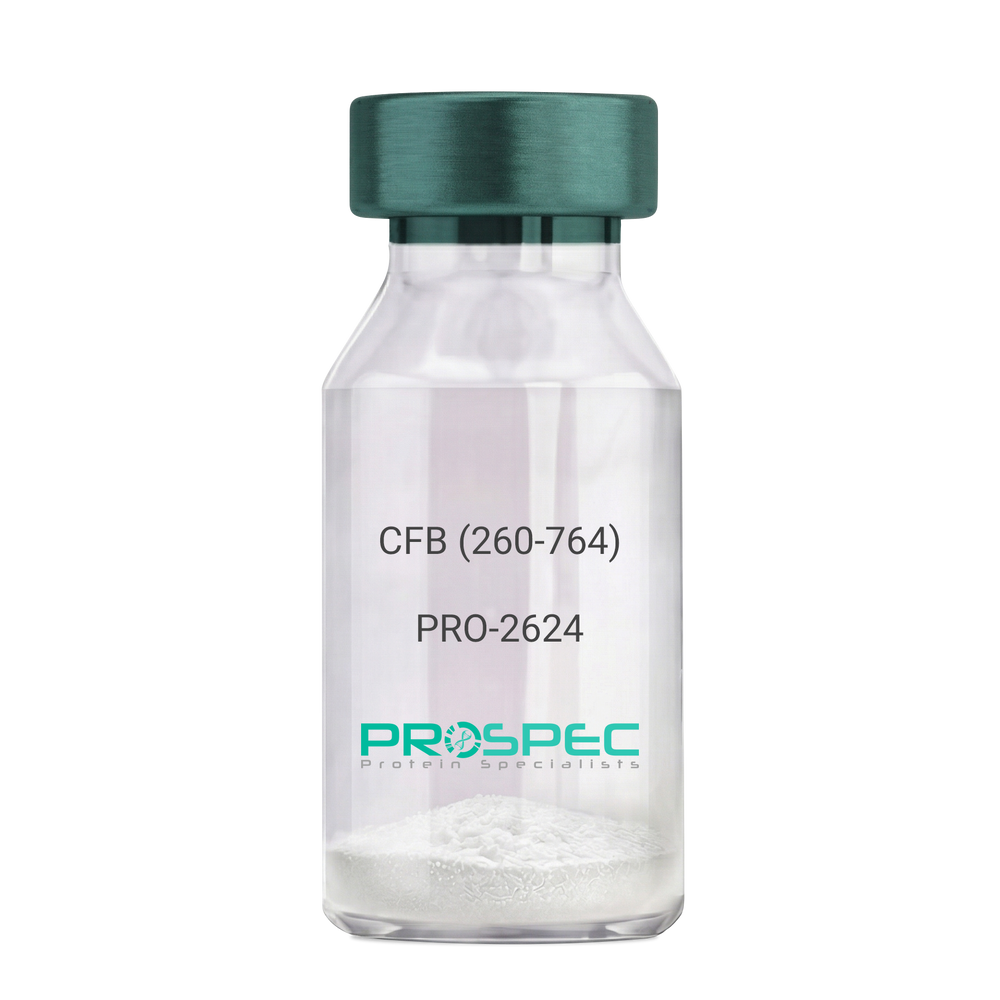
    CFB (260-764) Human, Sf9

    Description:

    Complement Factor B (260-764 a.a.) Human Recombinant, Sf9

    CFB, C3/C5 convertase, Glycine-rich beta glycoprotein, GBG, PBF2, Properdin factor B, BF, BFD, AHUS4, ARMD14, CFAB, CFBD, FB, FBI12, GBG, H2-Bf.

    Product # :

    PRO-2624

    Price :

    Quantity :

    Shipping Method :

    Ice Icon

    Shipped with Ice Packs

    Add To Cart

    More Info

    • description
    • source
    • formulation
    • purity
    • More Info

    Description

    CFB Human Recombinant produced in Sf9 Baculovirus cells is a single, glycosylated polypeptide chain containing 514 amino acids (260-764 a.a) and having a molecular mass of 58.1kDa.CFB is fused to an 9 amino acid His-tag at C-terminus & purified by proprietary chromatographic techniques.

    Source

    Sf9, Baculovirus cells.

    Formulation

    CFB protein solution (0.5mg/ml) containing Phosphate Buffered Saline (pH 7.4) and 20% glycerol.

    Purity

    Greater than 90.0% as determined by SDS-PAGE.

    More Info

    • Introduction

      Factor B is a protein, build from a single chain that circulates in the blood. Complement factor D cleaves and activates factor B, thus creating a catalytic subunit Bb and noncatalytic chain Ba. Subunit Bb is a serine protease that can bind to C3b and forms alternative pathway C3 convertase. Subunit Ba acts as an inhibitor for proliferation of lymphocytes, when Bb is part of the lymphocytes’ proliferation. The gene that codes for this protein is located on chromosome six.

    • Synonyms

      CFB, C3/C5 convertase, Glycine-rich beta glycoprotein, GBG, PBF2, Properdin factor B, BF, BFD, AHUS4, ARMD14, CFAB, CFBD, FB, FBI12, GBG, H2-Bf.

    • Physical Appearance

      Sterile Filtered colorless solution.

    • Stability

      Store at 4°C if entire vial will be used within 2-4 weeks. Store, frozen at -20°C for longer periods of time. For long term storage it is recommended to add a carrier protein (0.1% HSA or BSA).Avoid multiple freeze-thaw cycles.

    • Amino Acid Sequence

      ADPKIVLDPS GSMNIYLVLD GSDSIGASNF TGAKKCLVNL IEKVASYGVK PRYGLVTYAT
      YPKIWVKVSE ADSSNADWVT KQLNEINYED HKLKSGTNTK KALQAVYSMM SWPDDVPPEG
      WNRTRHVIIL MTDGLHNMGG DPITVIDEIR DLLYIGKDRK NPREDYLDVY VFGVGPLVNQ
      VNINALASKK DNEQHVFKVK DMENLEDVFY QMIDESQSLS LCGMVWEHRK GTDYHKQPWQ
      AKISVIRPSK GHESCMGAVV SEYFVLTAAH CFTVDDKEHS IKVSVGGEKR DLEIEVVLFH
      PNYNINGKKE AGIPEFYDYD VALIKLKNKL KYGQTIRPIC LPCTEGTTRA LRLPPTTTCQ
      QQKEELLPAQ DIKALFVSEE EKKLTRKEVY IKNGDKKGSC ERDAQYAPGY DKVKDISEVV
      TPRFLCTGGV SPYADPNTCR GDSGGPLIVH KRSRFIQVGV ISWGVVDVCK NQKRQKQVPA HARDFHINLF QVLPWLKEKL QDEDLGFLHH HHHH

    ProSpec's products are furnished for LABORATORY RESEARCH USE ONLY. The product may not be used as drugs, agricultural or pesticidal products, food additives or household chemicals.

    Complement Factor B
  • View Data Sheet

    Name :

    CBX5 Human

    Description:

    Chromobox Homolog 5 Human Recombinant

    Chromobox homolog 5 (HP1 alpha homolog, Drosophila), Heterochromatin protein 1 homolog alpha, HP1Hs alpha, HP1 alpha homolog.

    Product # :

    PRO-1062

    Price :

    Quantity :

    Shipping Method :

    Ice Icon

    Shipped with Ice Packs

    Add To Cart

    More Info

    • description
    • source
    • formulation
    • purity
    • More Info

    Description

    CBX5 Human Recombinant produced in E. coli is a single polypeptide chain containing 215 amino acids (1-191) and having a molecular mass of 24.8kDa.CBX5 is fused to a 24 amino acid His-tag at N-terminus & purified by proprietary chromatographic techniques.

    Source

    E.coli.

    Formulation

    The CBX5 solution (1mg/1ml) contains 20mM Tris-HCl buffer (pH 8.0), 100mM Nacl, 1mM DTT and 30% glycerol.

    Purity

    Greater than 90% as determined by SDS-PAGE.

    More Info

    • Introduction

      CBX5 belongs to the heterochromatin protein family. CBX5 is enriched in the heterochromatin and linked with centromeres. CBX5 takes part in the creation of functional kinetochore by interaction with important kinetochore proteins. CBX5 has a pseudogene situated on chromosome 3. Additionaly, The CBX5 proteins reconnect with chromatin at the end of mitosis, as Histone H3 is dephosphorylated.

    • Synonyms

      Chromobox homolog 5 (HP1 alpha homolog, Drosophila), Heterochromatin protein 1 homolog alpha, HP1Hs alpha, HP1 alpha homolog.

    • Physical Appearance

      Sterile Filtered colorless solution.

    • Stability

      Store at 4°C if entire vial will be used within 2-4 weeks. Store, frozen at -20°C for longer periods of time. For long term storage it is recommended to add a carrier protein (0.1% HSA or BSA).Avoid multiple freeze-thaw cycles.

    • Amino Acid Sequence

      MGSSHHHHHH SSGLVPRGSH MGSHMGKKTK RTADSSSSED EEEYVVEKVL DRRVVKGQVE YLLKWKGFSE EHNTWEPEKN LDCPELISEF MKKYKKMKEG ENNKPREKSE SNKRKSNFSN SADDIKSKKK REQSNDIARG FERGLEPEKI IGATDSCGDL MFLMKWKDTD EADLVLAKEA NVKCPQIVIA FYEERLTWHA YPEDAENKEK ETAKS.

    ProSpec's products are furnished for LABORATORY RESEARCH USE ONLY. The product may not be used as drugs, agricultural or pesticidal products, food additives or household chemicals.

    Cbx5 Human
  • View Data Sheet

    Name :

    Avidin Protein

    Description:

    Avidin

    Avidin, AVD, AVID.

    Product # :

    PRO-500

    Price :

    Quantity :

    Shipping Method :

    Room Temp Icon

    Shipped at Room temp

    Add To Cart

    More Info

    • description
    • source
    • biological activity
    • More Info

    Description

    Avidin is a glycosylated polypeptide chain having a molecular mass of 68kDa and containing 4 subunits each with a binding site for biotin. The Avidin is purified by affinity chromatographic techniques.The purification procedure ensures minimal contamination by other proteins or DNA.The resulting high activity and purity of the product gives very low non-specific binding (NSB).

    Source

    Hen's egg white.

    Biological Activity

    15.0 units/mg protein, 1 unit binds 1µg biotin.

    More Info

    • Introduction

      Avidin is a tetrameric protein of 4 identical subunits (homotetramer) each of which can bind to biotin with a high degree of affinity and specificity. Avidin molecular weight in its tetrameric form is estimated to be between 66-69 kDa. Avidin is produced in the oviducts of birds, reptiles and amphibians and is subsequently deposited in the whites of their eggs. In the chicken egg white, avidin makes up roughly 0.05% of total protein (approximately 1.8 mg per egg). 10% of Avidin’s molecular weight is ascribed to carbohydrate content which is composed of four to five mannose and three N-acetylglucosamine residues. Avidin has at least three distinctive oligosaccharide structural type which are similar in structure and composition. The dissociation constant (KD) of avidin is approximately 10-15M, making it one of the strongest known non-covalent bonds.

    • Synonyms

      Avidin, AVD, AVID.

    • Physical Appearance

      Sterile Filtered white lyophilized powder.

    • Stability

      Lyophilized Avidin although stable at room temperature for 3 weeks, should be stored desiccated below -18°C. Upon reconstitution Avidin should be stored at 4°C between 2-7 days and for future use below -18°C.For long term storage it is recommended to add a carrier protein (0.1% HSA or BSA).Please prevent freeze-thaw cycles.

    • Solubility

      It is recommended to reconstitute the lyophilized Avidin in sterile 18MΩ-cm H2O not less than 100µg/ml or more than 10mg/ml solutions.

    ProSpec's products are furnished for LABORATORY RESEARCH USE ONLY. The product may not be used as drugs, agricultural or pesticidal products, food additives or household chemicals.

    Avidin Avid
  • View Data Sheet

    Name :

    FLT1 D7 Human

    Description:

    Vascular Endothelial Growth Factor Receptor-1 D1-7 Human Recombinant

    FLT-1, FLT1, Tyrosine-protein kinase receptor FLT, Flt-1, Tyrosine-protein kinase FRT, Fms-like tyrosine kinase 1, VEGFR-1.

    Product # :

    PKA-241

    Price :

    Quantity :

    Shipping Method :

    Room Temp Icon

    Shipped at Room temp

    Add To Cart

    More Info

    • description
    • source
    • formulation
    • purity
    • biological activity
    • More Info

    Description

    Soluble FLT1 Human Recombinant fused with the Fc part of human IgG1 produced in baculovirus is disulfide-linked homodimeric, glycosylated, polypeptide containing 751 amino acids and having a molecular mass of 130 kDa. The soluble receptor protein contains only the first 7 extracellular domains (Met1-Thr751), which contain all the information necessary for high affinity ligand binding. The FLT1 fc/Chimera is purified by proprietary chromatographic techniques.

    Source

    Insect Cells.

    Formulation

    FLT1 D1-7 was lyophilized from a concentrated (1 mg/ml) sterile solution containing PBS Buffer, pH 7.4.

    Purity

    Greater than 95.0% as determined by SDS-PAGE.

    Biological Activity

    The activity of FLT1/Fc was determined by its ability to inhibit the VEGF-dependent proliferation of human umbilical vein endothelial cells. The ED50 for this effect is typically 10-30 ng/ml, corresponding to a specific activity of 33,333.33-100,000 units/mg.

    More Info

    • Introduction

      Endothelial cells express three different vascular endothelial growth factor (VEGF) receptors, belonging to the family of receptor tyrosine kinases (RTKs). They are named VEGFR-1 (Flt-1), VEGFR-2 (KDR/Flk-1), and VEGFR-3 (Flt-4). Their expression is almost exclusively restricted to endothelial cells, but VEGFR-1 can also be found on monocytes. All VEGF-receptors have seven immunoglobulin-like extracellular domains, a single transmembrane region and an intracellular split tyrosine kinase domain. VEGFR-2 has a lower affinity for VEGF than the Flt-1 receptor, but a higher signalling activity. Mitogenic activity in endothelial cells is mainly mediated by VEGFR-2 leading to their proliferation. Differential splicing of the flt-1 gene leads to the formation of a secreted, soluble variant of VEGFR-1 (sVEGFR-1). No naturally occurring, secreted forms of VEGFR-2 have so far been reported. The binding of VEGF165 to VEGFR-2 is dependent on heparin.

    • Synonyms

      FLT-1, FLT1, Tyrosine-protein kinase receptor FLT, Flt-1, Tyrosine-protein kinase FRT, Fms-like tyrosine kinase 1, VEGFR-1.

    • Physical Appearance

      Sterile Filtered White lyophilized (freeze-dried) powder.

    • Stability

      Lyophilized FLT-1 although stable at room temperature for 3 weeks, should be stored desiccated below -18C. Upon reconstitution FLT1 should be stored at 4C between 2-7 days and for future use below -18C. For long term storage it is recommended to add a carrier protein (0.1% HSA or BSA). Please prevent freeze-thaw cycles.

    • Solubility

      It is recommended to reconstitute the lyophilized FLT1 Fc/Chimera in PBS not less than 50µg/ml, which can then be further diluted to other aqueous solutions.

    • Amino Acid Sequence

      MVSYWDTGVL LCALLSCLLL TGSSSGSKLK DPELSLKGTQ HIMQAGQTLH LQCRGEAAHK WSLPEMVSKE SERLSITKSA CGRNGKQFCS TLTLNTAQAN HTGFYSCKYL AVPTSKKKET ESAIYIFISD TGRPFVEMYS EIPEIIHMTE GRELVIPCRV TSPNITVTLK KFPLDTLIPD GKRIIWDSRK GFIISNATYK EIGLLTCEAT VNGHLYKTNY LTHRQTNTII DVQISTPRPV KLLRGHTLVL NCTATTPLNT RVQMTWSYPD EKNKRASVRR RIDQSNSHAN IFYSVLTIDK MQNKDKGLYT CRVRSGPSFK SVNTSVHIYD KAFITVKHRK QQVLETVAGK RSYRLSMKVK AFPSPEVVWL KDGLPATEKS ARYLTRGYSL IIKDVTEEDA GNYTILLSIK QSNVFKNLTA TLIVNVKPQI YEKAVSSFPD PALYPLGSRQ ILTCTAYGIP QPTIKWFWHP CNHNHSEARC DFCSNNEESF ILDADSNMGN RIESITQRMA IIEGKNKMAS TLVVADSRIS GIYICIASNK VGTVGRNISF YITDVPNGFH VNLEKMPTEG EDLKLSCTVN KFLYRDVTWI LLRTVNNRTM HYSISKQKMA ITKEHSITLN LTIMNVSLQD SGTYACRARN VYTGEEILQK KEITIRDQEA PYLLRNLSDH TVAISSSTTL DCHANGVPEP QITWFKNNHK IQQEPGIILG PGSSTLFIER VTEEDEGVYH CKATNQKGSV ESSAYLTVQG TAASDKTHTC PPCPAPELLG GPSVFLFPPK PKDTLMISRT PEVTCVVVDV SHEDPEVKFN WYVDGVEVHN AKTKPREEQY NSTYRVVSVL TVLHQDWLNG KEYKCKVSNK ALPAPIEKTI S.

    ProSpec's products are furnished for LABORATORY RESEARCH USE ONLY. The product may not be used as drugs, agricultural or pesticidal products, food additives or household chemicals.

    Flt1 D7 Human
  • 1
  • 2
  • 3
  • 4
  • 5
  • …
  • 42
Your browser does not support the video tag.

Products

  • Cytokines
  • Growth Factors
  • Chemokines
  • CD Antigens
  • Neurotrophins
  • Hormones
  • Enzymes
  • Viral Antigens
  • Recombinant Proteins
  • Natural Proteins
  • Monoclonal Antibodies
  • Polyclonal Antibodies
  • Thermal Packaging

About

  • Company
  • Founder
  • Contribution
  • Contact Us
  • My Account

Ordering

  • Ordering Method
  • Ordering - Credit Card
  • Ordering PO
  • Shipping Fees
  • FAQ's

Legal

  • Terms & Conditions
  • Privacy
  • Site Map
  • info@prospecbio.com
  • Facebook
  • Instagram
  • Linkedin Profile

© 2026 ProSpec-Tany TechnoGene Ltd. All Rights Reserved.

  • Choosing a selection results in a full page refresh.
  • Opens in a new window.