Search results
1000 results found for “tsh”
Name
Description
Product #
Price
Quantity
Shipping Method
- View Data Sheet
Name :
PTPRC HumanDescription:
Protein Tyrosine Phosphatase Receptor Type C Human Recombinant
LCA, LY5, B220, CD45, T200, GP180, Receptor-type tyrosine-protein phosphatase C, Leukocyte common antigen, L-CA, PTPRC.
Product # :
ENZ-775Price :
Quantity :
Shipping Method :
Shipped with Ice Packs
More Info
- description
- source
- formulation
- purity
- More Info
- Citations
Description
PTPRC Human Recombinant produced in E.coli is a single, non-glycosylated polypeptide chain containing 257 amino acids (1031-1251a.a) and having a molecular mass of 29.6kDa. PTPRC is fused to a 36 amino acid His-tag at N-terminus & purified by proprietary chromatographic techniques.
Source
Escherichia Coli.
Formulation
The PTPRC solution (0.25mg/ml) contains 20mM Tris-HCl buffer (pH 8.0), 0.15M NaCl, 10% glycerol and 1mM DTT.
Purity
Greater than 95% as determined by SDS-PAGE.
Citations
More Info
-
Introduction
Protein Tyrosine Phosphatase Receptor Type C, also known as PTPRC, which is a vital regulator of T- and B-cell antigen receptor signaling, is also a part of the protein tyrosine phosphatase (PTP) family. PTPs are signaling molecules which regulate a variety of cellular processes including differentiation, cell growth, mitosis, and oncogenic transformation. PTPRC contains an extracellular domain, a single transmembrane segment and 2 tandem intracytoplasmic catalytic domains, and therefore is classified as a receptor type PTP.
-
Synonyms
LCA, LY5, B220, CD45, T200, GP180, Receptor-type tyrosine-protein phosphatase C, Leukocyte common antigen, L-CA, PTPRC.
-
Physical Appearance
Sterile Filtered colorless solution.
-
Stability
Store at 4°C if entire vial will be used within 2-4 weeks. Store, frozen at -20°C for longer periods of time. For long term storage it is recommended to add a carrier protein (0.1% HSA or BSA). Avoid multiple freeze-thaw cycles.
-
Amino Acid Sequence
MRGSHHHHHH GMASMTGGQQ MGRDLYDDDD KDRWGSVMIA AQGPLKETIG DFWQMIFQRK VKVIVMLTEL KHGDQEICAQ YWGEGKQTYG DIEVDLKDTD KSSTYTLRVF ELRHSKRKDS RTVYQYQYTN WSVEQLPAEP KELISMIQVV KQKLPQKNSS EGNKHHKSTP LLIHCRDGSQ QTGIFCALLN LLESAETEEV VDIFQVVKAL RKARPGMVST FEQYQFLYDV IASTYPAQNG QVKKNNHQED KIEFDNE.
ProSpec's products are furnished for LABORATORY RESEARCH USE ONLY. The product may not be used as drugs, agricultural or pesticidal products, food additives or household chemicals.
- View Data Sheet
Name :
LA/SS-B Human, BiotinDescription:
LA / SS-B Human Recombinant, Biotinylated
Lupus La protein, Sjoegren syndrome type B antigen, SS-B, La ribonucleoprotein, La autoantigen, SSB, La, LARP3, LA/SS-B, La(SS-B).
Product # :
PRO-2562Price :
Quantity :
Shipping Method :
Shipped with Ice Packs
More Info
- description
- source
- formulation
- purity
- More Info
- Citations
Description
LA/SS-B Human Recombinant produced in SF9 is a single, glycosylated, polypeptide chain having a calculated molecular mass of 48 kDa. The LA/SS-B is expressed with a -6x His tag at N-terminus and purified by proprietary chromatographic techniques.
Source
Sf9 insect cells.
Formulation
The protein solution contains 20mM HEPES, pH 7.5, 400mM NaCl, 20% Glycerol.
Purity
Greater than 80.0% as determined by SDS-PAGE.
Citations
More Info
-
Introduction
The La protein is a 47 kDa polypeptide that frequently acts as an autoantigen in systemic lupus erythematosus and Sjogren's syndrome patients. La is involved in various aspects of RNA metabolism, including binding and protecting 3-prime UUU(OH) elements of newly RNA polymerase III - transcribed RNA, processing 5-prime and 3-prime ends of pre-tRNA precursors, acting as an RNA chaperone, and binding viral RNAs linked to hepatitis C virus. It occurs in both the nucleus and the cytoplasm, where it assumes different roles. In the nucleus, La protein facilitates the production of tRNAs, acting as an RNA polymerase III (RNAP III) transcription factor by attaching to the U-rich 3'UTR of nascent transcripts, aiding in their folding and maturation. In the cytoplasm, La protein facilitates the translation of specific mRNAs, acting as a translation factor. As an RNA binding protein (RBP), La protein associates with subsets of mRNAs which contain a 5'-terminal oligopyrimidine (5'TOP) motif known to direct protein synthesis. The binding of La protein to particular classes of RNA molecules regulates their downstream processing, guards them from endonuclease digestion, and organizes their export from the nucleus. La/SS-B appears to be readily disposed to proteolysis, which results in many smaller (42kD, 320, and 270) nevertheless still immunoreactive polypeptides. La/SS-B antigen is strongly conserved across species. Anti-La/SS-B autoantibodies were originally found as precipitating autoantibodies in sera of Sjogren's Syndrome patients and referred to as SjT. Anti-La/SS-B precipitins are most frequently found in Sjogren's Syndrome, Systemic Lupus Erythematosus (SLE) and Subacute Cutaneous Lupus. Also, there seems to be a correlation between anti-La/SS-B and the absence of nephritis in SLE patients.
-
Synonyms
Lupus La protein, Sjoegren syndrome type B antigen, SS-B, La ribonucleoprotein, La autoantigen, SSB, La, LARP3, LA/SS-B, La(SS-B).
-
Physical Appearance
Sterile Filtered clear solution.
-
Stability
Store at 4°C if entire vial will be used within 2-4 weeks. Store, frozen at -20°C for longer periods of time. Avoid multiple freeze-thaw cycles.
-
Immunological Functions
1. Binds IgG type human auto antibodies.2. Functional Streptavidin based ELISA test (analysis of positive/negative samples.)
ProSpec's products are furnished for LABORATORY RESEARCH USE ONLY. The product may not be used as drugs, agricultural or pesticidal products, food additives or household chemicals.
- View Data Sheet
Name :
TBCB AntibodyDescription:
Tubulin Folding Cofactor B, Mouse Anti Human
Tubulin folding cofactor B, Cytoskeleton-associated protein CKAPI, cytoskeleton associated protein 1, CKAP1, CG22, Tubulin-specific chaperone B.
Product # :
ANT-526Price :
Quantity :
Shipping Method :
Shipped with Ice Packs
More Info
- formulation
- More Info
- Citations
Formulation
1mg/ml containing PBS, pH-7.4, 10% Glycerol and 0.02% Sodium Azide.
Citations
More Info
-
Introduction
TBCB is a member of the Microtubules family. Biosynthesis of functional microtubules include the involvement of a number of chaperones, termed Tubulin folding cofactors A (TBCA), B (TBCB), C (TBCC), D (TBCD) and E (TBCE), which operate on folding intermediates downstream of the cytosolic chaperon, known as TCP. The 244 amino acid cytoplasmic protein TBCB has one CAP-Gly domain and is universally expressed. TBCB takes part in the regulation of tubulin heterodimer dissociation and operates as a negative regulator of axonal growth.
-
Synonyms
Tubulin folding cofactor B, Cytoskeleton-associated protein CKAPI, cytoskeleton associated protein 1, CKAP1, CG22, Tubulin-specific chaperone B.
-
Physical Appearance
Sterile filtered colorless solution.
-
Immunogen
Anti-human TBCB mAb, clone PAT1F6AT, is derived from hybridization of mouse F0 myeloma cells with spleen cells from BALB/c mice immunized with a recombinant human TBCB protein 1-244 amino acids purified from E. coli.
-
Ig Subclass
Mouse IgG2b heavy chain and k light chain.
-
Clone
PAT1F6AT.
-
Applications
The antibody has been tested by ELISA, Western blot analysis to assure specificity and reactivity. Since application varies, however, each investigation should be titrated by the reagent to obtain optimal results. Recommended starting dilution is 1:1500.
-
Type
Mouse Anti Human Monoclonal.
-
Storage Procedures
For periods up to 1 month store at 4°C, for longer periods of time, store at -20°C. Prevent freeze thaw cycles.
-
Purification Method
TBCB antibody was purified from mouse ascitic fluids by protein-A affinity chromatography.
ProSpec's products are furnished for LABORATORY RESEARCH USE ONLY. The product may not be used as drugs, agricultural or pesticidal products, food additives or household chemicals.
- View Data Sheet
Name :
PTPN7 HumanDescription:
Protein Tyrosine Phosphatase Non Receptor Type-7 Human Recombinant
Protein tyrosine phosphatase non-receptor type 7, HEPTP, LC-PTP, Hematopoietic protein-tyrosine phosphatase, Protein-tyrosine phosphatase LC-PTP, PTPNI, LPTP, BPTP-4, dual specificity phosphatase 1, protein-tyrosine phosphatase non-receptor-type, stress induced, EC 3.1.3.48.
Product # :
PKA-025Price :
Quantity :
Shipping Method :
Shipped with Ice Packs
More Info
- description
- source
- formulation
- purity
- More Info
- Citations
Description
PTPN7 Human Recombinant produced in E. coli is a single polypeptide chain containing 384 amino acids (1-360) and having a molecular mass of 43.1kDa.PTPN7 is fused to a 24 amino acid His-tag at N-terminus & purified by proprietary chromatographic techniques.
Source
E.coli.
Formulation
The PTPN7 solution (0.5mg/ml) contains 20mM Tris-HCl buffer (pH 8.0), 100mM NaCl, 1mM DTT and 20% glycerol.
Purity
Greater than 90% as determined by SDS-PAGE.
Citations
More Info
-
Introduction
PTPN7 performs specifically on tyrosine-phosphorylated MAPK1. PTPN7 takes part in the regulation of signal transduction and T and B-lymphocyte development.
-
Synonyms
Protein tyrosine phosphatase non-receptor type 7, HEPTP, LC-PTP, Hematopoietic protein-tyrosine phosphatase, Protein-tyrosine phosphatase LC-PTP, PTPNI, LPTP, BPTP-4, dual specificity phosphatase 1, protein-tyrosine phosphatase non-receptor-type, stress induced, EC 3.1.3.48.
-
Physical Appearance
Sterile Filtered colorless solution.
-
Stability
Store at 4°C if entire vial will be used within 2-4 weeks. Store, frozen at -20°C for longer periods of time. For long term storage it is recommended to add a carrier protein (0.1% HSA or BSA).Avoid multiple freeze-thaw cycles.
-
Amino Acid Sequence
MGSSHHHHHH SSGLVPRGSH MGSHMVQAHG GRSRAQPLTL SLGAAMTQPP PEKTPAKKHV RLQERRGSNV ALMLDVRSLG AVEPICSVNT PREVTLHFLR TAGHPLTRWA LQRQPPSPKQ LEEEFLKIPS NFVSPEDLDI PGHASKDRYK TILPNPQSRV CLGRAQSQED GDYINANYIR GYDGKEKVYI ATQGPMPNTV SDFWEMVWQE EVSLIVMLTQ LREGKEKCVH YWPTEEETYG PFQIRIQDMK ECPEYTVRQL TIQYQEERRS VKHILFSAWP DHQTPESAGP LLRLVAEVEE SPETAAHPGP IVVHCSAGIG RTGCFIATRI GCQQLKARGE VDILGIVCQL RLDRGGMIQT AEQYQFLHHT LALYAGQLPE EPSP
ProSpec's products are furnished for LABORATORY RESEARCH USE ONLY. The product may not be used as drugs, agricultural or pesticidal products, food additives or household chemicals.
- View Data Sheet
Name :
TNFA BovineDescription:
Tumor Necrosis Factor-alpha Bovine Recombinant
TNF-alpha, Tumor necrosis factor ligand superfamily member 2, TNF-a, Cachectin, DIF, TNFA, TNFSF2.
Product # :
CYT-1104Price :
Quantity :
Shipping Method :
Shipped with Ice Packs
More Info
- description
- source
- formulation
- purity
- biological activity
- More Info
- Citations
Description
TNFA Bovine produced in E.Coli is a single, non-glycosylated polypeptide chain containing 158 amino acids (78-234 a.a.) and having a molecular mass of 17.5kDa. TNFA is purified by proprietary chromatographic techniques.
Source
Escherichia Coli.
Formulation
The TNFA (1mg/ml) contains Phosphate buffer saline(pH7.4) and 10% glycerol.
Purity
Greater than 90.0% as determined by SDS-PAGE.
Biological Activity
The ED50 Is < 15 ng/ml and is measured in a cytotoxicity assay using L929 mouse fibrosarcoma cells in the presence of the metabolic inhibitor actinomycin D.
Citations
More Info
-
Introduction
Tumor necrosis factor is a member of a group of cytokines that all stimulate the acute phase reaction. TNF is involved in systemic inflammationand secreted mainly by macrophages.
TNF causes apoptotic cell death, cellular proliferation, differentiation, inflammation, tumorigenesis and viral replication, TNF is also involved in lipid metabolism, and coagulation. TNF's primary role is in the regulation of immune cells.
Dysregulation and, in particular, overproduction of TNF have been implicated in a variety of human diseases- autoimmune diseases, insulin resistance, and cancer. -
Synonyms
TNF-alpha, Tumor necrosis factor ligand superfamily member 2, TNF-a, Cachectin, DIF, TNFA, TNFSF2.
-
Physical Appearance
Sterile filtered colorless solution.
-
Stability
Store at 4°C if entire vial will be used within 2-4 weeks. Store, frozen at -20°C for longer periods of time. For long term storage it is recommended to add a carrier protein (0.1% HSA or BSA).Avoid multiple freeze-thaw cycles.
-
Amino Acid Sequence
MLRSSSQASS NKPVAHVVAD INSPGQLRWW DSYANALMAN GVKLEDNQLV VPADGLYLIY SQVLFRGQGC PSTPLFLTHT ISRIAVSYQT KVNILSAIKS PCHRETPEWA EAKPWYEPIY QGGVFQLEKG DRLSAEINLP DYLDYAESGQ VYFGIIAL.
ProSpec's products are furnished for LABORATORY RESEARCH USE ONLY. The product may not be used as drugs, agricultural or pesticidal products, food additives or household chemicals.
- View Data Sheet
Name :
TNFR2 Human, Sf9Description:
Tumor Necrosis Factor Receptor Type 2 Human Recombinant, Sf9
Tumor Necrosis Factor Receptor Superfamily, Member 1B, TNFR2, TNFBR, Tumor Necrosis Factor Receptor Type II, Tumor Necrosis Factor Receptor 2, P80 TNF-Alpha Receptor, TNF-RII, TNF-R2, P75, Tumor Necrosis Factor Receptor Superfamily Member 1B, Tumor Necrosis Factor Binding Protein 2, Tumor Necrosis Factor Beta Receptor, Soluble TNFR1B Variant 1, P75 TNF Receptor, CD120b Antigen, Etanercept, TNF-R-II, TNF-R75, P75TNFR, TNFR-II, CD120b, TNFR1B, TNFR80, TBPII.
Product # :
CYT-908Price :
Quantity :
Shipping Method :
Shipped with Ice Packs
More Info
- description
- source
- formulation
- purity
- biological activity
- More Info
- Citations
Description
TNFR2 produced in Sf9 Baculovirus cells is a single, glycosylated polypeptide chain (23-257 a.a.) and fused to a 6 aa His Tag at C-terminus containing a total of 241 amino acids and having a molecular mass of 25.9kDa. TNFR2 is purified by proprietary chromatographic techniques.
Source
Sf9, Baculovirus cells.
Formulation
TNFR2 protein solution (1mg/ml) contains Phosphate buffered saline (pH7.4) and 10% glycerol.
Purity
Greater than 95.0% as determined by SDS-PAGE.
Biological Activity
The ED50 range ≤ 0.2 μg/ml and is measured by its ability to inhibit cytotoxicity using L-929 mouse fibroblast cells in the presence of Human TNF-α.
Citations
More Info
-
Introduction
TNFR2 belongs to the TNF-receptor superfamily. TNFR2 is receptor with high affinity for TNFSF2/TNF-alpha and approximately 5-fold lower affinity for homotrimeric TNFSF1/lymphotoxin-alpha. TNFR2 mediates the majority of the metabolic effects of TNF-alpha. In addition, knockout studies in mice propose a role for TNFR2 in protecting neurons from apoptosis by stimulating antioxidative pathways. TNFR2 expression might have a significant role in the angiogenesis, tumor cell proliferation and metastasis of Invasive micropapillary carcinoma of the breast.
There are 2 types of soluble TNF receptors: sTNFR-I and sTNFR-II, which act to neutralize the biological activities of TNF alpha and TNF beta. The levels of these soluble receptors seem to increase as a result of shedding of the extracellular domains of the membrane bound receptors. High levels of soluble TNF receptors are found in the amniotic fluid of pregnant women. TNFR2 and TNFR1 form a heterocomplex which mediates the recruitment of 2 anti-apoptotic proteins, c-IAP1 and c-IAP2, which possess E3 ubiquitin ligase activity. IAPs’ function in TNF-receptor signaling is unknown; nevertheless, c-IAP1 is believed to potentiate TNF-induced apoptosis by the ubiquitination and degradation of TNF-receptor-associated factor 2, which mediates anti-apoptotic signals. Oxidative stress promotes TNFR1 and TNFR2 self-interaction, ligand-independent and enhanced ligand-dependent TNF signaling. TNF-a, TNFR1 and TNFR2 have roles in cellular differentiation. TNFR1 and TNFR2 function in cell type-specific renal injury. -
Synonyms
Tumor Necrosis Factor Receptor Superfamily, Member 1B, TNFR2, TNFBR, Tumor Necrosis Factor Receptor Type II, Tumor Necrosis Factor Receptor 2, P80 TNF-Alpha Receptor, TNF-RII, TNF-R2, P75, Tumor Necrosis Factor Receptor Superfamily Member 1B, Tumor Necrosis Factor Binding Protein 2, Tumor Necrosis Factor Beta Receptor, Soluble TNFR1B Variant 1, P75 TNF Receptor, CD120b Antigen, Etanercept, TNF-R-II, TNF-R75, P75TNFR, TNFR-II, CD120b, TNFR1B, TNFR80, TBPII.
-
Physical Appearance
Sterile filtered colorless solution.
-
Stability
Store at 4°C if entire vial will be used within 2-4 weeks. Store, frozen at -20°C for longer periods of time. For long term storage it is recommended to add a carrier protein (0.1% HSA or BSA).Avoid multiple freeze-thaw cycles.
-
Amino Acid Sequence
LPAQVAFTPY APEPGSTCRL REYYDQTAQM CCSKCSPGQH AKVFCTKTSD TVCDSCEDST YTQLWNWVPE CLSCGSRCSS DQVETQACTR EQNRICTCRP GWYCALSKQE GCRLCAPLRK CRPGFGVARP GTETSDVVCK PCAPGTFSNT TSSTDICRPH QICNVVAIPG NASMDAVCTS TSPTRSMAPG AVHLPQPVST RSQHTQPTPE PSTAPSTSFL LPMGPSPPAE GSTGDHHHHH H
ProSpec's products are furnished for LABORATORY RESEARCH USE ONLY. The product may not be used as drugs, agricultural or pesticidal products, food additives or household chemicals.
- View Data Sheet
Name :
SemaglutideDescription:
Semaglutide
Product # :
HOR-048Price :
Quantity :
Shipping Method :
Shipped at Room temp
More Info
- description
- formulation
- purity
- More Info
- HPLC, MS
- Citations
Description
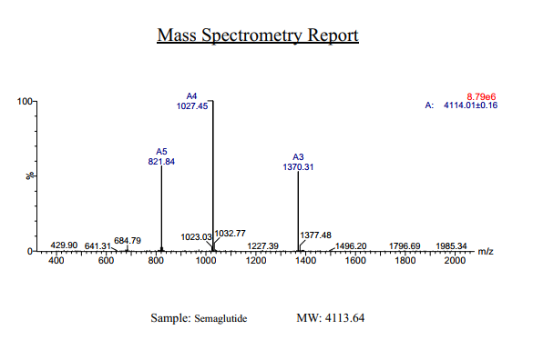
Semaglutide Synthetic is a single, non-glycosylated polypeptide chain containing 31 amino acids, having a molecular mass of 4113 Dalton and a Molecular formula of C187H291N45O59.
Formulation
The protein was lyophilized with no additives.
Purity
Greater than 97.0% as determined by analysis by RP-HPLC.
HPLC, MS
Citations
More Info
-
Physical Appearance
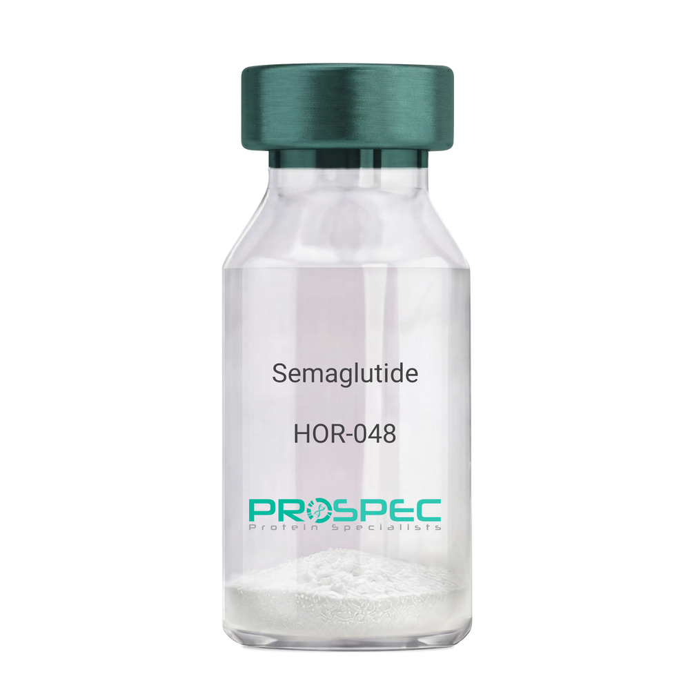
Sterile Filtered White lyophilized (freeze-dried) powder.
-
Stability
Lyophilized Semaglutide although stable at room temperature for 3 weeks, should be stored desiccated below -18°C. Upon reconstitution Semaglutide should be stored at 4°C between 2-7 days and for future use below -18°C. For long term storage it is recommended to add a carrier protein (0.1% HSA or BSA). Please prevent freeze-thaw cycles.
-
Solubility
It is recommended to reconstitute the lyophilized Semaglutide in sterile 18MΩ-cm H2O not less than 100 µg/ml, which can then be further diluted to other aqueous solutions.
-
Amino Acid Sequence
H-His-Aib-Glu-Gly-Thr-Phe-Thr-Ser-Asp-Val-Ser-Ser-Tyr-Leu-Glu-Gly-Gln-Ala-Ala-Lys(AEEAc-AEEAc-γ-Glu-17-carboxyheptadecanoyl)-Glu-Phe-Ile-Ala-Trp-Leu-Val-Arg-Gly-Arg-Gly-OH.
-
Background
Semaglutide, a glucagon-like peptide-1 receptor agonist (GLP-1 RA), has emerged as a breakthrough in the field of diabetes management and metabolic disorders. It is recognized for its potent glucose-lowering effects, weight management properties, and cardiovascular benefits. Beyond diabetes, semaglutide is under investigation for its potential applications in obesity treatment and other related conditions. This research aims to comprehensively explore semaglutide, shedding light on its multifaceted mechanisms of action and its broader therapeutic implications.
The primary objective of this research is to elucidate the mechanisms underlying the glucose-lowering and metabolic effects of semaglutide. In vitro and in vivo experiments will be conducted to investigate how semaglutide interacts with GLP-1 receptors, influences insulin secretion, and modulates glucose homeostasis. Understanding these mechanisms is crucial for harnessing the full therapeutic potential of semaglutide.
The second objective is to assess the clinical relevance of semaglutide in diabetes management. Clinical trials involving individuals with type 2 diabetes will be conducted to evaluate the efficacy, safety, and long-term outcomes of semaglutide treatment. These investigations may provide valuable insights into its use as a monotherapy or adjunct therapy in diabetes care.
The third objective is to explore the potential applications of semaglutide beyond diabetes. Research will investigate its role in obesity treatment, cardiovascular risk reduction, and non-alcoholic fatty liver disease (NAFLD) management. Understanding the multifaceted properties of semaglutide may open new avenues for therapeutic interventions in various metabolic and cardiovascular conditions.
By delving into the diverse functions of semaglutide, this research aims to expand our understanding of its therapeutic potential and clinical applications. The findings may contribute to improved treatment strategies for individuals affected by diabetes, obesity, and related metabolic disorders.
ProSpec's products are furnished for LABORATORY RESEARCH USE ONLY. The product may not be used as drugs, agricultural or pesticidal products, food additives or household chemicals.
- View Data Sheet
Name :
HPRT AntibodyDescription:
Hypoxanthine-Guanine Phosphoribosyltransferase, Mouse Anti Human
Hypoxanthine-Guanine Phosphoribosyltransferase, EC 2.4.2.8, HGPRT, HGPRTase, HPRT, HPRT1.
Product # :
ANT-046Price :
Quantity :
Shipping Method :
Shipped with Ice Packs
More Info
- formulation
- More Info
- Citations
Formulation
1mg/ml containing PBS, pH-7.4, & 0.1% Sodium Azide.
Citations
More Info
-
Introduction
HPRT1 has a main part in the generation of purine nucleotides through the purine salvage pathway. HPRT1 primarily functions to salvage purines from degraded DNA to renewed purine synthesis. Therefore, it performs as a catalyst in the reaction between guanine and phosphoribosyl pyrophosphate to form GMP.
-
Synonyms
Hypoxanthine-Guanine Phosphoribosyltransferase, EC 2.4.2.8, HGPRT, HGPRTase, HPRT, HPRT1.
-
Immunogen
Anti-human HPRT mAb, is derived from hybridization of mouse FO myeloma cells with spleen cells from BALB/c mice immunized with recombinant human HPRT amino acids 1-218 purified from E. coli.
-
Ig Subclass
Mouse IgG1 heavy chain and k light chain.
-
Clone
PAT2G8AT.
-
Applications
HPRT antibody has been tested by ELISA and Western blot analysis to assure specificity and reactivity. Since application varies, however, each investigation should be titrated by the reagent to obtain optimal results. Recommended dilution range for Western blot analysis is 1:500~1:5000.
Recommended starting dilution is 1:1000. -
Type
Mouse Anti Human Monoclonal.
-
Storage Procedures
For periods up to 1 month store at 4°C, for longer periods of time, store at -20°C. Prevent freeze thaw cycles.
-
Purification Method
HPRT antibody was purified from mouse ascitic fluids by protein-G affinity chromatography.
ProSpec's products are furnished for LABORATORY RESEARCH USE ONLY. The product may not be used as drugs, agricultural or pesticidal products, food additives or household chemicals.
- View Data Sheet
Name :
NTRK2 Human , HEKDescription:
Neurotrophic Receptor Tyrosine Kinase 2, HEK Human Recombinant
GP145-TrkB, trk-B, TRKB, NTRK2.
Product # :
CYT-1230Price :
Quantity :
Shipping Method :
Shipped with Ice Packs
More Info
- description
- source
- formulation
- purity
- More Info
- Citations
Description
NTRK2 Human Recombinant is a single, glycosylated, polypeptide chain (32-430 a.a) containing a total of 632 amino acids and having a molecular mass of 70.3 kDa. NTRK2 is fused to a 233 a.a hIgG-His-Tag at C-terminus and is purified by proprietary chromatographic techniques.
Source
HEK293 Cells.
Formulation
The NTRK2 solution (1mg/ml) contains 10% Glycerol and Phosphate-Buffered Saline (pH 7.4).
Purity
Greater than 90.0% as determined by SDS-PAGE.
Citations
More Info
-
Synonyms
GP145-TrkB, trk-B, TRKB, NTRK2.
-
Physical Appearance
Sterile Filtered colorless solution.
-
Stability
Store at 4°C if entire vial will be used within 2-4 weeks. Store, frozen at -20°C for longer periods of time. For long term storage it is recommended to add a carrier protein (0.1% HSA or BSA). Avoid multiple freeze-thaw cycles.
-
Amino Acid Sequence
CPTSCKCSAS RIWCSDPSPG IVAFPRLEPN SVDPENITEI FIANQKRLEI INEDDVEAYV GLRNLTIVDS GLKFVAHKAF LKNSNLQHIN FTRNKLTSLS RKHFRHLDLS ELILVGNPFT CSCDIMWIKT LQEAKSSPDT QDLYCLNESS KNIPLANLQI PNCGLPSANL AAPNLTVEEG KSITLSCSVA GDPVPNMYWD VGNLVSKHMN ETSHTQGSLR ITNISSDDSG KQISCVAENL VGEDQDSVNL TVHFAPTITF LESPTSDHHW CIPFTVKGNP KPALQWFYNG AILNESKYIC TKIHVTNHTE YHGCLQLDNP THMNNGDYTL IAKNEYGKDE KQISAHFMGW PGIDDGANPN YPDVIYEDYG TAANDIGDTT NRSNEIPSTD VTDKTGREH LEPKSCDKTHT CPPCPAPELL GGPSVFLFPP KPKDTLMISR TPEVTCVVVD VSHEDPEVKF NWYVDGVEVH NAKTKPREEQ YNSTYRVVSV LTVLHQDWLN GKEYKCKVSN KALPAPIEKT ISKAKGQPRE PQVYTLPPSR DELTKNQVSL TCLVKGFYPS DIAVEWESNG QPENNYKTTP PVLDSDGSFF LYSKLTVDKS RWQQGNVFSC SVMHEALHNH YTQKSLSLSP GK.
-
Background
NTRK2, also known as TrkB, is a high-affinity receptor for BDNF and other neurotrophins such as neurotrophin-4 (NT-4) and neurotrophin-3 (NT-3). It plays a crucial role in the development, function, and plasticity of the nervous system. Dysregulation of NTRK2 signaling is associated with various neurological disorders, including neurodegenerative diseases, psychiatric disorders, and cancers. NTRK2 has been developed to facilitate research into its biological functions and potential therapeutic applications. This paper provides a comprehensive overview of the scientific research on recombinant human NTRK2, focusing on its biological functionality, detailed description, and physical properties.
NTRK2 is critical for synaptic plasticity, which underlies learning and memory. Research with rhNTRK2 has demonstrated that BDNF-NTRK2 signaling enhances synaptic strength and facilitates long-term potentiation (LTP) in hippocampal neuron.
ProSpec's products are furnished for LABORATORY RESEARCH USE ONLY. The product may not be used as drugs, agricultural or pesticidal products, food additives or household chemicals.
- View Data Sheet
Name :
TNFR (22-211) HumanDescription:
Tumor Necrosis Factor Receptor (22-211 a.a.) Human Recombinant
CD120a, FPF, MS5, p55, p55-R, p60, TBP1, TNF-R, TNF-R-I, TNF-R55, TNFAR, TNFR1, TNFR1-d2,TNFR55, TNFR60, Tumor necrosis factor receptor superfamily member 1A, Tumor necrosis factor receptor superfamily member 1A.
Product # :
CYT-851Price :
Quantity :
Shipping Method :
Shipped with Ice Packs
More Info
- description
- source
- formulation
- purity
- More Info
- Citations
Description
TNFR Human Recombinant produced in E.Coli is a single, non-glycosylated polypeptide chain containing 213 amino acids (22-211 a.a) and having a molecular mass of 23.6kDa.TNFR is fused to a 23 amino acid His-tag at N-terminus & purified by proprietary chromatographic techniques.
Source
Escherichia Coli.
Formulation
TNFR protein solution (1mg/ml) containing 20mM Tirs-HCl (pH8.0) and 10% glycerol.
Purity
Greater than 85% as determined by SDS-PAGE.
Citations
More Info
-
Introduction
TNFR1 belongs to the TNF-receptor superfamily. TNFR1 is a receptor for TNFSF2/TNF-alpha and homotrimeric TNFSF1/lymphotoxin-alpha.
There are 2 types of soluble TNF receptors: sTNFR-I and sTNFR-II, which act to neutralize the biological activities of TNF alpha and TNF beta. The levels of these soluble receptors seem to increase as a result of shedding of the extracellular domains of the membrane bound receptors. TNF-a, TNFR1 and TNFR2 have roles in cellular differentiation. TNFR1 and TNFR2 function in cell type-specific renal injury.
TNFR1 is capable of signaling both cell survival and apoptosis. TNFR1-induced apoptosis requires 2 sequential signaling complexes. TNFR1 is capable of activating NF-kappaB, mediate apoptosis, and function as a regulator of inflammation. Oxidative stress promotes TNFR1 and TNFR2 self-interaction, ligand-independent and enhanced ligand-dependent TNF signaling. TNFR1 contributes to the induction of non-cytocidal TNF effects including anti-viral state and activation of the acid sphingomyelinase. Human TNFR1 has a major region which controls cell surface expression. High levels of soluble TNF receptors are found in the amniotic fluid of pregnant women.
Germline mutations of the extracellular domains of TNFR1 are linked to the autosomal dominant periodic fever syndrome. The impaired receptor clearance is believed to be a mechanism of the disease. Familial hibernian fever (FHF) is caused by defects in TNFRSF1A gene. -
Synonyms
CD120a, FPF, MS5, p55, p55-R, p60, TBP1, TNF-R, TNF-R-I, TNF-R55, TNFAR, TNFR1, TNFR1-d2,TNFR55, TNFR60, Tumor necrosis factor receptor superfamily member 1A, Tumor necrosis factor receptor superfamily member 1A.
-
Physical Appearance
Sterile filtered colorless solution.
-
Stability
Store at 4°C if entire vial will be used within 2-4 weeks. Store, frozen at -20°C for longer periods of time. For long term storage it is recommended to add a carrier protein (0.1% HSA or BSA).Avoid multiple freeze-thaw cycles.
-
Amino Acid Sequence
MGSSHHHHHH SSGLVPRGSH MGSIYPSGVI GLVPHLGDRE KRDSVCPQGK YIHPQNNSIC CTKCHKGTYL YNDCPGPGQD TDCRECESGS FTASENHLRH CLSCSKCRKE MGQVEISSCT VDRDTVCGCR KNQYRHYWSE NLFQCFNCSL CLNGTVHLSC QEKQNTVCTC HAGFFLRENE CVSCSNCKKS LECTKLCLPQ IENVKGTEDS GTT.
ProSpec's products are furnished for LABORATORY RESEARCH USE ONLY. The product may not be used as drugs, agricultural or pesticidal products, food additives or household chemicals.
- View Data Sheet
Name :
TROVE2 Human, His BiotinDescription:
TROVE Domain Family Member 2 Human Recombinant, His Tag Biotinylated
60 kDa SS-A/Ro ribonucleoprotein, 60 kDa ribonucleoprotein Ro, RoRNP, 60 kDa Ro protein, Ro 60 kDa autoantigen, TROVE domain family member 2, Sjoegren syndrome type A antigen, SS-A, Sjoegren syndrome antigen A2, TROVE2, RO60, SSA2, RO-60.
Product # :
PRO-2560Price :
Quantity :
Shipping Method :
Shipped with Ice Packs
More Info
- description
- source
- formulation
- purity
- More Info
- Citations
Description
TROVE2 Human Recombinant Biotin produced in E.Coli is a single, non-glycosylated, polypeptide chain having a molecular mass of 56kDa. The TROVE2 is expressed with a 6xHis tag at N-terminus and purified by proprietary chromatographic techniques.
Source
Escherichia Coli.
Formulation
The protein solution contains 20mM HEPES (pH 8.0), 20% Glycerol and 150mM NaCl.
Purity
Greater than 89% as determined by Capillary Electrophoresis.
Citations
More Info
-
Introduction
Ro 60 kDa autoantigen is an RNA-binding protein that binds to several small cytoplasmic RNA molecules known as Y RNAs. SSA2 may stabilize these RNAs from degradation. Sera taken from patients with Systemic Lupus Erythematosus (SLE) often contains antibodies that react with the normal cellular SSA2 protein as if this antigen was foreign.
-
Synonyms
60 kDa SS-A/Ro ribonucleoprotein, 60 kDa ribonucleoprotein Ro, RoRNP, 60 kDa Ro protein, Ro 60 kDa autoantigen, TROVE domain family member 2, Sjoegren syndrome type A antigen, SS-A, Sjoegren syndrome antigen A2, TROVE2, RO60, SSA2, RO-60.
-
Physical Appearance
Sterile Filtered clear solution.
-
Stability
Store at 4°C if entire vial will be used within 2-4 weeks. Store, frozen at -20°C for longer periods of time. Avoid multiple freeze-thaw cycles.
ProSpec's products are furnished for LABORATORY RESEARCH USE ONLY. The product may not be used as drugs, agricultural or pesticidal products, food additives or household chemicals.
- View Data Sheet
Name :
HBV-X, His TagDescription:
Hepatitis B Virus X Recombinant, His Tag
Product # :
HBV-271Price :
Quantity :
Shipping Method :
Shipped at Room temp
More Info
- description
- source
- formulation
- purity
- More Info
- Citations
Description
HBV-X Recombinant produced in E. coli is a single polypeptide chain containing 165 amino acids (2-154) and having a molecular mass of 17.8 kDa.HBV-X is fused to a 12 amino acid His-tag at N-terminus & purified by proprietary chromatographic techniques.
Source
Escherichia Coli.
Formulation
Filtered (0.4µm) and lyophilized from 0.5mg/ml in 50mM acetate buffer pH4 and 5% trehalose.
Purity
Greater than 90%.
Citations
More Info
-
Introduction
Hepatitis B virus X protein (HBx) is a 17 kD transcriptional coactivator that plays a significant role in the regulation of genes involved in inflammation and cell survival. It regulates many transcription factors including nuclear factor kappa B (NF-kappaB) and plays a key role in hepatocarcinogenesis. rHBx facilitates the binding of cAMP response element binding protein (CREB) to its responsive element. rHBx stabilizes the cellular coactivator ASC-2 through direct protein-protein interaction, affecting the regulation of genes actively transcribed in liver cancer cells. HBx transactivates both JNK and MAPK signal transduction pathways in association with the mobilization of cytosolic Ca2+. The communication between HBx and general transcription factor TFIIB is also one of the mechanisms which account for its transcriptional transactivation. HBx decreased the expression of PTEN a known tumor suppressor and a negative regulator of phosphatidylinositol 3'-kinase/AKT and HBx decreased the expression of PTEN in HBx-transfected cells. The etiology of hepatocellular carcinoma (HCC) is involved with hepatitis B virus (HBV) infection and HBx in particular plays a role in the development of HBV-related HCC. The persistence of HBx is important to the pathogenesis of early HCC and HBx expression in the liver during chronic HBV infection may be an important prognostic marker for the development of HCC.
-
Stability
For long term storage lyophilized protein should be stored at -20°C. Aliquot the product after reconstitution to avoid repeated freezing/thawing cycles. Reconstituted protein can be stored at 4°C for a limited period of time; it does not show any change after two weeks at 4°C.
-
Solubility
It is recommended to add 0.1M Acetate buffer pH4 to prepare a working stock solution of approximately 0.5mg/ml and let the lyophilized pellet dissolve completely. For conversion into higher pH value, we recommend intensive dilution by relevant buffer to a concentration of 10µg/ml. In higher concentrations the solubility of the HBV X antigen is limited. Filter sterilize your culture media/working solutions containing this non-sterile product before using in cell culture.
-
Amino Acid Sequence
MRGSHHHHHH GSAARVCCQL DPARDVLCLR PVGAESRGRP VSGPFGTLPS PSSSAVPADH GAHLSLRGLP VCAFSSAGPC ALRFTSARRM ETTVNAHQVL PKVLHKRTLG LSAMSTTDLE AYFKDCLFKD WEELGEEIRL KVFVLGGCRH KLVCSPAPCN FFTSA.
ProSpec's products are furnished for LABORATORY RESEARCH USE ONLY. The product may not be used as drugs, agricultural or pesticidal products, food additives or household chemicals.
- View Data Sheet
Name :
T.gondii p40Description:
Toxoplasma Gondii p40 Recombinant
Product # :
TOX-268Price :
Quantity :
Shipping Method :
Shipped with Ice Packs
More Info
- description
- source
- formulation
- purity
- More Info
- Citations
Description
The E.Coli derived recombinant Toxoplasma Gondii p40 having a Mw of 35kDa and fused to a 6 His tag at C-terminus can be used to test the specific IgG and IgM antibody for the diagnosis of Toxoplasma gondii infection.
Source
Escherichia Coli.
Formulation
1x PBS and 25mM arginine.
Purity
Toxoplasma protein is >95% pure as determined by 10% PAGE (coomassie staining).
Citations
More Info
-
Introduction
The life cycle of Toxoplasma gondii has two phases. The coccidia like takes place only in members of the Felidae family which makes these animals the parasite's primary host. The asexual part of the life cycle can take place in any warm-blooded animal, like other mammals(including felines) and birds. T. gondii constructing daughter scaffolds within the mother cell. In the intermediate hosts (including felines), the parasite invades cells, forming intracellular so-called parasitophorous vacuoles containing bradyzoites, the slowly replicating form of the parasit. Vacuoles form tissue cysts mainly within the muscles and brain. Since they are within cells, the host's immune system does not detect these cysts. Resistance to antibiotics varies, but the cysts are very difficult to eradicate entirely. Within these vacuoles T. gondii propagates by a series of binary fissions until the infected cell eventually bursts and tachyzoites are released. Tachyzoites are the motile, asexually reproducing form of the parasite. Unlike the bradyzoites, the free tachyzoites are usually efficiently cleared by the host's immune response, although some manage to infect cells and form bradyzoites, thus maintaining the infection.
-
Physical Appearance
Sterile Filtered clear solution.
-
Stability
T.gondii p40 although stable at 4°C for 1 week, should be stored below -18°C. Please prevent freeze thaw cycles.
-
Purification Method
Purified by proprietary chromatographic technique.
ProSpec's products are furnished for LABORATORY RESEARCH USE ONLY. The product may not be used as drugs, agricultural or pesticidal products, food additives or household chemicals.
- View Data Sheet
Name :
TUBB3 HumanDescription:
Tubulin, Beta 3 Class III Human Recombinant
Beta-4, CDCBM, CFEOM3A, TUBB4 , Tubulin beta-3 chain, Tubulin beta-4 chain, Tubulin beta-III, TUBB3.
Product # :
PRO-1388Price :
Quantity :
Shipping Method :
Shipped with Ice Packs
More Info
- description
- source
- formulation
- purity
- More Info
- Citations
Description
TUBB3 Human Recombinant produced in E.Coli is a single, non-glycosylated polypeptide chain containing 473 amino acids (1-450 a.a.) and having a molecular mass of 52.8kDa. TUBB3 is fused to a 23 amino acid His-tag at N-terminus & purified by proprietary chromatographic techniques.
Source
E.coli.
Formulation
TUBB3 protein solution (0.25mg/ml) contains 20mM Tris-HCl buffer (pH 8.0), 0.15M NaCl, 10% glycerol and 1mM DTT.
Purity
Greater than 90% as determined by SDS-PAGE.
Citations
More Info
-
Introduction
Tubulin, beta 3 class III (TUBB3), which is the major constituent of microtubules, is primarily Expressed in neurons and is involved in neurogenesis and in axon guidance and maintenance. TUBB3 binds two moles of GTP, one at an exchangeable site on the beta chain and one at a non-exchangeable site on the alpha-chain.
-
Synonyms
Beta-4, CDCBM, CFEOM3A, TUBB4 , Tubulin beta-3 chain, Tubulin beta-4 chain, Tubulin beta-III, TUBB3.
-
Physical Appearance
Sterile Filtered colorless solution.
-
Stability
Store at 4°C if entire vial will be used within 2-4 weeks. Store, frozen at -20°C for longer periods of time. For long term storage it is recommended to add a carrier protein (0.1% HSA or BSA).Avoid multiple freeze-thaw cycles.
-
Amino Acid Sequence
MGSSHHHHHH SSGLVPRGSH MGSMREIVHI QAGQCGNQIG AKFWEVISDE HGIDPSGNYV GDSDLQLERI SVYYNEASSH KYVPRAILVD LEPGTMDSVR SGAFGHLFRP DNFIFGQSGA GNNWAKGHYT EGAELVDSVL DVVRKECENC DCLQGFQLTH SLGGGTGSGM GTLLISKVRE EYPDRIMNTF SVVPSPKVSD TVVEPYNATL SIHQLVENTD ETYCIDNEAL YDICFRTLKL ATPTYGDLNH LVSATMSGVT TSLRFPGQLN ADLRKLAVNM VPFPRLHFFM PGFAPLTARG SQQYRALTVP ELTQQMFDAK NMMAACDPRH GRYLTVATVF RGRMSMKEVD EQMLAIQSKN SSYFVEWIPN NVKVAVCDIP PRGLKMSSTF IGNSTAIQEL FKRISEQFTA MFRRKAFLHW YTGEGMDEME FTEAESNMND LVSEYQQYQD ATAEEEGEMY EDDEEESEAQ GPK.
ProSpec's products are furnished for LABORATORY RESEARCH USE ONLY. The product may not be used as drugs, agricultural or pesticidal products, food additives or household chemicals.
- View Data Sheet
Name :
LH Paired AntibodyDescription:
Anti Human Leutenizing Hormone Paired Antibody Mouse
Product # :
ANT-787Price :
Quantity :
Shipping Method :
Shipped with Ice Packs
More Info
- description
- formulation
- More Info
- Citations
Description
Paired LH monoclonal antibodies are composed of anti-LH α and β subunits, the anti-LH α is used for the coating and antibody to β subunit is used for colloid gold conjugation. Please note that when ordering for example: 100µg paired antibody, you receive 50µg from each antibody (100µg in total).
Formulation
* LH α (Coating) antibody in 50mM NaCl, 10mM PBS, 0.1% NaN3, PH 8 (11.26mg/ml).
* LH β (Labeling) antibody in 50mM NaCl, 10mM PBS, 0.1% NaN3, PH 8 (18.39mg/ml).
Citations
More Info
-
Physical Appearance
2 vials of sterile Filtered clear colorless solution.
-
Stability
For periods up to 1month LHPaired Antibody should be stored at 4°C, for longer periods of time, store at -20°C. Prevent freeze thaw cycles.
-
Applications
Lateral flow immunoassay.
-
Background
Luteinizing hormone (LH) is produced by the pituitary gland and takes an important part in the reproductive system. LH is necessary for sexual development and fertility in both males and females. In females, LH triggers ovulation and the production of progesterone. In males, LH stimulates testosterone production by the Leydig cells in the testes. LH testing can diagnose conditions related to excessive or insufficient LH production, such as infertility or pituitary gland disorders. LH takes an important part in the reproductive system, influencing sex hormone production, ovulation and in general reproductive function in both males and females.
-
Type
Mouse antibody Monoclonal.
-
Purification Method
Purified by Ion Exchange (IEX) Chromatography.
ProSpec's products are furnished for LABORATORY RESEARCH USE ONLY. The product may not be used as drugs, agricultural or pesticidal products, food additives or household chemicals.
- View Data Sheet
Name :
TNFR2 MouseDescription:
Tumor Necrosis Factor Receptor Type 2 Mouse Recombinant
Tumor necrosis factor receptor superfamily member 1B, Tumor necrosis factor receptor 2, TNF-R2, Tumor necrosis factor receptor type II, TNF-RII, TNFR-II, p75, p80 TNF-alpha receptor, CD120b, Tnfrsf1b, Tnfr-2, Tnfr2, TNFBR, TNFR80, TNFRII, TNF-R75, TNF-R-II, TNF-alphaR2, TNFalpha-R2.
Product # :
CYT-770Price :
Quantity :
Shipping Method :
Shipped at Room temp
More Info
- description
- source
- formulation
- purity
- biological activity
- More Info
- Citations
Description
TNFR2 Mouse Recombinant produced in E.coli is a single, non-glycosylated polypeptide chain containing 236 amino acids and having a molecular mass of 25.3kDa.The TNFR2 is purified by proprietary chromatographic techniques.
Source
Escherichia Coli.
Formulation
TNFR2 protein was lyophilized from a 0.2µm filtered concentrated solution in PBS, pH 7.4.
Purity
Greater than 97.0% as determined by SDS-PAGE.
Biological Activity
The ED50 as determined by its ability to inhibit the TNF-a mediated cytotoxicity in the L-929 cells is less than 2µg/ml, corresponding to a specific activity of > 500IU/mg in the presence of 0.1ng/mL of rHuTNF-a.
Citations
More Info
-
Introduction
TNFR2 belongs to the TNF-receptor superfamily. TNFR2 is receptor with high affinity for TNFSF2/TNF-alpha and approximately 5-fold lower affinity for homotrimeric TNFSF1/lymphotoxin-alpha. TNFR2 mediates the majority of the metabolic effects of TNF-alpha. In addition, knockout studies in mice propose a role for TNFR2 in protecting neurons from apoptosis by stimulating antioxidative pathways. TNFR2 expression might have a significant role in the angiogenesis, tumor cell proliferation and metastasis of Invasive micropapillary carcinoma of the breast.
There are 2 types of soluble TNF receptors: sTNFR-I and sTNFR-II, which act to neutralize the biological activities of TNF alpha and TNF beta. The levels of these soluble receptors seem to increase as a result of shedding of the extracellular domains of the membrane bound receptors. High levels of soluble TNF receptors are found in the amniotic fluid of pregnant women. TNFR2 and TNFR1 form a heterocomplex which mediates the recruitment of 2 anti-apoptotic proteins, c-IAP1 and c-IAP2, which possess E3 ubiquitin ligase activity. IAPs’ function in TNF-receptor signaling is unknown; nevertheless, c-IAP1 is believed to potentiate TNF-induced apoptosis by the ubiquitination and degradation of TNF-receptor-associated factor 2, which mediates anti-apoptotic signals. Oxidative stress promotes TNFR1 and TNFR2 self-interaction, ligand-independent and enhanced ligand-dependent TNF signaling. TNF-a, TNFR1 and TNFR2 have roles in cellular differentiation. TNFR1 and TNFR2 function in cell type-specific renal injury. -
Synonyms
Tumor necrosis factor receptor superfamily member 1B, Tumor necrosis factor receptor 2, TNF-R2, Tumor necrosis factor receptor type II, TNF-RII, TNFR-II, p75, p80 TNF-alpha receptor, CD120b, Tnfrsf1b, Tnfr-2, Tnfr2, TNFBR, TNFR80, TNFRII, TNF-R75, TNF-R-II, TNF-alphaR2, TNFalpha-R2.
-
Physical Appearance
Sterile Filtered White lyophilized (freeze-dried) powder.
-
Stability
Lyophilized TNFR2 although stable at room temperature for 3 weeks, should be stored desiccated below -18C. Upon reconstitution TNFR2 should be stored at 4C between 2-7 days and for future use below -18C. For long term storage it is recommended to add a carrier protein (0.1% HSA or BSA). Please prevent freeze-thaw cycles.
-
Solubility
It is recommended to reconstitute the lyophilized TNFR2 in sterile 18M-cm H2O not less than 100µg/ml, which can then be further diluted to other aqueous solutions.
-
Amino Acid Sequence
VPAQVVLTPY KPEPGYECQI SQEYYDRKAQ MCCAKCPPGQ YVKHFCNKTS DTVCADCEAS MYTQVWNQFR TCLSCSSSCT TDQVEIRACT KQQNRVCACE AGRYCALKTH SGSCRQCMRL SKCGPGFGVA SSRAPNGNVL CKACAPGTFS DTTSSTDVCR PHRICSILAI PGNASTDAVC APESPTLSAI PRTLYVSQPE PTRSQPLDQE PGPSQTPSIL TSLGSTPIIE QSTKGG.
ProSpec's products are furnished for LABORATORY RESEARCH USE ONLY. The product may not be used as drugs, agricultural or pesticidal products, food additives or household chemicals.
- View Data Sheet
Name :
TRAIL Human (114-281 a.a.)Description:
TRAIL/APO 2 Ligand (114-281 a.a.) Human Recombinant
Tumor necrosis factor ligand superfamily member 10, TNF-related apoptosis-inducing ligand, Protein TRAIL, Apo-2 ligand, Apo-2L, CD253 antigen, TL2, APO2L, TNFSF10.
Product # :
CYT-546Price :
Quantity :
Shipping Method :
Shipped with Ice Packs
More Info
- description
- source
- formulation
- purity
- More Info
- Citations
Description
Soluble TNF-related apoptosis-inducing ligand Human Recombinant produced in E.Coli is a single, non-glycosylated polypeptide chain containing 169 amino acids (114-281) and having a molecular mass of 19.6 kDa. The sTRAIL is purified by proprietary chromatographic techniques.
Source
Escherichia Coli.
Formulation
1mg/ml in 20mM Tris-HCl pH-7.5, 300mM NaCl, 0.1mM DTT & 10% glycerol.
Purity
Greater than 95.0% as determined by SDS-PAGE.
Citations
More Info
-
Introduction
TNF-related apoptosis-inducing ligand (TRAIL) is a ligand molecule which induces apoptosis. It is a type II transmembrane protein with homology to other members of the tumor necrosis factor family.
In humans, the gene that encodes for TRAIL is located at chromosome 3q26.
TRAIL binds to the death receptors, DR4 and DR5. The process of apoptosis is caspase-8-dependent. This protein preferentially induces apoptosis in transformed and tumor cells, but does not appear to kill normal cells although it is expressed at a significant level in most normal tissues. -
Synonyms
Tumor necrosis factor ligand superfamily member 10, TNF-related apoptosis-inducing ligand, Protein TRAIL, Apo-2 ligand, Apo-2L, CD253 antigen, TL2, APO2L, TNFSF10.
-
Physical Appearance
Sterile Filtered colorless liquid.
-
Stability
Store at 4°C if entire vial will be used within 2-4 weeks. Store, frozen at -20°C for longer periods of time. For long term storage it is recommended to add a carrier protein (0.1% HSA or BSA).Avoid multiple freeze-thaw cycles.
-
Amino Acid Sequence
MVRERGPQRV AAHITGTRGR SNTLSSPNSK NEKALGRKIN SWESSRSGHS FLSNLHLRNGELVIHEKGFY YIYSQTYFRF QEEIKENTKN DKQMVQYIYK YTSYPDPILL MKSARNSCWSKDAEYGLYSI YQGGIFELKE NDRIFVSVTN EHLIDMDHEA SFFGAFLVG.
ProSpec's products are furnished for LABORATORY RESEARCH USE ONLY. The product may not be used as drugs, agricultural or pesticidal products, food additives or household chemicals.
- View Data Sheet
Name :
RARRES2 Human, HisDescription:
Retinoic Acid Receptor Responder 2 Human Recombinant, His Tag
Chemerin, TIG2, Tazarotene-induced gene 2 protein, Retinoic acid receptor responder protein 2, RAR-responsive protein TIG2, RARRES2, HP10433.
Product # :
PRO-1458Price :
Quantity :
Shipping Method :
Shipped with Ice Packs
More Info
- description
- source
- formulation
- purity
- More Info
- Citations
Description
RARRES2 Human Recombinant produced in E.Coli is a single, non-glycosylated, polypeptide chain containing 158 amino acids (21-157a.a) and having a total molecular mass of 18 kDa. RARRES2 is fused to a 21 amino acid His-tag at N-terminus & purified by proprietary chromatographic techniques.
Source
Escherichia Coli.
Formulation
RARRES2 protein solution (1mg/ml) containing 20mM Tris-HCl buffer (pH 8.0), 0.4M Urea and 10% glycerol.
Purity
Greater than 85.0% as determined by SDS-PAGE.
Citations
More Info
-
Introduction
RARRES2 is a secreted chemotactic protein that initiates chemotaxis through the ChemR23 G protein-coupled seven-transmembrane domain ligand. RARRES2 is upregulated by the synthetic retinoid tazarotene and found in a vast variety of tissues. RARRES2 acts as an adipokine, and is truncated on both termini from the proprotein. RARRES2 is structurally related to the cathelicidin precursors, cystatin C and kininogens. RARRES2 promotes calcium mobilization and chemotaxis of immature dendritic cells and macrophages. RARRES2 is secreted as a precursor of little biological activity, which requires proteolytic cleavage of its COOH-terminal domain to be exchangeed into a potent and highly specific agonist of ChemR23. RARRES2 signals via its receptor, ChemR23 (CMKLR1), as a positive regulator of adipocyte differentiation and metabolic function. The Chemerin receptor acts as a coreceptor for SIV and some primary HIV-1 strains. The Chemerin receptor has another ligand, called tazarotene-induced gene.
-
Synonyms
Chemerin, TIG2, Tazarotene-induced gene 2 protein, Retinoic acid receptor responder protein 2, RAR-responsive protein TIG2, RARRES2, HP10433.
-
Physical Appearance
Sterile Filtered colorless solution.
-
Stability
Store at 4°C if entire vial will be used within 2-4 weeks. Store, frozen at -20°C for longer periods of time. For long term storage it is recommended to add a carrier protein (0.1% HSA or BSA).Avoid multiple freeze-thaw cycles.
-
Amino Acid Sequence
MGSSHHHHHH SSGLVPRGSH MELTEAQRRG LQVALEEFHK HPPVQWAFQE TSVESAVDTP FPAGIFVRLE FKLQQTSCRK RDWKKPECKV RPNGRKRKCL ACIKLGSEDK VLGRLVHCPI ETQVLREAEE HQETQCLRVQ RAGEDPHSFY FPGQFAFS.
ProSpec's products are furnished for LABORATORY RESEARCH USE ONLY. The product may not be used as drugs, agricultural or pesticidal products, food additives or household chemicals.
- View Data Sheet
Name :
TBEV gEDescription:
Tick-Borne Encephalitis Virus gE Recombinant
Product # :
TBE-281Price :
Quantity :
Shipping Method :
Shipped with Ice Packs
More Info
- description
- source
- formulation
- purity
- More Info
- Citations
Description
The E.coli derived recombinant protein contains the Tick-borne Encephalitis Virus glycoprotein E regions, 95-229 amino acids.
Source
Escherichia Coli.
Formulation
20mM Tris-MES pH 6.5, 8M urea, 200mM NaCl and 0.05% Tween-20.
Purity
Protein is >90% pure as determined by SDS- PAGE.
Citations
More Info
-
Introduction
TBE is caused by tick-borne encephalitis virus (TBEV), a member of the family Flaviviridae.
A closely related virus in Far Eastern Eurasia, Russian spring-summer encephalitis virus (RSSEV).
The family Flaviviridae includes other tick-borne viruses are closely related to TBEV and RSSEV, such as Omsk hemorrhagic fever virus & Kyasanur Forest virus.
Louping ill virus is also a member of this family. -
Stability
Encephalitis protein although stable at 4°C for 1 week, should be stored below -18°C. Please prevent freeze thaw cycles.
-
Applications
Encephalitis antigen in ELISA and Western blots, excellent antigen for detection of Tick-borne encephalitis virus with minimal specificity problems.
-
Specificity
Immunoreactive with sera of encephalitis virus infected individuals.
-
Purification Method
Encephalitis protein was purified by proprietary chromatographic technique.
ProSpec's products are furnished for LABORATORY RESEARCH USE ONLY. The product may not be used as drugs, agricultural or pesticidal products, food additives or household chemicals.
- View Data Sheet
Name :
Activin A Human PlantDescription:
Activin A Human Recombinant, Plant
Inhba, Inhibin beta A, FSH releasing protein.
Product # :
CYT-052Price :
Quantity :
Shipping Method :
Shipped at Room temp
More Info
- description
- source
- formulation
- purity
- More Info
- Citations
Description
Activin A human Recombinant produced in Nicotiana benthamiana plant is a disulfide-linked homodimers of two betaA chains, each containing 116 amino residues (molecular formula C600H911N173O174S13) and 6-His-tag at the N-terminal having the total molecular mass of 27.4kDa.
Source
Nicotiana benthamiana plant
Formulation
Lyophilized from 1mg/ml solution in Tris HCl 0.05M buffer at pH 7.4.
Purity
Greater than 97.0% as determined by Analysis by SDS-PAGE.
Citations
More Info
-
Introduction
Activins are homodimers or heterodimers of the different ? subunit isoforms, part of the TGF? family. Mature Activin A has two 116 amino acids residues betaA subunits (bA-bA). Activin displays an extensive variety of biological activities, including mesoderm induction, neural cell differentiation, bone remodelling, haematopoiesis, and reproductive physiology. Activins takes part in the production and regulation of hormones such as FSH, LH, GnRH and ACTH. Cells that are identified to express Activin A include fibroblasts, endothelial cells, hepatocytes, vascular smooth muscle cells, macrophages, keratinocytes, osteoclasts, bone marrow monocytes, prostatic epithelium, neurons, chondrocytes, osteoblasts, Leydig cells, Sertoli cells, and ovarian granulosa cells.
-
Synonyms
Inhba, Inhibin beta A, FSH releasing protein.
-
Physical Appearance
Sterile Filtered White lyophilized (freeze-dried) powder.
-
Stability
Lyophilized Activin A although stable at room temperature for 3 weeks, should be stored desiccated below -18°C. Upon reconstitution Activin A should be stored at 4°C between 2-7 days and for future use below -18°C. For long term storage it is recommended to add a carrier protein (0.1% HSA or BSA). Please prevent freeze-thaw cycles.
-
Solubility
It is recommended to reconstitute the lyophilized Activin A in sterile 18MΩ-cm H2O not less than 100µg/ml, which can then be further diluted to other aqueous solutions.
-
Amino Acid Sequence
HHHHHHGLEC DGKVNICCKK QFFVSFKDIG WNDWIIAPSG YHANYCEGEC PSHIAGTSGS SLSFHSTVIN HYRMRGHSPF ANLKSCCVPT KLRPMSMLYY DDGQNIIKKD IQNMIVEECG CS
-
Background
What is the molecular weight / Mw of Activin A Protein?
Activin A Protein has a total Mw of 27.4 kDa.
What is the source or expression system of Activin A Protein?
Nicotinia
What is the Purity of Activin A Protein?
Activin A Protein is >97% pure as determined by SDS-PAGE.
What is the Biological Activity of Activin A Protein?
The biological functionality of Activin-A Protein will be determined in the future.
What is the endotoxin level for Activin A Protein?
The endotoxin level is minimal, ACTIVIN A Protein was purified using conventional chromatography techniques.
What is the amino acid sequence of ACTIVIN A Protein?
HHHHHHGLEC DGKVNICCKK QFFVSFKDIG WNDWIIAPSG YHANYCEGEC PSHIAGTSGS SLSFHSTVIN HYRMRGHSPF ANLKSCCVPT KLRPMSMLYY DDGQNIIKKD IQNMIVEECG CS
What applications can ACTIVIN A Protein be used in?
ACTIVIN A Protein can probably be used in western blot, ELISA and Lateral Flow.
-
Serological Identification
The protein was electrophoresed under reducing condition on a 15% SDS-polyacrylamide gel, transferred by electroblotting to a NC membrane and visualized by immune-detection with specific antibody Activin A.
ProSpec's products are furnished for LABORATORY RESEARCH USE ONLY. The product may not be used as drugs, agricultural or pesticidal products, food additives or household chemicals.
- View Data Sheet
Name :
SCF Human, HEKDescription:
Stem Cell Factor Human Recombinant, HEK
Kit ligand Precursor, C-kit ligand, SCF, Mast cell growth factor, MGF, SF, KL-1, Kitl, DKFZp686F2250.
Product # :
CYT-111Price :
Quantity :
Shipping Method :
Shipped at Room temp
More Info
- description
- source
- formulation
- purity
- biological activity
- More Info
- Citations
Description
SCF Human Recombinant produced in HEK cells is a glycosylated monomer, having a molecular weight range of 35-45kDa due to glycosylation.The SCF is purified by proprietary chromatographic techniques.
Source
HEK.
Formulation
SCF was lyophilized from a 0.2µm filtered solution (1mg/ml) containing 1xPBS.
Purity
Greater than 95% as obsereved by SDS-PAGE.
Biological Activity
The specific activity was determined by the dose-dependent stimulation of the proliferation of human TF-1 cells (human erythroleukemic indicator cell line).
The EC50 is 15.25ng/ml.Citations
More Info
-
Introduction
Stem cell factor / KIT ligand (SCF) is a cytokine which binds CD117(c-Kit). SCF is also known as "steel factor" or "c-kit ligand". SCF exists in two forms, cell surface bound SCF and soluble (or free) SCF. Soluble SCF is produced by the cleavage of surface bound SCF by metalloproteases. SCF is a growth factor important for the survival, proliferation, and differentiation of hematopoietic stem cells and other hematopoietic progenitor cells. One of its roles is to change the BFU-E (burst-forming unit-erythroid) cells, which are the earliest erythrocyte precursors in the erythrocytic series, into the CFU-E (colony-forming unit-erythroid).
-
Synonyms
Kit ligand Precursor, C-kit ligand, SCF, Mast cell growth factor, MGF, SF, KL-1, Kitl, DKFZp686F2250.
-
Physical Appearance
Sterile Filtered White lyophilized (freeze-dried) powder.
-
Stability
Lyophilized SCF although stable at room temperature for 3 weeks, should be stored desiccated below -18°C. Upon reconstitution SCF should be stored at 4°C between 2-7 days and for future use below -18°C.For long term storage it is recommended to add a carrier protein (0.1% HSA or BSA).Please prevent freeze-thaw cycles.
-
Solubility
It is recommended to reconstitute the lyophilized SCF in sterile PBS containing 0.1% endotoxin-free recombinant HSA.
ProSpec's products are furnished for LABORATORY RESEARCH USE ONLY. The product may not be used as drugs, agricultural or pesticidal products, food additives or household chemicals.
- View Data Sheet
Description:
Myostatin Propeptide Human Recombinant, HEK
GDF-8, MSTN, Growth Differentiation Factor 8, MSTN Muscle Hypertrophy.
Product # :
CYT-936Price :
Quantity :
Shipping Method :
Shipped with Ice Packs
More Info
- description
- source
- formulation
- purity
- More Info
- Citations
Description
Myostatin Propetide Human Recombinant produced in HEK cells is a single, glycosylated, polypeptide chain (Asn24-Arg266) containing a total of 253 amino acids, having a calculated molecular mass of 29.1kDa. Myostatin Propetide is fused to a 10 aa C-terminal His tag.
Source
HEK 293.
Formulation
Myostatin Propetide solution at a concentration of 0.25mg/ml in phosphate buffered saline (PBS) pH 8.0 and 20% (w/v) glycerol.
Purity
Greater than 80.0% as determined by SDS-PAGE.
Citations
More Info
-
Introduction
GDF8 is a member of the bone morphogenetic protein (BMP) family and the TGF-beta superfamily. This group of proteins is characterized by a polybasic proteolytic processing site which is cleaved to produce a mature protein containing seven conserved cysteine residues. The members of this family are regulators of cell growth and differentiation in both embryonic and adult tissues. This gene is thought to encode a secreted protein which negatively regulates skeletal muscle growth.
-
Synonyms
GDF-8, MSTN, Growth Differentiation Factor 8, MSTN Muscle Hypertrophy.
-
Physical Appearance
Filtered colorless solution.
-
Stability
Store at 4°C if entire vial will be used within 2-4 weeks. Store, frozen at -20°C for longer periods of time. For long term storage it is recommended to add a carrier protein (0.1% HSA or BSA).Avoid multiple freeze-thaw cycles.
-
Amino Acid Sequence
NENSEQKENV EKEGLCNACT WRQNTKSSRI EAIKIQILSK LRLETAPNIS KDVIRQLLPK APPLRELIDQ YDVQRDDSSD GSLEDDDYHA TTETIITMPT ESDFLMQVDG KPKCCFFKFS SKIQYNKVVK AQLWIYLRPV ETPTTVFVQI LRLIKPMKDG TRYTGIRSLK LDMNPGTGIW QSIDVKTVLQ NWLKQPESNL GIEIKALDEN GHDLAVTFPG PGEDGLNPFL EVKVTDTPKR SRR HHHHHHH HHH.
ProSpec's products are furnished for LABORATORY RESEARCH USE ONLY. The product may not be used as drugs, agricultural or pesticidal products, food additives or household chemicals.
- View Data Sheet
Name :
NARS Human, Sf9Description:
Asparaginyl-TRNA Synthetase Human Recombinant, Sf9
Asparagine--tRNA ligase, cytoplasmic, EC 6.1.1.22, Asparaginyl-tRNA synthetase, AsnRS, NARS, NARS1.
Product # :
ENZ-716Price :
Quantity :
Shipping Method :
Shipped with Ice Packs
More Info
- description
- source
- formulation
- purity
- More Info
- Citations
Description
NARS Human Recombinant produced in SF9 is a glycosylated, polypeptide chain having a calculated molecular mass of 63,853 Dalton. NARS is expressed with a -6xHis tag at N-terminus and purified by proprietary chromatographic techniques.
Source
Sf9 Insect Cells.
Formulation
NARS is supplied in 20mM HEPES buffer pH-7.6, 250mM NaCl and 20% glycerol.
Purity
Greater than 80.0% as determined by SDS-PAGE.
Citations
More Info
-
Introduction
Aminoacyl-tRNA synthetases are a class of enzymes which charge tRNAs with their cognate amino acids. Asparaginyl-tRNA synthetase (NARS) is localized to the cytoplasm and is a member of the class II family of tRNA synthetases. The N-terminal domain characterizes the signature sequence for the eukaryotic asparaginyl-tRNA synthetases.
-
Synonyms
Asparagine--tRNA ligase, cytoplasmic, EC 6.1.1.22, Asparaginyl-tRNA synthetase, AsnRS, NARS, NARS1.
-
Physical Appearance
Sterile Filtered clear solution.
-
Stability
Store at 4°C if entire vial will be used within 2-4 weeks. Store, frozen at -20°C for longer periods of time. Avoid multiple freeze-thaw cycles.
ProSpec's products are furnished for LABORATORY RESEARCH USE ONLY. The product may not be used as drugs, agricultural or pesticidal products, food additives or household chemicals.
- View Data Sheet
Name :
BST2 HumanDescription:
Bone Marrow Stromal Cell Antigen 2 Human Recombinant
Bone marrow stromal cell antigen 2, CD317 antigen, BST-2, HM1.24 antigen, Tetherin, NPC-A-7.
Product # :
CYT-059Price :
Quantity :
Shipping Method :
Shipped with Ice Packs
More Info
- description
- source
- formulation
- purity
- More Info
- sds-page
- Citations
Description
BST2 Human Recombinant produced in E.coli is a single, non-glycosylated polypeptide chain containing 133 amino acids (50-161) and having a molecular mass of 14.8 kDa.The BST2 is fused to a 21 amino acid His-Tag at N-terminus and purified by proprietary chromatographic techniques.
Source
Escherichia Coli.
Formulation
BST2 protein 0.5mg/ml is supplied in 20mM Tris-HCl, pH-8, 0.1M NaCl, 1mM DTT and 20% Glycerol.
Purity
Greater than 80.0% as determined by SDS-PAGE.
sds-page
Citations
More Info
-
Introduction
BST2 takes part in the growth and development of B-cells. The human cellular protein BST2 inhibits retrovirus infection by maintaining the diffusion of virus particles after budding from infected cells. BST2 was originally discovered as an inhibitor to HIV-1 infection in the absence of Vpu, but it is also known to inhibit the release of other viruses such as the Lassa and Marburg virions. In addition, BST2 has a part in B-cell activation in rheumatoid arthritis.
-
Synonyms
Bone marrow stromal cell antigen 2, CD317 antigen, BST-2, HM1.24 antigen, Tetherin,
NPC-A-7. -
Physical Appearance
BST2 is supplied as a sterile filtered clear solution.
-
Stability
Store at 4°C if entire vial will be used within 2-4 weeks. Store, frozen at -20°C for longer periods of time. For long term storage it is recommended to add a carrier protein (0.1% HSA or BSA).Avoid multiple freeze-thaw cycles.
-
Amino Acid Sequence
MGSSHHHHHH SSGLVPRGSH MSEACRDGLR AVMECRNVTH LLQQELTEAQ KGFQDVEAQA ATCNHTVMAL MASLDAEKAQ GQKKVEELEG EITTLNHKLQ DASAEVERLR RENQVLSVRI ADKKYYPSSQ DSS
-
Background
The Impact of Bone Marrow Stromal Cell Antigen 2 Human Recombinant in Regenerative Medicine
Introduction
As regenerative medicine progresses from the realm of imagination to tangible reality, Bone Marrow Stromal Cell Antigen 2 (BST-2) human recombinant surfaces as a noteworthy contributor with the potential to reshape the future of therapeutic practices.
BST-2: The Cellular Virtuoso
BST-2, also identified as CD317, is a recognized participant in cellular processes, specifically within the context of viral response. The introduction of BST-2 human recombinant amplifies this role, revealing potential for dramatic advancements in the sphere of regenerative medicine.
Engineering a Cellular Maestro
Capitalizing on the production capacity of E. coli, we successfully synthesized BST-2 human recombinant. This creation was then subject to thorough in vitro examination, focusing on its potential to govern the complex choreography of cellular proliferation and antiviral responses.
Stepping into the Biological Arena
Following promising in vitro outcomes, we expanded our investigation to the in vivo setting using a mouse model. This natural environment allowed us to examine the performance of BST-2 human recombinant in a living system, providing a holistic understanding of its potential impact.
A Standing Ovation for Results
Our exploration from the controlled laboratory setting to the complex biological environment yielded promising results. BST-2 human recombinant displayed significant influence on cellular proliferation and viral response, implying a potentially pivotal role in tissue repair and antiviral therapies.
Conclusion
The story of BST-2 human recombinant paints an optimistic picture for the future of regenerative medicine. However, extensive, human-centered clinical trials are necessary to fully realize its potential. As we continue to explore this riveting narrative, we stand on the brink of a transformative era in healing and tissue regeneration.
What is the molecular weight/Mw of BST2 Protein?
BST2 Protein has a total Mw of 14.8kDa.
What is the source or expression system of BST2 Protein?
Escherichia Coli.
What is the Purity of BST2 Protein?
BST2 Protein is >80% pure as determined by SDS-PAGE.
What is the Biological Activity of BST2 Protein?
The biological functionality of BST2 Protein will be determined in the future.
What is the amino acid sequence of BST2 Protein?
MGSSHHHHHH SSGLVPRGSH MSEACRDGLR AVMECRNVTH LLQQELTEAQ KGFQDVEAQA ATCNHTVMAL MASLDAEKAQ GQKKVEELEG EITTLNHKLQ DASAEVERLR RENQVLSVRI ADKKYYPSSQ DSS
What applications can BST2 Protein be used in?
BST2 Protein can probably be used in western blot, ELISA and Lateral Flow.
What is the endotoxin level for BST2 Protein?
The endotoxin level is minimal, BST2 Protein was purified using conventional chromatography techniques.
ProSpec's products are furnished for LABORATORY RESEARCH USE ONLY. The product may not be used as drugs, agricultural or pesticidal products, food additives or household chemicals.