Skip to content

High-quality research proteins.

  • 1-800-805-6531

  • Mon - Sat 8.00-18.00, Sun - Closed

  • info@prospecbio.com

  • PO Box 6591 East Brunswick NJ 08816

  • Products
    • Cytokines
    • Growth factors
    • Chemokines
    • Cd antigens
    • Neurotrophins
    • Hormones
    • Enzymes
    • Viral antigens
    • Recombinant proteins
    • Natural proteins
    • Monoclonal antibodies
    • Polyclonal antibodies
    • Thermal Packaging
  • About
    • Company
    • Founder
    • Contribution
  • Customers
    • Eastern Asia
    • Eastern Europe
    • North America
    • South America
    • South East Asia
    • Southern Asia
    • Western Europe
  • Ordering
    • Ordering Method
    • Ordering - Credit Card
    • Ordering - PO
    • Shipping Fees
    • FAQs
  • Services
    • DNA Cloning
    • Protein Expression
    • Monoclonal Antibody
    • Fermentation
    • Technology License
  • Contact us
Log in

Country/region

  • USD $ | Albania
  • USD $ | Andorra
  • USD $ | Argentina
  • USD $ | Armenia
  • USD $ | Australia
  • USD $ | Austria
  • USD $ | Azerbaijan
  • USD $ | Belarus
  • USD $ | Belgium
  • USD $ | Bolivia
  • USD $ | Brazil
  • USD $ | Bulgaria
  • USD $ | Cambodia
  • USD $ | Canada
  • USD $ | Chile
  • USD $ | China
  • USD $ | Colombia
  • USD $ | Costa Rica
  • USD $ | Croatia
  • USD $ | Cyprus
  • USD $ | Czechia
  • USD $ | Denmark
  • USD $ | Estonia
  • USD $ | Finland
  • USD $ | France
  • USD $ | Georgia
  • USD $ | Germany
  • USD $ | Greece
  • USD $ | Hong Kong SAR
  • USD $ | Hungary
  • USD $ | Iceland
  • USD $ | India
  • USD $ | Ireland
  • USD $ | Israel
  • USD $ | Italy
  • USD $ | Japan
  • USD $ | Kazakhstan
  • USD $ | Latvia
  • USD $ | Liechtenstein
  • USD $ | Lithuania
  • USD $ | Madagascar
  • USD $ | Malta
  • USD $ | Mexico
  • USD $ | Moldova
  • USD $ | Monaco
  • USD $ | Netherlands
  • USD $ | New Zealand
  • USD $ | North Macedonia
  • USD $ | Norway
  • USD $ | Panama
  • USD $ | Paraguay
  • USD $ | Peru
  • USD $ | Philippines
  • USD $ | Poland
  • USD $ | Portugal
  • USD $ | Romania
  • USD $ | Singapore
  • USD $ | Slovakia
  • USD $ | Slovenia
  • USD $ | South Africa
  • USD $ | South Korea
  • USD $ | Spain
  • USD $ | Sri Lanka
  • USD $ | Sweden
  • USD $ | Switzerland
  • USD $ | Taiwan
  • USD $ | Thailand
  • USD $ | Türkiye
  • USD $ | Ukraine
  • USD $ | United Kingdom
  • USD $ | United States
  • USD $ | Uruguay
  • USD $ | Vatican City
  • USD $ | Venezuela
  • USD $ | Vietnam
  • Facebook
  • Instagram
logoProSpec - Recombinant Protein Manufacturer for academic and R&D industry
Become a distributor
Account Log in
Wishlist
Cart Cart(0)
  • Products
    +
    • Cytokines
    • Growth factors
    • Chemokines
    • Cd antigens
    • Neurotrophins
    • Hormones
    • Enzymes
    • Viral antigens
    • Recombinant proteins
    • Natural proteins
    • Monoclonal antibodies
    • Polyclonal antibodies
    • Thermal Packaging
  • About
    +
    • Company
    • Founder
    • Contribution
  • Customers
    +
    • Eastern Asia
    • Eastern Europe
    • North America
    • South America
    • South East Asia
    • Southern Asia
    • Western Europe
  • Ordering
    +
    • Ordering Method
    • Ordering - Credit Card
    • Ordering - PO
    • Shipping Fees
    • FAQs
  • Services
    +
    • DNA Cloning
    • Protein Expression
    • Monoclonal Antibody
    • Fermentation
    • Technology License
  • Contact us

Item added to your cart

View cart
View All
  • Cytokines
  • Growth factors
  • Chemokines
  • Cd antigens
  • Neurotrophins
  • Hormones
  • Enzymes
  • Viral antigens
  • Recombinant proteins
  • Natural proteins
  • Monoclonal antibodies
  • Polyclonal antibodies
  • Thermal Packaging
  • Tumor Necrosis Factor

    Tumor Necrosis Factor

    View All
  • 4-1BB

    4-1BB

    View All
  • Adiponectin

    Adiponectin

    View All
  • AIF1

    AIF1

    View All
  • Other Cytokines

    Other Cytokines

    View All
  • AITRL

    AITRL

    View All
  • Angiopoietin

    Angiopoietin

    View All
  • Apolipoprotein

    Apolipoprotein

    View All
  • B-Cell Activating Factor

    B-Cell Activating Factor

    View All
  • Beta Defensin

    Beta Defensin

    View All
  • Bone Morphogenetic Protein

    Bone Morphogenetic Protein

    View All
  • B type Natriuretic Peptide

    B type Natriuretic Peptide

    View All
  • BST

    BST

    View All
  • Betacellulin

    Betacellulin

    View All
  • Cardiotrophin

    Cardiotrophin

    View All
  • Activin

    Activin

    View All
  • Fibroblast Growth Factor

    Fibroblast Growth Factor

    View All
  • Other Growth Factors

    Other Growth Factors

    View All
  • CSF

    CSF

    View All
  • CTGF

    CTGF

    View All
  • IGFBP

    IGFBP

    View All
  • VEGF Protein

    VEGF Protein

    View All
  • EGF

    EGF

    View All
  • Epigen

    Epigen

    View All
  • Erythropoietin

    Erythropoietin

    View All
  • Insulin-Like Growth Factor

    Insulin-Like Growth Factor

    View All
  • Growth Hormone

    Growth Hormone

    View All
  • HDGF

    HDGF

    View All
  • Hepatocyte Growth Factor

    Hepatocyte Growth Factor

    View All
  • Insulin

    Insulin

    View All
  • BCA-1/ BLC (CXCL13)

    BCA-1/ BLC (CXCL13)

    View All
  • BRAK (CXCL14)

    BRAK (CXCL14)

    View All
  • C-10 (CCL6)

    C-10 (CCL6)

    View All
  • Other Chemokines

    Other Chemokines

    View All
  • MEC (CCL28)

    MEC (CCL28)

    View All
  • LD78-beta (CCL3L1)

    LD78-beta (CCL3L1)

    View All
  • CTACK (CCL27)

    CTACK (CCL27)

    View All
  • CXCL16

    CXCL16

    View All
  • CXCL17

    CXCL17

    View All
  • Platelet Factor-4 (CXCL4)

    Platelet Factor-4 (CXCL4)

    View All
  • ENA-78 (CXCL5)

    ENA-78 (CXCL5)

    View All
  • Eotaxin (CCL11,24,26)

    Eotaxin (CCL11,24,26)

    View All
  • Exodus-2 (CCL21)

    Exodus-2 (CCL21)

    View All
  • Fractalkine (CX3CL1)

    Fractalkine (CX3CL1)

    View All
  • CXCL6

    CXCL6

    View All
  • Other CD Antigens

    Other CD Antigens

    View All
  • CD14

    CD14

    View All
  • CD164

    CD164

    View All
  • CD1

    CD1

    View All
  • CD2

    CD2

    View All
  • CD200

    CD200

    View All
  • CD207

    CD207

    View All
  • CD226

    CD226

    View All
  • CD23

    CD23

    View All
  • CD244

    CD244

    View All
  • CD247

    CD247

    View All
  • CD27

    CD27

    View All
  • CD274

    CD274

    View All
  • CD300

    CD300

    View All
  • CD33

    CD33

    View All
  • Other Neurotrophins

    Other Neurotrophins

    View All
  • Beta-NGF

    Beta-NGF

    View All
  • BDNF

    BDNF

    View All
  • CDNF

    CDNF

    View All
  • CNTF

    CNTF

    View All
  • GDNF

    GDNF

    View All
  • Glia Maturation Factor

    Glia Maturation Factor

    View All
  • MANF

    MANF

    View All
  • Midkine

    Midkine

    View All
  • Neuroglobin

    Neuroglobin

    View All
  • Neurotrophic factors

    Neurotrophic factors

    View All
  • Neuregulin

    Neuregulin

    View All
  • Neuropilin

    Neuropilin

    View All
  • Pigment Epithelium-Derived Factor

    Pigment Epithelium-Derived Factor

    View All
  • Pleiotrophin

    Pleiotrophin

    View All
  • Peptide Hormones

    Peptide Hormones

    View All
  • Other Hormones

    Other Hormones

    View All
  • HCG

    HCG

    View All
  • Endothelin

    Endothelin

    View All
  • Exendin

    Exendin

    View All
  • FSH

    FSH

    View All
  • Glucagon

    Glucagon

    View All
  • GHRP

    GHRP

    View All
  • GLP

    GLP

    View All
  • Thyrostimulin

    Thyrostimulin

    View All
  • Inhibin A

    Inhibin A

    View All
  • LHRH

    LHRH

    View All
  • Melanotan

    Melanotan

    View All
  • Procalcitonin

    Procalcitonin

    View All
  • PTH

    PTH

    View All
  • Peroxiredoxin

    Peroxiredoxin

    View All
  • Peptidase

    Peptidase

    View All
  • Synthetase

    Synthetase

    View All
  • Transferase

    Transferase

    View All
  • Hydrolase

    Hydrolase

    View All
  • Dehydrogenase

    Dehydrogenase

    View All
  • ACE2

    ACE2

    View All
  • Esterase

    Esterase

    View All
  • Protein Kinases

    Protein Kinases

    View All
  • Other Enzymes

    Other Enzymes

    View All
  • Phosphatase

    Phosphatase

    View All
  • Deaminase

    Deaminase

    View All
  • Oxygenase

    Oxygenase

    View All
  • Adenylate Kinase

    Adenylate Kinase

    View All
  • Lyase

    Lyase

    View All
  • Chagas

    Chagas

    View All
  • SARS

    SARS

    View All
  • Influenza

    Influenza

    View All
  • ASFV

    ASFV

    View All
  • Astrovirus

    Astrovirus

    View All
  • Borrelia

    Borrelia

    View All
  • Helicobacter Pylori

    Helicobacter Pylori

    View All
  • Chikungunya

    Chikungunya

    View All
  • Chlamydia

    Chlamydia

    View All
  • Cytomegalo

    Cytomegalo

    View All
  • Dengue

    Dengue

    View All
  • EBV

    EBV

    View All
  • Ebola

    Ebola

    View All
  • Feline Leukemia Virus

    Feline Leukemia Virus

    View All
  • Hepatitis A

    Hepatitis A

    View All
  • Other

    Other

    View All
  • Actin

    Actin

    View All
  • ADAM

    ADAM

    View All
  • Complement Component

    Complement Component

    View All
  • Ag85

    Ag85

    View All
  • Eukaryotic Translation Initiation Factor

    Eukaryotic Translation Initiation Factor

    View All
  • Anterior Gradient Protein

    Anterior Gradient Protein

    View All
  • Heat Shock Protein

    Heat Shock Protein

    View All
  • Alpha-2-HS-Glycoprotein

    Alpha-2-HS-Glycoprotein

    View All
  • Hemoglobin

    Hemoglobin

    View All
  • Allergy

    Allergy

    View All
  • Anaplasma

    Anaplasma

    View All
  • Angiogenin

    Angiogenin

    View All
  • Ankyrin Repeat Domain

    Ankyrin Repeat Domain

    View All
  • Annexin

    Annexin

    View All
  • Other Natural Proteins

    Other Natural Proteins

    View All
  • Aprotinin

    Aprotinin

    View All
  • Transferrin

    Transferrin

    View All
  • Avidin

    Avidin

    View All
  • Anti Coagulation Factors

    Anti Coagulation Factors

    View All
  • Natural Albumin

    Natural Albumin

    View All
  • Natural Coagulation Factors

    Natural Coagulation Factors

    View All
  • Fibronectin

    Fibronectin

    View All
  • Thrombin

    Thrombin

    View All
  • Anti Human Enzyme

    Anti Human Enzyme

    View All
  • Other Monoclonal Antibodies

    Other Monoclonal Antibodies

    View All
  • Anti Human Cytokine

    Anti Human Cytokine

    View All
  • Anti Human Heat Shock Protein

    Anti Human Heat Shock Protein

    View All
  • Anti Mouse Lymphocyte

    Anti Mouse Lymphocyte

    View All
  • Anti Human Chemokine

    Anti Human Chemokine

    View All
  • Anti Human Lymphocyte

    Anti Human Lymphocyte

    View All
  • Anti Viral Monoclonal

    Anti Viral Monoclonal

    View All
  • Anti Mouse Cytokine

    Anti Mouse Cytokine

    View All
  • Anti-GST Monoclonal

    Anti-GST Monoclonal

    View All
  • Other Polyclonal Antibodies

    Other Polyclonal Antibodies

    View All
  • Anti Viral

    Anti Viral

    View All
  • Anti-GST Monoclonal

    Anti-GST Monoclonal

    View All
Back

Search results

1000 results found for “other monoclonal antibodies”

Name

Description

Product #

Price

Quantity

Shipping Method

  • View Data Sheet

    Name :

    TGFB3 Antibody

    Description:

    Transforming Growth Factor-beta 3 Polyclonal Rabbit Anti Human Antibody

    Transforming Growth Factor-beta3, TGFB3, ARVD, FLJ16571, TGF-beta3.

    Product # :

    ANT-040

    Price :

    Quantity :

    Shipping Method :

    Ice Icon

    Shipped with Ice Packs

    Add To Cart

    More Info

    • formulation
    • More Info

    Formulation

    lyophilized from 1mg/ml solution in 0.2µm sterile filtered solution in phosphate buffered saline.

    More Info

    • Introduction

      Transforming growth factor betas (TGF Betas) mediate many cell-cell interactions that occur during embryonic development. Three TGF Betas have been identified in mammals. TGF Beta 1, TGF Beta 2 and TGF Beta 3 are each synthesized as precursor proteins that are very similar in that each is cleaved to yield a 112 amino acid polypeptide that remains associated with the latent portion of the molecule.

    • Synonyms

      Transforming Growth Factor-beta3, TGFB3, ARVD, FLJ16571, TGF-beta3.

    • Stability

      Store at 4°C. For long term storage freezes in working aliquots at -20°C. Repeated freezing and thawing is not recommended.

    • Solubility

      Reconstitute with H20. Mix gently, wash the sides of the vial and wait 30-60 seconds before use.

    • Immunogen

      IgG Anti Human TGFb-3 is developed in rabbit using recombinant Human TGFb-3 produced in plants.

    • Applications

      To detect Human TGFb-3 by WB analysis this IgG can be used in a dilution of 1/500-1/1000.

    • Antigen Amino Acid Sequence

      ALDTNYCFRNLEENCCVRPLYIDFRQDLGWKWVHEPKGYYANFCSGPCPYLRSADTTHSTVLGLYNTLNPEASASPCCVPQDLEPLTILYYVGRTPKVEQLSNMVVKSCKCS

    • Type

      Polyclonal Rabbit Antibody.

    • Purification Method

      Purified IgG prepared by affinity chromatography on protein G.

    ProSpec's products are furnished for LABORATORY RESEARCH USE ONLY. The product may not be used as drugs, agricultural or pesticidal products, food additives or household chemicals.

    Tgfb3 Antibody
  • View Data Sheet

    Name :

    SSU72 Antibody

    Description:

    SSU72 RNA Polymerase II CTD Phosphatase, Mouse Anti Human

    SSU72 RNA polymerase II CTD phosphatase homolog (S. cerevisiae), HSPC182, CTD phosphatase SSU72, Ssu72 RNA polymerase II CTD phosphatase homolog (yeast), PNAS-120, RNA polymerase II subunit A C-terminal domain phosphatase SSU72, EC 3.1.3.16.

    Product # :

    ANT-700

    Price :

    Quantity :

    Shipping Method :

    Ice Icon

    Shipped with Ice Packs

    Add To Cart

    More Info

    • formulation
    • More Info

    Formulation

    1mg/ml containing PBS, pH-7.4, 10% Glycerol and 0.02% Sodium Azide.

    More Info

    • Introduction

      SSU72 is an extremely conserved homologue of yeast Ssu72, a CTD phosphatase and a component of the polyadenylation/termination machinery. SSU72 interacts with TFIIB, Rb and DNAM-1 and operates to catalyze the dephosphorylation of target proteins, and taking part in RNA processing and termination via dephosphorylation of Pol II. SSU72 is found in multiple alternatively spliced isoforms.

    • Synonyms

      SSU72 RNA polymerase II CTD phosphatase homolog (S. cerevisiae), HSPC182, CTD phosphatase SSU72, Ssu72 RNA polymerase II CTD phosphatase homolog (yeast), PNAS-120, RNA polymerase II subunit A C-terminal domain phosphatase SSU72, EC 3.1.3.16.

    • Physical Appearance

      Sterile filtered colorless solution.

    • Immunogen

      Anti-human SSU72 mAb, is derived from hybridization of mouse F0 myeloma cells with spleen cells from BALB/c mice immunized with recombinant human SSU72 amino acids 1-194 purified from E. coli.

    • Ig Subclass

      Mouse IgG2b heavy chain and κ light chain.

    • Clone

      PAT66D10AT.

    • Applications

      SSU72 antibody has been tested by ELISA and Western blot analysis to assure specificity and reactivity. Since application varies, however, each investigation should be titrated by the reagent to obtain optimal results.

    • Type

      Mouse Anti Human Monoclonal.

    • Storage Procedures

      For periods up to 1 month store at 4°C, for longer periods of time, store at -20°C. Prevent freeze thaw cycles.

    • Purification Method

      SSU72 antibody was purified from mouse ascitic fluids by protein-A affinity chromatography.

    ProSpec's products are furnished for LABORATORY RESEARCH USE ONLY. The product may not be used as drugs, agricultural or pesticidal products, food additives or household chemicals.

    Ssu72 Antibody
  • View Data Sheet

    Name :

    IFN b Antibody

    Description:

    Interferon-beta, Mouse Anti-Human

    Leukocyte interferon, B cell interferon, Type I interferon, IFNB1, IFB, IFF, IFNB, IFN-b 1a MGC96956.

    Product # :

    ANT-185

    Price :

    Quantity :

    Shipping Method :

    Room Temp Icon

    Shipped at Room temp

    Add To Cart

    More Info

    • formulation
    • More Info

    Formulation

    1mg/ml in PBS (after reconstitution).

    More Info

    • Introduction

      Interferon-beta has antiviral, antibacterial and anticancer activities.

    • Synonyms

      Leukocyte interferon, B cell interferon, Type I interferon, IFNB1, IFB, IFF, IFNB, IFN-b 1a MGC96956.

    • Solubility

      Reconstitute with sterile H20. Mix gently, wash the sides of the vial and wait 30-60 seconds before use.

    • Immunogen

      r.Human IFN-b.

    • Ig Subclass

      Mouse IgG.

    • Clone

      NYRhIFN-b.

    • Titer

      By direct ELISA, 1:10,000 dilution will yield 0.5 O.D using alkaline phosphatase conjugated rabbit anti-mouse Ig (Jackson Laboratories).

    • Shipping Conditions

      Antibody is shipped lyophilized at ambient temperature.

    • Type

      Mouse Anti Human Monoclonal.

    • Storage Procedures

      In lyophilized form, for long periods, store at 4oC in a dry environment. After reconstitution, if not intended for use within a month, aliquot and store at -20oC.

    • Purification Method

      Protein-A.

    ProSpec's products are furnished for LABORATORY RESEARCH USE ONLY. The product may not be used as drugs, agricultural or pesticidal products, food additives or household chemicals.

    Interferon Beta Antibody
  • View Data Sheet

    Name :

    HGH Antibody

    Description:

    Growth Hormone Polyclonal Rabbit Anti Human Antibody

    GH1, GH, GHN, GH-N, hGH-N,Pituitary growth hormone, Growth hormone 1, Somatotropin.

    Product # :

    ANT-036

    Price :

    Quantity :

    Shipping Method :

    Ice Icon

    Shipped with Ice Packs

    Add To Cart

    More Info

    • formulation
    • purity
    • More Info

    Formulation

    Lyophilized from a sterile filtered (0.2µm) solution containing phosphate buffered saline.

    Purity

    Greater than 98%.

    More Info

    • Introduction

      GH is a member of the somatotropin/prolactin family of hormones which play an important role in growth control. The gene, along with four other related genes, is located at the growth hormone locus on chromosome 17 where they are interspersed in the same transcriptional orientation; an arrangement which is thought to have evolved by a series of gene duplications. The five genes share a remarkably high degree of sequence identity. Alternative splicing generates additional isoforms of each of the five growth hormones, leading to further diversity and potential for specialization. This particular family member is expressed in the pituitary but not in placental tissue as is the case for the other four genes in the growth hormone locus. Mutations in or deletions of the gene lead to growth hormone deficiency and short stature.

    • Synonyms

      GH1, GH, GHN, GH-N, hGH-N,Pituitary growth hormone, Growth hormone 1, Somatotropin.

    • Stability

      Store at -20°C. For long term storage freezes in working aliquots at -20°C. Repeated freezing and thawing is not recommended.

    • Solubility

      Add distilled water at 1:2 ratio and let the lyophilized pellet dissolve completely.

    • Immunogen

      IgG Anti HGH has been developed in rabbit using highly pure (>98%) recombinant human HGH expressed in plants.

    • Applications

      ELISA: to detect HGH by indirect ELISA, a dilution of at least 1/1,000 of this antibody is required. This antibody, in conjunction with compatible secondary reagents (anti rabbit AP conjugated), allows the detection of 0.2-1 ng/well of HGH.
      Western Blot: to detect HGH by WB analysis this antibody can be used in a dilution of 1/1,500.

    • Antigen Amino Acid Sequence

      FPTI PLSRLFDNAM LRAHRLHQLAFDTYQ EFEEAYIPKEQKYSFLQNPQTSLCFSESIPTPSNRE ETQQKSNLE LLRISLLLIQSWLEPVQFLRSVFANSLVYGASDSNVYDLL KDLEEGIQTLMGRLE DGSPRTGQIFKQ TYSK DTNSHNDDALLKNYGLLYCFRK DM DKVETFLRIVQCRSVEGSCGF

    • Type

      Polyclonal Rabbit Antibody.

    • Purification Method

      Purified IgG prepared by affinity chromatography on protein G.

    ProSpec's products are furnished for LABORATORY RESEARCH USE ONLY. The product may not be used as drugs, agricultural or pesticidal products, food additives or household chemicals.

    Hgh Antibody
  • View Data Sheet

    Name :

    Zika Envelope Antibody

    Description:

    Polyclonal Rabbit Anti Zika envelope

    Product # :

    ANT-659

    Price :

    Quantity :

    Shipping Method :

    Ice Icon

    Shipped with Ice Packs

    Add To Cart

    More Info

    • description
    • formulation
    • purity
    • More Info

    Description

    Zika virus Full length envelope DNA was cloned into an expression vector containing a recombinant mammalian cell promoter vector which was injected into rabbit for in vivo expression of Zika envelope inducing the production of the specific Zika envelope antibody in rabbit. High titer anti-Zika envelope antibody was detected, dilute 1:4000 to 5000 before use.

    Formulation

    Protein solution in PBS and 0.1M Glycine pH8.0.

    Purity

    Pure Rabbit IgG.

    More Info

    • Introduction

      Zika virus (ZIKV) belongs to the family Flaviviridae and the genus Flavivirus, it is transmitted by daytime-active Aedes mosquitoes, such as A. aegypti and A. albopictus. The Zika virus is related to the dengue, yellow fever, Japanese encephalitis, and West Nile viruses. Much like the other flaviviruses, Zika virus is enveloped and icosahedral and has a nonsegmented, single-stranded, positive-sense RNA genome. Zika fever is an infection, which often causes no symptoms or only mild ones, like a mild form of dengue fever, and it is treated by rest. As of February 2016, there has been mounting evidence that Zika fever in pregnant women can cause abnormal brain development in their fetuses by mother-to-child transmission, which may result in miscarriage or microcephaly, however it is not yet known whether Zika virus causes microcephaly. Furthermore, a connection has been established with neurologic conditions in infected adults, including Guillain–Barre syndrome.

    • Physical Appearance

      Sterile Filtered clear colorless solution.

    • Stability

      Store at 4°C if entire vial will be used within 2-4 weeks. Store, frozen at -20°C for longer periods of time. For long term storage it is recommended to add a carrier protein (0.1% HSA or BSA).Avoid multiple freeze-thaw cycles.

    • Purification Method

      Purified by affinity chromatographic technique.

    ProSpec's products are furnished for LABORATORY RESEARCH USE ONLY. The product may not be used as drugs, agricultural or pesticidal products, food additives or household chemicals.

    Zika Envelope Antibody
  • View Data Sheet

    Name :

    PPA1 Antibody

    Description:

    Pyrophosphatase-1, Mouse Anti Human

    Pyrophosphatase (inorganic) 1, PP, PP1, IOPPP, SID6-8061, Pyrophosphate phospho-hydrolase, PPase, cytosolic inorganic pyrophosphatase, diphosphate phosphohydrolase, inorganic diphosphatase, EC 3.6.1.1.

    Product # :

    ANT-658

    Price :

    Quantity :

    Shipping Method :

    Ice Icon

    Shipped with Ice Packs

    Add To Cart

    More Info

    • formulation
    • More Info

    Formulation

    1mg/ml containing PBS, pH-7.4, 10% Glycerol and 0.02% Sodium Azide.

    More Info

    • Introduction

      PPA1 is a member of the PPase family. PPA1 catalyzes the conversion of one molecule of pyrophosphate to two phosphate ions. Many biochemical pathways use the hydrolysis of PPi to two phosphate ions to make the reactions permanently irreversible. A prominent illustration of this phenomenon is the inorganic pyrophosphatase catalyzation in the hydrolysis reaction at the beginning of lipid degradation. Inorganic pyrophosphatase provides the motivation for the activation of fatty acids destined for oxidation by stimulating the rapid hydrolysis of pyrophosphate.

    • Synonyms

      Pyrophosphatase (inorganic) 1, PP, PP1, IOPPP, SID6-8061, Pyrophosphate phospho-hydrolase, PPase, cytosolic inorganic pyrophosphatase, diphosphate phosphohydrolase, inorganic diphosphatase, EC 3.6.1.1.

    • Physical Appearance

      Sterile filtered colorless solution.

    • Immunogen

      Anti-human PPA1 mAb, is derived from hybridization of mouse F0 myeloma cells with spleen cells from BALB/c mice immunized with a recombinant human PPA1 protein 1-289 amino acids purified from E. coli.

    • Ig Subclass

      Mouse IgG2b Lambda.

    • Clone

      PAT4G4AT.

    • Applications

      The antibody has been tested by ELISA, Western blot analysis, Flow cytometry and ICC/IF to assure specificity and reactivity. Since application varies, however, each investigation should be titrated by the reagent to obtain optimal results.

    • Type

      Mouse Anti Human Monoclonal.

    • Storage Procedures

      For periods up to 1 month store at 4°C, for longer periods of time, store at -20°C. Prevent freeze thaw cycles.

    • Purification Method

      PPA1 antibody was purified from mouse ascitic fluids by protein-A affinity chromatography.

    ProSpec's products are furnished for LABORATORY RESEARCH USE ONLY. The product may not be used as drugs, agricultural or pesticidal products, food additives or household chemicals.

    Ppa1 Antibody
  • View Data Sheet

    Name :

    HB-Pre-sAg Antibody

    Description:

    Hepatitis-B Pre Surface , Mouse antibody

    Product # :

    ANT-774

    Price :

    Quantity :

    Shipping Method :

    Ice Icon

    Shipped with Ice Packs

    Add To Cart

    More Info

    • formulation
    • More Info

    Formulation

    PBS and 0.01% NaAzide.

    More Info

    • Introduction

      HBsAg is the surface antigen of the Hepatitis-B-Virus (HBV). The capsid of a virus has different surface proteins from the rest of the virus. The antigen is a protein that binds specifically on one of these surface proteins. It is commonly referred to as the Australian Antigen.

    • Physical Appearance

      Sterile Filtered solution.

    • Immunogen

      HBsAg purified from human sera pool

    • Ig Subclass

      Mouse IgG1.

    • Clone

      PS26VN

    • Specificity

      HBsAg pre-S antigen

    • Shipping Conditions

      Antibody is shipped as in liquid form with ice packs.

    • Type

      Mouse antibody Monoclonal.

    • Storage Procedures

      For periods up to 1 month store at 4°C, for longer periods of time, store at -20°C. Prevent freeze thaw cycles.

    ProSpec's products are furnished for LABORATORY RESEARCH USE ONLY. The product may not be used as drugs, agricultural or pesticidal products, food additives or household chemicals.

    Hepatitis B Pre Surface Antibody
  • View Data Sheet

    Name :

    TDG Antibody

    Description:

    Thymine-DNA Glycosylase, Mouse Anti Human

    Thymine-DNA Glycosylase, G/T Mismatch-Specific Thymine DNA Glycosylase, EC 3.2.2.29.

    Product # :

    ANT-469

    Price :

    Quantity :

    Shipping Method :

    Ice Icon

    Shipped with Ice Packs

    Add To Cart

    More Info

    • formulation
    • More Info

    Formulation

    1mg/ml containing PBS, pH-7.4, 10% Glycerol and 0.02% Sodium Azide.

    More Info

    • Introduction

      Thymine-DNA glycosylase (TDG) is a member of the TDG/mug DNA glycosylase family.
      TDG is a nuclear protein that fixes G/T mismatches to G/C pairs by hydrolyzing the carbon-nitrogen bond between the sugar-phosphate backbone of the DNA and the mispaired thymin. In addition, TDG removes uracil and 5-bromouracil from mispairings with guanine. The TDG enzyme has an essential role in cellular defense against genetic mutation triggered by the spontaneous deamination of 5-methylcytosine and cytosine.

    • Synonyms

      Thymine-DNA Glycosylase, G/T Mismatch-Specific Thymine DNA Glycosylase, EC 3.2.2.29.

    • Physical Appearance

      Sterile filtered colorless solution.

    • Immunogen

      Anti-human TDG mAb, clone PAT2F7A, is derived from hybridization of mouse F0 myeloma cells with spleen cells from BALB/c mice immunized with a recombinant human TDG protein 1-410 amino acids purified from E. coli.

    • Ig Subclass

      Mouse IgG1 heavy chain and Kappa light chain.

    • Clone

      PAT2F7A.

    • Applications

      The antibody has been tested by ELISA, Western blot analysis to assure specificity and reactivity. Since application varies, however, each investigation should be titrated by the reagent to obtain optimal results. Recommended starting dilution is 1:250.

    • Type

      Mouse Anti Human Monoclonal.

    • Storage Procedures

      For periods up to 1 month store at 4°C, for longer periods of time, store at -20°C. Prevent freeze thaw cycles.

    ProSpec's products are furnished for LABORATORY RESEARCH USE ONLY. The product may not be used as drugs, agricultural or pesticidal products, food additives or household chemicals.

    Tdg Antibody
  • View Data Sheet

    Name :

    Leptin Antibody

    Description:

    Leptin, Mouse Anti-Human

    OB Protein, Obesity Protein, OBS, Obesity factor.

    Product # :

    ANT-172

    Price :

    Quantity :

    Shipping Method :

    Room Temp Icon

    Shipped at Room temp

    Add To Cart

    More Info

    • More Info

    More Info

    • Introduction

      A 16-kDa peptide hormone secreted from white adipocytes and implicated in the regulation of food intake and energy balance. Leptin provides the key afferent signal from fat cells in the feedback system that controls body fat stores.

    • Synonyms

      OB Protein, Obesity Protein, OBS, Obesity factor.

    • Solubility

      Reconstitute with sterile H20. Mix gently, wash the sides of the vial and wait 30-60 seconds before use.

    • Immunogen

      r.Human Leptin.

    • Ig Subclass

      Mouse IgG1.

    • Clone

      YD-HLEP4B6.

    • Applications

      Direct ELISA, Western Blot, Immuneprecipitation.

    • Titer

      By direct ELISA, 1:10,000 dilution will yield 0.8 O.D using alkaline phosphatase conjugated rabbit anti-mouse Ig (Jackson Laboratories).

    • Shipping Conditions

      Antibody is shipped lyophilized at ambient temperature.

    • Type

      Mouse Anti Human Monoclonal.

    • Storage Procedures

      In lyophilized form, for long periods, store at 4C in a dry environment. After reconstitution, if not intended for use within a month, aliquot and store at -20C.

    • Purification Method

      Ion exchange.

    ProSpec's products are furnished for LABORATORY RESEARCH USE ONLY. The product may not be used as drugs, agricultural or pesticidal products, food additives or household chemicals.

    Leptin Antibody
  • View Data Sheet

    Name :

    LTA Antibody

    Description:

    Lymphotoxin-alpha, Mouse Anti Human

    Lymphotoxin-alpha, LT-alpha, TNF-beta, Tumor necrosis factor ligand superfamily member 1, LTA, TNFB, TNFSF1, LT.

    Product # :

    ANT-014

    Price :

    Quantity :

    Shipping Method :

    Ice Icon

    Shipped with Ice Packs

    Add To Cart

    More Info

    • formulation
    • More Info

    Formulation

    1mg/ml containing PBS, pH-7.4, & 0.1% Sodium Azide.

    More Info

    • Introduction

      Lymphotoxin-alpha (LT-alpha or LTA) belongs to the TNF ligand superfamily, which bind the same TNF receptor and mediate similar pleiotropic effect. LTA is a proinflammatory cytokine with important biological activity and immunomodulatory function and is known to influence a variety of cellular response. Furthermore, LTA mediates a large variety of inflammatory, immunostimulatory, and antiviral responses, is involved in the formation of secondary lymphoid organs during development and has a role in apoptosis. LTA is highly inducible, secreted, and forms heterotrimers with lymphotoxin-beta which anchors lymphotoxin-alpha to the cell surface. LTA, which is located within the MHC III region of chromosome 6, shows close relation to the HLA class I (HLA-B) and class II (HLA-DR) genes. Polymorphism in the LTA gene appears to contribute to infectious disease susceptibility and infection. Genetic variations in the LTA gene are linked to susceptibility to leprosy type 4 and psoriatic arthritis.

    • Synonyms

      Lymphotoxin-alpha, LT-alpha, TNF-beta, Tumor necrosis factor ligand superfamily member 1, LTA, TNFB, TNFSF1, LT.

    • Physical Appearance

      Sterile Filtered clear solution.

    • Immunogen

      Anti-human LTA mAb, is derived from hybridization of mouse F0 myeloma cells with spleen cells from BALB/c mice immunized with recombinant human LTA amino acids 35-205 purified from E. coli.

    • Ig Subclass

      Mouse IgG2b heavy chain and ? light chain.

    • Clone

      PAT15A3AT.

    • Applications

      LTA antibody has been tested by ELISA, Western blot and Immunofluorescence analysis to assure specificity and reactivity. Since application varies, however, each investigation should be titrated by the reagent to obtain optimal results. Recommended dilution range for Western blot analysis and Immunofluorescence is 1:250 ~ 500.
      Recommended starting dilution is 1:250.

    • Type

      Mouse Anti Human Monoclonal.

    • Storage Procedures

      For periods up to 1 month store at 4°C, for longer periods of time, store at -20°C. Prevent freeze thaw cycles.

    • Purification Method

      LTA antibody was purified from mouse ascitic fluids by protein-G affinity chromatography.

    ProSpec's products are furnished for LABORATORY RESEARCH USE ONLY. The product may not be used as drugs, agricultural or pesticidal products, food additives or household chemicals.

    Lta Antibody
  • View Data Sheet

    Name :

    PKM2 Antibody

    Description:

    Tumor Type M2 Pyruvate Kinase, Mouse Anti Human

    Pyruvate kinase isozymes M1/M2, EC 2.7.1.40, Pyruvate kinase muscle isozyme, Pyruvate kinase 2/3, Cytosolic thyroid hormone-binding protein, CTHBP, THBP1, M2PK, PKM2, PK3, PK2, PKM, TCB, OIP3, MGC3932, Tumor Type M2 Pyruvate Kinase.

    Product # :

    ANT-009

    Price :

    Quantity :

    Shipping Method :

    Ice Icon

    Shipped with Ice Packs

    Add To Cart

    More Info

    • formulation
    • More Info

    Formulation

    1mg/ml containing PBS, pH-7.4, & 0.02% Sodium Azide and 10% glycerol.

    More Info

    • Introduction

      Pyruvate kinase is a key enzyme in the glycolytic pathway. The M2 isoenzyme of pyruvate kinase is specifically expressed at high levels in tumor cells, and can be measured in plasma of patients with advanced breast cancer. The marker is useful for measuring disease activity, sensitivity to chemotherapy and recurrence.

    • Synonyms

      Pyruvate kinase isozymes M1/M2, EC 2.7.1.40, Pyruvate kinase muscle isozyme, Pyruvate kinase 2/3, Cytosolic thyroid hormone-binding protein, CTHBP, THBP1, M2PK, PKM2, PK3, PK2, PKM, TCB, OIP3, MGC3932, Tumor Type M2 Pyruvate Kinase.

    • Immunogen

      Anti-human PKM2 mAb, is derived from hybridization of mouse F0 myeloma cells with spleen cells from BALB/c mice immunized with recombinant human PKM2 amino acids 1-531 purified from E. coli.

    • Ig Subclass

      Mouse IgG1 heavy chain and κ light chain.

    • Clone

      PAT1B10AT.

    • Applications

      PKM2 antibody has been tested by ELISA, Western blot analysis, Flow cytometry and ICC/IF to assure specificity and reactivity. Since application varies, however, each investigation should be titrated by the reagent to obtain optimal results.

    • Type

      Mouse Anti Human Monoclonal.

    • Storage Procedures

      For periods up to 1 month store at 4°C, for longer periods of time, store at -20°C. Prevent freeze thaw cycles.

    • Purification Method

      PKM2 antibody was purified from mouse ascitic fluids by protein-G affinity chromatography.

    ProSpec's products are furnished for LABORATORY RESEARCH USE ONLY. The product may not be used as drugs, agricultural or pesticidal products, food additives or household chemicals.

    Pkm2 Antibody
  • View Data Sheet

    Name :

    AK2 Antibody

    Description:

    Adenylate Kinase 2, Mouse Anti Human

    ADK2, AK-2, Adenylate kinase isoenzyme 2 mitochondrial, ATP-AMP transphosphorylase 2, adenylate kinase 2.

    Product # :

    ANT-616

    Price :

    Quantity :

    Shipping Method :

    Ice Icon

    Shipped with Ice Packs

    Add To Cart

    More Info

    • formulation
    • More Info

    Formulation

    1mg/ml containing PBS, pH-7.4, 10% Glycerol and 0.02% Sodium Azide.

    More Info

    • Introduction

      Adenylate kinases plays a role in regulating the adenine nucleotide composition within a cell by catalyzing the reversible transfer of phosphate groups among adenine nucleotides. There are 3 types of adenylate kinase isozymes, AK1, AK2, and AK3 in vertebrates. Expression of these isozymes are tissue-specific and developmentally regulated. AK2 is localized in the mitochondrial intermembrane space and is involved in apoptosis. AK2 is mutated in individuals with reticular dysgenesis.

    • Synonyms

      ADK2, AK-2, Adenylate kinase isoenzyme 2 mitochondrial, ATP-AMP transphosphorylase 2, adenylate kinase 2.

    • Physical Appearance

      Sterile filtered colorless solution.

    • Immunogen

      Anti-human AK2 mAb, is derived from hybridization of mouse F0 myeloma cells with spleen cells from BALB/c mice immunized with a recombinant human AK2 protein 1-239 amino acids purified from E. coli.

    • Ig Subclass

      Mouse IgG1 heavy chain and k light chain.

    • Clone

      PAT7E7AT.

    • Applications

      The antibody has been tested by ELISA, Western blot analysis, Flow cytometry and ICC/IF to assure specificity and reactivity. Since application varies, however, each investigation should be titrated by the reagent to obtain optimal results.

    • Type

      Mouse Anti Human Monoclonal.

    • Storage Procedures

      For periods up to 1 month store at 4°C, for longer periods of time, store at -20°C. Prevent freeze thaw cycles.

    • Purification Method

      AK2 antibody was purified from mouse ascitic fluids by protein-A affinity chromatography.

    ProSpec's products are furnished for LABORATORY RESEARCH USE ONLY. The product may not be used as drugs, agricultural or pesticidal products, food additives or household chemicals.

    Ak2 Antibody
  • View Data Sheet

    Name :

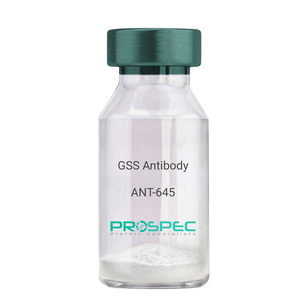
    GSS Antibody

    Description:

    Glutathione Synthetase, Mouse Anti Human

    Glutathione synthetase, GSH synthetase, GSH-S, Glutathione synthase, GSHS, MGC14098, GSS.

    Product # :

    ANT-645

    Price :

    Quantity :

    Shipping Method :

    Ice Icon

    Shipped with Ice Packs

    Add To Cart

    More Info

    • formulation
    • More Info

    Formulation

    1mg/ml containing PBS, pH-7.4, 10% Glycerol and 0.02% Sodium Azide.

    More Info

    • Introduction

      Glutathione synthetase (GSS) is the second enzyme in the glutathione biosynthesis pathway. GSS catalyses the condensation of gamma-glutamylcysteine and glycine to form glutathione. Defects in the GSS are the cause of glutathione synthetase deficiency aka GSS deficiency or 5-oxoprolinuria or pyroglutamic aciduria, which is a severe form characterized by an increased rate of hemolysis and defective function of the central nervous system.

    • Synonyms

      Glutathione synthetase, GSH synthetase, GSH-S, Glutathione synthase, GSHS, MGC14098, GSS.

    • Physical Appearance

      Sterile filtered colorless solution.

    • Immunogen

      Anti-human GSS mAb, is derived from hybridization of mouse F0 myeloma cells with spleen cells from BALB/c mice immunized with recombinant human GSS amino acids 1-474 purified from E. coli.

    • Ig Subclass

      Mouse IgG1 heavy chain and k light chain.

    • Clone

      PAT12F2AT.

    • Applications

      GSS antibody has been tested by ELISA, Western blot analysis and ICC/IF to assure specificity and reactivity. Since application varies, however, each investigation should be titrated by the reagent to obtain optimal results.

    • Type

      Mouse Anti Human Monoclonal.

    • Storage Procedures

      For periods up to 1 month store at 4°C, for longer periods of time, store at -20°C. Prevent freeze thaw cycles.

    • Purification Method

      GSS antibody was purified from mouse ascitic fluids by protein-A affinity chromatography.

    ProSpec's products are furnished for LABORATORY RESEARCH USE ONLY. The product may not be used as drugs, agricultural or pesticidal products, food additives or household chemicals.

    Gss Antibody
  • View Data Sheet

    Name :

    ALDOC Antibody

    Description:

    Aldolase C Fructose-Bisphosphate, Mouse Anti Human

    Fructose-bisphosphate aldolase C, Brain-type aldolase, ALDOC, ALDC.

    Product # :

    ANT-620

    Price :

    Quantity :

    Shipping Method :

    Ice Icon

    Shipped with Ice Packs

    Add To Cart

    More Info

    • formulation
    • More Info

    Formulation

    1mg/ml containing PBS, pH-7.4, 10% Glycerol and 0.02% Sodium Azide.

    More Info

    • Introduction

      Aldolase C Fructose-Bisphosphate (ALDOC) belongs to the class I fructose-bisphosphate aldolase family. ALDOC is a glycolytic enzyme which catalyzes the reversible aldol cleavage of fructose-1,6-biphosphate and fructose 1-phosphate to dihydroxyacetone phosphate and either glyceraldehyde-3-phosphate or glyceraldehydes respectively. ALDOC is expressed exclusively in the hippocampus and Purkinje cells of the brain.

    • Synonyms

      Fructose-bisphosphate aldolase C, Brain-type aldolase, ALDOC, ALDC.

    • Physical Appearance

      Sterile filtered colorless solution.

    • Immunogen

      Anti-human ALDOC mAb, is derived from hybridization of mouse F0 myeloma cells with spleen cells from BALB/c mice immunized with a recombinant human ALDOC protein 1-364 amino acids purified from E. coli.

    • Ig Subclass

      Mouse IgG1 heavy chain and k light chain.

    • Clone

      PAT2E11AT.

    • Applications

      The antibody has been tested by ELISA, Western blot analysis, Flow cytometry and ICC/IF to assure specificity and reactivity. Since application varies, however, each investigation should be titrated by the reagent to obtain optimal results.

    • Type

      Mouse Anti Human Monoclonal.

    • Storage Procedures

      For periods up to 1 month store at 4°C, for longer periods of time, store at -20°C. Prevent freeze thaw cycles.

    • Purification Method

      ALDOC antibody was purified from mouse ascitic fluids by protein-A affinity chromatography.

    ProSpec's products are furnished for LABORATORY RESEARCH USE ONLY. The product may not be used as drugs, agricultural or pesticidal products, food additives or household chemicals.

    Aldoc Antibody
  • View Data Sheet

    Name :

    ACAA1 Antibody

    Description:

    Acetyl-COA Acyltransferase, Mouse Anti Human

    ACAA, PTHIO, THIO.

    Product # :

    ANT-654

    Price :

    Quantity :

    Shipping Method :

    Ice Icon

    Shipped with Ice Packs

    Add To Cart

    More Info

    • formulation
    • More Info

    Formulation

    1mg/ml containing PBS, pH-7.4, 10% Glycerol and 0.02% Sodium Azide.

    More Info

    • Introduction

      ACAA1 is part of the thiolase family of enzymes and is takes part in lipid metabolism. ACAA1 enzyme is localized to the peroxisome and catalyzes the conversion of acyl-CoA and acetyl-CoA to 3-oxoacyl-CoA in the fatty acid oxidation pathway. ACAA1 shows high enzymatic activity in liver, kidney, intestine and white adipose tissue in rats. ACAA1 deficiency causes pseudo-Zellweger syndrome.

    • Synonyms

      ACAA, PTHIO, THIO.

    • Physical Appearance

      Sterile filtered colorless solution.

    • Immunogen

      Anti-human ACAA1 mAb, is derived from hybridization of mouse F0 myeloma cells with spleen cells from BALB/c mice immunized with a recombinant human ACAA1 protein 27-424 amino acids purified from E. coli.

    • Ig Subclass

      Mouse IgG2b heavy chain and k light chain.

    • Clone

      PAT9E5AT.

    • Applications

      The antibody has been tested by ELISA, Western blot analysis, Flow cytometry and ICC/IF to assure specificity and reactivity. Since application varies, however, each investigation should be titrated by the reagent to obtain optimal results.

    • Type

      Mouse Anti Human Monoclonal.

    • Storage Procedures

      For periods up to 1 month store at 4°C, for longer periods of time, store at -20°C. Prevent freeze thaw cycles.

    • Purification Method

      ACAA1 antibody was purified from mouse ascitic fluids by protein-A affinity chromatography.

    ProSpec's products are furnished for LABORATORY RESEARCH USE ONLY. The product may not be used as drugs, agricultural or pesticidal products, food additives or household chemicals.

    Acaa1 Antibody
  • View Data Sheet

    Name :

    PPCS Antibody

    Description:

    Phosphopantothenoylcysteine Synthetase, Mouse Anti Human

    Phosphopantothenate--cysteine ligase, Phosphopantothenoylcysteine synthetase, PPC synthetase, PPCS, COAB, RP11-163G10.1.

    Product # :

    ANT-070

    Price :

    Quantity :

    Shipping Method :

    Ice Icon

    Shipped with Ice Packs

    Add To Cart

    More Info

    • formulation
    • More Info

    Formulation

    1mg/ml containing PBS, pH-7.4, 10% Glycerol and 0.02% Sodium Azide.

    More Info

    • Introduction

      Phosphopantothenate-cysteine ligase (PPCS) catalyzes the first step in the biosynthesis of coenzyme A (CoA) from pantothenic acid (vitamin B5), which is a vital universal pathway in prokaryotes and eukaryotes. PPCS, being one of the last enzymes in this pathway, converts phosphopantothenate to phosphopantothenoylcysteine.

    • Synonyms

      Phosphopantothenate--cysteine ligase, Phosphopantothenoylcysteine synthetase, PPC synthetase, PPCS, COAB, RP11-163G10.1.

    • Physical Appearance

      Sterile filtered colorless solution.

    • Immunogen

      Anti-human PPCS mAb, is derived from hybridization of mouse F0 myeloma cells with spleen cells from BALB/c mice immunized with recombinant human PPCS amino acids 1-311 purified from E. coli.

    • Ig Subclass

      Mouse IgG1 heavy chain and ? light chain.

    • Clone

      PAT10D11AT.

    • Applications

      PPCS antibody has been tested by ELISA, Western blot analysis to assure specificity and reactivity. Since application varies, however, each investigation should be titrated by the reagent to obtain optimal results.

    • Type

      Mouse Anti Human Monoclonal.

    • Storage Procedures

      For periods up to 1 month store at 4°C, for longer periods of time, store at -20°C. Prevent freeze thaw cycles.

    • Purification Method

      PPCS antibody was purified from mouse ascitic fluids by protein-A affinity chromatography.

    ProSpec's products are furnished for LABORATORY RESEARCH USE ONLY. The product may not be used as drugs, agricultural or pesticidal products, food additives or household chemicals.

    Ppcs Antibody
  • View Data Sheet

    Name :

    SARS MERS Spike Antibody

    Description:

    Mouse Anti MERS-CoV Spike

    Middle East respiratory syndrome coronavirus, Human betacoronavirus 2c EMC/2012, MERS-CoV, MERS, MERSCoV SP, Spike glycoprotein, S glycoprotein, S, Spike protein, E2, Peplomer protein.

    Product # :

    ANT-768

    Price :

    Quantity :

    Shipping Method :

    Ice Icon

    Shipped with Ice Packs

    Add To Cart

    More Info

    • formulation
    • More Info

    Formulation

    1mg/ml containing Phosphate-Buffered Saline (pH 7.4), 0.02% Sodium Azide and 10% Glycerol.

    More Info

    • Introduction

      Since April 2012, cases of the Middle East Respiratory Syndrome Coronavirus (MERS-CoV) have been identified in various countries. Coronaviruses are the cause of the common cold, SARS (severe acute respiratory syndrome) and other severe illnesses with high mortality rates, all are classified into coronavirus family. MERS-CoV is a new type of SARS found in the coronavirus family causing severe pneumonia with sudden and serious respiratory illness with high mortality rates as well. Since January 27th 2015, the WHO has reported 956 human cases, including 351 deaths. More cases of the new coronavirus strain are expected. Like in other coronaviruses, large surface spike glycoprotein is a central structural protein of this virus; it is located above the virion surface to bind and enter into the target cell. Spike protein has 2 domains- S1 and S2. The S1 domain is responsible for cellular tropism and interaction with target cell, while the S2 domain is responsible for membrane fusion. The C-terminal of S1 domain contains a receptor binding domain, and is also a potential target for vaccine development and an antigen for diagnosis.

    • Synonyms

      Middle East respiratory syndrome coronavirus, Human betacoronavirus 2c EMC/2012, MERS-CoV, MERS, MERSCoV SP, Spike glycoprotein, S glycoprotein, S, Spike protein, E2, Peplomer protein.

    • Physical Appearance

      Sterile filtered colorless solution.

    • Immunogen

      Recombinant MERS-CoV Spike (18-1296aa) purified from Baculovirus.

    • Ig Subclass

      IgG1 kappa.

    • Clone

      PAT40G7AT

    • Applications

      SARS MERS Spike antibody has been tested by ELISA analysis to assure specificity and reactivity. Since application varies, however, each investigation should be titrated by the reagent to obtain optimal results.

    • Type

      Mouse antibody Monoclonal.

    • Storage Procedures

      For periods up to 1-month store at 4°C, for longer periods of time, store at -20°C. Prevent freeze thaw cycles.

    • Purification Method

      SARS MERS Spike antibody was purified from mouse ascitic fluids by protein-A affinity chromatography.

    ProSpec's products are furnished for LABORATORY RESEARCH USE ONLY. The product may not be used as drugs, agricultural or pesticidal products, food additives or household chemicals.

    Cov Mers Spike Antibody
  • View Data Sheet

    Name :

    SSU72 PAT45E2AT Antibody

    Description:

    SSU72 RNA Polymerase II CTD Phosphatase, Clone PAT45E2AT, Mouse Anti Human

    SSU72 RNA polymerase II CTD phosphatase homolog (S. cerevisiae), HSPC182, CTD phosphatase SSU72, Ssu72 RNA polymerase II CTD phosphatase homolog (yeast), PNAS-120, RNA polymerase II subunit A C-terminal domain phosphatase SSU72, EC 3.1.3.16.

    Product # :

    ANT-701

    Price :

    Quantity :

    Shipping Method :

    Ice Icon

    Shipped with Ice Packs

    Add To Cart

    More Info

    • formulation
    • More Info

    Formulation

    1mg/ml containing PBS, pH-7.4, 10% Glycerol and 0.02% Sodium Azide.

    More Info

    • Introduction

      SSU72 is an extremely conserved homologue of yeast Ssu72, a CTD phosphatase and a component of the polyadenylation/termination machinery. SSU72 interacts with TFIIB, Rb and DNAM-1 and operates to catalyze the dephosphorylation of target proteins, and taking part in RNA processing and termination via dephosphorylation of Pol II. SSU72 is found in multiple alternatively spliced isoforms.

    • Synonyms

      SSU72 RNA polymerase II CTD phosphatase homolog (S. cerevisiae), HSPC182, CTD phosphatase SSU72, Ssu72 RNA polymerase II CTD phosphatase homolog (yeast), PNAS-120, RNA polymerase II subunit A C-terminal domain phosphatase SSU72, EC 3.1.3.16.

    • Physical Appearance

      Sterile filtered colorless solution.

    • Immunogen

      Anti-human SSU72 mAb, is derived from hybridization of mouse F0 myeloma cells with spleen cells from BALB/c mice immunized with recombinant human SSU72 amino acids 1-194 purified from E. coli.

    • Ig Subclass

      Mouse IgG1 heavy chain and κ light chain.

    • Clone

      PAT45E2AT.

    • Applications

      SSU72 antibody has been tested by ELISA and Western blot analysis to assure specificity and reactivity. Since application varies, however, each investigation should be titrated by the reagent to obtain optimal results.

    • Type

      Mouse Anti Human Monoclonal.

    • Storage Procedures

      For periods up to 1 month store at 4°C, for longer periods of time, store at -20°C. Prevent freeze thaw cycles.

    • Purification Method

      SSU72 antibody was purified from mouse ascitic fluids by protein-A affinity chromatography.

    ProSpec's products are furnished for LABORATORY RESEARCH USE ONLY. The product may not be used as drugs, agricultural or pesticidal products, food additives or household chemicals.

    Anti Ssu72
  • View Data Sheet

    Name :

    ACP1 Antibody

    Description:

    Acid Phosphatase-1, Mouse Anti Human

    HAAP, MGC3499, MGC111030, ACP1, Low molecular weight phosphotyrosine protein phosphatase, LMW-PTPase, LMW-PTP, Low molecular weight cytosolic acid phosphatase, Red cell acid phosphatase 1, Adipocyte acid phosphatase, Acid Phosphatase-1.

    Product # :

    ANT-610

    Price :

    Quantity :

    Shipping Method :

    Ice Icon

    Shipped with Ice Packs

    Add To Cart

    More Info

    • formulation
    • More Info

    Formulation

    1mg/ml containing PBS, pH-7.4, 10% Glycerol and 0.02% Sodium Azide.

    More Info

    • Introduction

      ACP1 is part of the phosphotyrosine protein. ACP1 functions as an acid phosphatase and a protein tyrosine phosphatase (PTPase) existing in all human tissues, including adipocytes. ACP1 enzyme hydrolyzes protein tyrosine phosphate to protein tyrosine and orthophosphate, and also orthophosphoric monoesters to alcohol and orthophosphate. ACP1 is present in adipocytes, thus playing a specific role in the regulation of adipose tissue. High levels of the ACP1 negatively regulate cell proliferation and growth of leiomyomas during dephosphorylation of the PDGF receptor. High significant differences in birth weight-placental weight relationships were observed among acid phosphatase locus 1 phenotypes.

    • Synonyms

      HAAP, MGC3499, MGC111030, ACP1, Low molecular weight phosphotyrosine protein phosphatase, LMW-PTPase, LMW-PTP, Low molecular weight cytosolic acid phosphatase, Red cell acid phosphatase 1, Adipocyte acid phosphatase, Acid Phosphatase-1.

    • Physical Appearance

      Sterile filtered colorless solution.

    • Immunogen

      Anti-human ACP1 mAb, is derived from hybridization of mouse F0 myeloma cells with spleen cells from BALB/c mice immunized with a recombinant human ACP1 protein 1-158 amino acids purified from E. coli.

    • Ig Subclass

      Mouse IgG1 heavy chain and k light chain.

    • Clone

      PAT5C11AT.

    • Applications

      ACP1 antibody has been tested by ELISA, Western blot analysis, Flow cytometry and ICC/IF to assure specificity and reactivity. Since application varies, however, each investigation should be titrated by the reagent to obtain optimal results.

    • Type

      Mouse Anti Human Monoclonal.

    • Storage Procedures

      For periods up to 1 month store at 4°C, for longer periods of time, store at -20°C. Prevent freeze thaw cycles.

    • Purification Method

      ACP1 antibody was purified from mouse ascitic fluids by protein-A affinity chromatography.

    ProSpec's products are furnished for LABORATORY RESEARCH USE ONLY. The product may not be used as drugs, agricultural or pesticidal products, food additives or household chemicals.

    Acp1 Antibody
  • View Data Sheet

    Name :

    HK1 Antibody

    Description:

    Hexokinase-1, Mouse Anti Human

    Hexokinase-1, EC 2.7.1.1, Hexokinase type I, HK I, Brain form hexokinase, HK1-ta, HK1-tb, HXK1, HK1.

    Product # :

    ANT-375

    Price :

    Quantity :

    Shipping Method :

    Ice Icon

    Shipped with Ice Packs

    Add To Cart

    More Info

    • formulation
    • More Info

    Formulation

    1mg/ml containing PBS, pH-7.4, & 0.1% Sodium Azide.

    More Info

    • Introduction

      Hexokinases phosphorylate glucose to produce glucose-6-phosphate, thus committing glucose to the glycolytic pathway. Hexokinase1 encodes a ubiquitous form of hexokinase which localizes to the outer membrane of mitochondria. Mutations in this gene have been associated with hemolytic anemia due to hexokinase deficiency. Alternative splicing of HXK1 results in five transcript variants which encode different isoforms, some of which are tissue-specific. Each isoform has a distinct N-terminus; the remainder of the protein is identical among all the isoforms. A sixth transcript variant has been described, but due to the presence of several stop codons, it is not thought to encode a protein.

    • Synonyms

      Hexokinase-1, EC 2.7.1.1, Hexokinase type I, HK I, Brain form hexokinase, HK1-ta, HK1-tb, HXK1, HK1.

    • Physical Appearance

      Sterile Filtered clear solution.

    • Immunogen

      Anti-human Hexokinase-1 mAb is derived from hybridization of mouse SP2/0 myeloma cells with spleen cells from BALB/c mice immunized with recombinant human Hexokinase-1 amino acids 1-917 purified from E. coli.

    • Ig Subclass

      Mouse IgG2a heavy chain and κ light chain.

    • Clone

      P4D7AT.

    • Applications

      Hexokinase-1 antibody has been tested by ELISA and Western blot analysis to assure specificity and reactivity. Since application varies, however, each investigation should be titrated by the reagent to obtain optimal results. Recommended dilution range for Western blot analysis is 1:1,000 ~ 2,000. Recommended starting dilution is 1:1,000.

    • Type

      Mouse Anti Human Monoclonal.

    • Storage Procedures

      For periods up to 1 month store at 4°C, for longer periods of time, store at -20°C. Prevent freeze thaw cycles.

    • Purification Method

      Hexokinase-1 antibody was purified from mouse ascitic fluids by protein-G affinity chromatography.

    ProSpec's products are furnished for LABORATORY RESEARCH USE ONLY. The product may not be used as drugs, agricultural or pesticidal products, food additives or household chemicals.

    Hk1 Antibody
  • View Data Sheet

    Name :

    ODC1 antibody

    Description:

    Ornithine Decarboxylase 1, Mouse Anti Human

    ODC, Ornithine decarboxylase 1, EC 4.1.1.17.

    Product # :

    ANT-739

    Price :

    Quantity :

    Shipping Method :

    Ice Icon

    Shipped with Ice Packs

    Add To Cart

    More Info

    • formulation
    • More Info

    Formulation

    1mg/ml containing PBS, pH-7.4, 10% Glycerol and 0.02% Sodium Azide.

    More Info

    • Introduction

      Ornithine decarboxylase is the preliminary and rate-limiting enzyme in the biosynthetic pathway of polyamines and it takes part in the alteration of ornithine to putrescine. ODC1 is a key member of various biological processes, such as cell growth, transformation, differentiation and apoptosis. Overexpression of the ODC1 gene has a vital part in cell proliferation and the progress of cancer.

    • Synonyms

      ODC, Ornithine decarboxylase 1, EC 4.1.1.17.

    • Physical Appearance

      Sterile filtered colorless solution.

    • Immunogen

      Anti-human ODC1 mAb, is derived from hybridization of mouse F0 myeloma cells with spleen cells from BALB/c mice immunized with recombinant human ODC1 amino acids 1-461 purified from E. coli.

    • Ig Subclass

      Mouse IgG2b heavy chain and k light chain.

    • Clone

      PAT9B3AT.

    • Applications

      ODC1 antibody has been tested by ELISA, Western blot analysis, Flow cytometry and ICC/IF to assure specificity and reactivity. Since application varies, however, each investigation should be titrated by the reagent to obtain optimal results.

    • Type

      Mouse Anti Human Monoclonal.

    • Storage Procedures

      For periods up to 1 month store at 4°C, for longer periods of time, store at -20°C. Prevent freeze thaw cycles.

    • Purification Method

      ODC1 antibody was purified from mouse ascitic fluids by protein-A affinity chromatography.

    ProSpec's products are furnished for LABORATORY RESEARCH USE ONLY. The product may not be used as drugs, agricultural or pesticidal products, food additives or household chemicals.

    Odc1 Antibody
  • View Data Sheet

    Name :

    G6PD Antibody

    Description:

    Glucose-6-Phosphate Dehydrogenase, Mouse Anti Human

    G6PD, G6PD1, Glucose-6-phosphate 1-dehydrogenase.

    Product # :

    ANT-391

    Price :

    Quantity :

    Shipping Method :

    Ice Icon

    Shipped with Ice Packs

    Add To Cart

    More Info

    • formulation
    • More Info

    Formulation

    1mg/ml containing PBS, pH-7.4, & 0.1% Sodium Azide.

    More Info

    • Introduction

      G6PD is the rate-limiting enzyme of the pentose phosphate pathway, a metabolic pathway that supplies reducing energy to cells by maintaining the level of NADPH. G6PD converts glucose-6-phosphate into 6-phosphoglucono-δ-lactone and at the same time produces NADPH. The NADPH maintains the level of glutathione in these cells that helps protect the red blood cells against oxidative damage. G6PD deficiency causes acute hemolytic anemia, neonatal jaundice or acute hemolysis. G6PD is a cytosolic enzyme encoded by an X-linked gene whose main function is to produce NADPH, a crucial electron donor in the defense against oxidizing agents and in reductive biosynthetic reactions. G6PD produces pentose sugars for nucleic acid synthesis and is a main producer of NADPH reducing power.

    • Synonyms

      G6PD, G6PD1, Glucose-6-phosphate 1-dehydrogenase.

    • Immunogen

      Anti-human G6PD mAb is derived from hybridization of mouse F0 myeloma cells with spleen cells from BALB/c mice immunized with recombinant human G6PD amino acids 35-506 purified from E. coli.

    • Ig Subclass

      Mouse IgG2b heavy chain and κ light chain.

    • Clone

      PAT2F6AT.

    • Type

      Mouse Anti Human Monoclonal.

    • Storage Procedures

      For periods up to 1 month store at 4°C, for longer periods of time, store at -20°C. Prevent freeze thaw cycles.

    • Purification Method

      G6PD antibody was purified from mouse ascitic fluids by protein-G affinity chromatography.

    ProSpec's products are furnished for LABORATORY RESEARCH USE ONLY. The product may not be used as drugs, agricultural or pesticidal products, food additives or household chemicals.

    G6Pd Antibody
  • View Data Sheet

    Name :

    FAS Antibody

    Description:

    FAS Blocking/Activating antibody (CD95), Mouse anti Human

    FASLG receptor, Apoptosis-mediating surface antigen FAS, Apo-1 antigen, CD95, Tumor necrosis factor receptor superfamily member 6 TNR6, APT1, FAS1, TNFRSF6.

    Product # :

    ANT-204

    Price :

    Quantity :

    Shipping Method :

    Room Temp Icon

    Shipped at Room temp

    Add To Cart

    More Info

    • formulation
    • More Info

    Formulation

    1 mg/ml in PBS (after reconstitution).

    More Info

    • Introduction

      The Fas receptor (CD95) mediates apoptotic signaling by Fas-ligand expressed on the surface of other cells. The Fas-FasL interaction plays an important role in the immune system and lack of this system leads to autoimmunity, indicating that Fas-mediated apoptosis removes self-reactive lymphocytes. Fas signaling is also involved in immune surveillance to remove transformed cells and virus infected cells. Binding of FAS to oligimerized FasL on another cell activates apoptotic signaling through a cytoplasmic domain termed the death domain that interacts with signaling adaptors including FAF, FADD and DAX to activate the caspase proteolytic cascade. Caspase-8 and caspase-10 are first activated, to then cleave and activate downstream caspases, and a variety of cellular substrates that lead to cell death. Caspases cleave nuclear lamins, causing the nucleus to break down and lose its normal structure and another caspase substrate is DFF, inducing cleavage and degradation of the genome. Other caspase substrates are involved in cytoskeletal structure, cell cycle regulation and signaling pathways. Activation of JNK kinase, activation of Jun, and production of ceramide may also play roles in Fas-mediated apoptosis. Activation of fas-mediated apoptosis is opposed by I-FLICE and FAP. Viruses and tumors may escape immune surveillance in part through suppression of fas-mediated apoptosis using similar mechanisms.

    • Synonyms

      FASLG receptor, Apoptosis-mediating surface antigen FAS, Apo-1 antigen, CD95, Tumor necrosis factor receptor superfamily member 6 TNR6, APT1, FAS1, TNFRSF6.

    • Solubility

      Reconstitute with H20. Mix gently, wash the sides of the vial and wait 30-60 seconds before use.

    • Immunogen

      Recombinant Human FAS.

    • Ig Subclass

      mouse IgG1.

    • Clone

      NYRhFAS.

    • Note

      This antibody is a BLOCKING antibody when in soluble form and will block FAS-mediated apoptosis. It will ACTIVATE FAS-mediated killing when immobilized.

    • Titer

      By direct ELISA, 1:10,000 dilution will yield 0.5 O.D using alkaline phosphatase conjugated rabbit anti-mouse Ig (Jackson Laboratories).

    • Shipping Conditions

      Antibody is shipped lyophilized at ambient temperature.

    • Type

      Mouse Anti Human Monoclonal.

    • Storage Procedures

      In lyophilized form, for long periods, store at 4°C in a dry environment. After reconstitution, if not intended for use within a month, aliquot and store at -20°C.

    • Purification Method

      Protein-A column.

    ProSpec's products are furnished for LABORATORY RESEARCH USE ONLY. The product may not be used as drugs, agricultural or pesticidal products, food additives or household chemicals.

    Fas B A Antibody
  • View Data Sheet

    Name :

    GroEL Antibody

    Description:

    GroEL (HSP60), Mouse Anti Human

    CPN60, GROEL, HSP60, HSP65, SPG13, CHA60, GROL, crpA, mopA, 60 kDa heat shock protein mitochondrial, Heat shock protein 60, HSP-60, 60 kDa chaperonin, Chaperonin 60, Mitochondrial matrix protein P1, P60 lymphocyte protein, HuCHA60, HSPD1.

    Product # :

    ANT-327

    Price :

    Quantity :

    Shipping Method :

    Ice Icon

    Shipped with Ice Packs

    Add To Cart

    More Info

    • formulation
    • More Info

    Formulation

    1mg/ml containing PBS, pH-7.4, & 0.1% Sodium Azide.

    More Info

    • Introduction

      GroEL, HSP60 is a chaperonin located in the mitochondria which is responsible for the transportation & refolding of proteins from the cytoplasm directly into the mitochondrial matrix. GroEL is regulated by the HSP10 cochaperonin, which is a single heptameric protein ring having a molecular mass of 10 kDa which form a unique complex with HSP60. HSP10, GroES coordinates the ATPase activity of the HSP60 subunits in order to allow the release of bound polypeptide in a manner that is productive for its correct folding.

    • Synonyms

      CPN60, GROEL, HSP60, HSP65, SPG13, CHA60, GROL, crpA, mopA, 60 kDa heat shock protein mitochondrial, Heat shock protein 60, HSP-60, 60 kDa chaperonin, Chaperonin 60, Mitochondrial matrix protein P1, P60 lymphocyte protein, HuCHA60, HSPD1.

    • Physical Appearance

      Sterile Filtered solution.

    • Immunogen

      Anti-human GroEL mAb, is derived from hybridization of mouse F0 myeloma cells with spleen cells from BALB/c mice immunized with recombinant human GroEL amino acids 1-573 purified from E. coli.

    • Ig Subclass

      Mouse IgG1 heavy chain and κ light chain.

    • Clone

      P2E4AT.

    • Applications

      GroEL antibody has been tested by ELISA, Western blot and immunohistochemistry analysis to assure specificity and reactivity. Since application varies, however, each investigation should be titrated by the reagent to obtain optimal results. Recommended dilution range for Western blot is 1:1,000 ~ 1:2,000 and immunohistochemistry analysis is 1:50~100. Recommended starting dilution for Western blot is 1:1,000 and Immunohistochemistry is 1:50.

    • Type

      Mouse Anti Human Monoclonal.

    • Storage Procedures

      For periods up to 1 month store at 4°C, for longer periods of time, store at -20°C. Prevent freeze thaw cycles.

    • Purification Method

      GroEL antibody was purified from mouse ascitic fluids by protein-G affinity chromatography.

    ProSpec's products are furnished for LABORATORY RESEARCH USE ONLY. The product may not be used as drugs, agricultural or pesticidal products, food additives or household chemicals.

    Groel Antibody
  • 1
  • …
  • 27
  • 28
  • 29
  • 30
  • 31
  • …
  • 42
Your browser does not support the video tag.

Products

  • Cytokines
  • Growth Factors
  • Chemokines
  • CD Antigens
  • Neurotrophins
  • Hormones
  • Enzymes
  • Viral Antigens
  • Recombinant Proteins
  • Natural Proteins
  • Monoclonal Antibodies
  • Polyclonal Antibodies
  • Thermal Packaging

About

  • Company
  • Founder
  • Contribution
  • Contact Us
  • My Account

Ordering

  • Ordering Method
  • Ordering - Credit Card
  • Ordering PO
  • Shipping Fees
  • FAQ's

Legal

  • Terms & Conditions
  • Privacy
  • Site Map
  • info@prospecbio.com
  • Facebook
  • Instagram
  • Linkedin Profile

© 2026 ProSpec-Tany TechnoGene Ltd. All Rights Reserved.

  • Choosing a selection results in a full page refresh.
  • Opens in a new window.