Skip to content

High-quality research proteins.

  • 1-800-805-6531

  • Mon - Sat 8.00-18.00, Sun - Closed

  • info@prospecbio.com

  • PO Box 6591 East Brunswick NJ 08816

  • Products
    • Cytokines
    • Growth factors
    • Chemokines
    • Cd antigens
    • Neurotrophins
    • Hormones
    • Enzymes
    • Viral antigens
    • Recombinant proteins
    • Natural proteins
    • Monoclonal antibodies
    • Polyclonal antibodies
    • Thermal Packaging
  • About
    • Company
    • Founder
    • Contribution
  • Customers
    • Eastern Asia
    • Eastern Europe
    • North America
    • South America
    • South East Asia
    • Southern Asia
    • Western Europe
  • Ordering
    • Ordering Method
    • Ordering - Credit Card
    • Ordering - PO
    • Shipping Fees
    • FAQs
  • Services
    • DNA Cloning
    • Protein Expression
    • Monoclonal Antibody
    • Fermentation
    • Technology License
  • Contact us
Log in

Country/region

  • USD $ | Albania
  • USD $ | Andorra
  • USD $ | Argentina
  • USD $ | Armenia
  • USD $ | Australia
  • USD $ | Austria
  • USD $ | Azerbaijan
  • USD $ | Belarus
  • USD $ | Belgium
  • USD $ | Bolivia
  • USD $ | Brazil
  • USD $ | Bulgaria
  • USD $ | Cambodia
  • USD $ | Canada
  • USD $ | Chile
  • USD $ | China
  • USD $ | Colombia
  • USD $ | Costa Rica
  • USD $ | Croatia
  • USD $ | Cyprus
  • USD $ | Czechia
  • USD $ | Denmark
  • USD $ | Estonia
  • USD $ | Finland
  • USD $ | France
  • USD $ | Georgia
  • USD $ | Germany
  • USD $ | Greece
  • USD $ | Hong Kong SAR
  • USD $ | Hungary
  • USD $ | Iceland
  • USD $ | India
  • USD $ | Ireland
  • USD $ | Israel
  • USD $ | Italy
  • USD $ | Japan
  • USD $ | Kazakhstan
  • USD $ | Latvia
  • USD $ | Liechtenstein
  • USD $ | Lithuania
  • USD $ | Madagascar
  • USD $ | Malta
  • USD $ | Mexico
  • USD $ | Moldova
  • USD $ | Monaco
  • USD $ | Netherlands
  • USD $ | New Zealand
  • USD $ | North Macedonia
  • USD $ | Norway
  • USD $ | Panama
  • USD $ | Paraguay
  • USD $ | Peru
  • USD $ | Philippines
  • USD $ | Poland
  • USD $ | Portugal
  • USD $ | Romania
  • USD $ | Singapore
  • USD $ | Slovakia
  • USD $ | Slovenia
  • USD $ | South Africa
  • USD $ | South Korea
  • USD $ | Spain
  • USD $ | Sri Lanka
  • USD $ | Sweden
  • USD $ | Switzerland
  • USD $ | Taiwan
  • USD $ | Thailand
  • USD $ | Türkiye
  • USD $ | Ukraine
  • USD $ | United Kingdom
  • USD $ | United States
  • USD $ | Uruguay
  • USD $ | Vatican City
  • USD $ | Venezuela
  • USD $ | Vietnam
  • Facebook
  • Instagram
logoProSpec - Recombinant Protein Manufacturer for academic and R&D industry
Become a distributor
Account Log in
Wishlist
Cart Cart(0)
  • Products
    +
    • Cytokines
    • Growth factors
    • Chemokines
    • Cd antigens
    • Neurotrophins
    • Hormones
    • Enzymes
    • Viral antigens
    • Recombinant proteins
    • Natural proteins
    • Monoclonal antibodies
    • Polyclonal antibodies
    • Thermal Packaging
  • About
    +
    • Company
    • Founder
    • Contribution
  • Customers
    +
    • Eastern Asia
    • Eastern Europe
    • North America
    • South America
    • South East Asia
    • Southern Asia
    • Western Europe
  • Ordering
    +
    • Ordering Method
    • Ordering - Credit Card
    • Ordering - PO
    • Shipping Fees
    • FAQs
  • Services
    +
    • DNA Cloning
    • Protein Expression
    • Monoclonal Antibody
    • Fermentation
    • Technology License
  • Contact us

Item added to your cart

View cart
View All
  • Cytokines
  • Growth factors
  • Chemokines
  • Cd antigens
  • Neurotrophins
  • Hormones
  • Enzymes
  • Viral antigens
  • Recombinant proteins
  • Natural proteins
  • Monoclonal antibodies
  • Polyclonal antibodies
  • Thermal Packaging
  • Tumor Necrosis Factor

    Tumor Necrosis Factor

    View All
  • 4-1BB

    4-1BB

    View All
  • Adiponectin

    Adiponectin

    View All
  • AIF1

    AIF1

    View All
  • Other Cytokines

    Other Cytokines

    View All
  • AITRL

    AITRL

    View All
  • Angiopoietin

    Angiopoietin

    View All
  • Apolipoprotein

    Apolipoprotein

    View All
  • B-Cell Activating Factor

    B-Cell Activating Factor

    View All
  • Beta Defensin

    Beta Defensin

    View All
  • Bone Morphogenetic Protein

    Bone Morphogenetic Protein

    View All
  • B type Natriuretic Peptide

    B type Natriuretic Peptide

    View All
  • BST

    BST

    View All
  • Betacellulin

    Betacellulin

    View All
  • Cardiotrophin

    Cardiotrophin

    View All
  • Activin

    Activin

    View All
  • Fibroblast Growth Factor

    Fibroblast Growth Factor

    View All
  • Other Growth Factors

    Other Growth Factors

    View All
  • CSF

    CSF

    View All
  • CTGF

    CTGF

    View All
  • IGFBP

    IGFBP

    View All
  • VEGF Protein

    VEGF Protein

    View All
  • EGF

    EGF

    View All
  • Epigen

    Epigen

    View All
  • Erythropoietin

    Erythropoietin

    View All
  • Insulin-Like Growth Factor

    Insulin-Like Growth Factor

    View All
  • Growth Hormone

    Growth Hormone

    View All
  • HDGF

    HDGF

    View All
  • Hepatocyte Growth Factor

    Hepatocyte Growth Factor

    View All
  • Insulin

    Insulin

    View All
  • BCA-1/ BLC (CXCL13)

    BCA-1/ BLC (CXCL13)

    View All
  • BRAK (CXCL14)

    BRAK (CXCL14)

    View All
  • C-10 (CCL6)

    C-10 (CCL6)

    View All
  • Other Chemokines

    Other Chemokines

    View All
  • MEC (CCL28)

    MEC (CCL28)

    View All
  • LD78-beta (CCL3L1)

    LD78-beta (CCL3L1)

    View All
  • CTACK (CCL27)

    CTACK (CCL27)

    View All
  • CXCL16

    CXCL16

    View All
  • CXCL17

    CXCL17

    View All
  • Platelet Factor-4 (CXCL4)

    Platelet Factor-4 (CXCL4)

    View All
  • ENA-78 (CXCL5)

    ENA-78 (CXCL5)

    View All
  • Eotaxin (CCL11,24,26)

    Eotaxin (CCL11,24,26)

    View All
  • Exodus-2 (CCL21)

    Exodus-2 (CCL21)

    View All
  • Fractalkine (CX3CL1)

    Fractalkine (CX3CL1)

    View All
  • CXCL6

    CXCL6

    View All
  • Other CD Antigens

    Other CD Antigens

    View All
  • CD14

    CD14

    View All
  • CD164

    CD164

    View All
  • CD1

    CD1

    View All
  • CD2

    CD2

    View All
  • CD200

    CD200

    View All
  • CD207

    CD207

    View All
  • CD226

    CD226

    View All
  • CD23

    CD23

    View All
  • CD244

    CD244

    View All
  • CD247

    CD247

    View All
  • CD27

    CD27

    View All
  • CD274

    CD274

    View All
  • CD300

    CD300

    View All
  • CD33

    CD33

    View All
  • Other Neurotrophins

    Other Neurotrophins

    View All
  • Beta-NGF

    Beta-NGF

    View All
  • BDNF

    BDNF

    View All
  • CDNF

    CDNF

    View All
  • CNTF

    CNTF

    View All
  • GDNF

    GDNF

    View All
  • Glia Maturation Factor

    Glia Maturation Factor

    View All
  • MANF

    MANF

    View All
  • Midkine

    Midkine

    View All
  • Neuroglobin

    Neuroglobin

    View All
  • Neurotrophic factors

    Neurotrophic factors

    View All
  • Neuregulin

    Neuregulin

    View All
  • Neuropilin

    Neuropilin

    View All
  • Pigment Epithelium-Derived Factor

    Pigment Epithelium-Derived Factor

    View All
  • Pleiotrophin

    Pleiotrophin

    View All
  • Peptide Hormones

    Peptide Hormones

    View All
  • Other Hormones

    Other Hormones

    View All
  • HCG

    HCG

    View All
  • Endothelin

    Endothelin

    View All
  • Exendin

    Exendin

    View All
  • FSH

    FSH

    View All
  • Glucagon

    Glucagon

    View All
  • GHRP

    GHRP

    View All
  • GLP

    GLP

    View All
  • Thyrostimulin

    Thyrostimulin

    View All
  • Inhibin A

    Inhibin A

    View All
  • LHRH

    LHRH

    View All
  • Melanotan

    Melanotan

    View All
  • Procalcitonin

    Procalcitonin

    View All
  • PTH

    PTH

    View All
  • Peroxiredoxin

    Peroxiredoxin

    View All
  • Peptidase

    Peptidase

    View All
  • Synthetase

    Synthetase

    View All
  • Transferase

    Transferase

    View All
  • Hydrolase

    Hydrolase

    View All
  • Dehydrogenase

    Dehydrogenase

    View All
  • ACE2

    ACE2

    View All
  • Esterase

    Esterase

    View All
  • Protein Kinases

    Protein Kinases

    View All
  • Other Enzymes

    Other Enzymes

    View All
  • Phosphatase

    Phosphatase

    View All
  • Deaminase

    Deaminase

    View All
  • Oxygenase

    Oxygenase

    View All
  • Adenylate Kinase

    Adenylate Kinase

    View All
  • Lyase

    Lyase

    View All
  • Chagas

    Chagas

    View All
  • SARS

    SARS

    View All
  • Influenza

    Influenza

    View All
  • ASFV

    ASFV

    View All
  • Astrovirus

    Astrovirus

    View All
  • Borrelia

    Borrelia

    View All
  • Helicobacter Pylori

    Helicobacter Pylori

    View All
  • Chikungunya

    Chikungunya

    View All
  • Chlamydia

    Chlamydia

    View All
  • Cytomegalo

    Cytomegalo

    View All
  • Dengue

    Dengue

    View All
  • EBV

    EBV

    View All
  • Ebola

    Ebola

    View All
  • Feline Leukemia Virus

    Feline Leukemia Virus

    View All
  • Hepatitis A

    Hepatitis A

    View All
  • Other

    Other

    View All
  • Actin

    Actin

    View All
  • ADAM

    ADAM

    View All
  • Complement Component

    Complement Component

    View All
  • Ag85

    Ag85

    View All
  • Eukaryotic Translation Initiation Factor

    Eukaryotic Translation Initiation Factor

    View All
  • Anterior Gradient Protein

    Anterior Gradient Protein

    View All
  • Heat Shock Protein

    Heat Shock Protein

    View All
  • Alpha-2-HS-Glycoprotein

    Alpha-2-HS-Glycoprotein

    View All
  • Hemoglobin

    Hemoglobin

    View All
  • Allergy

    Allergy

    View All
  • Anaplasma

    Anaplasma

    View All
  • Angiogenin

    Angiogenin

    View All
  • Ankyrin Repeat Domain

    Ankyrin Repeat Domain

    View All
  • Annexin

    Annexin

    View All
  • Other Natural Proteins

    Other Natural Proteins

    View All
  • Aprotinin

    Aprotinin

    View All
  • Transferrin

    Transferrin

    View All
  • Avidin

    Avidin

    View All
  • Anti Coagulation Factors

    Anti Coagulation Factors

    View All
  • Natural Albumin

    Natural Albumin

    View All
  • Natural Coagulation Factors

    Natural Coagulation Factors

    View All
  • Fibronectin

    Fibronectin

    View All
  • Thrombin

    Thrombin

    View All
  • Anti Human Enzyme

    Anti Human Enzyme

    View All
  • Other Monoclonal Antibodies

    Other Monoclonal Antibodies

    View All
  • Anti Human Cytokine

    Anti Human Cytokine

    View All
  • Anti Human Heat Shock Protein

    Anti Human Heat Shock Protein

    View All
  • Anti Mouse Lymphocyte

    Anti Mouse Lymphocyte

    View All
  • Anti Human Chemokine

    Anti Human Chemokine

    View All
  • Anti Human Lymphocyte

    Anti Human Lymphocyte

    View All
  • Anti Viral Monoclonal

    Anti Viral Monoclonal

    View All
  • Anti Mouse Cytokine

    Anti Mouse Cytokine

    View All
  • Anti-GST Monoclonal

    Anti-GST Monoclonal

    View All
  • Other Polyclonal Antibodies

    Other Polyclonal Antibodies

    View All
  • Anti Viral

    Anti Viral

    View All
  • Anti-GST Monoclonal

    Anti-GST Monoclonal

    View All
Back

Search results

1000 results found for “encephalitis”

Name

Description

Product #

Price

Quantity

Shipping Method

  • View Data Sheet

    Name :

    SHMT1 Antibody

    Description:

    Serine Hydroxymethyltransferase 1, Mouse Anti Human

    Serine hydroxymethyltransferase 1 (soluble), CSHMT, Glycine hydroxymethyltransferase, Serine methylase, 14 kDa protein, cytoplasmic serine hydroxymethyltransferase, serine hydroxymethyltransferase cytosolic, EC 2.1.2.1.

    Product # :

    ANT-636

    Price :

    Quantity :

    Shipping Method :

    Ice Icon

    Shipped with Ice Packs

    Add To Cart

    More Info

    • formulation
    • More Info
    • Citations

    Formulation

    1mg/ml containing PBS, pH-7.4, 10% Glycerol and 0.02% Sodium Azide.

    Citations

    More Info

    • Introduction

      SHMT1 is a member of the SHMT family. SHMT1 is the cellular form of serine hydroxymethyltransferase, a pyridoxal phosphate-containing enzyme which catalyzes the reversible conversion of serine and tetrahydrofolate to glycine and 5 10-methylene tetrahydrofolate. In addition, SHMT1 specifically provides one-carbon units for thymidylate biosynthesis, reduces methylenetetrahydrofolate pools for S-adenosylmethionine (SAM) synthesis by synthesizing serine, sequesters 5-methyltetrahydrofolate and inhibits SAM synthesis.

    • Synonyms

      Serine hydroxymethyltransferase 1 (soluble), CSHMT, Glycine hydroxymethyltransferase, Serine methylase, 14 kDa protein, cytoplasmic serine hydroxymethyltransferase, serine hydroxymethyltransferase cytosolic, EC 2.1.2.1.

    • Physical Appearance

      Sterile filtered colorless solution.

    • Immunogen

      Anti-human SHMT1 mAb, is derived from hybridization of mouse F0 myeloma cells with spleen cells from BALB/c mice immunized with recombinant human SHMT1 amino acids 1-483 purified from E. coli.

    • Ig Subclass

      Mouse IgG1 heavy chain and k light chain.

    • Clone

      PAT26E5AT.

    • Applications

      SHMT1 antibody has been tested by ELISA, Western blot analysis, Flow cytometry and ICC/IF to assure specificity and reactivity. Since application varies, however, each investigation should be titrated by the reagent to obtain optimal results.

    • Type

      Mouse Anti Human Monoclonal.

    • Storage Procedures

      For periods up to 1 month store at 4°C, for longer periods of time, store at -20°C. Prevent freeze thaw cycles.

    • Purification Method

      SHMT1 antibody was purified from mouse ascitic fluids by protein-A affinity chromatography.

    ProSpec's products are furnished for LABORATORY RESEARCH USE ONLY. The product may not be used as drugs, agricultural or pesticidal products, food additives or household chemicals.

    Shmt1 Antibody
  • View Data Sheet

    Name :

    NMT1 Antibody

    Description:

    N-Myristoyltransferase 1, Mouse Anti Human

    N-Myristoyltransferase 1, NMT, Myristoyl-CoA:Protein N-Myristoyltransferase 1, Type I N-Myristoyltransferase, EC 2.3.1.97, Myristoyl-CoA:Protein,N- Myristoyltransferase, Glycylpeptide N-Tetradecanoyltransferase 1,Peptide N-Myristoyltransferase 1,Alternative, Short Form NMT-S, Short Form NMT-S,Long Form, NMT-L, Alternative, Long Form, NMT-L, NMT 1.

    Product # :

    ANT-731

    Price :

    Quantity :

    Shipping Method :

    Ice Icon

    Shipped with Ice Packs

    Add To Cart

    More Info

    • formulation
    • More Info
    • Citations

    Formulation

    1mg/ml containing PBS, pH-7.4, 10% Glycerol and 0.02% Sodium Azide.

    Citations

    More Info

    • Introduction

      Myristate, a rare 14-carbon saturated fatty acid, is co-translationally attached by an amide linkage to the N-terminal glycine residue of cellular & viral proteins with various functions. N-Myristoyltransferase 1, also known as NMT1 catalyzes the transfer of myristate from CoA to proteins. NMT1 seems to be irreversible and is essential for full expression of the biologic activities of several N-myristoylated proteins, as well as the alpha subunit of the signal-transducing guanine nucleotide-binding protein, G protein.

    • Synonyms

      N-Myristoyltransferase 1, NMT, Myristoyl-CoA:Protein N-Myristoyltransferase 1, Type I N-Myristoyltransferase, EC 2.3.1.97, Myristoyl-CoA:Protein,N- Myristoyltransferase, Glycylpeptide N-Tetradecanoyltransferase 1,Peptide N-Myristoyltransferase 1,Alternative, Short Form NMT-S, Short Form NMT-S,Long Form, NMT-L, Alternative, Long Form, NMT-L, NMT 1.

    • Physical Appearance

      Sterile filtered colorless solution.

    • Immunogen

      Anti-human NMT1 mAb, is derived from hybridization of mouse F0 myeloma cells with spleen cells from BALB/c mice immunized with recombinant human NMT1 amino acids 1-496 purified from E. coli.

    • Ig Subclass

      Mouse IgG2a heavy chain and κ light chain.

    • Clone

      PAT2C8AT.

    • Applications

      NMT1 antibody has been tested by ELISA, Western blot analysis, ICC/IF and Flow cytometry to assure specificity and reactivity. Since application varies, however, each investigation should be titrated by the reagent to obtain optimal results.

    • Type

      Mouse Anti Human Monoclonal.

    • Storage Procedures

      For periods up to 1 month store at 4°C, for longer periods of time, store at -20°C. Prevent freeze thaw cycles.

    • Purification Method

      NMT1 antibody was purified from mouse ascitic fluids by protein-A affinity chromatography.

    ProSpec's products are furnished for LABORATORY RESEARCH USE ONLY. The product may not be used as drugs, agricultural or pesticidal products, food additives or household chemicals.

    Nmt1 Antibody
  • View Data Sheet

    Name :

    ODC1 antibody

    Description:

    Ornithine Decarboxylase 1, Mouse Anti Human

    ODC, Ornithine decarboxylase 1, EC 4.1.1.17.

    Product # :

    ANT-739

    Price :

    Quantity :

    Shipping Method :

    Ice Icon

    Shipped with Ice Packs

    Add To Cart

    More Info

    • formulation
    • More Info
    • Citations

    Formulation

    1mg/ml containing PBS, pH-7.4, 10% Glycerol and 0.02% Sodium Azide.

    Citations

    More Info

    • Introduction

      Ornithine decarboxylase is the preliminary and rate-limiting enzyme in the biosynthetic pathway of polyamines and it takes part in the alteration of ornithine to putrescine. ODC1 is a key member of various biological processes, such as cell growth, transformation, differentiation and apoptosis. Overexpression of the ODC1 gene has a vital part in cell proliferation and the progress of cancer.

    • Synonyms

      ODC, Ornithine decarboxylase 1, EC 4.1.1.17.

    • Physical Appearance

      Sterile filtered colorless solution.

    • Immunogen

      Anti-human ODC1 mAb, is derived from hybridization of mouse F0 myeloma cells with spleen cells from BALB/c mice immunized with recombinant human ODC1 amino acids 1-461 purified from E. coli.

    • Ig Subclass

      Mouse IgG2b heavy chain and k light chain.

    • Clone

      PAT9B3AT.

    • Applications

      ODC1 antibody has been tested by ELISA, Western blot analysis, Flow cytometry and ICC/IF to assure specificity and reactivity. Since application varies, however, each investigation should be titrated by the reagent to obtain optimal results.

    • Type

      Mouse Anti Human Monoclonal.

    • Storage Procedures

      For periods up to 1 month store at 4°C, for longer periods of time, store at -20°C. Prevent freeze thaw cycles.

    • Purification Method

      ODC1 antibody was purified from mouse ascitic fluids by protein-A affinity chromatography.

    ProSpec's products are furnished for LABORATORY RESEARCH USE ONLY. The product may not be used as drugs, agricultural or pesticidal products, food additives or household chemicals.

    Odc1 Antibody
  • View Data Sheet

    Name :

    PA2G4 Antibody

    Description:

    Proliferation-associated protein 2G4, Mouse Anti Human

    Proliferation-associated protein 2G4, Cell cycle protein p38-2G4 homolog, hG4-1, ErbB3-binding protein 1, PA2G4, EBP1, p38-2G4.

    Product # :

    ANT-655

    Price :

    Quantity :

    Shipping Method :

    Ice Icon

    Shipped with Ice Packs

    Add To Cart

    More Info

    • formulation
    • More Info
    • Citations

    Formulation

    1mg/ml containing PBS, pH-7.4, 10% Glycerol and 0.02% Sodium Azide.

    Citations

    More Info

    • Introduction

      PA2G4 belongs to the peptidase M24C family and functions as an RNA-binding protein involved in cellular proliferation and differentiation processes. PA2G4 is a component of pre-ribosomal ribonucleoprotein complexes, participating in ribosome assembly and regulating the later steps of rRNA processing. Also, PA2G4 interacts with ErbB-3 and may function as a modulator of the ErbB-3 mediated signal transduction pathway by regulating the effects of Neuregulin-1. Furthermore, PA2G4 is a transcriptional co-repressor of androgen receptor-regulated genes and other cell cycle regulatory genes through its interactions with histone deacetylases. PA2G4 is implicated in growth inhibition and the induction of differentiation of human cancer cells. In addition, PA2G4 mediates cap-independent translation of specific viral IRESs (internal ribosomal entry site). PA2G4 associates with 28S, 18S and 5.8S mature rRNAs, several rRNA precursors and probably U3 small nucleolar RNA.

    • Synonyms

      Proliferation-associated protein 2G4, Cell cycle protein p38-2G4 homolog, hG4-1, ErbB3-binding protein 1, PA2G4, EBP1, p38-2G4.

    • Physical Appearance

      Sterile filtered colorless solution.

    • Immunogen

      Anti-human PA2G4 mAb, is derived from hybridization of mouse F0 myeloma cells with spleen cells from BALB/c mice immunized with a recombinant human PA2G4 protein 1-394 amino acids purified from E. coli.

    • Ig Subclass

      Mouse IgG1 heavy chain and k light chain.

    • Clone

      PAT5E7AT.

    • Applications

      The antibody has been tested by ELISA, Western blot analysis, Flow cytometry and ICC/IF to assure specificity and reactivity. Since application varies, however, each investigation should be titrated by the reagent to obtain optimal results.

    • Type

      Mouse Anti Human Monoclonal.

    • Storage Procedures

      For periods up to 1 month store at 4°C, for longer periods of time, store at -20°C. Prevent freeze thaw cycles.

    • Purification Method

      PA2G4 antibody was purified from mouse ascitic fluids by protein-A affinity chromatography.

    ProSpec's products are furnished for LABORATORY RESEARCH USE ONLY. The product may not be used as drugs, agricultural or pesticidal products, food additives or household chemicals.

    Pa2G4 Antibody
  • View Data Sheet

    Name :

    FKBP6 Antibody

    Description:

    FK506 Binding Protein 6, Mouse Anti Human

    FKBP36, EC 5.2.1.8, FKBP-6, PPIase, MGC87179, FK506-binding protein 6, Rotamase.

    Product # :

    ANT-499

    Price :

    Quantity :

    Shipping Method :

    Ice Icon

    Shipped with Ice Packs

    Add To Cart

    More Info

    • formulation
    • More Info
    • Citations

    Formulation

    1mg/ml containing PBS, pH-7.4, 10% Glycerol and 0.02% Sodium Azide.

    Citations

    More Info

    • Introduction

      FKBP6 is part of the immunophilin protein family, and is involved in immunoregulation and basic cellular functions involving protein folding and trafficking. FKBP6 is widely expressed in all tissues, FKBP6 is present at utmost levels in testis, liver, kidney, skeletal muscle and heart. Deletion of FKBP6 contributes to hypercalcemia and growth delay in Williams-Beuren syndrome.

    • Synonyms

      FKBP36, EC 5.2.1.8, FKBP-6, PPIase, MGC87179, FK506-binding protein 6, Rotamase.

    • Physical Appearance

      Sterile filtered colorless solution.

    • Immunogen

      Anti-human FKBP6 mAb, clone PAT9B7A, is derived from hybridization of mouse F0 myeloma cells with spleen cells from BALB/c mice immunized with a recombinant human FKBP6 protein 1-327 amino acids purified from E. coli.

    • Ig Subclass

      Mouse IgG1 heavy chain and Kappa light chain.

    • Clone

      PAT9B7A

    • Applications

      The antibody has been tested by ELISA, Western blot analysis to assure specificity and reactivity. Since application varies, however, each investigation should be titrated by the reagent to obtain optimal results. Recommended starting dilution is 1:1000.

    • Type

      Mouse Anti Human Monoclonal.

    • Storage Procedures

      For periods up to 1 month store at 4°C, for longer periods of time, store at -20°C. Prevent freeze thaw cycles.

    • Purification Method

      NCK1 antibody was purified from mouse ascitic fluids by protein-A affinity chromatography.

    ProSpec's products are furnished for LABORATORY RESEARCH USE ONLY. The product may not be used as drugs, agricultural or pesticidal products, food additives or household chemicals.

    Fkbp6 Antibody
  • View Data Sheet

    Name :

    JAM2 antibody

    Description:

    Mouse Anti Human Junctional Adhesion Molecule 2

    Junctional adhesion molecule B, JAM-B, Junctional adhesion molecule 2, JAM-2, Vascular endothelial junction-associated molecule, VE-JAM, CD322.

    Product # :

    ANT-738

    Price :

    Quantity :

    Shipping Method :

    Ice Icon

    Shipped with Ice Packs

    Add To Cart

    More Info

    • formulation
    • More Info
    • Citations

    Formulation

    1mg/ml containing PBS, pH-7.4, 10% Glycerol and 0.02% Sodium Azide.

    Citations

    More Info

    • Introduction

      Junctional Adhesion Molecule 2 (JAM2) is a member of the immunoglobulin superfamily, and the junctional adhesion molecule (JAM) family. JAM2 is a type I membrane protein which is localized at the tight junctions of both epithelial and endothelial cells. JAM2 functions as an adhesive ligand for interacting with various immune cell types, and may have a role in lymphocyte homing to secondary lymphoid organs.

    • Synonyms

      Junctional adhesion molecule B, JAM-B, Junctional adhesion molecule 2, JAM-2, Vascular endothelial junction-associated molecule, VE-JAM, CD322.

    • Physical Appearance

      Sterile filtered colorless solution.

    • Immunogen

      Anti-human JAM2 mAb, is derived from hybridization of mouse F0 myeloma cells with spleen cells from BALB/c mice immunized with a recombinant human JAM2 protein 21-238 amino acids purified from E. coli.

    • Ig Subclass

      Mouse IgG2a heavy chain and k light chain.

    • Clone

      PAT21D7AT.

    • Applications

      JAM2 antibody has been tested by ELISA and Western blot analysis to assure specificity and reactivity. Since application varies, however, each investigation should be titrated by the reagent to obtain optimal results.

    • Type

      Mouse Anti Human Monoclonal.

    • Storage Procedures

      For periods up to 1 month store at 4°C, for longer periods of time, store at -20°C. Prevent freeze thaw cycles.

    • Purification Method

      JAM2 antibody was purified from mouse ascitic fluids by protein-A affinity chromatography.

    ProSpec's products are furnished for LABORATORY RESEARCH USE ONLY. The product may not be used as drugs, agricultural or pesticidal products, food additives or household chemicals.

    Jam2 Antibody
  • View Data Sheet

    Name :

    PSMD10 Antibody

    Description:

    Gankyrin, Mouse Anti Human

    26S proteasome non-ATPase regulatory subunit 10, 26S proteasome regulatory subunit p28, Gankyrin, PSMD10, p28, dJ889N15.2.

    Product # :

    ANT-609

    Price :

    Quantity :

    Shipping Method :

    Ice Icon

    Shipped with Ice Packs

    Add To Cart

    More Info

    • formulation
    • More Info
    • Citations

    Formulation

    1mg/ml containing PBS, pH-7.4, 10% Glycerol and 0.02% Sodium Azide.

    Citations

    More Info

    • Introduction

      Gankyrin (proteasome 26S subunit) is a multicatalytic proteinase oncoprotein commonly overexpressed in most hepatocellular carcinomas. Proteasomes are found throughout eukaryotic cells at a high concentrations and cleave peptides in an ATP/ubiquitin-dependent process in a non-lysosomal pathway. Gankyrin interacts with S6 ATPase of the 19S regulatory particle of the 26S proteasome. Gankyrin is involved in theregulation of the phosphorylation of the retinoblastoma protein by CDK4, and to enhance the ubiquitinylation of p53 by MDM2. Gankyrin consists of 7 ankyrin repeats and is structurally similar to I kappa Bs. Gankyrin acts as a regulatory subunit of the 26s proteasome which is involved in the atp-dependent degradation of ubiquitinated proteins. Gankyrin is involved in progression of esophageal squamous cell carcinoma. gankyrin plays an oncogenic role especially in early stages of human epatocarcinogenesis. Gankyrin binds to NF-kappaB and suppresses its activity at the transcription level by modulating acetylation through SIRT1. Structural comparison between Gankyrin & p16(INK4A) identified numerous residues of gankyrin that are potentially important for CDK4 binding.

    • Synonyms

      26S proteasome non-ATPase regulatory subunit 10, 26S proteasome regulatory subunit p28, Gankyrin, PSMD10, p28, dJ889N15.2.

    • Physical Appearance

      Sterile filtered colorless solution.

    • Immunogen

      Anti-human PSMD10 mAb, is derived from hybridization of mouse F0 myeloma cells with spleen cells from BALB/c mice immunized with a recombinant human PSMD10 protein 1-226 amino acids purified from E.coli.

    • Ig Subclass

      Mouse IgG1 heavy chain and k light chain.

    • Clone

      PAT1F4AT.

    • Applications

      PSMD10 antibody has been tested by ELISA, Western blot analysis, Flow cytometry and ICC/IF to assure specificity and reactivity. Since application varies, however, each investigation should be titrated by the reagent to obtain optimal results.

    • Type

      Mouse Anti Human Monoclonal.

    • Storage Procedures

      For periods up to 1 month store at 4°C, for longer periods of time, store at -20°C. Prevent freeze thaw cycles.

    • Purification Method

      PSMD10 antibody was purified from mouse ascitic fluids by protein-A affinity chromatography.

    ProSpec's products are furnished for LABORATORY RESEARCH USE ONLY. The product may not be used as drugs, agricultural or pesticidal products, food additives or household chemicals.

    Psmd10 Antibody
  • View Data Sheet

    Name :

    GSTP1 Antibody

    Description:

    Glutathione S-Transferase pi 1, Mouse Anti Human

    Glutathione S-transferase P 1, Gst P1, GST YF-YF, GST class-pi, GST-piB, Preadipocyte growth factor.

    Product # :

    ANT-729

    Price :

    Quantity :

    Shipping Method :

    Ice Icon

    Shipped with Ice Packs

    Add To Cart

    More Info

    • formulation
    • More Info
    • Citations

    Formulation

    1mg/ml containing PBS, pH-7.4, 10% Glycerol and 0.02% Sodium Azide.

    Citations

    More Info

    • Introduction

      GSTP1 is a polymorphic gene encoding active, functionally different GSTP1 variant proteins that are believed to function in xenobiotic metabolism and have a part in susceptibility to cancer, and other diseases. GSTP1 is a glutathione S-transferase belonging to the pi class. GST family enzymes play a significant role in detoxification by catalyzing the conjugation of many hydrophobic and electrophilic compounds with reduced glutathione. Based upon the biochemical, immunologic and structural properties of the soluble GSTs they are grouped into 4 main classes: alpha, mu, pi, and theta. The GSTP1 enzyme acts by catalyzing the reaction of glutathione with an acceptor molecule to form a Sulfur-substituted glutathione. The reactions employing glutathione contribute the transformation of a broad range of electrophiles, including reactive products of lipid, protein, carcinogens, therapeutic drugs, environmental toxins, and products of oxidative stress.
      The GSTP1 inactivation through CpG hypermethylation is frequent in pituitary adenomas and may be a factor in aggressive pituitary tumor behavior. GSTP1 is may be a transcriptional target of the p53 tumor suppressor gene. Single-nucleotide polymorphism in GSTP1 is linked to modified protein binding, which influence GSTP1's contribution to carcinogen and drug metabolism, and possibly disease pathogenesis and/or drug response. GST-pi might have central roles in proliferation of androgen-independent human prostate cancer cells.

    • Synonyms

      Glutathione S-transferase P 1, Gst P1, GST YF-YF, GST class-pi, GST-piB, Preadipocyte growth factor.

    • Physical Appearance

      Sterile filtered colorless solution.

    • Immunogen

      Anti-human GSTP1 mAb, is derived from hybridization of mouse F0 myeloma cells with spleen cells from BALB/c mice immunized with recombinant human GSTP1 amino acids 1-210 purified from E. coli.

    • Ig Subclass

      Mouse IgG1 heavy chain and κ light chain.

    • Clone

      PAT12C10AT

    • Applications

      GSTP1 antibody has been tested by ELISA, Western blot analysis, ICC/IF and Flow cytometry to assure specificity and reactivity. Since application varies, however, each investigation should be titrated by the reagent to obtain optimal results.

    • Type

      Mouse Anti Human Monoclonal.

    • Storage Procedures

      For periods up to 1 month store at 4°C, for longer periods of time, store at -20°C. Prevent freeze thaw cycles.

    • Purification Method

      GSTP1 antibody was purified from mouse ascitic fluids by protein-A affinity chromatography.

    ProSpec's products are furnished for LABORATORY RESEARCH USE ONLY. The product may not be used as drugs, agricultural or pesticidal products, food additives or household chemicals.

    Gstp1 Antibody
  • View Data Sheet

    Name :

    GSTT1 Antibody

    Description:

    Glutathione S-Transferase Theta-1, Mouse Anti Human

    Glutathione S-transferase theta-1, GST class-theta-1, Glutathione transferase T1-1, GSTT1.

    Product # :

    ANT-591

    Price :

    Quantity :

    Shipping Method :

    Ice Icon

    Shipped with Ice Packs

    Add To Cart

    More Info

    • formulation
    • More Info
    • Citations

    Formulation

    1mg/ml containing PBS, pH-7.4, 10% Glycerol and 0.02% Sodium Azide.

    Citations

    More Info

    • Introduction

      GSTT1 belongs to a superfamily of proteins which catalyze the conjugation of reduced glutathione to a variety of electrophilic and hydrophobic compounds. GSTT1 is one of the GSTs’ four main classes: alpha, mu, pi and theta (which includes GSTT1 and GSTT2). GSTT1 is involved in activation and detoxification reactions and catalyzes the conjugation of industrial chemicals, such as epoxybutane, ethylene oxides, halomethane with glutathione. GSTT1 is found in erythrocytes, at low levels in the liver as well as in Clara and ciliated cells at the alveolar/bronchiolar junction in the lung. The GSTT1 gene is deficient in 38% of the population. The GSTTI enzyme deficiency might influence the individual risk for development of acquired aplastic anemia and acute myeloid leukemia. The presence or absence of the GSTT1 gene is concurrent with GSST1+ (the conjugator) and GSTT1- (the non-conjugator) phenotypes correspondingly. The GSTT1+ phenotype is able to catalyze the glutathione conjugation of dichloromethane. GSTT1-null genotypes are seen as having a higher risk of developing leukoplakia. Germline genetic polymorphism in GSTT1 is linked to breast cancer.

    • Synonyms

      Glutathione S-transferase theta-1, GST class-theta-1, Glutathione transferase T1-1, GSTT1.

    • Physical Appearance

      Sterile filtered colorless solution.

    • Immunogen

      Anti-human GSTT1 mAb, is derived from hybridization of mouse F0 myeloma cells with spleen cells from BALB/c mice immunized with recombinant human GSTT1 amino acids 1-240 purified from E. coli.

    • Ig Subclass

      Mouse IgG2b heavy chain and k light chain.

    • Clone

      PAT38D11AT.

    • Applications

      GSTT1 antibody has been tested by ELISA, Western blot analysis, ICC/IF and Flow cytometry to assure specificity and reactivity. Since application varies, however, each investigation should be titrated by the reagent to obtain optimal results.

    • Type

      Mouse Anti Human Monoclonal.

    • Storage Procedures

      For periods up to 1 month store at 4°C, for longer periods of time, store at -20°C. Prevent freeze thaw cycles.

    • Purification Method

      GSTT1 antibody was purified from mouse ascitic fluids by protein-A affinity chromatography.

    ProSpec's products are furnished for LABORATORY RESEARCH USE ONLY. The product may not be used as drugs, agricultural or pesticidal products, food additives or household chemicals.

    Gstt1 Antibody
  • View Data Sheet

    Name :

    PfHSP70

    Description:

    Plasmodium Falciparum HSP70 Recombinant

    Product # :

    MAL-001

    Price :

    Quantity :

    Shipping Method :

    Ice Icon

    Shipped with Ice Packs

    Add To Cart

    More Info

    • description
    • formulation
    • purity
    • More Info
    • Citations

    Description

    The E.Coli derived recombinant Plasmodium falciparum contains the Plasmodium falciparum HSP70 protein epitopes 33-114 amino acids.

    Formulation

    150mM Imidazole, pH 8.0, 150mM NaCl, 150mM Sodium Phosphate and 50% glycerol.

    Purity

    Protein is >95% pure as determined by 10% PAGE (coomassie staining) and RP-HPLC.

    Citations

    More Info

    • Introduction

      Plasmodium falciparum heat shock proteins (PfHSPs) contribute to immunopathological alterations in infected hosts PfHSP70s may have a significant function during parasite adaptation in its environments. They have also been a focus of considerable attention due to their immunodominant antigenic nature and their properties as mediators of protective immunity.

    • Physical Appearance

      Sterile Filtered clear solution.

    • Stability

      PfHSP70 although stable at 4°C for 1 week, should be stored below -18°C. Please prevent freeze thaw cycles.

    • Applications

      Use as an antigen in ELISA and Western Blots.

    • Purification Method

      Purified by proprietary chromatographic technique

    ProSpec's products are furnished for LABORATORY RESEARCH USE ONLY. The product may not be used as drugs, agricultural or pesticidal products, food additives or household chemicals.

    Pfhsp70
  • View Data Sheet

    Name :

    Y.Enterocolitica (O:9) YopB

    Description:

    Yersinia Enterocolitica (O:9) YopB Recombinant

    Protein YopD, pYV0054.

    Product # :

    PRO-2575

    Price :

    Quantity :

    Shipping Method :

    Ice Icon

    Shipped with Ice Packs

    Add To Cart

    More Info

    • description
    • source
    • formulation
    • purity
    • More Info
    • Citations

    Description

    Recombinant Yersinia Enterocolitica (O:9) YopB produced in Sf9 Baculovirus cells is a single, glycosylated polypeptide chain having a calculated molecular mass of 43kDa. Y.Enterocolitica (O:9) YopB is expressed with a -10x His tag at N-terminus and purified by proprietary chromatographic techniques.

    Source

    Sf9, Baculovirus cells.

    Formulation

    Y.Enterocolitica (O:9) YopB is supplied in 20mM HEPES buffer pH-7.6, 250mM NaCl and 20% glycerol.

    Purity

    Greater than 80.0% as determined by SDS-PAGE.

    Citations

    More Info

    • Introduction

      Gastroenteritis which has been identified as caused by Yersinia Enterocolitica has been discovered by virulent isolates of the bacteria. During infection, pores are being formed in the presence of adapter protein Yersiniosis. YopB's Interaction with the target membrane to form pores enables the entrance of YopE effectors to the cytoplasmic space of the host’s cell.

    • Synonyms

      Protein YopD, pYV0054.

    • Physical Appearance

      Sterile Filtered clear solution.

    • Stability

      Store at 4°C if entire vial will be used within 2-4 weeks. Store, frozen at -20°C for longer periods of time. Avoid multiple freeze-thaw cycles.

    • Immunological Activity

      1. Binds IgG- and IgM-type human antibodies.2. Immunodot test with positive/negative samples.

    ProSpec's products are furnished for LABORATORY RESEARCH USE ONLY. The product may not be used as drugs, agricultural or pesticidal products, food additives or household chemicals.

    Yopb Protein
  • View Data Sheet

    Name :

    Eptifibatide Human

    Description:

    Eptifibatide Human

    Product # :

    PRO-358

    Price :

    Quantity :

    Shipping Method :

    Room Temp Icon

    Shipped at Room temp

    Add To Cart

    More Info

    • description
    • formulation
    • purity
    • More Info
    • Citations

    Description

    Synthetic Eptifibatide is a 6 amino acid peptide having a MW of 832.5 Dalton the following sequence Mpr-Har-Gly-Asp-Trp-Pro-Cys-NH2.

    Formulation

    Eptifibatide was lyophilized with no additives.

    Purity

    Greater than 98.0% as determined by:
    (a) Analysis by RP-HPLC.
    (b) Analysis by SDS-PAGE.

    Citations

    More Info

    • Introduction

      Eptifibatide binds to the platelet receptor glycoprotein (GP) IIb/IIIa of human platelets and inhibits platelet aggregation. Eptifibatide is a cyclic peptide derived from a protein discovered in the venom of the southeastern pygmy rattlesnake (Sistrurus miliarus barbouri). Eptifibatide belongs to the arginin-glycin-aspartat-mimetics and reversibly binds to platelets.

    • Physical Appearance

      Sterile Filtered White lyophilized (freeze-dried) powder.

    • Stability

      Lyophilized Eptifibatide although stable at room temperature for 3 weeks, should be stored desiccated below -18°C. Upon reconstitution Eptifibatide should be stored at 4°C between 2-7 days and for future use below -18°C.For long term storage it is recommended to add a carrier protein (0.1% HSA or BSA).Please prevent freeze-thaw cycles.

    • Solubility

      It is recommended to reconstitute the lyophilized Eptifibatide in sterile 18MΩ-cm H2O not less than 100µg/ml, which can then be further diluted to other aqueous solutions.

    ProSpec's products are furnished for LABORATORY RESEARCH USE ONLY. The product may not be used as drugs, agricultural or pesticidal products, food additives or household chemicals.

    Eptifibatide
  • View Data Sheet

    Name :

    EPHA2 PAT66G9AT Antibody

    Description:

    EPH receptor A2 PAT66G9AT, Mouse Anti Human

    EPHA2, EPH Receptor A2, ECK, Tyrosine-Protein Kinase Receptor ECK, EC 2.7.10.1, CTRCT6, ARCC2, CTPP1, CTPA, Epithelial Cell Receptor Protein Tyrosine Kinase, Ephrin Type-A Receptor 2, Soluble EPHA2 Variant 1, Epithelial Cell Kinase, EC 2.7.10, EphA2.

    Product # :

    ANT-672

    Price :

    Quantity :

    Shipping Method :

    Ice Icon

    Shipped with Ice Packs

    Add To Cart

    More Info

    • formulation
    • More Info
    • Citations

    Formulation

    1mg/ml containing PBS, pH-7.4, 10% Glycerol and 0.02% Sodium Azide.

    Citations

    More Info

    • Introduction

      EPH Receptor A2 (EPHA2) is a member of the ephrin receptor subfamily of the protein-tyrosine kinase family. EPHA2 is a protein which binds ephrin-A ligands. EPH and EPH-related receptors are associated with mediating developmental events, particularly in the nervous system. Receptors in the EPH subfamily normally have a single kinase domain and an extracellular region containing a Cys-rich domain and 2 fibronectin type III repeats. The ephrin receptors are divided into two groups based on the similarity of their extracellular domain sequences and their affinities for binding ephrin-A and ephrin-B ligands. EPHA2 gene mutations are the cause of certain genetically-related cataract disorders.

    • Synonyms

      EPHA2, EPH Receptor A2, ECK, Tyrosine-Protein Kinase Receptor ECK, EC 2.7.10.1, CTRCT6, ARCC2, CTPP1, CTPA, Epithelial Cell Receptor Protein Tyrosine Kinase, Ephrin Type-A Receptor 2, Soluble EPHA2 Variant 1, Epithelial Cell Kinase, EC 2.7.10, EphA2.

    • Physical Appearance

      Sterile filtered colorless solution.

    • Immunogen

      Anti-human EPHA2 mAb, is derived from hybridization of mouse F0 myeloma cells with spleen cells from BALB/c mice immunized with a recombinant human EPHA2 protein 27-537 amino acids purified from E. coli.

    • Ig Subclass

      Mouse IgG2b heavy chain and k light chain.

    • Clone

      PAT66G9AT.

    • Applications

      The antibody has been tested by ELISA, Western blot analysis, Flow cytometry and ICC/IF to assure specificity and reactivity. Since application varies, however, each investigation should be titrated by the reagent to obtain optimal results.

    • Type

      Mouse Anti Human Monoclonal.

    • Storage Procedures

      For periods up to 1 month store at 4°C, for longer periods of time, store at -20°C. Prevent freeze thaw cycles.

    • Purification Method

      EPHA2 antibody was purified from mouse ascitic fluids by protein-A affinity chromatography.

    ProSpec's products are furnished for LABORATORY RESEARCH USE ONLY. The product may not be used as drugs, agricultural or pesticidal products, food additives or household chemicals.

    Epha2 Pat66G9At Antibody
  • View Data Sheet

    Name :

    sCD23 Human

    Description:

    Soluble CD23 Human Recombinant

    Low affinity immunoglobulin epsilon Fc receptor, Lymphocyte IgE receptor, Fc-epsilon-RII, BLAST-2, Immunoglobulin E-binding factor, CD23 antigen, FCER2, CD23, FCE2, CD23A, IGEBF, CLEC4J.

    Product # :

    PRO-1789

    Price :

    Quantity :

    Shipping Method :

    Room Temp Icon

    Shipped at Room temp

    Add To Cart

    More Info

    • description
    • source
    • formulation
    • purity
    • biological activity
    • More Info
    • Citations

    Description

    sCD23 Human Recombinant produced in E.Coli is a single, non-glycosylated, polypeptide chain containing 172 amino acids and having a molecular mass of 19.2kDa.The sCD23 is purified by proprietary chromatographic techniques.

    Source

    Escherichia Coli.

    Formulation

    Lyophilized from a 0.2µm filtered concentrated solution in PBS, pH 7.4.

    Purity

    Greater than 97.0% as determined by:
    (a) Analysis by RP-HPLC.
    (b) Analysis by SDS-PAGE.

    Biological Activity

    The ED50 is determined by its ability to induce TNF-alpha production by human PBMCs. 

    Citations

    More Info

    • Introduction

      CD23 is a 45kDa glycoprotein, which is present on a subpopulation of freshly isolated peripheral blood and tonsil B cells and strongly expressed on EBV-transformed B lymphoblasts. The CD23 molecule is identical to the low affinity IgE receptor found on B cells. Expression of CD23 has been detected in neoplastic cells from cases of B cell chronic lymphocyctic leukaemia and some cases of centroblastic/centrocytic lymphoma.

    • Synonyms

      Low affinity immunoglobulin epsilon Fc receptor, Lymphocyte IgE receptor, Fc-epsilon-RII, BLAST-2, Immunoglobulin E-binding factor, CD23 antigen, FCER2, CD23, FCE2, CD23A, IGEBF, CLEC4J.

    • Physical Appearance

      Sterile Filtered White lyophilized (freeze-dried) powder.

    • Stability

      Lyophilized sCD23 although stable at room temperature for 3 weeks, should be stored desiccated below -18°C. Upon reconstitution sCD23 should be stored at 4°C between 2-7 days and for future use below -18°C. For long term storage it is recommended to add a carrier protein (0.1% HSA or BSA). Please prevent freeze-thaw cycles.

    • Solubility

      It is recommended to reconstitute the lyophilized sCD23 in sterile 18M-cm H2O not less than 100µg/ml, which can then be further diluted to other aqueous solutions.

    • Amino Acid Sequence

      MELQVSSGFV CNTCPEKWIN FQRKCYYFGK GTKQWVHARY ACDDMEGQLV SIHSPEEQDF LTKHASHTGS WIGLRNLDLK GEFIWVDGSH VDYSNWAPGE PTSRSQGEDC VMMRGSGRWN DAFCDRKLGA WVCDRLATCT PPASEGSAES MGPDSRPDPD GRLPTPSAPL HS

    ProSpec's products are furnished for LABORATORY RESEARCH USE ONLY. The product may not be used as drugs, agricultural or pesticidal products, food additives or household chemicals.

    Scd23 Human
  • View Data Sheet

    Name :

    CNTF Human

    Description:

    Ciliary-Neurotrophic Factor Human Recombinant

    HCNTF, CNTF, Ciliary Neurotrophic Factor.

    Product # :

    CYT-272

    Price :

    Quantity :

    Shipping Method :

    Room Temp Icon

    Shipped at Room temp

    Add To Cart

    More Info

    • description
    • source
    • formulation
    • purity
    • biological activity
    • More Info
    • Citations

    Description

    Ciliary Neurotrophic Factor Recombinant Human produced in E.Coli is a single, non-glycosylated polypeptide chain containing 199 amino acids and having a molecular mass of 22706 Dalton. The CNTF is purified by proprietary chromatographic techniques.

    Source

    Escherichia Coli.

    Formulation

    Lyophilized from a concentrated (1mg/ml) solution in water containing 5mM sodium Phosphate buffer pH=7.5 and 5mM sodium chloride.

    Purity

    Greater than 98.0% as determined by(a) Analysis by RP-HPLC.
    (b) Analysis by SDS-PAGE.

    Biological Activity

    The ED50 as determined by the dose-dependant stimulation of TF-1 cells is < 2 ng/ml, corresponding to a Specific Activity of 500,000IU/mg.

    Citations

    More Info

    • Introduction

      CNTF is a polypeptide hormone whose actions appear to be restricted to the nervous system where it promotes neurotransmitter synthesis and neurite outgrowth in certain neuronal populations. The protein is a potent survival factor for neurons and oligodendrocytes and may be relevant in reducing tissue destruction during inflammatory attacks. A mutation in this gene, which results in aberrant splicing, leads to ciliary neurotrophic factor deficiency, but this phenotype is not causally related to neurologic disease. In addition to the predominant monocistronic transcript originating from this locus, the gene is also co-transcribed with the upstream ZFP91 gene. Co-transcription from the two loci results in a transcript that contains a complete coding region for the zinc finger protein but lacks a complete coding region for ciliary neurotrophic factor.
      CNTF is a survival factor for various neuronal cell types. Seems to prevent the degeneration of motor axons after axotomy.

    • Synonyms

      HCNTF, CNTF, Ciliary Neurotrophic Factor.

    • Physical Appearance

      Sterile Filtered White lyophilized (freeze-dried) powder.

    • Stability

      Lyophilized Ciliary Neurotrophic Factor although stable at room temperature for 3 weeks, should be stored desiccated below -18°C. Upon reconstitution CNTF should be stored at 4°C between 2-7 days and for future use below -18°C.Please prevent freeze-thaw cycles.

    • Solubility

      It is recommended to reconstitute the lyophilized HCNTF in sterile 18MΩ-cm H2O not less than 100µg/ml, which can then be further diluted to other aqueous solutions.

    • Amino Acid Sequence

      The sequence of the first five N-terminal amino acids was determined and was found to be Met-Ala-Phe-Thr-Glu.

    • Background

      Exploring the Potential of Human Recombinant Ciliary-Neurotrophic Factor: Implications and Applications

      Abstract:

      Ciliary-Neurotrophic Factor (CNTF) holds remarkable promise in neurobiology and therapeutic development due to its neuroprotective and regenerative properties. This paper delves into the significance of Human Recombinant CNTF, its production methodologies, and its potential applications in treating neurodegenerative disorders. The review sheds light on the therapeutic potential of CNTF and its role in advancing neuroregeneration research.

      Introduction:

      CNTF, a neurotrophic cytokine, is known for its pivotal role in neuronal survival and growth. The availability of Human Recombinant CNTF allows researchers to investigate its therapeutic potential and explore avenues for developing novel treatments for neurodegenerative diseases. CNTF's ability to support neuronal health and promote regeneration makes it a promising candidate for medical interventions.

      Mechanisms of Action:

      CNTF interacts with specific receptor complexes, activating various downstream signaling pathways, including Janus kinase (JAK) and Signal Transducer and Activator of Transcription (STAT) pathways. These pathways contribute to cell survival, differentiation, and axonal growth, forming the foundation for CNTF's neuroprotective effects.

      Production Methods:

      Human Recombinant CNTF is produced by introducing the CNTF gene into suitable expression systems, often employing bacterial or mammalian cells. Ensuring proper post-translational modifications is essential for maintaining the protein's biological activity and therapeutic potential.

      Therapeutic Applications:

      CNTF's neuroprotective and regenerative effects offer potential therapeutic applications in neurodegenerative disorders, such as amyotrophic lateral sclerosis (ALS), retinal degeneration, and Parkinson's disease. It holds promise for preserving and restoring neuronal function, thereby improving the quality of life for affected individuals.

      Challenges and Future Directions:

      While Human Recombinant CNTF shows great potential, challenges include precise dosing, delivery methods, and potential side effects. Further research is needed to optimize CNTF-based therapies and assess their long-term safety and efficacy in clinical settings.

      Conclusion:

      Human Recombinant Ciliary-Neurotrophic Factor emerges as a critical tool in advancing our understanding of neuroprotection and neuroregeneration. Its potential in treating neurodegenerative disorders highlights the ongoing quest for innovative therapeutic approaches that harness the body's inherent ability to heal and regenerate.

      What is the molecular weight/Mw of CNTF Protein?
      CNTF Protein has a total Mw of 22kDa.

      What is the source or expression system of CNTF Protein?
      Escherichia Coli.

      What is the Purity of CNTF Protein?
      CNTF Protein is >98% pure as determined by SDS-PAGE.

      What is the Biological Activity of CNTF Protein?
      The ED50 as determined by the dose-dependant stimulation of TF-1 cells is < 2 ng/ml, corresponding to a Specific Activity of 500,000IU/mg.

      What is the amino acid sequence of CNTF Protein?
      CNTF Protein is composed from 199 amino acids.

      What applications can CNTF Protein be used in?
      CNTF Protein can probably be used in western blot, ELISA and Lateral Flow.

      What is the endotoxin level for CNTF Protein?
      The endotoxin level is minimal, CNTF Protein was purified using conventional chromatography techniques.

    • Protein content

      CNTF quantitation was carried out by two independent methods1. UV spectroscopy at 280 nm using the absorbency value of 1.28 as the extinction coefficient for a 0.1% (1mg/ml) solution. This value is calculated by the PC GENE computer analysis program of protein sequences (IntelliGenetics). 2. Analysis by RP-HPLC, using a standard solution of CNTF Recombinant as a Reference Standard.

    ProSpec's products are furnished for LABORATORY RESEARCH USE ONLY. The product may not be used as drugs, agricultural or pesticidal products, food additives or household chemicals.

    Cntf Human
  • View Data Sheet

    Name :

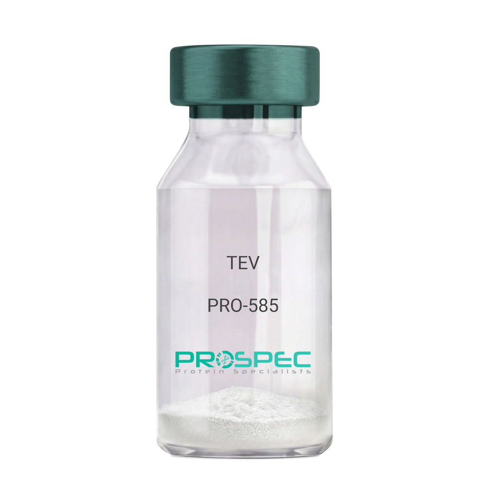
    TEV

    Description:

    Tobacco Etch Virus Protease Recombinant

    rTEV, TEV, P1 protease.

    Product # :

    PRO-585

    Price :

    Quantity :

    Shipping Method :

    Ice Icon

    Shipped with Ice Packs

    Add To Cart

    More Info

    • description
    • source
    • formulation
    • purity
    • More Info
    • Citations

    Description

    Recombinant TEV Protease (rTEV) is a site-specific protease purified from E. coli. The protease can be used for the removal of affinity tags from fusion proteins. The seven-amino-acid recognition site for rTEV is Glu-Asn-Leu-Tyr-Phe-Gln-Gly with cleavage occurring between Gln and Gly. The optimal temperature for cleavage is 30°C; however, the enzyme can be used at temperatures as low as 4°C. The rTEV contains His tag.The rTEV is purified by proprietary chromatographic techniques.

    Source

    Escherichia Coli.

    Formulation

    The rTEV contains 25mM Tris, Ph 8.0, 75mM NaCl, 5mM EDTA, 10mM GSH, 50% Glycerol.

    Purity

    Greater than 90.0% as determined by analysis by SDS-PAGE.

    Citations

    More Info

    • Introduction

      TEV protease is the common name for the 27 kDa catalytic domain of the Nuclear Inclusion a (NIa) protein encoded by the tobacco etch virus (TEV). Because its sequence specificity is far more stringent than that of factor Xa, thrombin, or enterokinase, TEV protease is a very useful reagent for cleaving fusion proteins. TEV protease recognizes a linear epitope of the general form E-Xaa-Xaa-Y -Xaa-Q-(G/S), with cleavage occurring between Q and G or Q and S. The most commonly used sequence is ENLYFQG.

    • Synonyms

      rTEV, TEV, P1 protease.

    • Physical Appearance

      Sterile liquid formulation.

    • Stability

      rTEV although stable at 4°C for 1 week, should be stored below -18°C. Please prevent freeze thaw cycles.

    • Cleavage Conditions

      A number of variables can be changed to optimize the cleavage of any specific protein. The amount of rTEV, the temperature of the incubation, and the time needed for cleavage may be examined. If the protein of interest is heat-labile, then 4°C incubations are recommended. Reactions at 4°C will require longer incubation times and/or more rTEV.

    • Unit Definition

      One unit is defined as the amount of enzyme needed to cleave 3 ug of fusion protein in 1 hour to 85 % completion at 30°C in a buffer containing 50 mM Tris-HCl, pH 8.0, 0.5 mM EDTA, and 1 mM DTT.

    ProSpec's products are furnished for LABORATORY RESEARCH USE ONLY. The product may not be used as drugs, agricultural or pesticidal products, food additives or household chemicals.

    Tev Protease
  • View Data Sheet

    Name :

    HCV Core Genotype-3/10

    Description:

    Hepatitis C Virus Core Genotype-3/10 Recombinant

    Product # :

    HCV-201

    Price :

    Quantity :

    Shipping Method :

    Ice Icon

    Shipped with Ice Packs

    Add To Cart

    More Info

    • description
    • source
    • formulation
    • purity
    • More Info
    • Citations

    Description

    The E.coli derived recombinant protein contains the HCV core nucleocapsid immunodominant regions, amino acids 2-119.

    Source

    Escherichia Coli.

    Formulation

    50mM Tris-HCl, pH-8, 60mM NaCl, 10mM glutathione, 0.25% sarkosil & 50% glycerol.

    Purity

    HCV Core Genotype-3/10 protein is >95% pure as determined by 10% PAGE (coomassie staining).

    Citations

    More Info

    • Introduction

      HCV is a small 50nm, enveloped, single-stranded, positive sense RNAvirus in the family Flaviviridae.
      HCV has a high rate of replication with approximately one trillion particles produced each day in an infected individual. Due to lack of proofreading by the HCV RNA polymerase, the HCV has an exceptionally high mutation rate, a factor that may help it elude the host's immune response. Hepatitis C virus is classified into six genotypes(1-6) with several subtypes within each genotype. The preponderance and distribution of HCV genotypes varies globally. Genotype is clinically important in determining potential response to interferon-based therapy and the required duration of such therapy. Genotypes 1 and 4 are less responsive to interferon-based treatment than are the other genotypes (2, 3, 5 and 6).

    • Stability

      HCV Core Genotype-3/10 although stable at 4°C for 1 week, should be stored below -18°C. Please prevent freeze thaw cycles.

    • Applications

      HCV Core Genotype-3/10 antigen is suitable for ELISA and Western blots, excellent antigen for detection of HCV with minimal specificity problems.

    • Specificity

      Immunoreactive with sera of HCV-infected individuals.

    • Purification Method

      HCV Core Genotype-3/10 protein was purified by proprietary chromatographic technique.

    ProSpec's products are furnished for LABORATORY RESEARCH USE ONLY. The product may not be used as drugs, agricultural or pesticidal products, food additives or household chemicals.

    Hcv Core Genotype 3 10
  • View Data Sheet

    Name :

    HCV NS3 Genotype-6a

    Description:

    Hepatitis C Virus NS3 Genotype-6a, (1192-1459 a.a.) Recombinant

    Product # :

    HCV-211

    Price :

    Quantity :

    Shipping Method :

    Ice Icon

    Shipped with Ice Packs

    Add To Cart

    More Info

    • description
    • formulation
    • purity
    • More Info
    • Citations

    Description

    The E.coli derived recombinant protein fused to a His tag contains the HCV NS3 immunodominant regions, amino acids 1192-1459.

    Formulation

    1.5M urea, 25mM Tris-HCl pH-8, 0.2% Triton-X & 50% Glycerol.

    Purity

    HCV NS3 Genotype-6a protein is >95% pure as determined by 10% PAGE (coomassie staining).

    Citations

    More Info

    • Introduction

      HCV is a small 50nm, enveloped, single-stranded, positive sense RNAvirus in the family Flaviviridae.
      HCV has a high rate of replication with approximately one trillion particles produced each day in an infected individual. Due to lack of proofreading by the HCV RNA polymerase, the HCV has an exceptionally high mutation rate, a factor that may help it elude the host's immune response. Hepatitis C virus is classified into six genotypes(1-6) with several subtypes within each genotype. The preponderance and distribution of HCV genotypes varies globally. Genotype is clinically important in determining potential response to interferon-based therapy and the required duration of such therapy. Genotypes 1 and 4 are less responsive to interferon-based treatment than are the other genotypes (2, 3, 5 and 6).

    • Stability

      HCV NS3 Genotype-6a although stable at 4°C for 1 week, should be stored below -18°C. Please prevent freeze thaw cycles.

    • Applications

      HCV NS3 Genotype-6a antigen is suitable for ELISA and Western blots, excellent antigen for detection of HCV with minimal specificity problems.

    • Specificity

      Immunoreactive with sera of HCV-infected individuals.

    • Purification Method

      HCV NS3 Genotype-6a protein was purified by proprietary chromatographic technique.

    ProSpec's products are furnished for LABORATORY RESEARCH USE ONLY. The product may not be used as drugs, agricultural or pesticidal products, food additives or household chemicals.

    Hcv Ns3 Genotype 6A
  • View Data Sheet

    Name :

    T.pallidum p15 (Partial)

    Description:

    Treponema pallidum p15 (Partial) Recombinant

    Product # :

    TRP-275

    Price :

    Quantity :

    Shipping Method :

    Ice Icon

    Shipped with Ice Packs

    Add To Cart

    More Info

    • description
    • source
    • formulation
    • purity
    • More Info
    • Citations

    Description

    The E.Coli derived recombinant protein is fused at N-terminus with GST tag and contains the Trp. Pallidum p15 immunodominant regions.

    Source

    Escherichia Coli.

    Formulation

    70mM Tris-HCl pH-8, 84mM NaCl, 14mM Glutathione, 30% Glycerol & 0.2% Sarcosil.

    Purity

    Treponema Pallidum protein is >95% pure as determined by 10% PAGE (coomassie staining).

    Citations

    More Info

    • Introduction

      Treponema pallidum is a gram-negative spirochaete bacterium and is considered to be metabolically crippled. There are at least four known subspecies: T. pallidum pallidum, T. pallidum pertenue, T. pallidum carateum and T. pallidum endemicum. The helical structure of T. pallidum pallidum allows it to move in a corkscrew motion through viscous mediums such as mucus. Treponema pallidum sub sp. pallidum has one of the smallest bacterial genomes at 1.14 million base pairs (Mb) and has limited metabolic capabilities, reflecting its adaptation through genome reduction to the rich environment of mammalian tissue.

    • Stability

      Treponema Pallidum protein although stable at 4°C for 1 week, should be stored below -18°C. Please prevent freeze thaw cycles.

    • Applications

      Treponema Pallidum protein is suitable for ELISA and Western blots, excellent antigen for detection of Trp. Pallidum with minimal specificity problems.

    • Specificity

      Immunoreactive with sera of Trp. Pallidum infected individuals.

    • Purification Method

      Treponema Pallidum protein was purified by proprietary chromatographic technique.

    ProSpec's products are furnished for LABORATORY RESEARCH USE ONLY. The product may not be used as drugs, agricultural or pesticidal products, food additives or household chemicals.

    Tpallidum P15 Partial
  • View Data Sheet

    Name :

    FeLV

    Description:

    Feline Leukemia Virus p27 Recombinant

    Product # :

    FLV-001

    Price :

    Quantity :

    Shipping Method :

    Ice Icon

    Shipped with Ice Packs

    Add To Cart

    More Info

    • description
    • source
    • formulation
    • purity
    • More Info
    • Citations

    Description

    The E.Coli truncated derived recombinant protein contains the p27 immunodominant regions, containing 248 amino acids (271-519) and having a Mw of 31kda as visualized by SDS-PAGE. Recombinant Feline Leukemia Virus p27 is fused to a His Tag and purified by conventional chromatography techniques.

    Source

    Escherichia Coli.

    Formulation

    1x PBS, 25MmK2CO3 and 10% glycerol.

    Purity

    Protein is >90% pure as determined by 10% PAGE (coomassie staining).

    Citations

    More Info

    • Introduction

      FeLV is an RNA virus, a member of the Retroviridae family and Oncovirinae subfamily. FeLV retrovirus infects cats and is transferred from infected cats by saliva or nasal secretions. FeLV causes immunosuppression in domestic cats and larger wild cat populations such as cheetahs and lions and is the cause for a form of cancer of the blood cells called lymphocytes (a leukemia) which can be lethal.

    • Physical Appearance

      Sterile Filtered clear solution.

    • Stability

      FeLV p27 although stable at 4°C for 1 week, should be stored below -18°C. Please prevent freeze thaw cycles.

    • Applications

      Immunoassay.

    • Purification Method

      Purified by proprietary chromatographic technique.

    ProSpec's products are furnished for LABORATORY RESEARCH USE ONLY. The product may not be used as drugs, agricultural or pesticidal products, food additives or household chemicals.

    Felv
  • View Data Sheet

    Name :

    M.Pneumoniae P30

    Description:

    Mycoplasma Pneumoniae P30 Recombinant

    Product # :

    PRO-2738

    Price :

    Quantity :

    Shipping Method :

    Ice Icon

    Shipped with Ice Packs

    Add To Cart

    More Info

    • description
    • source
    • formulation
    • purity
    • More Info
    • Citations

    Description

    M.Pneumoniae P30 Recombinant produced in E.Coli is a non-glycosylated polypeptide chain having a molecular mass of 18-19kDa.M.Pneumoniae P30 is fused to a 6 amino acid His-tag at C-terminus & purified by proprietary chromatographic techniques.

    Source

    Escherichia Coli.

    Formulation

    MP-P30c solution contains 25mM K2CO3, 0.025% NaN3 and PBS.

    Purity

    Protein is >95% pure as determined by 10% PAGE (coomassie staining).

    Citations

    More Info

    • Introduction

      Mycoplasma pneumonia is part of the atypical pneumonia subtype which is caused by the bacteria M. pneumoniae. Mycoplasma pneumonia affects individuals younger than 40. It makes up 15 - 50% of all pneumonia cases in adults and especially in school-aged children. People at great risk for mycoplasma pneumonia comprise of those living or working in busy areas such as schools and homeless shelters, although many people who contract mycoplasma pneumonia have no identifiable risk factor. P1, P30, and P116 of mycoplasma pneumonia membrane proteins have been recognized as adhesive factors, P1 is considered as a main adhesion protein of the organism colonization.

    • Physical Appearance

      Sterile Filtered clear solution.

    • Stability

      The Recombinant MP-P30c protein although stable at 4°C for 1 week, should be stored below -18°C. For long term storage it is recommended to add a carrier protein (0.1% HSA or BSA). Please prevent freeze-thaw cycles.

    • Applications

      Immunoassay.

    ProSpec's products are furnished for LABORATORY RESEARCH USE ONLY. The product may not be used as drugs, agricultural or pesticidal products, food additives or household chemicals.

    Pneumoniae P30
  • View Data Sheet

    Name :

    DDAVP

    Description:

    Desmopressin

    Product # :

    HOR-270

    Price :

    Quantity :

    Shipping Method :

    Room Temp Icon

    Shipped at Room temp

    Add To Cart

    More Info

    • description
    • formulation
    • purity
    • More Info
    • Citations

    Description

    Desmopressin also called ADH (Anti-Diuretic Hormone) has a molecular formula of C46H64N14O12S2 , Mpr-Tyr-Phe-Gln-Asn-Cys-Pro-D-Arg-Gly-NH2 having a Mw of 1069.23 Dalton.

    Formulation

    The Desmopressin peptide was lyophilized with no additives.

    Purity

    Greater than 98.0% as determined by:
    (a) Analysis by RP-HPLC.
    (b) Analysis by SDS-PAGE.

    Citations

    More Info

    • Introduction

      Desmopressin is the first vasopressin analog with a very high and very specific antidiuretic effect, has been widely used for different therapeutic purposes and is believed to be partly responsible for the formation of memories learning and memory processes. Desmopressin increases urine concentration and decreases urine production. Desmopressin is used to prevent and control excessive thirst, urination, and dehydration.

    • Physical Appearance

      Sterile Filtered White lyophilized (freeze-dried) powder.

    • Stability

      Lyophilized Desmopressin although stable at room temperature for 3 weeks, should be stored desiccated below -18°C. Upon reconstitution DDAVP should be stored at 4°C between 2-7 days and for future use below -18°C. For long term storage it is recommended to add a carrier protein (0.1% HSA or BSA).Please prevent freeze-thaw cycles.

    • Solubility

      It is recommended to reconstitute the lyophilized DDAVP in sterile 18MΩ-cm H2O not less than 100 µg/ml, which can then be further diluted to other aqueous solutions.

    ProSpec's products are furnished for LABORATORY RESEARCH USE ONLY. The product may not be used as drugs, agricultural or pesticidal products, food additives or household chemicals.

    Desmopressin
  • View Data Sheet

    Name :

    EPHA3 Mouse

    Description:

    EPH Receptor A3 Mouse Recombinant

    Cek4, EK4, End3, ETK1, Hek, Hek4, Mek4, Tyro4, ephrin type-A receptor 3, ephrin type-A receptor 3 isoform1, receptor protein-tyrosine kinase, EPH-like kinase 4, tyrosine-protein kinase TYRO4, tyrosine-protein kinase receptor ETK1, Eph receptor A3, AW492086.

    Product # :

    PRO-2817

    Price :

    Quantity :

    Shipping Method :

    Ice Icon

    Shipped with Ice Packs

    Add To Cart

    More Info

    • description
    • source
    • formulation
    • purity
    • biological activity
    • More Info
    • Citations

    Description

    EPHA3 Mouse Recombinant produced in HEK293 cells is a single, polypeptide chain containing 527 amino acids (21-541 a.a.) and having a molecular mass of 59.5kDa. EPHA3 is expressed with a 6 amino acid His tag at C-Terminus and purified by proprietary chromatographic techniques.

    Source

    HEK293 Cells.

    Formulation

    EPHA3 protein solution (1mg/ml) containing Phosphate Buffered Saline (pH 7.4) and 10% glycerol.

    Purity

    Greater than 95% as determined by SDS-PAGE.

    Biological Activity

    Measured by its binding ability in a functional ELISA with cat#pro-2816.

    Citations

    More Info

    • Synonyms

      Cek4, EK4, End3, ETK1, Hek, Hek4, Mek4, Tyro4, ephrin type-A receptor 3, ephrin type-A receptor 3 isoform1, receptor protein-tyrosine kinase, EPH-like kinase 4, tyrosine-protein kinase TYRO4, tyrosine-protein kinase receptor ETK1, Eph receptor A3, AW492086.

    • Physical Appearance

      Sterile Filtered colorless solution.

    • Stability

      Store at 4°C if entire vial will be used within 2-4 weeks. Store, frozen at -20°C for longer periods of time. For long term storage it is recommended to add a carrier protein (0.1% HSA or BSA). Avoid multiple freeze-thaw cycles.

    • Amino Acid Sequence

      ELSPQPSNEV NLLDSKTIQG ELGWISYPSH GWEEISGVDE HYTPIRTYQV CNVMDHSQNN WLRTNWVPRN SAQKIYVELK FTLRDCNSIP LVLGTCKETF NLYYMESDDD HGVKFREHQF TKIDTIAADE SFTQMDLGDR ILKLNTEIRE VGPVNKKGFY LAFQDVGACV ALVSVRVYFK KCPFTVKNLA MFPDTVPMDS QSLVEVRGSC VNNSKEEDPP RMYCSTEGEW LVPIGKCTCN AGYEERGFIC QACRPGFYKA SDGAAKCAKC PPHSSTQEDG SMNCRCENNY FRAEKDPPSM ACTRPPSAPR NVISNINETS VILDWSWPLD TGGRKDITFN IICKKCGWNV RQCEPCSPNV RFLPRQLGLT NTTVTVTDLL AHTNYTFEID AVNGVSELSS PPRQYAAVSI TTNQAAPSPV MTIKKDRTSR NSISLSWQEP EHPNGIILDY EVKYYEKQEQ ETSYTILRAR GTNVTISSLK PDTTYVFQIR ARTAAGYGTN SRKFEFETSP DSFSISGENS HHHHHHH.

    • Background

      Research on EphA3 in mice has highlighted its critical involvement in the development of the nervous system, cardiovascular system, and skeletal system. EphA3 signaling influences axon pathfinding, synaptic plasticity, and the formation of tissue boundaries, ensuring proper tissue and organ development. Dysregulation of EphA3 has been implicated in various pathologies, including cancer, where it contributes to tumor growth, angiogenesis, and metastasis.

    ProSpec's products are furnished for LABORATORY RESEARCH USE ONLY. The product may not be used as drugs, agricultural or pesticidal products, food additives or household chemicals.

    Epha3 Mouse
  • View Data Sheet

    Name :

    DBI Mouse

    Description:

    Diazepam Binding Inhibitor Mouse Recombinant

    Acyl-CoA-binding protein, ACBP, Diazepam-binding inhibitor, DBI, Endozepine, EP, Dbi.  

    Product # :

    PRO-2527

    Price :

    Quantity :

    Shipping Method :

    Ice Icon

    Shipped with Ice Packs

    Add To Cart

    More Info

    • description
    • source
    • formulation
    • purity
    • More Info
    • Citations

    Description

    DBI Mouse Recombinant produced in E.Coli is a single, non-glycosylated polypeptide chain containing 110 amino acids (1-87 a.a) and having a molecular mass of 12.4kDa. DBI is fused to a 23 amino acid His-tag at N-terminus & purified by proprietary chromatographic techniques.

    Source

    Escherichia Coli.

    Formulation

    DBI protein solution (1mg/ml) contains Phosphate buffer saline (pH7.4).

    Purity

    Greater than 95.0% as determined by SDS-PAGE.

    Citations

    More Info

    • Introduction

      DBI, also known as Acyl-CoA-binding protein isoform 2, is a diazepam binding inhibitor, which is regulated by hormones. DBI participates in lipid metabolism and in the displacement of beta-carbolines & benzodiazepines, which modulate signal transduction at type A gamma-aminobutyric acid receptors located in brain synapses. Moreover, during adipocyte differentiation the expression of DBI is significantly induced. DBI preforms as an acyl-CoA pool former and regulates LCFA (long-chain fatty acids) metabolism in peripheral tissues.

    • Synonyms

      Acyl-CoA-binding protein, ACBP, Diazepam-binding inhibitor, DBI, Endozepine, EP, Dbi.

    • Physical Appearance

      Sterile Filtered clear solution.

    • Stability

      Store at 4°C if entire vial will be used within 2-4 weeks. Store, frozen at -20°C for longer periods of time. For long term storage it is recommended to add a carrier protein (0.1% HSA or BSA).Avoid multiple freeze-thaw cycles.

    • Amino Acid Sequence

      MGSSHHHHHH SSGLVPRGSH MGSMSQAEFD KAAEEVKRLK TQPTDEEMLF IYSHFKQATV GDVNTDRPGL LDLKGKAKWD SWNKLKGTSK ESAMKTYVEK VDELKKKYGI

    ProSpec's products are furnished for LABORATORY RESEARCH USE ONLY. The product may not be used as drugs, agricultural or pesticidal products, food additives or household chemicals.

    Dbi Mouse
  • 1
  • …
  • 27
  • 28
  • 29
  • 30
  • 31
  • …
  • 42
Your browser does not support the video tag.

Products

  • Cytokines
  • Growth Factors
  • Chemokines
  • CD Antigens
  • Neurotrophins
  • Hormones
  • Enzymes
  • Viral Antigens
  • Recombinant Proteins
  • Natural Proteins
  • Monoclonal Antibodies
  • Polyclonal Antibodies
  • Thermal Packaging

About

  • Company
  • Founder
  • Contribution
  • Contact Us
  • My Account

Ordering

  • Ordering Method
  • Ordering - Credit Card
  • Ordering PO
  • Shipping Fees
  • FAQ's

Legal

  • Terms & Conditions
  • Privacy
  • Site Map
  • info@prospecbio.com
  • Facebook
  • Instagram
  • Linkedin Profile

© 2026 ProSpec-Tany TechnoGene Ltd. All Rights Reserved.

  • Choosing a selection results in a full page refresh.
  • Opens in a new window.