Search results
1000 results found for “cebp”
Name
Description
Product #
Price
Quantity
Shipping Method
- View Data Sheet
Name :
CST6 HumanDescription:
Cystatin E/M Human Recombinant
Cystatin E/M, cystatin 6, Cystatin M, Cystatin-E, Cysteine proteinase inhibitor.
Product # :
PRO-892Price :
Quantity :
Shipping Method :
Shipped with Ice Packs
More Info
- description
- source
- formulation
- purity
- More Info
Description
CST6 Human Recombinant produced in E.coli is a single, non-glycosylated polypeptide chain containing 142 amino acids (29-149) and having a molecular mass of 15.9 kDa.The CST6 is fused to a 21 amino acid His-Tag at N-terminus and purified by proprietary chromatographic techniques.
Source
E.coli.
Formulation
The CST6 solution (0.5mg/ml) contains 20mM Tris-HCl buffer (pH 8.0), 1mM DTT, 0.1M NaCl and 10% glycerol.
Purity
Greater than 95% as determined by SDS-PAGE.
More Info
-
Introduction
CST6 belongs to the type 2 cystatin family. Unlike other family members that act as active cysteine protease inhibitors, CST6 restrain the inhibition of cathepsin B but is not active against cathepsin C. CST6 are secretable proteins that influence osteogenesis and bone resorption, regulation of the hepatocyte growth factor receptors, and the response to systemic inflammation.
-
Synonyms
Cystatin E/M, cystatin 6, Cystatin M, Cystatin-E, Cysteine proteinase inhibitor.
-
Physical Appearance
Sterile Filtered colorless solution.
-
Stability
Store at 4°C if entire vial will be used within 2-4 weeks. Store, frozen at -20°C for longer periods of time. For long term storage it is recommended to add a carrier protein (0.1% HSA or BSA).Avoid multiple freeze-thaw cycles.
-
Amino Acid Sequence
MGSSHHHHHH SSGLVPRGSH MRPQERMVGE LRDLSPDDPQ VQKAAQAAVA SYNMGSNSIY YFRDTHIIKA QSQLVAGIKY FLTMEMGSTD CRKTRVTGDH VDLTTCPLAA GAQQEKLRCD FEVLVVPWQN SSQLLKHNCV QM.
ProSpec's products are furnished for LABORATORY RESEARCH USE ONLY. The product may not be used as drugs, agricultural or pesticidal products, food additives or household chemicals.
- View Data Sheet
Name :
SNRPB HumanDescription:
Small Nuclear Ribonucleoprotein Polypeptides B & B1 Human Recombinant
Small nuclear ribonucleoprotein-associated proteins B and B', snRNP-B, Sm protein B/B', Sm-B/B', SmB/B', SNRPB, COD, SNRPB1, SmB/SmB'.
Product # :
PRO-1511Price :
Quantity :
Shipping Method :
Shipped with Ice Packs
More Info
- description
- source
- formulation
- purity
- More Info
Description
SNRPB Human Recombinant produced in SF9 is a glycosylated, polypeptide chain having a calculated molecular mass of 25.4 kDa which migrates at 30kDa on SDS-PAGE. SNRPB is expressed with a -6x His tag and purified by proprietary chromatographic techniques.
Source
Sf9 insect cells.
Formulation
SNRPB is supplied in 20mM HEPES buffer pH-7.5, 0.01mM EDTA and 0.02% SDS.
Purity
Greater than 95% as determined by SDS-PAGE.
More Info
-
Introduction
SNRPB is one of several nuclear proteins which are found in common among U1, U2, U4/U6, and U5 small ribonucleoprotein particles (snRNPs). These snRNPs are involved in pre-mRNA splicing, and the SNRPB protein may also have a role in pre-mRNA splicing or snRNP structure. SNRPB binds to the downstream cleavage product (DCP) of histone pre-mRNA in a U7 snRNP dependent manner. SNRPB functions in the U7 snRNP complex which is involved in histone 3'-end processing.
-
Synonyms
Small nuclear ribonucleoprotein-associated proteins B and B', snRNP-B, Sm protein B/B', Sm-B/B', SmB/B', SNRPB, COD, SNRPB1, SmB/SmB'.
-
Physical Appearance
Sterile Filtered clear solution.
-
Stability
Store at 4°C if entire vial will be used within 2-4 weeks. Store, frozen at -20°C for longer periods of time. Avoid multiple freeze-thaw cycles.
ProSpec's products are furnished for LABORATORY RESEARCH USE ONLY. The product may not be used as drugs, agricultural or pesticidal products, food additives or household chemicals.
- View Data Sheet
Name :
SNRPG HumanDescription:
Small Nuclear Ribonucleoprotein Polypeptide G Human Recombinant
Small nuclear ribonucleoprotein G, snRNP-G, Sm protein G, Sm-G, SmG, SNRPG, PBSCG, MGC117317.
Product # :
PRO-196Price :
Quantity :
Shipping Method :
Shipped with Ice Packs
More Info
- description
- source
- formulation
- purity
- More Info
Description
SNRPG Human Recombinant fused with a 20 amino acid His tag at N-terminus produced in E.Coli is a single, non-glycosylated, polypeptide chain containing 96 amino acids (1-76 a.a.) and having a molecular mass of 10.6kDa. The SNRPG is purified by proprietary chromatographic techniques.
Source
Escherichia Coli.
Formulation
The SNRPG solution (1 mg/ml) contains 20mM Tris-HCl buffer (pH 8.0), 1mM DTT, 50% glycerol and 0.1M NaCl.
Purity
Greater than 95.0% as determined by SDS-PAGE.
More Info
-
Introduction
Small nuclear ribonucleoprotein polypeptide G (SNRPG) is a member of the snRNP Sm proteins family. There are at least 7 isoforms, B/B, E, F, G, D1, D2, and D3. This class of common proteins has a vital role in the biogenesis of the snRNPs. The human Sm G genes are mapped to chromosomes 2. Furthermore, these proteins represent the major targets for the supposed anti-Sm auto-antibodies which are diagnostic for SLE (systemic lupus erythematosus). One class of these autoantibodies reacts specifically with native Sm E-F-G complexes.
-
Synonyms
Small nuclear ribonucleoprotein G, snRNP-G, Sm protein G, Sm-G, SmG, SNRPG, PBSCG, MGC117317.
-
Physical Appearance
Sterile Filtered colorless solution.
-
Stability
Store at 4°C if entire vial will be used within 2-4 weeks. Store, frozen at -20°C for longer periods of time. For long term storage it is recommended to add a carrier protein (0.1% HSA or BSA).Avoid multiple freeze-thaw cycles.
-
Amino Acid Sequence
MGSSHHHHHH SSGLVPRGSH MSKAHPPELK KFMDKKLSLK LNGGRHVQGI LRGFDPFMNL VIDECVEMAT SGQQNNIGMV VIRGNSIIML EALERV.
ProSpec's products are furnished for LABORATORY RESEARCH USE ONLY. The product may not be used as drugs, agricultural or pesticidal products, food additives or household chemicals.
- View Data Sheet
Name :
ETFB HumanDescription:
Electron-Transfer-Flavoprotein Beta Polypeptide Human Recombinant
Electron-transfer-flavoprotein beta polypeptide, MADD, beta-ETF.
Product # :
PRO-220Price :
Quantity :
Shipping Method :
Shipped with Ice Packs
More Info
- description
- source
- formulation
- purity
- More Info
Description
ETFB Human Recombinant fused with a 20 amino acid His tag at N-terminus produced in E.Coli is a single, non-glycosylated, polypeptide chain containing 275 amino acids (1-255a.a.) and having a molecular mass of 30.0kDa. The ETFB is purified by proprietary chromatographic techniques.
Source
Escherichia Coli.
Formulation
The ETFB solution (0.25mg/ml) contains 20mM Tris-HCl buffer (pH 8.0), 40% glycerol and 0.1M NaCl.
Purity
Greater than 90.0% as determined by SDS-PAGE.
More Info
-
Introduction
ETF is a heterodimer composed of alpha and beta subunit. ETFB protein is electron-transfer-flavoprotein, beta polypeptide that transports electrons between primary flavoprotein dehydrogenases involved in mitochondrial fatty acid and amino acid catabolism and the membrane-bound electron transfer flavoprotein ubiquinone oxidoreductase.
-
Synonyms
Electron-transfer-flavoprotein beta polypeptide, MADD, beta-ETF.
-
Physical Appearance
Sterile Filtered colorless solution.
-
Stability
Store at 4°C if entire vial will be used within 2-4 weeks. Store, frozen at -20°C for longer periods of time. For long term storage it is recommended to add a carrier protein (0.1% HSA or BSA).Avoid multiple freeze-thaw cycles.
-
Amino Acid Sequence
MGSSHHHHHH SSGLVPRGSH MAELRVLVAV KRVIDYAVKI RVKPDRTGVV TDGVKHSMNP FCEIAVEEAV RLKEKKLVKE VIAVSCGPAQ CQETIRTALA MGADRGIHVE VPPAEAERLG PLQVARVLAK LAEKEKVDLV LLGKQAIDDD CNQTGQMTAG FLDWPQGTFA SQVTLEGDKL
KVEREIDGGL ETLRLKLPAV VTADLRLNEP RYATLPNIMK AKKKKIEVIK PGDLGVDLTS KLSVISVEDP PQRTAGVKVE TTEDLVAKLK EIGRI.
ProSpec's products are furnished for LABORATORY RESEARCH USE ONLY. The product may not be used as drugs, agricultural or pesticidal products, food additives or household chemicals.
- View Data Sheet
Name :
FAM19A5 HumanDescription:
Family With Sequence Similarity 19 Member-A5 Human Recombinant
Family With Sequence Similarity 19 (Chemokine (C-C Motif)-Like), Member A5, Chemokine-Like Protein TAFA-5, TAFA5, TAFA Protein 5, QLLK5208, UNQ5208, TAFA-5, FAM19A5.
Product # :
CHM-284Price :
Quantity :
Shipping Method :
Shipped with Ice Packs
More Info
- description
- source
- formulation
- purity
- More Info
Description
FAM19A5 Human Recombinant (isoform 2) produced in E.Coli is a single, non-glycosylated, polypeptide chain (Gln26-Ser125) containing 110 amino acids including a 10 aa His tag at N-terminus. The total calculated molecular mass is 12.2kDa.
Source
Escherichia Coli.
Formulation
FAM19A5 filtered (0.4µm) solution in 20mM Tris buffer, 50mM NaCl, pH 7.5 and 10% (w/v) glycerol.
Purity
Greater than 95.0% as determined by SDS-PAGE.
More Info
-
Introduction
Family With Sequence Similarity 19 Member-A5 (FAM19A5) belongs to the TAFA family which is comprised of 5 highly homologous genes encoding small secreted proteins. These proteins have conserved cysteine residues at fixed positions, and are remotely related to MIP-1alpha, which is a member of the CC-chemokine family. The TAFA proteins are primarily expressed in specific regions of the brain, and are assumed to serve as brain-specific chemokines or neurokines which act as regulators of immune and nervous cells.
-
Synonyms
Family With Sequence Similarity 19 (Chemokine (C-C Motif)-Like), Member A5, Chemokine-Like Protein TAFA-5, TAFA5, TAFA Protein 5, QLLK5208, UNQ5208, TAFA-5, FAM19A5.
-
Physical Appearance
Filtered colorless solution.
-
Stability
Store at 4°C if entire vial will be used within 2-4 weeks. Store, frozen at -20°C for longer periods of time. For long term storage it is recommended to add a carrier protein (0.1% HSA or BSA).Avoid multiple freeze-thaw cycles.
-
Amino Acid Sequence
MKHHHHHHASQFLKEGQLAA GTCEIVTLDR DSSQPRRTIA RQTARCACRK GQIAGTTRAR PACVDARIIK TKQWCDMLPC LEGEGCDLLI NRSGWTCTQP GGRIKTTTVS.
-
Background
What is the molecular weight/Mw of FAM19A5 HUMAN Protein?
FAM19A5 HUMAN Protein has a total Mw of 12.2kDa.
What is the source or expression system of FAM19A5 HUMAN Protein?
Escherichia Coli.
What is the Purity of FAM19A5 HUMAN Protein?
FAM19A5 HUMAN Protein is >95% pure as determined by SDS-PAGE.
What is the Biological Activity of FAM19A5 HUMAN Protein?
The biological functionality of FAM19A5 HUMAN Protein will be determined in the future.
What is the amino acid sequence of FAM19A5 HUMAN Protein?
MKHHHHHHASQFLKEGQLAA GTCEIVTLDR DSSQPRRTIA RQTARCACRK GQIAGTTRAR PACVDARIIK TKQWCDMLPC LEGEGCDLLI NRSGWTCTQP GGRIKTTTVS.
What applications can FAM19A5 HUMAN Protein be used in?
FAM19A5 HUMAN Protein can probably be used in western blot, ELISA and Lateral Flow.
What is the endotoxin level for FAM19A5 HUMAN Protein?
The endotoxin level is minimal, FAM19A5 HUMAN Protein was purified using conventional chromatography techniques.
ProSpec's products are furnished for LABORATORY RESEARCH USE ONLY. The product may not be used as drugs, agricultural or pesticidal products, food additives or household chemicals.
- View Data Sheet
Name :
CXCL13 MouseDescription:
BCA-1/BLC Mouse Recombinant (CXCL13)
C-X-C motif chemokine 13, B lymphocyte chemoattractant, CXC chemokine BLC, Small-inducible cytokine B13, Cxcl13, Blc, Scyb13.
Product # :
CHM-030Price :
Quantity :
Shipping Method :
Shipped at Room temp
More Info
- description
- source
- formulation
- purity
- biological activity
- More Info
Description
CXCL13 Mouse Recombinant (22-109) produced in E.Coli is a single, non-glycosylated, polypeptide chain containing 88 amino acids and having a molecular mass of 10kDa.The BCA-1 is purified by proprietary chromatographic techniques.
Source
Escherichia Coli.
Formulation
The BCA1 protein was lyophilized from a 0.2µm filtered solution in Acetonitrile and TFA.
Purity
Greater than 97.0% as determined by SDS-PAGE.
Biological Activity
The ED50, as measured by its ability to chemoattract human CXCR5-transfected mouse BaF3 cells, is less than 2µg/ml.More Info
-
Introduction
BCA-1 is a CXC chemokine that is highly expressed in thesecondary lymphoid organs, such as follicles of the spleen, lymph nodes, and Peyer's patches. CXCL13 promotes the migration of B lymphocytes (compared to T cells and macrophages), by stimulating calcium influx into, and chemotaxis of, cells expressing Burkitt's lymphoma receptor 1 (BLR1). BCA1 therefore function in the homing of B lymphocytes to follicles. Human BCA-1 shares a 64% amino acid sequence similarity with the mouse protein and 23 - 34% amino acid sequence identity with other known CXC chemokines. Recombinant or chemically synthesized BCA1 is a potent chemoattractant for B lymphocytes but not T lymphocytes, monocytes or neutrophils. BLR1, a G protein-coupled receptor originally isolated from Burkitt’s lymphoma cells, has now been shown to be the specific receptor for BCA1. Among cells of the hematopoietic lineages, the expression of BLR-1, now designated CXCR-5, is restricted to B lymphocytes and a subpopulation of T helper memory cells.
-
Synonyms
C-X-C motif chemokine 13, B lymphocyte chemoattractant, CXC chemokine BLC, Small-inducible cytokine B13, Cxcl13, Blc, Scyb13.
-
Physical Appearance
Sterile Filtered White lyophilized (freeze-dried) powder.
-
Stability
Lyophilized BCA1 although stable at room temperature for 3 weeks, should be stored desiccated below -18°C. Upon reconstitution BCA1 should be stored at 4°C between 2-7 days and for future use below -18°C.For long term storage it is recommended to add a carrier protein (0.1% HSA or BSA).Please prevent freeze-thaw cycles.
-
Solubility
It is recommended to reconstitute the lyophilized CXCL13 in sterile 18MΩ-cm H2O not less than 100µg/ml, which can then be further diluted to other aqueous solutions.
-
Amino Acid Sequence
ILEAHYTNLK CRCSGVISTV VGLNIIDRIQ VTPPGNGCPK TEVVIWTKMK KVICVNPRAK WLQRLLRHVQ SKSLSSTPQA PVSKRRAA.
-
Background
What is the molecular weight/Mw of CXCL13 MOUSE Protein?
CXCL13 MOUSE Protein has a total Mw of 10kDa.
What is the source or expression system of CXCL13 MOUSE Protein?
Escherichia Coli.
What is the Purity of CXCL13 MOUSE Protein?
CXCL13 MOUSE Protein is >97% pure as determined by SDS-PAGE.
What is the Biological Activity of CXCL13 MOUSE Protein?
The ED50, as measured by its ability to chemoattract human CXCR5-transfected mouse BaF3 cells, is less than 2µg/ml.
What is the amino acid sequence of CXCL13 MOUSE Protein?
ILEAHYTNLK CRCSGVISTV VGLNIIDRIQ VTPPGNGCPK TEVVIWTKMK KVICVNPRAK WLQRLLRHVQ SKSLSSTPQA PVSKRRAA.
What applications can CXCL13 MOUSE Protein be used in?
CXCL13 MOUSE Protein can probably be used in western blot, ELISA and Lateral Flow.
What is the endotoxin level for CXCL13 MOUSE Protein?
The endotoxin level is minimal, CXCL13 MOUSE Protein was purified using conventional chromatography techniques.
ProSpec's products are furnished for LABORATORY RESEARCH USE ONLY. The product may not be used as drugs, agricultural or pesticidal products, food additives or household chemicals.
- View Data Sheet
Name :
IGFBP 1 HumanDescription:
Insulin-Like Growth Factor Binding Protein-1 Human Recombinant
IBP-1, IGF-Binding Protein 1, AFBP, PP12, IGF-BP25, hIGFBP-1, IGFBP-1.
Product # :
CYT-299Price :
Quantity :
Shipping Method :
Shipped at Room temp
More Info
- description
- source
- formulation
- purity
- biological activity
- More Info
Description
IGFBP-1 Human Recombinant (26-259 a.a.) produced in NS0 is a single, glycosylated, polypeptide chain containing 234 amino acids and having a molecular mass of 25kDa. The IGFBP1 is purified by proprietary chromatographic techniques.
Source
Mouse myeloma cell line, NS0.
Formulation
IGFBP-1 protein was lyophilized from a 0.2µm filtered concentrated solution in PBS.
Purity
Greater than 95.0% as determined by SDS-PAGE.
Biological Activity
The ED50, as determined by the inhibition of rHuIGF-I-induced proliferation of human MCF-7 cells, is less than 4µg/ml.More Info
-
Introduction
IGFBP1 is a member of the insulin-like growth factor binding protein (IGFBP) family and encodes a protein with an IGFBP domain and a thyroglobulin type-I domain. The protein binds both insulin-like growth factors (IGFs) I and II and circulates in the plasma. Binding of this protein prolongs the half-life of the IGFs and alters their interaction with cell surface receptors. Alternate transcriptional splice variants, encoding different isoforms, have been characterized.
-
Synonyms
IBP-1, IGF-Binding Protein 1, AFBP, PP12, IGF-BP25, hIGFBP-1, IGFBP-1.
-
Physical Appearance
Sterile Filtered White lyophilized (freeze-dried) powder.
-
Stability
Lyophilized IGFBP1 although stable at room temperature for 3 weeks, should be stored desiccated below -18°C. Upon reconstitution IGF-BP1 should be stored at 4°C between 2-7 days and for future use below -18°C.For long term storage it is recommended to add a carrier protein (0.1% HSA or BSA).Please prevent freeze-thaw cycles.
-
Solubility
It is recommended to reconstitute the lyophilized IBP-1 in sterile 18MΩ-cm H2O not less than 100µg/ml, which can then be further diluted to other aqueous solutions.
-
Amino Acid Sequence
APWQCAPCSA EKLALCPPVS ASCSEVTRSA GCGCCPMCAL PLGAACGVAT ARCARGLSCR ALPGEQQPLH ALTRGQGACV QESDASAPHA AEAGSPESPE STEITEEELL DNFHLMAPSE EDHSILWDAI STYDGSKALH VTNIKKWKEP CRIELYRVVE SLAKAQETSG EEISKFYLPN CNKNGFYHSR QCETSMDGEA GLCWCVYPWN GKRIPGSPEI RGDPNCQIYF NVQN.
-
Background
What is the molecular weight/Mw of IGFBP1 HUMAN Protein?
IGFBP1 HUMAN Protein has a total Mw of 25kDa.
What is the source or expression system of IGFBP1 HUMAN Protein?
Mouse myeloma cell line, NS0.
What is the Purity of IGFBP1 HUMAN Protein?
IGFBP1 HUMAN Protein is >95% pure as determined by SDS-PAGE.
What is the Biological Activity of IGFBP1 HUMAN Protein?
The ED50, as determined by the inhibition of rHuIGF-I-induced proliferation of human MCF-7 cells, is less than 4µg/ml.
What is the amino acid sequence of IGFBP1 HUMAN Protein?
APWQCAPCSA EKLALCPPVS ASCSEVTRSA GCGCCPMCAL PLGAACGVAT ARCARGLSCR ALPGEQQPLH ALTRGQGACV QESDASAPHA AEAGSPESPE STEITEEELL DNFHLMAPSE EDHSILWDAI STYDGSKALH VTNIKKWKEP CRIELYRVVE SLAKAQETSG EEISKFYLPN CNKNGFYHSR QCETSMDGEA GLCWCVYPWN GKRIPGSPEI RGDPNCQIYF NVQN.
What applications can IGFBP1 HUMAN Protein be used in?
IGFBP1 HUMAN Protein can probably be used in western blot, ELISA and Lateral Flow.
What is the endotoxin level for IGFBP1 HUMAN Protein?
The endotoxin level is minimal, IGFBP1 HUMAN Protein was purified using conventional chromatography techniques.
ProSpec's products are furnished for LABORATORY RESEARCH USE ONLY. The product may not be used as drugs, agricultural or pesticidal products, food additives or household chemicals.
- View Data Sheet
Name :
SELE Human, HEKDescription:
E-Selectin Human Recombinant, HEK
E-selectin, Endothelial leukocyte adhesion molecule 1, ELAM-1, Leukocyte-endothelial cell adhesion molecule 2, LECAM2, CD62E antigen, SELE, ELAM1, ELAM, ESEL, CD62E.
Product # :
PRO-1645Price :
Quantity :
Shipping Method :
Shipped at Room temp
More Info
- description
- source
- formulation
- purity
- More Info
Description
SELE Human Recombinant produced by mammalian expression system in human cells is a single polypeptide chain containing 543 amino acids (22-556). SELE is fused to an 8 amino acid His-tag at C-terminus is purified by proprietary chromatographic techniques.
Source
HEK293 cells.
Formulation
SELE was lyophilized from a 0.2 µM filtered solution of PBS and 4% Mannitol, pH 7.5.
Purity
Greater than 95% as determined by SDS-PAGE.
More Info
-
Introduction
E-selectin which is also called Endothelial leukocyte adhesion molecule 1, ELAM1, ELAM belongs to a family of divalent cation-dependent carbohydrate-binding glycoproteins or adhesion molecules. Eselectin is expressed on the surface of endothelial cells and mediates the interaction of leukocytes and platelets with endothelial cells during an inflammatory response. E-selectin is present in single copy in the human genome and contains 14 exons spanning about 13 kb of DNA.
-
Synonyms
E-selectin, Endothelial leukocyte adhesion molecule 1, ELAM-1, Leukocyte-endothelial cell adhesion molecule 2, LECAM2, CD62E antigen, SELE, ELAM1, ELAM, ESEL, CD62E.
-
Physical Appearance
Sterile Filtered White lyophilized (freeze-dried) powder.
-
Stability
Lyophilized SELE although stable at room temperature for 3 weeks, should be stored desiccated below -18°C. Upon reconstitution SELE should be stored at 4°C between 2-7 days and for future use below -18°C. Please prevent freeze-thaw cycles.
-
Solubility
It is recommended to reconstitute the lyophilized SELE in 1xPBS to a concentration no less than 100 µg/ml, which can then be further diluted to other aqueous solutions.
-
Amino Acid Sequence
WSYNTSTEAMTYDEASAYCQQRYTHLVAIQNKEEIEYLNSILSYSPSYYWIGIRKVNNVW
VWVGTQKPLTEEAKNWAPGEPNNRQKDEDCVEIYIKREKDVGMWNDERCSKKKLALCYTA
ACTNTSCSGHGECVETINNYTCKCDPGFSGLKCEQIVNCTALESPEHGSLVCSHPLGNFSY
NSSCSISCDRGYLPSSMETMQCMSSGEWSAPIPACNVVECDAVTNPANGFVECFQNPGSFPW
NTTCTFDCEEGFELMGAQSLQCTSSGNWDNEKPTCKAVTCRAVRQPQNGSVRCSHSPAGEFT
FKSSCNFTCEEGFMLQGPAQVECTTQGQWTQQIPVCEAFQCTALSNPERGYMNCLPSASGSFR
YGSSCEFSCEQGFVLKGSKRLQCGPTGEWDNEKPTCEAVRCDAVHQPPKGLVRCAHSPIGEFTY
KSSCAFSCEEGFELHGSTQLECTSQGQWTEEVPSCQVVKCSSLAVPGKINMSCSGEPVFGTVCKF
ACPEGWTLNGSAARTCGATGHWSGLLPTCEAPTESNIPVDHHHHHH.
ProSpec's products are furnished for LABORATORY RESEARCH USE ONLY. The product may not be used as drugs, agricultural or pesticidal products, food additives or household chemicals.
- View Data Sheet
Name :
SEP15 HumanDescription:
15 KDa Selenoprotein Human Recombinant
15 KDa Selenoprotein, SEP15.
Product # :
PRO-1468Price :
Quantity :
Shipping Method :
Shipped with Ice Packs
More Info
- description
- source
- formulation
- purity
- More Info
Description
SEP15 Human Recombinant produced in E.Coli is a single, non-glycosylated polypeptide chain containing 160 amino acids (29-165 a.a) and having a molecular mass of 17.7kDa.SEP15 is fused to a 23 amino acid His-tag at N-terminus & purified by proprietary chromatographic techniques.
Source
Escherichia Coli.
Formulation
SEP15 protein solution (0.5mg/ml) containing 20mM Tris-HCl buffer (pH 8.0), 0.15M NaCl, 10% glycerol and 1mM DTT.
Purity
Greater than 95.0% as determined by SDS-PAGE.
More Info
-
Introduction
15 KDa Selenoprotein (SEP15) is a protein-coding gene which contains a selenocysteine (Sec) residue at its active site. Theselenocysteine is encoded by the UGA codon that usually signals translation termination. The 3' UTR ofselenoprotein genes have a conventional stem-loop structure, the sec insertion sequence (SECIS), which isnecessary for the recognition of UGA as a Sec codon rather than as a stop signal. Studies in mouse propose thatthis selenoprotein may have redox function and may be implicated in the quality control of protein folding. This geneis localized on chromosome 1p31, a genetic locus usually mutated or deleted in human cancers.Diseases associated with SEP15 include lung cancer susceptibility, and chronic lymphocytic leukemia.
-
Synonyms
15 KDa Selenoprotein, SEP15.
-
Physical Appearance
Sterile Filtered clear solution.
-
Stability
Store at 4°C if entire vial will be used within 2-4 weeks. Store, frozen at -20°C for longer periods of time. For long term storage it is recommended to add a carrier protein (0.1% HSA or BSA).Avoid multiple freeze-thaw cycles.
-
Amino Acid Sequence
MGSSHHHHHH SSGLVPRGSH MGSVSAFGAE FSSEACRELG FSSNLLCSSC DLLGQFNLLQ LDPDCRGCCQ EEAQFETKKL YAGAILEVCG CKLGRFPQVQ AFVRSDKPKL FRGLQIKYVR GSDPVLKLLD DNGNIAEELS ILKWNTDSVE EFLSEKLERI
ProSpec's products are furnished for LABORATORY RESEARCH USE ONLY. The product may not be used as drugs, agricultural or pesticidal products, food additives or household chemicals.
- View Data Sheet
Name :
ANGPT1 HumanDescription:
Angiopoietin-1 Human Recombinant
Angiopoietin 1, KIAA0003, ANG-1, AGP1, AGPT.
Product # :
CYT-074Price :
Quantity :
Shipping Method :
Shipped with Ice Packs
More Info
- description
- source
- formulation
- purity
- More Info
- sds-page
Description
ANGPT1 Human Recombinant produced in E.coli is a single, non-glycosylated polypeptide chain containing 480 amino acids (20-498) and having a molecular mass of 55.6 kDa.The ANGPT1 is purified by proprietary chromatographic techniques.
Source
E.coli.
Formulation
The ANGPT1 solution (1mg/ml) contains 20mM Tris-HCl buffer (pH 8.0), 1M Urea and 5% glycerol.
Purity
Greater than 85% as determined by SDS-PAGE.
sds-page
More Info
-
Introduction
ANGPT1 is an angiogenic factor which intervenes in blood vessel maturation and takes part in endothelial development. ANGPT1 is a secreted ligand for Tie-2, a cell surface receptor tyrosine kinase expressed in endothelial and hemopoietic cells. The glycosylated ANGPT1 protein has a coiled-coil region in the amino terminus and a fibrinogen-like domain at the carboxy terminus.
-
Synonyms
Angiopoietin 1, KIAA0003, ANG-1, AGP1, AGPT.
-
Physical Appearance
Sterile Filtered colorless solution.
-
Stability
Store at 4°C if entire vial will be used within 2-4 weeks. Store, frozen at -20°C for longer periods of time. For long term storage it is recommended to add a carrier protein (0.1% HSA or BSA).Avoid multiple freeze-thaw cycles.
-
Amino Acid Sequence
MSNQRRSPEN SGRRYNRIQH GQCAYTFILP EHDGNCREST TDQYNTNALQ RDAPHVEPDF SSQKLQHLEH VMENYTQWLQ KLENYIVENM KSEMAQIQQN AVQNHTATML EIGTSLLSQT AEQTRKLTDV ETQVLNQTSR LEIQLLENSL STYKLEKQLL QQTNEILKIH EKNSLLEHKI LEMEGKHKEE LDTLKEEKEN LQGLVTRQTY IIQELEKQLN RATTNNSVLQ KQQLELMDTV HNLVNLCTKE GVLLKGGKRE EEKPFRDCAD VYQAGFNKSG IYTIYINNMP EPKKVFCNMD VNGGGWTVIQ HREDGSLDFQ RGWKEYKMGF GNPSGEYWLG NEFIFAITSQ RQYMLRIELM DWEGNRAYSQ YDRFHIGNEK QNYRLYLKGH TGTAGKQSSL ILHGADFSTK DADNDNCMCK CALMLTGGWW FDACGPSNLN GMFYTAGQNH GKLNGIKWHY FKGPSYSLRS TTMMIRPLDF
-
Background
This factor was found in the conditioned medium of the human neuroepithelioma cell line SHEP1 and the mouse myoblast cell line C2C12ras. The cDNA encoding a protein of 498 amino acids was isolated by using a secretion-trap expression cloning procedure exploiting the presence of a signal sequence present in growth factors that are secreted by producer cells (Davis et al, 1996). Murine and human factors show 97 % identity at the amino acid level. For a related factor see: CDT6. The human gene has been mapped to chromosome 8q22.3-q23 (Cheung et al, 1998).
The expression of angiopoietin-1 mRNA is downregulated by PDGF, EGF, IL1-beta, and TGF-beta (Enholm et al, 1997).
Angiopoietin-1 is a ligand for the receptor-like tyrosine kinase designated TIE-2 (Davis et al, 1996). Binding of Angiopoietin-1 to its receptor induces tyrosine phosphorylation of the cytoplasmic receptor domain. A naturally occuring antagonist of Angiopoietin-1 binding to TIE-2 is Angiopoietin-2. Angiopoietin-1 also binds to the TIE-1 receptor and this interaction appears to be critical for the development of the right-hand side venous system. It is dispensable for the left-hand side venous system, suggesting that right-hand and left-hand side vascular networks are established early before asymmetrical features of the network become morphologically discernible Loughna and Sato, 2001).
Angiopoietin-1 does not directly promote the growth of cultured endothelial cells. Angiopoietin-1 is chemotactic for endothelial cells (Witzenbichler et al, 1998). Excess soluble TIE-2 receptors abolish the chemotactic response of endothelial cells toward angiopoietin-1. Angiopoietin-1 has been shown to counteract cell death by apoptosis in cultured endothelial cells (Holash et al, 1999). Angiopoietin-1 also acts as an apoptosis survival factor for endothelial cells and this effect is augmented by the presence of VEGF (Kwak et al, 1999).
Angiopoietin-2 dose-dependently blocks directed migration toward Angiopoietin-1. Carlson et al (2001) have shown that Ang-1 binds rather selectively to vitronectin and that Ang-1 can directly support adhesion of human umbilical vein endothelial cells and fibroblasts in a process mediated by integrins.
The physiologic roles of Angiopoietin-1 and its receptor are limited to angiogenic processes that occur subsequent to the earlier vasculogenic and angiogenic actions of the VEGF family and their receptors. However, it is unlike most of the known angiogenesis factors such as VEGF and other classical endothelial cell growth factors in that addition of the factor to cultures of endothelial cells does not directly promote cell growth even though the TIE-2 receptor becomes activated. Angiopoietin-1 also appears to be incapable of inducing the formation of tubules by endothelial cells.
Angiopoietins can potentiate the effects of other angiogenic cytokines. An investigation of the impact of angiopoietins on neovascularization in vivo in the cornea micropocket assay of neovascularization demonstrates that neither Angiopoietin-1 nor Angiopoietin-2 alone promote neovascularization. Holash et al (1999) have shown that a subset of tumors initially grows by coopting existing host vessels. Regression of these vessels via a process that involves disruption of interactions between endothelial cells and smooth muscle cells as well as cell death by apoptosis of endothelial cells first causes loss of tumour cells before angiogenesis begins at the tumor margin and the tumor is rescued under the influence of VEGF, Angiopoietin-1, and probably other angiogenic stimuli.
The embryonic expression pattern of Angiopoietin-1 suggests that it plays a particularly important role in the developing heart. Angiopoietin-1 is expressed highly in the myocardial wall surrounding the endocardium expressing TIE-2. Expression of Angiopoietin-1 becomes much more widespread later in development.
ANGPT1 Protein has a total Mw of 55.6kDa.
What is the source or expression system of ANGPT1 Protein?
Escherichia Coli.
What is the Purity of ANGPT1 Protein?
ANGPT1 Protein is >85% pure as determined by SDS-PAGE.
What is the Biological Activity of ANGPT1 Protein?
The biological functionality of ANGPT1 Protein will be determined in the future.
What is the amino acid sequence of ANGPT1 Protein?
MSNQRRSPEN SGRRYNRIQH GQCAYTFILP EHDGNCREST TDQYNTNALQ RDAPHVEPDF SSQKLQHLEH VMENYTQWLQ KLENYIVENM KSEMAQIQQN AVQNHTATML EIGTSLLSQT AEQTRKLTDV ETQVLNQTSR LEIQLLENSL STYKLEKQLL QQTNEILKIH EKNSLLEHKI LEMEGKHKEE LDTLKEEKEN LQGLVTRQTY IIQELEKQLN RATTNNSVLQ KQQLELMDTV HNLVNLCTKE GVLLKGGKRE EEKPFRDCAD VYQAGFNKSG IYTIYINNMP EPKKVFCNMD VNGGGWTVIQ HREDGSLDFQ RGWKEYKMGF GNPSGEYWLG NEFIFAITSQ RQYMLRIELM DWEGNRAYSQ YDRFHIGNEK QNYRLYLKGH TGTAGKQSSL ILHGADFSTK DADNDNCMCK CALMLTGGWW FDACGPSNLN GMFYTAGQNH GKLNGIKWHY FKGPSYSLRS TTMMIRPLDF
What applications can ANGPT1 Protein be used in?
ANGPT1 Protein can probably be used in western blot, ELISA and Lateral Flow.
What is the endotoxin level for ANGPT1 Protein?
The endotoxin level is minimal, ANGPT1 Protein was purified using conventional chromatography techniques.
ProSpec's products are furnished for LABORATORY RESEARCH USE ONLY. The product may not be used as drugs, agricultural or pesticidal products, food additives or household chemicals.
- View Data Sheet
Name :
DsbE E.ColiDescription:
Thiol Disulfide Interchange Protein E.Coli Recombinant DsbE
Thiol:disulfide interchange protein dsbE, Cytochrome c biogenesis protein ccmG, dsbE, ccmG, yejQ, b2195, JW2183.
Product # :
HSP-025Price :
Quantity :
Shipping Method :
Shipped with Ice Packs
More Info
- description
- source
- formulation
- purity
- More Info
Description
Recombinant DsbE produced in E.Coli is a single, non-glycosylated polypeptide chain containing 161 amino acids and having a molecular mass of 18.1 kDa. DsbE is purified by conventional chromatography techniques.
Source
Escherichia Coli.
Formulation
The DsbE protein solution contains 20mM Tris-HCl, pH-7.5, 2mM EDTA and 10% Glycerol.
Purity
Greater than 95.0% as determined by SDS-PAGE.
More Info
-
Introduction
DsbE is a reducing Dsb protein involved in electron transfer for cytochrome c maturation in the periplasm of Escherichia coli. DsbE is one of 12 proteins required for their assembly in the periplasm. DsbE functions is to decrease disulphide bonds formed among correctly paired cysteine residues in the cytochrome c apoproteins prior to haem attachment by CcmF and CcmH.
-
Synonyms
Thiol:disulfide interchange protein dsbE, Cytochrome c biogenesis protein ccmG, dsbE, ccmG, yejQ, b2195, JW2183.
-
Physical Appearance
Sterile filtered colorless solution.
-
Stability
Store at 4°C if entire vial will be used within 2-4 weeks. Store, frozen at -20°C for longer periods of time. For long term storage it is recommended to add a carrier protein (0.1% HSA or BSA).Avoid multiple freeze-thaw cycles.
-
Amino Acid Sequence
MRNAEGDDPT NLESALIGKP VPKFRLESLD NPGQFYQADV LTQGKPVLLN VWATWCPTCR AEHQYLNQLS AQGIRVVGMN YKDDRQKAIS WLKELGNPYA LSLFDGDGML GLDLGVYGAP ETFLIDGNGI IRYRHAGDLN PRVWEEEIKP LWEKYSKEAA Q.
ProSpec's products are furnished for LABORATORY RESEARCH USE ONLY. The product may not be used as drugs, agricultural or pesticidal products, food additives or household chemicals.
- View Data Sheet
Name :
ATP1B1 HumanDescription:
ATPase Transporting Beta 1 Human Recombinant
Sodium/potassium-transporting ATPase subunit beta-1, ATPase, Na+/K+ transporting, beta 1 polypeptide, ATP1B, ATPBS, Sodium/potassium-dependent ATPase subunit beta-1, ATP1B1, ATPaseTransporting Beta 1.
Product # :
PRO-1665Price :
Quantity :
Shipping Method :
Shipped with Ice Packs
More Info
- description
- source
- formulation
- purity
- More Info
Description
ATP1B1 Human Recombinant produced in E.coli is a single, non-glycosylated polypeptide chain containing 264 amino acids (63-303) and having a molecular mass of 30.4 kDa.ATP1B1 is fused to a 23 amino acid His-tag at N-terminus & purified by proprietary chromatographic techniques.
Source
Escherichia Coli.
Formulation
The ATP1B1 solution (1mg/ml) contains 20mM Tris-HCl buffer (pH 8.0), 10% glycerol and 0.4M Urea.
Purity
Greater than 90.0% as determined by SDS-PAGE.
More Info
-
Introduction
ATPaseTransporting Beta 1 (ATP1B1) is a part of the family of Na+/K+ and H+/K+ ATPases beta chain proteins, and the subfamily of Na+/K+ -ATPases. Na+/K+ -ATPase is an essential membrane protein accountable for establishing and maintaining the electrochemical gradients of Na and K ions over the plasma membrane. These gradients are vital for osmoregulation, for sodium-coupled transport of a range of organic and inorganic molecules, and for electrical excitability of muscle and nerve. ATP1B1is combined of 2 subunits, a large catalytic subunit (alpha) and a smaller glycoprotein subunit (beta). The beta subunit regulates the number of sodium pumps transported to the plasma membrane through assembly of alpha/beta heterodimers. ATP1B1 is a beta 1 subunit.
-
Synonyms
Sodium/potassium-transporting ATPase subunit beta-1, ATPase, Na+/K+ transporting, beta 1 polypeptide, ATP1B, ATPBS, Sodium/potassium-dependent ATPase subunit beta-1, ATP1B1, ATPaseTransporting Beta 1.
-
Physical Appearance
Sterile Filtered colorless solution.
-
Stability
Store at 4°C if entire vial will be used within 2-4 weeks. Store, frozen at -20°C for longer periods of time. For long term storage it is recommended to add a carrier protein (0.1% HSA or BSA).Avoid multiple freeze-thaw cycles.
-
Amino Acid Sequence
MGSSHHHHHH SSGLVPRGSH MGSEFKPTYQ DRVAPPGLTQ IPQIQKTEIS FRPNDPKSYE AYVLNIVRFL EKYKDSAQRD DMIFEDCGDV PSEPKERGDF NHERGERKVC RFKLEWLGNC SGLNDETYGY KEGKPCIIIK LNRVLGFKPK PPKNESLETY PVMKYNPNVL PVQCTGKRDE DKDKVGNVEY FGLGNSPGFP LQYYPYYGKL LQPKYLQPLL AVQFTNLTMD TEIRIECKAY GENIGYSEKD RFQGRFDVKI EVKS.
ProSpec's products are furnished for LABORATORY RESEARCH USE ONLY. The product may not be used as drugs, agricultural or pesticidal products, food additives or household chemicals.
- View Data Sheet
Name :
SNUPN HumanDescription:
Snurportin 1 Human Recombinant
KPNBL, RNUT1, Snurportin1, SPN1, RNA U transporter 1.
Product # :
PRO-866Price :
Quantity :
Shipping Method :
Shipped with Ice Packs
More Info
- description
- source
- formulation
- purity
- More Info
Description
SNUPN Recombinant Human produced in E.Coli is a single, non-glycosylated polypeptide chain containing 380 amino acids (1-360 a.a.) and having a molecular mass of 43.3 kDa. The SNUPN is fused to 20 amino acid His-Tag at N-terminus and purified by proprietary chromatographic techniques.
Source
Escherichia Coli.
Formulation
SNUPN Human solution containing 20mM Tris pH-8, 2mM DTT, 0.1M NaCl, & 10% glycerol.
Purity
Greater than 90.0% as determined by SDS-PAGE.
More Info
-
Introduction
SNUPN is a nuclear import adaptor protein which is part of the Snurportin family.
SNUPN is Localized to the cytoplasm and nucleus and contains an N-terminal IBB domain and a trimethylguanosine (m3G)-cap binding domain. SNUPN binds specifically the terminal 2,2,7-m3G-cap at the 5'' end of U snRNPs and is involved in transport of U snRNPs into the nucleus through an association with Importin β. -
Synonyms
KPNBL, RNUT1, Snurportin1, SPN1, RNA U transporter 1.
-
Physical Appearance
Sterile filtered colorless solution.
-
Stability
Store at 4°C if entire vial will be used within 2-4 weeks. Store, frozen at -20°C for longer periods of time. For long term storage it is recommended to add a carrier protein (0.1% HSA or BSA). Avoid multiple freeze-thaw cycles.
-
Amino Acid Sequence
MGSSHHHHHH SSGLVPRGSH MEELSQALAS SFSVSQDLNS TAAPHPRLSQ YKSKYSSLEQ SERRRRLLEL QKSKRLDYVN HARRLAEDDW TGMESEEENK KDDEEMDIDT VKKLPKHYAN QLMLSEWLID VPSDLGQEWI VVVCPVGKRA LIVASRGSTS AYTKSGYCVN RFSSLLPGGN RRNSTAKDYT ILDCIYNEVN QTYYVLDVMC WRGHPFYDCQ TDFRFYWMHS KLPEEEGLGE KTKLNPFKFV GLKNFPCTPE SLCDVLSMDF PFEVDGLLFY HKQTHYSPGS TPLVGWLRPY MVSDVLGVAV PAGPLTTKPD YAGHQLQQIM EHKKSQKEGM KEKLTHKASE NGHYELEHLS
TPKLKGSSHS PDHPGCLMEN.
ProSpec's products are furnished for LABORATORY RESEARCH USE ONLY. The product may not be used as drugs, agricultural or pesticidal products, food additives or household chemicals.
- View Data Sheet
Name :
CCDC23 HumanDescription:
Coiled-Coil Domain Containing 23 Human Recombinant
Coiled-Coil Domain Containing 23, CCDC23.
Product # :
PRO-2132Price :
Quantity :
Shipping Method :
Shipped with Ice Packs
More Info
- description
- source
- formulation
- purity
- More Info
Description
CCDC23 Human Recombinant produced in E.Coli is a single, non-glycosylated polypeptide chain containing 91 amino acids (1-66 a.a) and having a molecular mass of 10.5kDa.CCDC23 is fused to a 25 amino acid His-tag at N-terminus & purified by proprietary chromatographic techniques.
Source
E.coli.
Formulation
CCDC23 protein solution (0.5mg/ml) containing Phosphate buffered saline (pH7.4), 20% glycerol and 1mM DTT.
Purity
Greater than 90% as determined by SDS-PAGE.
More Info
-
Introduction
Coiled-Coil Domain Containing 23, also known as CCDC23 is a 66 amino acid protein which is encoded by a gene mapping tohuman chromosome 1. Chromosome 1 is the biggest human chromosome spanning approximately 260 million base pairsand making up 8% of the human genome. In addition, there are about 3,000 genes on chromosome 1, and taking into account thehuge number of genes there are also a big number of diseases connected with chromosome 1. One of the diseasesthat have been associated with CCDC23 is endotheliitis.
-
Synonyms
Coiled-Coil Domain Containing 23, CCDC23.
-
Physical Appearance
Sterile Filtered colorless solution.
-
Stability
Store at 4°C if entire vial will be used within 2-4 weeks. Store, frozen at -20°C for longer periods of time. For long term storage it is recommended to add a carrier protein (0.1% HSA or BSA).Avoid multiple freeze-thaw cycles.
-
Amino Acid Sequence
MGSSHHHHHH SSGLVPRGSH MGSEFMDPPA RKEKTKVKES VSRVEKAKQK SAQQELKQRQ RAEIYALNRV MTELEQQQFD EFCKQMQPPG E.
ProSpec's products are furnished for LABORATORY RESEARCH USE ONLY. The product may not be used as drugs, agricultural or pesticidal products, food additives or household chemicals.
- View Data Sheet
Name :
PMP2 HumanDescription:
Peripheral Myelin Protein-2 Human Recombinant
P2, MP2, FABP8, M-FABP, Myelin P2 protein, PMP2, Peripheral Myelin Protein-2.
Product # :
PRO-651Price :
Quantity :
Shipping Method :
Shipped with Ice Packs
More Info
- description
- source
- formulation
- purity
- More Info
Description
PMP2 Human Recombinant produced in E.Coli is a single, non-glycosylated polypeptide chain containing 132 amino acids and having a molecular mass of 14.9 kDa.
Source
Escherichia Coli.
Formulation
The PMP2 protein solution contains 20mM Tris pH-7.5, 2mM EDTA, 1mM DTT and 10% glycerol.
Purity
Greater than 95.0% as determined by:
(a) Analysis by RP-HPLC.
(b) Analysis by SDS-PAGE.More Info
-
Introduction
PMP2 is a small protein found in peripheral nerve myelin and spinal cord myelin, belongs to a family of fatty acid binding proteins. PMP2 partly decreases the inhibitory effect of T suppressors in the culture of immune lymph node cells. PMP2 protein is a lipid transport protein in schwann cells.
-
Synonyms
P2, MP2, FABP8, M-FABP, Myelin P2 protein, PMP2, Peripheral Myelin Protein-2.
-
Physical Appearance
Sterile Filtered colorless solution.
-
Stability
Store at 4°C if entire vial will be used within 2-4 weeks.Store, frozen at -20°C for longer periods of time. For long term storage it is recommended to add a carrier protein (0.1% HSA or BSA).Avoid multiple freeze-thaw cycles.
-
Amino Acid Sequence
MSNKFLGTWK LVSSENFDDY MKALGVGLAT RKLGNLAKPT VIISKKGDII TIRTESTFKN EISFKLGQE FEETTADNRK TKSIVTLQRG SLNQVQRWDG KETTIKRKLV NGKMVAECKM KGVVCTRIYE KV.
ProSpec's products are furnished for LABORATORY RESEARCH USE ONLY. The product may not be used as drugs, agricultural or pesticidal products, food additives or household chemicals.
- View Data Sheet
Name :
CNTF RatDescription:
Ciliary Neurotrophic Factor Rat Recombinant
HCNTF, CNTF, Ciliary Neurotrophic Factor.
Product # :
CYT-654Price :
Quantity :
Shipping Method :
Shipped at Room temp
More Info
- description
- source
- formulation
- purity
- biological activity
- More Info
Description
CNTF Recombinant Rat produced in E.Coli is a single, non-glycosylated polypeptide chain containing 200 amino acids and having a molecular mass of 22834 Dalton. The CNTF is purified by proprietary chromatographic techniques.
Source
Escherichia Coli.
Formulation
Lyophilized from a concentrated (1mg/ml) solution in water containing 0.025% NaHCO3.
Purity
Greater than 99.0% as determined by:
(a) Analysis by Gel Filtration.
(b) Analysis by SDS-PAGE.Biological Activity
Fully biologically active by its ability to phosphorylate STAT3 in several cells lines.More Info
-
Introduction
CNTF is a polypeptide hormone whose actions appear to be restricted to the nervous system where it promotes neurotransmitter synthesis and neurite outgrowth in certain neuronal populations. The protein is a potent survival factor for neurons and oligodendrocytes and may be relevant in reducing tissue destruction during inflammatory attacks. A mutation in this gene, which results in aberrant splicing, leads to ciliary neurotrophic factor deficiency, but this phenotype is not causally related to neurologic disease. In addition to the predominant monocistronic transcript originating from this locus, the gene is also co-transcribed with the upstream ZFP91 gene. Co-transcription from the two loci results in a transcript that contains a complete coding region for the zinc finger protein but lacks a complete coding region for ciliary neurotrophic factor.
CNTF is a survival factor for various neuronal cell types. Seems to prevent the degeneration of motor axons after axotomy. -
Synonyms
HCNTF, CNTF, Ciliary Neurotrophic Factor.
-
Physical Appearance
Sterile Filtered White lyophilized (freeze-dried) powder.
-
Stability
Lyophilized CNTF although stable at room temperature for 3 weeks, should be stored desiccated below -18°C. Upon reconstitution CNTF should be stored at 4°C between 2-7 days and for future use below -18°C.Please prevent freeze-thaw cycles.
-
Solubility
It is recommended to reconstitute the lyophilized CNTF in sterile water or 0.4% NaHCO3 adjusted to pH 8-9, not less than 100µg/ml, which can then be further diluted to other aqueous solutions, preferably in presence of carrier protein.
-
Amino Acid Sequence
AFAEQTPLTL HRRDLSSRSIWLARKIRSDLTALMESYVKHQGLNKNI
NLDSVDGVPVASTDRWSEMTEAERLQENLQAYRTFQGMLTKLLEDQRV
HFTPTEGDFHQAIHTLMLQVSAFAYQLEELMVLLEQKIPENEADGMPA
TVGDGGLFEKKLWGLKVLQELSQWTVRSIHDLRVISSHQMGISALESH
YGAKDKQM. -
Background
What is the molecular weight/Mw of CNTF Protein?
CNTF Protein has a total Mw of 22kDa.
What is the source or expression system of CNTF Protein?
Escherichia Coli.
What is the Purity of CNTF Protein?
CNTF Protein is >99% pure as determined by SDS-PAGE.
What is the Biological Activity of CNTF Protein?
Fully biologically active by its ability to phosphorylate STAT3 in several cells lines.
What is the amino acid sequence of CNTF Protein?
AFAEQTPLTL HRRDLSSRSIWLARKIRSDLTALMESYVKHQGLNKNI
NLDSVDGVPVASTDRWSEMTEAERLQENLQAYRTFQGMLTKLLEDQRV
HFTPTEGDFHQAIHTLMLQVSAFAYQLEELMVLLEQKIPENEADGMPA
TVGDGGLFEKKLWGLKVLQELSQWTVRSIHDLRVISSHQMGISALESH YGAKDKQM.
What applications can CNTF Protein be used in?
CNTF Protein can probably be used in western blot, ELISA and Lateral Flow.
What is the endotoxin level for CNTF Protein?
The endotoxin level is minimal, CNTF Protein was purified using conventional chromatography techniques.
ProSpec's products are furnished for LABORATORY RESEARCH USE ONLY. The product may not be used as drugs, agricultural or pesticidal products, food additives or household chemicals.
- View Data Sheet
Name :
Visfatin HumanDescription:
Visfatin Human Recombinant
PBEF, Pre-B cell colony-enhancing factor, Nicotinamide phosphoribosyltransferase NAmPRTase, Nampt, MGC117256, DKFZP666B131, 1110035O14Rik.
Product # :
CYT-318Price :
Quantity :
Shipping Method :
Shipped at Room temp
More Info
- description
- source
- formulation
- purity
- biological activity
- More Info
Description
Visfatin Human Recombinant produced in E.Coli is a single, non-glycosylated, polypeptide chain containing 466 amino acids. The total molecular mass is 52.6kDa (calculated). The Visfatin is purified by Flag-affinity chromatography.
Source
Escherichia Coli.
Formulation
Visfatin was lyophilized with no additives.
Purity
Greater than 90.0% as determined by SDS-PAGE.
Biological Activity
The activity is determined by its ability to induce IL-6, IL-1 beta and TNF alpha production from human PBMCs at 100ng/ml.More Info
-
Introduction
Excess adiposity is the most important risk in the development of type 2 diabetes mellitus (T2DM). Adipose tissue produces several proteins (adipocytokines) such as leptin, adiponectin, resistin, tumor necrosis factor-a, and IL-6, that modulate sensitivity and appear to play an important role in the pathogenesis, diabetes, dyslipidemia, inflammation, and atherosclerosis. Visfatin, also known as pre-B cell colony-enhancing factor (PBEF), is a cytokine that is highly expressed in visceral fat and was originally isolated as a secreted factor that synergizes with IL-7 and stem cell factors to promote the growth of B cell precursors. Visfatin homologs have been identified in carp, invertebrate mollusks, and bacteria, as well as in vertebrates, including humans and the mouse. It has been postulated to play a role in innate immunity.
Visfatin exerts mimetic effects that are dose-dependent and quantitatively similar to stimulating muscle and adipocyte glucose transport, and in inhibiting hepatocyte glucose production. Intravenous injection of recombinant visfatin in mice decreased plasma glucose in a dose-dependent fashion. In keeping with its mimetic effects, visfatin was as effective in reducing hyperglycemia in deficient diabetic mice. Visfatin was also found to be bound to and activate receptor, causing receptor phosphorylation and the activation of downstream signaling molecules. However, visfatin did not compete for binding to the receptor, indicating that the two proteins were recognized by different regions of the receptor. Thus, visfatin might play a role in glucose homeostasis and dysregulation in biosynthesis or signal transduction, and might contribute to the pathogenesis of diabetes. -
Synonyms
PBEF, Pre-B cell colony-enhancing factor, Nicotinamide phosphoribosyltransferase NAmPRTase, Nampt, MGC117256, DKFZP666B131, 1110035O14Rik.
-
Physical Appearance
Filtered White lyophilized (freeze-dried) powder.
-
Stability
Lyophilized Visfatin although stable at room temperature for 3 weeks, should be stored desiccated below -18°C. Upon reconstitution Visfatin should be stored at 4°C between 2-7 days and for future use below -18°C.For long term storage it is recommended to add a carrier protein (0.1% HSA or BSA).Please prevent freeze-thaw cycles.
-
Solubility
Centrifuge vial before opening. When reconstituting the product, gently pipet and wash down the sides of the vial to ensure full recovery of the protein into solution. It is recommended to reconstitute the lyophilized product with 20 mM HCl at a concentration of 0.1 mg/mL, which can be further diluted into other aqueous solutions. Wait several minutes for full reconstitution and solubility.
-
Amino Acid Sequence
MPPNTSKVYS YFECREKKTE NSKLRKVKYE ETVFYGLQYI LNKYLKGKVV TKEKIQEAKD VYKEHFQDDV FNEKGWNYIL EKYDGHLPIE IKAVPEGFVI PRGNVLFTVE NTDPECYWLT NWIETILVQS WYPITVATNS REQKKILAKY LLETSGNLDG LEYKLHDFGY RGVSSQETAG IGASAHLVNF KGTDTVAGLA LIKKYYGTKD PVPGYSVPAA EHSTITAWGK DHEKDAFEHI VTQFSSVPVS VVSDSYDIYN ACEKIWGEDL RHLIVSRSTQ APLIIRPDSG NPLDTVLKVL EILGKKFPVT ENSKGYKLLP PYLRVIQGDG VDINTLQEIV EGMKQKMWSI ENIAFGSGGG LLQKLTRDLL NCSFKCSYVV TNGLGINVFK DPVADPNKRS KKGRLSLHRT PAGNFVTLEE GKGDLEEYGQ DLLHTVFKNG KVTKSYSFDE IRKNAQLNIE LEAAHH.
-
Background
About Visfatin Human
Visfatin is a cytokine expressed in visceral fat that was originally isolated as a secreted
element that synergized with stem cell factors and IL-7. One of its main functions is to
enhance the development of B cell precursors.The cytokine is also known as the “Pre-B Cell Colony-Enhancing Factor (PBEF).” It has been
identified in vertebrates, including mice and humans, and it’s being studied due to its link
to inflammatory conditions, beta cell function, and cardiovascular disease.
What’s the Function of Visfatin Human Recombinant?Visfatin human recombinant is produced in E. Coli. It’s a single, non-glycosylated,
polypeptide chain that contains 466 amino acids, it’s purified by FLAG-affinity
chromatography, and it contains a total molecular mass of 52.6 kDa.
What Are the Main Applications of Visfatin Human Recombinant?The cytokine is being researched because of its involvement in glucose homeostasis,
dysregulation in biosynthesis and signal transduction, and the pathogenesis of diabetes.
Visfatin human recombinant is tailored exclusively for laboratory research, ensuring
experts can get further answers regarding the cytokine’s involvement in different
processes, including pathogenesis, diabetes, inflammation, dyslipidemia, and
atherosclerosis.Findings can also help during the identification of high-risk people for cardiovascular
disease and diabetes.
ProSpec's products are furnished for LABORATORY RESEARCH USE ONLY. The product may not be used as drugs, agricultural or pesticidal products, food additives or household chemicals.
- View Data Sheet
Name :
RPL26 HumanDescription:
Ribosomal Protein L26 Human Recombinant
L26, Ribosomal Protein L26, 60S Ribosomal Protein L26.
Product # :
PRO-1538Price :
Quantity :
Shipping Method :
Shipped with Ice Packs
More Info
- description
- source
- formulation
- purity
- More Info
Description
RPL26 Human Recombinant produced in E. coli is a single polypeptide chain containing 168 amino acids (1-145) and having a molecular mass of 19.6kDa. RPL26 is fused to a 23 amino acid His-tag at N-terminus & purified by proprietary chromatographic techniques.
Source
Escherichia Coli.
Formulation
The RPL26 solution (0.25mg/ml) contains 20mM Tris-HCl buffer (pH 8.0), 0.15M NaCl, 40% glycerol and 1mM DTT.
Purity
Greater than 85% as determined by SDS-PAGE.
More Info
-
Introduction
Ribosomal Protein L26 (RPL26) is a ribosomal protein which is a component of the 60S subunit (the large subunit of the ribosome). RPL26 is a part of the L24P family of ribosomal proteins which is located in the cytoplasm. There are numerous processed pseudogenes of RPL26 dispersed through the genome as usual for genes encoding ribosomal proteins.
-
Synonyms
L26, Ribosomal Protein L26, 60S Ribosomal Protein L26.
-
Physical Appearance
Sterile Filtered clear solution.
-
Stability
Store at 4°C if entire vial will be used within 2-4 weeks. Store frozen at -20°C for longer periods of time. For long term storage it is recommended to add a carrier protein (0.1% HSA or BSA).Avoid multiple freeze-thaw cycles.
-
Amino Acid Sequence
MGSSHHHHHH SSGLVPRGSH MGSMKFNPFV TSDRSKNRKR HFNAPSHIRR KIMSSPLSKE LRQKYNVRSM PIRKDDEVQV VRGHYKGQQI GKVVQVYRKK YVIYIERVQR EKANGTTVHV GIHPSKVVIT RLKLDKDRKK ILERKAKSRQ VGKEKGKYKE ETIEKMQE.
ProSpec's products are furnished for LABORATORY RESEARCH USE ONLY. The product may not be used as drugs, agricultural or pesticidal products, food additives or household chemicals.
- View Data Sheet
Name :
OmpADescription:
Outer Membrane Protein-A Bacterial Recombinant
Outer Membrane Protein-A, OmpA.
Product # :
PRO-571Price :
Quantity :
Shipping Method :
Shipped at Room temp
More Info
- description
- source
- formulation
- purity
- biological activity
- More Info
Description
The recombinant form was found to be undistinguishable from the wild type when examined by SDS-PAGE and gel filtration chromatography yielding a 50.5 kDa monomeric protein. The immunological similarity of the protein samples was demonstrated by employing polyclonal and monoclonal antibodies in ELISA and Western Blot techniques. All forms of A-protein were found to activate the secretion of tumour necrosis factor alpha from murine macrophage. For ref see Maurice et al. (1999) Protein Expression and Purification 16, 396-404.The OmpA is purified by proprietary chromatographic techniques.
Source
Escherichia Coli.
Formulation
The OmpA protein was lyophilized from a concentrated (1mg/ml) solution with 0.02% NaHCO3.
Purity
Greater than 98.0% as determined by:
(a) Analysis by Gel filtration.
(b) Analysis by SDS-PAGE.Biological Activity
The interaction of bacterial and recombinant A-layer protein with murine macrophages was directed at determining the effect of A-protein on intracellular events that occur in primed macrophages. This was accomplished by measuring the cytotoxic product produced by peritoneal macrophages when exposed to A-protein coated latex beads. Thioglycolate elicited macrophages exhibited a low level of activation (18% cytotoxicity) that was significantly increased (48% cytotoxicity) in the presence of latex beads. Coating of the latex beads with each of the three A-protein products resulted in an increase of cytoxicity (mean +/- SEM) from 48% to 91%.More Info
-
Introduction
The OmpA protein is one of the main outer-membrane proteins of a large array of Gram-negative bacteria such as A.salmonicida, Shigella dysenteriae and E.coli.OmpA’s major physiological functions include maintenance of the structural integrity and morphology of the cells and porin activity, as well as a role in conjugation and bacteriophage binding.Achromogenic atypical Aeromonas salmonicida is the causative agent of goldfish ulcer disease.Virulence of this bacterium is associated with the production of a paracrystalline outer membrane A-layer protein.The species specific structural gene for the monomeric form of A-protein was cloned into a pET-3d plasmid in order to express and produce a recombinant form of the protein in E.coli BL21(DE3). The induced protein was isolated from inclusion bodies by a simple solubilization-renaturation procedure and purified by ion exchange chromatography on Q-Sepharose to over 95% pure monomeric protein.Recombinant A-protein was compared by biochemical, immunological and molecular methods with the A-protein isolated from atypical A.salmonicida bacterial cells by the glycine and the membrane extraction methods.
-
Synonyms
Outer Membrane Protein-A, OmpA.
-
Physical Appearance
Sterile Filtered White lyophilized (freeze-dried) powder.
-
Stability
Lyophilized Bacterial Outer Membrane Protein-A although stable at room temperature for 3 weeks, should be stored desiccated below -18°C. Upon C between 2-7 days and for future use reconstituted OmpA should be stored at 4 below -18°C. For long term storage it is recommended to add a carrier protein (0.1% HSA or BSA). Please avoid freeze-thaw cycles.
-
Solubility
It is recommended to reconstitute the lyophilized OmpA in sterile 0.4% NaHCO3, not less than 100µg/ml, which can then be further diluted to other aqueous solutions.
-
Amino Acid Sequence
mdvvispndn tfvttslasv tkqpvldfst aqqnltlnfs evgdlknngf ivleiqgegq fndaeirqwl sngfwrrpft gllvnpndhg nfansgevnd vrkffkiisd gtqltivhti dsngkrlrla lasdveetin fadaevelkl nlanqafklt sgsqgtvalt agalwnasyt adpvatkplf klgklfqlsl tnagkatalv segflklnig danisatdfa itnvttnqti qrdkvnltlt gdvsafkkda ngnlvnkaga sigwkaaadg qsatavlgag nmaggvqnal aafgtlyvaa dntvpvpavn fnvkaeiqgd sqatynyfkd eladlfiltr dgmkfdtitt gttsanlihi rdvsnilpte ggkifvtite yadhaangrg egtvlvtrka lsvtlpsgga vtlkpadvaa dvgasitagr qarlvfevet nqgevavkks naegvdiqng trgtaplvdf tl.
ProSpec's products are furnished for LABORATORY RESEARCH USE ONLY. The product may not be used as drugs, agricultural or pesticidal products, food additives or household chemicals.
- View Data Sheet
Name :
CHIKV E2Description:
Chikungunya E2 Recombinant
Product # :
CHI-003Price :
Quantity :
Shipping Method :
Shipped with Ice Packs
More Info
- description
- source
- formulation
- purity
- More Info
Description
Recombinant Chikungunya E2 produced in E.coli having a molecular weight of 38kDa (migrates at 38-40kDa on 10% SDS-PAGE).
Source
Escherichia Coli.
Formulation
Sterile Filtered solution containing PBS and 25mM K2CO3.
Purity
Protein is >95% pure as determined by SDS-PAGE.
More Info
-
Introduction
Chikungunya is an infection caused by the chikungunya virus which is passed to humans by two species of mosquito of the genus Aedes: A. albopictus and A. aegypti. Animal reservoirs of the virus include monkeys, birds, cattle, and rodents. The features of the disease are a sudden onset of fever 2-4 days after exposure. The fever typically lasts 2-7 days, while the associated joint pains usually last weeks or months but sometimes years. The mortality rate is a little less than 1 in 1,000. The disease has occurred in outbreaks in Asia, Europe and the Americas since 2004. CHIKV is a single-stranded positive-sense RNA genome, 11,800 nts long which encodes 2 open reading frames. The nucleocapsid is tightly enveloped by a host-derived lipid bilayer (envelope) supporting the virus-encoded envelope proteins. 80 glycoprotein spikes are C- terminally anchored within the viral envelope. The structural polyprotein is translated from a viral sub genomic mRNA, while as the 5 structural proteins (capsid, E3, E2, 6K, E1) are translated as a single polyprotein, from which capsid (C) is cleaved off to encapsidate. The envelope polyprotein precursor E3-E2-6K-E1 is translocated to the endoplasmatic reticulum. Polyprotein is processed by host signalases, resulting in E3, E2 & E1 forming viral hetero-trimeric spikes. The viral spikes majorly contains E2 and E1 facilitate cell receptor recognition, cell entry thru pH-dependent endocytosis and support viral budding.
-
Physical Appearance
Sterile filtered colorless solution.
-
Stability
CHIKV E2 although stable at 4°C for 1 week, should be stored below -18°C. Please prevent freeze thaw cycles.
-
Applications
Rapid test and Immunoassay.
-
Purification Method
Purified by proprietary chromatographic technique.
ProSpec's products are furnished for LABORATORY RESEARCH USE ONLY. The product may not be used as drugs, agricultural or pesticidal products, food additives or household chemicals.
- View Data Sheet
Name :
IGFBP2 MouseDescription:
Insulin Like Growth Factor Binding Protein-2 Mouse Recombinant
IBP-2, IGF-binding protein 2, IGFBP-2, mIGFBP-2, Igfbp-2, insulin-like growth factor binding protein 2 isoform 1, insulin-like growth factor binding protein 2.
Product # :
CYT-1229Price :
Quantity :
Shipping Method :
Shipped with Ice Packs
More Info
- description
- source
- formulation
- purity
- biological activity
- More Info
- SDS-PAGE
Description
IGFBP2 Mouse Recombinant produced in HEK293 cells is a single, glycosylated polypeptide chain (a.a 35-305) containing 277 amino acids and having a molecular mass of 30.3kDa. IGFBP2 is fused to a 6 amino acid His tag at C-terminus and purified by proprietary chromatographic techniques.
Source
HEK293 cells.
Formulation
IGFBP2 protein (1mg/ml) contains Phosphate-Buffered Saline (pH 7.4) and 10% glycerol.
Purity
Greater than 85.0% as determined by SDS-PAGE.
Biological Activity
The ED50 range is ≤ 0.7 ug/ml which measured by its ability to inhibit proliferation using MCF-7 human breast cancer cells in the presence of Mouse IGF-2.
SDS-PAGE
More Info
-
Synonyms
IBP-2, IGF-binding protein 2, IGFBP-2, mIGFBP-2, Igfbp-2, insulin-like growth factor binding protein 2 isoform 1, insulin-like growth factor binding protein 2.
-
Physical Appearance
Sterile Filtered colorless solution.
-
Stability
Store at 4°C if entire vial will be used within 2-4 weeks. Store, frozen at -20°C for longer periods of time. For long term storage it is recommended to add a carrier protein (0.1% HSA or BSA). Avoid multiple freeze-thaw cycles.
-
Amino Acid Sequence
EVLFRCPPCT PERLAACGPP PDAPCAELVR EPGCGCCSVC ARQEGEACGV YIPRCAQTLR CYPNPGSELP LKALVTGAGT CEKRRVGTTP QQVADSDDDH SEGGLVENHV DGTMNMLGGG SSAGRKPLKS GMKELAVFRE KVNEQHRQMG KGAKHLSLEE PKKLRPPPAR TPCQQELDQV LERISTMRLP DDRGPLEHLY SLHIPNCDKH GRYNLKQCKM SLNGQRGECW CVNPNTGKPI QGAPTIRGDP ECHLFYNEQQ ETGGAHAQSV QHHHHHH.
-
Background
Insulin-like growth factor-binding protein 2 (IGFBP2) is a key regulator of insulin-like growth factor (IGF) signaling pathway, playing crucial roles in cell proliferation, differentiation, and survival. IGFBP2, a member of the IGFBP family, modulates the bioavailability and activity of IGFs by binding to them and regulating their interaction with cell surface receptors. In recent years, IGFBP2 has emerged as a promising target for research due to its involvement in various physiological processes and its implications in several diseases, including cancer, metabolic disorders, and neurodegenerative conditions.
What is the molecular weight/Mw of IGFBP2 MOUSE Protein?
IGFBP2 MOUSE Protein has a total Mw of 30.3kDa.
What is the source or expression system of IGFBP2 MOUSE Protein?
HEK293 cells.
What is the Purity of IGFBP2 MOUSE Protein?
IGFBP2 MOUSE Protein is >85% pure as determined by SDS-PAGE.
What is the Biological Activity of IGFBP2 MOUSE Protein?
The ED50 range is ≤ 0.7 ug/ml which measured by its ability to inhibit proliferation using MCF-7 human breast cancer cells in the presence of Mouse IGF-2.
What is the amino acid sequence of IGFBP2 MOUSE Protein?
EVLFRCPPCT PERLAACGPP PDAPCAELVR EPGCGCCSVC ARQEGEACGV YIPRCAQTLR CYPNPGSELP LKALVTGAGT CEKRRVGTTP QQVADSDDDH SEGGLVENHV DGTMNMLGGG SSAGRKPLKS GMKELAVFRE KVNEQHRQMG KGAKHLSLEE PKKLRPPPAR TPCQQELDQV LERISTMRLP DDRGPLEHLY SLHIPNCDKH GRYNLKQCKM SLNGQRGECW CVNPNTGKPI QGAPTIRGDP ECHLFYNEQQ ETGGAHAQSV QHHHHHH.
What applications can IGFBP2 MOUSE Protein be used in?
IGFBP2 MOUSE Protein can probably be used in western blot, ELISA and Lateral Flow.
What is the endotoxin level for IGFBP2 MOUSE Protein?
The endotoxin level is minimal, IGFBP2 MOUSE Protein was purified using conventional chromatography techniques.
ProSpec's products are furnished for LABORATORY RESEARCH USE ONLY. The product may not be used as drugs, agricultural or pesticidal products, food additives or household chemicals.
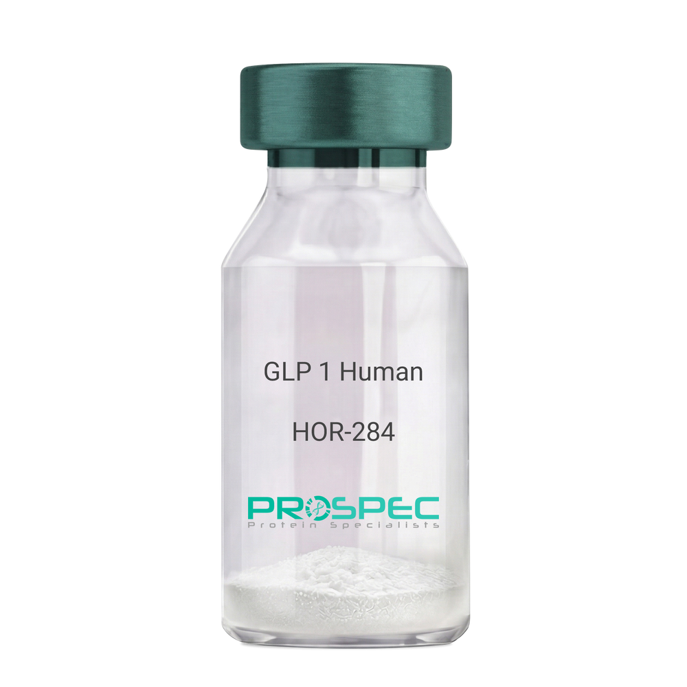
- View Data Sheet
Name :
GLP 1 HumanDescription:
Human Glucagon Like Peptide-1
GLP1, Glucagon Like Peptide-1, Incretin hormone.
Product # :
HOR-284Price :
Quantity :
Shipping Method :
Shipped at Room temp
More Info
- description
- formulation
- purity
- More Info
Description
Glucagon Like Peptide-1 is a single, non-glycosylated, polypeptide chain containing 30 amino acids and having a molecular mass of 3297.7 Dalton.The GLP-1 is purified by proprietary chromatographic techniques.
Formulation
The GLP-1 peptide was lyophilized with no additives.
Purity
Greater than 96.0% as determined by RP-HPLC.
More Info
-
Introduction
Glucagon-like peptide-1 (GLP-1) is derived from the transcription product of the proglucagon gene. The major source of GLP-1 in the body is the intestinal L cell that secretes GLP-1 as a guthormone. The biologically active forms of GLP-1 are: GLP-1-(7-37) and GLP-1-(7-36)NH2.
GLP-1 secretion by L cells is dependent on the presence of nutrients in the lumen of the small intestine. The secretagogues (agents that causes or stimulates secretion) of this hormone include major nutrients like carbohydrate, proteinand lipid. Once in the circulation, GLP-1 has a half life of less than 2 minutes, due to rapid degradation by the enzyme dipeptidyl peptidase-4.
GLP-1 possesses several physiological properties that make it a subject of intensive investigation as a potential treatment of diabetes mellitus. The known physiological functions of GLP-1 include: Increases insulin secretion from the pancreas in a glucose-dependent manner, decreases glucagon secretion from the pancreas, increases beta cells mass and insulin gene expression, inhibits acid secretion and gastric emptying in the stomach, decreases food intake by increasing satiety. -
Synonyms
GLP1, Glucagon Like Peptide-1, Incretin hormone.
-
Physical Appearance
Sterile Filtered White lyophilized (freeze-dried) powder.
-
Stability
Lyophilized Glucagon Like Peptide-1 although stable at room temperature for 3 weeks, should be stored desiccated below -18°C. Upon reconstitution GLP-1 should be stored at 4°C between 2-7 days and for future use below -18°C. For long term storage it is recommended to add a carrier protein (0.1% HSA or BSA).Please prevent freeze-thaw cycles.
-
Solubility
It is recommended to reconstitute the lyophilized Glucagon Like Peptide-1 in sterile H2O at 0.5mg/ml, which can then be further diluted to other aqueous solutions.
-
Amino Acid Sequence
H-His-Ala-Glu-Gly-Thr-Phe-Thr-Ser-Asp-Val-Ser-Ser-Tyr-Leu-Glu-Gly-Gln-Ala-Ala-Lys-Glu-Phe-Ile-Ala-Trp-Leu-Val-Lys-Gly-Arg-NH2.
ProSpec's products are furnished for LABORATORY RESEARCH USE ONLY. The product may not be used as drugs, agricultural or pesticidal products, food additives or household chemicals.
- View Data Sheet
Name :
Cys-Protein-GDescription:
Cys-Protein G Recombinant
Product # :
PRO-1238Price :
Quantity :
Shipping Method :
Shipped at Room temp
More Info
- description
- source
- formulation
- purity
- More Info
Description
Cys-Protein G Recombinant produced in E.Coli, is a single non-glycosylated polypeptide chain containing 201 amino acids and having a cys on N-terminal. Cys-Protein G has a predicted molecular mass of approximately 21.9kDa but it migrates with an apparent molecular mass of 40kDa in SDS-PAGE. The Cys-Protein G is purified by proprietary chromatographic techniques.
Source
Escherichia Coli.
Formulation
Lyophilized without any additives.
Purity
Greater than 95.0% as determined by SDS-PAGE and HPLC analyses.
More Info
-
Physical Appearance
Sterile Filtered White lyophilized (freeze-dried) powder.
-
Stability
Lyophilized Protein G although stable at room temperature for 3 weeks, should be stored desiccated below -18°C. Upon reconstitution Protein G should be stored at 4°C between 2-7 days and for future use below -18°C.Please prevent freeze-thaw cycles.
-
Solubility
It is recommended to reconstitute the lyophilized Protein G in sterile 18M-cm H2O not less than 100µg/ml, which can then be further diluted to other aqueous solutions.
-
Amino Acid Sequence
CLPKTDTYKL ILNGKTLKGE TTTEAVDAAT AEKVFKQYAN DNGVDGEWTY DDATKTFTVT EKPEVIDASE LTPAVTTYKL VINGKTLKGE TTTEAVDAAT AEKVFKQYAN DNGVDGEWTY DDATKTFTVT EKPEVIDASE LTPAVTTYKL VINGKTLKGE TTTKAVDAET AEKAFKQYAN DNGVDGVWTY DDATKTFTVT E.
-
Specificity
The recombinant Protein G is a genetically engineered protein contains 3 IgG-binding regions of protein G.
ProSpec's products are furnished for LABORATORY RESEARCH USE ONLY. The product may not be used as drugs, agricultural or pesticidal products, food additives or household chemicals.
- View Data Sheet
Name :
MMP 3 Human, HEKDescription:
Matrix Metalloproteinase-3 Human Recombinant, HEK
Stromelysin-1, EC 3.4.24.17, Matrix metalloproteinase-3, MMP-3, Transin-1, SL-1, STMY, STR1, STMY1, MGC126102, MGC126103, MGC126104.
Product # :
ENZ-284Price :
Quantity :
Shipping Method :
Shipped with Ice Packs
More Info
- description
- source
- formulation
- purity
- biological activity
- More Info
- sds-page
Description
MMP-3 Human Recombinant produced in HEK293 cells is a proform of the Human MMP3 [Tyr18-Cys477 (Lys45Glu)] and fused with a ployhistide tag at the C-terminus, having an Mw of 52kDa. MMP-3 is purified by proprietary chromatographic techniques.
Source
HEK293 cells.
Formulation
The MMP-3 is supplied as a 0.2µm filtered solution in 20mM Tris-HCl, 150mM NaCl and 0.05% Brij35, pH 7.5.
Purity
Greater than 95% as determined by SDS-PAGE.
Biological Activity
The activity was measured by its ability to cleave the fluorogenic peptide substrate, Mca-RPKPVE-Nval-WRK(Dnp)-NH2. The specific activity is > 150 pmoles/min/µg.
Recombinant Human MMP-3 protein pro form needs to be activated with Chymotrypsin.
Activation Protocol:
1. Dilute MMP3 to 20µg/ml in the Assay Buffer: 50mM Tris, 10mM CaCl2, 150mM NaCl, 0.05% (w/v) and Brij 35, pH 7.5.
2. Activate MMP3 by adding Chymotrypsin(Sigma, Catalog#C3142,1mg/ml stock in 1mM HCl) to a final concentration of 5ug/ml.
3. Incubate at 37°C for 30 minutes.
4. Stop activation with 2mM PMSF. Pre-warm the PMSF to 37°C prior to adding to sample.sds-page
More Info
-
Introduction
MMP-3 enzyme is also known as Stromelysin-1or as Transin-1 which hydrolyzes natural collagen at physiological pH and temperature. It dissolves the intervertebral nucleus pulposus and annulus fibrosus of Herniated Lumbar Intervertebral Disk . MMP-3 hydrolyzes components of the extracellular matrix like proteoglycan, laminin, fibronectin, gelatin and collagen types III, IV and IX. It also activates pro-MMP-9 and pro-MMP-8 and superactivates plasmin activated MMP-1. MMP-3 is secreted as a latent proenzyme and is activated by a variety of proteinases, e.g. plasmin, trypsin, chymotrypsin, cathepsin G or human neutrophil elastase. MMP-3 was found to be capable of activating the precursor of IL1-beta.
-
Synonyms
Stromelysin-1, EC 3.4.24.17, Matrix metalloproteinase-3, MMP-3, Transin-1, SL-1, STMY, STR1, STMY1, MGC126102, MGC126103, MGC126104.
-
Physical Appearance
Sterile Filtered colorless solution.
-
Stability
Store at 4°C if entire vial will be used within 2-4 weeks. Store, frozen at -20°C for longer periods of time.Avoid multiple freeze-thaw cycles.
ProSpec's products are furnished for LABORATORY RESEARCH USE ONLY. The product may not be used as drugs, agricultural or pesticidal products, food additives or household chemicals.