Search results
1000 results found for “angiogenin”
Name
Description
Product #
Price
Quantity
Shipping Method
- View Data Sheet
Name :
F9 HumanDescription:
Coagulation Factor IX Human
Coagulation factor IX, EC 3.4.21.22, Christmas factor, Plasma thromboplastin component, PTC, F9, FIX, HEMB, MGC129641, MGC129642, GLA domain, Factor IX.
Product # :
PRO-353Price :
Quantity :
Shipping Method :
Shipped with Ice Packs
More Info
- description
- source
- formulation
- purity
- biological activity
- More Info
- Citations
Description
Human Factor-IX produced from fresh frozen human plasma is a glycosylated polypeptide chain having a molecular mass of 56 kDa.
Source
Human Plasma.
Formulation
The Factor-IX is provided in a sterile solution containing 20mM Tris-HCl pH-7.4, 100mM NaCl and 1mM Benzamidine.
Purity
Greater than 95.0% as determined by SDS-PAGE.
Biological Activity
The activity per mg was tested and found to be 380 PEU/mg.Citations
More Info
-
Introduction
Human Factor IX also called Christmas-Factor is a glycoprotein, which is synthesized in the liver and belongs to the serine proteases system and is part of the S1 peptidase family.
Lack of Factor-IX causes Hemophilia-B meaning Christmas Disease. Factor-IX has a N-terminus region which contains 12xGla residues which asist the calcium dpendant binding of Factor-IX to the phospholipid surface. Factor-IX is activated by either factor XIa or the factor VIIa/tissue factor/phospholipid complex. Cleavage yields the intermediate IXa, which is subsequently converted to the fully active form IXab.
Factor-IX binds initially to exosites on the factor XIa heavy chain, followed by interaction at the active site with subsequent bond cleavage. Coagulation factor IX is activated by interaction with the erythrocyte membrane, causing intrinsic coagulation. Chaperones & lectins act simultaniously to guarantee the proper folding of Factor-IX and the retention of mutant molecules. Human Factor IX, activated by either the Contact or Tissue Factor Pathway, is responsible for the activation of Factor X to Xa. -
Synonyms
Coagulation factor IX, EC 3.4.21.22, Christmas factor, Plasma thromboplastin component, PTC, F9, FIX, HEMB, MGC129641, MGC129642, GLA domain, Factor IX.
-
Physical Appearance
Sterile filtered colorless solution.
-
Stability
Store at 4°C if entire vial will be used within 2-4 weeks. Store, frozen at -20°C for longer periods of time. For long term storage it is recommended to add a carrier protein (0.1% HSA or BSA). Avoid multiple freeze-thaw cycles. Please prevent freeze-thaw cycles..
-
Human Virus Test
Human plasma was tested and found negative for HIV-1, HIV-2, Hepatitis B Surface antigen and HCV. Donors are screened for CJD (Creutzfeldt-Jakob Disease).
ProSpec's products are furnished for LABORATORY RESEARCH USE ONLY. The product may not be used as drugs, agricultural or pesticidal products, food additives or household chemicals.
- View Data Sheet
Name :
AlarelinDescription:
Alarelin
Alarelin, Alarelin Acetate.
Product # :
HOR-291Price :
Quantity :
Shipping Method :
Shipped at Room temp
More Info
- description
- source
- formulation
- purity
- More Info
- Citations
Description
Alarelin acetate peptide is a single, non-glycosylated polypeptide chain containing 9 amino acids, having a molecular mass of 1167.3 Dalton and a Molecular formula of C56H78N16O12 x C2H4O2. The CAS No. is 79561-22-1.
Source
Synthetic.
Formulation
The Alarelin was lyophilized with no additives.
Purity
Greater than 99.0% as determined by Analysis by RP-HPLC.
Citations
More Info
-
Introduction
Alarelin (Gonadotrophin-releasing hormone) is a synthetic LH-RH agonist that is found in higher amounts than that of LH-RH in rat hypophyseal stimulation of gonadotropin secretion in vivo and in vitro and in ovulation inductions. Alarelin is known for its induction of ovulation. Alarelin acetate is the acetate form of a hypothalamic peptide that stimulates the release of FSH and LH from the pituitary gland.
-
Synonyms
Alarelin, Alarelin Acetate.
-
Physical Appearance
Sterile Filtered White lyophilized (freeze-dried) powder.
-
Stability
Lyophilized Alarelin e although stable at room temperature for 3 weeks, should be stored desiccated below -18°C. Upon reconstitution Alarelin should be stored at 4°C between 2-7 days and for future use below -18°C. For long term storage it is recommended to add a carrier protein (0.1% HSA or BSA).Please prevent freeze-thaw cycles.
-
Solubility
It is recommended to reconstitute the lyophilized Argipressin in sterile 18MΩ-cm H2O not less than 100 µg/ml, which can then be further diluted to other aqueous solutions.
-
Amino Acid Sequence
5-oxo-pro-His-Trp-Ser-Tyr-D-Ala-Leu-Arg-Pro-Nhet x CH3COOH.
-
Background
What is the molecular weight/Mw of ALARELIN Protein?
ALARELIN Protein has a total Mw of 1.16kDa.
What is the source or expression system of ALARELIN Protein?
Synthetic.
What is the Purity of ALARELIN Protein?
ALARELIN Protein is >99% pure as determined by SDS-PAGE.
What is the Biological Activity of ALARELIN Protein?
The biological functionality of ALARELIN Protein will be determined in the future.
What is the amino acid sequence of ALARELIN Protein?
5-oxo-pro-His-Trp-Ser-Tyr-D-Ala-Leu-Arg-Pro-Nhet x CH3COOH.
What applications can ALARELIN Protein be used in?
ALARELIN Protein can probably be used in western blot, ELISA and Lateral Flow.
What is the endotoxin level for ALARELIN Protein?
The endotoxin level is minimal, ALARELIN Protein was purified using conventional chromatography techniques.
Who supplies ALARELIN that is in stock and available to order?
ProSpec has the protein in stock and available for immediate delivery.
Where can I order bulk mg quantities of ALARELIN?
ProSpec supplies bulk mg quantities at affordable prices that meets research and academic budgets.
ProSpec's products are furnished for LABORATORY RESEARCH USE ONLY. The product may not be used as drugs, agricultural or pesticidal products, food additives or household chemicals.
- View Data Sheet
Name :
Cys-Protein-GDescription:
Cys-Protein G Recombinant
Product # :
PRO-1238Price :
Quantity :
Shipping Method :
Shipped at Room temp
More Info
- description
- source
- formulation
- purity
- More Info
- Citations
Description
Cys-Protein G Recombinant produced in E.Coli, is a single non-glycosylated polypeptide chain containing 201 amino acids and having a cys on N-terminal. Cys-Protein G has a predicted molecular mass of approximately 21.9kDa but it migrates with an apparent molecular mass of 40kDa in SDS-PAGE. The Cys-Protein G is purified by proprietary chromatographic techniques.
Source
Escherichia Coli.
Formulation
Lyophilized without any additives.
Purity
Greater than 95.0% as determined by SDS-PAGE and HPLC analyses.
Citations
More Info
-
Physical Appearance
Sterile Filtered White lyophilized (freeze-dried) powder.
-
Stability
Lyophilized Protein G although stable at room temperature for 3 weeks, should be stored desiccated below -18°C. Upon reconstitution Protein G should be stored at 4°C between 2-7 days and for future use below -18°C.Please prevent freeze-thaw cycles.
-
Solubility
It is recommended to reconstitute the lyophilized Protein G in sterile 18M-cm H2O not less than 100µg/ml, which can then be further diluted to other aqueous solutions.
-
Amino Acid Sequence
CLPKTDTYKL ILNGKTLKGE TTTEAVDAAT AEKVFKQYAN DNGVDGEWTY DDATKTFTVT EKPEVIDASE LTPAVTTYKL VINGKTLKGE TTTEAVDAAT AEKVFKQYAN DNGVDGEWTY DDATKTFTVT EKPEVIDASE LTPAVTTYKL VINGKTLKGE TTTKAVDAET AEKAFKQYAN DNGVDGVWTY DDATKTFTVT E.
-
Specificity
The recombinant Protein G is a genetically engineered protein contains 3 IgG-binding regions of protein G.
ProSpec's products are furnished for LABORATORY RESEARCH USE ONLY. The product may not be used as drugs, agricultural or pesticidal products, food additives or household chemicals.
- View Data Sheet
Name :
TNF alpha humanDescription:
Tumor Necrosis Factor-Alpha Human Recombinant
TNF-alpha, Tumor necrosis factor ligand superfamily member 2, TNF-a, Cachectin, DIF, TNFA, TNFSF2.
Product # :
CYT-223Price :
Quantity :
Shipping Method :
Shipped at Room temp
More Info
- description
- source
- formulation
- purity
- biological activity
- More Info
- Citations
Description
Tumor Necrosis Factor-a Human Recombinant produced in E.Coli is a single, non-glycosylated, polypeptide chain containing 158 amino acids (157 a.a. of the mature human TNF-alpha and an N-terminal methionine) and having a molecular mass of 17.5kDa. The TNF-alpha is purified by standard chromatographic techniques.
Source
Escherichia Coli.
Formulation
TNF-a Human was lyophilized from a concentrated 1mg/ml solution containing 20mM PB, pH-7.2, and 100mM NaCl.
Purity
Greater than 95.0% as determined by:
(a) Analysis by RP-HPLC.
(b) Analysis by SDS-PAGE.Biological Activity
The Specific Activity is >5.0×10⁷ IU/mg as determined by the cytolysis of murine L929 cells in the presence of Actinomycin D.Citations
More Info
-
Introduction
Tumor necrosis factor is a cytokine involved in systemic inflammation and is a member of a group of cytokines that all stimulate the acute phase reaction. TNF is mainly secreted by macrophages.
TNF causes apoptotic cell death, cellular proliferation, differentiation, inflammation, tumorigenesis and viral replication, TNF is also involved in lipid metabolism, and coagulation. TNF's primary role is in the regulation of immune cells.
Dysregulation and, in particular, overproduction of TNF have been implicated in a variety of human diseases- autoimmune diseases, INS resistance, and cancer. -
Synonyms
TNF-alpha, Tumor necrosis factor ligand superfamily member 2, TNF-a, Cachectin, DIF, TNFA, TNFSF2.
-
Physical Appearance
Sterile Filtered White lyophilized (freeze-dried) powder.
-
Stability
Lyophilized Tumor Necrosis Factor-a although stable at room temperature for 3 weeks, should be stored desiccated below -18°C. Upon reconstitution TNF-a should be stored at 4°C between 2-7 days and for future use below -18°C.For long term storage it is recommended to add a carrier protein (0.1% HSA or BSA).Please prevent freeze-thaw cycles.
-
Solubility
It is recommended to reconstitute the lyophilized Tumor Necrosis Factor-alpha in sterile 18MΩ-cm H2O not less than 100µg/ml, which can then be further diluted to other aqueous solutions.
-
Amino Acid Sequence
MVRSSSRTPS DKPVAHVVAN PQAEGQLQWL NRRANALLAN GVELRDNQLV VPSEGLYLIY SQVLFKGQGC PSTHVLLTHT ISRIAVSYQT KVNLLSAIKS PCQRETPEGA E AKPWYEPIY LGGVFQLEKG DRLSAEINRP DYLDFAESGQ VYFGIIAL.
-
Background
TNF Alpha Human: An Overview of Its Role and Importance in Immunology
TNF alpha human, also known as Tumor Necrosis Factor-alpha, is a critical cytokine in the immune system. Macrophages mainly produce this protein, and it plays a key role in inflammation and the acute phase reaction.
This protein is involved in various cellular functions, including cell death, differentiation, proliferation, and immune regulation.
Production and Properties
Tumor Necrosis Factor-alpha is produced recombinantly in E. coli and consists of a single, non-glycosylated polypeptide chain. It includes 157 amino acids of the mature human TNF-alpha and an N-terminal methionine, resulting in a molecular mass of approximately 17.5 kDa.
Furthermore, the protein is purified through standard chromatographic techniques to ensure high purity and biological activity.
Solubility and Usage
The lyophilized form of TNF appears as a sterile, white powder. It is recommended to reconstitute this powder in sterile water to achieve a solution of no less than 100µg/ml.
This solution can then be further diluted for various experimental applications. TNF alpha is used extensively in research, particularly for studying its effects on cell signaling and immune response.
Storage and Stability
For long-term storage, TNF should be kept desiccated below -18°C. Once reconstituted, it should be used within a week if stored at 4°C or kept below -18°C for future use. Avoiding freeze-thaw cycles is crucial to maintain the protein's functionality.
Biological Role and Implications
TNF alpha human is involved in the regulation of immune cells and is known for its role in inflammatory processes.
Dysregulation of TNF alpha production is linked to various diseases, such as autoimmune disorders, insulin resistance, and cancer. It is also a target for therapeutic interventions, particularly in conditions like rheumatoid arthritis and inflammatory bowel disease.
Mechanism of Action
TNF alpha can induce fever, apoptotic cell death, and can inhibit tumorigenesis and viral replication. Moreover, it is a potent mediator of the acute phase reaction, which influences the activity of various cells involved in systemic inflammation.
Research and Clinical Importance
Scientific research on TNF has provided insights into its complex role in disease mechanisms. Its interaction with receptors such as TNFRSF1A underscores its multifaceted effects across different organ systems, from liver function to brain activity.
Ongoing studies continue to explore its therapeutic potential, especially how it can be modulated to treat diseases without harmful side effects.
In essence, TNF alpha human is a versatile and powerful component of the immune system, important for both health and disease. Understanding its pathways and functions helps scientists develop better treatments for various inflammatory and autoimmune diseases.
ProSpec's products are furnished for LABORATORY RESEARCH USE ONLY. The product may not be used as drugs, agricultural or pesticidal products, food additives or household chemicals.
- View Data Sheet
Name :
Aln G 1.0101Description:
Major pollen allergen Aln g 1 Recombinant
Major pollen allergen Aln g 1, Allergen Aln g I, Aln g 1.
Product # :
PRO-2280Price :
Quantity :
Shipping Method :
Shipped with Ice Packs
More Info
- description
- source
- formulation
- purity
- More Info
- Citations
Description
Recombinant Major pollen allergen Aln g 1 produced in SF9 is a glycosylated, polypeptide chain having a calculated molecular mass of 18,710 Dalton. Aln G 1.0101 is expressed with a 10xHis tag at N-terminus and purified by proprietary chromatographic techniques.
Source
Sf9 insect cells.
Formulation
Aln G 1.0101 is supplied in 20mM HEPES buffer pH-7.9 and 6M Urea.
Purity
Greater than 80.0% as determined by SDS-PAGE.
Citations
More Info
-
Introduction
Major pollen allergen Aln g 1 (Aln G 1.0101) causes an allergic reaction in humans.
-
Synonyms
Major pollen allergen Aln g 1, Allergen Aln g I, Aln g 1.
-
Physical Appearance
Sterile Filtered clear solution.
-
Stability
Store at 4°C if entire vial will be used within 2-4 weeks. Store, frozen at -20°C for longer periods of time. Avoid multiple freeze-thaw cycles.
-
Immunological Functions
1. Binds IgE type human antibodies. 2. Immunodot test with positive/negative sera panels.
-
Molar extinction coefficient
7450; A280(1mg/ml)=0.398
ProSpec's products are furnished for LABORATORY RESEARCH USE ONLY. The product may not be used as drugs, agricultural or pesticidal products, food additives or household chemicals.
- View Data Sheet
Name :
BMP4 Human, ActiveDescription:
Bone Morphogenetic protein-4 Active Active, Human Recombinant
BMP4, ZYME, BMP2B, BMP2B1.
Product # :
CYT-1293Price :
Quantity :
Shipping Method :
Shipped at Room temp
More Info
- description
- source
- formulation
- purity
- biological activity
- More Info
- Citations
Description
Bone Morphogenetic protein-4 Active Human Recombinant produced in E.coli is a homodimer , non-glycosylated, polypeptide chain containing 2x116 amino acids (ser293-arg408) and having a total molecular mass of 26.2kDa. BMP4 is purified by proprietary chromatographic techniques.
Source
Escherichia Coli.
Formulation
The protein was lyophilized from a sterile (0.2µm) filtered solution containing 4mM HCL.
Purity
Greater than 95.0% as determined by SDS-PAGE.
Biological Activity
The ED50, as calculated by Alkaline phosphatase activity induced in ATDC-5 cells is 157.2ng/ml corresponding to a specific activity which is 6361 units/mg.
Citations
More Info
-
Synonyms
BMP4, ZYME, BMP2B, BMP2B1.
-
Physical Appearance
Sterile Filtered White lyophilized (freeze-dried) powder.
-
Stability
Lyophilized BMP4 although stable at room temperature for 3 weeks, should be stored desiccated below -18°C. Upon reconstitution BMP4 should be stored at 4°C between 2-7 days and for future use below -18°C. Please prevent freeze-thaw cycles.
-
Solubility
It is recommended to reconstitute the lyophilized BMP4 in sterile 4mM HCl at a concentration of 0.1 mg/ml, which can then be further diluted to other aqueous solutions
-
Amino Acid Sequence
SPKHHSQRAR KKNKNCRRHS LYVDFSDVGW NDWIVAPPGY QAFYCHGDCP FPLADHLNST NHAI VQT LVN SVNSSIPKAC CVPTELSAIS MLYLDEYDKV VLKNYQEMVV EGCGCR.
-
Background
The protein encoded by this gene is a member of the bone morphogenetic protein family which is part of the transforming growth factor-beta superfamily.
The superfamily includes large families of growth and differentiation factors.
Bone morphogenetic proteins were originally identified by an ability of demineralized bone extract to induce endochondral osteogenesis in vivo in an extraskeletal site.
This particular family member plays an important role in the onset of endochondral bone formation in humans, and a reduction in expression has been associated with a variety of bone diseases, including the heritable disorder Fibrodysplasia Ossificans Progressiva.
What is the molecular weight/Mw of BMP4 Protein?
BMP4 Protein has a total Mw of 26.2kDa.
What is the source or expression system of BMP4 Protein?
Escherichia Coli.
What is the Purity of BMP4 Protein?
BMP4 Protein is >95% pure as determined by SDS-PAGE.
What is the Biological Activity of BMP4 Protein?
The ED50, as calculated by Alkaline phosphatase activity induced in ATDC-5 cells is 157.2ng/ml corresponding to a specific activity which is 6361 units/mg.
What is the amino acid sequence of BMP4 Protein?
SPKHHSQRAR KKNKNCRRHS LYVDFSDVGW NDWIVAPPGY QAFYCHGDCP FPLADHLNST NHAI VQT LVN SVNSSIPKAC CVPTELSAIS MLYLDEYDKV VLKNYQEMVV EGCGCR.
What applications can BMP4 Protein be used in?
BMP4 Protein can probably be used in western blot, ELISA and Lateral Flow.
What is the endotoxin level for BMP4 Protein?
The endotoxin level is minimal, BMP4 Protein was purified using conventional chromatography techniques.
ProSpec's products are furnished for LABORATORY RESEARCH USE ONLY. The product may not be used as drugs, agricultural or pesticidal products, food additives or household chemicals.
- View Data Sheet
Name :
OPG Human, HisDescription:
Osteoprotegerin Human Recombinant, His Tag
TNFRSF11B, OPG, OCIF, Osteoclastogenesis inhibitory factor, TR1, MGC29565.
Product # :
CYT-290Price :
Quantity :
Shipping Method :
Shipped at Room temp
More Info
- description
- source
- formulation
- purity
- More Info
- Citations
Description
Recombinant Human OCIF produced in E.coli cells is a single, non-glycosylated, polypeptide chain containing amino acids 201-401 and having a molecular mass of 31 kDa which includes a 4 kDa His tag.The OPG is purified by proprietary chromatographic techniques.
Source
Escherichia Coli.
Formulation
The protein was lyophilized from a concentrated (1mg/ml) solution with 1X PBS, 0.1% SDS and 1mM DTT.
Purity
Greater than 80.0% as determined by(a) Analysis by RP-HPLC.
(b) Analysis by SDS-PAGE.Citations
More Info
-
Introduction
Osteoprotegerin, which is a member of the tumor necrosis factor receptor superfamily and is involved in the regulation of bone metabolism. OPGand its ligand (OPGL) are cytokines regulating osteoclasto-genesis. OPGL binds to receptors on the surface of preosteoclasts and stimulates their differentiation into active osteoclasts. This leads to osteoresorption. OPG inhibits this osteoclasto-genesis (OPG is secreted by osteoblasts, and binds to OPGL, thus inhibiting maturation of osteoclasts and osteoresorption). The degree and activity of osteoresorption depend mainly on the balance between OPG and its ligand (OPGL); factors increasing OPGL expression mostly reduce OPG expression and vice versa.
-
Synonyms
TNFRSF11B, OPG, OCIF, Osteoclastogenesis inhibitory factor, TR1, MGC29565.
-
Physical Appearance
Sterile Filtered White lyophilized (freeze-dried) powder.
-
Stability
Lyophilized Osteoprotegerin although stable at room temperature for 3 weeks, should be stored desiccated below -18°C. Upon reconstitution OCIF should be stored at 4°C between 2-7 days and for future use below -18°C.For long term storage it is recommended to add a carrier protein (0.1% HSA or BSA).Please prevent freeze-thaw cycles.
-
Solubility
It is recommended to reconstitute the lyophilized Osteoprotegerin in sterile 18MΩ-cm H2O not less than 100µg/ml, which can then be further diluted to other aqueous solutions.
-
Applications
1. Positive control for Western blot.
2. Antibody production.
3. Protein assay.
ProSpec's products are furnished for LABORATORY RESEARCH USE ONLY. The product may not be used as drugs, agricultural or pesticidal products, food additives or household chemicals.
- View Data Sheet
Name :
GDNF HumanDescription:
Glial-Derived Neurotrophic Factor Human Recombinant
ATF1, ATF2, HFB1-GDNF, GDNF.
Product # :
CYT-305Price :
Quantity :
Shipping Method :
Shipped at Room temp
More Info
- description
- source
- formulation
- purity
- biological activity
- More Info
- sds-page
- Citations
Description
Glial derived Neurotrophic Factor Human Recombinant produced in E.Coli is a non-glycosylated disulfide-linked homodimer containing 2 x 135 amino acids and having a total molecular mass of approximately 30kDa. GDNF is purified by proprietary chromatographic techniques.
Source
Escherichia Coli.
Formulation
GDNF was lyophilized from a 0.2µm filtered concentrated solution in 1×PBS, pH 7.4 and 5% Trehalose.
Purity
Greater than 95.0% as determined by analysis by SDS-PAGE.
Biological Activity
The ED50 was determined by the proliferation of rat C6 cells is < 0.1 ng/ml, corresponding to a specific activity of > 1.0x10⁷ units/mg.
sds-page
Citations
More Info
-
Introduction
GDNF promotes the survival and differentiation of minergic neurons in culture, and is able to prevent apoptosis of motor neurons induced by axotomy. The encoded protein is processed to a mature secreted form that exists as a homodimer. The mature form of the protein is a ligand for the product of the RET (rearranged during transfection) protooncogene. In addition to the transcript encoding GDNF, two additional alternative transcripts encoding distinct proteins, referred to as astrocyte-derived trophic factors, have also been described. Mutations in this gene may be associated with Hirschsprung disease.
GDNF enhances survival and morphological differentiation of minergic neurons and increases their high-affinity uptake. -
Synonyms
ATF1, ATF2, HFB1-GDNF, GDNF.
-
Physical Appearance
Sterile Filtered White lyophilized (freeze-dried) powder.
-
Stability
Lyophilized Glial-derived Neurotrophic Factor although stable at room temperature for 3 weeks, should be stored desiccated below -18°C. Upon reconstitution GDNF should be stored at 4°C between 2-7 days and for future use below -18°C.For long term storage it is recommended to add a carrier protein (0.1% HSA or BSA).Please prevent freeze-thaw cycles.
-
Solubility
It is recommended to reconstitute the lyophilized Glial Derived Neurotrophic Factor in sterile 18MΩ-cm H2O not less than 100µg/ml, which can then be further diluted to other aqueous solutions.
-
Amino Acid Sequence
SPDKQMAVLP RRERNRQAAA ANPENSRGKG RRGQRGKNRG CVLTAIHLNV TDLGLGYETK EELIFRYCSG SCDAAETTYD KILKNLSRNR RLVSDKVGQA CCRPIAFDDD LSFLDDNLVY HILRKHSAKR CGCI.
-
Background
What is the molecular weight/Mw of GDNF HUMAN Protein?
GDNF HUMAN Protein has a total Mw of 30kDa.
What is the source or expression system of GDNF HUMAN Protein?
Escherichia Coli.
What is the Purity of GDNF HUMAN Protein?
GDNF HUMAN Protein is >95% pure as determined by SDS-PAGE.
What is the Biological Activity of GDNF HUMAN Protein?
The ED50 was determined by the proliferation of rat C6 cells is < 0.1 ng/ml, corresponding to a specific activity of > 1.0x10⁷ units/mg.
What is the amino acid sequence of GDNF HUMAN Protein?
SPDKQMAVLP RRERNRQAAA ANPENSRGKG RRGQRGKNRG CVLTAIHLNV TDLGLGYETK EELIFRYCSG SCDAAETTYD KILKNLSRNR RLVSDKVGQA CCRPIAFDDD LSFLDDNLVY HILRKHSAKR CGCI.
What applications can GDNF HUMAN Protein be used in?
GDNF HUMAN Protein can probably be used in western blot, ELISA and Lateral Flow.
What is the endotoxin level for GDNF HUMAN Protein?
The endotoxin level is minimal, GDNF HUMAN Protein was purified using conventional chromatography techniques.
ProSpec's products are furnished for LABORATORY RESEARCH USE ONLY. The product may not be used as drugs, agricultural or pesticidal products, food additives or household chemicals.
- View Data Sheet
Name :
TGFB1 Human, HEKDescription:
Transforming Growth Factor-Beta 1 Human Recombinant, HEK
Transforming growth factor beta-1, TGF-beta-1, CED, DPD1, TGFB, TGF-b 1, LAP, TGFB1.
Product # :
CYT-1260Price :
Quantity :
Shipping Method :
Shipped at Room temp
More Info
- description
- source
- formulation
- purity
- biological activity
- More Info
- Citations
Description
TGFB1 Human Recombinant produced in 293 cells is a glycosylated homodimer polypeptide chain containing 2 x 112 amino acids and having a total molecular mass of 25.0kDa. The TGFB1 is purified by proprietary chromatographic techniques.
Source
HEK 293 cells.
Formulation
Lyophilized from a sterile filtered solution containing TFA (0.1%).
Purity
Greater than 98.0% as determined by: (a) Analysis by HPLC. (b) Analysis by SDS-PAGE.
Biological Activity
The ED50 as determined by the dose-dependent inhibition of IL-4-induced proliferation of HT-2 cells is ≤ 0.05 ng/ml, corresponding to a specific activity of ≥ 2 x 10⁷ units/mg.
Citations
More Info
-
Synonyms
Transforming growth factor beta-1, TGF-beta-1, CED, DPD1, TGFB, TGF-b 1, LAP, TGFB1.
-
Physical Appearance
Sterile Filtered White lyophilized (freeze-dried) powder.
-
Stability
Lyophilized TGFB1 although stable at room temperature for 3 weeks, should be stored desiccated below -18°C. Upon reconstitution TGFB1 Human should be stored at 4°C between 2-7 days and for future use below -18°C. For long term storage it is recommended to add a carrier protein (0.1% HSA or BSA). Please prevent freeze-thaw cycles.
-
Solubility
It is recommended to reconstitute the lyophilized TGFB1 in sterile in 18MΩ-cm H2O at a concentration of 0.1 mg/ml, which can then be further diluted to other aqueous solutions.
-
Amino Acid Sequence
ALDTNYCFSS TEKNCCVRQL YIDFRKDLGW KWIHEPKGYH ANFCLGPCPY IWSLDTQYSK VLALYNQHNP GASAAPCCVP QALEPLPIVY YVGRKPKVEQ LSNMIVRSCK CS.
-
Background
Transforming growth factor betas (TGF Betas) mediate many cell-cell interactions that occur during embryonic development. 3 TGF Betas have been identified in mammals: TGF Beta 1, TGF Beta 2 and TGF Beta 3. each are synthesized as precursor proteins that are very similar in that each is cleaved to yield a 112 amino acid polypeptide that remains associated with the latent portion of the molecule.
ProSpec's products are furnished for LABORATORY RESEARCH USE ONLY. The product may not be used as drugs, agricultural or pesticidal products, food additives or household chemicals.
- View Data Sheet
Name :
Ferritin HumanDescription:
Human Liver Ferritin
Product # :
PRO-564Price :
Quantity :
Shipping Method :
Shipped with Ice Packs
More Info
- description
- source
- formulation
- purity
- More Info
- Citations
Description
Ferritin is a glycoprotein produced in Human Liver having a molecular mass of 440- 450kDa and pI of 5.5, which stores iron atoms in the ferric state. It is predominantly intracellular, where it forms an exchangeable pool of iron acting as an iron store. Ferritin level in serum is directly proportional to body iron stores and serum levels are an excellent indicator in monitoring iron status in anemia. It can be used as a marker for inflammation and also used for monitoring and prediction of future events in coronary artery disease.
Source
Human Liver.
Formulation
The protein solution is in 0.05M TRIS buffer pH 7.5 containing 1.0M NaCl and 0.09% NaN3.
Purity
Greater than 96.0%.
Citations
More Info
-
Introduction
Ferritin is the main intracellular iron storage protein in prokaryotes and eukaryotes. Ferritin’s major functions are the storage of iron in a soluble and nontoxic state and its release in a controlled fashion. An iron-containing protein complex is found mostly in the intestinal mucosa, spleen, and liver. Ferritin is composed of 24 subunits of the heavy and light chains. Variation in ferritin subunit composition may influence the rates of iron uptake and release in different tissues. Defects in the light chain ferritin gene are linked to a number of neurodegenerative diseases and hyperferritinemia-cataract syndrome. The genes that encode the light and heavy chains are on located different chromosomes. The light chain genes are in chromosome region 19q13.3-q13.4 whilst those for the heavy chain are in chromosome region 11q12-q13. Ferritin is shaped like a hollow sphere, inside which the iron is stored in the Fe(III) oxidation state. The iron is integrated in the mineral ferrihydrite, [FeO(OH)]8[FeO(H2PO4)], which is attached to the inner wall of the sphere. To release iron once the body needs it, the iron must be altered from the Fe(III) to the Fe(II) oxidation state. Subsequently, the iron leaves through channels in the spherical structure. Therefore, the structure of ferritin is tremendously important for the protein's ability to store and release iron in a controlled mode.
The amount of ferritin in the blood (serum ferritin level) is directly related to the amount of iron stored in the body. The body has a "buffer" against iron deficiency (if the blood has too little iron, ferritin can release more) and, to a lesser extent, iron overload (if the blood and tissues of the body have too much iron, ferritin can help store the excess iron). -
Physical Appearance
Sterile Filtered brownish solution.
-
Stability
Human Ferritin should be stored at 2-8°C.
-
Human Virus Test
Tissue sample tested and found negative for HIV-1 & 2 antibodies, Hepatatis B surface antigen, Syphilis RPR and Hepatatis C antibodies.
ProSpec's products are furnished for LABORATORY RESEARCH USE ONLY. The product may not be used as drugs, agricultural or pesticidal products, food additives or household chemicals.
- View Data Sheet
Name :
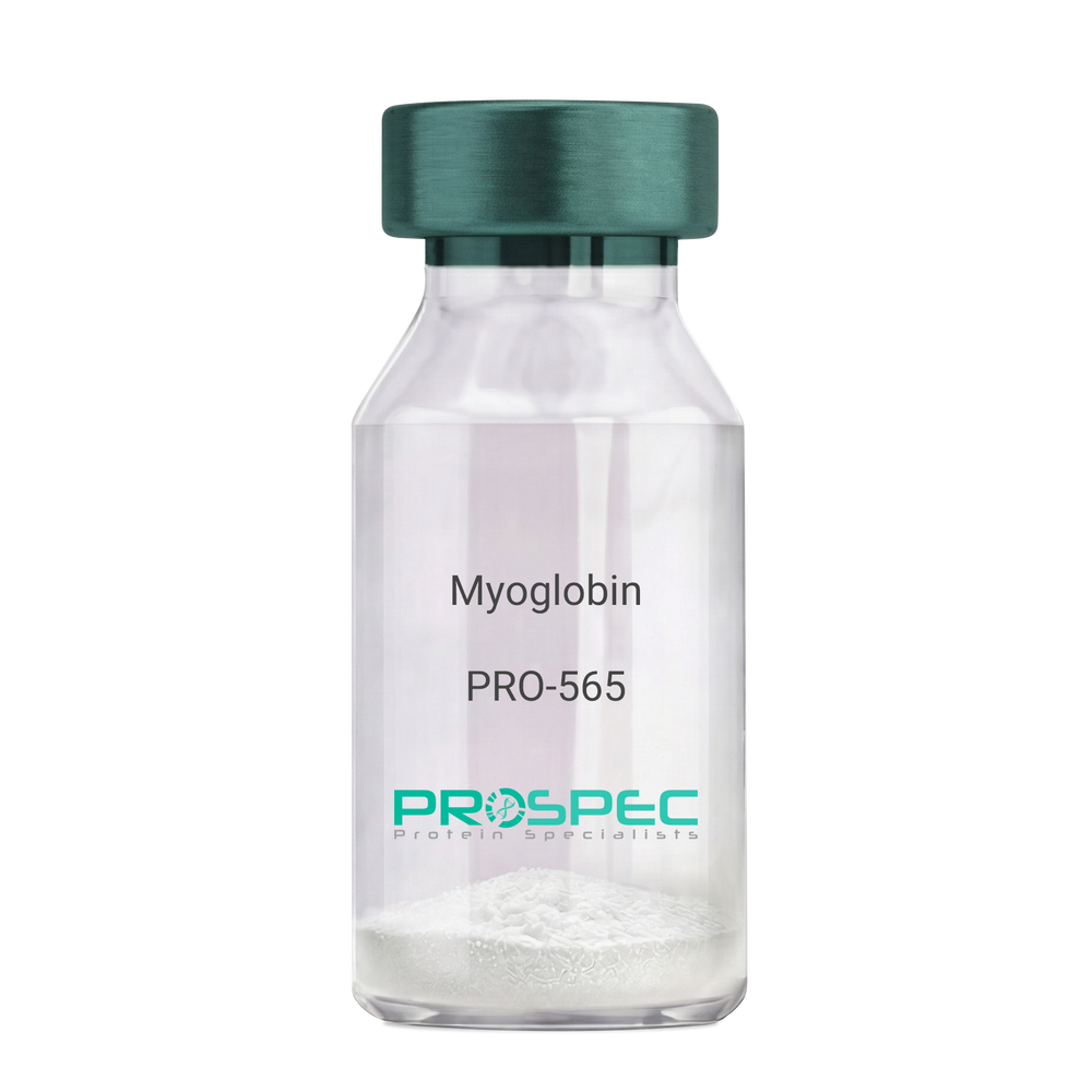
MyoglobinDescription:
Myoglobin Human
Myoglobin, MB, PVALB, MGC13548.
Product # :
PRO-565Price :
Quantity :
Shipping Method :
Shipped with Ice Packs
More Info
- description
- source
- formulation
- purity
- More Info
- Citations
Description
Human Myoglobin produced in Human Cardiac Tissues having a molecular mass of 17.5kDa. Myoglobin is released from recently injured myocardial cells within a few hours of Infarction. Peak levels are reached more quickly than CK-MB or Troponin complex.
Source
Human Cardiac Tissues.
Formulation
The protein solution is in 0.05M phosphate buffer pH 7.5 containing 0.15M NaCl and 0.09% NaN3. Filtered through a 0.2µM membrane.
Purity
Greater than 96.0%.
Citations
More Info
-
Introduction
Myoglobin is a member of the globin superfamily and can be found in skeletal and cardiac muscles. It is a haemoprotein that contributs to intracellular oxygen storage and transcellular facilitated diffusion of oxygen. Myoglobin has a single-chain globular structure of 153 amino acids, containing a heme prosthetic group (iron-containing porphyrin) in the core around which the remaining apoprotein folds. Myoglobin has 8 alpha helices and a hydrophobic core. Myoglobin’s molecular weight is 16.7 kDa, and it is the primary oxygen-carrying pigment of muscle tissues. The binding of oxygen in myoglobin is different from the cooperative oxygen binding in hemoglobin, since positive collaboration is a property of multimeric/oligomeric proteins only. Instead, the binding of oxygen by myoglobin is uninfluenced by the oxygen pressure in the surrounding tissue. Myoglobin is frequently referred to as having an "instant binding tenacity" to oxygen given its hyperbolic oxygen dissociation curve. Different organisms are able to hold their breaths longer due to high concentrations of myoglobin in their muscle cells. Myoglobin is responsible for the pigments that make meat red. The color of the meat is partly determined by the charge of the iron atom in myoglobin and the oxygen attached to it. Myoglobin is found in Type I muscle, Type II A and Type II B, but it is mostly deemed that myoglobin is not found in smooth muscle. Myoglobin is discharged from damaged muscle tissue (rhabdomyolysis), which contains very high concentrations of myoglobin. Even though the released myoglobin is filtered by the kidneys, it is toxic to the renal tubular epithelium and thus may cause acute renal failure.
-
Synonyms
Myoglobin, MB, PVALB, MGC13548.
-
Physical Appearance
Sterile Filtered red solution.
-
Stability
Human Myoglobin should be stored at 2-8°C.
-
Human Virus Test
Starting material donor tested and certified negative for HIV I & II antibodies, Hepatitis B surface antigen, and Hepatitis C antibodies.
ProSpec's products are furnished for LABORATORY RESEARCH USE ONLY. The product may not be used as drugs, agricultural or pesticidal products, food additives or household chemicals.
- View Data Sheet
Name :
Prolactin HumanDescription:
Prolactin Human Recombinant
Mammotropin, Luteotropic hormone, Luteotropin, PRL.
Product # :
CYT-267Price :
Quantity :
Shipping Method :
Shipped at Room temp
More Info
- description
- source
- formulation
- purity
- biological activity
- More Info
- Citations
Description
Prolactin Human Recombinant produced in E.Coli is a single, non-glycosylated polypeptide chain containing 200 amino acids and having a molecular mass of 23007 Dalton. The Prolactin is purified by proprietary chromatographic techniques.
Source
Escherichia Coli.
Formulation
The protein was lyophilized from a concentrated (1mg/ml) solution with 10mM sodium Phosphate buffer pH=8 and 50mM Nacl.
Purity
Greater than 95.0% as determined by:
(a) Analysis by SEC-HPLC.
(b) Analysis by SDS-PAGE.Biological Activity
The ED50 as determined by the dose-dependant stimulation of the proliferation of rat lymphoma, Nb2-11 was found to be < 0.065ng/ml corresponding to a Specific Activity of 15,385,000IU/mg.Citations
More Info
-
Introduction
Prolactin is a neuroendocrine hormone synthesized primarily by the pituitary gland but also a variety of other cell types including the placenta, brain and uterus. Its primary function is to promote and maintain lactation but has also been shown to have a role in breast cancer development, regulation of reproductive function and immunoregulation.
-
Synonyms
Mammotropin, Luteotropic hormone, Luteotropin, PRL.
-
Physical Appearance
Sterile Filtered White lyophilized (freeze-dried) powder.
-
Stability
Lyophilized Prolactin although stable at room temperature for 3 weeks, should be stored desiccated below -18°C. Upon reconstitution Prolactin should be stored at 4°C between 2-7 days and for future use below -18°C.For long term storage it is recommended to add a carrier protein (0.1% HSA or BSA).Please prevent freeze-thaw cycles.
-
Solubility
It is recommended to reconstitute the lyophilized Prolactin in sterile 18MΩ-cm H2O not less than 100µg/ml, which can then be further diluted to other aqueous solutions.
-
Amino Acid Sequence
The sequence of the first five N-terminal amino acids was determined and was found to be Met-Leu-Pro-Ile-Cys.
ProSpec's products are furnished for LABORATORY RESEARCH USE ONLY. The product may not be used as drugs, agricultural or pesticidal products, food additives or household chemicals.
- View Data Sheet
Name :
Dengue Envelope-1 & 3Description:
Dengue Virus Subtype 1 & 3 fused Envelope 58kDa Recombinant
Product # :
DEN-001Price :
Quantity :
Shipping Method :
Shipped with Ice Packs
More Info
- description
- source
- formulation
- purity
- More Info
- Citations
Description
The E.coli derived recombinant 58kDa protein is a genetically engineered peptide which is derived from Dengue Type-1 and 3 to be expressed as a fused envelope, each part in this fusion contains 170 a.a (positions 46-217), it is used in ELISA assay. This fusion protein is connected to a 6xHis Tag. Dengue Type-1 and 3 is purified by proprietary chromatographic technique.
Source
Escherichia Coli.
Formulation
The protein solution contains Phosphate buffered saline, pH-7.4 and 0.05% sodium azide.
Purity
Protein is >95% pure as determined by 12% PAGE (coomassie staining).
Citations
More Info
-
Introduction
Caused by one of four closely related virus serotypes of the genus Flavivirus, family Flaviviridae, each serotype is sufficiently different that there is no cross-protection and epidemics caused by multiple serotypes (hyperendemicity) can occur. In cell culture experiments and mice Morpholino antisense oligos have shown specific activity against Dengue virus.
-
Physical Appearance
Sterile filtered colorless solution.
-
Stability
Dengue Envelope-1 & 3 Recombinant although stable at 4°C for 1 week, should be stored below -18°C. Please prevent freeze thaw cycles.
ProSpec's products are furnished for LABORATORY RESEARCH USE ONLY. The product may not be used as drugs, agricultural or pesticidal products, food additives or household chemicals.
- View Data Sheet
Name :
CHIKV E2Description:
Chikungunya E2 Recombinant
Product # :
CHI-003Price :
Quantity :
Shipping Method :
Shipped with Ice Packs
More Info
- description
- source
- formulation
- purity
- More Info
- Citations
Description
Recombinant Chikungunya E2 produced in E.coli having a molecular weight of 38kDa (migrates at 38-40kDa on 10% SDS-PAGE).
Source
Escherichia Coli.
Formulation
Sterile Filtered solution containing PBS and 25mM K2CO3.
Purity
Protein is >95% pure as determined by SDS-PAGE.
Citations
More Info
-
Introduction
Chikungunya is an infection caused by the chikungunya virus which is passed to humans by two species of mosquito of the genus Aedes: A. albopictus and A. aegypti. Animal reservoirs of the virus include monkeys, birds, cattle, and rodents. The features of the disease are a sudden onset of fever 2-4 days after exposure. The fever typically lasts 2-7 days, while the associated joint pains usually last weeks or months but sometimes years. The mortality rate is a little less than 1 in 1,000. The disease has occurred in outbreaks in Asia, Europe and the Americas since 2004. CHIKV is a single-stranded positive-sense RNA genome, 11,800 nts long which encodes 2 open reading frames. The nucleocapsid is tightly enveloped by a host-derived lipid bilayer (envelope) supporting the virus-encoded envelope proteins. 80 glycoprotein spikes are C- terminally anchored within the viral envelope. The structural polyprotein is translated from a viral sub genomic mRNA, while as the 5 structural proteins (capsid, E3, E2, 6K, E1) are translated as a single polyprotein, from which capsid (C) is cleaved off to encapsidate. The envelope polyprotein precursor E3-E2-6K-E1 is translocated to the endoplasmatic reticulum. Polyprotein is processed by host signalases, resulting in E3, E2 & E1 forming viral hetero-trimeric spikes. The viral spikes majorly contains E2 and E1 facilitate cell receptor recognition, cell entry thru pH-dependent endocytosis and support viral budding.
-
Physical Appearance
Sterile filtered colorless solution.
-
Stability
CHIKV E2 although stable at 4°C for 1 week, should be stored below -18°C. Please prevent freeze thaw cycles.
-
Applications
Rapid test and Immunoassay.
-
Purification Method
Purified by proprietary chromatographic technique.
ProSpec's products are furnished for LABORATORY RESEARCH USE ONLY. The product may not be used as drugs, agricultural or pesticidal products, food additives or household chemicals.
- View Data Sheet
Name :
CAV1 AntibodyDescription:
Caveolin-1, Mouse Anti Human
Caveolin-1, CAV1, CAV, CGL3, BSCL3, VIP21, MSTP085.
Product # :
ANT-027Price :
Quantity :
Shipping Method :
Shipped with Ice Packs
More Info
- formulation
- More Info
- Citations
Formulation
1mg/ml containing PBS, pH-7.4, & 0.1% Sodium Azide.
Citations
More Info
-
Introduction
The scaffolding protein encoded by the CAV1 gene is the key component of the caveolae plasma membranes found in most cell types. Cholesterol is vital for caveolae formation and maintenance, and caveolae structure is highly sensitive to cholesterol depletion or treatment with cholesterol binding drugs. CAV1 can tightly and specifically bind free cholesterol and artificial cholesterol containing vesicles. Caveolin 1 links integrin subunits to the tyrosine kinase FYN, which is an initiating step in coupling integrins to the Ras-ERK pathway and promoting cell cycle progression. CAV1 is a tumor suppressor gene candidate and a negative regulator of the Ras-p42/44 mitogen-activated kinase. Caveolin 1 and caveolin 2 are positioned next to each other on chromosome 7 and express co-localizing proteins which form a stable hetero-oligomeric complex cascade. Mutations in the CAV1 gene are linked to Berardinelli-Seip congenital lipodystrophy.
-
Synonyms
Caveolin-1, CAV1, CAV, CGL3, BSCL3, VIP21, MSTP085.
-
Physical Appearance
Sterile Filtered clear solution.
-
Immunogen
Anti-human CAV1 mAb, is derived from hybridization of mouse FO myeloma cells with spleen cells from BALB/c mice immunized with recombinant human CAV1 amino acids 1-104 purified from E. coli.
-
Ig Subclass
Mouse IgG2b heavy chain and k light chain.
-
Clone
PAT4C1AT.
-
Applications
CAV1 antibody has been tested by ELISA, Western blot to assure specificity and reactivity. Since application varies, however, each investigation should be titrated by the reagent to obtain optimal results. Recommended dilution range for Western blot analysis is 1:250 ~ 1000.
Recommended starting dilution is 1:500. -
Type
Mouse Anti Human Monoclonal.
-
Storage Procedures
For periods up to 1 month store at 4°C, for longer periods of time, store at -20°C. Prevent freeze thaw cycles.
-
Purification Method
CAV1 antibody was purified from mouse ascitic fluids by protein-G affinity chromatography.
ProSpec's products are furnished for LABORATORY RESEARCH USE ONLY. The product may not be used as drugs, agricultural or pesticidal products, food additives or household chemicals.
- View Data Sheet
Name :
CagrilintideDescription:
Cagrilintide
Product # :
HOR-059Price :
Quantity :
Shipping Method :
Shipped at Room temp
More Info
- description
- formulation
- purity
- More Info
- Citations
Description
Cagrilintide is a synthetic single, non-glycosylated polypeptide chain containing 37 amino acids, having a molecular mass of 4409 Dalton and a Molecular formula of C194H312N54O59.
Formulation
The protein was lyophilized with no additives.
Purity
Greater than 97.0% as determined by analysis by RP-HPLC.
Citations
More Info
-
Physical Appearance
Sterile Filtered White lyophilized (freeze-dried) powder.
-
Stability
Lyophilized Cagrilintide although stable at room temperature for 3 weeks, should be stored desiccated below -18°C. Upon reconstitution Cagrilintide should be stored at 4°C between 2-7 days and for future use below -18°C.
For long term storage it is recommended to add a carrier protein (0.1% HSA or BSA).
Please prevent freeze-thaw cycles.
-
Solubility
It is recommended to reconstitute the lyophilized Cagrilintide in sterile 18MΩ-cm H2O not less than 100 µg/ml, which can then be further diluted to other aqueous solutions.
-
Amino Acid Sequence
(Eicosanedioic acid-γ-Glu)-Lys-CysAsn-Thr-Ala-Thr-Cys-Ala-Thr-Gln-Arg-Leu-Ala-Glu-Phe-Leu-Arg-HisSer-Ser-Asn-Asn-Phe-Gly-Pro-Ile-Leu-Pro-Pro-Thr-Asn-Val-Gly-SerAsn-Thr-Pro-NH2 (Disulfide bridge:Cys3-Cys8).
-
Background
Cagrilintide plays a role as a pioneering long-acting amylin analogue and integrates into the fixed-dose combination CagriSema (Cagrilintide + Semaglutide). Unlike GLP-1 agonists, Cagrilintide mimics amylin, a hormone co-secreted with insulin takes part in regulating satiety and slows gastric emptying through brainstem pathways.
ProSpec's products are furnished for LABORATORY RESEARCH USE ONLY. The product may not be used as drugs, agricultural or pesticidal products, food additives or household chemicals.
- View Data Sheet
Name :
Secretin HumanDescription:
Secretin Human
Product # :
HOR-273Price :
Quantity :
Shipping Method :
Shipped at Room temp
More Info
- description
- formulation
- purity
- More Info
- Citations
Description
Secretin has a molecular formula of C130H220N44O41, a.a. sequence of H-His-Ser-Asp-Gly-Thr-Phe-Thr-Ser-Glu-Leu-Ser-Arg-Leu-Arg- Asp-Ser-Ala-Arg-Leu-Gln-Arg-Leu-Leu-Gln-Gly-Leu-Val-NH2 and having an Mw of 3055.4 Dalton.
Formulation
The protein (1mg/ml) was lyophilized with no additives.
Purity
Greater than 98.0% as determined by:
(a) Analysis by RP-HPLC.
(b) Analysis by SDS-PAGE.Citations
More Info
-
Introduction
Human Secretin stimulates the secretion of bicarbonate by the pancreas and inhibits the production of gastrin and acid production in the stomach. It also potentiates the release of digestive enzymes from the pancreas triggered by cholecystokinin.
-
Physical Appearance
Sterile Filtered White lyophilized (freeze-dried) powder.
-
Stability
Lyophilized Secretin although stable at room temperature for 3 weeks, should be stored desiccated below -18°C. Upon reconstitution Secretin should be stored at 4°C between 2-7 days and for future use below -18°C.For long term storage it is recommended to add a carrier protein (0.1% HSA or BSA).Please prevent freeze-thaw cycles.
-
Solubility
It is recommended to reconstitute the lyophilized Secretin in sterile 18MΩ-cm H2O not less than 100 µg/ml, which can then be further diluted to other aqueous solutions.
ProSpec's products are furnished for LABORATORY RESEARCH USE ONLY. The product may not be used as drugs, agricultural or pesticidal products, food additives or household chemicals.
- View Data Sheet
Name :
Protein-A/G/LDescription:
Protein A/G/L Recombinant
Product # :
PRO-1936Price :
Quantity :
Shipping Method :
Shipped at Room temp
More Info
- description
- source
- formulation
- purity
- More Info
- Citations
Description
Recombinant Protein-A/G/L produced in E.Coli is a single non-glycosylated polypeptide chain. Protein- A/G/L is comprised of 5 IgG-binding regions of Protein A (E-D-A-B-C), 2 of protein G (C1-C3) and 5 of Protein L (B1-B2-B3-B4-B5) containing 805 amino acids in total and having a molecular mass of 89.2kDa. Cell wall binding region, cell membrane binding region and albumin binding region have been eliminated from the recombinant Protein- A/G/L to guarantee the maximum specific IgG binding.
Source
Escherichia Coli.
Formulation
Protein- A/G/L was lyophilized without any additives.
Purity
Greater than 95.0% as determined by:
(a) Analysis by SEC-HPLC.
(b) Analysis by SDS-PAGE.Citations
More Info
-
Introduction
The recombinant Protein A/G/L is a genetically engineered protein which combines the IgG binding profiles of all Protein A, Protein G and Protein L. Protein A/G/L is a gene fusion product. Recombinant fusion protein A/G/L is comprised of 5 Ig-binding regions of protein L (B1-B2-B3-B4-B5), 5 IgG binding domains from Protein A (E-D-A-B-C) and 2 Ig-binding region of protein G (C1-C3). The recombinant Protein A/G/L is ideal for purification of monoclonal or polyclonal IgG antibodies. Protein A/G/L binds to IgG from humans, mice, rats, cows, goats, sheep, rabbits, guinea pigs, pigs, dogs and cats.
-
Physical Appearance
Sterile Filtered White lyophilized (freeze-dried) powder.
-
Stability
Lyophilized Protein-A/G/L although stable at room temperature for 3 weeks, should be stored desiccated below -18°C. Upon reconstitution Protein-A/G/L should be stored at 4°C between 2-7 days and for future use below -18°C. For long term storage it is recommended to add a carrier protein (0.1% HSA or BSA). Please prevent freeze-thaw cycles.
-
Solubility
It is recommended to reconstitute the lyophilized Protein-A/G/L in sterile 18M-cm H2O not less than 0.1mg/ml, which can then be further diluted to other aqueous solutions.
-
Amino Acid Sequence
NAAQHDEAQQ NAFYQVLNMP NLNADQRNGF IQSLKDDPSQ SANVLGEAQK LNDSQAPKAD AQQNNFNKDQ QSAFYEILNM PNLNEAQRNG FIQSLKDDPS QSTNVLGEAK KLNESQAPKA DNNFNKEQQN AFYEILNMPN LNEEQRNGFI QSLKDDPSQS ANLLSEAKKL NESQAPKADN KFNKEQQNAF YEILHLPNLN EEQRNGFIQS LKDDPSQSAN LLAEAKKLND AQAPKADNKF NKEQQNAFYE ILHLPNLTEE QRNGFIQSLK DDPSVSKEIL AEAKKLNDAQ APKEEDSLEG SGSGTYKLIL NGKTLKGETT TEAVDAATAE KVFKQYANDN GVDGEWTYDD ATKTFTVTEK PEVIDASELT PAVTTYKLVI NGKTLKGETT TKAVDAETAE KAFKQYANDN GVDGVWTYDD ATKTFTVTEE PRARPGSGSG KEETPETPET DSEEEVTIKA NLIFANGSTQ TAEFKGTFEK ATSEAYAYAD TLKKDNGEYT VDVADKGYTL NIKFAGKEKT PEEPKEEVTI KANLIYADGK TQTAEFKGTF EEATAEAYRY ADALKKDNGE YTVDVADKGY TLNIKFAGKE KTPEEPKEEV TIKANLIYAD GKTQTAEFKG TFEEATAEAY RYADLLAKEN GKYTVDVADK GYTLNIKFAG KEKTPEEPKE EVTIKANLIY ADGKTQTAEF KGTFAEATAE AYRYADLLAK ENGKYTADLE DGGYTINIRF AGKKVDEKPE EKEQVTIKEN IYFEDGTVQT ATFKGTFAEA TAEAYRYADL LSKEHGKYTA DLEDGGYTIN IRFAG.
ProSpec's products are furnished for LABORATORY RESEARCH USE ONLY. The product may not be used as drugs, agricultural or pesticidal products, food additives or household chemicals.
- View Data Sheet
Name :
Protein-A/G/L-CysDescription:
Protein A/G/L-Cys Recombinant
Product # :
PRO-1934Price :
Quantity :
Shipping Method :
Shipped at Room temp
More Info
- description
- source
- formulation
- purity
- More Info
- Citations
Description
Recombinant Protein-A/G/L produced in E.Coli is a single non-glycosylated polypeptide chain fused with a Cys at C-terminus. Protein- A/G/L is comprised of 5 IgG-binding regions of Protein A (E-D-A-B-C), 2 of protein G (C1-C3) and 5 of Protein L (B1-B2-B3-B4-B5) containing 806 amino acids in total and having a molecular mass of 89.3kDa. Cell wall binding region, cell membrane binding region and albumin binding region have been eliminated from the recombinant Protein- A/G/L to guarantee the maximum specific IgG binding.
Source
Escherichia Coli.
Formulation
Protein- A/G/L was lyophilized without any additives.
Purity
Greater than 95.0% as determined by:
(a) Analysis by SEC-HPLC.
(b) Analysis by SDS-PAGE.Citations
More Info
-
Introduction
The recombinant Protein A/G/L is a genetically engineered protein which combines the IgG binding profiles of all Protein A, Protein G and Protein L. Protein A/G/L is a gene fusion product. Recombinant fusion protein A/G/L is comprised of 5 Ig-binding regions of protein L (B1-B2-B3-B4-B5), 5 IgG binding domains from Protein A (E-D-A-B-C) and 2 Ig-binding region of protein G (C1-C3). The recombinant Protein A/G/L is ideal for purification of monoclonal or polyclonal IgG antibodies. Protein A/G/L binds to IgG from humans, mice, rats, cows, goats, sheep, rabbits, guinea pigs, pigs, dogs and cats.
-
Physical Appearance
Sterile Filtered White lyophilized (freeze-dried) powder.
-
Stability
Lyophilized Protein-A/G/L although stable at room temperature for 3 weeks, should be stored desiccated below -18°C. Upon reconstitution Protein-A/G/L should be stored at 4°C between 2-7 days and for future use below -18°C. For long term storage it is recommended to add a carrier protein (0.1% HSA or BSA). Please prevent freeze-thaw cycles.
-
Solubility
It is recommended to reconstitute the lyophilized Protein-A/G/L in sterile 18M-cm H2O not less than 0.1mg/ml, which can then be further diluted to other aqueous solutions.
-
Amino Acid Sequence
NAAQHDEAQQ NAFYQVLNMP NLNADQRNGF IQSLKDDPSQ SANVLGEAQK LNDSQAPKAD AQQNNFNKDQ QSAFYEILNM PNLNEAQRNG FIQSLKDDPS QSTNVLGEAK KLNESQAPKA DNNFNKEQQN AFYEILNMPN LNEEQRNGFI QSLKDDPSQS ANLLSEAKKL NESQAPKADN KFNKEQQNAF YEILHLPNLN EEQRNGFIQS LKDDPSQSAN LLAEAKKLND AQAPKADNKF NKEQQNAFYE ILHLPNLTEE QRNGFIQSLK DDPSVSKEIL AEAKKLNDAQ APKEEDSLEG SGSGTYKLIL NGKTLKGETT TEAVDAATAE KVFKQYANDN GVDGEWTYDD ATKTFTVTEK PEVIDASELT PAVTTYKLVI NGKTLKGETT TKAVDAETAE KAFKQYANDN GVDGVWTYDD ATKTFTVTEE PRARPGSGSG KEETPETPET DSEEEVTIKA NLIFANGSTQ TAEFKGTFEK ATSEAYAYAD TLKKDNGEYT VDVADKGYTL NIKFAGKEKT PEEPKEEVTI KANLIYADGK TQTAEFKGTF EEATAEAYRY ADALKKDNGE YTVDVADKGY TLNIKFAGKE KTPEEPKEEV TIKANLIYAD GKTQTAEFKG TFEEATAEAY RYADLLAKEN GKYTVDVADK GYTLNIKFAG KEKTPEEPKE EVTIKANLIY ADGKTQTAEF KGTFAEATAE AYRYADLLAK ENGKYTADLE DGGYTINIRF AGKKVDEKPE EKEQVTIKEN IYFEDGTVQT ATFKGTFAEA TAEAYRYADL LSKEHGKYTA DLEDGGYTIN IRFAGC.
ProSpec's products are furnished for LABORATORY RESEARCH USE ONLY. The product may not be used as drugs, agricultural or pesticidal products, food additives or household chemicals.
- View Data Sheet
Name :
AHSG MouseDescription:
Alpha-2-HS-Glycoprotein Mouse Recombinant
Alpha-2-HS-glycoprotein, Countertrypin, Fetuin-A.
Product # :
PRO-2220Price :
Quantity :
Shipping Method :
Shipped with Ice Packs
More Info
- description
- source
- formulation
- purity
- More Info
- Citations
Description
AHSG produced in Sf9 Baculovirus cells is a single, glycosylated polypeptide chain containing 333 amino acids (19-345.a.) and having a molecular mass of 36.1kDa (Molecular size on SDS-PAGE will appear at approximately 40-57kDa). AHSG is expressed with a 6 amino acid His tag at C-Terminus and purified by proprietary chromatographic techniques.
Source
Sf9, Baculovirus cells.
Formulation
AHSG protein solution (0.25mg/ml) contains phosphate buffered saline (pH7.4) and 10% glycerol.
Purity
Greater than 90.0% as determined by SDS-PAGE.
Citations
More Info
-
Introduction
AHSG (fetuin-A) which is a glycoprotein present in the serum, is synthesized by hepatocytes. AHSG is one of the fetuin class of plasma binding proteins. It is involved in a number of functions, such as endocytosis, brain development and the formation of bone tissue. It is usually present in the cortical plate of the immature cerebral cortex and bone marrow hemopoietic matrix, hence it has been postulated that it participates in the development of the tissues. Yet, its exact significance is still vague. AHSG promotes endocytosis, hold opsonic properties and influences the mineral phase of the bone. Affinity for calcium and barium ions has been shown.
-
Synonyms
Alpha-2-HS-glycoprotein, Countertrypin, Fetuin-A.
-
Physical Appearance
Sterile Filtered colorless solution.
-
Stability
Store at 4°C if entire vial will be used within 2-4 weeks. Store, frozen at -20°C for longer periods of time. For long term storage it is recommended to add a carrier protein (0.1% HSA or BSA). Avoid multiple freeze-thaw cycles.
-
Amino Acid Sequence
APQGTGLGFR ELACDDPEAE QVALLAVDYL NNHLLQGFKQ VLNQIDKVKV WSRRPFGVVY EMEVDTLETT CHALDPTPLA NCSVRQLTEH AVEGDCDFHI LKQDGQFRVM HTQCHSTPDS AEDVRKLCPR CPLLTPFNDT NVVHTVNTAL AAFNTQNNGT YFKLVEISRA QNVPLPVSTL VEFVIAATDC TAKEVTDPAK CNLLAEKQHG FCKANLMHNL GGEEVSVACK LFQTQPQPAN ANAVGPVPTA NAALPADPPA SVVVGPVVVP RGLSDHRTYH DLRHAFSPVA SVESASGETL HSPKVGQPGA AGPVSPMCPG RIRHFKIHHH HHH.
ProSpec's products are furnished for LABORATORY RESEARCH USE ONLY. The product may not be used as drugs, agricultural or pesticidal products, food additives or household chemicals.
- View Data Sheet
Name :
NME1 Human, ActiveDescription:
Non-Metastatic Cells 1 Human Recombinant, BioActive
Non-metastatic cells 1, Nucleoside diphosphate kinase A, NDP kinase A, AWD, GAAD, NB, NBS, NDPK-A, NM23, NM23-H1.
Product # :
PRO-2639Price :
Quantity :
Shipping Method :
Shipped with Ice Packs
More Info
- description
- source
- formulation
- purity
- biological activity
- More Info
- Citations
Description
NME1 Human Recombinant produced in E.Coli is a single, non-glycosylated polypeptide chain containing 152 amino acids (1-152 a.a.) and having a molecular mass of 17.1kDa.
Source
E.coli.
Formulation
The NME1 solution (1mg/ml) contains 10% glycerol, 20mM Tris-HCl buffer (pH 7.5) and 1mM DTT.
Purity
Greater than 90.0% as determined by SDS-PAGE.
Biological Activity
Specific activity is > 1,200unit/mg, and is defined as the amount of enzyme that convert 1.0 umole each of ATP and TDP to ADP and TTP per minute at pH 7.5 at 25C in a couple system with PK/LDH.
Citations
More Info
-
Introduction
Non-metastatic cells 1 or NME1 is a protein, found at first as a suppressor gene for candidate metastasis. The protein can be found in various types of tumor, potential of metastatic may increase or decrease as the protein’s levels changes. When the protein’s concentration is low, an aggressive carcinoma (colon, breast, gastric and melanoma) appears. High levels of NME1 have been linked to advanced thyroid cancer.
-
Synonyms
Non-metastatic cells 1, Nucleoside diphosphate kinase A, NDP kinase A, AWD, GAAD, NB, NBS, NDPK-A, NM23, NM23-H1.
-
Physical Appearance
Sterile Filtered colorless solution.
-
Stability
Store at 4°C if entire vial will be used within 2-4 weeks. Store, frozen at -20°C for longer periods of time. For long term storage it is recommended to add a carrier protein (0.1% HSA or BSA).Avoid multiple freeze-thaw cycles.
-
Amino Acid Sequence
MANCERTFIA IKPDGVQRGL VGEIIKRFEQ KGFRLVGLKF MQASEDLLKE HYVDLKDRPF FAGLVKYMHS GPVVAMVWEG LNVVKTGRVM LGETNPADSK PGTIRGDFCI QVGRNIIHGS DSVESAEKEI GLWFHPEELV DYTSCAQNWI YE
ProSpec's products are furnished for LABORATORY RESEARCH USE ONLY. The product may not be used as drugs, agricultural or pesticidal products, food additives or household chemicals.
- View Data Sheet
Name :
BST1 HumanDescription:
Bone Marrow Stromal Cell Antigen 1 Human Recombinant
Bone Marrow Stromal Cell Antigen 1, ADP-Ribosyl Cyclase 2, Bone Marrow Stromal Antigen 1, Cyclic ADP-Ribose Hydrolase 2, NAD(+) Nucleosidase, CADPr Hydrolase 2, ADP-Ribosyl Cyclase/Cyclic ADP-Ribose Hydrolase 2, CD157 Antigen, EC 3.2.2.6, CD157, BST-1, ADP-ribosyl cyclase/cyclic ADP-ribose hydrolase 2, ADP-ribosyl cyclase 2, Bone marrow stromal antigen 1, Cyclic ADP-ribose hydrolase 2, cADPr hydrolase 2.
Product # :
CYT-1071Price :
Quantity :
Shipping Method :
Shipped with Ice Packs
More Info
- description
- source
- formulation
- purity
- More Info
- Citations
Description
BST1 produced in Sf9 Baculovirus cells is a single, glycosylated polypeptide chain containing 267 amino acids (33-293a.a.) and having a molecular mass of 30.5kDa. (Molecular size on SDS-PAGE will appear at approximately 40-57kDa).BST1 is expressed with a 6 amino acid His tag at C-Terminus and purified by proprietary chromatographic techniques.
Source
Sf9, Baculovirus cells.
Formulation
BST1 protein solution (0.5mg/ml) contains 10% glycerol & Phosphate Buffered Saline (pH 7.4).
Purity
Greater than 95.0% as determined by SDS-PAGE.
Citations
More Info
-
Introduction
BST1 (Bone Marrow Stromal Cell Antigen 1), is a GPI (glycosylphosphatidylinositol) anchored membrane protein which is part of the CD38 family. BST1 was initially recognized as a bone marrow stromal cell molecule. BST1 is an ectoenzyme sharing more than a few features with ADP-ribosyl cyclase CD38. BST1 together with CD38, exhibit both DP-ribosyl cyclase and cyclinc ADP ribose hydrolase activities. BST1 participates in rheumatoid arthritis due to its enhanced expression in RA-derived bone marrow stromal cell lines. Moreover, BST1 is expressed by cells of the myeloid lineage and could perform as a receptor with a signal transduction capability.
-
Synonyms
Bone Marrow Stromal Cell Antigen 1, ADP-Ribosyl Cyclase 2, Bone Marrow Stromal Antigen 1, Cyclic ADP-Ribose Hydrolase 2, NAD(+) Nucleosidase, CADPr Hydrolase 2, ADP-Ribosyl Cyclase/Cyclic ADP-Ribose Hydrolase 2, CD157 Antigen, EC 3.2.2.6, CD157, BST-1, ADP-ribosyl cyclase/cyclic ADP-ribose hydrolase 2, ADP-ribosyl cyclase 2, Bone marrow stromal antigen 1, Cyclic ADP-ribose hydrolase 2, cADPr hydrolase 2.
-
Physical Appearance
Sterile filtered colorless solution.
-
Stability
Store at 4°C if entire vial will be used within 2-4 weeks. Store, frozen at -20°C for longer periods of time. For long term storage it is recommended to add a carrier protein (0.1% HSA or BSA).Avoid multiple freeze-thaw cycles.
-
Amino Acid Sequence
RWRGEGTSAH LRDIFLGRCA EYRALLSPEQ RNKNCTAIWE AFKVALDKDP CSVLPSDYDL FINLSRHSIP RDKSLFWENS HLLVNSFADN TRRFMPLSDV LYGRVADFLS WCRQKNDSGL DYQSCPTSED CENNPVDSFW KRASIQYSKD SSGVIHVMLN GSEPTGAYPI KGFFADYEIP NLQKEKITRI EIWVMHEIGG PNVESCGEGS MKVLEKRLKD MGFQYSCIND YRPVKLLQCV DHSTHPDCAL KSAAAATQRK AHHHHHH.
-
Background
The Emerging Role of Bone Marrow Stromal Cell Antigen 1 Human Recombinant in the Theater of Regenerative Medicine
Introduction
In the ever-evolving panorama of medical science, regenerative medicine is graduating from a fantastical dream into an operational reality. Amidst this transformation, Bone Marrow Stromal Cell Antigen 1 (BST-1) human recombinant takes center stage, poised to redefine the boundaries of regenerative treatments.
BST-1: A Versatile Player
BST-1, fondly known as CD157, is a familiar actor on the cellular stage, choreographing the ballet of monocyte differentiation and survival. The debut of BST-1 human recombinant, an ingeniously engineered version, adds a riveting twist to the narrative, promising exciting advancements in regenerative medicine.
Engineering a Cellular Conductor
With E. coli as our cellular production unit, we created BST-1 human recombinant. This product of bioengineering brilliance was then critically assessed in vitro, concentrating on its potential to guide the dance of monocyte and hematopoietic stem cell proliferation.
Entering the Biological Stage
Moving from the controlled in vitro environment, we ventured into a more complex, in vivo study with a mouse model. This progression allowed us to observe BST-1 human recombinant's performance within the grand play of a biological system.
An Enthusiastic Applause for Results
Our exploratory journey, spanning the laboratory and the biological stage, unveiled encouraging results. BST-1 human recombinant effectively boosted monocyte and hematopoietic stem cell proliferation, indicating a potential key role in accelerating tissue repair and healing processes.
Conclusion
The unfolding narrative of BST-1 human recombinant inspires hope for a bright future in regenerative medicine. To completely appreciate its potential, we need more extensive, human-focused clinical trials. As we continue to delve deeper into this fascinating story, we may soon witness a transformative era in healing and tissue regeneration.
What is the molecular weight/Mw of BST1 Protein?
BST1 Protein has a total Mw of 30.5kDa.
What is the source or expression system of BST1 Protein?
Sf9, Baculovirus cells.
What is the Purity of BST1 Protein?
BST1 Protein is >95% pure as determined by SDS-PAGE.
What is the Biological Activity of BST1 Protein?
The biological functionality of BST1 Protein will be determined in the future.
What is the amino acid sequence of BST1 Protein?
RWRGEGTSAH LRDIFLGRCA EYRALLSPEQ RNKNCTAIWE AFKVALDKDP CSVLPSDYDL FINLSRHSIP RDKSLFWENS HLLVNSFADN TRRFMPLSDV LYGRVADFLS WCRQKNDSGL DYQSCPTSED CENNPVDSFW KRASIQYSKD SSGVIHVMLN GSEPTGAYPI KGFFADYEIP NLQKEKITRI EIWVMHEIGG PNVESCGEGS MKVLEKRLKD MGFQYSCIND YRPVKLLQCV DHSTHPDCAL KSAAAATQRK AHHHHHH.
What applications can BST1 Protein be used in?
BST1 Protein can probably be used in western blot, ELISA and Lateral Flow.
What is the endotoxin level for BST1 Protein?
The endotoxin level is minimal, BST1 Protein was purified using conventional chromatography techniques.
ProSpec's products are furnished for LABORATORY RESEARCH USE ONLY. The product may not be used as drugs, agricultural or pesticidal products, food additives or household chemicals.
- View Data Sheet
Name :
FAM19A5 HumanDescription:
Family With Sequence Similarity 19 Member-A5 Human Recombinant
Family With Sequence Similarity 19 (Chemokine (C-C Motif)-Like), Member A5, Chemokine-Like Protein TAFA-5, TAFA5, TAFA Protein 5, QLLK5208, UNQ5208, TAFA-5, FAM19A5.
Product # :
CHM-284Price :
Quantity :
Shipping Method :
Shipped with Ice Packs
More Info
- description
- source
- formulation
- purity
- More Info
- Citations
Description
FAM19A5 Human Recombinant (isoform 2) produced in E.Coli is a single, non-glycosylated, polypeptide chain (Gln26-Ser125) containing 110 amino acids including a 10 aa His tag at N-terminus. The total calculated molecular mass is 12.2kDa.
Source
Escherichia Coli.
Formulation
FAM19A5 filtered (0.4µm) solution in 20mM Tris buffer, 50mM NaCl, pH 7.5 and 10% (w/v) glycerol.
Purity
Greater than 95.0% as determined by SDS-PAGE.
Citations
More Info
-
Introduction
Family With Sequence Similarity 19 Member-A5 (FAM19A5) belongs to the TAFA family which is comprised of 5 highly homologous genes encoding small secreted proteins. These proteins have conserved cysteine residues at fixed positions, and are remotely related to MIP-1alpha, which is a member of the CC-chemokine family. The TAFA proteins are primarily expressed in specific regions of the brain, and are assumed to serve as brain-specific chemokines or neurokines which act as regulators of immune and nervous cells.
-
Synonyms
Family With Sequence Similarity 19 (Chemokine (C-C Motif)-Like), Member A5, Chemokine-Like Protein TAFA-5, TAFA5, TAFA Protein 5, QLLK5208, UNQ5208, TAFA-5, FAM19A5.
-
Physical Appearance
Filtered colorless solution.
-
Stability
Store at 4°C if entire vial will be used within 2-4 weeks. Store, frozen at -20°C for longer periods of time. For long term storage it is recommended to add a carrier protein (0.1% HSA or BSA).Avoid multiple freeze-thaw cycles.
-
Amino Acid Sequence
MKHHHHHHASQFLKEGQLAA GTCEIVTLDR DSSQPRRTIA RQTARCACRK GQIAGTTRAR PACVDARIIK TKQWCDMLPC LEGEGCDLLI NRSGWTCTQP GGRIKTTTVS.
-
Background
What is the molecular weight/Mw of FAM19A5 HUMAN Protein?
FAM19A5 HUMAN Protein has a total Mw of 12.2kDa.
What is the source or expression system of FAM19A5 HUMAN Protein?
Escherichia Coli.
What is the Purity of FAM19A5 HUMAN Protein?
FAM19A5 HUMAN Protein is >95% pure as determined by SDS-PAGE.
What is the Biological Activity of FAM19A5 HUMAN Protein?
The biological functionality of FAM19A5 HUMAN Protein will be determined in the future.
What is the amino acid sequence of FAM19A5 HUMAN Protein?
MKHHHHHHASQFLKEGQLAA GTCEIVTLDR DSSQPRRTIA RQTARCACRK GQIAGTTRAR PACVDARIIK TKQWCDMLPC LEGEGCDLLI NRSGWTCTQP GGRIKTTTVS.
What applications can FAM19A5 HUMAN Protein be used in?
FAM19A5 HUMAN Protein can probably be used in western blot, ELISA and Lateral Flow.
What is the endotoxin level for FAM19A5 HUMAN Protein?
The endotoxin level is minimal, FAM19A5 HUMAN Protein was purified using conventional chromatography techniques.
Who supplies recombinant FAM19A5 HUMAN that is in stock and available to order?
ProSpec has the protein in stock and available for immediate delivery.
Where can I order bulk mg quantities of FAM19A5 HUMAN?
ProSpec supplies bulk mg quantities at affordable prices that meets research and academic budgets.
ProSpec's products are furnished for LABORATORY RESEARCH USE ONLY. The product may not be used as drugs, agricultural or pesticidal products, food additives or household chemicals.
- View Data Sheet
Name :
LGALS7 HumanDescription:
Galectin-7 Human Recombinant
Galectin-7, Gal-7, HKL-14, PI7, p53-induced gene 1 protein, LGALS7, PIG1, LGALS7B, GAL7, LGALS7A.
Product # :
CYT-016Price :
Quantity :
Shipping Method :
Shipped at Room temp
More Info
- description
- source
- formulation
- purity
- More Info
- Citations
Description
Galectin-7 Human Recombinant produced in E.Coli is a single, non-glycosylated, Polypeptide chain containing 136 amino acids and having a molecular mass of 15kDa.The LGALS7 is purified by proprietary chromatographic techniques.
Source
Escherichia Coli.
Formulation
LGALS7 was lyophilized from a concentrated (1mg/ml) solution in 20mM Tris, 150mM NaCl, 1mM EDTA and 5% Trehalose, pH 8.
Purity
Greater than 95.0% as determined by SDS-PAGE.
Citations
More Info
-
Introduction
Galectins are a family of animal lectins with an affinity for beta-galactosides. This family has at least 14 identified members. Galectins share similarities in the CRD (the carbohydrate recognition domain). Galectins are synthesized as cytosolic proteins. Though localized principally in the cytoplasm and lacking a classical signal peptide, galectins can also be stimulated to secretion by non-classical pathways or alternatively targeted to the nucleus. Galectins are involved in modulating cell-cell and cell-matrix interactions. Human Galectin-7 belongs to the prototypical Galectins containing a single CRD, which is initially identified in human epidermis as a monomer. The Galectin-7 expression is induced by tumor suppressor protein p53 and associated with apoptosis. Galectin-7 is a pro-apoptotic protein which functions intracellularlly upstream of JNK activation and mitochondrial cytochrome c release. The correlation of Galectin-7 with the UV-induced apoptosis of keratinocytes presents a critical mechanism in the maintenance of epidermal homeostasis. Human Galectin-7 is localized in both nucleus and cytoplasm.
-
Synonyms
Galectin-7, Gal-7, HKL-14, PI7, p53-induced gene 1 protein, LGALS7, PIG1, LGALS7B, GAL7, LGALS7A.
-
Physical Appearance
Sterile Filtered White lyophilized (freeze-dried) powder.
-
Stability
Lyophilized LGALS7 although stable at room temperature for 3 weeks, should be stored desiccated below -18°C. Upon reconstitution Galectin-7 should be stored at 4°C between 2-7 days and for future use below -18°C.Please prevent freeze-thaw cycles.
-
Solubility
It is recommended to reconstitute the lyophilized Galectin-7 in sterile distilled H2O not less than 100µg/ml, which can then be further diluted to other aqueous solutions.
-
Amino Acid Sequence
MSNVPHKSSLPEGIRPGTVLRIRGLVPPNASRFHVNLLCGEEQGSDAALHFNP
RLDTSEVVFNSKEQGSWGREERGPGVPFQRGQPFEVLIIASDDGFKAVVGDAQ
YHHFRHRLPLARVRLVEVGGDVQLDSVRIF -
Background
What is the molecular weight/Mw of LGALS7 HUMAN Protein?
LGALS7 HUMAN Protein has a total Mw of 15kDa.
What is the source or expression system of LGALS7 HUMAN Protein?
Escherichia Coli.
What is the Purity of LGALS7 HUMAN Protein?
LGALS7 HUMAN Protein is >95% pure as determined by SDS-PAGE.
What is the Biological Activity of LGALS7 HUMAN Protein?
The biological functionality of LGALS7 HUMAN Protein will be determined in the future.
What is the amino acid sequence of LGALS7 HUMAN Protein?
MSNVPHKSSLPEGIRPGTVLRIRGLVPPNASRFHVNLLCGEEQGSDAALHFNP
RLDTSEVVFNSKEQGSWGREERGPGVPFQRGQPFEVLIIASDDGFKAVVGDAQ
YHHFRHRLPLARVRLVEVGGDVQLDSVRIF
What applications can LGALS7 HUMAN Protein be used in?
LGALS7 HUMAN Protein can probably be used in western blot, ELISA and Lateral Flow.
What is the endotoxin level for LGALS7 HUMAN Protein?
The endotoxin level is minimal, LGALS7 HUMAN Protein was purified using conventional chromatography techniques.
ProSpec's products are furnished for LABORATORY RESEARCH USE ONLY. The product may not be used as drugs, agricultural or pesticidal products, food additives or household chemicals.