Search results
1000 results found for “HDGF”
Name
Description
Product #
Price
Quantity
Shipping Method
- View Data Sheet
Name :
IGFBP 4 HumanDescription:
Insulin-Like Growth Factor Binding Protein-4 Human
Insulin-like growth factor-binding protein 4, IBP-4, IGF-binding protein 4, IGFBP-4, IGFBP4, IBP4, BP-4, HT29-IGFBP.
Product # :
CYT-030Price :
Quantity :
Shipping Method :
Shipped at Room temp
More Info
- description
- source
- formulation
- purity
- biological activity
- More Info
Description
IGFBP-4 Human Recombinant amino acids Asp22- Glu258, produced in HEK293 cells and fused with a polyhistidine tag at the C-terminus. IGFBP4 predicted Mw is 27kDa and on SDS-PAGE appears as a 32kDa band under denaturing conditions.IGFBP4 is purified by proprietary chromatographic techniques.
Source
HEK293 cells.
Formulation
IGFBP 4 was lyophilized after extensive dialysis against PBS.
Purity
Greater than 95.0% as determined by SDS-PAGE.
Biological Activity
The ED50 range is 0.01-0.09µg/ml as measured by its ability to inhibit the biological activity of IGFII (20ng/ml) on MCF7 human breast adenocarcinoma cells.More Info
-
Introduction
Insulin-like growth factor-binding protein 4 (IGFBP-4) belongs to the insulin-like growth factor binding protein (IGFBP) family. IGFBP4 includes an IGFBP domain and a thyroglobulin type-I domain. IGFBP4 binds both insulin-like growth factors (IGFs) I and II. IGFBP-4 circulates in the plasma in both glycosylated and non-glycosylated forms. IGFBPs can either inhibit or enhance the biological activities of IGF, or act in an IGF independent manner. IGFBP-4 is exceptional since it consistently inhibits several cancer cells in vivo and in vitro, suggesting that it may function as an apoptotic factor. IGFBP4 is produced by all colon cancer cells. Binding of IGFBP-4 prolongs the half-life of the IGFs and changes their interaction with cell surface receptors.
-
Synonyms
Insulin-like growth factor-binding protein 4, IBP-4, IGF-binding protein 4, IGFBP-4, IGFBP4, IBP4, BP-4, HT29-IGFBP.
-
Physical Appearance
Sterile Filtered White lyophilized (freeze-dried) powder.
-
Stability
Lyophilized IGFBP4 although stable at room temperature for 3 weeks, should be stored desiccated below -18°C. Upon reconstitution IGFBP-4 should be stored at 4°C between 2-7 days and for future use below -18°C. For long term storage it is recommended to add a carrier protein (0.1% HSA or BSA).Please prevent freeze-thaw cycles.
-
Solubility
It is recommended to reconstitute the lyophilized IGFBP4 in sterile 18M-cm H2O not less than 100µg/ml, which can then be further diluted to other aqueous solutions.
-
Amino Acid Sequence
DEAIHCPPCS EEKLARCRPP VGCEELVREP GCGCCATCAL GLGMPCGVYT PRCGSGLRCY PPRGVEKPLH TLMHGQGVCM ELAEIEAIQE SLQPSDKDEG DHPNNSFSPC SAHDRRCLQK HFAKIRDRST SGGKMKVNGA PREDARPVPQ GSCQSELHRA LERLAASQSR THEDLYIIPI PNCDRNGNFH PKQCHPALDG QRGKCWCVDR KTGVKLPGGL EPKGELDCHQ LADSFRE + His Tag.
ProSpec's products are furnished for LABORATORY RESEARCH USE ONLY. The product may not be used as drugs, agricultural or pesticidal products, food additives or household chemicals.
- View Data Sheet
Name :
FASLG Human, HEKDescription:
FAS Ligand Human Recombinant, HEK
Fas ligand (TNF superfamily, member 6), APT1LG1, FASL, TNFSF6, CD178, tumor necrosis factor (ligand) superfamily member 6, Apoptosis antigen ligand, Fas antigen ligand, APTL, CD95-L.
Product # :
CYT-051Price :
Quantity :
Shipping Method :
Shipped with Ice Packs
More Info
- description
- source
- formulation
- purity
- biological activity
- More Info
Description
Recombinant Human FAS Ligand produced in HEK293 cells is a polypeptide chain containing 147 amino acids (134-281a.a).FASLG is fused to a 6 amino acid His-tag at N-terminus and purified by proprietary chromatographic techniques.
Source
HEK293 cells.
Formulation
The FASLG solution (0.6mg/ml) contains 1xPBS.
Purity
Greater than 95.0% as determined by:
(a) Analysis by SEC-HPLC.
(b) Analysis by SDS-PAGE.Biological Activity
Fas ligand is biologically active as determined by its ability to induce cytotoxicity in Jurkat cells in the absence of any cross-linking. The expected ED50< 10 ng/ml, corresponding to a specific activity of 1x10⁵ units/mg.More Info
-
Introduction
The type II transmembrane protein FASLG is a member of the tumor necrosis factor (TNF) superfamily. A fas ligand/receptor interaction has a significant part in the regulation of the immune system and the advancement of cancer. FASLG is expressed on the activated T cell surface as a nondisulfidelinked homotrimer. FASLG binding to Fas/CD95/TNFRSF6 on a nearby cell prompts apoptosis in the Fas expressing cell. FASLG is released from the cell surface by metalloproteinases as a soluble molecule that stays trimeric and is able to bind with Fas, but its capability to activate apoptosis is radically reduced. In addition, FASLG binds to DcR3 - a soluble trap receptor with no signal transduction capabilities. Flawed Fas-mediated apoptosis causes oncogenesis in addition to drug resistance in existing tumors. Constitutive expression of FASLG in a variety of tumors enables their immune evasion. Both mouse and human FASLG are active on mouse and human cells.
-
Synonyms
Fas ligand (TNF superfamily, member 6), APT1LG1, FASL, TNFSF6, CD178, tumor necrosis factor (ligand) superfamily member 6, Apoptosis antigen ligand, Fas antigen ligand, APTL, CD95-L.
-
Physical Appearance
Sterile Filtered colorless solution.
-
Stability
FASLG Human Recombinant although stable at 4°C for 1 week, should be stored below -18°C. Please prevent freeze thaw cycles.
-
Background
What is the source or expression system of FASL Protein?
HEK293 cells.
What is the Purity of FASL Protein?
FASL Protein is >95% pure as determined by SDS-PAGE.
What is the Biological Activity of FASL Protein?
Fas ligand is biologically active as determined by its ability to induce cytotoxicity in Jurkat cells in the absence of any cross-linking. The expected ED50< 10 ng/ml, corresponding to a specific activity of 1x10⁵ units/mg.
What is the amino acid sequence of FASL Protein?
FASL Protein is composed from 147 amino acids.
What applications can FASL Protein be used in?
FASL Protein can probably be used in western blot, ELISA and Lateral Flow.
What is the endotoxin level for FASL Protein?
The endotoxin level is minimal, FASL Protein was purified using conventional chromatography techniques.
ProSpec's products are furnished for LABORATORY RESEARCH USE ONLY. The product may not be used as drugs, agricultural or pesticidal products, food additives or household chemicals.
- View Data Sheet
Name :
TFF3 HumanDescription:
Trefoil Factor-3 Human Recombinant
TFF-3, ITF, TFI, HITF, hP1.B, TFF3, Trefoil factor 3, Intestinal trefoil factor.
Product # :
CYT-005Price :
Quantity :
Shipping Method :
Shipped at Room temp
More Info
- description
- source
- formulation
- purity
- biological activity
- More Info
Description
TFF-3 Human Recombinant produced in E.Coli is a homodimeric, non-glycosylated, polypeptide chain containing 2 x 59 amino acid chains which includes a 40 amino acid trefoil motif containing 3 conserved interamolecular disulfide bonds and having a total molecular mass of 13.2kDa. TFF-3 Human Recombinant is purified by proprietary chromatographic techniques.
Source
Escherichia Coli.
Formulation
Lyophilized from a 0.2µm filtered concentrated solution in PBS, pH 7.4.
Purity
Greater than 97.0% as determined by RP-HPLC and SDS-PAGE analysis.
Biological Activity
The ED50 as determined by a chemotaxis bioassay using human MCF-7 cells is less than 10µg/ml, corresponding to a specific activity of > 100IU/mg.More Info
-
Introduction
Proteins of the TFF family are characterized by obtaining a minimum of 1 copy of the trefoil motif, a 40-amino acid domain that contains 3 conserved disulfides. Trefoil Factors are stable secretory proteins expressed in gastrointestinal mucosa which protect the mucosa from insults, stabilize the mucus layer and affect healing of the epithelium.TFF2 inhibits gastric acid motility & secretion. TFF2 stabilizes glycoproteins in the mucus gel through interactions with carbohydrate side chains. TFF3 induces ciliogenesis and promotes airway epithelial ciliated cell differentiation, relatively through an epidermal growth factor receptor-dependent pathway. TFF3 overexpression is crucial for progression in mouse and human hepatocellular carcinogenesis. TFF-3 is normally expressed in hepatocellular carcinoma and its expression associates with tumor grade.
-
Synonyms
TFF-3, ITF, TFI, HITF, hP1.B, TFF3, Trefoil factor 3, Intestinal trefoil factor.
-
Physical Appearance
Sterile Filtered White lyophilized (freeze-dried) powder.
-
Stability
Lyophilized TFF3 although stable at room temperature for 3 weeks, should be stored desiccated below -18°C. Upon reconstitution TFF3 should be stored at 4°C between 2-7 days and for future use below -18°C.For long term storage it is recommended to add a carrier protein (0.1% HSA or BSA).Please prevent freeze-thaw cycles.
-
Solubility
It is recommended to reconstitute the lyophilized TFF3 in sterile 18M-cm H2O not less than 100µg/ml, which can then be further diluted to other aqueous solutions.
-
Amino Acid Sequence
EEYVGLSANQ CAVPAKDRVD CGYPHVTPKE CNNRGCCFDS RIPGVPWCFK PLQEAECTF.
ProSpec's products are furnished for LABORATORY RESEARCH USE ONLY. The product may not be used as drugs, agricultural or pesticidal products, food additives or household chemicals.
- View Data Sheet
Name :
PDHX AntibodyDescription:
Pyruvate Dehydrogenase Complex, Component X, Mouse Anti Human
DLDBP, E3BP, OPDX, PDX1, proX, Component X.
Product # :
ANT-725Price :
Quantity :
Shipping Method :
Shipped with Ice Packs
More Info
- formulation
- More Info
Formulation
1mg/ml containing PBS, pH-7.4, 10% Glycerol and 0.02% Sodium Azide.
More Info
-
Introduction
Pyruvate Dehydrogenase Complex, Component X, also known as PDHX, encodes the E3 binding protein subunit of the PDH complex which contains 3 catalytic subunits. PDHX tethers E3 dimers to the E2 core of the pyruvate dehydrogenase complexes of eukaryotes. This specific binding is critical for a functional PDH complex.
-
Synonyms
DLDBP, E3BP, OPDX, PDX1, proX, Component X.
-
Physical Appearance
Sterile filtered colorless solution.
-
Immunogen
Anti-human PDHX mAb, is derived from hybridization of mouse F0 myeloma cells with spleen cells from BALB/c mice immunized with recombinant human PDHX amino acids 54-501 purified from E. coli.
-
Ig Subclass
Mouse IgG1 heavy chain and κ light chain.
-
Clone
PAT1E11AT
-
Applications
PDHX antibody has been tested by ELISA and Western blot analysis to assure specificity and reactivity. Since application varies, however, each investigation should be titrated by the reagent to obtain optimal results.
-
Type
Mouse Anti Human Monoclonal.
-
Storage Procedures
For periods up to 1 month store at 4°C, for longer periods of time, store at -20°C. Prevent freeze thaw cycles.
-
Purification Method
PDHX antibody was purified from mouse ascitic fluids by protein-A affinity chromatography.
ProSpec's products are furnished for LABORATORY RESEARCH USE ONLY. The product may not be used as drugs, agricultural or pesticidal products, food additives or household chemicals.
- View Data Sheet
Name :
FGF 2 BovineDescription:
Fibroblast Growth Factor-Basic Bovine Recombinant
HBGH-2, HBGF-2, Prostatropin, FGF-2, FGB-b.
Product # :
CYT-288Price :
Quantity :
Shipping Method :
Shipped at Room temp
More Info
- description
- source
- formulation
- purity
- biological activity
- More Info
Description
FGF-2 Bovine Recombinant produced in E.Coli is a single, non-glycosylated, polypeptide chain containing 155 amino acids and having a molecular mass of 17250 Dalton. The Fibroblast Growth Factor 2 is purified by proprietary chromatographic techniques.
Source
Escherichia Coli.
Formulation
The FGF-b Bovine was lyophilized from a concentrated (1mg/ml) sterile solution containing 1% HSA.
Purity
Greater than 97.0% as determined by:
(a) Analysis by RP-HPLC.
(b) Analysis by SDS-PAGE.Biological Activity
The ED50, measured in a mitogenic assay using quiescent NR6R-3T3 fibroblasts was found to be <0.1ng/ml, corresponding to a specific activity of 3 x 10⁶ Units/mg.More Info
-
Introduction
FGF-basic is a member of the fibroblast growth factor (FGF) family. FGF family members bind heparin and possess broad mitogenic and angiogenic activities. This protein has been implicated in diverse biological processes, such as limb and nervous system development, wound healing, and tumor growth. The mRNA for this gene contains multiple polyadenylation sites, and is alternatively translated from AUG and non-AUG (CUG) initiation codons resulting in five different isoforms with distinct properties. The CUG-initiated isoforms are localized in the nucleus and are responsible for the intracrine effect, whereas, the AUG-initiated form is mostly cytosolic and is responsible for the paracrine and autocrine effects of this FGF.
The heparin-binding growth factors are angiogenic agents in vivo and are potent mitogens for a variety of cell types in vitro. there are differences in the tissue distribution and concentration of these 2 growth factors. -
Synonyms
HBGH-2, HBGF-2, Prostatropin, FGF-2, FGB-b.
-
Physical Appearance
Sterile Filtered White lyophilized (freeze-dried) powder.
-
Stability
Lyophilized Fibroblast Growth Factor 2 Bovine although stable at room temperature for 3 weeks, should be stored desiccated below -18°C. Upon reconstitution FGF-b Bovine Recombinant should be stored at 4°C between 2-7 days and for future use below -18°C.Please prevent freeze-thaw cycles.
-
Solubility
It is recommended to reconstitute the lyophilized Fibroblast Growth Factor-2 Bovine Recombinant in sterile 18MΩ-cm H2O not less than 100µg/ml, which can then be further diluted to other aqueous solutions.
-
Amino Acid Sequence
The sequence of the first five N-terminal amino acids was determined and was found to be Met-Ala-Ala-Gly-Ser.
-
Background
What is the molecular weight/Mw of FGF-2 BOVINE Protein?
FGF-2 BOVINE Protein has a total Mw of 17.5kDa.
What is the source or expression system of FGF-2 BOVINE Protein?
Escherichia Coli.
What is the Purity of FGF-2 BOVINE Protein?
FGF-2 BOVINE Protein is >97% pure as determined by SDS-PAGE.
What is the Biological Activity of FGF-2 BOVINE Protein?
The ED50, measured in a mitogenic assay using quiescent NR6R-3T3 fibroblasts was found to be <0.1ng/ml, corresponding to a specific activity of 3 x 10⁶ Units/mg.
What is the amino acid sequence of FGF-2 BOVINE Protein?
FGF-2 BOVINE Protein is composed from 155 amino acids.
What applications can FGF-2 BOVINE Protein be used in?
FGF-2 BOVINE Protein can probably be used in western blot, ELISA and Lateral Flow.
What is the endotoxin level for FGF-2 BOVINE Protein?
The endotoxin level is minimal, FGF-2 BOVINE Protein was purified using conventional chromatography techniques.
-
Protein content
Protein quantitation was carried out by two independent methods:1. UV spectroscopy at 280 nm using the absorbency value of 0.85 as the extinction coefficient for a 0.1% (1mg/ml) solution. This value is calculated by the PC GENE computer analysis program of protein sequences (IntelliGenetics).2. Analysis by RP-HPLC, using a calibrated solution of FGF-2 Bovine as a Reference Standard.
ProSpec's products are furnished for LABORATORY RESEARCH USE ONLY. The product may not be used as drugs, agricultural or pesticidal products, food additives or household chemicals.
- View Data Sheet
Name :
TNFRSF10A Human FcDescription:
TRAIL Receptor-1 Human Recombinant Fc
Tumor Necrosis Factor Receptor Superfamily Member 10a, TRAILR1, APO2, DR4, TNF-Related Apoptosis-Inducing Ligand Receptor 1, Death Receptor 4, TRAIL Receptor 1, TRAIL-R1, Tumor Necrosis Factor Receptor Superfamily Member 10a Variant 2, Tumor Necrosis Factor Receptor Superfamily Member 10A, Cytotoxic TRAIL Receptor, CD261 Antigen, TRAILR-1, CD261, TNFRSF10A.
Product # :
CYT-877Price :
Quantity :
Shipping Method :
Shipped at Room temp
More Info
- description
- source
- formulation
- purity
- biological activity
- More Info
Description
TRAIL R1 Fc Human Recombinant (24-240) produced in E.Coli is a non-glycosylated, disulfide-linked homodimer containing two polypeptide chains of 217 amino acids each and having a molecular mass of 41kDa [including tag] each chain. TRAILR1 is fused to a 236 amino acid Fc tag at C-terminal and purified by proprietary chromatographic techniques.
Source
Escherichia Coli.
Formulation
Lyophilized from a 0.2µm filtered solution in PBS.
Purity
Greater than 95.0% as determined by SDS-PAGE.
Biological Activity
The ED50, as measured by its ability to inhibit Trail-mediated cytotoxicity in mouse L-929 cells, is less than 1.25ng/ml.More Info
-
Introduction
Human TNFRSF10A is a type 1 transmembrane protein in the TNF R superfamily. TNFRSF10A is not expressed in rodents. The trimeric ligand Trail binds TNFRSF10A and induces apoptosis, and recombinant, soluble forms of the receptor inhibit Trail-induced apoptosis. TNFRSF10A is expressed generally in damaged, infected, and malignant cells. TNFRSF10A functions in immune surveillance, inducing apoptosis in cancer cells but not normal cells.
-
Synonyms
Tumor Necrosis Factor Receptor Superfamily Member 10a, TRAILR1, APO2, DR4, TNF-Related Apoptosis-Inducing Ligand Receptor 1, Death Receptor 4, TRAIL Receptor 1, TRAIL-R1, Tumor Necrosis Factor Receptor Superfamily Member 10a Variant 2, Tumor Necrosis Factor Receptor Superfamily Member 10A, Cytotoxic TRAIL Receptor, CD261 Antigen, TRAILR-1, CD261, TNFRSF10A.
-
Physical Appearance
Sterile Filtered White lyophilized (freeze-dried) powder.
-
Stability
Lyophilized TNFRSF10A although stable at room temperature for 3 weeks, should be stored desiccated below -18°C. Upon reconstitution TRAIL-R1 should be stored at 4°C between 2-7 days and for future use below -18°C.For long term storage it is recommended to add a carrier protein (0.1% HSA or BSA).Please prevent freeze-thaw cycles.
-
Solubility
It is recommended to reconstitute the lyophilized TNFRSF10A in sterile 18M-cm H2O not less than 100µg/ml, which can then be further diluted to other aqueous solutions.
-
Amino Acid Sequence
ASGTEAAAAT PSKVWGSSAG RIEPRGGGRG ALPTSMGQHG PSARARAGRA PGPRPAREAS PRLRVHKTFK FVVVGVLLQV VPSSAATIKL HDQSIGTQQW EHSPLGELCP PGSHRSERPG ACNRCTEGVG YTNASNNLFA CLPCTACKSD EEERSPCTTT RNTACQCKPG TFRNDNSAEM CRKCSTGCPR GMVKVKDCTP WSDIECVHKE SGNGHNIEGR MDPKSCDKTH TCPPCPAPEL LGGPSVFLFP PKPKDTLMIS RTPEVTCVVV DVSHEDPEVK FNWYVDGVEV HNAKTKPREE QYNSTYRVVS VLTVLHQDWL NGKEYKCKVS NKALPAPIEK TISKAKGQPR EPQVYTLPPS RDELTKNQVS LTCLVKGFYP SDIAVEWESN GQPENNYKTT PPVLDSDGSF FLYSKLTVDK SRWQQGNVFS CSVMHEALHN HYTQKSLSLS PGK.
ProSpec's products are furnished for LABORATORY RESEARCH USE ONLY. The product may not be used as drugs, agricultural or pesticidal products, food additives or household chemicals.
- View Data Sheet
Name :
VEGF Human, BaculovirusDescription:
Vascular Endothelial Growth Factor Human Recombinant, Baculovirus
Vascular Endothelial Growth Factor A, VEGF, Vascular Permeability Factor, MVCD1, VPF, Vascular Endothelial Growth Factor, VEGF-A, Vascular endothelial growth factor A.
Product # :
CYT-849Price :
Quantity :
Shipping Method :
Shipped with Ice Packs
More Info
- description
- source
- formulation
- purity
- More Info
Description
VEGF produced in Sf9 Baculovirus cells is a single, glycosylated polypeptide chain containing 171 amino acids (27-191 a.a.) and having a molecular mass of 19.9 kDa. VEGF is expressed with a 6 amino acid His tag at C-Terminus and purified by proprietary chromatographic techniques.
Source
Sf9, Baculovirus cells.
Formulation
VEGF protein solution (0.25mg/ml) contains Phosphate buffered saline (pH7.4), 30% glycerol, 1mM DTT and 0.1mM PMSF.
Purity
Greater than 85.0% as determined by SDS-PAGE.
More Info
-
Introduction
Vascular endothelial growth factor is an important signaling protein involved in both vasculogenesis and angiogenesis. As its name implies, VEGF activity has been mostly studied on cells of the vascular endothelium, although it does have effects on a number of other cell types (e.g. stimulation monocyte/ macrophagemigration, neurons, cancer cells, kidney epithelial cells ).VEGF mediates increased vascular permeability, induces angiogenesis, vasculogenesis and endothelial cell growth, promotes cell migration, and inhibits apoptosis. In vitro, VEGF has been shown to stimulate endothelial cell mitogenesisand cell migration. VEGF is also a vasodilator and increases microvascular permeability and was originally referred to as vascular permeability factor. Elevated levels of this protein are linked to POEMS syndrome, also known as Crow-Fukase syndrome. Mutations in this gene have been associated with proliferative and nonproliferative diabetic retinopathy.
-
Synonyms
Vascular Endothelial Growth Factor A, VEGF, Vascular Permeability Factor, MVCD1, VPF, Vascular Endothelial Growth Factor, VEGF-A, Vascular endothelial growth factor A.
-
Physical Appearance
Sterile filtered colorless solution.
-
Stability
Store at 4°C if entire vial will be used within 2-4 weeks. Store, frozen at -20°C for longer periods of time. For long term storage it is recommended to add a carrier protein (0.1% HSA or BSA). Avoid multiple freeze-thaw cycles.
-
Amino Acid Sequence
APMAEGGGQN HHEVVKFMDV YQRSYCHPIE TLVDIFQEYP DEIEYIFKPS CVPLMRCGGC CNDEGLECVP TEESNITMQI MRIKPHQGQH IGEMSFLQHN KCECRPKKDR ARQENPCGPC SERRKHLFVQ DPQTCKCSCK NTDSRCKARQ LELNERTCRC DKPRRHHHHH H.
ProSpec's products are furnished for LABORATORY RESEARCH USE ONLY. The product may not be used as drugs, agricultural or pesticidal products, food additives or household chemicals.
- View Data Sheet
Name :
SNRPG HumanDescription:
Small Nuclear Ribonucleoprotein Polypeptide G Human Recombinant
Small nuclear ribonucleoprotein G, snRNP-G, Sm protein G, Sm-G, SmG, SNRPG, PBSCG, MGC117317.
Product # :
PRO-196Price :
Quantity :
Shipping Method :
Shipped with Ice Packs
More Info
- description
- source
- formulation
- purity
- More Info
Description
SNRPG Human Recombinant fused with a 20 amino acid His tag at N-terminus produced in E.Coli is a single, non-glycosylated, polypeptide chain containing 96 amino acids (1-76 a.a.) and having a molecular mass of 10.6kDa. The SNRPG is purified by proprietary chromatographic techniques.
Source
Escherichia Coli.
Formulation
The SNRPG solution (1 mg/ml) contains 20mM Tris-HCl buffer (pH 8.0), 1mM DTT, 50% glycerol and 0.1M NaCl.
Purity
Greater than 95.0% as determined by SDS-PAGE.
More Info
-
Introduction
Small nuclear ribonucleoprotein polypeptide G (SNRPG) is a member of the snRNP Sm proteins family. There are at least 7 isoforms, B/B, E, F, G, D1, D2, and D3. This class of common proteins has a vital role in the biogenesis of the snRNPs. The human Sm G genes are mapped to chromosomes 2. Furthermore, these proteins represent the major targets for the supposed anti-Sm auto-antibodies which are diagnostic for SLE (systemic lupus erythematosus). One class of these autoantibodies reacts specifically with native Sm E-F-G complexes.
-
Synonyms
Small nuclear ribonucleoprotein G, snRNP-G, Sm protein G, Sm-G, SmG, SNRPG, PBSCG, MGC117317.
-
Physical Appearance
Sterile Filtered colorless solution.
-
Stability
Store at 4°C if entire vial will be used within 2-4 weeks. Store, frozen at -20°C for longer periods of time. For long term storage it is recommended to add a carrier protein (0.1% HSA or BSA).Avoid multiple freeze-thaw cycles.
-
Amino Acid Sequence
MGSSHHHHHH SSGLVPRGSH MSKAHPPELK KFMDKKLSLK LNGGRHVQGI LRGFDPFMNL VIDECVEMAT SGQQNNIGMV VIRGNSIIML EALERV.
ProSpec's products are furnished for LABORATORY RESEARCH USE ONLY. The product may not be used as drugs, agricultural or pesticidal products, food additives or household chemicals.
- View Data Sheet
Name :
FGFBP HumanDescription:
Fibroblast Growth Factor Binding Protein 1 Human Recombinant
Fibroblast Growth Factor Binding Protein 1, FGFBP, HBP17, 17 KDa Heparin-Binding Growth Factor-Binding Protein, 17 KDa HBGF-Binding Protein, FGF-Binding Protein 1, FGF-BP1, FGFBP-1, FGF-BP, Heparin-Binding Growth Factor Binding Protein, Fibroblast Growth Factor-Binding Protein 1, Fibroblast growth factor-binding protein 1.
Product # :
CYT-860Price :
Quantity :
Shipping Method :
Shipped with Ice Packs
More Info
- description
- source
- formulation
- purity
- More Info
- sds-page
Description
FGFBP Human Recombinant produced in E.Coli is a single, non-glycosylated polypeptide chain containing 234 amino acids (24-234 a.a) and having a molecular mass of 26.2kDa. FGFBP is fused to a 23 amino acid His-tag at N-terminus & purified by proprietary chromatographic techniques.
Source
Escherichia Coli.
Formulation
FGFBP protein solution (0.25mg/ml) containing Phosphate buffered saline (pH7.4), 10% glycerol and 1mM DTT.
Purity
Greater than 85.0% as determined by SDS-PAGE.
sds-page
More Info
-
Introduction
Fibroblast Growth Factor Binding Protein 1, also known as FGFBP1 is a secreted fibroblast growth factor carrier protein. FGFBP1 plays a vital part in cell proliferation, differentiation and migration by binding to fibroblast growth factors and potentiating their biological effects on target cells. In addition, FGFBP1 also takes part in tumor growth as an angiogenic switch molecule, furthermore an expression of FGFBP1 has been associated with more than a few types of cancer as well as pancreatic and colorectal adenocarcinoma.
-
Synonyms
Fibroblast Growth Factor Binding Protein 1, FGFBP, HBP17, 17 KDa Heparin-Binding Growth Factor-Binding Protein, 17 KDa HBGF-Binding Protein, FGF-Binding Protein 1, FGF-BP1, FGFBP-1, FGF-BP, Heparin-Binding Growth Factor Binding Protein, Fibroblast Growth Factor-Binding Protein 1, Fibroblast growth factor-binding protein 1.
-
Physical Appearance
Sterile Filtered colorless clear solution.
-
Stability
Store at 4°C if entire vial will be used within 2-4 weeks. Store, frozen at -20°C for longer periods of time. For long term storage it is recommended to add a carrier protein (0.1% HSA or BSA).Please avoid freeze thaw cycles.
-
Amino Acid Sequence
MGSSHHHHHH SSGLVPRGSH MGSKKKVKNG LHSKVVSEQK DTLGNTQIKQ KSRPGNKGKF VTKDQANCRW AATEQEEGIS LKVECTQLDH EFSCVFAGNP TSCLKLKDER VYWKQVARNL RSQKDICRYS KTAVKTRVCR KDFPESSLKL VSSTLFGNTK PRKEKTEMSP REHIKGKETT PSSLAVTQTM ATKAPECVED PDMANQRKTA LEFCGETWSS LCTFFLSIVQ DTSC.
-
Background
What is the molecular weight/Mw of CYT-860 Protein?
CYT-860 Protein has a total Mw of 26.2kDa.
What is the source or expression system of CYT-860 Protein?
Escherichia Coli.
What is the Purity of CYT-860 Protein?
CYT-860 Protein is >85% pure as determined by SDS-PAGE.
What is the Biological Activity of CYT-860 Protein?
The biological functionality of CYT-860 Protein will be determined in the future.
What is the amino acid sequence of CYT-860 Protein?
MGSSHHHHHH SSGLVPRGSH MGSKKKVKNG LHSKVVSEQK DTLGNTQIKQ KSRPGNKGKF VTKDQANCRW AATEQEEGIS LKVECTQLDH EFSCVFAGNP TSCLKLKDER VYWKQVARNL RSQKDICRYS KTAVKTRVCR KDFPESSLKL VSSTLFGNTK PRKEKTEMSP REHIKGKETT PSSLAVTQTM ATKAPECVED PDMANQRKTA LEFCGETWSS LCTFFLSIVQ DTSC.
What applications can CYT-860 Protein be used in?
CYT-860 Protein can probably be used in western blot, ELISA and Lateral Flow.
What is the endotoxin level for CYT-860 Protein?
The endotoxin level is minimal, CYT-860 Protein was purified using conventional chromatography techniques.
ProSpec's products are furnished for LABORATORY RESEARCH USE ONLY. The product may not be used as drugs, agricultural or pesticidal products, food additives or household chemicals.
- View Data Sheet
Name :
DKK1 Human Sf9Description:
Dickkopf-Related Protein 1 Human Recombinant, Sf9
DKK-1, DKK1, HDKK1, HDKK-1, Dickkopf-1, Dickkopf-related protein 1, SK.
Product # :
PRO-673Price :
Quantity :
Shipping Method :
Shipped with Ice Packs
More Info
- description
- source
- formulation
- purity
- More Info
Description
DKK1 produced in Sf9, Baculovirus cells is a single, glycosylated polypeptide chain containing 253 amino acids (32-266 a.a.) and having a molecular mass of 27.8 kDa (Molecular size on DSD-PAGE will appear at approximately 40kDa). DKK1 is expressed with a 15 amino acid His tag at C-Terminus and purified by proprietary chromatographic techniques.
Source
Sf9, Baculovirus cells.
Formulation
The DKK1 protein solution (0.25mg/ml) contains PBS (pH 7.4), 50% Glycerol, 2mM DTT, 2mM EDTA, & 0.1mM PMSF.
Purity
Greater than 85.0% as determined by SDS-PAGE.
More Info
-
Introduction
DKK1 is part of the dickkopf family. DKK1 is a secreted protein with 2 cysteine rich regions and takes part in embryonic development through its inhibition of the WNT signaling pathway. High levels of DKK1 in bone marrow plasma and peripheral blood is associated with the presence of osteolytic bone lesions in patients with multiple myeloma. Induction of DKK1 contributes to the pathological cascade triggered by beta-amyloid and is significantly involved in the development of tau phosphorylation.
DKK1 reduces the differentiation of osteoblastic precursor cells. DKK1 is an important member in multiple myeloma bone disease. DKK1 expression is reduced in human colon tumors, implying that DKK-1 plays a role as a tumor suppressor gene in this neoplasia; the Wnt/beta-catenin pathway is downregulated by the induction of DKK1 expression, a mechanism that is missing in colon cancer. -
Synonyms
DKK-1, DKK1, HDKK1, HDKK-1, Dickkopf-1, Dickkopf-related protein 1, SK.
-
Physical Appearance
Sterile filtered colorless solution.
-
Stability
Store at 4°C if entire vial will be used within 2-4 weeks. Store, frozen at -20°C for longer periods of time. For long term storage it is recommended to add a carrier protein (0.1% HSA or BSA). Avoid multiple freeze-thaw cycles.
-
Amino Acid Sequence
ADPTLNSVLN SNAIKNLPPP LGGAAGHPGS AVSAAPGILY PGGNKYQTID NYQPYPCAED EECGTDEYCA SPTRGGDAGV QICLACRKRR KRCMRHAMCC PGNYCKNGIC VSSDQNHFRG EIEETITESF GNDHSTLDGY SRRTTLSSKM YHTKGQEGSV CLRSSDCASG LCCARHFWSK ICKPVLKEGQ VCTKHRRKGS HGLEIFQRCY CGEGLSCRIQ KDHHQASNSS RLHTCQRHSG RLVPRGSHHH HHH.
ProSpec's products are furnished for LABORATORY RESEARCH USE ONLY. The product may not be used as drugs, agricultural or pesticidal products, food additives or household chemicals.
- View Data Sheet
Name :
ADH5 HumanDescription:
Alcohol Dehydrogenase 5 Human Recombinant
Alcohol dehydrogenase 5 (class III) chi polypeptide, Alcohol dehydrogenase class chi chain, Glutathione-dependent formaldehyde dehydrogenase, S-(hydroxymethyl) glutathione dehydrogenase, FDH, ADHX, ADH-3, FALDH, GSH-FDH, GSNOR, EC 1.1.1.1, EC 1.1.1.284.
Product # :
ENZ-595Price :
Quantity :
Shipping Method :
Shipped with Ice Packs
More Info
- description
- source
- formulation
- purity
- More Info
Description
ADH5 Recombinant produced in E. coli is a single polypeptide chain containing 398 amino acids (1-374) and having a molecular mass of 42.3kDa.ADH5 is fused to a 24 amino acid His-tag at N-terminus & purified by proprietary chromatographic techniques.
Source
E.coli.
Formulation
The ADH5 solution (1mg/ml) contains 20mM Tris-HCl buffer (pH 8.0), 100mM NaCl, 1mM DTT and 20% glycerol.
Purity
Greater than 90% as determined by SDS-PAGE.
More Info
-
Introduction
ADH5 belongs to the alcohol dehydrogenase family which metabolizes a large selection of substrates, such as retinol, ethanol, other aliphatic alcohols, hydroxysteroids, and lipid peroxidation products. ADH5 has practically no activity for ethanol oxidation, but displays high activity for oxidation of long-chain primary alcohols and for oxidation of S-hydroxymethyl-glutathione, a spontaneous adduct between glutathione and formaldehyde. ADH5 enzyme is a key factor of cellular metabolism for the removal of formaldehyde, a powerful aggravating and alerting mediator which causes pharyngitis, lacrymation, rhinitis and contact dermatitis.
-
Synonyms
Alcohol dehydrogenase 5 (class III) chi polypeptide, Alcohol dehydrogenase class chi chain, Glutathione-dependent formaldehyde dehydrogenase, S-(hydroxymethyl) glutathione dehydrogenase, FDH, ADHX, ADH-3, FALDH, GSH-FDH, GSNOR, EC 1.1.1.1, EC 1.1.1.284.
-
Physical Appearance
Sterile Filtered colorless solution.
-
Stability
Store at 4°C if entire vial will be used within 2-4 weeks. Store, frozen at -20°C for longer periods of time. For long term storage it is recommended to add a carrier protein (0.1% HSA or BSA).Avoid multiple freeze-thaw cycles.
-
Amino Acid Sequence
MGSSHHHHHH SSGLVPRGSH MGSHMANEVI KCKAAVAWEA GKPLSIEEIE VAPPKAHEVR IKIIATAVCH TDAYTLSGAD PEGCFPVILG HEGAGIVESV GEGVTKLKAG DTVIPLYIPQ CGECKFCLNP KTNLCQKIRV TQGKGLMPDG TSRFTCKGKT ILHYMGTSTF SEYTVVADIS VAKIDPLAPL DKVCLLGCGI STGYGAAVNT AKLEPGSVCA VFGLGGVGLA VIMGCKVAGA SRIIGVDINK DKFARAKEFG ATECINPQDF SKPIQEVLIE MTDGGVDYSF ECIGNVKVMR AALEACHKGW GVSVVVGVAA SGEEIATRPF QLVTGRTWKG TAFGGWKSVE SVPKLVSEYM SKKIKVDEFV THNLSFDEIN KAFELMHSGK SIRTVVKI
ProSpec's products are furnished for LABORATORY RESEARCH USE ONLY. The product may not be used as drugs, agricultural or pesticidal products, food additives or household chemicals.
- View Data Sheet
Name :
HERC5 HumanDescription:
HECT and RLD Domain Containing E3 Ubiquitin Protein Ligase 5 Human Recombinant
HERC5, HECT and RLD Domain Containing E3 Ubiquitin Protein Ligase 5, CEB1, Hect Domain and RLD 5, Cyclin-E-Binding Protein 1, CEBP1, HECT Domain and RCC1-Like Domain-Containing Protein 5, E3 ISG15--Protein Ligase HERC5, Probable E3 Ubiquitin-Protein Ligase HERC5, EC 6.3.2.- , EC 6.3.2.
Product # :
ENZ-797Price :
Quantity :
Shipping Method :
Shipped with Ice Packs
More Info
- description
- source
- formulation
- purity
- More Info
Description
HERC5 Human Recombinant produced in E.Coli is a single, non-glycosylated polypeptide chain containing 367 amino acids (681-1024 a.a.) and having a molecular mass of 43kDa. HERC5 is fused to a 23 amino acid His-tag at N-terminus.
Source
Escherichia Coli.
Formulation
HERC5 protein solution (1mg/ml) contains 20mM Tris-HCl buffer (pH 8.0), 0.4M Urea and 10% glycerol.
Purity
Greater than 90.0% as determined by SDS-PAGE.
More Info
-
Introduction
HECT and RLD Domain Containing E3 Ubiquitin Protein Ligase 5 (HERC5) is a member of the HERC family of ubiquitin ligases, found in a cluster of HERC family genes on chromosome 4. HERC5 is a protein with a HECT domain and 5 RCC1 repeats. The HERC5 protein localizes to the cytoplasm and perinuclear region and serves as an INF-induced E3 protein ligase that mediates ISGylation of protein targets. HERC5 exhibits antiviral activity towards HIV-1, influenza A virus and human papillomavirus. HERC5 is a major E3 ligase for ISG15 conjugation. HERC5 also serves as a positive regulator of innate antiviral response in cells induced by INF. Pro-inflammatory cytokines upregulate HERC5 in endothelial cells. HERC5 is physically connected with polyribosomes, broadly modifies recently synthesized proteins in a cotranslational fashion.
-
Synonyms
HERC5, HECT and RLD Domain Containing E3 Ubiquitin Protein Ligase 5, CEB1, Hect Domain and RLD 5, Cyclin-E-Binding Protein 1, CEBP1, HECT Domain and RCC1-Like Domain-Containing Protein 5, E3 ISG15--Protein Ligase HERC5, Probable E3 Ubiquitin-Protein Ligase HERC5, EC 6.3.2.- , EC 6.3.2.
-
Physical Appearance
Sterile Filtered colorless solution.
-
Stability
Store at 4°C if entire vial will be used within 2-4 weeks. Store, frozen at -20°C for longer periods of time. For long term storage it is recommended to add a carrier protein (0.1% HSA or BSA).Avoid multiple freeze-thaw cycles.
-
Amino Acid Sequence
MGSSHHHHHH SSGLVPRGSH MGSFDLTVRR NHLIEDVLNQ LSQFENEDLR KELWVSFSGE IGYDLGGVKK EFFYCLFAEM IQPEYGMFMY PEGASCMWFP VKPKFEKKRY FFFGVLCGLS LFNCNVANLP FPLALFKKLL DQMPSLEDLK ELSPDLGKNL QTLLDDEGDN FEEVFYIHFN VHWDRNDTNL IPNGSSITVN QTNKRDYVSK YINYIFNDSV KAVYEEFRRG FYKMCDEDII KLFHPEELKD VIVGNTDYDW KTFEKNARYE PGYNSSHPTI VMFWKAFHKL TLEEKKKFLV FLTGTDRLQM KDLNNMKITF CCPESWNERD PIRALTCFSV LFLPKYSTME TVEEALQEAI NNNRGFG.
ProSpec's products are furnished for LABORATORY RESEARCH USE ONLY. The product may not be used as drugs, agricultural or pesticidal products, food additives or household chemicals.
- View Data Sheet
Name :
SDF 1b FelineDescription:
Stromal Cell-Derived Factor-1 beta Feline Recombinant (CXCL12)
SDF-1, CXCL12, Pre-B cell growth-stimulating factor, PBSF, hIRH, chemokine (C-X-C motif) ligand 12, SDF1, SDF1B, TPAR1, SCYB12, SDF-1b, TLSF-b, 12-O-tetradecanoylphorbol 13-acetate repressed protein 1, Thymic lymphoma cell-stimulating factor, TLSF.
Product # :
CHM-247Price :
Quantity :
Shipping Method :
Shipped at Room temp
More Info
- description
- source
- formulation
- purity
- biological activity
- More Info
Description
Stromal Cell-Derived Factor-1 beta Feline Recombinant produced in E.Coli is a non-glycosylated, Polypeptide chain containing 72 amino acids and having a molecular mass of 8526 Dalton. The SDF-1b is purified by proprietary chromatographic techniques.
Source
Escherichia Coli.
Formulation
The protein was lyophilized from a concentrated (1 mg/ml) sterile solution containing no additives.
Purity
Greater than 97.0% as determined by(a) Analysis by RP-HPLC.
(b) Analysis by SDS-PAGE.Biological Activity
The specific activity as determined by its ability to chemoattract human peripheral T cells stimulated with IL-2 and PHA in a concentration range from 10-75 ng/ml corresponding to a Specific Activity of 13,334-100,000IU/mg.More Info
-
Introduction
SDF-1 (stromal cell-derived factor-1) is small cytokine belonging to the chemokine family that is officially designated Chemokine (C-X-C motif) ligand 12 (CXCL12). It is produced in two forms, SDF-1?/CXCL12a and SDF-1?/CXCL12b, by alternate splicing of the same gene. Chemokines are characterized by the presence of four conserved cysteines, which form two disulfide bonds. The CXCL12 proteins belong to the group of CXC chemokines, whose initial pair of cysteines are separated by one intervening amino acid. CXCL12 is strongly chemotactic for lymphocytes and has been implicated as an important cell co-ordinator during development. During embryogenesis it directs the migration of hematopoietic cells from foetal liver to bone marrow. Mice which were knocked-out for CXCL12 gene were lethal before the birth or within just 1 hour of life. As another role, CXCL12a alters also the electrophysiology of neurons. CXCL12 was shown to be expressend in many tissues in mice (including brain, thymus, heart, lung, liver, kidney, spleen and bone marrow).
The receptor for this chemokine is CXCR4, which was previously called fusin. This CXCL12-CXCR4 interaction used to be considered exclusive (unlike for other chemokines and their receptors), but recently it was suggested that CXCL12 is also bound by CXCR7 receptor.
The gene for CXCL12 is located on human chromosome 10. In human and mouse both CXCL12 and CXCR4 show high identity of sequence: 99% and 90%, respectively. -
Synonyms
SDF-1, CXCL12, Pre-B cell growth-stimulating factor, PBSF, hIRH, chemokine (C-X-C motif) ligand 12, SDF1, SDF1B, TPAR1, SCYB12, SDF-1b, TLSF-b, 12-O-tetradecanoylphorbol 13-acetate repressed protein 1, Thymic lymphoma cell-stimulating factor, TLSF.
-
Physical Appearance
Sterile Filtered White lyophilized (freeze-dried) powder.
-
Stability
Lyophilized SDF-1b although stable at room temperature for 3 weeks, should be stored desiccated below -18°C. Upon reconstitution CXCL12 should be stored at 4°C between 2-7 days and for future use below -18°C.For long term storage it is recommended to add a carrier protein (0.1% HSA or BSA).Please prevent freeze-thaw cycles.
-
Solubility
It is recommended to reconstitute the lyophilized SDF-1b in sterile 18MΩ-cm H2O not less than 100µg/ml, which can then be further diluted to other aqueous solutions.
-
Amino Acid Sequence
KPVSLSYRCP CRFFESHVAR ANVKHLKILN TPNCALQIVA RLKNNNRQVC IDPKLKWIQE YLEKALNKRF KM.
ProSpec's products are furnished for LABORATORY RESEARCH USE ONLY. The product may not be used as drugs, agricultural or pesticidal products, food additives or household chemicals.
- View Data Sheet
Name :
HRPDescription:
Horseradish Peroxidase
Horseradish Peroxidase, HRP, EC 1.11.1.7.
Product # :
ENZ-321Price :
Quantity :
Shipping Method :
Shipped at Room temp
More Info
- description
- source
- purity
- biological activity
- More Info
Description
HRP consists of the basic isoenzyme having a molecular weight of 44 kDa.The Horseradish Peroxidase is purified by affinity chromatography, which results in an enzyme of high specific activity and purity.
Source
Root extracts of horseradish.
Purity
(A403/A275) = RZ: 3.0.
Biological Activity
276 U/mg (25°C, guaiacol as the hydrogen donor, pH-7 and H2O2 as substrates).
More Info
-
Introduction
The enzyme horseradish peroxidase, found in horseradish, is used extensively in molecular biologyand in antibody amplification and detection, among other things. For example, "In recent years the technique of marking neurons with the enzyme horseradish peroxidase (HRP) has become a major tool. In its brief history, this method has probably been used by more neurobiologists than have used the Golgi stainsince its discovery in 1870." Horseradish peroxidase is also highly used in techniques such as Western blottingand ELISAs.
HRP is widely used as an enzymatic label in immunoassays. Usually, the enzyme is coupled to antibodies, lectins or haptens. Coupling to antibodies etc. may be performed through the carbohydrate side chains of the HRP. -
Synonyms
Horseradish Peroxidase, HRP, EC 1.11.1.7.
-
Physical Appearance
Sterile Filtered red-brown lyophilized powder.
-
Stability
Lyophilized HRP although stable at room temperature for 3 weeks, should be stored desiccated below -18°C. Upon reconstitution HRP should be stored at 4°C between 2-7 days and for future use below -18°C.Please prevent freeze-thaw cycles.
-
Solubility
It is recommended to reconstitute the lyophilized HRP in sterile 18MΩ-cm H2O not less than 100 µg/ml.
ProSpec's products are furnished for LABORATORY RESEARCH USE ONLY. The product may not be used as drugs, agricultural or pesticidal products, food additives or household chemicals.
- View Data Sheet
Name :
ARF1 HumanDescription:
ADP-Ribosylation Factor 1 Human Recombinant
ARF-1, ADP-ribosylation factor 1.
Product # :
PRO-867Price :
Quantity :
Shipping Method :
Shipped with Ice Packs
More Info
- description
- source
- formulation
- purity
- More Info
Description
ARF1 Recombinant Human produced in E.Coli is a single, non-glycosylated polypeptide chain containing 201 amino acids (1-181 a.a.) and having a molecular mass of 22.8 kDa. The ARF1 is fused to a 20 amino acid His-Tag at N-terminus and purified by proprietary chromatographic techniques.
Source
Escherichia Coli.
Formulation
The ARF1 protein solution (1mg/ml) containing 20mM Tris-HCl pH-8, 1mM DTT, 0.1M NaCl & 20% glycerol.
Purity
Greater than 90.0% as determined by SDS-PAGE.
More Info
-
Introduction
ARF1 is a small guanine nucleotide-binding protein that increases the enzymatic activities of cholera toxin. ARF1 is necessary and ubiquitous in eukaryotes. ARF1 takes part in vesicular transport and functioning via phospholipase D activation. ARF1 is involved in membrane traffic and organelle integrity which are closely tied to their reversible association with membranes and distinct interactions with membrane phospholipids.
-
Synonyms
ARF-1, ADP-ribosylation factor 1.
-
Physical Appearance
Sterile filtered colorless solution.
-
Stability
Store at 4°C if entire vial will be used within 2-4 weeks.Store, frozen at -20°C for longer periods of time. For long term storage it is recommended to add a carrier protein (0.1% HSA or BSA).Avoid multiple freeze-thaw cycles.
-
Amino Acid Sequence
MGSSHHHHHH SSGLVPRGSH MGNIFANLFK GLFGKKEMRI LMVGLDAAGK TTILYKLKLG EIVTTIPTIG FNVETVEYKN ISFTVWDVGG QDKIRPLWRH YFQNTQGLIF VVDSNDRERV NEAREELMRM LAEDELRDAV LLVFANKQDL PNAMNAAEIT DKLGLHSLRH RNWYIQATCA TSGDGLYEGL DWLSNQLRNQ K.
ProSpec's products are furnished for LABORATORY RESEARCH USE ONLY. The product may not be used as drugs, agricultural or pesticidal products, food additives or household chemicals.
- View Data Sheet
Name :
NDUFS5 HumanDescription:
Histidine NADH Dehydrogenase Fe-S Protein 5 Human Recombinant
CI-15k, CI15K, NADH dehydrogenase [ubiquinone] iron-sulfur protein 5, Complex I-15 kDa, NADH-ubiquinone oxidoreductase 15 kDa subunit, CI-15 kDa, NDUFS5.
Product # :
ENZ-771Price :
Quantity :
Shipping Method :
Shipped with Ice Packs
More Info
- description
- source
- formulation
- purity
- More Info
Description
NDUFS5 Human Recombinant produced in E.coli is a single, non-glycosylated polypeptide chain containing 129 amino acids (1-106a.a) and having a molecular mass of 14.9kDa. NDUFS5 is fused to a 23 amino acid His-tag at N-terminus & purified by proprietary chromatographic techniques.
Source
Escherichia Coli.
Formulation
The NDUFS5 solution (1mg/ml) contains 20mM Tris-HCl buffer (pH8.0), 10% glycerol and 0.4M Urea.
Purity
Greater than 85% as determined by SDS-PAGE.
More Info
-
Introduction
Histidine NADH Dehydrogenase Fe-S Protein 5 (NDUFS5) belongs to the NADH dehydrogenase (ubiquinone) iron-sulfur protein family. NDUFS5 is a subunit of the mitochondrial membrane respiratory chain NADH dehydrogenase (Complex I), which doesn’t take part in catalysis. Complex I is transferring the electrons from NADH to the respiratory chain.
-
Synonyms
CI-15k, CI15K, NADH dehydrogenase [ubiquinone] iron-sulfur protein 5, Complex I-15 kDa, NADH-ubiquinone oxidoreductase 15 kDa subunit, CI-15 kDa, NDUFS5.
-
Physical Appearance
Sterile Filtered colorless solution.
-
Stability
Store at 4°C if entire vial will be used within 2-4 weeks. Store, frozen at -20°C for longer periods of time. For long term storage it is recommended to add a carrier protein (0.1% HSA or BSA). Avoid multiple freeze-thaw cycles.
-
Amino Acid Sequence
MGSSHHHHHH SSGLVPRGSH MGSMPFLDIQ KRFGLNIDRW LTIQSGEQPY KMAGRCHAFE KEWIECAHGI GYTRAEKECK IEYDDFVECL LRQKTMRRAG TIRKQRDKLI KEGKYTPPPH HIGKGEPRP.
ProSpec's products are furnished for LABORATORY RESEARCH USE ONLY. The product may not be used as drugs, agricultural or pesticidal products, food additives or household chemicals.
- View Data Sheet
Name :
IDH1 HumanDescription:
Isocitrate Dehydrogenase-1 Human Recombinant
Isocitrate dehydrogenase [NADP] cytoplasmic, EC 1.1.1.42, Cytosolic NADP-isocitrate dehydrogenase, Oxalosuccinate decarboxylase, IDH, NADP(+)-specific ICDH, IDP, PICD.
Product # :
ENZ-193Price :
Quantity :
Shipping Method :
Shipped with Ice Packs
More Info
- description
- source
- formulation
- purity
- biological activity
- More Info
Description
IDH1 Human Recombinant produced in E.coli is a single, non-glycosylated polypeptide chain containing 434 amino acids (1-414) and having a molecular mass of 48.8 kDa.IDH1 is fused to a 20 amino acid His-tag at N-terminus & purified by proprietary chromatographic techniques.
Source
E.coli.
Formulation
The IDH1 solution (1mg/ml) contains 20mM Tris-HCl buffer (pH 8.0), 0.1M NaCl,
1mM DTT, 0.1mM PMSF and 20% glycerol.Purity
Greater than 95% as determined by SDS-PAGE.
Biological Activity
The Specific activity is > 0.7 units/ml. One unit will convert 1.0 umole of isocitrate to alpha-ketoglutarate per minute at pH7.5 at 25C.More Info
-
Introduction
Isocitrate Dehydrogenase is an enzyme of the oxidoreductase class that catalyzes the conversion of isocitrate and NAD+ to yield 2-ketoglutarate, carbon dioxide, and NADH. It occurs in cell mitochondria. The enzyme requires Mg2+, Mn2+; it is activated by ADP, citrate, and Ca2+, and inhibited by NADH, NADPH, and ATP. The reaction is the key rate-limiting step of the citric acid (tricarboxylic) cycle.
-
Synonyms
Isocitrate dehydrogenase [NADP] cytoplasmic, EC 1.1.1.42, Cytosolic NADP-isocitrate dehydrogenase, Oxalosuccinate decarboxylase, IDH, NADP(+)-specific ICDH, IDP, PICD.
-
Physical Appearance
Sterile Filtered colorless solution.
-
Stability
Store at 4°C if entire vial will be used within 2-4 weeks. Store, frozen at -20°C for longer periods of time. For long term storage it is recommended to add a carrier protein (0.1% HSA or BSA).Avoid multiple freeze-thaw cycles.
-
Amino Acid Sequence
MGSSHHHHHH SSGLVPRGSH MSKKISGGSV VEMQGDEMTR IIWELIKEKL IFPYVELDLH SYDLGIENRD ATNDQVTKDA AEAIKKHNVG VKCATITPDE KRVEEFKLKQ MWKSPNGTIR NILGGTVFRE AIICKNIPRL VSGWVKPIII GRHAYGDQYR ATDFVVPGPG KVEITYTPSD GTQKVTYLVH NFEEGGGVAM GMYNQDKSIE DFAHSSFQMA LSKGWPLYLS TKNTILKKYD GRFKDIFQEI YDKQYKSQFE AQKIWYEHRL IDDMVAQAMK SEGGFIWACK NYDGDVQSDS VAQGYGSLGM MTSVLVCPDG KTVEAEAAHG TVTRHYRMYQ KGQETSTNPI ASIFAWTRGL AHRAKLDNNK ELAFFANALE EVSIETIEAG FMTKDLAACI KGLPNVQRSD YLNTFEFMDK LGENLKIKLA QAKL
ProSpec's products are furnished for LABORATORY RESEARCH USE ONLY. The product may not be used as drugs, agricultural or pesticidal products, food additives or household chemicals.
- View Data Sheet
Name :
TGFBI HumanDescription:
Transforming Growth Factor Beta Induced Human Recombinant
Transforming growth factor-beta-induced protein ig-h3, Beta ig-h3, Kerato-epithelin, RGD-containing collagen-associated protein, RGD-CAP, TGFBI, BIGH3, CSD, CDB1, CDG2, CSD1, CSD2, CSD3, EBMD, LCD1, CDGG1.
Product # :
PRO-568Price :
Quantity :
Shipping Method :
Shipped with Ice Packs
More Info
- description
- source
- formulation
- purity
- More Info
Description
TGFBI Human Recombinant produced in e.Coli is a single, non-glycosylated, polypeptide containing 135 amino acids (502-636) and having a molecular mass of 14.5 kDa. The TGFBI recombinant Human protein is purified by proprietary chromatographic techniques.
Source
Escherichia Coli.
Formulation
The TGFBI recombinant Human is formulated 20mM Tris-HCl pH-8.
Purity
Greater than 95.0% as determined by SDS-PAGE.
More Info
-
Introduction
Transforming Growth Factor Beta Induced protein also known as TGFBI is an extracellular matrix protein induced by transforming growth factor (TGF)-beta 1. TGFBI protein is involved in cell growth, cell differentiation, wound healing and cell adhesion. In addition, some missense mutations of TGFBI were identified in families affected with human autosomal dominant corneal dystrophies. TGFBI gene encodes for a 683 amino-acid protein containing an RGD motif and four internal repeated domains which have highly conserved sequences founded in several species (Fasciclin domain).
-
Synonyms
Transforming growth factor-beta-induced protein ig-h3, Beta ig-h3, Kerato-epithelin, RGD-containing collagen-associated protein, RGD-CAP, TGFBI, BIGH3, CSD, CDB1, CDG2, CSD1, CSD2, CSD3, EBMD, LCD1, CDGG1.
-
Physical Appearance
Sterile filtered liquid formulation.
-
Stability
Store at 4°C if entire vial will be used within 2-4 weeks. Store, frozen at -20°C for longer periods of time. For long term storage it is recommended to add a carrier protein (0.1% HSA or BSA).Avoid multiple freeze-thaw cycles.
-
Amino Acid Sequence
MGTVMDVLKG DNRFSMLVAA IQSAGLTETL NREGVYTVFA PTNEAFRALP PRERSRLLGD AKELANILKY HIGDEILVSG GIGALVRLKS LQGDKLEVSL KNNVVSVNKE PVAEPDIMAT NGVVHVITNV LQPPA.
ProSpec's products are furnished for LABORATORY RESEARCH USE ONLY. The product may not be used as drugs, agricultural or pesticidal products, food additives or household chemicals.
- View Data Sheet
Name :
KLF3 HumanDescription:
Kruppel-Like Factor 3 Human Recombinant
ATP Synthase H+ Transporting, Mitochondrial Fo Complex Subunit D, ATP Synthase D Chain Mitochondrial, ATP Synthase H+ Transporting Mitochondrial F1F0 Subunit D, ATPase Subunit D, My032 Protein, ATPQ. Kruppel-Like Factor 3 (Basic), CACCC-Box-Binding Protein BKLF , Basic Kruppel Like Factor, Krueppel-Like Factor 3, Transcript Ch138, BKLF, TEF-2.
Product # :
PRO-1632Price :
Quantity :
Shipping Method :
Shipped with Ice Packs
More Info
- description
- source
- formulation
- purity
- More Info
Description
KLF3 Human Recombinant produced in E.coli is a single, non-glycosylated polypeptide chain containing 368 amino acids (1-345) and having a molecular mass of 41.2kDa.KLF3 is fused to a 23 amino acid His-tag at N-terminus & purified by proprietary chromatographic techniques.
Source
Escherichia Coli.
Formulation
The KLF3 solution contains 20mM Tris-HCl buffer (pH 8.0), 0.4M Urea and 10% glycerol.
Purity
Greater than 90% as determined by SDS-PAGE.
More Info
-
Introduction
KLF3 belongs to the Sp1-like/KLF family. KLFs is a family of evolutionarily conserved zinc finger-having transcription factors with various regulatory purposes in cell growth, embryogenesis, proliferation and differentiation. KLF3 is Kruppel-like zinc finger which contain transcription factors and take part in hematopoiesis.
-
Synonyms
ATP Synthase H+ Transporting, Mitochondrial Fo Complex Subunit D, ATP Synthase D Chain Mitochondrial, ATP Synthase H+ Transporting Mitochondrial F1F0 Subunit D, ATPase Subunit D, My032 Protein, ATPQ. Kruppel-Like Factor 3 (Basic), CACCC-Box-Binding Protein BKLF , Basic Kruppel Like Factor, Krueppel-Like Factor 3, Transcript Ch138, BKLF, TEF-2.
-
Physical Appearance
Sterile filtered colorless solution.
-
Stability
Store at 4°C if entire vial will be used within 2-4 weeks. Store, frozen at -20°C for longer periods of time. For long term storage it is recommended to add a carrier protein (0.1% HSA or BSA).Avoid multiple freeze-thaw cycles.
-
Amino Acid Sequence
MGSSHHHHHH SSGLVPRGSH MGSMLMFDPV PVKQEAMDPV SVSYPSNYME SMKPNKYGVI YSTPLPEKFF QTPEGLSHGI QMEPVDLTVN KRSSPPSAGN SPSSLKFPSS HRRASPGLSM PSSSPPIKKY SPPSPGVQPF GVPLSMPPVM AAALSRHGIR SPGILPVIQP VVVQPVPFMY TSHLQQPLMV SLSEEMENSS SSMQVPVIES YEKPISQKKI KIEPGIEPQR TDYYPEEMSP PLMNSVSPPQ ALLQENHPSV IVQPGKRPLP VESPDTQRKR RIHRCDYDGC NKVYTKSSHL KAHRRTHTGE KPYKCTWEGC TWKFARSDEL TRHFRKHTGI KPFQCPDCDR SFSRSDHLAL HRKRHMLV
ProSpec's products are furnished for LABORATORY RESEARCH USE ONLY. The product may not be used as drugs, agricultural or pesticidal products, food additives or household chemicals.
- View Data Sheet
Name :
LGALS1 MouseDescription:
Galectin-1 Mouse Recombinant
Galectin-1, Gal-1, 14 kDa lectin, Beta-galactoside-binding lectin L-14-I, Galaptin, Lactose-binding lectin 1, Lectin galactoside-binding soluble 1, S-Lac lectin 1, Lgals1, Gbp, L14, Galbp, L-14.5, Lect14, AA410090.
Product # :
CYT-186Price :
Quantity :
Shipping Method :
Shipped with Ice Packs
More Info
- description
- source
- formulation
- purity
- More Info
- SDS-PAGE
Description
LGALS1 mouse Recombinant produced E. coli is a single polypeptide chain containing 159 amino acids (1-135) and having a molecular mass of 17kDa.LGALS1 is fused to a 24 amino acid His-tag at N-terminus & purified by proprietary chromatographic techniques.
Source
Escherichia Coli.
Formulation
The LGALS1 solution (1mg/ml) contains 20mM Tris-HCl buffer (pH 8.0), 0.15M NaCl and 10% glycerol.
Purity
Greater than 95.0% as determined by SDS-PAGE.
SDS-PAGE
More Info
-
Introduction
The galectins are a family of beta-galactoside-binding proteins implicated in modulating cell-cell and cell-matrix interactions. Galectin-1 is an autocrine negative growth factor that regulates cell proliferation. Galectin-1 regulates cell apoptosis and cell differentiation. Galectin-1 binds CD45, CD3 and CD4 & inhibits CD45 protein phosphatase activity and therefore the dephosphorylation of lyn kinase. Galectin-1 and its ligands are one of the master regulators of immune responses as T-cell homeostasis and survival, T-cell immune disorders, inflammation and allergies as well as host–pathogen interactions. Galectin-1 expression or overexpression in tumors and/or the tissue surrounding them must be considered as a sign of the malignant tumor progression that is often related to the long-range dissemination of tumoral cells (metastasis), to their dissemination into the surrounding normal tissue, and to tumor immune-escape. Galectin-1 in its oxidized form plays a number of important roles in the regeneration of the central nervous system after injury. The targeted overexpression (or delivery) of Galectin-1 should be considered as a method of choice for the treatment of some kinds of inflammation-related diseases, neurodegenerative pathologies and muscular dystrophies. In contrast, the targeted inhibition of Galectin-1 expression is what should be developed for therapeutic applications against cancer progression. Galectin-1 is thus a promising molecular target for the development of new and original therapeutic tools. There is 88% homology between the human and mouse galectin-1.
-
Synonyms
Galectin-1, Gal-1, 14 kDa lectin, Beta-galactoside-binding lectin L-14-I, Galaptin, Lactose-binding lectin 1, Lectin galactoside-binding soluble 1, S-Lac lectin 1, Lgals1, Gbp, L14, Galbp, L-14.5, Lect14, AA410090.
-
Physical Appearance
Sterile Filtered colorless solution.
-
Stability
Store at 4°C if entire vial will be used within 2-4 weeks. Store, frozen at -20°C for longer periods of time. For long term storage it is recommended to add a carrier protein (0.1% HSA or BSA).Avoid multiple freeze-thaw cycles.
-
Amino Acid Sequence
MGSSHHHHHH SSGLVPRGSH MGSHMACGLV ASNLNLKPGE CLKVRGEVAS DAKSFVLNLG KDSNNLCLHF NPRFNAHGDA NTIVCNTKED GTWGTEHREP AFPFQPGSIT EVCITFDQAD LTIKLPDGHE FKFPNRLNME AINYMAADGD FKIKCVAFE.
-
Background
What is the molecular weight/Mw of LGALS1 MOUSE Protein?
LGALS1 MOUSE Protein has a total Mw of 17kDa.
What is the source or expression system of LGALS1 MOUSE Protein?
Escherichia Coli.
What is the Purity of LGALS1 MOUSE Protein?
LGALS1 MOUSE Protein is >95% pure as determined by SDS-PAGE.
What is the Biological Activity of LGALS1 MOUSE Protein?
The biological functionality of LGALS1 MOUSE Protein will be determined in the future.
What is the amino acid sequence of LGALS1 MOUSE Protein?
MGSSHHHHHH SSGLVPRGSH MGSHMACGLV ASNLNLKPGE CLKVRGEVAS DAKSFVLNLG KDSNNLCLHF NPRFNAHGDA NTIVCNTKED GTWGTEHREP AFPFQPGSIT EVCITFDQAD LTIKLPDGHE FKFPNRLNME AINYMAADGD FKIKCVAFE.
What applications can LGALS1 MOUSE Protein be used in?
LGALS1 MOUSE Protein can probably be used in western blot, ELISA and Lateral Flow.
What is the endotoxin level for LGALS1 MOUSE Protein?
The endotoxin level is minimal, LGALS1 MOUSE Protein was purified using conventional chromatography techniques.
ProSpec's products are furnished for LABORATORY RESEARCH USE ONLY. The product may not be used as drugs, agricultural or pesticidal products, food additives or household chemicals.
- View Data Sheet
Name :
LITAF HumanDescription:
Lipopolysaccharide-Induced TNF Factor Human Recombinant
Lipopolysaccharide-induced TNF-alpha factor, PIG7, SIMPLE, Lipopolysaccharide-induced tumor necrosis factor-alpha factor, LPS-induced TNF-alpha factor, p53-induced gene 7 protein, Small integral membrane protein of lysosome/late endosome, LITAF.
Product # :
PRO-1350Price :
Quantity :
Shipping Method :
Shipped with Ice Packs
More Info
- description
- source
- formulation
- purity
- More Info
Description
LITAF Human Recombinant produced in E.coli is a single, non-glycosylated polypeptide chain containing 181 amino acids (1-161) and having a molecular mass of 19.2 kDa. LITAF is fused to a 20 amino acid His-tag at N-terminus & purified by proprietary chromatographic techniques.
Source
Escherichia Coli.
Formulation
The LITAF solution (0.25mg/ml) contains 20mM Tris-HCl buffer (pH 8.0), 0.4M Urea and 10% glycerol.
Purity
Greater than 85% as determined by SDS-PAGE.
More Info
-
Introduction
Lipopolysaccharide-induced TNF-alpha factor (LITAF) is a small integral membrane protein of lysosome/late endosome. The expression of inflammatory cytokines such as TNF-alpha in Lipopolysaccharide-induced processes is mediated by LITAF. LITAF connects to STAT6B, which belongs to the STAT6 family forming a complex on the TNF-alpha promoter that modifies TNF activity. High levels of expression of LITAF mRNA are observed mostly in the placenta, peripheral blood leukocytes, lymph nodes and spleen.
-
Synonyms
Lipopolysaccharide-induced TNF-alpha factor, PIG7, SIMPLE, Lipopolysaccharide-induced tumor necrosis factor-alpha factor, LPS-induced TNF-alpha factor, p53-induced gene 7 protein, Small integral membrane protein of lysosome/late endosome, LITAF.
-
Physical Appearance
Sterile Filtered colorless solution.
-
Stability
Store at 4°C if entire vial will be used within 2-4 weeks. Store, frozen at -20°C for longer periods of time. For long term storage it is recommended to add a carrier protein (0.1% HSA or BSA).Avoid multiple freeze-thaw cycles.
-
Amino Acid Sequence
MGSSHHHHHH SSGLVPRGSH MSVPGPYQAA TGPSSAPSAP PSYEETVAVN SYYPTPPAPM PGPTTGLVTG PDGKGMNPPS YYTQPAPIPN NNPITVQTVY VQHPITFLDR PIQMCCPSCN KMIVSQLSYN AGALTWLSCG SLCLLGCIAG CCFIPFCVDA LQDVDHYCPN CRALLGTYKR L.
ProSpec's products are furnished for LABORATORY RESEARCH USE ONLY. The product may not be used as drugs, agricultural or pesticidal products, food additives or household chemicals.
- View Data Sheet
Name :
CHODL HumanDescription:
Chondrolectin Human Recombinant
Chondrolectin, Transmembrane Protein MT75, C21orf68, Chromosome 21 Open Reading Frame 68, PRED12, MT75, CHODL.
Product # :
PRO-1964Price :
Quantity :
Shipping Method :
Shipped with Ice Packs
More Info
- description
- source
- formulation
- purity
- More Info
Description
CHODL Human Recombinant produced in E.coli is a single, non-glycosylated polypeptide chain containing 220 amino acids (22-216) and having a molecular mass of 24.6 kDa.CHODL is fused to a 25 amino acid His-tag at N-terminus & purified by proprietary chromatographic techniques.
Source
Escherichia Coli.
Formulation
The CHODL solution (1mg/ml) contains 20mM Tris-HCl buffer (pH 8.0), 0.15M NaCl, 10% glycerol and 1mM DTT.
Purity
Greater than 90.0% as determined by SDS-PAGE.
More Info
-
Introduction
Chondrolectin (CHODL) is a type I membrane protein with a carbohydrate recognition domain characteristic of C-type lectins in its extracellular portion. In other proteins, this domain is involved in endocytosis of glycoproteins and exogenous sugar-bearing pathogens. The CHODL protein localizes mainly to the perinuclear region.
-
Synonyms
Chondrolectin, Transmembrane Protein MT75, C21orf68, Chromosome 21 Open Reading Frame 68, PRED12, MT75, CHODL.
-
Physical Appearance
Sterile Filtered colorless solution.
-
Stability
Store at 4°C if entire vial will be used within 2-4 weeks. Store, frozen at -20°C for longer periods of time. For long term storage it is recommended to add a carrier protein (0.1% HSA or BSA).Avoid multiple freeze-thaw cycles.
-
Amino Acid Sequence
MGSSHHHHHH SSGLVPRGSH MGSFCRRVVS GQKVCFADFK HPCYKMAYFH ELSSRVSFQE ARLACESEGG VLLSLENEAE QKLIESMLQN LTKPGTGISD GDFWIGLWRN GDGQTSGACP DLYQWSDGSN SQYRNWYTDE PSCGSEKCVV MYHQPTANPG LGGPYLYQWN DDRCNMKHNY ICKYEPEINP TAPVEKPYLT NQPGDTHQNV VVTEAGIIPN.
ProSpec's products are furnished for LABORATORY RESEARCH USE ONLY. The product may not be used as drugs, agricultural or pesticidal products, food additives or household chemicals.
- View Data Sheet
Name :
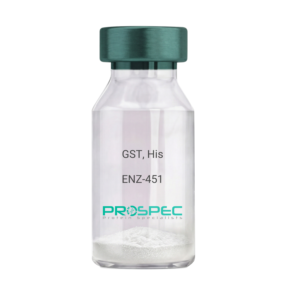
GST, HisDescription:
Glutathione S-Transferase Recombinant, His Tag
Glutathione S-Transferase, GST, Glutathione S-transferase class-mu 28 kDa isozyme, GST 28, EC 2.5.1.18, Sj28GST, Sj28 antigen.
Product # :
ENZ-451Price :
Quantity :
Shipping Method :
Shipped with Ice Packs
More Info
- description
- source
- formulation
- purity
- biological activity
- More Info
Description
Recombinant Schistosoma japonicum GST full length protein contains a total of 244 amino acids (1-218 a.a.) expressed in E.coli, having a molecular mass of 28.3kDa. The GST protein is fused to a 20 amino acids His-Tag at N-terminus. The GST protein is purified by proprietary chromatographic techniques.
Source
Escherichia Coli.
Formulation
GST is supplied in PBS pH 7.4 & 10% glycerol.
Purity
Greater than 90% as determined by SDS-PAGE.
Biological Activity
>10 units/mg, & is defined as the amount of enzyme that conjugate 1.0 u mole of 1-chloro-2,4-dinitrobenzene (CDNB) with reduced glutathione per minute at pH-6.5 at 25C.
More Info
-
Introduction
Antioxidant enzyme Glutathione S- Transferase (GST) is thought to do the primary cellular defense mechanism against reactive oxygen species. GST reduces lipid hydroperoxides through its Se-independent glutathione peroxidase activity. The enzyme also detoxifies lipid peroxidation end products such as 4-hydroxynonenal (4-HNE).
The soluble GST is a 26 kDa protein which occurs as a dimer in all aerobic organisms. Each monomer has two domains, one that binds GSH and is an /-structure similar to thioredoxin and the other, all helical, that binds the hydrophobic substrate. The GST -fusion protein expression system is a widely used recombinant protein expression system that allows a peptide or a regulatory protein domain to be expressed as a fusion to the C-terminus of Schistosoma japonicum GST. Fusion proteins also possess GST -enzymatic activity and can undergo dimerization similar to in vivo. The fusion protein can be purified via GST -affinity column chromatography. In most cases, the desired peptides or domains are removed from GST by applying a specific protease that recognizes and cleaves the linker between the protein domain and GST. The technique has been widely used to generate different kinds of proteins for crystallization, molecular immunology studies, the production of vaccines and studies involving protein-protein and protein-DNA interactions. -
Synonyms
Glutathione S-Transferase, GST, Glutathione S-transferase class-mu 28 kDa isozyme, GST 28, EC 2.5.1.18, Sj28GST, Sj28 antigen.
-
Physical Appearance
Sterile Filtered clear solution.
-
Stability
Store at 4°C if entire vial will be used within 2-4 weeks. Store, frozen at -20°C for longer periods of time. For long term storage it is recommended to add a carrier protein (0.1% HSA or BSA).Avoid multiple freeze-thaw cycles.
-
Amino Acid Sequence
MGSSHHHHHH SSGLVPRGSH MSPILGYWKI KGLVQPTRLL LEYLEEKYEE HLYERDEGDK WRNKKFELGL EFPNLPYYID GDVKLTQSMAIIRYIADKHN MLGGCPKERA EISMLEGAVL DIRYGVSRIA YSKDFETLKV DFLSKLPEML KMFEDRLCHK TYLNGDHVTH PDFMLYDALDVVLYMDPMCL DAFPKLVCFK KRIEAIPQID KYLKSSKYIA WPLQGWQATF GGGDHPPKSD LVPR.
ProSpec's products are furnished for LABORATORY RESEARCH USE ONLY. The product may not be used as drugs, agricultural or pesticidal products, food additives or household chemicals.
- View Data Sheet
Name :
rGFPDescription:
Green Fluorescent Protein Recombinant
Green fluorescent protein, GFP.
Product # :
PRO-687Price :
Quantity :
Shipping Method :
Shipped with Ice Packs
More Info
- description
- source
- formulation
- purity
- More Info
Description
rGFP Aequorea victoria produced in E.Coli is a single, non-glycosylated polypeptide chain containing 238 amino acids (1-238 a.a.) and having a molecular mass of 26.8 kDa.rGFP is purified by proprietary chromatographic techniques.
Source
Escherichia Coli.
Formulation
The Green Fluorescent Protein solution (1mg/ml) contains 20mM Tris-HCl, pH-8 and 10% Glycerol.
Purity
Greater than 95.0% as determined by SDS-PAGE.
More Info
-
Introduction
rGFP, also known as Green Fluorescent Protein, is a protein produced by the jellyfish (Aequorea Victoria) that produces bioluminescence in the green zone of the noticeable spectrum. Green Fluorescent Protein is a useful and ubiquitous instrument for producing chimeric proteins, where it functions as a fluorescent protein tag. rGFP is expressed in most known cell types and is used as a noninvasive fluorescent marker in living cells and organisms. Green Fluorescent Protein permits a broad range of applications where it has functioned as a cell lineage tracer, reporter of gene expression, or as a measure of protein-protein interactions.
-
Synonyms
Green fluorescent protein, GFP.
-
Physical Appearance
Sterile filtered yellowish solution.
-
Stability
Store at 4°C if entire vial will be used within 2-4 weeks. Store, frozen at -20°C for longer periods of time. For long term storage it is recommended to add a carrier protein (0.1% HSA or BSA).Avoid multiple freeze-thaw cycles.
-
Amino Acid Sequence
MSKGEELFTG VVPILVELDG DVNGHKFSVS GEGEGDATYG KLTLKFICTT GKLPVPWPTL VTTFSYGVQC FSRYPDHMKQ HDFFKSAMPEGYVQERTIFF KDDGNYKTRA EVKFEGDTLV NRIELKGIDF KEDGNILGHK LEYNYNSHNV YIMADKQKNG IKVNFKIRHN IEDGSVQLAD
HYQQNTPIGD GPVLLPDNHY LSTQSALSKD PNEKRDHMVL LEFVTAAGIT HGMDELYK.
ProSpec's products are furnished for LABORATORY RESEARCH USE ONLY. The product may not be used as drugs, agricultural or pesticidal products, food additives or household chemicals.