Skip to content

High-quality research proteins.

  • 1-800-805-6531

  • Mon - Sat 8.00-18.00, Sun - Closed

  • info@prospecbio.com

  • PO Box 6591 East Brunswick NJ 08816

  • Products
    • Cytokines
    • Growth factors
    • Chemokines
    • Cd antigens
    • Neurotrophins
    • Hormones
    • Enzymes
    • Viral antigens
    • Recombinant proteins
    • Natural proteins
    • Monoclonal antibodies
    • Polyclonal antibodies
    • Thermal Packaging
  • About
    • Company
    • Founder
    • Contribution
  • Customers
    • Eastern Asia
    • Eastern Europe
    • North America
    • South America
    • South East Asia
    • Southern Asia
    • Western Europe
  • Ordering
    • Ordering Method
    • Ordering - Credit Card
    • Ordering - PO
    • Shipping Fees
    • FAQs
  • Services
    • DNA Cloning
    • Protein Expression
    • Monoclonal Antibody
    • Fermentation
    • Technology License
  • Contact us
Log in

Country/region

  • USD $ | Albania
  • USD $ | Andorra
  • USD $ | Argentina
  • USD $ | Armenia
  • USD $ | Australia
  • USD $ | Austria
  • USD $ | Azerbaijan
  • USD $ | Belarus
  • USD $ | Belgium
  • USD $ | Bolivia
  • USD $ | Brazil
  • USD $ | Bulgaria
  • USD $ | Cambodia
  • USD $ | Canada
  • USD $ | Chile
  • USD $ | China
  • USD $ | Colombia
  • USD $ | Costa Rica
  • USD $ | Croatia
  • USD $ | Cyprus
  • USD $ | Czechia
  • USD $ | Denmark
  • USD $ | Estonia
  • USD $ | Finland
  • USD $ | France
  • USD $ | Georgia
  • USD $ | Germany
  • USD $ | Greece
  • USD $ | Hong Kong SAR
  • USD $ | Hungary
  • USD $ | Iceland
  • USD $ | India
  • USD $ | Ireland
  • USD $ | Israel
  • USD $ | Italy
  • USD $ | Japan
  • USD $ | Kazakhstan
  • USD $ | Latvia
  • USD $ | Liechtenstein
  • USD $ | Lithuania
  • USD $ | Madagascar
  • USD $ | Malta
  • USD $ | Mexico
  • USD $ | Moldova
  • USD $ | Monaco
  • USD $ | Netherlands
  • USD $ | New Zealand
  • USD $ | North Macedonia
  • USD $ | Norway
  • USD $ | Panama
  • USD $ | Paraguay
  • USD $ | Peru
  • USD $ | Philippines
  • USD $ | Poland
  • USD $ | Portugal
  • USD $ | Romania
  • USD $ | Singapore
  • USD $ | Slovakia
  • USD $ | Slovenia
  • USD $ | South Africa
  • USD $ | South Korea
  • USD $ | Spain
  • USD $ | Sri Lanka
  • USD $ | Sweden
  • USD $ | Switzerland
  • USD $ | Taiwan
  • USD $ | Thailand
  • USD $ | Türkiye
  • USD $ | Ukraine
  • USD $ | United Kingdom
  • USD $ | United States
  • USD $ | Uruguay
  • USD $ | Vatican City
  • USD $ | Venezuela
  • USD $ | Vietnam
  • Facebook
  • Instagram
logoProSpec - Recombinant Protein Manufacturer for academic and R&D industry
Become a distributor
Account Log in
Wishlist
Cart Cart(0)
  • Products
    +
    • Cytokines
    • Growth factors
    • Chemokines
    • Cd antigens
    • Neurotrophins
    • Hormones
    • Enzymes
    • Viral antigens
    • Recombinant proteins
    • Natural proteins
    • Monoclonal antibodies
    • Polyclonal antibodies
    • Thermal Packaging
  • About
    +
    • Company
    • Founder
    • Contribution
  • Customers
    +
    • Eastern Asia
    • Eastern Europe
    • North America
    • South America
    • South East Asia
    • Southern Asia
    • Western Europe
  • Ordering
    +
    • Ordering Method
    • Ordering - Credit Card
    • Ordering - PO
    • Shipping Fees
    • FAQs
  • Services
    +
    • DNA Cloning
    • Protein Expression
    • Monoclonal Antibody
    • Fermentation
    • Technology License
  • Contact us

Item added to your cart

View cart
View All
  • Cytokines
  • Growth factors
  • Chemokines
  • Cd antigens
  • Neurotrophins
  • Hormones
  • Enzymes
  • Viral antigens
  • Recombinant proteins
  • Natural proteins
  • Monoclonal antibodies
  • Polyclonal antibodies
  • Thermal Packaging
  • Tumor Necrosis Factor

    Tumor Necrosis Factor

    View All
  • 4-1BB

    4-1BB

    View All
  • Adiponectin

    Adiponectin

    View All
  • AIF1

    AIF1

    View All
  • Other Cytokines

    Other Cytokines

    View All
  • AITRL

    AITRL

    View All
  • Angiopoietin

    Angiopoietin

    View All
  • Apolipoprotein

    Apolipoprotein

    View All
  • B-Cell Activating Factor

    B-Cell Activating Factor

    View All
  • Beta Defensin

    Beta Defensin

    View All
  • Bone Morphogenetic Protein

    Bone Morphogenetic Protein

    View All
  • B type Natriuretic Peptide

    B type Natriuretic Peptide

    View All
  • BST

    BST

    View All
  • Betacellulin

    Betacellulin

    View All
  • Cardiotrophin

    Cardiotrophin

    View All
  • Activin

    Activin

    View All
  • Fibroblast Growth Factor

    Fibroblast Growth Factor

    View All
  • Other Growth Factors

    Other Growth Factors

    View All
  • CSF

    CSF

    View All
  • CTGF

    CTGF

    View All
  • IGFBP

    IGFBP

    View All
  • VEGF Protein

    VEGF Protein

    View All
  • EGF

    EGF

    View All
  • Epigen

    Epigen

    View All
  • Erythropoietin

    Erythropoietin

    View All
  • Insulin-Like Growth Factor

    Insulin-Like Growth Factor

    View All
  • Growth Hormone

    Growth Hormone

    View All
  • HDGF

    HDGF

    View All
  • Hepatocyte Growth Factor

    Hepatocyte Growth Factor

    View All
  • Insulin

    Insulin

    View All
  • BCA-1/ BLC (CXCL13)

    BCA-1/ BLC (CXCL13)

    View All
  • BRAK (CXCL14)

    BRAK (CXCL14)

    View All
  • C-10 (CCL6)

    C-10 (CCL6)

    View All
  • Other Chemokines

    Other Chemokines

    View All
  • MEC (CCL28)

    MEC (CCL28)

    View All
  • LD78-beta (CCL3L1)

    LD78-beta (CCL3L1)

    View All
  • CTACK (CCL27)

    CTACK (CCL27)

    View All
  • CXCL16

    CXCL16

    View All
  • CXCL17

    CXCL17

    View All
  • Platelet Factor-4 (CXCL4)

    Platelet Factor-4 (CXCL4)

    View All
  • ENA-78 (CXCL5)

    ENA-78 (CXCL5)

    View All
  • Eotaxin (CCL11,24,26)

    Eotaxin (CCL11,24,26)

    View All
  • Exodus-2 (CCL21)

    Exodus-2 (CCL21)

    View All
  • Fractalkine (CX3CL1)

    Fractalkine (CX3CL1)

    View All
  • CXCL6

    CXCL6

    View All
  • Other CD Antigens

    Other CD Antigens

    View All
  • CD14

    CD14

    View All
  • CD164

    CD164

    View All
  • CD1

    CD1

    View All
  • CD2

    CD2

    View All
  • CD200

    CD200

    View All
  • CD207

    CD207

    View All
  • CD226

    CD226

    View All
  • CD23

    CD23

    View All
  • CD244

    CD244

    View All
  • CD247

    CD247

    View All
  • CD27

    CD27

    View All
  • CD274

    CD274

    View All
  • CD300

    CD300

    View All
  • CD33

    CD33

    View All
  • Other Neurotrophins

    Other Neurotrophins

    View All
  • Beta-NGF

    Beta-NGF

    View All
  • BDNF

    BDNF

    View All
  • CDNF

    CDNF

    View All
  • CNTF

    CNTF

    View All
  • GDNF

    GDNF

    View All
  • Glia Maturation Factor

    Glia Maturation Factor

    View All
  • MANF

    MANF

    View All
  • Midkine

    Midkine

    View All
  • Neuroglobin

    Neuroglobin

    View All
  • Neurotrophic factors

    Neurotrophic factors

    View All
  • Neuregulin

    Neuregulin

    View All
  • Neuropilin

    Neuropilin

    View All
  • Pigment Epithelium-Derived Factor

    Pigment Epithelium-Derived Factor

    View All
  • Pleiotrophin

    Pleiotrophin

    View All
  • Peptide Hormones

    Peptide Hormones

    View All
  • Other Hormones

    Other Hormones

    View All
  • HCG

    HCG

    View All
  • Endothelin

    Endothelin

    View All
  • Exendin

    Exendin

    View All
  • FSH

    FSH

    View All
  • Glucagon

    Glucagon

    View All
  • GHRP

    GHRP

    View All
  • GLP

    GLP

    View All
  • Thyrostimulin

    Thyrostimulin

    View All
  • Inhibin A

    Inhibin A

    View All
  • LHRH

    LHRH

    View All
  • Melanotan

    Melanotan

    View All
  • Procalcitonin

    Procalcitonin

    View All
  • PTH

    PTH

    View All
  • Peroxiredoxin

    Peroxiredoxin

    View All
  • Peptidase

    Peptidase

    View All
  • Synthetase

    Synthetase

    View All
  • Transferase

    Transferase

    View All
  • Hydrolase

    Hydrolase

    View All
  • Dehydrogenase

    Dehydrogenase

    View All
  • ACE2

    ACE2

    View All
  • Esterase

    Esterase

    View All
  • Protein Kinases

    Protein Kinases

    View All
  • Other Enzymes

    Other Enzymes

    View All
  • Phosphatase

    Phosphatase

    View All
  • Deaminase

    Deaminase

    View All
  • Oxygenase

    Oxygenase

    View All
  • Adenylate Kinase

    Adenylate Kinase

    View All
  • Lyase

    Lyase

    View All
  • Chagas

    Chagas

    View All
  • SARS

    SARS

    View All
  • Influenza

    Influenza

    View All
  • ASFV

    ASFV

    View All
  • Astrovirus

    Astrovirus

    View All
  • Borrelia

    Borrelia

    View All
  • Helicobacter Pylori

    Helicobacter Pylori

    View All
  • Chikungunya

    Chikungunya

    View All
  • Chlamydia

    Chlamydia

    View All
  • Cytomegalo

    Cytomegalo

    View All
  • Dengue

    Dengue

    View All
  • EBV

    EBV

    View All
  • Ebola

    Ebola

    View All
  • Feline Leukemia Virus

    Feline Leukemia Virus

    View All
  • Hepatitis A

    Hepatitis A

    View All
  • Other

    Other

    View All
  • Actin

    Actin

    View All
  • ADAM

    ADAM

    View All
  • Complement Component

    Complement Component

    View All
  • Ag85

    Ag85

    View All
  • Eukaryotic Translation Initiation Factor

    Eukaryotic Translation Initiation Factor

    View All
  • Anterior Gradient Protein

    Anterior Gradient Protein

    View All
  • Heat Shock Protein

    Heat Shock Protein

    View All
  • Alpha-2-HS-Glycoprotein

    Alpha-2-HS-Glycoprotein

    View All
  • Hemoglobin

    Hemoglobin

    View All
  • Allergy

    Allergy

    View All
  • Anaplasma

    Anaplasma

    View All
  • Angiogenin

    Angiogenin

    View All
  • Ankyrin Repeat Domain

    Ankyrin Repeat Domain

    View All
  • Annexin

    Annexin

    View All
  • Other Natural Proteins

    Other Natural Proteins

    View All
  • Aprotinin

    Aprotinin

    View All
  • Transferrin

    Transferrin

    View All
  • Avidin

    Avidin

    View All
  • Anti Coagulation Factors

    Anti Coagulation Factors

    View All
  • Natural Albumin

    Natural Albumin

    View All
  • Natural Coagulation Factors

    Natural Coagulation Factors

    View All
  • Fibronectin

    Fibronectin

    View All
  • Thrombin

    Thrombin

    View All
  • Anti Human Enzyme

    Anti Human Enzyme

    View All
  • Other Monoclonal Antibodies

    Other Monoclonal Antibodies

    View All
  • Anti Human Cytokine

    Anti Human Cytokine

    View All
  • Anti Human Heat Shock Protein

    Anti Human Heat Shock Protein

    View All
  • Anti Mouse Lymphocyte

    Anti Mouse Lymphocyte

    View All
  • Anti Human Chemokine

    Anti Human Chemokine

    View All
  • Anti Human Lymphocyte

    Anti Human Lymphocyte

    View All
  • Anti Viral Monoclonal

    Anti Viral Monoclonal

    View All
  • Anti Mouse Cytokine

    Anti Mouse Cytokine

    View All
  • Anti-GST Monoclonal

    Anti-GST Monoclonal

    View All
  • Other Polyclonal Antibodies

    Other Polyclonal Antibodies

    View All
  • Anti Viral

    Anti Viral

    View All
  • Anti-GST Monoclonal

    Anti-GST Monoclonal

    View All
Back

Search results

1000 results found for “Dynactin”

Name

Description

Product #

Price

Quantity

Shipping Method

  • View Data Sheet

    Name :

    PON1 Human, HEK

    Description:

    Paraoxonase-1 Human Recombinant, HEK

    Serum paraoxonase/arylesterase 1, Serum aryldialkylphosphatase 1, Aromatic esterase 1, A-esterase 1 , Serum aryldialkylphosphatase 1, paraoxonase 1, K-45, ESA, PON, MVCD5

    Product # :

    ENZ-1154

    Price :

    Quantity :

    Shipping Method :

    Ice Icon

    Shipped with Ice Packs

    Add To Cart

    More Info

    • description
    • source
    • formulation
    • purity
    • biological activity
    • More Info

    Description

    PON1 Human Recombinant produced in HEK cells is a single, glycosylated, polypeptide chain (16-355 a.a) containing a total of 346 amino acids, having a molecular mass of 39.0kDa. PON1 is fused to a 6 amino acid His-tag at C-terminus,and is purified by proprietary chromatographic techniques.

    Source

    HEK293 Cells.

    Formulation

    The PON1 solution (0.25mg/ml) contains 20% Glycerol and Phosphate-Buffered Saline (pH 7.4).

    Purity

    Greater than 90.0% as determined by SDS-PAGE.

    Biological Activity

    Specific activity is > 2,500 pmol/min/ug. Defined by the amount of enzyme that  hydrolyzes 1pmole of pnitrophenyl acetate to p-nitrophenol per minute at pH 7.5 at 37˚C.

    More Info

    • Introduction

      Paraoxonase-1 or PON1 is part of the paraoxonase group of proteins. PON1 is an enzyme, responsible to the toxic metabolites of a different of organophosphorus insecticides hydrolyzation. Furthermore, PON1 is a dominant anti-atherosclerotic part of HDL. The enzyme needs PPAR-gamma for activation, leading to synthesis and release of paraoxonase 1 from the liver tissue, resulting in atherosclerosis reduction. PON1 has many qualities for atheroprotective through inflammatory lipid peroxides metabolism. This enzyme can hydrolyze a large number of substrates, for example cyclic carbonates, lactones, nerve gases etc.

    • Synonyms

      Serum paraoxonase/arylesterase 1, Serum aryldialkylphosphatase 1, Aromatic esterase 1, A-esterase 1 , Serum aryldialkylphosphatase 1, paraoxonase 1, K-45, ESA, PON, MVCD5

    • Physical Appearance

      Sterile Filtered colorless solution.

    • Stability

      Store at 4°C if entire vial will be used within 2-4 weeks. Store, frozen at -20°C for longer periods of time. For long term storage it is recommended to add a carrier protein (0.1% HSA or BSA).Avoid multiple freeze-thaw cycles.

    • Amino Acid Sequence

      LFRNHQSSYQ TRLNALREVQ PVELPNCNLV KGIETGSEDL EILPNGLAFI SSGLKYPGIK SFNPNSPGKI LLMDLNEEDP TVLELGITGS KFDVSSFNPH GISTFTDEDN AMYLLVVNHP DAKSTVELFK FQEEEKSLLH LKTIRHKLLP NLNDIVAVGP EHFYGTNDHY FLDPYLQSWE MYLGLAWSYV VYYSPSEVRV VAEGFDFANG INISPDGKYV YIAELLAHKI HVYEKHANWT LTPLKSLDFN TLVDNISVDP ETGDLWVGCH PNGMKIFFYD SENPPASEVL RIQNILTEEP KVTQVYAENG TVLQGSTVAS VYKGKLLIGT VFHKALYCEL HHHHHH

    ProSpec's products are furnished for LABORATORY RESEARCH USE ONLY. The product may not be used as drugs, agricultural or pesticidal products, food additives or household chemicals.

    Pon1 Enzyme
  • View Data Sheet

    Name :

    HIV-2 gp32, Biotin

    Description:

    HIV-2 gp32 Recombinant, Biotin Labeled

    Product # :

    HIV-135

    Price :

    Quantity :

    Shipping Method :

    Ice Icon

    Shipped with Ice Packs

    Add To Cart

    More Info

    • description
    • source
    • formulation
    • purity
    • More Info

    Description

    HIV-2 gp32 Biotin Labeled recombinant- contains the full-length sequence of HIV-2 envelope immunodominant regions gp32 having a Mw of 32kDa and fused to a beta-galactosidase at N-terminus.

    Source

    Escherichia Coli.

    Formulation

    0.01M Na2CO3, 10mM EDTA, 14mM beta-ME and 0.02% Sarcosyl.

    Purity

    Greater than 95.0% as determined by SDS-PAGE.

    More Info

    • Introduction

      HIV-1 and HIV-2 appear to package their RNA differently. HIV-1 binds to any appropriate RNA whereas HIV-2 preferentially binds to mRNA which creates the Gag protein itself. This means that HIV-1 is better able to mutate. HIV-2 is transmitted in the same ways as HIV-1: Through exposure to bodily fluids such as blood, semen, tears and vaginal fluids.
      Immunodeficiency develops more slowly with HIV-2.
      HIV-2 is less infectious in the early stages of the virus than with HIV-1.
      The infectiousness of HIV-2 increases as the virus progresses.
      Major differences include reduced pathogenicity of HIV-2 relative to HIV-1, enhanced immune control of HIV-2 infection and often some degree of CD4-independence. Despite considerable sequence and phenotypic differences between HIV-1 and 2 envelopes, structurally they are quite similar. Both membrane-anchored proteins eventually form the 6-helix bundles from the N-terminal and C-terminal regions of the ectodomain, which is common to many viral and cellular fusion proteins and which seems to drive fusion.
      HIV-1 gp41 helical regions can form more stable 6-helix bundles than HIV-2 gp41 helical regions however HIV-2 fusion occurs at a lower threshold temperature (25°C), does not require Ca2+ in the medium, is insensitive to treatment of target cells with cytochalasin B, and is not affected by target membrane glycosphingolipid composition.

    • Physical Appearance

      Sterile filtered colorless clear solution.

    • Stability

      HIV-2 gp-32 although stable at room temperature for 3 weeks, should be stored at 4°C.

    ProSpec's products are furnished for LABORATORY RESEARCH USE ONLY. The product may not be used as drugs, agricultural or pesticidal products, food additives or household chemicals.

    Hiv 2 Gp 32 Biotin
  • View Data Sheet

    Name :

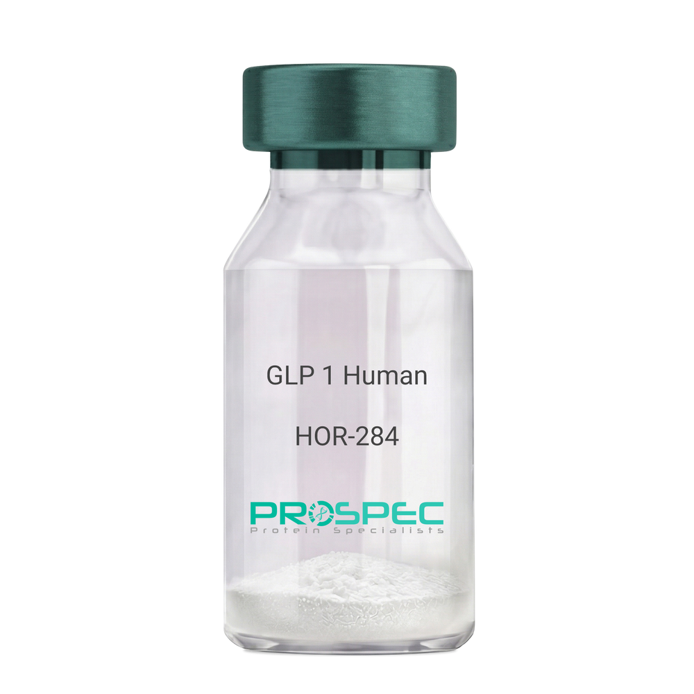
    GLP 1 Human

    Description:

    Human Glucagon Like Peptide-1

    GLP1, Glucagon Like Peptide-1, Incretin hormone.

    Product # :

    HOR-284

    Price :

    Quantity :

    Shipping Method :

    Room Temp Icon

    Shipped at Room temp

    Add To Cart

    More Info

    • description
    • formulation
    • purity
    • More Info

    Description

    Glucagon Like Peptide-1 is a single, non-glycosylated, polypeptide chain containing 30 amino acids and having a molecular mass of 3297.7 Dalton.The GLP-1 is purified by proprietary chromatographic techniques.

    Formulation

    The GLP-1 peptide was lyophilized with no additives.

    Purity

    Greater than 96.0% as determined by RP-HPLC.

    More Info

    • Introduction

      Glucagon-like peptide-1 (GLP-1) is derived from the transcription product of the proglucagon gene. The major source of GLP-1 in the body is the intestinal L cell that secretes GLP-1 as a guthormone. The biologically active forms of GLP-1 are: GLP-1-(7-37) and GLP-1-(7-36)NH2.
      GLP-1 secretion by L cells is dependent on the presence of nutrients in the lumen of the small intestine. The secretagogues (agents that causes or stimulates secretion) of this hormone include major nutrients like carbohydrate, proteinand lipid. Once in the circulation, GLP-1 has a half life of less than 2 minutes, due to rapid degradation by the enzyme dipeptidyl peptidase-4.
      GLP-1 possesses several physiological properties that make it a subject of intensive investigation as a potential treatment of diabetes mellitus. The known physiological functions of GLP-1 include: Increases insulin secretion from the pancreas in a glucose-dependent manner, decreases glucagon secretion from the pancreas, increases beta cells mass and insulin gene expression, inhibits acid secretion and gastric emptying in the stomach, decreases food intake by increasing satiety.

    • Synonyms

      GLP1, Glucagon Like Peptide-1, Incretin hormone.

    • Physical Appearance

      Sterile Filtered White lyophilized (freeze-dried) powder.

    • Stability

      Lyophilized Glucagon Like Peptide-1 although stable at room temperature for 3 weeks, should be stored desiccated below -18°C. Upon reconstitution GLP-1 should be stored at 4°C between 2-7 days and for future use below -18°C. For long term storage it is recommended to add a carrier protein (0.1% HSA or BSA).Please prevent freeze-thaw cycles.

    • Solubility

      It is recommended to reconstitute the lyophilized Glucagon Like Peptide-1 in sterile H2O at 0.5mg/ml, which can then be further diluted to other aqueous solutions.

    • Amino Acid Sequence

      H-His-Ala-Glu-Gly-Thr-Phe-Thr-Ser-Asp-Val-Ser-Ser-Tyr-Leu-Glu-Gly-Gln-Ala-Ala-Lys-Glu-Phe-Ile-Ala-Trp-Leu-Val-Lys-Gly-Arg-NH2.

    ProSpec's products are furnished for LABORATORY RESEARCH USE ONLY. The product may not be used as drugs, agricultural or pesticidal products, food additives or household chemicals.

    Glp 1 Human
  • View Data Sheet

    Name :

    EGF Long Human

    Description:

    Epidermal Growth Factor Long Human Recombinant

    Urogastrone, URG, EGF.

    Product # :

    CYT-798

    Price :

    Quantity :

    Shipping Method :

    Room Temp Icon

    Shipped at Room temp

    Add To Cart

    More Info

    • description
    • source
    • formulation
    • purity
    • biological activity
    • More Info

    Description

    Recombinant Human EGF Long produced in E.coli cells is a single non-glycosylated, polypeptide chain containing 106 amino acids and having a molecular mass of 12.3kDa. The EGF Long is purified by proprietary chromatographic techniques.

    Source

    Escherichia Coli.

    Formulation

    The EGF Long was lyophilized from a 0.2µm filtered concentrated solution in 10mM HCl.

    Purity

    Greater than 95.0% as determined by:
    (a) Analysis by RP-HPLC.
    (b) Analysis by SDS-PAGE.

    Biological Activity

    The ED50 as determined by a cell proliferation assay using murine Balb/c 3T3 cells is less than 1.0 ng/ml, corresponding to a specific activity of > 1.0 × 10⁶ IU/mg.

    More Info

    • Introduction

      Epidermal growth factor has a profound effect on the differentiation of specific cells in vivo and is a potent mitogenic factor for a variety of cultured cells of both ectodermal and mesodermal origin. The EGF precursor is believed to exist as a membrane-bound molecule which is proteolytically cleaved to generate the 53-amino acid peptide hormone that stimulates cells to divide. EGF stimulates the growth of various epidermal and epithelial tissues in vivo and in vitro and of some fibroblasts in cell culture. Long EGF is a recombinant analog of Human EGF developed as a replacement for use in therapeutic cell culture applications as a like-for-like supplement for Recombinant Human or native EGF. It includes the Human EGF amino acid sequence plus a 53 amino acid N-terminal extension peptide.

    • Synonyms

      Urogastrone, URG, EGF.

    • Physical Appearance

      Sterile Filtered White lyophilized (freeze-dried) powder.

    • Stability

      Lyophilized EGF Long although stable at room temperature for 3 weeks, should be stored desiccated below -18°C. Upon reconstitution EGF Long should be stored at 4°C between 2-7 days and for future use below -18°C.For long term storage it is recommended to add a carrier protein (0.1% HSA or BSA).Please prevent freeze-thaw cycles.

    • Solubility

      It is recommended to reconstitute the lyophilized EGF Long in sterile 100mM AcOH (acetic Acid) not less than 100µg/ml, which can then be further diluted to other aqueous solutions.

    • Amino Acid Sequence

      MFPAMPLSSL FANAVLRAQH LHQLAADTYK EFERAYIPEG QRYSIQVNFA HYGNSDSECP LSHDGYCLHD GVCMYIEALD KYACNCVVGY IGERCQYRDL KWWELR

    • Background

      What is the molecular weight/Mw of EGF Protein?
      EGF Protein has a total Mw of 12.3kDa.

      What is the source or expression system of EGF Protein?
      Escherichia Coli.

      What is the Purity of EGF Protein?
      EGF Protein is >95% pure as determined by SDS-PAGE.

      What is the Biological Activity of EGF Protein?
      The ED50 as determined by a cell proliferation assay using murine Balb/c 3T3 cells is less than 1.0 ng/ml, corresponding to a specific activity of > 1.0 × 10⁶ IU/mg.

      What is the amino acid sequence of EGF Protein?
      MFPAMPLSSL FANAVLRAQH LHQLAADTYK EFERAYIPEG QRYSIQVNFA HYGNSDSECP LSHDGYCLHD GVCMYIEALD KYACNCVVGY IGERCQYRDL KWWELR

      What applications can EGF Protein be used in?
      EGF Protein can probably be used in western blot, ELISA and Lateral Flow.

      What is the endotoxin level for EGF Protein?
      The endotoxin level is minimal, EGF Protein was purified using conventional chromatography techniques.

    ProSpec's products are furnished for LABORATORY RESEARCH USE ONLY. The product may not be used as drugs, agricultural or pesticidal products, food additives or household chemicals.

    Egf Long Human
  • View Data Sheet

    Name :

    TINAGL1 Human, Sf9

    Description:

    Tubulointerstitial Nephritis Antigen Like 1 Human Recombinant, Sf9

    Tubulointerstitial nephritis antigen-like isoform 1, TINAGL1, Tubulointerstitial nephritis antigen-like, Glucocorticoid-inducible protein 5, Oxidized LDL-responsive gene 2 protein, OLRG-2, Tubulointerstitial nephritis antigen-related protein, TIN Ag-related protein, TIN-Ag-RP, GIS5, LCN7, OLRG2, TINAGL, PP6614, PSEC0088, UNQ204/PRO230.

    Product # :

    PRO-2422

    Price :

    Quantity :

    Shipping Method :

    Ice Icon

    Shipped with Ice Packs

    Add To Cart

    More Info

    • description
    • source
    • formulation
    • purity
    • More Info

    Description

    TINAGL1 Human Recombinant produced in Sf9 Baculovirus cells is a single, glycosylated polypeptide chain containing 455 amino acids (22-467a.a.) and having a molecular mass of 51.2kDa (Molecular size on SDS-PAGE will appear at approximately 50-70kDa). TINAGL1 is expressed with a 6 amino acid His tag at C-Terminus and purified by proprietary chromatographic techniques.

    Source

    Sf9, Baculovirus cells.

    Formulation

    TINAGL1 protein solution (0.25mg/ml) contains Phosphate Buffered Saline (pH 7.4), 30% glycerol and 1mM DTT.

    Purity

    Greater than 90.0% as determined by SDS-PAGE.

    More Info

    • Introduction

      Tubulointerstitial Nephritis Antigen Like 1 also known as TINAGL1 is similar in sequence to tubulointerstitial nephritis antigen, a secreted glycoprotein which is identified by antibodies in several types of immune-related tubulointerstitial nephritis. Three transcript variants encoding different isoforms are known for TINAGL1.

    • Synonyms

      Tubulointerstitial nephritis antigen-like isoform 1, TINAGL1, Tubulointerstitial nephritis antigen-like, Glucocorticoid-inducible protein 5, Oxidized LDL-responsive gene 2 protein, OLRG-2, Tubulointerstitial nephritis antigen-related protein, TIN Ag-related protein, TIN-Ag-RP, GIS5, LCN7, OLRG2, TINAGL, PP6614, PSEC0088, UNQ204/PRO230.

    • Physical Appearance

      Sterile filtered colorless solution.

    • Stability

      Store at 4°C if entire vial will be used within 2-4 weeks. Store, frozen at -20°C for longer periods of time. For long term storage it is recommended to add a carrier protein (0.1% HSA or BSA).Avoid multiple freeze-thaw cycles.

    • Amino Acid Sequence

      ADLAQQGRGR RELAPGLHLR GIRDAGGRYC QEQDLCCRGR ADDCALPYLG AICYCDLFCN RTVSDCCPDF WDFCLGVPPP FPPIQGCMHG GRIYPVLGTY WDNCNRCTCQ ENRQWQCDQE PCLVDPDMIK AINQGNYGWQ AGNHSAFWGM TLDEGIRYRL GTIRPSSSVM NMHEIYTVLN PGEVLPTAFE ASEKWPNLIH EPLDQGNCAG SWAFSTAAVA SDRVSIHSLG HMTPVLSPQN LLSCDTHQQQ GCRGGRLDGA WWFLRRRGVV SDHCYPFSGR ERDEAGPAPP CMMHSRAMGR GKRQATAHCP NSYVNNNDIY QVTPVYRLGS NDKEIMKELM ENGPVQALME VHEDFFLYKG GIYSHTPVSL GRPERYRRHG THSVKITGWG EETLPDGRTL KYWTAANSWG PAWGERGHFR IVRGVNECDI ESFVLGVWGR VGMEDMGHHH HHHHH.

    ProSpec's products are furnished for LABORATORY RESEARCH USE ONLY. The product may not be used as drugs, agricultural or pesticidal products, food additives or household chemicals.

    Tinagl1 Human Sf9
  • View Data Sheet

    Name :

    Leptin Human, His

    Description:

    Leptin Human Recombinant, His Tag

    OB Protein, Obesity Protein, OBS, Obesity factor.

    Product # :

    CYT-287

    Price :

    Quantity :

    Shipping Method :

    Room Temp Icon

    Shipped at Room temp

    Add To Cart

    More Info

    • description
    • source
    • formulation
    • purity
    • More Info

    Description

    Leptin Human Recombinant produced in E.Coli is a single, non-glycosylated, polypeptide chain containing amino acids 48-167 and having a total molecular mass of 19 kDa including the 4 kDa His tag.The Leptin is purified by proprietary chromatographic techniques.

    Source

    Escherichia Coli.

    Formulation

    The protein was lyophilized from a concentrated (1mg/ml) solution with 1X PBS, 0.1% SDS and 1mM DTT.

    Purity

    Greater than 90.0% as determined by SDS-PAGE.

    More Info

    • Introduction

      Leptin is a protein hormone with important effects in regulating body weight, metabolism and reproductive function. The protein is approximately~16 kDa in mass and encoded by the obese (ob)gene. leptin is expressed predominantly by adipocytes, which fits with the idea that body weight is sensed as the total mass of fat in the body. Smaller amounts of leptin are also secreted by cellsin the epithelium of the stomach and in the placenta. Leptin receptors are highly expressed in areas of the hypothalamus known to be important in regulating body weight, as well as in T lymphocytes and vascular endothelial cells.

    • Synonyms

      OB Protein, Obesity Protein, OBS, Obesity factor.

    • Physical Appearance

      Sterile Filtered White lyophilized (freeze-dried) powder.

    • Stability

      Lyophilized Leptin although stable at room temperature, should be stored desiccated below 0°C. Reconstituted Leptin is best stored refrigerated at 4°C.Please avoid freeze-thaw cycles.

    • Solubility

      The lyophilized Leptin is very soluble in water and most aqueous buffers below and above the isoelectric point.

    ProSpec's products are furnished for LABORATORY RESEARCH USE ONLY. The product may not be used as drugs, agricultural or pesticidal products, food additives or household chemicals.

    Leptin Human His
  • View Data Sheet

    Name :

    Leptin tA Human

    Description:

    Leptin Antagonist Triple Mutant Human Recombinant

    Product # :

    CYT-352

    Price :

    Quantity :

    Shipping Method :

    Room Temp Icon

    Shipped at Room temp

    Add To Cart

    More Info

    • description
    • source
    • formulation
    • purity
    • biological activity
    • More Info

    Description

    Leptin Antagonist Triple Mutant Human Recombinant is a single non-glycosilated polypeptide chain containing 146 amino and additional Ala at N-terminus acids and having a molecular weight of 16 kDa, Leptin was mutated, resulting in L39A/D40A/F41A. Leptin Antagonist Triple Mutant Human Recombinant was purified by proprietary chromatographic techniques.

    Source

    Escherichia coli.

    Formulation

    The protein was lyophilized from a concentrated (1mg/ml) solution with 0.0045mM NaHCO3.

    Purity

    Greater than 98.0% as determined by:
    (a) Gel filtration analysis.
    (b) Analysis by SDS-PAGE.

    Biological Activity

    ProSpec’s Leptin triple antagonist is capable of inhibiting leptin-induced proliferation of BAF/3 cells stably transfected with the long form of human leptin receptor. It also inhibits various leptin effects in several in vitro bioassays.

    More Info

    • Physical Appearance

      White lyophilized (freeze-dried) powder.

    • Stability

      Lyophilized Leptin Antagonist Triple Mutant Human Recombinant although stable at room temperature for several weeks, should be stored desiccated below -18°C. Upon reconstitution at > 0.1 mg/ml and up to 2 mg/ml and filter sterilization Leptin mutant can be stored at 4°C or even room temperature for several weeks making it suitable for long term infusion studies using osmotic pumps. At lower concentration addition of a carrier protein (0.1% HSA or BSA) is suggested. Please prevent freeze-thaw cycles.

    • Solubility

      It is recommended to reconstitute the lyophilized Leptin Antagonist Triple Mutant Human Recombinant in sterile 0.4% NaHCO3, not less than 100µg/ml, which can then be further diluted to other aqueous solutions.

    • Amino Acid Sequence

      The sequence of the first five N-terminal amino acids was determined and was found to be Ala-Val-Pro-Ile-Gln.

    • Protein content

      Protein quantitation was carried out by UV spectroscopy at 280 nm using the absorbency value of 0.88 as the extinction coefficient for a 0.1% (1mg/ml) solution at pH 8.0. This value is calculated by the PC GENE computer analysis program of protein sequences (IntelliGenetics).

    ProSpec's products are furnished for LABORATORY RESEARCH USE ONLY. The product may not be used as drugs, agricultural or pesticidal products, food additives or household chemicals.

    Leptin Ta Human
  • View Data Sheet

    Name :

    Leptin Human, PEG

    Description:

    Leptin Human Recombinant, PEG

    OB Protein, Obesity Protein, OBS, Obesity factor.

    Product # :

    CYT-1108

    Price :

    Quantity :

    Shipping Method :

    Room Temp Icon

    Shipped at Room temp

    Add To Cart

    More Info

    • description
    • source
    • formulation
    • purity
    • biological activity
    • More Info

    Description

    Pegylated Leptin Human Recombinant produced in E.Coli is a single non-glycosilated polypeptide chain containing 146 amino acids, an additional Ala at N-terminus and one molecule of PEG 20 kDa at its N-terminus acids and having a molecular weight of 35.6kDa. However due to enlarged hydrodymanic volume it runs on the SDS-PAGE as 48 kDa protein and in gel-filtration on Superdex 200 as over 100 kDa protein. Pegylated Leptin Human Recombinant was purified by proprietary chromatographic techniques.

    Source

    Escherichia Coli.

    Formulation

    Lyophilized from a concentrated (1mg/ml) solution with 0.0045mM NaHCO3.

    Purity

    Greater than 98.0% as determined by:
    (a) Gel filtration analysis.
    (b) Analysis by SDS-PAGE.

    Biological Activity

    Biological Activity is < than 0.1% as determined by inducing proliferation of BAF/3 cells stably transfected with the long form of human leptin receptor. It’s in vitro activity is 5-7 fold lower than the non-pegylated recombinant human leptin but in vivo it has profound weight reducing effect, resulting mainly from reduced food intake.

    More Info

    • Introduction

      Leptin takes an important part in the regulation of energy balance and body weight control.After entering the circulation, Leptin binds LEPRwhich results in the activation of several major signalling pathways. In the hypothalamus Leptin acts as an appetite-regulating factor that induces a decrease in food intake and an increase in energy consumption and also regulates bone mass and secretion of hypothalamo-pituitary-adrenal hormones. In the periphery, increases basal metabolism, regulates pancreatic beta-cell function and insulin secretion and affects innate and adaptive immunity.

    • Synonyms

      OB Protein, Obesity Protein, OBS, Obesity factor.

    • Physical Appearance

      Sterile Filtered White lyophilized (freeze-dried) powder.

    • Stability

      Lyophilized Pegylated leptin although stable at room temperature for 3 weeks, should be stored desiccated below -18°C. Upon reconstitution leptin N82K should be stored at 4°C between 2-7 days and for future use below -18°C. For long term storage it is recommended to add a carrier protein (0.1% HSA or BSA). Please prevent freeze-thaw cycles.

    • Solubility

      It is recommended to reconstitute the lyophilized Pegylated leptin in sterile water or 0.4% NaHCO3 adjusted to pH 8-9, not less than 100µg/ml, which can then be further diluted to other aqueous solutions.

    • Protein content

      Protein quantitation was carried out by UV spectroscopy at 280 nm using the absorbency value of 0.87 as the extinction coefficient for a 0.1% (1mg/ml) solution at pH 8.0. This value is calculated by the PC GENE computer analysis program of protein sequences (IntelliGenetics).

    ProSpec's products are furnished for LABORATORY RESEARCH USE ONLY. The product may not be used as drugs, agricultural or pesticidal products, food additives or household chemicals.

    Leptin Mutant
  • View Data Sheet

    Name :

    Angiostatin K1-4

    Description:

    Angiostatin Kringles 1-4 Human

    Product # :

    PRO-604

    Price :

    Quantity :

    Shipping Method :

    Room Temp Icon

    Shipped at Room temp

    Add To Cart

    More Info

    • description
    • source
    • formulation
    • purity
    • biological activity
    • More Info

    Description

    Human Angiostatin kringles 1-4 is produced from Human Fluid is a glycosylated polypeptide chain which migrates as a doublet 50 kDa on SDS-PAGE. The Ang K1-4 is purified by proprietary chromatographic techniques.

    Source

    Human Fluid.

    Formulation

    Lyophilized from a (1mg/ml) solution in containing 20mM Hepes buffer pH-8.2 & 20mM NaCl.

    Purity

    Greater than 98.0% as determined by SDS-PAGE.

    Biological Activity

    Human Angiostatin Kringles 1-4 significantly inhibits basic-FGF induced endothelial cell proliferation and migration at concentration ranging from 300nM-1.0 uM.

    More Info

    • Introduction

      There are several proteolytic fragments or specific domains of proteins that act as inhibitors of angiogenesis. These include fragments of plasminogen such as Angiostatin protein kringles 1-4 and kringles 1-5, Endostatin, Restin, PEX, the N-terminal fragment of prolactin, and the Nterminally truncated platelet factor. Angiostatin is a proteolytic protein fragment of plasminogen that is comprised of the first 4 kringle regions. Angiostatin k1-4 prevents the growth of endothelial cells, and its systemic administration inhibits the growth of primary carcinomas in mice. Angiostatin Kringles 1-3 segment has a larger inhibitory activity than the Angiostatin kringles 1-4 fragment. The protease-activated angiostatin kringles 1-5 is the most potent plasminogen fragment with over 50 times larger endothelial cell specific inhibitory activity. Angiostatin kringles 1-5 systemic administration inhibits growth of fibrosarcoma and significantly reduces neovascularization.
      Angiostatin is an angiogenesis inhibitor in mouse serum and urine. Angiostatin is a 38 kDa protein fragment of the plasminogen composed of the 1st 4 kringle domains of plasminogen. Angiostatin K1-4 is also named plasminogen kringles 1-4 and PK1-4.
      Angiostatin protein is manufactured by the protelytic cleavage of plasminogen by a serine protease from several prostate carcinoma cell lines. The manufacturing of angiostatin by pancreatic cancer cells can be inhibited by TGF-beta 1 along with plasminogen activator inhibitor type-1 (PAI1).

    • Physical Appearance

      Sterile Filtered lyophilized powder.

    • Stability

      Lyophilized Angiostatin Kringles 1-4 although stable at room temperature for 3 weeks, should be stored desiccated below -18°C. Upon reconstitution Angiostatin Kringles1-4 should be stored at 4°C between 2-7 days and for future use below -18°C.For long term storage it is recommended to add a carrier protein (0.1% HSA or BSA).Please prevent freeze-thaw cycles.

    • Solubility

      It is recommended to reconstitute the lyophilized Angiostatin K1-4 in sterile 18MΩ-cm H2O not less than 100µg/ml, which can then be further diluted to other aqueous solutions.

    ProSpec's products are furnished for LABORATORY RESEARCH USE ONLY. The product may not be used as drugs, agricultural or pesticidal products, food additives or household chemicals.

    Angiostatin K1 4 Human
  • View Data Sheet

    Name :

    Visfatin Human, His

    Description:

    Visfatin Recombinant Human, His Tag

    PBEF, Pre-B cell colony-enhancing factor, Nicotinamide phosphoribosyltransferase NAmPRTase, Nampt, MGC117256, DKFZP666B131, 1110035O14Rik.

    Product # :

    CYT-563

    Price :

    Quantity :

    Shipping Method :

    Ice Icon

    Shipped with Ice Packs

    Add To Cart

    More Info

    • description
    • source
    • formulation
    • purity
    • More Info

    Description

    Visfatin Human Recombinant produced in E.Coli is a single, non-glycosylated, polypeptide chain containing 511 amino acids and having a molecular mass of 57 kDa. The recombinant human Visfatin is fused to His tag at N-Terminus.

    Source

    Escherichia Coli.

    Formulation

    Visfatin His tag protein contains 20mM Tris pH-8, 0.1mM DTT & 10% glycerol.

    Purity

    Greater than 95.0% as determined by(a) Analysis by RP-HPLC.
    (b) Analysis by SDS-PAGE.

    More Info

    • Introduction

      Excess adiposity is the most important risk in the development of insulin resistance and type 2 diabetes mellitus (T2DM). Adipose tissue produces several proteins (adipocytokines) such as leptin, adiponectin, resistin, tumor necrosis factor-?, and IL-6, that modulate insulin sensitivity and appear to play an important role in the pathogenesis of insulin resistance, diabetes, dyslipidemia, inflammation, and atherosclerosis. However, the mechanisms by which fat tissue induces insulin resistance and the role of adipocytokines in the pathogenesis of T2DM have not been well established.
      Visfatin, also known as pre-B cell colony-enhancing factor (PBEF), is a cytokine that is highly expressed in visceral fat and was originally isolated as a secreted factor that synergizes with IL-7 and stem cell factors to promote the growth of B cell precursors. Visfatin homologs have been identified in carp, invertebrate mollusks, and bacteria, as well as in vertebrates, including humans and the mouse. It has been postulated to play a role in innate immunity.
      Visfatin exerts insulin-mimetic effects that are dose-dependent and quantitatively similar to those of insulin in stimulating muscle and adipocyte glucose transport, and in inhibiting hepatocyte glucose production. Intravenous injection of recombinant visfatin in mice decreased plasma glucose in a dose-dependent fashion. In keeping with its insulin-mimetic effects, visfatin was as effective as insulin in reducing hyperglycemia in insulin-deficient diabetic mice. Visfatin was also found to be bound to and activate insulin receptor, causing receptor phosphorylation and the activation of downstream signaling molecules. However, visfatin and insulin did not compete for binding to the insulin receptor, indicating that the two proteins were recognized by different regions of the receptor. Thus, visfatin might play a role in glucose homeostasis and dysregulation in biosynthesis or signal transduction, and might contribute to the pathogenesis of diabetes.

    • Synonyms

      PBEF, Pre-B cell colony-enhancing factor, Nicotinamide phosphoribosyltransferase NAmPRTase, Nampt, MGC117256, DKFZP666B131, 1110035O14Rik.

    • Physical Appearance

      Sterile Filtered solution at a concentration of 1mg/ml.

    • Stability

      Store at 4°C if entire vial will be used within 2-4 weeks. Store, frozen at -20°C for longer periods of time. Please avoid freeze thaw cycles.

    • Amino Acid Sequence

      MGSSHHHHHH SSGLVPRGSH MNPAAEAEFN ILLATDSYKV THYKQYPPNT SKVYSYFECR EKKTENSKLR KVKYEETVFY GLQYILNKYL KGKVVTKEKI QEAKDVYKEH FQDDVFNEKG WNYILEKYDG HLPIEIKAVP EGFVIPRGNV LFTVENTDPE CYWLTNWIET ILVQSWYPIT VATNSREQKK ILAKYLLETS GNLDGLEYKL HDFGYRGVSS QETAGIGASA HLVNFKGTDT VAGLALIKKY YGTKDPVPGY SVPAAEHSTI TAWGKDHEKD AFEHIVTQFS SVPVSVVSDS YDIYNACEKI WGEDLRHLIV SRSTQAPLII RPDSGNPLDT VLKVLEILGK KFPVTENSKG YKLLPPYLRV IQGDGVDINT LQEIVEGMKQ KMWSIENIAF GSGGGLLQKL TRDLLNCSFK CSYVVTNGLG INVFKDPVAD PNKRSKKGRL SLHRTPAGNF VTLEEGKGDL EEYGQDLLHT VFKNGKVTKS YSFDEIRKNA QLNIELEAAH H.

    ProSpec's products are furnished for LABORATORY RESEARCH USE ONLY. The product may not be used as drugs, agricultural or pesticidal products, food additives or household chemicals.

    Visfatin Human His
  • View Data Sheet

    Name :

    SORBS3 Human

    Description:

    Sorbin And SH3 Domain Containing 3 Human Recombinant

    Sorbin And SH3 Domain Containing 3, SCAM1, Vinexin Beta (SH3-Containing Adaptor Molecule-1), vinexin, SH3-Containing Adapter Molecule 1.

    Product # :

    PRO-1829

    Price :

    Quantity :

    Shipping Method :

    Ice Icon

    Shipped with Ice Packs

    Add To Cart

    More Info

    • description
    • source
    • formulation
    • purity
    • More Info

    Description

    SORBS3 Human Recombinant produced in E.coli is a single, non-glycosylated polypeptide chain containing 352 amino acids (1-329) and having a molecular mass of 39.1 kDa. SORBS3 is fused to a 23 amino acid His-tag at N-terminus.

    Source

    Escherichia Coli.

    Formulation

    The SORBS3 solution (0.25mg/ml) contains 20mM Tris-HCl buffer (pH 8.0), 1mM DTT and 20% glycerol.

    Purity

    Greater than 90% as determined by SDS-PAGE.

    More Info

    • Introduction

      SORBS3 is an SH3 domain-containing adaptor protein. The existence of SH3 domains in the SORBS3 protein have a role in its capability to attach to other cytoplasmic molecules and contribute to cystoskeletal organization, cell adhesion and migration, signaling, and gene expression. Various transcript variants encoding different isoforms are known for this gene.

    • Synonyms

      Sorbin And SH3 Domain Containing 3, SCAM1, Vinexin Beta (SH3-Containing Adaptor Molecule-1), vinexin, SH3-Containing Adapter Molecule 1.

    • Physical Appearance

      Sterile Filtered clear solution.

    • Stability

      Store at 4°C if entire vial will be used within 2-4 weeks. Store, frozen at -20°C for longer periods of time. For long term storage it is recommended to add a carrier protein (0.1% HSA or BSA).Avoid multiple freeze-thaw cycles.

    • Amino Acid Sequence

      MGSSHHHHHH SSGLVPRGSH MGSMADGGSP FLGRRDFVYP SSTRDPSASN GGGSPARREE KKRKAARLKF DFQAQSPKEL TLQKGDIVYI HKEVDKNWLE GEHHGRLGIF PANYVEVLPA DEIPKPIKPP TYQVLEYGEA VAQYTFKGDL EVELSFRKGE HICLIRKVNE NWYEGRITGT GRQGIFPASY VQVSREPRLR LCDDGPQLPT SPRLTAAARS ARHPSSPSAL RSPADPIDLG GQTSPRRTGF SFPTQEPRPQ TQNLGTPGPA LSHSRGPSHP LDLGTSSPNT SQIHWTPYRA MYQYRPQNED ELELREGDRV DVMQQCDDGW FVGVSRRTQK FGTFPGNYVA PV

    ProSpec's products are furnished for LABORATORY RESEARCH USE ONLY. The product may not be used as drugs, agricultural or pesticidal products, food additives or household chemicals.

    Sorbs3 Human
  • View Data Sheet

    Name :

    EPYC Human

    Description:

    Epiphycan Human Recombinant

    Epiphycan, SLRR3B, DSPG3, EPYC Dermatan sulfate proteoglycan 3, Proteoglycan-Lb, PG-Lb, Small chondroitin/dermatan sulfate proteoglycan, PGLB, epiphycan proteoglycan.

    Product # :

    PRO-2778

    Price :

    Quantity :

    Shipping Method :

    Ice Icon

    Shipped with Ice Packs

    Add To Cart

    More Info

    • description
    • source
    • formulation
    • purity
    • biological activity
    • More Info

    Description

    EPYC Human Recombinant produced in HEK293 Cells.is a single, glycosylated polypeptide chain containing 309 amino acids (20-322 a.a.) and having a molecular mass of 35.5kDa. EPYC is fused to a 6 amino acid His-tag at C-terminus and is purified by proprietary chromatographic techniques.

    Source

    HEK293 Cells.

    Formulation

    EPYC protein solution (0.5mg/ml) containing 10% Glycerol and Phosphate-Buffered Saline (pH 7.4).

    Purity

    Greater than 95.0% as determined by SDS-PAGE.

    Biological Activity

    >60% and is measured by the ability of the immobilized protein to support the adhesion of Saos‑2 human osteosarcoma cells. When cells are added to human DSPG3 1.25 ug/ml and human fibronectin 0.5 ug/ml coated plates.

    More Info

    • Synonyms

      Epiphycan, SLRR3B, DSPG3, EPYC Dermatan sulfate proteoglycan 3, Proteoglycan-Lb, PG-Lb, Small chondroitin/dermatan sulfate proteoglycan, PGLB, epiphycan proteoglycan.

    • Physical Appearance

      Sterile Filtered colorless solution.

    • Stability

      Store at 4°C if entire vial will be used within 2-4 weeks. Store, frozen at -20°C for longer periods of time. For long term storage it is recommended to add a carrier protein (0.1% HSA or BSA).Avoid multiple freeze-thaw cycles.

    • Amino Acid Sequence

      APTLESINYD SETYDATLED LDNLYNYENI PVDKVEIEIA TVMPSGNREL LTPPPQPEKA QEEEEEEEST PRLIDGSSPQ EPEFTGVLGP HTNEDFPTCL LCTCISTTVY CDDHELDAIP PLPKNTAYFY SRFNRIKKIN KNDFASLSDL KRIDLTSNLI SEIDEDAFRK LPQLRELVLR DNKIRQLPEL PTTLTFIDIS NNRLGRKGIK QEAFKDMYDL HHLYLTDNNL DHIPLPLPEN LRALHLQNNN ILEMHEDTFC NVKNLTYIRK ALEDIRLDGN PINLSKTPQA YMCLPRLPVG SLVHHHHHH.

    • Background

      Epiphycan, a small leucine-rich proteoglycan, has gained recognition as a crucial player in tissue development, repair, and homeostasis. This research aims to explore the significance of epiphycan and its potential therapeutic applications in various physiological and pathological contexts. By investigating the properties and functions of epiphycan, we can uncover new insights into its role in tissue biology and its potential as a target for therapeutic interventions.

      Epiphycan is a proteoglycan primarily expressed in skeletal tissues, such as the growth plate of long bones, articular cartilage, and intervertebral discs. It is an extracellular matrix component that interacts with various growth factors, cytokines, and other matrix proteins, influencing cell behavior and tissue organization. The unique structure of epiphycan, comprising a core protein and attached glycosaminoglycan chains, endows it with diverse regulatory functions.

      The involvement of epiphycan in tissue development and repair has been highlighted in several studies. It plays a critical role in chondrogenesis, modulating the differentiation and proliferation of chondrocytes. Furthermore, epiphycan has been implicated in the maintenance of articular cartilage integrity and the prevention of osteoarthritis progression. Its ability to interact with growth factors, such as transforming growth factor-beta (TGF-β) and bone morphogenetic proteins (BMPs), suggests its potential as a modulator of tissue regeneration and repair.

      This research will delve into the molecular mechanisms underlying epiphycan's functions and its interactions with various signaling pathways. The paper will explore the regulatory effects of epiphycan on cell behavior, matrix remodeling, and tissue architecture. Additionally, it will examine the implications of epiphycan dysregulation in pathological conditions, such as osteoarthritis, and discuss its potential as a therapeutic target for promoting tissue regeneration and repair.

      The study will also investigate the diagnostic and prognostic value of epiphycan in various diseases and conditions. Understanding the expression patterns and alterations of epiphycan in different tissues and disease states may provide valuable insights into disease progression and aid in the development of personalized treatment strategies.

      By unraveling the multifaceted roles of epiphycan in tissue biology and its potential therapeutic applications, this research aims to contribute to our understanding of tissue development and repair mechanisms. Furthermore, it highlights the potential of epiphycan as a target for therapeutic interventions and emphasizes the importance of further investigations to harness its full therapeutic potential.

    ProSpec's products are furnished for LABORATORY RESEARCH USE ONLY. The product may not be used as drugs, agricultural or pesticidal products, food additives or household chemicals.

    Epyc Protein
  • View Data Sheet

    Name :

    JSRP1 Human

    Description:

    Junctional Sarcoplasmic Reticulum Protein 1 Human Recombinant

    Junctional Sarcoplasmic Reticulum Protein 1, Homolog of Mouse Skeletal Muscle Sarcoplasmic Reticulum Protein JP-45, Junctional-Face Membrane Protein Of 45 KDa Homolog, JP45, 2310032K21Rik, Skeletal Muscle Sarcoplasmic Reticulum Protein JP-45.

    Product # :

    PRO-1707

    Price :

    Quantity :

    Shipping Method :

    Ice Icon

    Shipped with Ice Packs

    Add To Cart

    More Info

    • description
    • source
    • formulation
    • purity
    • More Info

    Description

    JSRP1 Human Recombinant produced in E.coli is a single, non-glycosylated polypeptide chain containing 354 amino acids (1-331) and having a molecular mass of 38.0kDa.JSRP1 is fused to a 23 amino acid His-tag at N-terminus & purified by proprietary chromatographic techniques.

    Source

    Escherichia Coli.

    Formulation

    The JSRP1 solution (0.25mg/ml) contains 20mM Tris-HCl buffer (pH8.0), 0.15M NaCl and 10% glycerol.

    Purity

    Greater than 90.0% as determined by SDS-PAGE.

    More Info

    • Introduction

      JSRP1 takes part in excitation-contraction connection at the sarcoplasmic reticulum. JSRP1 is able to cooperate with CACNA1S, CACNB1 and calsequestrin to assist in regulating calcium influx and efflux in skeletal muscle.

    • Synonyms

      Junctional Sarcoplasmic Reticulum Protein 1, Homolog of Mouse Skeletal Muscle Sarcoplasmic Reticulum Protein JP-45, Junctional-Face Membrane Protein Of 45 KDa Homolog, JP45, 2310032K21Rik, Skeletal Muscle Sarcoplasmic Reticulum Protein JP-45.

    • Physical Appearance

      Sterile Filtered clear solution.

    • Stability

      Store at 4°C if entire vial will be used within 2-4 weeks. Store, frozen at -20°C for longer periods of time. For long term storage it is recommended to add a carrier protein (0.1% HSA or BSA).Avoid multiple freeze-thaw cycles.

    • Amino Acid Sequence

      MGSSHHHHHH SSGLVPRGSH MGSMSMTTRA WEELDGGLGS CQALEDHSAL AETQEDRASA TPRLADSGSV PHDSQVAEGP SVDTRPKKME KEPAARGTPG TGKERLKAGA SPRSVPARKK AQTAPPLQPP PPPPALSEEL PWGDLSLNKC LVLASLVALL GSAFQLCRDA VPGEAALQAR VPEPWVPPSS APREPSSPLP KFEAQAPPSA PPAPRAEAEV RPKIPGSREA AENDEEEPGE ATGEAVREDR VTLADRGPKE RPRREGKPRK EKPRKEERPK KERPRKEERP RAAREPREAL PQRWESREGG HRPWARDSRD AEPRKKQAWV SPRRPDEEQR PGSRQKLRAG KGRD

    ProSpec's products are furnished for LABORATORY RESEARCH USE ONLY. The product may not be used as drugs, agricultural or pesticidal products, food additives or household chemicals.

    Jsrp1 Human
  • View Data Sheet

    Name :

    CDC25A Human

    Description:

    Cell Division Cycle 25A Human Recombinant

    M-phase inducer phosphatase 1, Dual specificity phosphatase Cdc25A, CDC25A, CDC25A2.

    Product # :

    ENZ-091

    Price :

    Quantity :

    Shipping Method :

    Ice Icon

    Shipped with Ice Packs

    Add To Cart

    More Info

    • description
    • source
    • formulation
    • purity
    • More Info

    Description

    CDC25A Human Recombinant fused with a 36 amino acid His tag at N-terminus produced in E.Coli is a single, non-glycosylated, polypeptide chain containing 560 amino acids (1-524 a.a.) and having a molecular mass of 63.2kDa. The CDC25A is purified by proprietary chromatographic techniques.

    Source

    Escherichia Coli.

    Formulation

    The CDC25A solution (0.25 mg/ml) contains 20mM Tris-HCl buffer (pH 8.0), 5mM DTT, 20% glycerol, 0.2M NaCl and 1mM EDTA.

    Purity

    Greater than 80.0% as determined by SDS-PAGE.

    More Info

    • Introduction

      M-phase inducer phosphatase 1 (CDC25A) belongs to the CDC25 family of phosphatases. CDC25A is essential for progression from G1 to the S phase of the cell cycle. CDC25A activates the cyclin-dependent kinase CDC2 by eliminating 2 phosphate groups. CDC25A is specifically degraded in reaction to DNA damage, which inhibits cells with chromosomal abnormalities from progressing in the course of cell division. CDC25A is an oncogene, though its exact function in oncogenesis has not been determined.

    • Synonyms

      M-phase inducer phosphatase 1, Dual specificity phosphatase Cdc25A, CDC25A, CDC25A2.

    • Physical Appearance

      Sterile Filtered colorless solution.

    • Stability

      Store at 4°C if entire vial will be used within 2-4 weeks. Store, frozen at -20°C for longer periods of time. For long term storage it is recommended to add a carrier protein (0.1% HSA or BSA).Avoid multiple freeze-thaw cycles.

    • Amino Acid Sequence

      MRGSHHHHHH GMASMTGGQQ MGRDLYDDDD KDRWGSMELG PEPPHRRRLL FACSPPPASQ PVVKALFGAS AAGGLSPVTN LTVTMDQLQG LGSDYEQPLE VKNNSNLQRM GSSESTDSGF CLDSPGPLDS KENLENPMRR IHSLPQKLLG CSPALKRSHS DSLDHDIFQL IDPDENKENE AFEFKKPVRP VSRGCLHSHG LQEGKDLFTQ RQNSAPARML SSNERDSSEP GNFIPLFTPQ SPVTATLSDE DDGFVDLLDG ENLKNEEETP SCMASLWTAP LVMRTTNLDN RCKLFDSPSL CSSSTRSVLK RPERSQEESP PGSTKRRKSM SGASPKESTN PEKAHETLHQ SLSLASSPKG TIENILDNDP RDLIGDFSKG YLFHTVAGKH QDLKYISPEI MASVLNGKFA NLIKEFVIID CRYPYEYEGG HIKGAVNLHM EEEVEDFLLK KPIVPTDGKR VIVVFHCEFS SERGPRMCRY VRERDRLGNE YPKLHYPELY VLKGGYKEFF MKCQSYCEPP SYRPMHHEDF KEDLKKFRTK SRTWAGEKSK REMYSRLKKL.

    ProSpec's products are furnished for LABORATORY RESEARCH USE ONLY. The product may not be used as drugs, agricultural or pesticidal products, food additives or household chemicals.

    Cdc25A Human
  • View Data Sheet

    Name :

    GKN1 Human

    Description:

    Gastrokine 1 Human Recombinant

    BRICHOS domain containing 1, 18 kDa antrum mucosa protein, gastrokine 1, Protein CA11, AMP-18, BRICD1, foveolin, FOV.

    Product # :

    PRO-1206

    Price :

    Quantity :

    Shipping Method :

    Ice Icon

    Shipped with Ice Packs

    Add To Cart

    More Info

    • description
    • source
    • formulation
    • purity
    • More Info

    Description

    GKN1 Human Recombinant produced in E. coli is a single polypeptide chain containing 223 amino acids (1-199) and having a molecular mass of 24.5 kDa.GKN1 is fused to a 24 amino acid His-tag at N-terminus & purified by proprietary chromatographic techniques.

    Source

    E.coli.

    Formulation

    The GKN1 solution (0.25mg/1ml) contains 20mM Tris-HCl buffer (pH 8.0), 0.1M NaCl, 1mM DTT and 10% glycerol.

    Purity

    Greater than 85% as determined by SDS-PAGE.

    More Info

    • Introduction

      Gastrokine-1 (GKN1) is a member of the gastrokine family. GKN1 has mitogenic activity and is involved in preserving the integrity of the gastric mucosal epithelium. GKN1 is down-regulated in human gastric cancer tissue in comparison to normal gastric mucosa. GKN1 is expressed in the stomach; however no expression is detected in cancer tissue or gastric cancer cell lines.

    • Synonyms

      BRICHOS domain containing 1, 18 kDa antrum mucosa protein, gastrokine 1, Protein CA11, AMP-18, BRICD1, foveolin, FOV.

    • Physical Appearance

      Sterile Filtered colorless solution.

    • Stability

      Store at 4°C if entire vial will be used within 2-4 weeks. Store, frozen at -20°C for longer periods of time. For long term storage it is recommended to add a carrier protein (0.1% HSA or BSA).Avoid multiple freeze-thaw cycles.

    • Amino Acid Sequence

      MGSSHHHHHH SSGLVPRGSH MGSH MLAYSS VHCFREDKMK FTIVFAGLLG VFLAPALANY NINVNDDNNN AGSGQQSVSV NNEHNVANVD NNNGWDSWNS IWDYGNGFAA TRLFQKKTCI VHKMNKEVMP SIQSLDALVK EKKLQGKGPG GPPPKGLMYS VNPNKVDDLS KFGKNIANMC RGIPTYMAEE MQEASLFFYS GTCYTTSVLW IVDISFCGDT VEN.

    ProSpec's products are furnished for LABORATORY RESEARCH USE ONLY. The product may not be used as drugs, agricultural or pesticidal products, food additives or household chemicals.

    Gkn1 Human
  • View Data Sheet

    Name :

    Gliadin Native

    Description:

    Gliadin Triticum Aestivum Grain Native

    Product # :

    PRO-2675

    Price :

    Quantity :

    Shipping Method :

    Ice Icon

    Shipped with Ice Packs

    Add To Cart

    More Info

    • description
    • formulation
    • purity
    • More Info

    Description

    The native Gliadin Triticum Aestivum Grain is purified from wheat by protein chemical methods.

    Formulation

    Gliadin is supplied in 20mM HEPES buffer pH-7.4 and 6M Urea.

    Purity

    Greater than 95.0% as determined by SDS-PAGE.

    More Info

    • Introduction

      Gliadin is a common substrate of transglutaminase, which generates neo-epitopes by deamidation of glutamine side chains. In the past, serologic tests for gliadin antibodies usually were not very precise and were not enough for accurate diagnosis due to missing deamidated epitopes within the authentic gliadin fraction traditionally used in diagnostic test kits. ProSpec's deamidated Gliadin isoform matches to the deamidated neo-epitopes, which in the natural antigen are formed by transglutaminase-mediated glutamine side chain deamidation.

    • Physical Appearance

      Sterile Filtered clear solution.

    • Stability

      Store at 4°C if entire vial will be used within 2-4 weeks. Store, frozen at -20°C for longer periods of time. Avoid multiple freeze-thaw cycles.

    • Immunological Functions

      1. Binds IgG and IgA-type human auto antibodies in sera of patients diagnosed with celiac disease.2. Immunodot analysis with positive/negative samples.

    • Applications

      Western blot with patient sample.

    ProSpec's products are furnished for LABORATORY RESEARCH USE ONLY. The product may not be used as drugs, agricultural or pesticidal products, food additives or household chemicals.

    Gliadin Protein
  • View Data Sheet

    Name :

    Leptin-B Tilapia

    Description:

    Leptin-B Tilapia Recombinant

    Product # :

    CYT-1110

    Price :

    Quantity :

    Shipping Method :

    Room Temp Icon

    Shipped at Room temp

    Add To Cart

    More Info

    • description
    • source
    • formulation
    • purity
    • biological activity
    • More Info

    Description

    Leptin-B Tilapia Recombinant produced in E.Coli is a single, non-glycosylated polypeptide chain containing 152 amino acids and having a molecular mass of 15,243 Dalton. The Leptin-B Tilapia is purified by proprietary chromatographic techniques.

    Source

    Escherichia Coli.

    Formulation

    Lyophilized from a concentrated (1mg/ml) solution containing NaHCO3 at 1:2 salt: protein ratio.

    Purity

    Greater than 95.0% as determined by:
    (a) Gel filtration analysis.
    (b) Analysis by SDS-PAGE.

    Biological Activity

    Tilapia leptins were found to be biologically active in promoting proliferation of BAF/3 cells stably transfected with the long form of human leptin receptor, but their activity was lower than that of mammalian leptin. Furthermore, the Tilapia leptins were biologically active in promoting STAT‐LUC activation in COS7 cells transfected with Tilapia leptin receptor but not in cells transfected with human leptin receptor. Tilapia Leptin A was more active than Tilapia Leptin B.

    More Info

    • Introduction

      Leptin is a protein hormone. It is mainly produced in adipose cells that regulate energy homeostasis by restraining hunger. Leptin ties to nuclear receptors in the hypothalamus (arcuate nucleus). Similar to insulin resistance in type II diabetes, in obesity there is a decrease in the sensitivity towards leptin, ending in a failure to identify satiety, even in high levels of energy stores or leptin itself. Full-length cDNA encoding 2 leptin sequences (tLepA and tLepB) and 1 leptin receptor sequence (tLepR) exists in tilapia (Oreochromis niloticus). The full-length cDNA of tLepR is 3423 bp, encoding a protein of 1140 amino acid which contained all functionally important domains conserved among vertebrate leptin receptors. The cDNAs of tLepA and tLepB are 486 bp and 459 bp in length, encoding proteins of 161 aa and 152 aa, respectively. The three-dimensional structures of tLepA and tLepB demonstrates strong conservation of tertiary structure with that of human leptin comprised of 4 helixes.

    • Physical Appearance

      Sterile Filtered White lyophilized (freeze-dried) powder.

    • Stability

      Lyophilized Leptin-B Tilapia although stable at room temperature for 3 weeks, should be stored desiccated below -18°C. Upon reconstitution Leptin-B Tilapia should be stored at 4°C between 2-7 days and for future use below -18°C. For long term storage it is recommended to add a carrier protein (0.1% HSA or BSA). Please prevent freeze-thaw cycles.

    • Solubility

      It is recommended to reconstitute the lyophilized Leptin-B Tilapia in sterile water or 0.4% NaHCO3 adjusted to pH 8-9, not less than 100µg/ml, which can then be further diluted to other aqueous solutions.

    • Amino Acid Sequence

      The first six N-terminal amino acids of recombinant Tilapia leptin B are Ala-Leu-Leu-Thr-Lys-Gly.

    • Protein content

      Protein quantitation was carried out by UV spectroscopy at 280 nm using the absorbency value of 0.19 for 1 mg/ml Leptin-B Tilapia as the extinction coefficient for a 0.1% (1mg/ml) solution at pH 8.0. This value is calculated by the DNAman computer analysis program of protein sequences.

    ProSpec's products are furnished for LABORATORY RESEARCH USE ONLY. The product may not be used as drugs, agricultural or pesticidal products, food additives or household chemicals.

    Leptin B
  • View Data Sheet

    Name :

    Resistin Mutant Human

    Description:

    Resistin Mutant Human Recombinant

    Resistin, Cysteine-rich secreted protein FIZZ3, Adipose tissue-specific secretory factor, ADSF, C/EBP-epsilon-regulated myeloid-specific secreted cysteine-rich protein, Cysteine-rich secreted protein A12-alpha-like 2, RSTN, XCP1, RETN1, MGC126603, MGC126609.

    Product # :

    CYT-585

    Price :

    Quantity :

    Shipping Method :

    Room Temp Icon

    Shipped at Room temp

    Add To Cart

    More Info

    • description
    • source
    • formulation
    • purity
    • More Info

    Description

    9.9 kDa protein containing 93 amino acid residues, produced in E.coli. Mutant-Resistin has had a Cysteine residue mutated to prevent dimerization and possibly acts as an antagonist.

    Source

    Escherichia Coli.

    Formulation

    Recombinant Human Resistin was lyophilized from a concentrated (1mg/ml) solution containing 0.1% TFA.

    Purity

    Greater than 95% as determined by SDS-PAGE.

    More Info

    • Introduction

      Resistin, a product of the RSTN gene, is a peptide hormone belonging to the class of cysteine-rich secreted proteins which is termed the RELM family, and is also described as ADSF (Adipose Tissue- Specific Secretory Factor) and FIZZ3 (Found in Inflammatory Zone). Human resistin contains 108 amino acids as a prepeptide, and its hydrophobic signal peptide is cleaved before its secretion. Resistin circulates in human blood as a dimeric protein consisting of two 92 amino acid polypeptides, which are disulfide-linked via Cys26.
      Resistin may be an important link between obesity and insulin resistance. Mouse resistin, specifically produced and secreted by adipocyte, acts on skeletal muscle myocytes, hepatocytes and adipocytes themselves so that it reduces their sensitivity to insulin. Steppan et al. have suggested that resistin suppresses the ability of insulin to stimulate glucose uptake. They have also suggested that resistin is present at elevated levels in blood of obese mice, and is down regulated by fasting and antidiabetic drugs. Way et al., on the other hand, have found that resistin expression is severely suppressed in obesity and is stimulated by several antidiabetic drugs.
      Other studies have shown that mouse resistin increases during the differentiation of adipocytes, but it also seems to inhibit adipogenesis. In contrast, the human adipogenic differentiation is likely to be associated with a down regulation of resistin gene expression.

    • Synonyms

      Resistin, Cysteine-rich secreted protein FIZZ3, Adipose tissue-specific secretory factor, ADSF, C/EBP-epsilon-regulated myeloid-specific secreted cysteine-rich protein, Cysteine-rich secreted protein A12-alpha-like 2, RSTN, XCP1, RETN1, MGC126603, MGC126609.

    • Stability

      Store lyophilized protein at -20°C. Aliquot the product after reconstitution to avoid repeated freezing/thawing cycles. Reconstituted protein can be stored at 4°C for a limited period of time; it does not show any change after two weeks at 4°C.

    • Solubility

      Add 0.2 ml of deionized water and let the lyophilized pellet dissolve completely.

    • Amino Acid Sequence

      MSSKTLCSME EAINERIQEV AGSLIFRAIS SIGLECQSVT SRGDLATCPR GFAVTGCTCG SACGSWDVRA ETTCHCQCAG MDWTGARCCR VQP.

    ProSpec's products are furnished for LABORATORY RESEARCH USE ONLY. The product may not be used as drugs, agricultural or pesticidal products, food additives or household chemicals.

    Resistin Mutant Human
  • View Data Sheet

    Name :

    Ferritin Human, FTL

    Description:

    Ferritin Human Recombinant, Light Chain

    Ferritin, FTL, MGC71996, Ferritin light chain.

    Product # :

    PRO-650

    Price :

    Quantity :

    Shipping Method :

    Ice Icon

    Shipped with Ice Packs

    Add To Cart

    More Info

    • description
    • source
    • formulation
    • purity
    • More Info

    Description

    Ferritin Human Recombinant Light Chain produced in E.Coli is a single, non-glycosylated polypeptide chain containing 175 amino acids and having a molecular mass of 20 kDa.

    Source

    Escherichia Coli.

    Formulation

    The protein solution (1mg/ml) contains 20mM Tris-HCl pH-7.5.

    Purity

    Greater than 90.0% as determined by SDS-PAGE.

    More Info

    • Introduction

      Ferritin is a fairly large, iron-storage heteropolymeric protein composed of 2 subunit types, light Ferritin & heavy Ferritin polypeptides, which is expressed in most kinds of cells and co-assemble in different proportion in a tissue-specific manner. Ferritin is composed of 24 self-assembled polypeptide subunits of the heavy and light ferritin chains and is characterized by the capacity to remove Fe from solution in the presence of oxygen.
      Ferritin light polypeptide protein is the main intracellular iron storage protein in prokaryotes and eukaryotes. Variation in ferritin subunit composition influence the rates of iron uptake and release in various tissues. A key function of ferritin is the storage of iron in a soluble and nontoxic state. Defects in this light chain ferritin gene are associated with several neurodegenerative diseases and hyper ferrit anemia-cataract syndrome.
      Ferritin stores iron in a soluble, nontoxic, readily accessible form. Ferritin is needed for iron homeostasis. Iron is taken up in the ferrous form and deposited as ferric hydroxides after it has been oxidized.

    • Synonyms

      Ferritin, FTL, MGC71996, Ferritin light chain.

    • Physical Appearance

      Sterile Filtered solution.

    • Stability

      Store at 4°C if entire vial will be used within 2-4 weeks. Store, frozen at -20°C for longer periods of time. For long term storage it is recommended to add a carrier protein (0.1% HSA or BSA).Avoid multiple freeze-thaw cycles.

    • Amino Acid Sequence

      MSSQIRQNYS TDVEAAVNSL VNLYLQASYT YLSLGFYFDR DDVALEGVSH FFRELAEEKR EGYERLLKMQ NQRGGRALFQ DIKKPAEDEW GKTPDAMKAA MALEKKLNQA LLDLHALGSA RTDPHLCDFL ETHFLDEEVK LIKKMGDHLT NLHRLGGPEA GLGEYLFERL TLKHD.

    ProSpec's products are furnished for LABORATORY RESEARCH USE ONLY. The product may not be used as drugs, agricultural or pesticidal products, food additives or household chemicals.

    Ferritin Recombinant
  • View Data Sheet

    Name :

    Leptin tA Mouse, PEG (D23L)

    Description:

    Leptin Triple Antagonist (D23L) Pegylated Mouse Recombinant

    OB Protein, Obesity Protein, OBS, Obesity factor.

    Product # :

    CYT-1242

    Price :

    Quantity :

    Shipping Method :

    Room Temp Icon

    Shipped at Room temp

    Add To Cart

    More Info

    • description
    • source
    • formulation
    • purity
    • biological activity
    • More Info

    Description

    Leptin Antagonist Triple Mutant D23L Mouse Recombinant is a single non-glycosilated polypeptide chain containing 146 amino and additional Ala at N-terminus. The Leptin antagonist was mutated, resulting in L39A/D40A/F41A mutant. The Mouse Leptin antagonist is bound to 20 kDa mono-PEG at N-terminus, resulting in 35.6 kDa. The Mouse Leptin triple anatagonist runs as a 48 kDa. Leptin Antagonist Triple Mutant Mouse Recombinant was purified by proprietary chromatographic techniques.

    Source

    Escherichia coli.

    Formulation

    The Mouse Leptin triple anatagonist was lyophilized from a concentrated (0.65mg/ml) solution with 0.003mM NaHCO3.

    Purity

    Greater than 99.0% as determined by:

    (a) Gel filtration analysis.

    (b) Analysis by SDS-PAGE.

    Biological Activity

    Leptin Antagonist Triple Mutant D23L Mouse Recombinant is capable of stimulating proliferation of BAF/3 cells stably transfected with the long form of human leptin receptor. Its in vitro activity is only slightly lower than the non-pegylated recombinant mouse leptin but in vivo it has profound weight reducing effect (as compared to the non-pegylated recombinant mouse leptin), resulting mainly from reduced food intake.

    More Info

    • Synonyms

      OB Protein, Obesity Protein, OBS, Obesity factor.

    • Physical Appearance

      White lyophilized (freeze-dried) powder.

    • Stability

      Lyophilized Leptin Antagonist Triple Mutant Mouse Recombinant although stable at room temperature for several weeks, should be stored desiccated below -18°C. Upon reconstitution at > 0.1mg/ml and up to 2mM and filter sterilization LEP mutant can be stored at 4°C or even room temperature for several weeks making it suitable for long term infusion studies using osmotic pumps. At lower concentration addition of a carrier protein (0.1% HSA or BSA) is suggested. Please prevent freeze-thaw cycles.

    • Solubility

      It is recommended to reconstitute the lyophilized Leptin Antagonist Triple Mutant Mouse Recombinant in sterile water or sterile 0.4% NaHCO3adjusted to pH 8-9, not less than 100µg/ml, which can then be further diluted with other aqueous solutions.

    • Background

      Leptin is a hormone which mainly produced by adipocytes . Leptin’s main part is to regulate long-term energy balance. Leptin is encoded by the LEP gene. Leptin effects mainly on leptin receptors in the cell mambrane of various cells in the human body. The leptin receptor is found on a wide range of cell types. The leptin receptor is a single-transmembrane-domain type 1 cytokine receptor. leptin levels influence satiety, appetite and triggers behaviors which lead to energy savings High leptin levels are interpreted by the brain that energy reserves are high, whereas low leptin levels means that energy reserves are low, in the process adapting the organism to starvation through a variety of metabolic, neurobiochemical, endocrine and behavioral change.

    ProSpec's products are furnished for LABORATORY RESEARCH USE ONLY. The product may not be used as drugs, agricultural or pesticidal products, food additives or household chemicals.

    Leptin Mouse Peg Ta
  • View Data Sheet

    Name :

    Leptin Human, Mutant

    Description:

    Leptin Mutant D23L Human Recombinant

    OB Protein, Obesity Protein, OBS, Obesity factor.

    Product # :

    CYT-1243

    Price :

    Quantity :

    Shipping Method :

    Room Temp Icon

    Shipped at Room temp

    Add To Cart

    More Info

    • description
    • source
    • formulation
    • purity
    • biological activity
    • More Info

    Description

    Human Leptin Mutant D23L is a single non-glycosilated polypeptide chain containing 146 amino and additional Ala at N-terminus and having a molecular mass of ~ 16 kDa. Leptin Mutant was purified by proprietary chromatographic techniques.

    Source

    Escherichia coli.

    Formulation

    Leptin Mutant was lyophilized from a concentrated (1mg/ml) solution with 0.0045mM NaHCO3.

    Purity

    Greater than 95.0% as determined by:

    (a) Gel filtration analysis.

    (b) Analysis by SDS-PAGE.

    Biological Activity

    Human Leptin Mutant D23L is fully biologically active as evidenced by inducing proliferation of BAF/3 cells stably transfected with the long form of human leptin receptor.

    More Info

    • Synonyms

      OB Protein, Obesity Protein, OBS, Obesity factor.

    • Physical Appearance

      White lyophilized (freeze-dried) powder.

    • Stability

      Lyophilized Human Leptin Mutant D23L although stable at room temperature for several weeks, should be stored desiccated below -18°C. Upon reconstitution at > 0.1 Leptin mutant mg/ml and up to 2mM and filter sterilization LEP mutant can be stored at 4°C or even room temperature for several weeks making it suitable for long term infusion studies using osmotic pumps. At lower concentration addition of a carrier protein (0.1% HSA or BSA) is suggested. Please prevent freeze-thaw cycles.

    • Solubility

      It is recommended to reconstitute the lyophilized Leptin Mutant in sterile water or sterile 0.4% NaHCO3adjusted to pH 8-9, not less than 100µg/ml, which can then be further diluted with other aqueous solutions.

    • Amino Acid Sequence

      The sequence of the first five N-terminal amino acids of recombinant human leptin was determined and was found to be Ala-Val-Pro-Ile-Gln.

    • Background

      Leptin’s main part is to regulate long-term energy balance. Leptin is a hormone which mainly produced by adipocytes and is encoded by the LEP gene. Leptin effects mainly on leptin receptors in the cell mambrane of various cells in the human body. The leptin receptor is found on a wide range of cell types. The leptin receptor is a single-transmembrane-domain type 1 cytokine receptor. leptin levels influence satiety, appetite and triggers behaviors which lead to energy savings High leptin levels are interpreted by the brain that energy reserves are high, whereas low leptin levels means that energy reserves are low, in the process adapting the organism to starvation through a variety of metabolic, neurobiochemical, endocrine and behavioral change.

    ProSpec's products are furnished for LABORATORY RESEARCH USE ONLY. The product may not be used as drugs, agricultural or pesticidal products, food additives or household chemicals.

    Mutant Leptin
  • View Data Sheet

    Name :

    DACT3 Antibody

    Description:

    Dapper homolog 3, Mouse Anti Human

    Dapper homolog 3, Antagonist of beta-catenin Dapper homolog 3, Arginine-rich region 1 protein, DACT3, RRR1, MGC15476.

    Product # :

    ANT-432

    Price :

    Quantity :

    Shipping Method :

    Ice Icon

    Shipped with Ice Packs

    Add To Cart

    More Info

    • formulation
    • More Info

    Formulation

    1mg/ml containing PBS, pH-7.4, & 0.1% Sodium Azide.

    More Info

    • Introduction

      DACT3 which belongs to the DACT (Dpr/Frodo) gene family plays an imperative role in a negative regulator of Wnt/-catenin signaling. Unlike other Wnt signaling inhibitors that are silenced by DNA methylation, DACT3 repression is linked to bivalent histone modifications.

    • Synonyms

      Dapper homolog 3, Antagonist of beta-catenin Dapper homolog 3, Arginine-rich region 1 protein, DACT3, RRR1, MGC15476.

    • Immunogen

      Anti-human DACT3 mAb, is derived from hybridization of mouse F0 myeloma cells with spleen cells from BALB/c mice immunized with recombinant human DACT3 amino acids 89-202 purified from E. coli.

    • Ig Subclass

      Mouse IgG1 heavy chain and κ light chain.

    • Clone

      P2A5AT.

    • Applications

      DACT3 antibody has been tested by ELISA, Western blot and immunohistochemistry analysis to assure specificity and reactivity. Since application varies, however, each investigation should be titrated by the reagent to obtain optimal results. Recommended dilution range for Western blot is 1:250 ~ 1:500 and immunohistochemistry analysis is 1:50. Recommended starting dilution for Western blot is 1:250 and Immunohistochemistry is 1:50.

    • Type

      Mouse Anti Human Monoclonal.

    • Storage Procedures

      For periods up to 1 month store at 4°C, for longer periods of time, store at -20°C. Prevent freeze thaw cycles.

    • Purification Method

      DACT3 antibody was purified from mouse ascitic fluids by protein-G affinity chromatography.

    ProSpec's products are furnished for LABORATORY RESEARCH USE ONLY. The product may not be used as drugs, agricultural or pesticidal products, food additives or household chemicals.

    Dact3 Antibody
  • View Data Sheet

    Name :

    Insulin Human (20-110)

    Description:

    Insulin (20-110 a.a) Human Recombinant

    Product # :

    CYT-1237

    Price :

    Quantity :

    Shipping Method :

    Room Temp Icon

    Shipped at Room temp

    Add To Cart

    More Info

    • description
    • source
    • formulation
    • purity
    • More Info

    Description

    The Insulin Human is created as a recombinant protein with a 4kda N-terminal fusion of His Tag. The Insulin His-Tagged Fusion Protein, produced in E. coli, is a 13kDa protein containing 91 amino acid residues of the Insulin Human, 20-110 amino acids.

    Source

    Escherichia Coli.

    Formulation

    Each mg was lyophilized with 1xPBS, 0.4% SDS and 4mM DTT.

    Purity

    Greater than 90% as determined by SDS-PAGE.

    More Info

    • Physical Appearance

      Filtered White lyophilized (freeze-dried) powder.

    • Stability

      Store lyophilized Insulin at -20°C. Aliquot the product after reconstitution to avoid repeated freezing/thawing cycles. Reconstituted protein can be stored at 4°C for a limited period of time; it does not show any change after two weeks at 4°C.

    • Solubility

      It is recommended to add deionized water to prepare a working stock solution of approximately 0.5mg/ml and let the lyophilized pellet dissolve completely. Product is not sterile! Please filter the product by an appropriate sterile filter before using it on cell culture.

    • Amino Acid Sequence

      MALWMRLLPL LALLALWGPD PAAAFVNQHL CGSHLVEALY LVCGERGFFY TPKTRREAED LQVGQVELGG GPGAGSLQPL ALEGSLQKRG IVEQCCTSIC SLYQLENYCN

    • Background

      Insulin participates in the metabolism of carbohydrates, proteins and fats by regulating glucose homeostasis in the body. Insulin decreases blood glucose concentration. Insulin hormone facilitates the uptake of glucose into cells, mainly in muscle and fat tissues, and stimulates the liver to store glucose as glycogen. Insulin also inhibits the production of gluconeogenesis and promotes the synthesis of proteins and lipids. Insulin increases cell permeability to monosaccharides, fatty acids and amino acids. Insulin accelerates glycolysis, the pentose phosphate cycle and glycogen synthesis in liver.

    ProSpec's products are furnished for LABORATORY RESEARCH USE ONLY. The product may not be used as drugs, agricultural or pesticidal products, food additives or household chemicals.

    Insulin Recombinant
  • View Data Sheet

    Name :

    GDF15 D Human

    Description:

    Growth and Differentiation Factor 15 D-Variant Human Recombinant

    GDF-15, MIC1, MIC-1, NAG-1, PDF, PLAB, PTGFB, Growth/differentiation factor 15, Placental bone morphogenetic protein, Placental TGF-beta, Macrophage inhibitory cytokine 1, Prostate differentiation factor, NSAID-activated gene 1 protein, NSAID-regulated gene 1 protein, NRG-1, GDF15.

    Product # :

    CYT-314

    Price :

    Quantity :

    Shipping Method :

    Room Temp Icon

    Shipped at Room temp

    Add To Cart

    More Info

    • description
    • source
    • formulation
    • purity
    • More Info

    Description

    GDF15 D-variant (His substitutes Asp at position 7) Human Recombinant produced in E.Coli is a homodimeric, non-glycosylated, Polypeptide chain containing 2x113 amino acids and having a molecular mass of 24.5kDa. The GDF15 D-variant is purified by proprietary chromatographic techniques.

    Source

    Escherichia Coli.

    Formulation

    GDF15 D-variant is lyophilized without additives.

    Purity

    Greater than 95.0% as determined by
    (a) Analysis by RP-HPLC.
    (b) Analysis by SDS-PAGE.

    More Info

    • Introduction

      GDF15 is part of the TGF-Beta superfamily that is involved in regulating inflammatory and apoptotic pathways in injured tissues and throughout disease processes. GDF15 is most abundant in the liver. Its expression in liver can be considerably up-regulated in during injury of organs such as liver, kidney, heart and lung. GDF-15 promotes proliferation or growth arrest and differentiation due to differences in cellular differentiation. GDF15 prevents apoptosis in cerebellar granule neurons by activating Akt and inhibiting endogenously active ERK. GDF15 is a novel autocrine/endocrine factor that antagonizes the hypertrophic response and loss of ventricular performance.

    • Synonyms

      GDF-15, MIC1, MIC-1, NAG-1, PDF, PLAB, PTGFB, Growth/differentiation factor 15, Placental bone morphogenetic protein, Placental TGF-beta, Macrophage inhibitory cytokine 1, Prostate differentiation factor, NSAID-activated gene 1 protein, NSAID-regulated gene 1 protein, NRG-1, GDF15.

    • Physical Appearance

      Sterile Filtered White lyophilized (freeze-dried) powder.

    • Stability

      Lyophilized GDF15 D-variant although stable at room temperature for 3 weeks, should be stored desiccated below -18°C. Upon reconstitution GDF15 should be stored at 4°C between 2-7 days and for future use below -18°C.For long term storage it is recommended to add a carrier protein (0.1% HSA or BSA).Please prevent freeze-thaw cycles.

    • Solubility

      It is recommended to reconstitute the lyophilized GDF15 D-variant in sterile 5mM AcOH (acetic Acid) at a concentration of 100µg/ml, which can then be further diluted to other aqueous solutions.

    • Amino Acid Sequence

      MARNGDDCPL GPGRCCRLHT VRASLEDLGW ADWVLSPREV QVTMCIGACP SQFRAANMHA QIKTSLHRLK PDTVPAPCCV PASYNPMVLI QKTDTGVSLQ TYDDLLAKDC HCI.

    • Background

      What is the molecular weight/Mw of GDF15 D HUMAN Protein?
      GDF15 D HUMAN Protein has a total Mw of 24.5kDa.

      What is the source or expression system of GDF15 D HUMAN Protein?
      Escherichia Coli.

      What is the Purity of GDF15 D HUMAN Protein?
      GDF15 D HUMAN Protein is >95% pure as determined by SDS-PAGE.

      What is the Biological Activity of GDF15 D HUMAN Protein?
      The biological functionality of GDF15 D HUMAN Protein will be determined in the future.

      What is the amino acid sequence of GDF15 D HUMAN Protein?
      MARNGDDCPL GPGRCCRLHT VRASLEDLGW ADWVLSPREV QVTMCIGACP SQFRAANMHA QIKTSLHRLK PDTVPAPCCV PASYNPMVLI QKTDTGVSLQ TYDDLLAKDC HCI.

      What applications can GDF15 D HUMAN Protein be used in?
      GDF15 D HUMAN Protein can probably be used in western blot, ELISA and Lateral Flow.

      What is the endotoxin level for GDF15 D HUMAN Protein?
      The endotoxin level is minimal, GDF15 D HUMAN Protein was purified using conventional chromatography techniques.


    ProSpec's products are furnished for LABORATORY RESEARCH USE ONLY. The product may not be used as drugs, agricultural or pesticidal products, food additives or household chemicals.

    Gdf15 D Human
  • 1
  • …
  • 27
  • 28
  • 29
  • 30
  • 31
  • …
  • 42
Your browser does not support the video tag.

Products

  • Cytokines
  • Growth Factors
  • Chemokines
  • CD Antigens
  • Neurotrophins
  • Hormones
  • Enzymes
  • Viral Antigens
  • Recombinant Proteins
  • Natural Proteins
  • Monoclonal Antibodies
  • Polyclonal Antibodies
  • Thermal Packaging

About

  • Company
  • Founder
  • Contribution
  • Contact Us
  • My Account

Ordering

  • Ordering Method
  • Ordering - Credit Card
  • Ordering PO
  • Shipping Fees
  • FAQ's

Legal

  • Terms & Conditions
  • Privacy
  • Site Map
  • info@prospecbio.com
  • Facebook
  • Instagram
  • Linkedin Profile

© 2026 ProSpec-Tany TechnoGene Ltd. All Rights Reserved.

  • Choosing a selection results in a full page refresh.
  • Opens in a new window.