Search results
1000 results found for “receptor activity-modifying protein”
Name
Description
Product #
Price
Quantity
Shipping Method
- View Data Sheet
Name :
MINA HumanDescription:
MYC Induced Nuclear Antigen Human Recombinant
MYC Induced Nuclear Antigen, MINA53, MDIG, 60S Ribosomal Protein L27a Histidine Hydroxylase, Mineral Dust-Induced Gene Protein, Histone Lysine Demethylase MINA Ribosomal Oxygenase MINA, Nucleolar Protein 52, NO52, ROX, Bifunctional Lysine-Specific Demethylase And Histidyl-Hydroxylase MINA, Myc-Induced Nuclear Antigen, 53 KDa, Mineral Dust Induced Gene Protein, MYC-Induced Nuclear Antigen, EC 1.14.11.-, Bifunctional lysine-specific demethylase and histidyl-hydroxylase MINA.
Product # :
PRO-2078Price :
Quantity :
Shipping Method :
Shipped with Ice Packs
More Info
- description
- source
- formulation
- purity
- More Info
Description
MINA Human Recombinant produced in E.Coli is a single, non-glycosylated polypeptide chain containing 485 amino acids (1-465 a.a) and having a molecular mass of 54.9kDa. MINA is fused to a 20 amino acid His-tag at N-terminus & purified by proprietary chromatographic techniques.
Source
Escherichia Coli.
Formulation
MINA protein solution (0.25mg/ml) containing 20mM Tris-HCl buffer (pH 8.0), 0.15M NaCl, 10% glycerol and 1mM DTT.
Purity
Greater than 90.0% as determined by SDS-PAGE.
More Info
-
Introduction
MYC Induced Nuclear Antigen, also known as MINA is an oxygenase which can function both as a histone lysine demethylase and a ribosomal histidine hydroxylase. MINA is involved in the demethylation of trimethylated Lys-9 on histone H3 (H3K9me3), leading to an increase in ribosomal RNA expression. MINA also catalyzes the hydroxylation of 60S ribosomal protein L27a on His-39. In addition, MINA plays a significant role in cell growth and survival. MINA is implicated in ribosome biogenesis, probably in the duration of the assembly process of pre-ribosomal particles.
-
Synonyms
MYC Induced Nuclear Antigen, MINA53, MDIG, 60S Ribosomal Protein L27a Histidine Hydroxylase, Mineral Dust-Induced Gene Protein, Histone Lysine Demethylase MINA Ribosomal Oxygenase MINA, Nucleolar Protein 52, NO52, ROX, Bifunctional Lysine-Specific Demethylase And Histidyl-Hydroxylase MINA, Myc-Induced Nuclear Antigen, 53 KDa, Mineral Dust Induced Gene Protein, MYC-Induced Nuclear Antigen, EC 1.14.11.-, Bifunctional lysine-specific demethylase and histidyl-hydroxylase MINA.
-
Physical Appearance
Sterile Filtered clear solution.
-
Stability
Store at 4°C if entire vial will be used within 2-4 weeks. Store, frozen at -20°C for longer periods of time. For long term storage it is recommended to add a carrier protein (0.1% HSA or BSA).Avoid multiple freeze-thaw cycles.
-
Amino Acid Sequence
MGSSHHHHHH SSGLVPRGSH MPKKAKPTGS GKEEGPAPCK QMKLEAAGGP SALNFDSPSS LFESLISPIK TETFFKEFWE QKPLLIQRDD PALATYYGSL FKLTDLKSLC SRGMYYGRDV NVCRCVNGKK KVLNKDGKAH FLQLRKDFDQ KRATIQFHQP QRFKDELWRI QEKLECYFGS LVGSNVYITP AGSQGLPPHY DDVEVFILQL EGEKHWRLYH PTVPLAREYS VEAEERIGRP VHEFMLKPGD LLYFPRGTIH QADTPAGLAH STHVTISTYQ NNSWGDFLLD TISGLVFDTA KEDVELRTGI PRQLLLQVES TTVATRRLSG FLRTLADRLE GTKELLSSDM KKDFIMHRLP PYSAGDGAEL STPGGKLPRL DSVVRLQFKD HIVLTVLPDQ DQSDETQEKM VYIYHSLKNS RETHMMGNEE ETEFHGLRFP LSHLDALKQI WNSPAISVKD LKLTTDEEKE SLVLSLWTEC LIQVV.
ProSpec's products are furnished for LABORATORY RESEARCH USE ONLY. The product may not be used as drugs, agricultural or pesticidal products, food additives or household chemicals.
- View Data Sheet
Name :
Protein-A/G, HisDescription:
Protein A/G Recombinant, His Tag
Product # :
PRO-1927Price :
Quantity :
Shipping Method :
Shipped at Room temp
More Info
- description
- source
- formulation
- purity
- More Info
Description
Protein-A/G Recombinant produced in E.Coli is a single non-glycosylated polypeptide chain fused with a 6×His tag at C-terminus. Protein-A/G is comprised of 5 IgG-binding regions of protein A (E-D-A-B-C) and 3 of protein G (C1-C2-C3) containing 513 amino acids in total and having a molecular mass of 56.9kDa. Cell wall binding region, cell membrane binding region and albumin binding region have been eliminated from the recombinant Protein A/G to guarantee the maximum specific IgG binding.
Source
Escherichia Coli.
Formulation
Protein-A/G was lyophilized without any additives.
Purity
Greater than 96.0% as determined by:
(a) Analysis by RP-HPLC.
(b) Analysis by SDS-PAGE.More Info
-
Introduction
The recombinant Protein A/G is a genetically engineered protein comprised of 8 IgG-binding domains EDABC-C1C2C3, corresponding to the Protein A and G domains which are included in the recombinant sequence. The Protein A part is from Staphylococcus aureus segments E, D, A, B and C. The Protein G part is from Streptococcus segments C1, C2 and C3. The recombinant Protein A/G has a broader binding capacity than either Protein A or Protein G alone. The recombinant Protein A/G is ideal for purification of monoclonal or polyclonal IgG antibodies. Protein A/G binds to various human, mouse and rat IgG subclasses such as the human IgG1, IgG2, IgG3, IgG4; mouse IgG2a, IgG2b, IgG3 and rat IgG2a, IgG2c. In addition, Protein A/G binds to total IgG from cow, goat, sheep, horse, rabbit, guinea pig, pig, dog and cat.
-
Physical Appearance
Sterile Filtered White lyophilized (freeze-dried) powder.
-
Stability
Lyophilized Protein-A/G although stable at room temperature for 3 weeks, should be stored desiccated below -18°C. Upon reconstitution Protein-A/G should be stored at 4°C between 2-7 days and for future use below -18°C. For long term storage it is recommended to add a carrier protein (0.1% HSA or BSA). Please prevent freeze-thaw cycles.
-
Solubility
It is recommended to reconstitute the lyophilized Protein-A/G in sterile 18M-cm H2O not less than 0.1mg/ml, which can then be further diluted to other aqueous solutions.
-
Amino Acid Sequence
MNAAQHDEAQ QNAFYQVLNM PNLNADQRNG FIQSLKDDPS QSANVLGEAQ KLNDSQAPKA DAQQNNFNKD QQSAFYEILN MPNLNEAQRN GFIQSLKDDP SQSTNVLGEA KKLNESQAPK ADNNFNKEQQ NAFYEILNMP NLNEEQRNGF IQSLKDDPSQ SANLLSEAKK LNESQAPKAD NKFNKEQQNA FYEILHLPNL NEEQRNGFIQ SLKDDPSQSA NLLAEAKKLN DAQAPKADNK FNKEQQNAFY EILHLPNLTE EQRNGFIQSL KDDPSVSKEI LAEAKKLNDA QAPKEEDSLE GSGSGTYKLI LNGKTLKGET TTEAVDAATA EKVFKQYAND NGVDGEWTYD DATKTFTVTE KPEVIDASEL TPAVTTYKLV INGKTLKGET TTEAVDAATA EKVFKQYAND NGVDGEWTYD DATKTFTVTE KPEVIDASEL TPAVTTYKLV INGKTLKGET TTKAVDAETA EKAFKQYAND NGVDGVWTYD DATKTFTVTE KLAAALEHHH HHH.
ProSpec's products are furnished for LABORATORY RESEARCH USE ONLY. The product may not be used as drugs, agricultural or pesticidal products, food additives or household chemicals.
- View Data Sheet
Name :
ADPRH HumanDescription:
ADP-Ribosylarginine Hydrolase Human Recombinant
[Protein ADP-ribosylarginine] hydrolase, ADP-ribosylarginine hydrolase, ADP-ribose-L-arginine cleaving enzyme, ADPRH, ARH1.
Product # :
ENZ-631Price :
Quantity :
Shipping Method :
Shipped with Ice Packs
More Info
- description
- source
- formulation
- purity
- More Info
Description
ADPRH Human Recombinant produced in E.coli is a single, non-glycosylated polypeptide chain containing 381 amino acids (1-357) and having a molecular mass of 42.1kDa.ADPRH is fused to a 24 amino acid His-tag at N-terminus & purified by proprietary chromatographic techniques.
Source
E.coli.
Formulation
The ADPRH solution (0.5mg/ml) contains 20mM Tris-HCl buffer, pH8.0, 10% glycerol, 1mM DTT and 100mM NaCl.
Purity
Greater than 90% as determined by SDS-PAGE.
More Info
-
Introduction
ADP-ribosylarginine hydrolase (ADPRH) is a member of the ADP-ribosylglycohydrolase family. ADPRH catalyzes the removal of mono-ADP-ribose from arginine residues of proteins in the ADP-ribosylation cycle. The human ADPRH enzyme is DTT-independent as opposed to the rat and mouse enzymes, which require DTT for maximal activity.
-
Synonyms
[Protein ADP-ribosylarginine] hydrolase, ADP-ribosylarginine hydrolase, ADP-ribose-L-arginine cleaving enzyme, ADPRH, ARH1.
-
Physical Appearance
Sterile Filtered colorless solution.
-
Stability
Store at 4°C if entire vial will be used within 2-4 weeks. Store, frozen at -20°C for longer periods of time. For long term storage it is recommended to add a carrier protein (0.1% HSA or BSA).Avoid multiple freeze-thaw cycles.
-
Amino Acid Sequence
MGSSHHHHHH SSGLVPRGSH MGSHMEKYVA AMVLSAAGDA LGYYNGKWEF LQDGEKIHRQ LAQLGGLDAL DVGRWRVSDD TVMHLATAEA LVEAGKAPKL TQLYYLLAKH YQDCMEDMDG RAPGGASVHN AMQLKPGKPN GWRIPFNSHE GGCGAAMRAM CIGLRFPHHS QLDTLIQVSI ESGRMTHHHP TGYLGALASA LFTAYAVNSR PPLQWGKGLM ELLPEAKKYI VQSGYFVEEN LQHWSYFQTK WENYLKLRGI LDGESAPTFP ESFGVKERDQ FYTSLSYSGW GGSSGHDAPM IAYDAVLAAG DSWKELAHRA FFHGGDSDST AAIAGCWWGV MYGFKGVSPS NYEKLEYRNR
LEETARALYS LGSKEDTVIS L.
ProSpec's products are furnished for LABORATORY RESEARCH USE ONLY. The product may not be used as drugs, agricultural or pesticidal products, food additives or household chemicals.
- View Data Sheet
Name :
PVR HumanDescription:
Poliovirus Receptor Human Recombinant
Poliovirus receptor, Nectin-like protein 5, NECL-5, CD155, PVR, PVS.
Product # :
PRO-2463Price :
Quantity :
Shipping Method :
Shipped with Ice Packs
More Info
- description
- source
- formulation
- purity
- More Info
Description
PVR Human Recombinant produced in Sf9 Insect cells is a single, glycosylated polypeptide chain containing 331 amino acids (21-343 a.a.) and having a molecular mass of 36.1kDa (Molecular size on SDS-PAGE will appear at approximately 40-57kDa). PVR is expressed with an 8 amino acids His tag at C-Terminus and purified by proprietary chromatographic techniques.
Source
Sf9, Baculovirus cells.
Formulation
PVR protein solution (1mg/ml) contains Phosphate Buffered Saline (pH 7.4) and 10% glycerol.
Purity
Greater than 90.0% as determined by SDS-PAGE.
More Info
-
Introduction
Poliovirus receptor, also known as PVR, is a Type I transmembrane glycoprotein which belongs to the immunoglobulin superfamily. PVR catalyzes a big structural modification in the virus which exposes membrane-binding protein chains. PVR regulates helper T cell differentiation as well as allergic diseases. PVR is expressed in many kinds of human cells and has various functions. PVR is potentially functional as a biomarker for cancer development as well as progression. PVR takes a vital part through both immunological & non-immunological mechanisms in pancreatic cancer.
-
Synonyms
Poliovirus receptor, Nectin-like protein 5, NECL-5, CD155, PVR, PVS.
-
Physical Appearance
Sterile Filtered colorless solution.
-
Stability
Store at 4°C if entire vial will be used within 2-4 weeks. Store, frozen at -20°C for longer periods of time. For long term storage it is recommended to add a carrier protein (0.1% HSA or BSA). Avoid multiple freeze-thaw cycles.
-
Amino Acid Sequence
WPPPGTGDVV VQAPTQVPGF LGDSVTLPCY LQVPNMEVTH VSQLTWARHG ESGSMAVFHQ TQGPSYSESK RLEFVAARLG AELRNASLRM FGLRVEDEGN YTCLFVTFPQ GSRSVDIWLR VLAKPQNTAE VQKVQLTGEP VPMARCVSTG GRPPAQITWH SDLGGMPNTS QVPGFLSGTV TVTSLWILVP SSQVDGKNVT CKVEHESFEK PQLLTVNLTV YYPPEVSISG YDNNWYLGQN EATLTCDARS NPEPTGYNWS TTMGPLPPFA VAQGAQLLIR PVDKPINTTL ICNVTNALGA RQAELTVQVK EGPPSEHSGM SRNLEHHHHH H
ProSpec's products are furnished for LABORATORY RESEARCH USE ONLY. The product may not be used as drugs, agricultural or pesticidal products, food additives or household chemicals.
- View Data Sheet
Name :
Cor a 9Description:
Corylus Avellana Cor a 9
Product # :
ALR-021Price :
Quantity :
Shipping Method :
Shipped with Ice Packs
More Info
- description
- formulation
- purity
- More Info
Description
The native protein Corylus Avellana Cor a 9 is purified from hazelnut by protein chemical methods.
Formulation
Cor a 9 is supplied in 20mM HEPES buffer pH-8.0, 200mM NaCl and 20% glycerol.
Purity
Greater than 80.0% as determined by SDS-PAGE.
More Info
-
Introduction
Cor a 9, an 11S legumin-like seed storage globulin is a major allergen from Corylus avellana. Cor a 9 is associated with severe systemic reactions. Sensitization to Cor a 9 indicates primary hazelnut allergy.
-
Physical Appearance
Sterile Filtered clear solution.
-
Stability
Store at 4°C if entire vial will be used within 2-4 weeks.Store, frozen at -20°C for longer periods of time.Avoid multiple freeze-thaw cycles.
-
Immunological Functions
1. Binds IgE-type human antibodies.2. Immunodot test with positive/negative samples
-
Applications
Tested by LAL (Limulus Amoebocyte Lysate) chromogenic endotoxin assay.
ProSpec's products are furnished for LABORATORY RESEARCH USE ONLY. The product may not be used as drugs, agricultural or pesticidal products, food additives or household chemicals.
- View Data Sheet
Name :
AP3S1 HumanDescription:
Assembly Protein Complex 3 Subunit-1 Human Recombinant
CLAPS3, Sigma3A ,AP-3 complex subunit sigma-1, AP-3 complex subunit sigma-3A, Adapter-related protein complex 3 sigma-1 subunit, Clathrin-associated/assembly/adapter protein, small 3, Sigma-3A-adaptin, Sigma-adaptin 3a.
Product # :
PRO-1280Price :
Quantity :
Shipping Method :
Shipped with Ice Packs
More Info
- description
- source
- formulation
- purity
- More Info
Description
AP3S1 Human Recombinant produced in E. coli is a single polypeptide chain containing 213 amino acids (1-193) and having a molecular mass of 23.8 kDa. AP3S1 is fused to 20 amino acid His-tag at N-terminus & purified by proprietary chromatographic techniques.
Source
E.coli.
Formulation
The AP3S1 solution (1mg/1ml) contains 20mM Tris-HCl buffer (pH 8.0), 0.15M NaCl, 10% glycerol and 1mM DTT.
Purity
Greater than 90% as determined by SDS-PAGE.
More Info
-
Introduction
AP-3 complex subunit sigma-1 (AP3S1) belongs to the AP-3 complex, an adapter-related complex which is not clathrin-associated. AP3S1 is linkd with the Golgi region in addition to more peripheral structures. AP3S1 facilitates the budding of vesicles from the Golgi membrane and involved in trafficking to lysosomes.
-
Synonyms
CLAPS3, Sigma3A ,AP-3 complex subunit sigma-1, AP-3 complex subunit sigma-3A, Adapter-related protein complex 3 sigma-1 subunit, Clathrin-associated/assembly/adapter protein, small 3, Sigma-3A-adaptin, Sigma-adaptin 3a.
-
Physical Appearance
Sterile Filtered colorless solution.
-
Stability
Store at 4°C if entire vial will be used within 2-4 weeks. Store, frozen at -20°C for longer periods of time. For long term storage it is recommended to add a carrier protein (0.1% HSA or BSA).Avoid multiple freeze-thaw cycles.
-
Amino Acid Sequence
MGSSHHHHHH SSGLVPRGSH MIKAILIFNN HGKPRLSKFY QPYSEDTQQQ IIRETFHLVS RDENVCNFL EGGLLIGGSD NKLIYRHYAT LYFVFCVDSS ESELGILDLI QVFVETLDKC ENVCELDLI FHVDKVHNIL AEMVMGGMVL ETNMNEIVTQ IDAQNKLEKS EAGLAGAPAR AVSAVKNMNL PEIPRNINIG DISIKVPNLP SFK.
ProSpec's products are furnished for LABORATORY RESEARCH USE ONLY. The product may not be used as drugs, agricultural or pesticidal products, food additives or household chemicals.
- View Data Sheet
Name :
PF 4 ProteinDescription:
Platelet Factor-4 Human (CXCL4)
CXCL4, PF-4, PF4, Iroplact, Oncostatin-A, SCYB4, MGC138298.
Product # :
CHM-234Price :
Quantity :
Shipping Method :
Shipped at Room temp
More Info
- description
- source
- formulation
- purity
- More Info
Description
Human PF-4 a 7.8 kDa protein consisting of 70 amino acid residues.
Source
Human Platelets.
Formulation
The CXCL4 protein was lyophilized in PBS buffer pH-7.4.
Purity
Greater than 95.0% as determined by:
(a) Analysis by RP-HPLC.
(b) Analysis by SDS-PAGE.More Info
-
Introduction
Platelet factor-4 is a 70-amino acid protein that is released from the alpha-granules of activated platelets and binds with high affinity to heparin. Its major physiologic role appears to be neutralization of heparin-like molecules on the endothelial surface of blood vessels, thereby inhibiting local antithrombin III activity and promoting coagulation. As a strong chemoattractant for neutrophils and fibroblasts, PF4 probably has a role in inflammation and wound repair. Oncostatin-A is a member of the CXC chemokine family. Human PF4 is used for the proof of heparin-induced thrombocytopenia. Furthermore it is used as an inhibitor in the angiogenesis during tumor therapy.
-
Synonyms
CXCL4, PF-4, PF4, Iroplact, Oncostatin-A, SCYB4, MGC138298.
-
Physical Appearance
Sterile Filtered white lyophilized powder.
-
Stability
Human CXCL4 although stable at 25°C 1 week, should be stored desiccated below -18°C. Please prevent freeze-thaw cycles.
-
Solubility
It is recommended to reconstitute the lyophilized CXCL4 in sterile 18MΩ-cm H2O not less than 100µg/ml, which can then be further diluted to other aqueous solutions.
-
Amino Acid Sequence
The sequence of the first four N-terminal amino acids was determined and was found to be Glu-Ala-Glu-Glu.
ProSpec's products are furnished for LABORATORY RESEARCH USE ONLY. The product may not be used as drugs, agricultural or pesticidal products, food additives or household chemicals.
- View Data Sheet
Name :
MBP (27-392) E.ColiDescription:
Maltose Binding Protein E.coli Recombinant
Product # :
PRO-616Price :
Quantity :
Shipping Method :
Shipped with Ice Packs
More Info
- description
- source
- formulation
- purity
- More Info
Description
Maltose Binding Protein E.Coli Recombinant produced in E.Coli is a single, non-glycosylated polypeptide chain containing 387 amino acids (27-392) with additional 20aa linker and having a molecular mass of 42kDa.
Source
Escherichia Coli.
Formulation
The Maltose Binding Protein solution (1 mg/ml) contains 20mM Tris-HCl pH-8.
Purity
Greater than 95.0% as determined by SDS-PAGE.
More Info
-
Introduction
Maltose Binding Protein is a member of the maltose/maltodextrin system of E.Coli, which is accountable for the uptake and efficient catabolism of maltodextrins. The maltose/maltodextrin is a complex regulatory and transport system involving many proteins and protein complexes.
MBP elevates the yield of its fusion partner in many cases and is often able to promote the solubility of polypeptides to which it is fused. -
Physical Appearance
Sterile Filtered colorless solution.
-
Stability
Store at 4°C if entire vial will be used within 2-4 weeks. Store, frozen at -20°C for longer periods of time. For long term storage it is recommended to add a carrier protein (0.1% HSA or BSA).Avoid multiple freeze-thaw cycles.
-
Amino Acid Sequence
MKIEEGKLVI WINGDKGYNG LAEVGKKFEK DTGIKVTVEH PDKLEEKFPQ VAATGDGPDI IFWAHDRFGG YAQSGLLAEI TPDKAFQDKL YPFTWDAVRY NGKLIAYPIAVEALSLIYNK DLLPNPPKTW EEIPALDKEL KAKGKSALMF NLQEPYFTWP LIAADGGYAF KYENGKYDIK DVGVDNAGAK AGLTFLVDLI KNKHMNADTD YSIAEAAFNK GETAMTINGP WAWSNIDTSK VNYGVTVLPT FKGQPSKPFV GVLSAGINAA SPNKELAKEF LENYLLTDEG LEAVNKDKPL GAVALKSYEE ELAKDPRIAA TMENAQKGEI MPNIPQMSAF WYAVRTAVIN AASGRQTVDE ALKDAQTNSS SNNNNNNNNN NLGIEGR.
ProSpec's products are furnished for LABORATORY RESEARCH USE ONLY. The product may not be used as drugs, agricultural or pesticidal products, food additives or household chemicals.
- View Data Sheet
Name :
Resistin Human (64-110)Description:
Resistin (64-110) Human Recombinant
Cysteine-rich secreted protein FIZZ3, Adipose tissue-specific secretory factor, ADSF.
Product # :
CYT-1232Price :
Quantity :
Shipping Method :
Shipped at Room temp
More Info
- description
- source
- formulation
- purity
- More Info
Description
The Resistin Human is created as a recombinant protein with a 4kda N-terminal fusion of His Tag. The Resistin His-Tagged Fusion Protein, produced in E. coli, is a 12kDa protein containing 47 amino acid residues of the Resistin Human, 64-110 amino acids.
Source
Escherichia Coli.
Formulation
Each mg was lyophilized with 1xPBS, 0.4% SDS and 4mM DTT.
Purity
Greater than 80% as determined by SDS-PAGE.
More Info
-
Synonyms
Cysteine-rich secreted protein FIZZ3, Adipose tissue-specific secretory factor, ADSF.
-
Physical Appearance
Filtered White lyophilized (freeze-dried) powder.
-
Stability
Store lyophilized Resistin at -20°C. Aliquot the product after reconstitution to avoid repeated freezing/thawing cycles. Reconstituted protein can be stored at 4°C for a limited period of time; it does not show any change after two weeks at 4°C.
-
Solubility
It is recommended to add deionized water to prepare a working stock solution of approximately 0.5mg/ml and let the lyophilized pellet dissolve completely. Product is not sterile! Please filter the product by an appropriate sterile filter before using it on cell culture.
-
Background
Human resistin is an adipokine primarily secreted by adipose tissue, mainly in response to obesity and inflammatory conditions.
Resistin Function
Resistin takes part in insulin resistance, which can be the cause of the development of type 2 diabetes. Resistin can also affect glucose metabolism and insulin signalling.
Regulation
Levels of resistin are influenced by factors such as inflammation, obesity and certain hormones. It tends to increase in conditions associated with obesity and metabolic syndrome.
Clinical Relevance
Elevated levels of resistin have been associated with obesity-related conditions, cardiovascular diseases, and metabolic disorders. Resisting is considered as a potential biomarker for these conditions.
Resistin Mechanism
Resistin promotes insulin resistance through different pathways such as the modulation of inflammatory processes and the inhibition of insulin signaling in target tissues like liver and muscle.
Research
Ongoing studies are exploring resistin’s role in metabolic regulation, the exact mechanisms of action of resistin and its potential as a therapeutic target for treating metabolic diseases.
Overall, resistin is a critical factor in metabolic health, mainly in the context of diabetes and obesity.
ProSpec's products are furnished for LABORATORY RESEARCH USE ONLY. The product may not be used as drugs, agricultural or pesticidal products, food additives or household chemicals.
- View Data Sheet
Name :
MOAP1 HumanDescription:
Modulator Of Apoptosis 1 Human Recombinant
Modulator Of Apoptosis 1, MAP1, PNMA4, Paraneoplastic Ma Antigen Family Member 4, Paraneoplastic Antigen Ma4, MAP-1, Paraneoplastic Antigen Like 4.
Product # :
PRO-1750Price :
Quantity :
Shipping Method :
Shipped with Ice Packs
More Info
- description
- source
- formulation
- purity
- More Info
Description
MOAP1 Human Recombinant produced in E.Coli is a single, non-glycosylated polypeptide chain containing 374 amino acids (1-351a.a) and having a molecular mass of 41.9kDa.MOAP1 is fused to a 23 amino acid His-tag at N-terminus & purified by proprietary chromatographic techniques.
Source
Escherichia Coli.
Formulation
MOAP1 protein solution (0.5mg/ml) containing 20mM Tris-HCl buffer (pH 8.0), 0.4M Urea and 10% glycerol.
Purity
Greater than 85% as determined by SDS-PAGE.
More Info
-
Introduction
Modulator of apoptosis 1 (MOAP1), is a member of the PNMA family and contains one BH3-like domain and one RASSF1-binding domain. MOAP1 was recognized by its interaction with apoptosis regulator BAX protein. MOAP1 includes a Bcl-2 homology 3 (BH3)-like motif, which is essential for the association with BAX. In addition, once over expressed MOAP1 mediates caspase-dependent apoptosis.
-
Synonyms
Modulator Of Apoptosis 1, MAP1, PNMA4, Paraneoplastic Ma Antigen Family Member 4, Paraneoplastic Antigen Ma4, MAP-1, Paraneoplastic Antigen Like 4.
-
Physical Appearance
Sterile Filtered clear solution.
-
Stability
Store at 4°C if entire vial will be used within 2-4 weeks. Store, frozen at -20°C for longer periods of time.For long term storage it is recommended to add a carrier protein (0.1% HSA or BSA).Please avoid freeze thaw cycles.
-
Amino Acid Sequence
MGSSHHHHHH SSGLVPRGSH MGSMTLRLLE DWCRGMDMNP RKALLIAGIS QSCSVAEIEE ALQAGLAPLG EYRLLGRMFR RDENRKVALV GLTAETSHAL VPKEIPGKGG IWRVIFKPPD PDNTFLSRLN EFLAGEGMTV GELSRALGHE NGSLDPEQGM IPEMWAPMLA QALEALQPALQCLKYKKLRV FSGRESPEPG EEEFGRWMFH TTQMIKAWQV PDVEKRRRLL ESLRGPALDV IRVLKINNPL ITVDECLQAL EEVFGVTDNP RELQVKYLTT YQKDEEKLSA YVLRLEPLLQ KLVQRGAIER DAVNQARLDQ VIAGAVHKTI RRELNLPEDG PAPGFLQLLV LIKDYEAAEEEEALLQAILE GNFT
ProSpec's products are furnished for LABORATORY RESEARCH USE ONLY. The product may not be used as drugs, agricultural or pesticidal products, food additives or household chemicals.
- View Data Sheet
Name :
VEGI HumanDescription:
Human Vascular Endothelial Growth Inhibitor Recombinant
Tumor necrosis factor ligand superfamily member 15, TNFSF-15, TNFSF15, TNF ligand-related molecule 1, VEGI, TL-1, TL1, TL1A, VEGI192A, VEGI-192, MGC129934, MGC129935.
Product # :
CYT-517Price :
Quantity :
Shipping Method :
Shipped at Room temp
More Info
- description
- source
- formulation
- purity
- biological activity
- More Info
Description
TNFSF15 Human Recombinant produced in E.Coli is a single, non-glycosylated, polypeptide chain containing 180 amino acids and having a molecular mass of 20.5kDa. The TNFSF15 is purified by proprietary chromatographic techniques.
Source
Escherichia Coli.
Formulation
The TNFSF15 was lyophilized from a 0.2µm filtered concentrated solution in PBS, pH 7.4 with 0.02% Tween-20.
Purity
Greater than 95.0% as determined by:
(a) Analysis by RP-HPLC.
(b) Analysis by SDS-PAGE.Biological Activity
The ED50 as determined by its ability to induce apoptosis using human TF-1 cells is less than 20ng/ml, corresponding to a specific activity of > 5.0×10⁴ IU/mg.More Info
-
Introduction
TNFSF15 is a cytokine that belongs to the tumor necrosis factor (TNF) ligand family. This protein is abundantly expressed in endothelial cells, but is not expressed in either B or T cells. The expression of TNFSF15 is inducible by TNF and IL-1 alpha. This cytokine is a ligand for receptor TNFRSF25 and decoy receptor TNFRSF21/DR6. It can activate NF-kappaB and MAP kinases, and acts as an autocrine factor to induce apoptosis in endothelial cells. TNFSF15 is also found to inhibit endothelial cell proliferation, and thus may function as an angiogenesis inhibitor. An additional isoform encoded by an alternatively spliced transcript variant has been reported but the sequence of this transcript has not been determined.
-
Synonyms
Tumor necrosis factor ligand superfamily member 15, TNFSF-15, TNFSF15, TNF ligand-related molecule 1, VEGI, TL-1, TL1, TL1A, VEGI192A, VEGI-192, MGC129934, MGC129935.
-
Physical Appearance
Sterile Filtered White lyophilized (freeze-dried) powder.
-
Stability
TNFSF15 although stable at room temperature for 3 weeks, should be stored desiccated below -18°C. Upon reconstitution VEGI should be stored at 4°C between 2-7 days and for future use below -18°C.For long term storage it is recommended to add a carrier protein (0.1% HSA or BSA).Please prevent freeze-thaw cycles.
-
Solubility
It is recommended to reconstitute the lyophilized TNFSF15 in sterile 18MΩ-cm H2O not less than 100 µg/ml, which can then be further diluted to other aqueous solutions.
-
Amino Acid Sequence
MQLTKGRLHFSHPLSHTKHISPFVTDAPLRADGDKPRAHL
TVVRQTPTQHFKNQFPALHWEHELGLAFTKNRMNYTNKF
LLIPESGDYFIYSQVTFRGMTSECSEIRQAGRPNKPDSIT
VVITKVTDSYPEPTQLLMGTKSVCEVGSNWFQPIYLGAM
FSLQEGDKLMVNVSDISLVDYTKEDKTFFGAFLL.
ProSpec's products are furnished for LABORATORY RESEARCH USE ONLY. The product may not be used as drugs, agricultural or pesticidal products, food additives or household chemicals.
- View Data Sheet
Name :
CD116 Human, sf9Description:
GM-CSF Receptor Alpha Sf9 Human Recombinant
Colony Stimulating Factor 2 Receptor Alpha Subunit, Colony Stimulating Factor 2 Receptor, Alpha, Low-Affinity (Granulocyte-Macrophage), Alpha-GM-CSF Receptor, GM-CSF-R-Alpha, CD116 Antigen, GMCSFR-Alpha, GMR-Alpha, CDw116, CSF2RY, CSF2R, Granulocyte-Macrophage Colony-Stimulating Factor Receptor Subunit Alpha, Granulocyte-Macrophage Colony-Stimulating Factor Receptor Alpha Chain, GM-CSF Receptor Alpha Subunit, AlphaGMR, CSF2RAX, CSF2RAY, CSF2RX, GMCSFR, CD116, SMDP4, GMR.
Product # :
CYT-1044Price :
Quantity :
Shipping Method :
Shipped with Ice Packs
More Info
- description
- source
- formulation
- purity
- biological activity
- More Info
Description
CSF2RA produced in Sf9 Baculovirus cells is a single, glycosylated polypeptide chain containing 310 amino acids (20-320a.a.) and having a molecular mass of 35.9kDa. (Molecular size on SDS-PAGE will appear at approximately 40-57kDa). CSF2RA is expressed with a 9 amino acid His tag at C-Terminus and purified by proprietary chromatographic techniques.
Source
Sf9, Baculovirus cells.
Formulation
CSF2RA protein solution (0.5mg/ml) contains Phosphate Buffered Saline (pH 7.4) and 10% glycerol.
Purity
Greater than 90.0% as determined by SDS-PAGE.
Biological Activity
Measured by its ability to inhibit GM-CSF dependent proliferation of TF1 human erythroleukemic cells. The ED50 for this effect is less or equal to 10ug/ml in the presence of 0.5ng/ml GM-CSF.
More Info
-
Introduction
GM-CSF Receptor Alpha (CSF2RA) is the alpha subunit of the heterodimeric receptor for colony stimulating factor 2, a cytokine which controls the production, differentiation, and function of granulocytes and macrophages. CSFR2 is also a member of the cytokine family of receptors. In addition, this gene is found in the pseudoautosomal region (PAR) of the X and Y chromosomes. Multiple transcript variants encoding various isoforms have been found for this gene, while some of the isoforms being membrane-bound and others being soluble. Diseases associated with CSF2RA include surfactant metabolism dysfunction, pulmonary 4, and csf2ra-related pulmonary surfactant metabolism dysfunction.
-
Synonyms
Colony Stimulating Factor 2 Receptor Alpha Subunit, Colony Stimulating Factor 2 Receptor, Alpha, Low-Affinity (Granulocyte-Macrophage), Alpha-GM-CSF Receptor, GM-CSF-R-Alpha, CD116 Antigen, GMCSFR-Alpha, GMR-Alpha, CDw116, CSF2RY, CSF2R, Granulocyte-Macrophage Colony-Stimulating Factor Receptor Subunit Alpha, Granulocyte-Macrophage Colony-Stimulating Factor Receptor Alpha Chain, GM-CSF Receptor Alpha Subunit, AlphaGMR, CSF2RAX, CSF2RAY, CSF2RX, GMCSFR, CD116, SMDP4, GMR.
-
Physical Appearance
Sterile Filtered colorless solution.
-
Stability
Store at 4°C if entire vial will be used within 2-4 weeks. Store, frozen at -20°C for longer periods of time. For long term storage it is recommended to add a carrier protein (0.1% HSA or BSA). Avoid multiple freeze-thaw cycles.
-
Amino Acid Sequence
ADPLIPEKSD LRTVAPASSL NVRFDSRTMN LSWDCQENTT FSKCFLTDKK NRVVEPRLSN NECSCTFREI CLHEGVTFEV HVNTSQRGFQ QKLLYPNSGR EGTAAQNFSC FIYNADLMNC TWARGPTAPR DVQYFLYIRN SKRRREIRCP YYIQDSGTHV GCHLDNLSGL TSRNYFLVNG TSREIGIQFF DSLLDTKKIE RFNPPSNVTV RCNTTHCLVR WKQPRTYQKL SYLDFQYQLD VHRKNTQPGT ENLLINVSGD LENRYNFPSS EPRAKHSVKI RAADVRILNW SSWSEAIEFG SDDGHHHHHH
-
Background
GM-CSF Receptor Alpha Human Recombinant: A Glimpse into Its Potential and Implications
Abstract:
Granulocyte-Macrophage Colony Stimulating Factor (GM-CSF) receptor alpha, a pivotal component in the GM-CSF signaling pathway, has been the focal point of numerous studies pertaining to hematopoiesis and immune responses. This paper provides an overview of the GM-CSF receptor alpha human recombinant, exploring its characteristics, production techniques, and potential therapeutic applications.
Introduction
GM-CSF, a cytokine responsible for the differentiation and proliferation of white blood cells, functions by binding to its receptor, GM-CSF receptor. The alpha subunit (GM-CSFRα) of this receptor plays a crucial role in ligand binding and is essential for initiating cellular responses. Modern biotechnological advancements have led to the successful production of its human recombinant form, offering new avenues in medical research.
Recombinant GM-CSFRα:
Production and Features Recombinant GM-CSFRα is synthesized using cutting-edge recombinant DNA technologies, predominantly in bacterial or mammalian expression systems. This human recombinant form retains its ability to bind to GM-CSF, maintaining its biological functionality and providing myriad research opportunities.
Therapeutic and Clinical Prospects
- Autoimmune Diseases: GM-CSF is often overexpressed in various autoimmune disorders. By utilizing recombinant GM-CSFRα as a potential decoy receptor, it's feasible to mitigate the effects of excessive GM-CSF, offering a new therapeutic strategy.
- Hematopoietic Disorders: Given its integral role in white blood cell development, recombinant GM-CSFRα might hold promise in treatments or as a diagnostic tool for certain hematological conditions.
- Research Paradigm: Beyond therapeutic applications, the recombinant GM-CSFRα can serve as an invaluable research tool to elucidate the nuances of GM-CSF signaling, aiding in the understanding of immune response mechanisms.
Conclusion:
GM-CSF receptor alpha human recombinant stands at the forefront of exciting research and therapeutic potential. While its full capabilities are yet to be realized, current insights underscore its significance in the realms of immunology and medicine.
What is the molecular weight/Mw of CD116 Protein?
CD116 Protein has a total Mw of 35.9kDa.
What is the source or expression system of CD116 Protein?
Sf9, Baculovirus cells.
What is the Purity of CD116 Protein?
CD116 Protein is >90% pure as determined by SDS-PAGE.
What is the Biological Activity of CD116 Protein?
Measured by its ability to inhibit GM-CSF dependent proliferation of TF1 human erythroleukemic cells. The ED50 for this effect is less or equal to 10ug/ml in the presence of 0.5ng/ml GM-CSF.
What is the amino acid sequence of CD116 Protein?
CD116 Protein is composed from 310 amino acids.
What applications can CD116 Protein be used in?
CD116 Protein can probably be used in western blot, ELISA and Lateral Flow.
What is the endotoxin level for CD116 Protein?
The endotoxin level is minimal, CD116 Protein was purified using conventional chromatography techniques
ProSpec's products are furnished for LABORATORY RESEARCH USE ONLY. The product may not be used as drugs, agricultural or pesticidal products, food additives or household chemicals.
- View Data Sheet
Name :
FGFR1 Human, HisDescription:
Fibroblast Growth Factor Receptor-1 Human Recombinant, His Tag
FGFR-1, bFGF-R, C-FGR, CD331, fms-related tyrosine kinase 2, Pfeiffer syndrome, CEK, FLG, FLT2, KAL2, BFGFR, FGFBR, HBGFR, FGFR1/FGFR1OP2 FUSION GENE, FGFR1/ZNF198 FUSION GENE, FLG FGFR1/BCR FUSION GENE, FLG protein, FMS-LIKE GENE, N-sam tyrosine kinase, basic fibroblast growth factor receptor 1.
Product # :
PKA-100Price :
Quantity :
Shipping Method :
Shipped with Ice Packs
More Info
- description
- source
- formulation
- purity
- More Info
Description
FGFR Human Recombinant produced in Sf9 Baculovirus cells is a single, glycosylated polypeptide chain containing 363 amino acids (22-376) and having a molecular mass of 40.4kDa. (Molecular size on SDS-PAGE will appear at approximately 40-57kDa). FGFR is fused to a 8 amino acid His-tag at C-terminus & purified by proprietary chromatographic techniques.
Source
Sf9, Baculovirus cells.
Formulation
The FGFR solution (0.5mg/1ml) contains phosphate buffered Saline (pH7.4), and 10% glycerol.
Purity
Greater than 95.0% as determined by SDS-PAGE.
More Info
-
Introduction
Fibroblast Growth Factors (FGFs) comprise a family of at least eighteen structurally realted proteins that are involved in a multitude of physiological and pathological cellular processes, including cell growth, differentation, angiogenesis, wound healing and tumorgenesis. The biological activities of the FGFs are mediated by a family if type I transmembrane tyrosine kinases which undergo dimerization and autophosphorylation after ligand binding. Four distinct genes encoding closely related FGF receptors, FGFR-1to -4 are known. Multiple forms of FGFR-1 to -3 are generated by alternative splicing of the mRNAs. A frequent splicing event involving FGFR-1 and -2 results in receptors containing all three Ig domains, referred to as the alpha isoform, or only IgII and IgIII, referred to as the ? isoform. Only the alpha isoform has been identified for FGFR-3 and FGFR-4. Additional splicing events for FGFR-1 to -3, involving the C-terminal half of the IgIII domain encoded by two mutually exclusive alternative exons, generate FGF receptors with alternative IgIII domains (IIIb and IIIc). A IIIa isoform which is a secreted FGF binding protein containing only the N-terminal half of the IgIII domain plus some intron sequences has also been reported for FGFR-1. Mutations in FGFR-1 to -3 have been found in patients with birth defects involving craniosynostosis.
-
Synonyms
FGFR-1, bFGF-R, C-FGR, CD331, fms-related tyrosine kinase 2, Pfeiffer syndrome, CEK, FLG, FLT2, KAL2, BFGFR, FGFBR, HBGFR, FGFR1/FGFR1OP2 FUSION GENE, FGFR1/ZNF198 FUSION GENE, FLG FGFR1/BCR FUSION GENE, FLG protein, FMS-LIKE GENE, N-sam tyrosine kinase, basic fibroblast growth factor receptor 1.
-
Physical Appearance
Sterile Filtered colorless solution.
-
Stability
Store at 4°C if entire vial will be used within 2-4 weeks.Store, frozen at -20°C for longer periods of time. For long term storage it is recommended to add a carrier protein (0.1% HSA or BSA).Avoid multiple freeze-thaw cycles.
-
Amino Acid Sequence
RPSPTLPEQA QPWGAPVEVE SFLVHPGDLL QLRCRLRDDV QSINWLRDGV QLAESNRTRI TGEEVEVQDS VPADSGLYAC VTSSPSGSDT TYFSVNVSDA LPSSEDDDDD DDSSSEEKET DNTKPNRMPV APYWTSPEKM EKKLHAVPAA KTVKFKCPSS GTPNPTLRWL KNGKEFKPDH RIGGYKVRYA TWSIIMDSVV PSDKGNYTCI VENEYGSINH TYQLDVVERS PHRPILQAGL PANKTVALGS NVEFMCKVYS DPQPHIQWLK HIEVNGSKIG PDNLPYVQIL KTAGVNTTDK EMEVLHLRNV SFEDAGEYTC LAGNSIGLSH HSAWLTVLEA LEERPAVMTS PLYLELEHHH HHH.
ProSpec's products are furnished for LABORATORY RESEARCH USE ONLY. The product may not be used as drugs, agricultural or pesticidal products, food additives or household chemicals.
- View Data Sheet
Name :
RAB21 HumanDescription:
RAB21, Member RAS Oncogene Family Human Recombinant
Ras-related protein Rab-21, RAB21, KIAA0118.
Product # :
PRO-101Price :
Quantity :
Shipping Method :
Shipped with Ice Packs
More Info
- description
- source
- formulation
- purity
- More Info
Description
RAB21 Human Recombinant fused with a 37 amino acid His tag at N-terminus produced in E.Coli is a single, non-glycosylated, polypeptide chain containing 242 amino acids (18-222 a.a.) and having a molecular mass of 27.1kDa. The RAB21 is purified by proprietary chromatographic techniques.
Source
Escherichia Coli.
Formulation
The RAB21 solution (0.5 mg/ml) contains 20mM Tris-HCl buffer (pH8.0), 10% glycerol, 2mM DTT, 200mM NaCl and 1mM PMSF.
Purity
Greater than 90.0% as determined by SDS-PAGE.
More Info
-
Introduction
Ras-related protein Rab-21 (RAB21) belongs to the Ras superfamily of small GTP-binding proteins. Rab21 is an integrin-associated protein and a positive regulator of integrin traffic. Rab21 associates with the cytoplasmic domains of alpha-integrin chains and their expression influences the endo/exocytic traffic of integrins. This function of Rab21 depends on its GTP/GDP cycle and right membrane targeting.
-
Synonyms
Ras-related protein Rab-21, RAB21, KIAA0118.
-
Physical Appearance
Sterile Filtered colorless solution.
-
Stability
Store at 4°C if entire vial will be used within 2-4 weeks. Store, frozen at -20°C for longer periods of time. For long term storage it is recommended to add a carrier protein (0.1% HSA or BSA).Avoid multiple freeze-thaw cycles.
-
Amino Acid Sequence
MRGSHHHHHH GMASMTGGQQ MGRDLYDDDD KDRWGSMYSF KVVLLGEGCV GKTSLVLRYC ENKFNDKHIT TLQASFLTKK LNIGGKRVNL AIWDTAGQER FHALGPIYYR DSNGAILVYD ITDEDSFQKV KNWVKELRKM LGNEICLCIV GNKIDLEKER HVSIQEAESY AESVGAKHYH TSAKQNKGIE ELFLDLCKRM IETAQVDERA KGNGSSQPGT ARRGVQIIDD EPQAQTSGGG CC.
ProSpec's products are furnished for LABORATORY RESEARCH USE ONLY. The product may not be used as drugs, agricultural or pesticidal products, food additives or household chemicals.
- View Data Sheet
Name :
CST6 Human, ActiveDescription:
Cystatin E/M, BioActive Human Recombinant
Cystatin E/M, cystatin 6, Cystatin M, Cystatin-E, Cysteine proteinase inhibitor.
Product # :
PRO-2633Price :
Quantity :
Shipping Method :
Shipped with Ice Packs
More Info
- description
- source
- formulation
- purity
- biological activity
- More Info
Description
CST6 Human Recombinant produced in E.Coli is a single, non-glycosylated polypeptide chain containing 142 amino acids (29-149 a.a.) and having a molecular mass of 15.9kDa.CST6 is fused to a 21 amino acid His tag at N-Terminus and purified by proprietary chromatographic techniques.
Source
E.coli.
Formulation
The CST6 solution (0.5mg/1ml) contains 10% glycerol, 20mM Tris-HCl buffer (pH 8.0), 0.1M NaCl and 1mM DTT.
Purity
Greater than 90.0% as determined by SDS-PAGE.
Biological Activity
The IC50 value is < 10nM. The inhibitory function of Cystatin 6 on protease activity of papain was measured by a fluorometric assay using Z-FR-AMC at pH 7.5 at 25˚C.
More Info
-
Introduction
Cystatin E/M or CST6 is part of the cystatin type 2 family. Part of the cystatin type 2 family members can act as cysteine protease inhibitors, whereas cystatin E/M regulates cathepsin B inhibitors and not cathepsin C. cystatin E/M is a protein the when secreted, has an effect on osteogenesis and bone resorption, insulin regulation, response to systemic inflammation & hepatocyte growth factor receptors.
-
Synonyms
Cystatin E/M, cystatin 6, Cystatin M, Cystatin-E, Cysteine proteinase inhibitor.
-
Physical Appearance
Sterile Filtered colorless solution.
-
Stability
Store at 4°C if entire vial will be used within 2-4 weeks. Store, frozen at -20°C for longer periods of time. For long term storage it is recommended to add a carrier protein (0.1% HSA or BSA).Avoid multiple freeze-thaw cycles.
-
Amino Acid Sequence
MGSSHHHHHH SSGLVPRGSH MRPQERMVGE LRDLSPDDPQ VQKAAQAAVA SYNMGSNSIY YFRDTHIIKA QSQLVAGIKY FLTMEMGSTD CRKTRVTGDH VDLTTCPLAA GAQQEKLRCD FEVLVVPWQN SSQLLKHNCV QM
ProSpec's products are furnished for LABORATORY RESEARCH USE ONLY. The product may not be used as drugs, agricultural or pesticidal products, food additives or household chemicals.
- View Data Sheet
Name :
LBP MouseDescription:
Lipopolysaccaride Mouse Recombinant
Lipopolysaccharide-binding protein, LBP, Ly88.
Product # :
PRO-539Price :
Quantity :
Shipping Method :
Shipped at Room temp
More Info
- description
- source
- formulation
- biological activity
- More Info
Description
LBP is produced from mouse LBP transfected CHO-cells in serum free medium. For transfection we have cloned complete mouse LBP cDNA into expression vector pPOL-DHFR. Before transfection the complete mouse LBP cDNA was amplified by PCR and cloned into expression vector p-POL-DHFR. RmLBP was produced by his-tag by means of metal affinity purification with Talon and controlled by SDS page. Showing a 58kDa band on SDS-PAGE. Attention: His-tag at the c-terminal end of the LBP has no protease site and is not to split off.
Source
Chinese Hamster Ovarian Cells (CHO).
Formulation
Recombinant Mouse LBP was lyophilized from a protein solution (1 mg/ml) containing phosphate-buffered saline, pH 7.2.
Biological Activity
Up to 2µg/ml LBP mediates binding of FITC-LPS (0.5µg/ml) to CD14+CHO transfectants (2 x 10⁶/ml).
More Info
-
Introduction
Lipopolysaccharides (LPS) are a type of glycolipids on the outer cell wall of Gram-negative bacteria. Lipopolysaccharide binding protein (aka LBP) is a plasma protein which facilitates the diffusion of bacterial LPS (endotoxin). LBP is involved in the acute-phase immunologic response to gram-negative bacterial infections. In cooperation with bactericidal permeability-increasing protein (BPI), LBP binds LPS and interacts with the CD14 receptor, most likely playing a role in regulating LPS-dependent monocyte responses. LBP belongs to a family of structurally and functionally related proteins, including BPI, plasma cholesteryl ester transfer protein (CETP), and phospholipid transfer protein (PLTP). The LBP gene is found on chromosome 20, directly downstream of the BPI gene. LBP catalyzes the transfer of LPS monomers from LPS aggregates to HDL particles, to phospholipid bilayers, and to a binding site on soluble CD14 (sCD14). sCD14 is capable of speeding up the transfer by receiving an LPS monomer from an LPS aggregate, and then yielding it to an HDL particle, therefore acting as a soluble "shuttle" for an insoluble lipid.
-
Synonyms
Lipopolysaccharide-binding protein, LBP, Ly88.
-
Physical Appearance
Sterile Filtered White lyophilized (freeze-dried) powder.
-
Stability
Lyophilized LBP Mouse Recombinant although stable at room temperature for 3 weeks, should be stored desiccated below -18°C. Upon reconstitution LBP should be stored at 4°C between 2-7 days and for future use below -18°C.Please prevent freeze-thaw cycles.
-
Solubility
It is recommended to reconstitute the lyophilized LBP in sterile 18MΩ-cm H2O not less than 100µg/ml, which can then be further diluted to other aqueous solutions.
ProSpec's products are furnished for LABORATORY RESEARCH USE ONLY. The product may not be used as drugs, agricultural or pesticidal products, food additives or household chemicals.
- View Data Sheet
Name :
AGO2 HumanDescription:
Argonaute 2 Human Recombinant
Protein argonaute-2, Argonaute2, hAgo2, Argonaute RISC catalytic component 2, Eukaryotic translation initiation factor 2C 2, eIF-2C 2, eIF2C 2, PAZ Piwi domain protein, PPD, AGO2, EIF2C2, Protein slicer.
Product # :
PRO-2577Price :
Quantity :
Shipping Method :
Shipped with Ice Packs
More Info
- description
- source
- formulation
- purity
- More Info
Description
AGO2 Human Recombinant produced in E.Coli is a single, non-glycosylated, polypeptide chain (a.a 1-859) containing 869 amino acids including a 10 a.a N-terminal His tag. The total molecular mass is 98.4kDa (calculated).
Source
Escherichia Coli.
Formulation
AGO2 filltered solution in 50mM acetate buffer, pH 4.0 and 20% (w/v) glycerol.
Purity
Greater than 90.0% as determined by SDS-PAGE.
More Info
-
Introduction
The Argonaute protein is part of the RISC or RNA-induced silencing complex, as so, the protein has a key part in the slicing processes of RNA. The RNA interference (RNAi) is being held by RISC. Small non-coding RNA fragments bond to the Argonaute proteins, through base pairing, eventually leads to the cleavage of messenger RNA or translation suppression.
-
Synonyms
Protein argonaute-2, Argonaute2, hAgo2, Argonaute RISC catalytic component 2, Eukaryotic translation initiation factor 2C 2, eIF-2C 2, eIF2C 2, PAZ Piwi domain protein, PPD, AGO2, EIF2C2, Protein slicer.
-
Physical Appearance
Filtered colorless solution.
-
Stability
Store at 4°C if entire vial will be used within 2-4 weeks. Store, frozen at -20°C for longer periods of time. For long term storage it is recommended to add a carrier protein (0.1% HSA or BSA). Avoid multiple freeze-thaw cycles.
-
Amino Acid Sequence
MKHHHHHHAS MYSGAGPALA PPAPPPPIQG YAFKPPPRPD FGTSGRTIKL QANFFEMDIP KIDIYHYELD IKPEKCPRRV NREIVEHMVQ HFKTQIFGDR KPVFDGRKNL YTAMPLPIGR DKVELEVTLP GEGKDRIFKV SIKWVSCVSL QALHDALSGR LPSVPFETIQ ALDVVMRHLP SMRYTPVGRS FFTASEGCSN PLGGGREVWF GFHQSVRPSL WKMMLNIDVS ATAFYKAQPV IEFVCEVLDF KSIEEQQKPL TDSQRVKFTK EIKGLKVEIT HCGQMKRKYR VCNVTRRPAS HQTFPLQQES GQTVECTVAQ YFKDRHKLVL RYPHLPCLQV GQEQKHTYLP LEVCNIVAGQ RCIKKLTDNQ TSTMIRATAR SAPDRQEEIS KLMRSASFNT DPYVREFGIM VKDEMTDVTG RVLQPPSILY GGRNKAIATP VQGVWDMRNK QFHTGIEIKV WAIACFAPQR QCTEVHLKSF TEQLRKISRD AGMPIQGQPC FCKYAQGADS VEPMFRHLKN TYAGLQLVVV ILPGKTPVYA EVKRVGDTVL GMATQCVQMK NVQRTTPQTL SNLCLKINVK LGGVNNILLP QGRPPVFQQP VIFLGADVTH PPAGDGKKPS IAAVVGSMDA HPNRYCATVR VQQHRQEIIQ DLAAMVRELL IQFYKSTRFK PTRIIFYRDG VSEGQFQQVL HHELLAIREA CIKLEKDYQP GITFIVVQKR HHTRLFCTDK NERVGKSGNI PAGTTVDTKI THPTEFDFYL CSHAGIQGTS RPSHYHVLWD DNRFSSDELQ ILTYQLCHTY VRCTRSVSIP APAYYAHLVA FRARYHLVDK EHDSAEGSHT SGQSNGRDHQ ALAKAVQVHQ DTLRTMYFA.
ProSpec's products are furnished for LABORATORY RESEARCH USE ONLY. The product may not be used as drugs, agricultural or pesticidal products, food additives or household chemicals.
- View Data Sheet
Name :
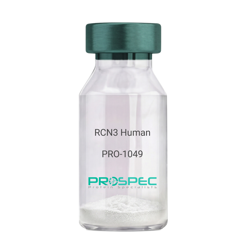
RCN3 HumanDescription:
Reticulocalbin 3 Human Recombinant
Reticulocalbin-3, EF-hand calcium-binding protein RLP49, RCN3, RLP49.
Product # :
PRO-1049Price :
Quantity :
Shipping Method :
Shipped with Ice Packs
More Info
- description
- source
- formulation
- purity
- More Info
Description
RCN3 Human Recombinant produced in E.Coli is a single, non-glycosylated polypeptide chain containing 333 amino acids (21-328 a.a) and having a molecular mass of 37.9kDa.RCN3 is fused to a 25 amino acid His-tag at N-terminus & purified by proprietary chromatographic techniques.
Source
Escherichia Coli.
Formulation
RCN3 protein solution (1mg/ml) containing 20mM Tris-HCl buffer (pH 8.0), 1mM DTT, 10% glycerol and 0.1M NaCl.
Purity
Greater than 95.0% as determined by SDS-PAGE.
More Info
-
Introduction
Reticulocalbin 3 (RCN3) belongs to the CREC (cab45/reticulocalbin/ ERC45/calumenin) family. RCN3 contains 5 Arg-Xaa-Xaa-Arg motifs, which function as target sequences of SPCs (subtilisin-like proprotein convertases), which is a family of serine endoproteases that proteolytically activate proproteins. The synthesis of PACE4 is induced by association and coexpression with RCN3.
-
Synonyms
Reticulocalbin-3, EF-hand calcium-binding protein RLP49, RCN3, RLP49.
-
Physical Appearance
Sterile Filtered colorless solution.
-
Stability
Store at 4°C if entire vial will be used within 2-4 weeks. Store, frozen at -20°C for longer periods of time. For long term storage it is recommended to add a carrier protein (0.1% HSA or BSA).Avoid multiple freeze-thaw cycles.
-
Amino Acid Sequence
MGSSHHHHHH SSGLVPRGSH MGSHMKPSPD AGPHGQGRVH QAAPLSDAPH DDAHGNFQYD HEAFLGREVA KEFDQLTPEE SQARLGRIVD RMDRAGDGDG WVSLAELRAW IAHTQQRHIR DSVSAAWDTY DTDRDGRVGW EELRNATYGH YAPGEEFHDV EDAETYKKML ARDERRFRVA DQDGDSMATR EELTAFLHPE EFPHMRDIVI AETLEDLDRN KDGYVQVEEY IADLYSAEPG EEEPAWVQTE RQQFRDFRDL NKDGHLDGSE VGHWVLPPAQ DQPLVEANHL LHESDTDKDG RLSKAEILGN WNMFVGSQAT NYGEDLTRHH DEL.
ProSpec's products are furnished for LABORATORY RESEARCH USE ONLY. The product may not be used as drugs, agricultural or pesticidal products, food additives or household chemicals.
- View Data Sheet
Name :
C5a ProteinDescription:
Complement C5a Human
Complement Component 5, C3 and PZP-Like Alpha-2-Macroglobulin Domain-Containing Protein 4, C5a Anaphylatoxin, Prepro-C5, CPAMD4, Anaphylatoxin C5a Analog, ECLZB, C5A, C5D, C5b, C5.
Product # :
PRO-2692Price :
Quantity :
Shipping Method :
Shipped with Ice Packs
More Info
- description
- source
- formulation
- purity
- More Info
Description
Human Complement C5a produced in Human plasma having a molecular mass of 10.4 kDa.
Source
Human Plasma.
Formulation
C5a protein solution contains 120 mM NaCl and 10mM HEPES, pH 7.2.
Purity
Greater than 95.0% as determined by SDS-PAGE.
More Info
-
Introduction
Complement Component C5a (C5a) is involved in the complement system and it is encoded by the C5 gene in human. Complement C5 is cleaved into C5a and C5b. C5a has an imperative role in chemotaxis and C5b forms the 1st part of the complement membrane attack complex. Complement C5 is the 5th component of complement, which plays a central role in inflammatory and cell killing processes. C5a protein is composed of alpha and beta polypeptide chains, which are linked by a disulfide bridge. An activation peptide, C5a, which is an anaphylatoxin, which has potent spasmogenic and chemotactic activity, is derivative from the alpha polypeptide via cleavage with a convertase. The C5b macromolecular cleavage product forms a complex with the C6 complement component, and this complex is the basis for creation of the membrane attack complex, which includes supplementary complement components.
-
Synonyms
Complement Component 5, C3 and PZP-Like Alpha-2-Macroglobulin Domain-Containing Protein 4, C5a Anaphylatoxin, Prepro-C5, CPAMD4, Anaphylatoxin C5a Analog, ECLZB, C5A, C5D, C5b, C5.
-
Physical Appearance
Sterile filtered solution.
-
Stability
C5a Human is stable at 4°C if entire vial will be used within 2-4 weeks.Store, frozen below -20°C for longer periods of time.For long term storage it is recommended to add a carrier protein (0.1% HSA or BSA).Avoid multiple freeze-thaw cycles.
-
Human Virus Test
Plasma from each donor has been tested and found negative for antibody to HIV-1, HIV-2, HCV, HTLV-I &II, STS and HBSAG.
ProSpec's products are furnished for LABORATORY RESEARCH USE ONLY. The product may not be used as drugs, agricultural or pesticidal products, food additives or household chemicals.
- View Data Sheet
Name :
ESM1 Human, HEKDescription:
Endothelial Cell-Specific Molecule 1 Human Recombinant, HEK
Endothelial cell-specific molecule 1, ESM-1, ESM1, endocan.
Product # :
PRO-2476Price :
Quantity :
Shipping Method :
Shipped at Room temp
More Info
- description
- source
- formulation
- purity
- More Info
Description
ESM1 Human Recombinant produced in HEK293 cells is a single, glycosylated polypeptide chain (a.a 20-184) containing 175 amino acids including a 10 a.a C-terminal His tag. The total molecular mass is 19.5kDa (calculated).
Source
HEK293 cells.
Formulation
ESM1 filtered (0.4 µm) and lyophilized from 0.5mg/ml in phosphate buffered saline.
Purity
Greater than 95.0% as determined by SDS-PAGE.
More Info
-
Introduction
Endothelial cell-specific molecule 1 (ESM1) is a proteoglycan secreted by endothelial cells (mostly in the human lung and kidney tissues) and its mRNA expression is regulated by inflammatory cytokines. ESM1 has potent implications in lung endothelial cell-leukocyte interactions. In addition, ESM1 expression is detected in various epithelia and in adipocytes. ESM1 is involved in angiogenesis and it also promotes angiogenic sprouting. ESM1 expression is upregulated by TNF alpha, IL1 beta, or lipopolysaccharide and downregulated by IFN gamma. Genetically engineered cells overexpressing ESM1 induce tumor formation, implying that ESM1 might be involved in the pathophysiology of tumor growth in vivo.
-
Synonyms
Endothelial cell-specific molecule 1, ESM-1, ESM1, endocan.
-
Physical Appearance
Filtered White lyophilized (freeze-dried) powder.
-
Stability
Store lyophilized protein at -20°C. Aliquot the product after reconstitution to avoid repeated freezing/thawing cycles. Reconstituted protein can be stored at 4°C for a limited period of time; it does not show any change after two weeks at 4°C.
-
Solubility
It is recommended to add deionized water to prepare a working stock solution of approximately 0.5mg/ml and let the lyophilized pellet dissolve completely. ESM1 is not sterile! Please filter the product by an appropriate sterile filter before using it in the cell culture.
-
Amino Acid Sequence
WSNNYAVDCP QHCDSSECKS SPRCKRTVLD DCGCCRVCAA GRGETCYRTV SGMDGMKCGP GLRCQPSNGE DPFGEEFGIC KDCPYGTFGM DCRETCNCQS GICDRGTGKC LKFPFFQYSV TKSSNRFVSL TEHDMASGDG NIVREEVVKE NAAGSPVMRK WLNPR HHHHH HHHHH.
ProSpec's products are furnished for LABORATORY RESEARCH USE ONLY. The product may not be used as drugs, agricultural or pesticidal products, food additives or household chemicals.
- View Data Sheet
Name :
SNAPIN HumanDescription:
SNAP Associated Protein Human Recombinant
SNAPAP, SNARE-associated protein Snapin, Synaptosomal-associated protein 25-binding protein, SNAP-associated protein, SNAPIN, SNAP25BP.
Product # :
PRO-663Price :
Quantity :
Shipping Method :
Shipped with Ice Packs
More Info
- description
- source
- formulation
- purity
- More Info
Description
SNAPIN Human Recombinant produced in E.Coli is a single, non-glycosylated polypeptide chain containing 156 amino acids (1-136) and having a molecular mass of 17 kDa. SNAPIN is fused to 20 amino acid His Tag at N-terminus and purified by standard chromatography techniques.
Source
Escherichia Coli.
Formulation
The protein solution contains 20mM Tris-HCl pH-8, 5mM DTT, 2mM EDTA, 0.2M NaCl, and 40% glycerol.
Purity
Greater than 90.0% as determined by SDS-PAGE.
More Info
-
Introduction
SNAPIN is involved in the neurotransmitter release process through its modulation of the sequential interactions between the SNAREs and synaptotagmin. SNAPIN is part of the SNARE complex of proteins that is needed for synaptic vesicle docking and fusion. SNAPAP is enriched in neurons and exclusively located on synaptic vesicle membrane protein. SNAPIN is also an important factor of the BLOC1 multisubunit protein complex. BLOC1 is required for normal biogenesis of specialized organelles of the endosomal-lysosomal system, such as melanosomes and platelet dense granules.
-
Synonyms
SNAPAP, SNARE-associated protein Snapin, Synaptosomal-associated protein 25-binding protein, SNAP-associated protein, SNAPIN, SNAP25BP.
-
Physical Appearance
Sterile Filtered colorless solution.
-
Stability
Store at 4°C if entire vial will be used within 2-4 weeks. Store, frozen at -20°C for longer periods of time. For long term storage it is recommended to add a carrier protein (0.1% HSA or BSA).Avoid multiple freeze-thaw cycles.
-
Amino Acid Sequence
MGSSHHHHHH SSGLVPRGSH MAGAGSAAVS GAGTPVAGPT GRDLFAEGLL EFLRPAVQQL DSHVHAVRES QVELREQIDN LATELCRINEDQKVALDLDP YVKKLLNARR RVVLVNNILQ NAQERLRRLN HSVAKETARR RAMLDSGIYP PGSPGK.
ProSpec's products are furnished for LABORATORY RESEARCH USE ONLY. The product may not be used as drugs, agricultural or pesticidal products, food additives or household chemicals.
- View Data Sheet
Name :
TNFRSF17 Human, HisDescription:
B-Cell Maturation Antigen Human Recombinant, His Tag
BCMA, CD269, Tumor Necrosis Factor Receptor Superfamily Member 17, BCM, TNFRSF17, B-cell maturation protein, CD269 antigen.
Product # :
CYT-190Price :
Quantity :
Shipping Method :
Shipped with Ice Packs
More Info
- description
- source
- formulation
- purity
- More Info
Description
TNFRSF17 Human Recombinant produced in E.Coli is a single, non-glycosylated polypeptide chain containing 130 amino acids (78-184 a.a) and having a molecular mass of 14.1kDa.TNFRSF17 is fused to a 23 amino acid His-tag at N-terminus & purified by proprietary chromatographic techniques.
Source
Escherichia Coli.
Formulation
TNFRSF17 protein solution (0.5mg/ml) containing 20mM Tris-HCl buffer (pH 8.0), 0.15M NaCl, 10% glycerol and 1mM DTT.
Purity
Greater than 90.0% as determined by SDS-PAGE.
More Info
-
Introduction
TNFRSF17 is a receptor for tnfsf13b/blys/baff and tnfsf13/april. TNFRSF17 promotes b-cell survival and plays a role in the regulation of humoral immunity. TNFRSF17 activates nf-kappa-b and jnk. TNFRSF17 is a member of the TNF-receptor superfamily. TNFRSF17 is expressed in mature B lymphocytes, and is invloved in B cell development and autoimmune response. TNFRSF17 specifically binds to the tumor TNFSF13B/TALL-1/BAFF, which causes NF-kappaB and MAPK8/JNK activation. TNFRSF17 binds to a variety of TRAF family members, and therefore transduces signals for cell survival and proliferation. TNFRSF17 is a type III membrane protein having 1 extracellular cysteine rich domain. Within the TNFRSF, it shares the highest homology with TACI. BCMA and TACI have both been shown to bind to APRIL and BAFF, members of the TNF ligand superfamily. BCMA expression has been found in immune organs. TNFRSF17 appears to be localized to the Golgi compartment. The binding of BCMA to APRIL or BAFF has been shown to stimulate IgM production in peripheral blood B cells and increase the survival of cultured B cells.
-
Synonyms
BCMA, CD269, Tumor Necrosis Factor Receptor Superfamily Member 17, BCM, TNFRSF17, B-cell maturation protein, CD269 antigen.
-
Physical Appearance
Sterile Filtered colorless liquid.
-
Stability
Store at 4°C if entire vial will be used within 2-4 weeks. Store, frozen at -20°C for longer periods of time. For long term storage it is recommended to add a carrier protein (0.1% HSA or BSA).Avoid multiple freeze-thaw cycles.
-
Amino Acid Sequence
MGSSHHHHHH SSGLVPRGSH MGSRKINSEP LKDEFKNTGS GLLGMANIDL EKSRTGDEII LPRGLEYTVE ECTCEDCIKS KPKVDSDHCF PLPAMEEGAT ILVTTKTNDY CKSLPAALSA TEIEKSISAR.
ProSpec's products are furnished for LABORATORY RESEARCH USE ONLY. The product may not be used as drugs, agricultural or pesticidal products, food additives or household chemicals.
- View Data Sheet
Name :
PNRC2 HumanDescription:
Proline-Rich Nuclear Receptor Coactivator 2 Human Recombinant
Proline-rich nuclear receptor coactivator 2, PNRC2.
Product # :
PRO-1534Price :
Quantity :
Shipping Method :
Shipped with Ice Packs
More Info
- description
- source
- formulation
- purity
- More Info
Description
PNRC2 Human Recombinant produced in E. coli is a single polypeptide chain containing 162 amino acids (1-139) and having a molecular mass of 18kDa. PNRC2 is fused to a 23 amino acid His-tag at N-terminus & purified by proprietary chromatographic techniques.
Source
Escherichia Coli.
Formulation
The PNRC2 solution (1mg/ml) contains 20mM Tris-HCl buffer (pH 8.0), 0.4M UREA and 10% glycerol.
Purity
Greater than 90% as determined by SDS-PAGE.
More Info
-
Introduction
Proline-Rich Nuclear Receptor Coactivator 2 (PNRC2) takes part in controlling the energy balance between energy storage and energy expenditure. PNRC2 is involved in nonsense-mediated mRNA decay (NMD) by operating as a bridge between the mRNA decapping complex and the NMD machinery. PNRC2 is needed for UPF1/RENT1 localization to the P-body and also plays a role as a nuclear receptor coactivator.
-
Synonyms
Proline-rich nuclear receptor coactivator 2, PNRC2.
-
Physical Appearance
Sterile Filtered clear solution.
-
Stability
Store at 4°C if entire vial will be used within 2-4 weeks. Store frozen at -20°C for longer periods of time. For long term storage it is recommended to add a carrier protein (0.1% HSA or BSA).Avoid multiple freeze-thaw cycles.
-
Amino Acid Sequence
MGSSHHHHHH SSGLVPRGSH MGSMGGGERY NIPAPQSRNV SKNQQQLNRQ KTKEQNSQMK IVHKKKERGH GYNSSAAAWQ AMQNGGKNKN FPNNQSWNSS LSGPRLLFKS QANQNYAGAK FSEPPSPSVL PKPPSHWVPV SFNPSDKEIM TFQLKTLLKV QV.
ProSpec's products are furnished for LABORATORY RESEARCH USE ONLY. The product may not be used as drugs, agricultural or pesticidal products, food additives or household chemicals.
- View Data Sheet
Name :
FAS AntibodyDescription:
FAS Blocking/Activating antibody (CD95), Mouse anti Human
FASLG receptor, Apoptosis-mediating surface antigen FAS, Apo-1 antigen, CD95, Tumor necrosis factor receptor superfamily member 6 TNR6, APT1, FAS1, TNFRSF6.
Product # :
ANT-204Price :
Quantity :
Shipping Method :
Shipped at Room temp
More Info
- formulation
- More Info
Formulation
1 mg/ml in PBS (after reconstitution).
More Info
-
Introduction
The Fas receptor (CD95) mediates apoptotic signaling by Fas-ligand expressed on the surface of other cells. The Fas-FasL interaction plays an important role in the immune system and lack of this system leads to autoimmunity, indicating that Fas-mediated apoptosis removes self-reactive lymphocytes. Fas signaling is also involved in immune surveillance to remove transformed cells and virus infected cells. Binding of FAS to oligimerized FasL on another cell activates apoptotic signaling through a cytoplasmic domain termed the death domain that interacts with signaling adaptors including FAF, FADD and DAX to activate the caspase proteolytic cascade. Caspase-8 and caspase-10 are first activated, to then cleave and activate downstream caspases, and a variety of cellular substrates that lead to cell death. Caspases cleave nuclear lamins, causing the nucleus to break down and lose its normal structure and another caspase substrate is DFF, inducing cleavage and degradation of the genome. Other caspase substrates are involved in cytoskeletal structure, cell cycle regulation and signaling pathways. Activation of JNK kinase, activation of Jun, and production of ceramide may also play roles in Fas-mediated apoptosis. Activation of fas-mediated apoptosis is opposed by I-FLICE and FAP. Viruses and tumors may escape immune surveillance in part through suppression of fas-mediated apoptosis using similar mechanisms.
-
Synonyms
FASLG receptor, Apoptosis-mediating surface antigen FAS, Apo-1 antigen, CD95, Tumor necrosis factor receptor superfamily member 6 TNR6, APT1, FAS1, TNFRSF6.
-
Solubility
Reconstitute with H20. Mix gently, wash the sides of the vial and wait 30-60 seconds before use.
-
Immunogen
Recombinant Human FAS.
-
Ig Subclass
mouse IgG1.
-
Clone
NYRhFAS.
-
Note
This antibody is a BLOCKING antibody when in soluble form and will block FAS-mediated apoptosis. It will ACTIVATE FAS-mediated killing when immobilized.
-
Titer
By direct ELISA, 1:10,000 dilution will yield 0.5 O.D using alkaline phosphatase conjugated rabbit anti-mouse Ig (Jackson Laboratories).
-
Shipping Conditions
Antibody is shipped lyophilized at ambient temperature.
-
Type
Mouse Anti Human Monoclonal.
-
Storage Procedures
In lyophilized form, for long periods, store at 4°C in a dry environment. After reconstitution, if not intended for use within a month, aliquot and store at -20°C.
-
Purification Method
Protein-A column.
ProSpec's products are furnished for LABORATORY RESEARCH USE ONLY. The product may not be used as drugs, agricultural or pesticidal products, food additives or household chemicals.