Skip to content

High-quality research proteins.

  • 1-800-805-6531

  • Mon - Sat 8.00-18.00, Sun - Closed

  • info@prospecbio.com

  • PO Box 6591 East Brunswick NJ 08816

  • Products
    • Cytokines
    • Growth factors
    • Chemokines
    • Cd antigens
    • Neurotrophins
    • Hormones
    • Enzymes
    • Viral antigens
    • Recombinant proteins
    • Natural proteins
    • Monoclonal antibodies
    • Polyclonal antibodies
    • Thermal Packaging
  • About
    • Company
    • Founder
    • Contribution
  • Customers
    • Eastern Asia
    • Eastern Europe
    • North America
    • South America
    • South East Asia
    • Southern Asia
    • Western Europe
  • Ordering
    • Ordering Method
    • Ordering - Credit Card
    • Ordering - PO
    • Shipping Fees
    • FAQs
  • Services
    • DNA Cloning
    • Protein Expression
    • Monoclonal Antibody
    • Fermentation
    • Technology License
  • Contact us
Log in

Country/region

  • USD $ | Albania
  • USD $ | Andorra
  • USD $ | Argentina
  • USD $ | Armenia
  • USD $ | Australia
  • USD $ | Austria
  • USD $ | Azerbaijan
  • USD $ | Belarus
  • USD $ | Belgium
  • USD $ | Bolivia
  • USD $ | Brazil
  • USD $ | Bulgaria
  • USD $ | Cambodia
  • USD $ | Canada
  • USD $ | Chile
  • USD $ | China
  • USD $ | Colombia
  • USD $ | Costa Rica
  • USD $ | Croatia
  • USD $ | Cyprus
  • USD $ | Czechia
  • USD $ | Denmark
  • USD $ | Estonia
  • USD $ | Finland
  • USD $ | France
  • USD $ | Georgia
  • USD $ | Germany
  • USD $ | Greece
  • USD $ | Hong Kong SAR
  • USD $ | Hungary
  • USD $ | Iceland
  • USD $ | India
  • USD $ | Ireland
  • USD $ | Israel
  • USD $ | Italy
  • USD $ | Japan
  • USD $ | Kazakhstan
  • USD $ | Latvia
  • USD $ | Liechtenstein
  • USD $ | Lithuania
  • USD $ | Madagascar
  • USD $ | Malta
  • USD $ | Mexico
  • USD $ | Moldova
  • USD $ | Monaco
  • USD $ | Netherlands
  • USD $ | New Zealand
  • USD $ | North Macedonia
  • USD $ | Norway
  • USD $ | Panama
  • USD $ | Paraguay
  • USD $ | Peru
  • USD $ | Philippines
  • USD $ | Poland
  • USD $ | Portugal
  • USD $ | Romania
  • USD $ | Singapore
  • USD $ | Slovakia
  • USD $ | Slovenia
  • USD $ | South Africa
  • USD $ | South Korea
  • USD $ | Spain
  • USD $ | Sri Lanka
  • USD $ | Sweden
  • USD $ | Switzerland
  • USD $ | Taiwan
  • USD $ | Thailand
  • USD $ | Türkiye
  • USD $ | Ukraine
  • USD $ | United Kingdom
  • USD $ | United States
  • USD $ | Uruguay
  • USD $ | Vatican City
  • USD $ | Venezuela
  • USD $ | Vietnam
  • Facebook
  • Instagram
logoProSpec - Recombinant Protein Manufacturer for academic and R&D industry
Become a distributor
Account Log in
Wishlist
Cart Cart(0)
  • Products
    +
    • Cytokines
    • Growth factors
    • Chemokines
    • Cd antigens
    • Neurotrophins
    • Hormones
    • Enzymes
    • Viral antigens
    • Recombinant proteins
    • Natural proteins
    • Monoclonal antibodies
    • Polyclonal antibodies
    • Thermal Packaging
  • About
    +
    • Company
    • Founder
    • Contribution
  • Customers
    +
    • Eastern Asia
    • Eastern Europe
    • North America
    • South America
    • South East Asia
    • Southern Asia
    • Western Europe
  • Ordering
    +
    • Ordering Method
    • Ordering - Credit Card
    • Ordering - PO
    • Shipping Fees
    • FAQs
  • Services
    +
    • DNA Cloning
    • Protein Expression
    • Monoclonal Antibody
    • Fermentation
    • Technology License
  • Contact us

Item added to your cart

View cart
View All
  • Cytokines
  • Growth factors
  • Chemokines
  • Cd antigens
  • Neurotrophins
  • Hormones
  • Enzymes
  • Viral antigens
  • Recombinant proteins
  • Natural proteins
  • Monoclonal antibodies
  • Polyclonal antibodies
  • Thermal Packaging
  • Tumor Necrosis Factor

    Tumor Necrosis Factor

    View All
  • 4-1BB

    4-1BB

    View All
  • Adiponectin

    Adiponectin

    View All
  • AIF1

    AIF1

    View All
  • Other Cytokines

    Other Cytokines

    View All
  • AITRL

    AITRL

    View All
  • Angiopoietin

    Angiopoietin

    View All
  • Apolipoprotein

    Apolipoprotein

    View All
  • B-Cell Activating Factor

    B-Cell Activating Factor

    View All
  • Beta Defensin

    Beta Defensin

    View All
  • Bone Morphogenetic Protein

    Bone Morphogenetic Protein

    View All
  • B type Natriuretic Peptide

    B type Natriuretic Peptide

    View All
  • BST

    BST

    View All
  • Betacellulin

    Betacellulin

    View All
  • Cardiotrophin

    Cardiotrophin

    View All
  • Activin

    Activin

    View All
  • Fibroblast Growth Factor

    Fibroblast Growth Factor

    View All
  • Other Growth Factors

    Other Growth Factors

    View All
  • CSF

    CSF

    View All
  • CTGF

    CTGF

    View All
  • IGFBP

    IGFBP

    View All
  • VEGF Protein

    VEGF Protein

    View All
  • EGF

    EGF

    View All
  • Epigen

    Epigen

    View All
  • Erythropoietin

    Erythropoietin

    View All
  • Insulin-Like Growth Factor

    Insulin-Like Growth Factor

    View All
  • Growth Hormone

    Growth Hormone

    View All
  • HDGF

    HDGF

    View All
  • Hepatocyte Growth Factor

    Hepatocyte Growth Factor

    View All
  • Insulin

    Insulin

    View All
  • BCA-1/ BLC (CXCL13)

    BCA-1/ BLC (CXCL13)

    View All
  • BRAK (CXCL14)

    BRAK (CXCL14)

    View All
  • C-10 (CCL6)

    C-10 (CCL6)

    View All
  • Other Chemokines

    Other Chemokines

    View All
  • MEC (CCL28)

    MEC (CCL28)

    View All
  • LD78-beta (CCL3L1)

    LD78-beta (CCL3L1)

    View All
  • CTACK (CCL27)

    CTACK (CCL27)

    View All
  • CXCL16

    CXCL16

    View All
  • CXCL17

    CXCL17

    View All
  • Platelet Factor-4 (CXCL4)

    Platelet Factor-4 (CXCL4)

    View All
  • ENA-78 (CXCL5)

    ENA-78 (CXCL5)

    View All
  • Eotaxin (CCL11,24,26)

    Eotaxin (CCL11,24,26)

    View All
  • Exodus-2 (CCL21)

    Exodus-2 (CCL21)

    View All
  • Fractalkine (CX3CL1)

    Fractalkine (CX3CL1)

    View All
  • CXCL6

    CXCL6

    View All
  • Other CD Antigens

    Other CD Antigens

    View All
  • CD14

    CD14

    View All
  • CD164

    CD164

    View All
  • CD1

    CD1

    View All
  • CD2

    CD2

    View All
  • CD200

    CD200

    View All
  • CD207

    CD207

    View All
  • CD226

    CD226

    View All
  • CD23

    CD23

    View All
  • CD244

    CD244

    View All
  • CD247

    CD247

    View All
  • CD27

    CD27

    View All
  • CD274

    CD274

    View All
  • CD300

    CD300

    View All
  • CD33

    CD33

    View All
  • Other Neurotrophins

    Other Neurotrophins

    View All
  • Beta-NGF

    Beta-NGF

    View All
  • BDNF

    BDNF

    View All
  • CDNF

    CDNF

    View All
  • CNTF

    CNTF

    View All
  • GDNF

    GDNF

    View All
  • Glia Maturation Factor

    Glia Maturation Factor

    View All
  • MANF

    MANF

    View All
  • Midkine

    Midkine

    View All
  • Neuroglobin

    Neuroglobin

    View All
  • Neurotrophic factors

    Neurotrophic factors

    View All
  • Neuregulin

    Neuregulin

    View All
  • Neuropilin

    Neuropilin

    View All
  • Pigment Epithelium-Derived Factor

    Pigment Epithelium-Derived Factor

    View All
  • Pleiotrophin

    Pleiotrophin

    View All
  • Peptide Hormones

    Peptide Hormones

    View All
  • Other Hormones

    Other Hormones

    View All
  • HCG

    HCG

    View All
  • Endothelin

    Endothelin

    View All
  • Exendin

    Exendin

    View All
  • FSH

    FSH

    View All
  • Glucagon

    Glucagon

    View All
  • GHRP

    GHRP

    View All
  • GLP

    GLP

    View All
  • Thyrostimulin

    Thyrostimulin

    View All
  • Inhibin A

    Inhibin A

    View All
  • LHRH

    LHRH

    View All
  • Melanotan

    Melanotan

    View All
  • Procalcitonin

    Procalcitonin

    View All
  • PTH

    PTH

    View All
  • Peroxiredoxin

    Peroxiredoxin

    View All
  • Peptidase

    Peptidase

    View All
  • Synthetase

    Synthetase

    View All
  • Transferase

    Transferase

    View All
  • Hydrolase

    Hydrolase

    View All
  • Dehydrogenase

    Dehydrogenase

    View All
  • ACE2

    ACE2

    View All
  • Esterase

    Esterase

    View All
  • Protein Kinases

    Protein Kinases

    View All
  • Other Enzymes

    Other Enzymes

    View All
  • Phosphatase

    Phosphatase

    View All
  • Deaminase

    Deaminase

    View All
  • Oxygenase

    Oxygenase

    View All
  • Adenylate Kinase

    Adenylate Kinase

    View All
  • Lyase

    Lyase

    View All
  • Chagas

    Chagas

    View All
  • SARS

    SARS

    View All
  • Influenza

    Influenza

    View All
  • ASFV

    ASFV

    View All
  • Astrovirus

    Astrovirus

    View All
  • Borrelia

    Borrelia

    View All
  • Helicobacter Pylori

    Helicobacter Pylori

    View All
  • Chikungunya

    Chikungunya

    View All
  • Chlamydia

    Chlamydia

    View All
  • Cytomegalo

    Cytomegalo

    View All
  • Dengue

    Dengue

    View All
  • EBV

    EBV

    View All
  • Ebola

    Ebola

    View All
  • Feline Leukemia Virus

    Feline Leukemia Virus

    View All
  • Hepatitis A

    Hepatitis A

    View All
  • Other

    Other

    View All
  • Actin

    Actin

    View All
  • ADAM

    ADAM

    View All
  • Complement Component

    Complement Component

    View All
  • Ag85

    Ag85

    View All
  • Eukaryotic Translation Initiation Factor

    Eukaryotic Translation Initiation Factor

    View All
  • Anterior Gradient Protein

    Anterior Gradient Protein

    View All
  • Heat Shock Protein

    Heat Shock Protein

    View All
  • Alpha-2-HS-Glycoprotein

    Alpha-2-HS-Glycoprotein

    View All
  • Hemoglobin

    Hemoglobin

    View All
  • Allergy

    Allergy

    View All
  • Anaplasma

    Anaplasma

    View All
  • Angiogenin

    Angiogenin

    View All
  • Ankyrin Repeat Domain

    Ankyrin Repeat Domain

    View All
  • Annexin

    Annexin

    View All
  • Other Natural Proteins

    Other Natural Proteins

    View All
  • Aprotinin

    Aprotinin

    View All
  • Transferrin

    Transferrin

    View All
  • Avidin

    Avidin

    View All
  • Anti Coagulation Factors

    Anti Coagulation Factors

    View All
  • Natural Albumin

    Natural Albumin

    View All
  • Natural Coagulation Factors

    Natural Coagulation Factors

    View All
  • Fibronectin

    Fibronectin

    View All
  • Thrombin

    Thrombin

    View All
  • Anti Human Enzyme

    Anti Human Enzyme

    View All
  • Other Monoclonal Antibodies

    Other Monoclonal Antibodies

    View All
  • Anti Human Cytokine

    Anti Human Cytokine

    View All
  • Anti Human Heat Shock Protein

    Anti Human Heat Shock Protein

    View All
  • Anti Mouse Lymphocyte

    Anti Mouse Lymphocyte

    View All
  • Anti Human Chemokine

    Anti Human Chemokine

    View All
  • Anti Human Lymphocyte

    Anti Human Lymphocyte

    View All
  • Anti Viral Monoclonal

    Anti Viral Monoclonal

    View All
  • Anti Mouse Cytokine

    Anti Mouse Cytokine

    View All
  • Anti-GST Monoclonal

    Anti-GST Monoclonal

    View All
  • Other Polyclonal Antibodies

    Other Polyclonal Antibodies

    View All
  • Anti Viral

    Anti Viral

    View All
  • Anti-GST Monoclonal

    Anti-GST Monoclonal

    View All
Back

Search results

1000 results found for “cysteine-rich”

Name

Description

Product #

Price

Quantity

Shipping Method

  • View Data Sheet

    Name :

    CNDP1 Human, Active

    Description:

    CNDP Dipeptidase 1 Human Recombinant, Active

    Carnosine Dipeptidase 1 (Metallopeptidase M20 Family), Glutamate Carboxypeptidase-Like Protein 2, CNDP Dipeptidase 1, Serum Carnosinase, Carnosinase 1, CPGL2, CN1, Carnosine Dipeptidase 1, EC 3.4.13.20, HsT2308.

    Product # :

    ENZ-1022

    Price :

    Quantity :

    Shipping Method :

    Ice Icon

    Shipped with Ice Packs

    Add To Cart

    More Info

    • description
    • source
    • formulation
    • purity
    • biological activity
    • More Info

    Description

    CNDP1 Human Recombinant produced in Sf9 Baculovirus cells is a single, glycosylated polypeptide chain containing 489 amino acids (27-507 a.a.) and having a molecular mass of 54.9kDa (Migrates at 50-70kDa on SDS-PAGE under reducing conditions).CNDP1 is expressed with a 8 amino acid His tag at C-Terminus and purified by proprietary chromatographic techniques.

    Source

    Sf9, Insect cells.

    Formulation

    CNDP1 protein solution (0.25mg/ml) contains Phosphate Buffered Saline (pH 7.4) and 10% glycerol.

    Purity

    Greater than 95.0% as determined by SDS-PAGE.

    Biological Activity

    Specific activity is >3,000 pmol/min/ug, and as measured by the hydrolysis of carnosine per minute at pH6.8 at 25°C.

    More Info

    • Introduction

      CNDP Dipeptidase 1, also known as CNDP1 is a member of the peptidase M20A family. CNDP1 Mannheim which is the shortest allelic form has been more common in the absence of nephropathy in addition to being associated with lower serum carnosinase levels. Furthermore, Carnosine inhibited the increased production of fibronectin as well as collagen type VI in podocytes and the increased production of TGF-beta in mesangial cells. Diabetic patients with the CNDP1 Mannheim variant are less at risk for nephropathy. In addition, on renal cells carnosine protects against the adverse effects of high glucose levels.

    • Synonyms

      Carnosine Dipeptidase 1 (Metallopeptidase M20 Family), Glutamate Carboxypeptidase-Like Protein 2, CNDP Dipeptidase 1, Serum Carnosinase, Carnosinase 1, CPGL2, CN1, Carnosine Dipeptidase 1, EC 3.4.13.20, HsT2308.

    • Physical Appearance

      Sterile filtered colorless solution.

    • Stability

      Store at 4°C if entire vial will be used within 2-4 weeks.Store, frozen at -20°C for longer periods of time. For long term storage it is recommended to add a carrier protein (0.1% HSA or BSA).Avoid multiple freeze-thaw cycles.

    • Amino Acid Sequence

      SPSPPPALLE KVFQYIDLHQ DEFVQTLKEW VAIESDSVQP VPRFRQELFR MMAVAADTLQ RLGARVASVD MGPQQLPDGQ SLPIPPVILA ELGSDPTKGT VCFYGHLDVQ PADRGDGWLT DPYVLTEVDG KLYGRGATDN KGPVLAWINA VSAFRALEQD LPVNIKFIIE GMEEAGSVAL EELVEKEKDR FFSGVDYIVI SDNLWISQRK PAITYGTRGN SYFMVEVKCR DQDFHSGTFG GILHEPMADL VALLGSLVDS SGHILVPGIY DEVVPLTEEE INTYKAIHLD LEEYRNSSRV EKFLFDTKEE ILMHLWRYPS LSIHGIEGAF DEPGTKTVIP GRVIGKFSIR LVPHMNVSAV EKQVTRHLED VFSKRNSSNK MVVSMTLGLH PWIANIDDTQ YLAAKRAIRT VFGTEPDMIR DGSTIPIAKM FQEIVHKSVV LIPLGAVDDG EHSQNEKINR WNYIEGTKLF AAFFLEMAQL HLEHHHHHH.

    ProSpec's products are furnished for LABORATORY RESEARCH USE ONLY. The product may not be used as drugs, agricultural or pesticidal products, food additives or household chemicals.

    Cndp1 Human Active
  • View Data Sheet

    Name :

    SYT1 Human

    Description:

    Synaptotagmin I Human Recombinant

    Synaptotagmin-1, Synaptotagmin I, SytI, p65, SYT1, SVP65, SYT.

    Product # :

    PRO-239

    Price :

    Quantity :

    Shipping Method :

    Ice Icon

    Shipped with Ice Packs

    Add To Cart

    More Info

    • description
    • source
    • formulation
    • purity
    • More Info

    Description

    SYT1 produced in E.Coli is a single, non-glycosylated polypeptide chain containing 256 amino acids (136-382 a.a) and having a molecular mass of 29.5kDa.SYT1 is fused to an 8 amino acid His-tag at C-terminus & purified by proprietary chromatographic techniques.

    Source

    Escherichia Coli.

    Formulation

    SYT1 protein solution (0.25mg/ml) containing 20mM Tris-HCl buffer (pH 8.0), 2mM DTT, 20% glycerol and 100mM NaCl.

    Purity

    Greater than 85.0% as determined by SDS-PAGE.

    More Info

    • Introduction

      Synaptotagmin-1(SYT1) is a member of the synaptotagmin family, which contains two C2 domains. The synaptotagmins are integral membrane proteins of synaptic vesicles assumed to function as Ca(2+) sensors in the process of vesicular trafficking and exocytosis. SYT1 is the principal regulator responsible for allowing the human brain to release neurotransmitters. SYT1 may have a regulatory role in the membrane interactions during trafficking of synaptic vesicles at the active zone of the synapse. SYT1 binds acidic phospholipids with a specificity which entails the presence of both an acidic head group and a diacyl backbone. SYT1 can also bind to at least 3 additional proteins in a Ca2+-independent manner; these being neurexins, syntaxin and AP2.

    • Synonyms

      Synaptotagmin-1, Synaptotagmin I, SytI, p65, SYT1, SVP65, SYT.

    • Physical Appearance

      Sterile Filtered colorless solution.

    • Stability

      Store at 4°C if entire vial will be used within 2-4 weeks. Store, frozen at -20°C for longer periods of time. For long term storage it is recommended to add a carrier protein (0.1% HSA or BSA).Avoid multiple freeze-thaw cycles.

    • Amino Acid Sequence

      MEPKEEEKLG KLQYSLDYDF QNNQLLVGII QAAELPALDM GGTSDPYVKV FLLPDKKKKF ETKVHRKTLN PVFNEQFTFK VPYSELGGKT LVMAVYDFDR FSKHDIIGEF KVPMNTVDFG HVTEEWRDLQ SAEKEEQEKL GDICFSLRYV PTAGKLTVVI LEAKNLKKMD VGGLSDPYVK IHLMQNGKRL KKKKTTIKKN TLNPYYNESF SFEVPFEQIQ KVQVVVTVLD YDKIGKNDAI GKVFVGYNLE HHHHHH.

    ProSpec's products are furnished for LABORATORY RESEARCH USE ONLY. The product may not be used as drugs, agricultural or pesticidal products, food additives or household chemicals.

    Syt1 Human
  • View Data Sheet

    Name :

    RBP4 Protein

    Description:

    Retinol Binding Protein-4 Human

    Retinol Binding Protein 4, RBP-4, RBP4, Plasma retinol-binding protein, PRBP, RBP.

    Product # :

    CYT-1218

    Price :

    Quantity :

    Shipping Method :

    Room Temp Icon

    Shipped at Room temp

    Add To Cart

    More Info

    • description
    • source
    • formulation
    • purity
    • More Info

    Description

    RBP4 Human produced in Pooled human plasma can be used as a calibrator in immunoassays. Immunoreactivity was checked using monoclonal antibodies specific to RBP4.

    Source

    Human Plasma.

    Formulation

    RBP4 was lyophilized from PBS, 150mM NaCl, and 10mM K-phosphate, pH 7.4.

    Purity

    Greater than 95.0% as determined by SDS-PAGE.

    More Info

    • Synonyms

      Retinol Binding Protein 4, RBP-4, RBP4, Plasma retinol-binding protein, PRBP, RBP.

    • Physical Appearance

      Sterile Filtered White lyophilized (freeze-dried) powder.

    • Stability

      Lyophilized Retinol Binding Protein-4 although stable at room temperature for 3 weeks, should be stored desiccated below -18°C. Upon reconstitution RBP4 should be stored at 4°C between 2-7 days and for future use below -18°C. For long term storage it is recommended to add a carrier protein (0.1% HSA or BSA). Please prevent freeze-thaw cycles.

    • Solubility

      It is recommended to reconstitute the lyophilized RBP4 in sterile 18MΩ-cm H2O not less than 100µg/ml, which can then be further diluted to other aqueous solutions.

    • Background

      Retinol Binding Protein 4 (RBP4) is a multifunctional protein that plays a crucial role in the transport of retinol (vitamin A) in the bloodstream. Beyond its traditional role in vitamin A metabolism, RBP4 has emerged as a key player in various physiological processes and pathological conditions. This research endeavors to explore the diverse facets of RBP4 in human biology, shedding light on its physiological functions, regulatory mechanisms, and implications in health and disease.

      Physiological Functions:

      At its core, RBP4 acts as a carrier protein, shuttling retinol from the liver, where it is stored, to peripheral tissues where it is utilized. Retinol is vital for vision, immune function, growth, and development, making RBP4 an essential component in these processes. By regulating the availability of retinol, RBP4 contributes significantly to maintaining normal vision, immune responses, and cellular differentiation, particularly in epithelial tissues.

      Metabolic Significance:

      Research has unveiled RBP4’s role in metabolic regulation. It has been associated with insulin resistance, a hallmark of type 2 diabetes mellitus. Elevated RBP4 levels are observed in individuals with obesity and insulin resistance, implicating its involvement in metabolic disorders. Understanding the interplay between RBP4, insulin signaling, and glucose metabolism is crucial for deciphering the complexities of diabetes and metabolic syndrome.

      Immunological Implications:

      Beyond its metabolic functions, RBP4 has been implicated in immune responses. Studies have suggested its involvement in modulating inflammatory processes and immune cell functions. By influencing immune cell differentiation and cytokine production, RBP4 may play a role in both immune defense and autoimmune disorders. Investigating these immunological implications provides insights into the crosstalk between metabolic and immune pathways.

      Genetic and Environmental Influences:

      Genetic variations and environmental factors, such as diet and lifestyle, can impact RBP4 levels and functions. Research into these influences is essential for understanding individual susceptibility to metabolic disorders and inflammatory conditions. Genetic studies shed light on the hereditary aspects of RBP4 regulation, providing valuable information for personalized medicine approaches.

      Clinical Relevance:

      RBP4’s involvement in various diseases, including diabetes, cardiovascular diseases, and certain cancers, underscores its clinical relevance. It serves as a potential biomarker for metabolic dysregulation and a target for therapeutic interventions. Moreover, RBP4-targeted therapies are being explored for their potential in managing metabolic disorders and related complications.

      Conclusion:

      RBP4, once primarily recognized for its role in vitamin A transport, has evolved into a multifaceted protein with intricate roles in metabolism, immunity, and disease. Its functions extend far beyond being a mere carrier of retinol, influencing diverse physiological processes and serving as a nexus between metabolic health and immunological responses. Unraveling the complexities of RBP4 opens avenues for understanding diseases like diabetes and offers promising prospects for innovative therapies, emphasizing its significance in human biology and medicine.

    ProSpec's products are furnished for LABORATORY RESEARCH USE ONLY. The product may not be used as drugs, agricultural or pesticidal products, food additives or household chemicals.

    Rbp4 Protein
  • View Data Sheet

    Name :

    C6 Human

    Description:

    Complement C6 Human

    Complement component C6, C6.

    Product # :

    PRO-2693

    Price :

    Quantity :

    Shipping Method :

    Ice Icon

    Shipped with Ice Packs

    Add To Cart

    More Info

    • description
    • source
    • formulation
    • purity
    • More Info

    Description

    Human Complement C6 produced in Human plasma having a molecular mass of 105 kDa.

    Source

    Human Plasma.

    Formulation

    C6 protein solution contains phosphate buffer saline, pH 7.2.

    Purity

    Greater than 90.0% as determined by SDS-PAGE.

    More Info

    • Introduction

      C6 is necessary for membrane attack complex formation and is activated by binding to recently-formed C5b at the cell membrane.Each pathway of complement activation generates proteolytic enzyme complexes which are bound to the target surface. These enzymes cleave a peptide bond in the larger alpha chain of C5 releasing C5a and activating C5b. Although C5b is unstable it remains bound to the activating complex for a few minutes during which it binds a single C6 from the surrounding fluid or it decays and is no longer capable of forming MAC. The C5b,6 complex may also remain connected to the C3/C5 convertase where the binding of a single C7 exposes a membrane-binding region and C5b,6,7 can enter into the bilipid layer of the target cell.

    • Synonyms

      Complement component C6, C6.

    • Physical Appearance

      Sterile filtered solution.

    • Stability

      C6 Human is stable at 4°C if entire vial will be used within 2-4 weeks.Store, frozen below -20°C for longer periods of time.For long term storage it is recommended to add a carrier protein (0.1% HSA or BSA).Avoid multiple freeze-thaw cycles.

    • Human Virus Test

      Plasma from each donor has been tested and found negative for antibody to HIV-1, HIV-2, HCV and HBSAG.

    ProSpec's products are furnished for LABORATORY RESEARCH USE ONLY. The product may not be used as drugs, agricultural or pesticidal products, food additives or household chemicals.

    C6 Human
  • View Data Sheet

    Name :

    LHRH Protein

    Description:

    Luteinizing Hormone Releasing Hormone Human Recombinant

    LHRH, GRH, GNRH, LNRH.

    Product # :

    HOR-268

    Price :

    Quantity :

    Shipping Method :

    Ice Icon

    Shipped with Ice Packs

    Add To Cart

    More Info

    • description
    • source
    • formulation
    • purity
    • More Info

    Description

    LHRH Human Recombinant produced in E.Coli is a single, non-glycosylated polypeptide chain containing 92 amino acids (24-92 a.a.) and having a molecular mass of 10.3kDa.LHRH is fused to a 23 amino acid His-tag at N-terminus & purified by proprietary chromatographic techniques.

    Source

    Escherichia Coli.

    Formulation

    LHRH protein solution (0.5mg/ml) containing 20mM Tris-HCl buffer (pH 8.0), 0.15M NaCl, 20% glycerol and 1mM DTT.

    Purity

    Greater than 90.0% as determined by SDS-PAGE.

    More Info

    • Introduction

      GNRH1 also known as Luteinising-hormone releasing hormone (LHRH), is a peptide hormone responsible for the release of FSH and LH from the anterior pituitary. GNRH1 is synthesized and released by the hypothalamus.
      At the pituitary, GNRH1 stimulates the synthesis and secretion of the follicle-stimulating hormone (FSH) and luteinizing hormone (LH). These processes are controlled by the size and frequency of GNRH1 pulses, as well as by feedback from androgens and estrogens. Low requency GNRH1 pulses lead to FSH release, whereas high frequency GNRH1 pulses stimulate LH release.
      There are differences in GNRH1 secretion between males and females. In males, GNRH1 is secreted in pulses at a constant frequency, but in females the frequency of the pulses varies during the menstrual cycle and there is a large surge of GNRH1 just before ovulation.
      GNRH1 secretion is pulsatile in all vertebrates, and is necessary for correct reproductive function. Thus, a single hormone, GNRH1, controls a complex process of follicular growth, ovulation, and corpus luteum maintenance in the female, and spermatogenesis in the male.

    • Synonyms

      LHRH, GRH, GNRH, LNRH.

    • Physical Appearance

      Sterile filtered colorless solution.

    • Stability

      Store at 4°C if entire vial will be used within 2-4 weeks. Store, frozen at -20°C for longer periods of time. For long term storage it is recommended to add a carrier protein (0.1% HSA or BSA).Avoid multiple freeze-thaw cycles.

    • Amino Acid Sequence

      MGSSHHHHHH SSGLVPRGSH MGSQHWSYGL RPGGKRDAEN LIDSFQEIVK EVGQLAETQR FECTTHQPRS PLRDLKGALE SLIEEETGQK KI.

    ProSpec's products are furnished for LABORATORY RESEARCH USE ONLY. The product may not be used as drugs, agricultural or pesticidal products, food additives or household chemicals.

    Lhrh Human Recombinant
  • View Data Sheet

    Name :

    CTSD Antibody

    Description:

    Cathepsin D, Mouse Anti Human

    Cathepsin D, EC 3.4.23.5, CTSD, CPSD, CLN10, MGC2311.

    Product # :

    ANT-324

    Price :

    Quantity :

    Shipping Method :

    Ice Icon

    Shipped with Ice Packs

    Add To Cart

    More Info

    • formulation
    • More Info

    Formulation

    1mg/ml containing PBS, pH-7.4, & 0.1% Sodium Azide.

    More Info

    • Introduction

      Cathepsin D is synthesized as a 54kDa precursor, which is proteolytically processed to an intermediate 48kDa single chain, which matures into more stable 34kDa and 14kDa two chain form. It is an estrogen-regulated lysosomal protease that has been suggested to facilitate cancer cell migration and invasion by digesting the basement membrane, extracellular matrix, and xonnective tissue. Because of its mitogenic and proteolytic activities, it has been implicated as a prognostic marker in many tumor types. Cathepsin D is expressed in epithelial cells as well as in macrophages.

    • Synonyms

      Cathepsin D, EC 3.4.23.5, CTSD, CPSD, CLN10, MGC2311.

    • Physical Appearance

      Sterile Filtered colorless solution.

    • Immunogen

      Anti-human CTSD mAb is derived from hybridization of mouse SP2/O myeloma cells with spleen cells from BALB/c mice immunized with recombinant human CTSD amino acids 21-412 purified from E. coli.

    • Ig Subclass

      Mouse IgG2b heavy chain and κ light chain.

    • Clone

      P4G2AT.

    • Applications

      CTSD antibody has been tested by ELISA and Western blot analysis to assure specificity and reactivity. Since application varies, however, each investigation should be titrated by the reagent to obtain optimal results. Recommended dilution range for Western blot analysis is 1:500 ~ 2,000. Recommended starting dilution is 1:1,000.

    • Type

      Mouse Anti Human Monoclonal.

    • Storage Procedures

      For periods up to 1 month store at 4°C, for longer periods of time, store at -20°C. Prevent freeze thaw cycles.

    • Purification Method

      CTSD antibody was purified from mouse ascitic fluids by protein-G affinity chromatography.

    ProSpec's products are furnished for LABORATORY RESEARCH USE ONLY. The product may not be used as drugs, agricultural or pesticidal products, food additives or household chemicals.

    Ctsd Antibody
  • View Data Sheet

    Name :

    CTSZ Antibody

    Description:

    Cathepsin-Z, Mouse Anti Human

    Cathepsin Z preproprotein, Cathepsin Z, CTSX, Cathepsin P, Cathepsin X, CTSZ, Cathepsin-Z.

    Product # :

    ANT-528

    Price :

    Quantity :

    Shipping Method :

    Ice Icon

    Shipped with Ice Packs

    Add To Cart

    More Info

    • formulation
    • More Info

    Formulation

    1mg/ml containing PBS, pH-7.4, 10% Glycerol and 0.02% Sodium Azide.

    More Info

    • Introduction

      Cathepsin-Z (CTSZ) is a lysosomal cysteine proteinase and member of the peptidase C1 family. CTSZ, which has been also known as cathepsin X and cathepsin P, exhibits carboxy-monopeptidase and carboxy-dipeptidase activities. CTSZ is expressed ubiquitously in cancer cell lines and primary tumors and, similar to other members of this family, takes part in tumorigenesis.

    • Synonyms

      Cathepsin Z preproprotein, Cathepsin Z, CTSX, Cathepsin P, Cathepsin X, CTSZ, Cathepsin-Z.

    • Physical Appearance

      Sterile filtered colorless solution.

    • Immunogen

      Anti-human CTSZ mAb, clone PAT6G11AT, is derived from hybridization of mouse F0 myeloma cells with spleen cells from BALB/c mice immunized with a recombinant human CTSZ protein 62-303 amino acids purified from E. coli.

    • Ig Subclass

      Mouse IgG2b heavy chain and k light chain.

    • Clone

      PAT6G11AT.

    • Applications

      The antibody has been tested by ELISA, Western blot analysis to assure specificity and reactivity. Since application varies, however, each investigation should be titrated by the reagent to obtain optimal results. Recommended starting dilution is 1:1000.

    • Type

      Mouse Anti Human Monoclonal.

    • Storage Procedures

      For periods up to 1 month store at 4°C, for longer periods of time, store at -20°C. Prevent freeze thaw cycles.

    • Purification Method

      CTSZ antibody was purified from mouse ascitic fluids by protein-A affinity chromatography.

    ProSpec's products are furnished for LABORATORY RESEARCH USE ONLY. The product may not be used as drugs, agricultural or pesticidal products, food additives or household chemicals.

    Ctsz Antibody
  • View Data Sheet

    Name :

    Leptin Chicken

    Description:

    Leptin Chicken Recombinant

    OB Protein, Obesity Protein, OBS, Obesity factor.

    Product # :

    CYT-505

    Price :

    Quantity :

    Shipping Method :

    Room Temp Icon

    Shipped at Room temp

    Add To Cart

    More Info

    • description
    • source
    • formulation
    • purity
    • biological activity
    • More Info

    Description

    Leptin Chicken Recombinant produced in E.Coli is a single, non-glycosylated, polypeptide chain containing 145 amino acids and having a molecular mass of 16 kDa.The Leptin is purified by proprietary chromatographic techniques.

    Source

    Escherichia Coli.

    Formulation

    The protein was lyophilized from a concentrated (1mg/ml) solution with 0.0045mM NaHCO3.

    Purity

    Greater than 95.0% as determined by:
    (a) Analysis by SEC-HPLC.
    (b) Analysis by SDS-PAGE.

    Biological Activity

    Biological active as evidenced by inducing proliferation of BAF/3 cells stably transfected with the long form of human leptin receptor. Its activity is however 5-10 fold lower as compared to mammalian leptins.

    More Info

    • Introduction

      A 16-kDa peptide hormone secreted from white adipocytes and implicated in the regulation of food intake and energy balance. Leptin provides the key afferent signal from fat cells in the feedback system that controls body fat stores.

    • Synonyms

      OB Protein, Obesity Protein, OBS, Obesity factor.

    • Physical Appearance

      Sterile Filtered White lyophilized (freeze-dried) powder.

    • Stability

      Lyophilized Leptin although stable at room temperature for 3 weeks, should be stored desiccated below -18°C. Upon reconstitution Leptin should be stored at 4°C between 2-7 days and for future use below -18°C.For long term storage it is recommended to add a carrier protein (0.1% HSA or BSA).Please prevent freeze-thaw cycles.

    • Solubility

      It is recommended to reconstitute the lyophilized Leptin in sterile 18MΩ-cm H2O not less than 100µg/ml, which can then be further diluted to other aqueous solutions.

    • Amino Acid Sequence

      The sequence of the first five N-terminal amino acids was determined and was found to be Ala-Val-Pro-Cys-Gln.

      Recombinant Chicken leptin was produced according to the a.a. sequence published by the groups of Taouis & McMutry, see Raver et al. Protein Expr Purif. 1998 Dec; 14(3):403-8.

    • Protein content

      Protein quantitation was carried out by two independent methods1. UV spectroscopy at 280 nm using the absorbency value of 0.19 as the extinction coefficient for a 0.1% (1mg/ml) solution. This value is calculated by the PC GENE computer analysis program of protein sequences (IntelliGenetics). 2. Analysis by RP-HPLC,using calibrated solution of Leptin Chicken as a Reference Standard.

    ProSpec's products are furnished for LABORATORY RESEARCH USE ONLY. The product may not be used as drugs, agricultural or pesticidal products, food additives or household chemicals.

    Leptin Chicken
  • View Data Sheet

    Name :

    Betacellulin Bovine

    Description:

    Betacellulin Bovine Recombinant

    Product # :

    CYT-406

    Price :

    Quantity :

    Shipping Method :

    Room Temp Icon

    Shipped at Room temp

    Add To Cart

    More Info

    • description
    • source
    • formulation
    • purity
    • biological activity
    • More Info

    Description

    Betacellulin Bovine Recombinant produced in E.Coli is a single, non-glycosylated, polypeptide chain containing 80 amino acids and having a molecular mass of 9003 Dalton. Betacellulin Bovine Recombinant is purified by proprietary chromatographic techniques.

    Source

    Escherichia Coli.

    Formulation

    The Betacellulin Bovine Recombinant was lyophilized after extensive dialysis against 50mM acetic acid.

    Purity

    Greater than 95.0% as determined by:
    (a) Analysis by RP-HPLC.
    (b) Analysis by SDS-PAGE.

    Biological Activity

    The ED50, calculated by the dose-dependent proliferation of murine BALB\C 3T3 cells (measured by 3H-thymidine uptake) is < 10.0 ng/ml, corresponding to a Specific Activity 100,000 units/mg.

    More Info

    • Introduction

      Btc is a potent mitogen for retinal pigment epithelial cells and vascular smooth muscle cells. The effects of betacellulin are probably mediated by the egf receptor and other related receptors.

    • Physical Appearance

      Sterile Filtered White lyophilized (freeze-dried) powder.

    • Stability

      Lyophilized Betacellulin Bovine Recombinant although stable at room temperature for 3 weeks, should be stored desiccated below -18°C. Upon reconstitution BTC Bovine should be stored at 4°C between 2-7 days and for future use below -18°C.For long term storage it is recommended to add a carrier protein (0.1% HSA or BSA).Please prevent freeze-thaw cycles.

    • Solubility

      It is recommended to reconstitute the lyophilized BTC Bovine in sterile 18MΩ-cm H2O not less than 100µg/ml, which can then be further diluted to other aqueous solutions.

    • Amino Acid Sequence

      The sequence of the first five N-terminal amino acids was determined and was found to be Asp-Gly-Asn-Ser-Thr.

    • Background

      What is the molecular weight/Mw of BETACELLULIN Protein?
      BETACELLULIN Protein has a total Mw of 9kDa.

      What is the source or expression system of BETACELLULIN Protein?
      Escherichia Coli.

      What is the Purity of BETACELLULIN Protein?
      BETACELLULIN Protein is >95% pure as determined by SDS-PAGE.

      What is the Biological Activity of BETACELLULIN Protein?
      The ED50, calculated by the dose-dependent proliferation of murine BALB\C 3T3 cells (measured by 3H-thymidine uptake) is < 10.0 ng/ml, corresponding to a Specific Activity 100,000 units/mg.

      What is the amino acid sequence of BETACELLULIN Protein?
      BETACELLULIN Protein is composed from 80 amino acids.

      What applications can BETACELLULIN Protein be used in?
      BETACELLULIN Protein can probably be used in western blot, ELISA and Lateral Flow.

      What is the endotoxin level for BETACELLULIN Protein?
      The endotoxin level is minimal, BETACELLULIN Protein was purified using conventional chromatography techniques.

    • Protein content

      Protein quantitation was carried out by two independent methods:1. UV spectroscopy at 280 nm using the absorbency value of 0.59 as the extinction coefficient for a 0.1% (1mg/ml) solution. This value is calculated by the PC GENE computer analysis program of protein sequences (IntelliGenetics). 2. Analysis by RP-HPLC, using a calibrated solution of BTC as a Reference Standard.

    ProSpec's products are furnished for LABORATORY RESEARCH USE ONLY. The product may not be used as drugs, agricultural or pesticidal products, food additives or household chemicals.

    Betacellulin Bovine
  • View Data Sheet

    Name :

    p59-Fyn Human

    Description:

    p59-Fyn Human Recombinant

    Proto-oncogene tyrosine-protein kinase Fyn, EC 2.7.10.2, p59-Fyn, Protooncogene Syn, SLK, FYN, MGC45350, Fyn p59.

    Product # :

    PRO-506

    Price :

    Quantity :

    Shipping Method :

    Ice Icon

    Shipped with Ice Packs

    Add To Cart

    More Info

    • description
    • source
    • formulation
    • More Info

    Description

    p59-Fyn Human Recombinant (a.a. 23-216) expressed in E.coli, shows a 50 kDa SDS-PAGE (Including GST tag).The p59-Fyn is purified by proprietary chromatographic techniques.

    Source

    Escherichia Coli.

    Formulation

    p59-Fyn in 50mM Tris-Acetate, pH7.5, 1mM EDTA and 20% Glycerol.

    More Info

    • Introduction

      p59-Fyn is a member of the protein-tyrosine kinase oncogene family. p59fyn is a membrane-associated non-receptor protein tyrosine kinase that belongs to the Src-family of kinases. p59-Fyn encodes a membrane-associated tyrosine kinase which is implicated in the control of cell growth. p59-Fyn associates with the p85 subunit of phosphatidylinositol 3-kinase and interacts with the fyn-binding protein. The unique N-terminal domain of p59fyn interacts with the CD3 and eta chains of the TcR. p59fyn can bind other proteins (p82 and p116) through its SH2 and SH3 domains, which may act as substrates or regulators of fyn activity. p59fyn is highly expressed in brain suggesting that it may have a role in the sensory nervous network and in myelination at early stages of CNS formation. Distinct isoforms exist due to alternative splicing.

    • Synonyms

      Proto-oncogene tyrosine-protein kinase Fyn, EC 2.7.10.2, p59-Fyn, Protooncogene Syn, SLK, FYN, MGC45350, Fyn p59.

    • Physical Appearance

      Sterile Filtered clear solution.

    • Stability

      Store vial at -20°C to -80°C. When stored at the recommended temperature, this protein is stable for 12 months.Please prevent freeze-thaw cycles.

    • Applications

      • ELISA
      • Inhibition Assays
      • Western Blotting.

    ProSpec's products are furnished for LABORATORY RESEARCH USE ONLY. The product may not be used as drugs, agricultural or pesticidal products, food additives or household chemicals.

    P59 Fyn Human
  • View Data Sheet

    Name :

    FHIT Human, GST

    Description:

    Fragile Histidine Triad Human Recombinant, GST Tag

    Bis(5'-adenosyl)-triphosphatase, EC 3.6.1.29, Diadenosine 5',5'''-P1,P3-triphosphate hydrolase, Dinucleosidetriphosphatase, AP3A hydrolase, AP3Aase, Fragile histidine triad protein, FHIT, FRA3B.

    Product # :

    PRO-297

    Price :

    Quantity :

    Shipping Method :

    Ice Icon

    Shipped with Ice Packs

    Add To Cart

    More Info

    • description
    • source
    • formulation
    • More Info

    Description

    FHIT Human Recombinant full length protein expressed in E.coli, shows a 43 kDa band on SDS-PAGE. The FHIT is fused to GST-Tag and purified by proprietary chromatographic techniques.

    Source

    Escherichia Coli.

    Formulation

    FHIT in 50mM Tris-Acetate, pH7.5, 1mM EDTA and 20% Glycerol.

    More Info

    • Introduction

      FHIT is a member of the histidine triad gene family. FHIT gene encodes a diadenosine 5',5'''-P1,P3-triphosphate hydrolase involved in purine metabolism. FHIT gene includes the common fragile site FRA3B on chromosome 3, where carcinogen-induced damage can lead to translocations and abnormal transcripts of this gene. The FHIT protein is a tumor suppressor with reduced or no expression in numerous types of cancer. FHIT may also act as a tumor suppressor in normal cells. Alterations and deletions of the FHIT (Fragile Histidine Triad) gene are strongly linked to the genesis and establishment of human tumors of the lung, cervix, breast, colon, stomach, and pancreas. The expression of FHIT is more often lost in cancers of individuals with familial mutations causing deficiency in DNA repair genes such as BRCA1, BRCA2 and MSH2. In vitro FHIT functions as a hydrolase that cleaves diadenosine triphosphate (Ap3A) to ADP and AMP. The FHIT -Ap3A enzyme-substrate complex seems to be the tumor suppressor signal. The restoration of FHIT expression in FHIT -deficient cancer cells leads to apoptosis, involving the intrinsic caspase pathway, in cancer-derived cells and in tumor xenografts.

    • Synonyms

      Bis(5'-adenosyl)-triphosphatase, EC 3.6.1.29, Diadenosine 5',5'''-P1,P3-triphosphate hydrolase, Dinucleosidetriphosphatase, AP3A hydrolase, AP3Aase, Fragile histidine triad protein, FHIT, FRA3B.

    • Physical Appearance

      Sterile Filtered clear solution.

    • Stability

      Store vial at -20°C to -80°C. When stored at the recommended temperature, this protein is stable for 12 months.Please prevent freeze-thaw cycles.

    • Applications

      • ELISA
      • Inhibition Assays
      • Western Blotting.

    ProSpec's products are furnished for LABORATORY RESEARCH USE ONLY. The product may not be used as drugs, agricultural or pesticidal products, food additives or household chemicals.

    Fhit Human Gst
  • View Data Sheet

    Name :

    CXCL8 Human, Pichia

    Description:

    Interleukin-8 (1-77 a.a.) Human Recombinant, (CXCL8) Pichia

    IL-8, CXCL8, Monocyte-derived neutrophil chemotactic factor, MDNCF, T-cell chemotactic factor, Neutrophil-activating protein 1, NAP-1, Protein 3-10C, Granulocyte chemotactic protein 1, GCP-1, Monocyte-derived neutrophil-activating peptide, MONAP, Emoctakin, K60, NAF, LECT, LUCT, 3-10C, LYNAP, SCYB8, TSG-1, AMCF-I, b-ENAP.

    Product # :

    CHM-349

    Price :

    Quantity :

    Shipping Method :

    Room Temp Icon

    Shipped at Room temp

    Add To Cart

    More Info

    • description
    • source
    • formulation
    • purity
    • biological activity
    • More Info

    Description

    Interleukin-8 Human Recombinant produced in Yeast is a single, glycosylated polypeptide chain containing 79 amino acids and having a molecular mass of 9 kDa. The IL-8 is purified by proprietary chromatographic techniques.

    Source

    Pichia Pastoris.

    Formulation

    Lyophilized from a concentrated (1mg/ml) solution in water containing 20mM sodium phosphate buffer pH-8.

    Purity

    Greater than 98.0% as determined by SDS-PAGE.

    Biological Activity

    Chemotactic activity was reached at 25ng/ml on human neutrophils.

    More Info

    • Introduction

      Interleukin-8 (IL-8) is a chemokine produced by macrophages and other cell types such as epithelial cells. It is also synthesized by endothelial cells, which store IL-8 in their storage vesicles, the Weibel-Palade bodies.
      When first encountering an antigen, the primary cells to encounter it are the macrophages who phagocytose the particle. Upon processing, they release chemokines to signal other immune cells to come in to the site of inflammation. IL-8 is one such chemokine. It serves as a chemical signal that attracts neutrophils at the site of inflammation, and therefore is also known as Neutrophil Chemotactic Factor.

    • Synonyms

      IL-8, CXCL8, Monocyte-derived neutrophil chemotactic factor, MDNCF, T-cell chemotactic factor, Neutrophil-activating protein 1, NAP-1, Protein 3-10C, Granulocyte chemotactic protein 1, GCP-1, Monocyte-derived neutrophil-activating peptide, MONAP, Emoctakin, K60, NAF, LECT, LUCT, 3-10C, LYNAP, SCYB8, TSG-1, AMCF-I, b-ENAP.

    • Physical Appearance

      Sterile Filtered White lyophilized (freeze-dried) powder.

    • Stability

      Lyophilized Interleukin-8 although stable at room temperature for 3 weeks, should be stored desiccated below -18°C. Upon reconstitution CXCL8 should be stored at 4°C between 2-7 days and for future use below -18°C.For long term storage it is recommended to add a carrier protein (0.1% HSA or BSA).Please prevent freeze-thaw cycles.

    • Solubility

      It is recommended to reconstitute the lyophilized Interleukin 8 in sterile 18MΩ-cm H2O not less than 100µg/ml, which can then be further diluted to other aqueous solutions.

    • Background

      What is the molecular weight/Mw of CXCL8 HUMAN, PICHIA Protein?
      CXCL8 HUMAN, PICHIA Protein has a total Mw of 9kDa.

      What is the source or expression system of CXCL8 HUMAN, PICHIA Protein?
      Pichia Pastoris.

      What is the Purity of CXCL8 HUMAN, PICHIA Protein?
      CXCL8 HUMAN, PICHIA Protein is >98% pure as determined by SDS-PAGE.

      What is the Biological Activity of CXCL8 HUMAN, PICHIA Protein?
      Chemotactic activity was reached at 25ng/ml on human neutrophils.

      What is the amino acid sequence of CXCL8 HUMAN, PICHIA Protein?
      CXCL8 HUMAN, PICHIA Protein is composed from 79 amino acids.

      What applications can CXCL8 HUMAN, PICHIA Protein be used in?
      CXCL8 HUMAN, PICHIA Protein can probably be used in western blot, ELISA and Lateral Flow.

      What is the endotoxin level for CXCL8 HUMAN, PICHIA Protein?
      The endotoxin level is minimal, CXCL8 HUMAN, PICHIA Protein was purified using conventional chromatography techniques.


    ProSpec's products are furnished for LABORATORY RESEARCH USE ONLY. The product may not be used as drugs, agricultural or pesticidal products, food additives or household chemicals.

    Il 8 77 Human Pichia
  • View Data Sheet

    Name :

    KRT17 Antibody

    Description:

    Cytokeratin 17, Mouse Anti Human

    Keratin 17, PCHC1, 39.1, CK-17, K17, PC2, PC, cytokeratin-17, Keratin 17 Epitope S1, Keratin 17 Epitope S2, Keratin 17 Epitope S4, Keratin, Type I Cytoskeletal 1, keratin-17, Cytokeratin-17, Keratin-17.

    Product # :

    ANT-566

    Price :

    Quantity :

    Shipping Method :

    Ice Icon

    Shipped with Ice Packs

    Add To Cart

    More Info

    • formulation
    • More Info

    Formulation

    1mg/ml containing PBS, pH-7.4, 10% Glycerol and 0.02% Sodium Azide.

    More Info

    • Introduction

      Cytokeratin 17 also known as KRT17 is a type I cytokeratin. KRT17 is found in nail beds, hair follicles, sebaceous glands, and other epidermal appendages. Mutations in KRT17 lead to Jackson-Lawler type pachyonychia congenita and steatocystoma multiplex. KRT17 takes part in the formation and maintenance of a variety of skin appendages, particularly in determining shape and orientation of hair. KRT17 also modulates the function of TNF-alpha in the precise context of hair cycling. Moreover, KRT17 regulates protein synthesis and epithelial cell growth all through binding to the adapter protein SFN and by stimulating Akt/mTOR pathway.

    • Synonyms

      Keratin 17, PCHC1, 39.1, CK-17, K17, PC2, PC, cytokeratin-17, Keratin 17 Epitope S1, Keratin 17 Epitope S2, Keratin 17 Epitope S4, Keratin, Type I Cytoskeletal 1, keratin-17, Cytokeratin-17, Keratin-17.

    • Physical Appearance

      Sterile filtered colorless solution.

    • Immunogen

      Anti-human KRT17 mAb, is derived from hybridization of mouse F0 myeloma cells with spleen cells from BALB/c mice immunized with a recombinant human KRT17 protein 1-432 amino acids purified from E. coli.

    • Ig Subclass

      Mouse IgG2a heavy chain and k light chain.

    • Clone

      PAT9F3AT.

    • Applications

      KRT17 antibody has been tested by ELISA, Western blot analysis to assure specificity and reactivity. Since application varies, however, each investigation should be titrated by the reagent to obtain optimal results. Recommended starting dilution is 1:1000.

    • Type

      Mouse Anti Human Monoclonal.

    • Storage Procedures

      For periods up to 1 month store at 4°C, for longer periods of time, store at -20°C. Prevent freeze thaw cycles.

    • Purification Method

      KRT17 antibody was purified from mouse ascitic fluids by protein-A affinity chromatography.

    ProSpec's products are furnished for LABORATORY RESEARCH USE ONLY. The product may not be used as drugs, agricultural or pesticidal products, food additives or household chemicals.

    Krt17 Antibody
  • View Data Sheet

    Name :

    IDH1

    Description:

    Isocitrate Dehydrogenase-1 Yeast Recombinant

    Isocitrate dehydrogenase [NADP] cytoplasmic, EC 1.1.1.42, Cytosolic NADP-isocitrate dehydrogenase, Oxalosuccinate decarboxylase, IDH, NADP(+)-specific ICDH, IDP, PICD.

    Product # :

    ENZ-289

    Price :

    Quantity :

    Shipping Method :

    Ice Icon

    Shipped with Ice Packs

    Add To Cart

    More Info

    • description
    • source
    • formulation
    • purity
    • biological activity
    • More Info

    Description

    Recombinant Saccharomyces Cerevisiae ICDH (NADP) derived from yeast host cells by using over-expression system, is full length same as designated ICD1 from Saccharomyces Cerevisiae. The N-terminal amino acid Phenylalanine residue next to Met is substituted with Alanine for overexpression. The ICDH is purified by proprietary chromatographic techniques.

    Source

    Yeast cells.

    Formulation

    One ml of solution contains 0.075 mol/l KPO4, 50% Glycerol, pH 7.1.

    Purity

    Greater than 90% as determined by SDS-PAGE.

    Biological Activity

    The specific activity was found to be 115 U/mg.

    More Info

    • Introduction

      Isocitrate Dehydrogenase is an enzyme of the oxidoreductase class that catalyzes the conversion of isocitrate and NAD+ to yield 2-ketoglutarate, carbon dioxide, and NADH. It occurs in cell mitochondria. The enzyme requires Mg2+, Mn2+; it is activated by ADP, citrate, and Ca2+, and inhibited by NADH, NADPH, and ATP. The reaction is the key rate-limiting step of the citric acid (tricarboxylic) cycle.

    • Synonyms

      Isocitrate dehydrogenase [NADP] cytoplasmic, EC 1.1.1.42, Cytosolic NADP-isocitrate dehydrogenase, Oxalosuccinate decarboxylase, IDH, NADP(+)-specific ICDH, IDP, PICD.

    • Physical Appearance

      Sterile Filtered clear solution.

    • Stability

      Store at 4°C if entire vial will be used within 2-4 weeks. Store, frozen at -20°C for longer periods of time. For long term storage it is recommended to add a carrier protein (0.1% HSA or BSA).Avoid multiple freeze-thaw cycles.

    • Unit Definition

      One unit is defined as 1 µmol of NAD+ production per minute under the assay conditions (25°C, pH 7.5).

    ProSpec's products are furnished for LABORATORY RESEARCH USE ONLY. The product may not be used as drugs, agricultural or pesticidal products, food additives or household chemicals.

    Isocitrate Dehydrogenase
  • View Data Sheet

    Name :

    SOD Human, 15N

    Description:

    Superoxide Dismutase, 15N Human Recombinant

    Superoxide dismutase [Cu-Zn], EC 1.15.1.1, SOD1, SOD, ALS, ALS1, IPOA.

    Product # :

    PRO-2613

    Price :

    Quantity :

    Shipping Method :

    Room Temp Icon

    Shipped at Room temp

    Add To Cart

    More Info

    • description
    • source
    • formulation
    • purity
    • More Info

    Description

    Recombinant Human Superoxide Dismutase, 15N produced in E.Coli is a single non-glycosylated polypeptide chain containing 153 amino acids and having a total molecular mass of 15.8kDa.

    Source

    Escherichia Coli.

    Formulation

    Lyophilized from a 0.2μm filtered concentrated (1mg/ml) solution in PBS, 0.1mM CuCl2 and 0.2mM ZnCl2.

    Purity

    Greater than 95.0% as determined by SDS-PAGE Analysis.

    More Info

    • Introduction

      Human Cu/Zn Superoxide Dismutase (SOD) catalyzes the reaction between superoxide anions and hydrogen to yield molecular oxygen and hydrogen peroxide. SOD protects the cell against dangerous levels of superoxide. SOD binds copper and zinc ions and is 1 of 3 isozymes accountable for destroying free superoxide radicals in the body. The encoded protein neutralizes supercharged oxygen molecules, which can damage cells if their levels are not controlled.

    • Synonyms

      Superoxide dismutase [Cu-Zn], EC 1.15.1.1, SOD1, SOD, ALS, ALS1, IPOA.

    • Physical Appearance

      Sterile Filtered White lyophilized (freeze-dried) powder.

    • Stability

      Lyophilized SOD although stable at room temperature for 3 weeks, should be stored desiccated below -18°C. Upon reconstitution SOD should be stored at 4°C between 2-7 days and for future use below -18°C.For long term storage it is recommended to add a carrier protein (0.1% HSA or BSA).Please prevent freeze-thaw cycles.

    • Solubility

      It is recommended to reconstitute the lyophilized SOD in sterile 18MΩ-cm H2O not less than 100µg/ml, which can then be further diluted to other aqueous solutions.

    • Amino Acid Sequence

      ATKAVCVLKGDGPVQGIINFEQKESNGPVKVWGSIKGLTEGLHGFHVHEFGD NTAGCTSAGPHFNPLSRKHGGPKDEERHVGDLGNVTADKDGVADVSIEDSV ISLSGDHCIIGRTLVVHEKADDLGKGGNEESTKTGNAGSRLACGVIGIAQ.

    ProSpec's products are furnished for LABORATORY RESEARCH USE ONLY. The product may not be used as drugs, agricultural or pesticidal products, food additives or household chemicals.

    Sod1 Protein
  • View Data Sheet

    Name :

    CXCL8 Human (1-77)

    Description:

    Interleukin-8 (1-77 a.a) Human Recombinant (CXCL8)

    IL-8, CXCL8, Monocyte-derived neutrophil chemotactic factor, MDNCF, T-cell chemotactic factor, Neutrophil-activating protein 1, NAP-1, Protein 3-10C, Granulocyte chemotactic protein 1, GCP-1, Monocyte-derived neutrophil-activating peptide, MONAP, Emoctakin, K60, NAF, LECT, LUCT, 3-10C, LYNAP, SCYB8, TSG-1, AMCF-I, b-ENAP.

    Product # :

    CHM-327

    Price :

    Quantity :

    Shipping Method :

    Room Temp Icon

    Shipped at Room temp

    Add To Cart

    More Info

    • description
    • source
    • formulation
    • purity
    • biological activity
    • More Info
    • sds-page

    Description

    Interleukin-8 Human Recombinant produced in E.Coli is a single, non-glycosylated polypeptide chain containing 77 amino acids and having a molecular mass of 8904 Dalton. The IL-8 is purified by proprietary chromatographic techniques.

    Source

    Escherichia Coli.

    Formulation

    Lyophilized from a 0.2µm filtered concentrated (1mg/ml) solution in PBS, pH 7.4.

    Purity

    Greater than 95.0% as determined by:
    (a) Analysis by RP-HPLC.
    (b) Analysis by SDS-PAGE.

    Biological Activity

    Determined by its ability to chemoattract human peripheral blood neutrophils using a concentration range of 25-150 ng/ml.

    sds-page

    IL8 Human sds-page - Product image 1

    More Info

    • Introduction

      Interleukin-8 (IL-8) is a chemokine produced by macrophages and other cell types such as epithelial cells. It is also synthesized by endothelial cells, which store IL-8 in their storage vesicles, the Weibel-Palade bodies. When first encountering an antigen, the primary cells to encounter it are the macrophages who phagocytose the particle. Upon processing, they release chemokines to signal other immune cells to come in to the site of inflammation. IL-8 is one such chemokine. It serves as a chemical signal that attracts neutrophils at the site of inflammation, and therefore is also known as Neutrophil Chemotactic Factor.

    • Synonyms

      IL-8, CXCL8, Monocyte-derived neutrophil chemotactic factor, MDNCF, T-cell chemotactic factor, Neutrophil-activating protein 1, NAP-1, Protein 3-10C, Granulocyte chemotactic protein 1, GCP-1, Monocyte-derived neutrophil-activating peptide, MONAP, Emoctakin, K60, NAF, LECT, LUCT, 3-10C, LYNAP, SCYB8, TSG-1, AMCF-I, b-ENAP.

    • Physical Appearance

      Sterile Filtered White lyophilized (freeze-dried) powder.

    • Stability

      Lyophilized Interleukin-8 although stable at room temperature for 3 weeks, should be stored desiccated below -18°C. Upon reconstitution CXCL8 should be stored at 4°C between 2-7 days and for future use below -18°C. For long term storage it is recommended to add a carrier protein (0.1% HSA or BSA).Please prevent freeze-thaw cycles.

    • Solubility

      It is recommended to reconstitute the lyophilized Interleukin 8 in sterile 18MΩ-cm H2O not less than 100µg/ml, which can then be further diluted to other aqueous solutions.

    • Amino Acid Sequence

      AVLPRSAKEL RCQCIKTYSK PFHPKFIKEL RVIESGPHCA NTEIIVKLSD GRELCLDPKE NWVQRVVEKF LKRAENS.

    • Background

      What is the molecular weight/Mw of CXCL8 HUMAN (1-77) Protein?
      CXCL8 HUMAN (1-77) Protein has a total Mw of 8.9kDa.

      What is the source or expression system of CXCL8 HUMAN (1-77) Protein?
      Escherichia Coli.

      What is the Purity of CXCL8 HUMAN (1-77) Protein?
      CXCL8 HUMAN (1-77) Protein is >95% pure as determined by SDS-PAGE.

      What is the Biological Activity of CXCL8 HUMAN (1-77) Protein?
      Determined by its ability to chemoattract human peripheral blood neutrophils using a concentration range of 25-150 ng/ml.

      What is the amino acid sequence of CXCL8 HUMAN (1-77) Protein?
      AVLPRSAKEL RCQCIKTYSK PFHPKFIKEL RVIESGPHCA NTEIIVKLSD GRELCLDPKE NWVQRVVEKF LKRAENS.

      What applications can CXCL8 HUMAN (1-77) Protein be used in?
      CXCL8 HUMAN (1-77) Protein can probably be used in western blot, ELISA and Lateral Flow.

      What is the endotoxin level for CXCL8 HUMAN (1-77) Protein?
      The endotoxin level is minimal, CXCL8 HUMAN (1-77) Protein was purified using conventional chromatography techniques.


    ProSpec's products are furnished for LABORATORY RESEARCH USE ONLY. The product may not be used as drugs, agricultural or pesticidal products, food additives or household chemicals.

    Il 8 77 Human
  • View Data Sheet

    Name :

    Protein-A/G/L

    Description:

    Protein A/G/L Recombinant

    Product # :

    PRO-1936

    Price :

    Quantity :

    Shipping Method :

    Room Temp Icon

    Shipped at Room temp

    Add To Cart

    More Info

    • description
    • source
    • formulation
    • purity
    • More Info

    Description

    Recombinant Protein-A/G/L produced in E.Coli is a single non-glycosylated polypeptide chain. Protein- A/G/L is comprised of 5 IgG-binding regions of Protein A (E-D-A-B-C), 2 of protein G (C1-C3) and 5 of Protein L (B1-B2-B3-B4-B5) containing 805 amino acids in total and having a molecular mass of 89.2kDa. Cell wall binding region, cell membrane binding region and albumin binding region have been eliminated from the recombinant Protein- A/G/L to guarantee the maximum specific IgG binding.

    Source

    Escherichia Coli.

    Formulation

    Protein- A/G/L was lyophilized without any additives.

    Purity

    Greater than 95.0% as determined by:
    (a) Analysis by SEC-HPLC.
    (b) Analysis by SDS-PAGE.

    More Info

    • Introduction

      The recombinant Protein A/G/L is a genetically engineered protein which combines the IgG binding profiles of all Protein A, Protein G and Protein L. Protein A/G/L is a gene fusion product. Recombinant fusion protein A/G/L is comprised of 5 Ig-binding regions of protein L (B1-B2-B3-B4-B5), 5 IgG binding domains from Protein A (E-D-A-B-C) and 2 Ig-binding region of protein G (C1-C3). The recombinant Protein A/G/L is ideal for purification of monoclonal or polyclonal IgG antibodies. Protein A/G/L binds to IgG from humans, mice, rats, cows, goats, sheep, rabbits, guinea pigs, pigs, dogs and cats.

    • Physical Appearance

      Sterile Filtered White lyophilized (freeze-dried) powder.

    • Stability

      Lyophilized Protein-A/G/L although stable at room temperature for 3 weeks, should be stored desiccated below -18°C. Upon reconstitution Protein-A/G/L should be stored at 4°C between 2-7 days and for future use below -18°C. For long term storage it is recommended to add a carrier protein (0.1% HSA or BSA). Please prevent freeze-thaw cycles.

    • Solubility

      It is recommended to reconstitute the lyophilized Protein-A/G/L in sterile 18M-cm H2O not less than 0.1mg/ml, which can then be further diluted to other aqueous solutions.

    • Amino Acid Sequence

      NAAQHDEAQQ NAFYQVLNMP NLNADQRNGF IQSLKDDPSQ SANVLGEAQK LNDSQAPKAD AQQNNFNKDQ QSAFYEILNM PNLNEAQRNG FIQSLKDDPS QSTNVLGEAK KLNESQAPKA DNNFNKEQQN AFYEILNMPN LNEEQRNGFI QSLKDDPSQS ANLLSEAKKL NESQAPKADN KFNKEQQNAF YEILHLPNLN EEQRNGFIQS LKDDPSQSAN LLAEAKKLND AQAPKADNKF NKEQQNAFYE ILHLPNLTEE QRNGFIQSLK DDPSVSKEIL AEAKKLNDAQ APKEEDSLEG SGSGTYKLIL NGKTLKGETT TEAVDAATAE KVFKQYANDN GVDGEWTYDD ATKTFTVTEK PEVIDASELT PAVTTYKLVI NGKTLKGETT TKAVDAETAE KAFKQYANDN GVDGVWTYDD ATKTFTVTEE PRARPGSGSG KEETPETPET DSEEEVTIKA NLIFANGSTQ TAEFKGTFEK ATSEAYAYAD TLKKDNGEYT VDVADKGYTL NIKFAGKEKT PEEPKEEVTI KANLIYADGK TQTAEFKGTF EEATAEAYRY ADALKKDNGE YTVDVADKGY TLNIKFAGKE KTPEEPKEEV TIKANLIYAD GKTQTAEFKG TFEEATAEAY RYADLLAKEN GKYTVDVADK GYTLNIKFAG KEKTPEEPKE EVTIKANLIY ADGKTQTAEF KGTFAEATAE AYRYADLLAK ENGKYTADLE DGGYTINIRF AGKKVDEKPE EKEQVTIKEN IYFEDGTVQT ATFKGTFAEA TAEAYRYADL LSKEHGKYTA DLEDGGYTIN IRFAG.

    ProSpec's products are furnished for LABORATORY RESEARCH USE ONLY. The product may not be used as drugs, agricultural or pesticidal products, food additives or household chemicals.

    Protein A G L
  • View Data Sheet

    Name :

    CRP Rat

    Description:

    C-Reactive Protein Rat Recombinant

    Ptx1, C-reactive protein, Pentraxin 1, C-Reactive Protein Pentraxin-Related.

    Product # :

    PRO-1421

    Price :

    Quantity :

    Shipping Method :

    Ice Icon

    Shipped with Ice Packs

    Add To Cart

    More Info

    • description
    • source
    • formulation
    • purity
    • More Info

    Description

    CRP produced in Sf9 Insect cells is a single, glycosylated polypeptide chain containing 217 amino acids (20-230 a.a.) and having a molecular mass of 24.1kDa. (Molecular size on SDS-PAGE will appear at approximately 28-40 kDa).CRP is expressed with a 6 amino acid His tag at C-Terminus and purified by proprietary chromatographic techniques.

    Source

    Sf9, Insect cells.

    Formulation

    CRP protein solution (0.5mg/ml) contains Phosphate Buffered Saline (pH 7.4) and 10% glycerol.

    Purity

    Greater than 95.0% as determined by SDS-PAGE.

    More Info

    • Introduction

      CRP is an acute phase protein that correlates with inflammatory disease and is synthesized by hepatocytes during the acute phase response by certain cytokines (IL-1 and TNF Alpha and Beta). CRP levels increase dramatically (up to 1,000 fold) and serve as a useful marker of inflammation in such conditions as bacterial infection, rheumatoid arthritis, viral infections, transplantation rejection, meningitis, myocardial infarction, septicemia, osteomyelitis and others. CRP is also highly correlated to Serum Amyloid A levels.

    • Synonyms

      Ptx1, C-reactive protein, Pentraxin 1, C-Reactive Protein Pentraxin-Related.

    • Physical Appearance

      Sterile filtered colorless solution.

    • Stability

      Store at 4°C if entire vial will be used within 2-4 weeks.Store, frozen at -20°C for longer periods of time. For long term storage it is recommended to add a carrier protein (0.1% HSA or BSA).Avoid multiple freeze-thaw cycles.

    • Amino Acid Sequence

      HEDMSKQAFV FPGVSATAYV SLEAESKKPL EAFTVCLYAH ADVSRSFSIF SYATKTSFNE ILLFWTRGQG FSIAVGGPEI LFSASEIPEV PTHICATWES ATGIVELWLD GKPRVRKSLQ KGYIVGTNAS IILGQEQDSY GGGFDANQSL VGDIGDVNMW DFVLSPEQIN AVYVGRVFSP NVLNWRALKY ETHGDVFIKP QLWPLTDCCE SHHHHHH.

    ProSpec's products are furnished for LABORATORY RESEARCH USE ONLY. The product may not be used as drugs, agricultural or pesticidal products, food additives or household chemicals.

    Crp Rat
  • View Data Sheet

    Name :

    SIRT6 Human

    Description:

    Sirtuin-6 Human Recombinant

    Mono-ADP-ribosyltransferase sirtuin-6, SIR2-like protein 6, SIRT6, SIR2L6, Sirtuin-6.

    Product # :

    PRO-282

    Price :

    Quantity :

    Shipping Method :

    Ice Icon

    Shipped with Ice Packs

    Add To Cart

    More Info

    • description
    • source
    • formulation
    • purity
    • More Info

    Description

    SIRT6 Human Recombinant produced in E.Coli is a single, non-glycosylated polypeptide chain containing 375 amino acids (1-355 a.a.) and having a molecular mass of 41 kDa. Recombinant SIRT6 is fused to 20 amino acids His-tag at N-terminus and purified by conventional chromatography techniques.

    Source

    Escherichia Coli.

    Formulation

    The SIRT6 protein solution (1mg/ml) contains 20mM Tris-HCl pH-8 & 10% glycerol.

    Purity

    Greater than 95.0% as determined by SDS-PAGE.

    More Info

    • Introduction

      SIRT6 is part of the sirtuin family of proteins (Class IV), homologs to the yeast Sir2 protein. SIRT6 is characterized by a sirtuin core domain. Yeast sirtuin proteins are recognized by their ability to regulate epigenetic gene silencing and suppress recombination of rDNA. SIRT6, a chromatin-associated protein is involved in DNA repair. Human Sirtuins function as intracellular regulatory proteins with mono-ADP-ribosyltransferase activity.

    • Synonyms

      Mono-ADP-ribosyltransferase sirtuin-6, SIR2-like protein 6, SIRT6, SIR2L6, Sirtuin-6.

    • Physical Appearance

      Sterile Filtered colorless solution.

    • Stability

      Store at 4°C if entire vial will be used within 2-4 weeks. Store, frozen at -20°C for longer periods of time. For long term storage it is recommended to add a carrier protein (0.1% HSA or BSA).Avoid multiple freeze-thaw cycles.

    • Amino Acid Sequence

      MGSSHHHHHH SSGLVPRGSH MSVNYAAGLS PYADKGKCGL PEIFDPPEEL ERKVWELARL VWQSSSVVFH TGAGISTASG IPDFRGPHGV WTMEERGLAP KFDTTFESAR PTQTHMALVQ LERVGLLRFL VSQNVDGLHV RSGFPRDKLA ELHGNMFVEE CAKCKTQYVR DTVVGTMGLK ATGRLCTVAK ARGLRACRGE LRDTILDWED SLPDRDLALA DEASRNADLS ITLGTSLQIR PSGNLPLATK RRGGRLVIVN LQPTKHDRHA DLRIHGYVDE VMTRLMEHLG LEIPAWDGPR
      VLERALPPLP RPPTPKLEPK EESPTRINGS IPAGPKQEPC AQHNGSEPAS PKRERPTSPA PHRPPKRVKA KAVPS.

    ProSpec's products are furnished for LABORATORY RESEARCH USE ONLY. The product may not be used as drugs, agricultural or pesticidal products, food additives or household chemicals.

    Sirt6 Human
  • View Data Sheet

    Name :

    C10ORF54 Human

    Description:

    Chromosome 10 Open Reading Frame 54 Human Recombinant

    C10orf54, Chromosome 10 Open Reading Frame 54, V-Domain Ig Suppressor Of T Cell Activation, Stress-Induced Secreted Protein-1, Sisp-1, SISP1, Stress Induced Secreted Protein 1, Death Domain1alpha, DD1alpha, PP2135, B7-H5, VISTA, B7H5, GI24.

    Product # :

    PRO-2259

    Price :

    Quantity :

    Shipping Method :

    Ice Icon

    Shipped with Ice Packs

    Add To Cart

    More Info

    • description
    • source
    • formulation
    • purity
    • More Info

    Description

    C10ORF54 Human Recombinant produced in Sf9 Baculovirus cells is a single, glycosylated polypeptide chain containing 170 amino acids (33-194 a.a) and having a molecular mass of 19.1kDa. (Migrates at 28-40kDa on SDS-PAGE under reducing conditions). C10ORF54 is fused to an 8 amino acid His-tag at C-terminus & purified by proprietary chromatographic techniques.

    Source

    Sf9, Baculovirus cells.

    Formulation

    C10ORF54 protein solution (0.5mg/ml) containing Phosphate Buffered Saline (pH 7.4) and 10% glycerol.

    Purity

    Greater than 95.0% as determined by SDS-PAGE.

    More Info

    • Introduction

      Chromosome 10 Open Reading Frame 54, also known as C10ORF54 is part of the immunoglobulin superfamily. Like a transmembrane molecule, C10ORF54 is expressed in the bone on embryonic stem cells (ESCs), and on tumor cell surface as well. Accordingly, C10ORF54 supports the differentiation of ESC and ehhances BMP4 induced signaling in ESC, however C10ORF54 is also down regulated following to BMP4 exposure.

    • Synonyms

      C10orf54, Chromosome 10 Open Reading Frame 54, V-Domain Ig Suppressor Of T Cell Activation, Stress-Induced Secreted Protein-1, Sisp-1, SISP1, Stress Induced Secreted Protein 1, Death Domain1alpha, DD1alpha, PP2135, B7-H5, VISTA, B7H5, GI24.

    • Physical Appearance

      Sterile Filtered colorless solution.

    • Stability

      Store at 4°C if entire vial will be used within 2-4 weeks. Store, frozen at -20°C for longer periods of time. For long term storage it is recommended to add a carrier protein (0.1% HSA or BSA).Avoid multiple freeze-thaw cycles.

    • Amino Acid Sequence

      FKVATPYSLY VCPEGQNVTL TCRLLGPVDK GHDVTFYKTW YRSSRGEVQT CSERRPIRNL TFQDLHLHHG GHQAANTSHD LAQRHGLESA SDHHGNFSIT MRNLTLLDSG LYCCLVVEIR HHHSEHRVHG AMELQVQTGK DAPSNCVVYP SSSQDSENIT AAVEHHHHHH.

    ProSpec's products are furnished for LABORATORY RESEARCH USE ONLY. The product may not be used as drugs, agricultural or pesticidal products, food additives or household chemicals.

    C10Orf54 Human
  • View Data Sheet

    Name :

    CCNB2 Human

    Description:

    Cyclin-B2 Human Recombinant

    G2/mitotic-specific cyclin-B2, HsT17299, cyclin B2.

    Product # :

    PKA-035

    Price :

    Quantity :

    Shipping Method :

    Ice Icon

    Shipped with Ice Packs

    Add To Cart

    More Info

    • description
    • source
    • formulation
    • purity
    • More Info

    Description

    CCNB2 Human Recombinant produced in E. coli is a single polypeptide chain containing 422 amino acids (1-398) and having a molecular mass of 47.9 kDa.CCNB2 is fused to a 24 amino acid His-tag at N-terminus & purified by proprietary chromatographic techniques.

    Source

    E.coli.

    Formulation

    The CCNB2 solution (0.25mg/ml) contains 20mM Tris-HCl buffer (pH 8.0), 0.2M NaCl, 5mM DTT and 50% glycerol.

    Purity

    Greater than 90% as determined by SDS-PAGE.

    More Info

    • Introduction

      CCNB2 is a member of the cyclin family. CCNB2 is vital for regulation of the cell cycle at the G2/M (mitosis) transition. CCNB2 cooperates with the CDK1 protein kinase to create a serine/threonine kinase holoenzyme complex recognized as maturation promoting factor (MPF).

    • Synonyms

      G2/mitotic-specific cyclin-B2, HsT17299, cyclin B2.

    • Physical Appearance

      Sterile Filtered colorless solution.

    • Stability

      Store at 4°C if entire vial will be used within 2-4 weeks. Store, frozen at -20°C for longer periods of time. For long term storage it is recommended to add a carrier protein (0.1% HSA or BSA).Avoid multiple freeze-thaw cycles.

    • Amino Acid Sequence

      MGSSHHHHHH SSGLVPRGSH MGSHMALLRR PTVSSDLENI DTGVNSKVKS HVTIRRTVLE EIGNRVTTRA AQVAKKAQNT KVPVQPTKTT NVNKQLKPTA SVKPVQMEKL APKGPSPTPE DVSMKEENLC QAFSDALLCK IEDIDNEDWE NPQLCSDYVK DIYQYLRQLE VLQSINPHFL DGRDINGRMR AILVDWLVQV HSKFRLLQET LYMCVGIMDR FLQVQPVSRK KLQLVGITAL LLASKYEEMF SPNIEDFVYI TDNAYTSSQI REMETLILKE LKFELGRPLP LHFLRRASKA GEVDVEQHTL AKYLMELTLI DYDMVHYHPS KVAAAASCLS QKVLGQGKWN LKQQYYTGYT ENEVLEVMQH MAKNVVKVNE NLTKFIAIKN KYASSKLLKI SMIPQLNSKA VKDLASPLIG RSc

    ProSpec's products are furnished for LABORATORY RESEARCH USE ONLY. The product may not be used as drugs, agricultural or pesticidal products, food additives or household chemicals.

    Ccnb2 Human
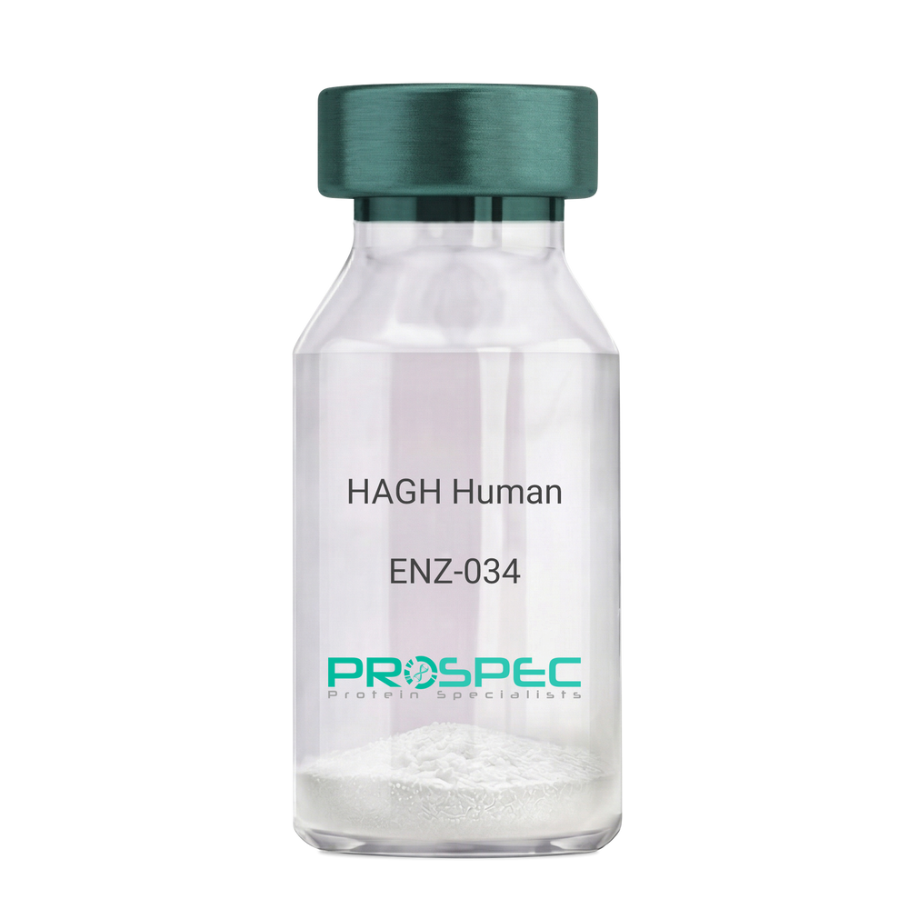
  • View Data Sheet

    Name :

    HAGH Human

    Description:

    Hydroxyacylglutathione Hydrolase Human Recombinant

    GLX2, Glyoxalase II, GLO2, Hydroxyacyl Glutathione Hydrolase, HAGH1, GLXII, Hydroxyacylglutathione Hydrolase, hydroxyacylglutathione hydroxylase.

    Product # :

    ENZ-034

    Price :

    Quantity :

    Shipping Method :

    Ice Icon

    Shipped with Ice Packs

    Add To Cart

    More Info

    • description
    • source
    • formulation
    • purity
    • More Info

    Description

    HAGH produced in E.Coli is a single, non-glycosylated polypeptide chain containing 284 amino acids (1-260a.a.) and having a molecular mass of 31.4kDa.HAGH is fused to a 24 amino acid His-tag at N-terminus & purified by proprietary chromatographic techniques.

    Source

    Escherichia Coli.

    Formulation

    The HAGH protein solution (0.5mg/1ml) is formulated in 20mM Tris-HCl Buffer (pH 8.5) and 10% Glycerol.

    Purity

    Greater than 95% as determined by SDS-PAGE.

    More Info

    • Introduction

      HAGH is a part of the glyoxalase family and a thiolesterase which hydrolyses S-lactoyl-glutathione to reduced glutathione and D-lactate. HAGH protein is a detoxifying enzyme of glycolysis byproduct methylglyoxal and a target of p63 and p73 and serves as a pro-survival factor of the p53 family. HAGH appears only as a monomer and binds two zinc ions per subunit.

    • Synonyms

      GLX2, Glyoxalase II, GLO2, Hydroxyacyl Glutathione Hydrolase, HAGH1, GLXII, Hydroxyacylglutathione Hydrolase, hydroxyacylglutathione hydroxylase.

    • Physical Appearance

      Sterile Filtered clear solution.

    • Stability

      Store at 4°C if entire vial will be used within 2-4 weeks. Store, frozen at -20°C for longer periods of time.For long term storage it is recommended to add a carrier protein (0.1% HSA or BSA).Please avoid freeze thaw cycles.

    • Amino Acid Sequence

      MGSSHHHHHH SSGLVPRGSH MGSHMKVEVL PALTDNYMYL VIDDETKEAA IVDPVQPQKV VDAARKHGVK LTTVLTTHHH WDHAGGNEKL VKLESGLKVY GGDDRIGALT HKITHLSTLQ VGSLNVKCLA TPCHTSGHIC YFVSKPGGSE PPAVFTGDTL FVAGCGKFYE GTADEMCKAL LEVLGRLPPD TRVYCGHEYT INNLKFARHV EPGNAAIREK LAWAKEKYSI GEPTVPSTLA EEFTYNPFMR VREKTVQQHA GETDPVTTMR AVRREKDQFK MPRD.

    ProSpec's products are furnished for LABORATORY RESEARCH USE ONLY. The product may not be used as drugs, agricultural or pesticidal products, food additives or household chemicals.

    Hagh Human
  • View Data Sheet

    Name :

    RAET1E Mouse

    Description:

    Retinoic Acid Early Transcript 1E Mouse Recombinant

    RAE-1-epsilon, Retinoic acid early-inducible protein 1-epsilon, Raet1e.

    Product # :

    PRO-2362

    Price :

    Quantity :

    Shipping Method :

    Ice Icon

    Shipped with Ice Packs

    Add To Cart

    More Info

    • description
    • source
    • formulation
    • purity
    • More Info

    Description

    RAET1E produced in Sf9 Insect cells is a single, glycosylated polypeptide chain containing 207 amino acids (29-227a.a.) and having a molecular mass of 23.5kDa (Molecular size on SDS-PAGE will appear at approximately 28-40kDa).RAET1E is expressed with an 8 amino acid His tag at C-Terminus and purified by proprietary chromatographic techniques.

    Source

    Sf9, Insect cells.

    Formulation

    RAET1E protein solution (0.5mg/ml) contains Phosphate Buffered Saline (pH 7.4) and 10% glycerol.

    Purity

    Greater than 95.0% as determined by SDS-PAGE.

    More Info

    • Introduction

      RAET1E is a member of the MHC class I family. MHC class I family contains main histocompatibility complex (MHC) class I-related genes positioned in a cluster on chromosome 6q24.2-q25.3. RAET1E and RAET1G protein are different from other RAET1 proteins since they have type I membrane-spanning sequences at their C termini instead glycosylphosphatidylinositol anchor sequences.RAET1E acts as a ligand for NKG2D receptor, expressed on the surface of numerous types of immune cells, involves in innate adaptive immune reactions.RAET1E delivers signals to NK cells and advances tumor immune surveillance by inducing the growth of anti-tumor cytotoxic lymphocyte.

    • Synonyms

      RAE-1-epsilon, Retinoic acid early-inducible protein 1-epsilon, Raet1e.

    • Physical Appearance

      Sterile Filtered clear solution.

    • Stability

      Store at 4°C if entire vial will be used within 2-4 weeks. Store, frozen at -20°C for longer periods of time. For long term storage it is recommended to add a carrier protein (0.1% HSA or BSA).Avoid multiple freeze-thaw cycles.

    • Amino Acid Sequence

      LDDAHSLRCN LTIKDPTSAD LPWCDVKCSV DEITILHLNN INKTMTSGDP GKMANATGKC LTQPLNDLCQ ELRDKVSNTK VDTHKTNGYP HLQVTMIYPQ SQGQTPSATW EFNISDSYFF TFYTENMSWR SANDESGVIM NKWKDDGDLV QQLKYFIPQC RQKIDEFLKQ SKEKPRSTSR SPSITQLTST SPLPPPSHSL EHHHHHH.

    ProSpec's products are furnished for LABORATORY RESEARCH USE ONLY. The product may not be used as drugs, agricultural or pesticidal products, food additives or household chemicals.

    Raet1E Mouse
  • View Data Sheet

    Name :

    KRT5 Antibody

    Description:

    Cytokeratin 5, Mouse Anti Human

    Keratin 5, KRT5, EBS2, Epidermolysis Bullosa Simplex 2 Dowling-Meara/Kobner/Weber-Cockayne Types, Keratin 5 (Epidermolysis Bullosa Simplex,Dowling-Meara/Kobner/Weber-Cockayne Types), 58 KDa Cytokeratin, Type-II Keratin Kb5, CK-5, K5, DDD1, CK5, DDD, KRT5A, cytokeratin-5, Keratin, Type II Cytoskeletal 5, Cytokeratin-5, Keratin-5.

    Product # :

    ANT-588

    Price :

    Quantity :

    Shipping Method :

    Ice Icon

    Shipped with Ice Packs

    Add To Cart

    More Info

    • formulation
    • More Info

    Formulation

    1mg/ml containing PBS, pH-7.4, 10% Glycerol and 0.02% Sodium Azide.

    More Info

    • Introduction

      Cytokeratin 5 (KRT5) belongs to the keratin gene family. The type II cytokeratins are comprised of basic or neutral proteins which are arranged in pairs of heterotypic keratin chains coexpressed throughout differentiation of simple and stratified epithelial tissues. The type II cytokeratins are clustered in a region of chromosome 12q12-q13. The KRT5 type II cytokeratin is specifically expressed in the basal layer of the epidermis with family member KRT14. Mutations in these genes are linked with a complex of diseases termed epidermolysis bullosa simplex.

    • Synonyms

      Keratin 5, KRT5, EBS2, Epidermolysis Bullosa Simplex 2 Dowling-Meara/Kobner/Weber-Cockayne Types, Keratin 5 (Epidermolysis Bullosa Simplex,Dowling-Meara/Kobner/Weber-Cockayne Types), 58 KDa Cytokeratin, Type-II Keratin Kb5, CK-5, K5, DDD1, CK5, DDD, KRT5A, cytokeratin-5, Keratin, Type II Cytoskeletal 5, Cytokeratin-5, Keratin-5.

    • Physical Appearance

      Sterile filtered colorless solution.

    • Immunogen

      Anti-human KRT5 mAb, is derived from hybridization of mouse F0 myeloma cells with spleen cells from BALB/c mice immunized with a recombinant human KRT5 protein 1-590 amino acids purified from E. coli.

    • Ig Subclass

      Mouse IgG2a heavy chain and k light chain.

    • Clone

      PAT12C4AT.

    • Applications

      KRT5 antibody has been tested by ELISA, Western blot analysis, ICC/IF and Flow cytometry to assure specificity and reactivity. Since application varies, however, each investigation should be titrated by the reagent to obtain optimal results.

    • Type

      Mouse Anti Human Monoclonal.

    • Storage Procedures

      For periods up to 1 month store at 4°C, for longer periods of time, store at -20°C. Prevent freeze thaw cycles.

    • Purification Method

      KRT5 antibody was purified from mouse ascitic fluids by protein-A affinity chromatography.

    ProSpec's products are furnished for LABORATORY RESEARCH USE ONLY. The product may not be used as drugs, agricultural or pesticidal products, food additives or household chemicals.

    Krt5 Antibody
  • 1
  • …
  • 26
  • 27
  • 28
  • 29
  • 30
  • …
  • 42
Your browser does not support the video tag.

Products

  • Cytokines
  • Growth Factors
  • Chemokines
  • CD Antigens
  • Neurotrophins
  • Hormones
  • Enzymes
  • Viral Antigens
  • Recombinant Proteins
  • Natural Proteins
  • Monoclonal Antibodies
  • Polyclonal Antibodies
  • Thermal Packaging

About

  • Company
  • Founder
  • Contribution
  • Contact Us
  • My Account

Ordering

  • Ordering Method
  • Ordering - Credit Card
  • Ordering PO
  • Shipping Fees
  • FAQ's

Legal

  • Terms & Conditions
  • Privacy
  • Site Map
  • info@prospecbio.com
  • Facebook
  • Instagram
  • Linkedin Profile

© 2026 ProSpec-Tany TechnoGene Ltd. All Rights Reserved.

  • Choosing a selection results in a full page refresh.
  • Opens in a new window.