Search results
1000 results found for “Syntaxin”
Name
Description
Product #
Price
Quantity
Shipping Method
- View Data Sheet
Name :
AIMP1 AntibodyDescription:
Aminoacyl tRNA Synthetase Complex-Interacting Multifunctional Protein 1, Mouse Anti Human
Aminoacyl tRNA synthase complex-interacting multifunctional protein 1, Multisynthase complex auxiliary component p43, AIMP1, EMAP2, SCYE1, p43, EMAPII.
Product # :
ANT-619Price :
Quantity :
Shipping Method :
Shipped with Ice Packs
More Info
- formulation
- More Info
Formulation
1mg/ml containing PBS, pH-7.4, 10% Glycerol and 0.02% Sodium Azide.
More Info
-
Introduction
AIMP1 (EMPA2 or p43) is a cytokine that is specifically induced by apoptosis, and it is involved in the control of angiogenesis, inflammation, and wound healing. The release of the AIMP1 cytokine renders the tumor-associated vasculature sensitive to tumor necrosis factor. Furthermore, AIMP1 is involved in the stimulation of inflammatory responses after proteolytic cleavage in tumor cells.
-
Synonyms
Aminoacyl tRNA synthase complex-interacting multifunctional protein 1, Multisynthase complex auxiliary component p43, AIMP1, EMAP2, SCYE1, p43, EMAPII.
-
Physical Appearance
Sterile filtered colorless solution.
-
Immunogen
Anti-human AIMP1 mAb, is derived from hybridization of mouse F0 myeloma cells with spleen cells from BALB/c mice immunized with a recombinant human AIMP1 protein 1-336 amino acids purified from E. coli.
-
Ig Subclass
Mouse IgG2a heavy chain and Kappa light chain.
-
Clone
PAT6D10AT.
-
Applications
The antibody has been tested by ELISA, Western blot analysis, Flow cytometry and ICC/IF to assure specificity and reactivity. Since application varies, however, each investigation should be titrated by the reagent to obtain optimal results.
-
Type
Mouse Anti Human Monoclonal.
-
Storage Procedures
For periods up to 1 month store at 4°C, for longer periods of time, store at -20°C. Prevent freeze thaw cycles.
-
Purification Method
AIMP1 antibody was purified by protein-A affinity chromatography.
ProSpec's products are furnished for LABORATORY RESEARCH USE ONLY. The product may not be used as drugs, agricultural or pesticidal products, food additives or household chemicals.
- View Data Sheet
Name :
HAX1 AntibodyDescription:
HCLS1-associated protein X-1, Mouse Anti Human
HCLS1-associated protein X-1, HS1-associating protein X-1, HS1-binding protein 1, HAX-1, HSP1BP-1, HAX1, HS1BP1.
Product # :
ANT-446Price :
Quantity :
Shipping Method :
Shipped with Ice Packs
More Info
- formulation
- More Info
Formulation
1mg/ml containing PBS, pH-7.4, & 0.1% Sodium Azide.
More Info
-
Introduction
HAX1 associates with hematopoietic cell-specific Lyn substrate 1, which is a substrate of Src family tyrosine kinases. HAX1 also interacts with the product of the polycystic kidney disease 2 gene, mutations in which are associated with autosomal-dominant polycystic kidney disease, and with the F-actin-binding protein, cortactin. It was earlier thought HAX1 is mainly localized to the mitochondria, however, recent studies indicate it to be localized in the cell body. Mutations in the HAX1 gene result in autosomal recessive severe congenital neutropenia, aka Kostmann disease.
-
Synonyms
HCLS1-associated protein X-1, HS1-associating protein X-1, HS1-binding protein 1, HAX-1, HSP1BP-1, HAX1, HS1BP1.
-
Immunogen
Anti-human HAX1 mAb is derived from hybridization of mouse F0 myeloma cells with spleen cells from BALB/c mice immunized with recombinant human HAX1 amino acids 1-279 purified from E. coli.
-
Ig Subclass
Mouse IgG2b heavy chain and κ light chain.
-
Clone
PAT3C5AT.
-
Applications
HAX1 antibody has been tested by ELISA and Western blot analysis to assure specificity and reactivity. Since application varies, however, each investigation should be titrated by the reagent to obtain optimal results. Recommended dilution range for Western blot analysis is 1:1000 ~ 2000. Recommended starting dilution is 1:1000.
-
Type
Mouse Anti Human Monoclonal.
-
Storage Procedures
For periods up to 1 month store at 4°C, for longer periods of time, store at -20°C. Prevent freeze thaw cycles.
-
Purification Method
HAX1 antibody was purified from mouse ascitic fluids by protein-G affinity chromatography.
ProSpec's products are furnished for LABORATORY RESEARCH USE ONLY. The product may not be used as drugs, agricultural or pesticidal products, food additives or household chemicals.
- View Data Sheet
Name :
Lysozyme HumanDescription:
Lysozyme Human Recombinant
EC 3.2.1.17, LYZ, Lysozyme.
Product # :
ENZ-1159Price :
Quantity :
Shipping Method :
Shipped at Room temp
More Info
- description
- source
- formulation
- purity
- biological activity
- More Info
Description
Recombinant Human Lysozyme produced in Plant is a non-glycosylated, polypeptide chain containing 130 amino acids and having a molecular mass of 14.7kDa. The Recombinant Human Lysozyme is purified by proprietary chromatographic techniques.
Source
Oryza sativa (rice).
Formulation
The protein (1mg/ml) was lyophilized with no additives.
Purity
Purity as determined by SDS-PAGE is greater than 90%.
Biological Activity
1.6x10⁵ U/mg.
More Info
-
Introduction
Lysozymeis an antimicrobial enzyme produced by animals that are part of the innate immune system. Lysozyme is a glycoside hydrolase that catalyzes the hydrolysis of 1,4-beta-linkages between N-acetylmuramic acid and N-acetyl-D-glucosamine residues in peptidoglycan, which is the major component of gram-positive bacterial cell wall.Lysozymes have primarily a bacteriolytic function - those in tissues and body fluids are associated with the monocyte-macrophage system and enhance the activity of immunoagents.
-
Synonyms
EC 3.2.1.17, LYZ, Lysozyme.
-
Physical Appearance
Sterile Filtered lyophilized (freeze-dried) off white powder.
-
Stability
Store the lyophilized Lysozyme between 2-8°C, do not freeze. Upon reconstitution Lysozyme should be stored at 4°C between 2-7 days and for future use below -18°C. Please prevent freeze-thaw cycles.
-
Solubility
Stock solutions can be prepared by dissolving gently into PBS for several minutes. Recommended stock concentrations are 1mg/ml in PBS.
ProSpec's products are furnished for LABORATORY RESEARCH USE ONLY. The product may not be used as drugs, agricultural or pesticidal products, food additives or household chemicals.
- View Data Sheet
Name :
Ganirelix peptideDescription:
Ganirelix
Product # :
HOR-276Price :
Quantity :
Shipping Method :
Shipped with Ice Packs
More Info
- description
- formulation
- purity
- More Info
Description
Ganirelix acetate is a synthetic decapeptide with high antagonistic activity against naturally occurring gonadotropin-releasing hormone (GnRH). Ganirelix acetate is derived from native GnRH with substitutions of amino acids at positions 1, 2, 3, 6, 8, and 10 to form the following molecular formula of the peptide: N-acetyl-3-(2-napthyl)-D-alanyl-4-chloro-D-phenylalanyl-3-(3-pyridyl)-D-alanyl-L-seryl-L-tyrosyl-N 9 ,N 10 -diethyl- D-homoarginyl-L-leucyl-N 9 ,N 10 -diethyl-L-homoarginyl-L-prolyl-D-alanylamide acetate. The molecular weight for Ganirelix acetate is 1570.4 Dalton as an anhydrous free base.
Formulation
The Ganirelix hormone (0.5mg/ml) contains 0.1mg acetic acid and 23.5mg mannitol pH-5.
Purity
Greater than 98.0% as determined by:
(a) Analysis by RP-HPLC.
(b) Analysis by SDS-PAGE.More Info
-
Physical Appearance
Sterile Filtered colorless liquid formulation.
-
Stability
Ganirelix should be stored between 2°C- 8°C at all time. DO NOT FREEZE.
-
Background
What is the molecular weight/Mw of GANIRELIX PEPTIDE Protein?
GANIRELIX PEPTIDE Protein has a total Mw of 1.57kDa.
What is the Purity of GANIRELIX PEPTIDE Protein?
GANIRELIX PEPTIDE Protein is >98% pure as determined by SDS-PAGE.
What is the Biological Activity of GANIRELIX PEPTIDE Protein?
The biological functionality of GANIRELIX PEPTIDE Protein will be determined in the future.
What applications can GANIRELIX PEPTIDE Protein be used in?
GANIRELIX PEPTIDE Protein can probably be used in western blot, ELISA and Lateral Flow.
What is the endotoxin level for GANIRELIX PEPTIDE Protein?
The endotoxin level is minimal, GANIRELIX PEPTIDE Protein was purified using conventional chromatography techniques.
ProSpec's products are furnished for LABORATORY RESEARCH USE ONLY. The product may not be used as drugs, agricultural or pesticidal products, food additives or household chemicals.
- View Data Sheet
Name :
SAE1 HumanDescription:
SUMO1 Activating Enzyme Subunit 1 Human Recombinant
AOS1, HSPC140, SUA1, UBLE1A, SAE1, SUMO1 Activating Enzyme Subunit 1, FLJ3091.
Product # :
ENZ-534Price :
Quantity :
Shipping Method :
Shipped with Ice Packs
More Info
- description
- source
- formulation
- purity
- More Info
Description
SAE1 Recombinant Human produced in E.Coli is a single, non-glycosylated polypeptide chain containing 378 amino acids (1-346 a.a.) and having a molecular mass of 42.2 kDa. The SAE1 is fused to 32 amino acid T7-Tag at N-terminus and purified by proprietary chromatographic techniques.
Source
Escherichia Coli.
Formulation
SAE1 Human solution containing 20mM Tris pH-8, 1mM DTT & 10% glycerol.
Purity
Greater than 90.0% as determined by SDS-PAGE.
More Info
-
Introduction
SAE1 is part of the ubiquitin-activating E1 family of proteins and participates in the significant first step of the UBL1 conjugation pathway. Proteins conjugated to Ub are marked for progressive degradation by the 26S Proteasome. SAE1 acts as a UBLI E1 ligase mediating the ATP-dependent activation of UBL1. SAE1 binds with UBLE1A and UBLE1B to form a heterodimer which can bind UBL1. SAE1 is a dimeric enzyme that takes part as a E1 ligase for SUMO1, SUMO2, SUMO3, and probably SUMO4. SAE1 regulates ATP-dependent activation of SUMO proteins and formation of a thioester with a conserved cysteine residue on SAE2.
-
Synonyms
AOS1, HSPC140, SUA1, UBLE1A, SAE1, SUMO1 Activating Enzyme Subunit 1, FLJ3091.
-
Physical Appearance
Sterile filtered colorless solution.
-
Stability
Store at 4°C if entire vial will be used within 2-4 weeks. Store, frozen at -20°C for longer periods of time. For long term storage it is recommended to add a carrier protein (0.1% HSA or BSA).Avoid multiple freeze-thaw cycles.
-
Amino Acid Sequence
MHHHHHHMAS MTGGQQMGRD LYDDDDKDRW GSMVEKEEAG GGISEEEAAQ YDRQIRLWGL EAQKRLRASR VLLVGLKGLG AEIAKNLILA GVKGLTMLDH EQVTPEDPGA QFLIRTGSVG RNRAEASLER AQNLNPMVDV KVDTEDIEKK PESFFTQFDA VCLTCCSRDV IVKVDQICHK NSIKFFTGDV FGYHGYTFAN LGEHEFVEEK TKVAKVSQGV EDGPDTKRAK LDSSETTMVK KKVVFCPVKE ALEVDWSSEK AKAALKRTTS DYFLLQVLLK FRTDKGRDPS SDTYEEDSEL LLQIRNDVLD SLGISPDLLP EDFVRYCFSE MAPVCAVVGG ILAQEIVKAL SQRDPPHNNF FFFDGMKGNG IVECLGPK.
ProSpec's products are furnished for LABORATORY RESEARCH USE ONLY. The product may not be used as drugs, agricultural or pesticidal products, food additives or household chemicals.
- View Data Sheet
Name :
HBXIP HumanDescription:
Hepatitis B Virus x Interacting Protein Human Recombinant
Ragulator complex protein LAMTOR5, Hepatitis B virus X-interacting protein, HBV X-interacting protein, HBX-interacting protein, Late endosomal/lysosomal adaptor and MAPK and MTOR activator 5, LAMTOR5, HBXIP, XIP.
Product # :
HBV-235Price :
Quantity :
Shipping Method :
Shipped with Ice Packs
More Info
- description
- source
- formulation
- purity
- More Info
Description
HBXIP Human Recombinant produced in E. coli is a single polypeptide chain containing 197 amino acids (aa 1-173) and having a molecular mass of 20.7kDa.HBXIP is fused to a 24 amino acid His-tag at N-terminus & purified by proprietary chromatographic techniques.
Source
Escherichia Coli.
Formulation
The HBXIP solution (1mg/ml) contains 20mM Tris-HCl buffer (pH 8.0), 0.1M NaCl, 10% glycerol and 1mM EDTA.
Purity
Greater than 90% as determined by SDS-PAGE.
More Info
-
Introduction
Hepatitis B virus x interacting protein (HBXIP) forms a complex with the C-terminus of hepatitis B virus X (HBX) protein. HBXIP negatively regulates HBX activity and changes the replicative life cycle of the virus. Furthermore, HBXIP is involved in bipolar spindle formation and regulates centrosome dynamics and cytokinesis in cells, possibly due to interaction with Dynein light chain. HBXIP is highly expressed in the skeletal and cardiac muscle, followed by pancreas, kidney, liver, brain, placenta and lung. HBXIP has elevated levels in both cancerous and non-cancerous liver tissue of patients with chronic HBV infection compared with hepatic tissue without HBV infection.
-
Synonyms
Ragulator complex protein LAMTOR5, Hepatitis B virus X-interacting protein, HBV X-interacting protein, HBX-interacting protein, Late endosomal/lysosomal adaptor and MAPK and MTOR activator 5, LAMTOR5, HBXIP, XIP.
-
Physical Appearance
Sterile filtered colorless solution.
-
Stability
Store at 4°C if entire vial will be used within 2-4 weeks. Store, frozen at -20°C for longer periods of time. For long term storage it is recommended to add a carrier protein (0.1% HSA or BSA).Avoid multiple freeze-thaw cycles.
-
Amino Acid Sequence
MGSSHHHHHH SSGLVPRGSH MGSHMEPGAG HLDGHRAGSP SLRQALCDGS AVMFSSKERG RCTVINFVPL EAPLRSTPRS RQVTEACGGE GRAVPLGSEP EWSVGGMEAT LEQHLEDTMK NPSIVGVLCT DSQGLNLGCR GTLSDEHAGV ISVLAQQAAK LTSDPTDIPV VCLESDNGNI MIQKHDGITV AVHKMAS.
ProSpec's products are furnished for LABORATORY RESEARCH USE ONLY. The product may not be used as drugs, agricultural or pesticidal products, food additives or household chemicals.
- View Data Sheet
Name :
DBI MouseDescription:
Diazepam Binding Inhibitor Mouse Recombinant
Acyl-CoA-binding protein, ACBP, Diazepam-binding inhibitor, DBI, Endozepine, EP, Dbi.
Product # :
PRO-2527Price :
Quantity :
Shipping Method :
Shipped with Ice Packs
More Info
- description
- source
- formulation
- purity
- More Info
Description
DBI Mouse Recombinant produced in E.Coli is a single, non-glycosylated polypeptide chain containing 110 amino acids (1-87 a.a) and having a molecular mass of 12.4kDa. DBI is fused to a 23 amino acid His-tag at N-terminus & purified by proprietary chromatographic techniques.
Source
Escherichia Coli.
Formulation
DBI protein solution (1mg/ml) contains Phosphate buffer saline (pH7.4).
Purity
Greater than 95.0% as determined by SDS-PAGE.
More Info
-
Introduction
DBI, also known as Acyl-CoA-binding protein isoform 2, is a diazepam binding inhibitor, which is regulated by hormones. DBI participates in lipid metabolism and in the displacement of beta-carbolines & benzodiazepines, which modulate signal transduction at type A gamma-aminobutyric acid receptors located in brain synapses. Moreover, during adipocyte differentiation the expression of DBI is significantly induced. DBI preforms as an acyl-CoA pool former and regulates LCFA (long-chain fatty acids) metabolism in peripheral tissues.
-
Synonyms
Acyl-CoA-binding protein, ACBP, Diazepam-binding inhibitor, DBI, Endozepine, EP, Dbi.
-
Physical Appearance
Sterile Filtered clear solution.
-
Stability
Store at 4°C if entire vial will be used within 2-4 weeks. Store, frozen at -20°C for longer periods of time. For long term storage it is recommended to add a carrier protein (0.1% HSA or BSA).Avoid multiple freeze-thaw cycles.
-
Amino Acid Sequence
MGSSHHHHHH SSGLVPRGSH MGSMSQAEFD KAAEEVKRLK TQPTDEEMLF IYSHFKQATV GDVNTDRPGL LDLKGKAKWD SWNKLKGTSK ESAMKTYVEK VDELKKKYGI
ProSpec's products are furnished for LABORATORY RESEARCH USE ONLY. The product may not be used as drugs, agricultural or pesticidal products, food additives or household chemicals.
- View Data Sheet
Name :
DACT3 AntibodyDescription:
Dapper homolog 3, Mouse Anti Human
Dapper homolog 3, Antagonist of beta-catenin Dapper homolog 3, Arginine-rich region 1 protein, DACT3, RRR1, MGC15476.
Product # :
ANT-432Price :
Quantity :
Shipping Method :
Shipped with Ice Packs
More Info
- formulation
- More Info
Formulation
1mg/ml containing PBS, pH-7.4, & 0.1% Sodium Azide.
More Info
-
Introduction
DACT3 which belongs to the DACT (Dpr/Frodo) gene family plays an imperative role in a negative regulator of Wnt/-catenin signaling. Unlike other Wnt signaling inhibitors that are silenced by DNA methylation, DACT3 repression is linked to bivalent histone modifications.
-
Synonyms
Dapper homolog 3, Antagonist of beta-catenin Dapper homolog 3, Arginine-rich region 1 protein, DACT3, RRR1, MGC15476.
-
Immunogen
Anti-human DACT3 mAb, is derived from hybridization of mouse F0 myeloma cells with spleen cells from BALB/c mice immunized with recombinant human DACT3 amino acids 89-202 purified from E. coli.
-
Ig Subclass
Mouse IgG1 heavy chain and κ light chain.
-
Clone
P2A5AT.
-
Applications
DACT3 antibody has been tested by ELISA, Western blot and immunohistochemistry analysis to assure specificity and reactivity. Since application varies, however, each investigation should be titrated by the reagent to obtain optimal results. Recommended dilution range for Western blot is 1:250 ~ 1:500 and immunohistochemistry analysis is 1:50. Recommended starting dilution for Western blot is 1:250 and Immunohistochemistry is 1:50.
-
Type
Mouse Anti Human Monoclonal.
-
Storage Procedures
For periods up to 1 month store at 4°C, for longer periods of time, store at -20°C. Prevent freeze thaw cycles.
-
Purification Method
DACT3 antibody was purified from mouse ascitic fluids by protein-G affinity chromatography.
ProSpec's products are furnished for LABORATORY RESEARCH USE ONLY. The product may not be used as drugs, agricultural or pesticidal products, food additives or household chemicals.
- View Data Sheet
Name :
NKp46 AntibodyDescription:
Natural Cytotoxicity Receptor NKp46, Mouse Anti Human
Natural cytotoxicity triggering receptor 1, Natural killer cell p46-related protein, hNKp46, NK-p46, NKp46, NK cell-activating receptor, Lymphocyte antigen 94 homolog, CD335 antigen, NCR1, LY94, NCRNKp46, CD335.
Product # :
ANT-303Price :
Quantity :
Shipping Method :
Shipped with Ice Packs
More Info
- formulation
- More Info
Formulation
1mg/ml containing PBS, pH-7.4, & 0.1% Sodium Azide.
More Info
-
Introduction
A natural cytotoxicity receptor (NCR) NKp46 has been shown to represent a novel NK cell-specific molecule involved in human NK cell activation. The natural cytotoxicity receptors (NCRs) are a recently characterized family of Ig-like activation receptors that appear to be major triggering receptors in tumor cell recognition. The three known NCRs include NKp46 and NKp30, which are expressed on circulating NKcells, and NKp44, which is expressed only on activating NK cells. NKp46 has been implicated in NK cell-mediated lysis of several autologous tumor cells, pathogen-infected cell lines and mononuclear phagocytes infected with an intracellular bacterium. The Lysis of tumor cells by NK-cells involves recognition by NKp46 of heparan sulfate moieties of membrane heparan sulfate proteoglycans. Furthermore, NKp46 is a surface receptor involved in NK-cell cell death by apoptosis. NKp46 has two extracellular Ig-like domains followed by a ~40 residue stalk region, a type I transmembrane domain, and a short cytoplasmic tail. The extracellular Ig-like domain of NKp46 (22-255aa) is purified by FPLC gel-filtration chromatography, after refolding of the isolated inclusion bodies in a redox buffer. In addition, engagement of the antigen with the monoclonal antibody stimulates intracellular calcium levels and the synthesis of cytokines. CD59 is an NKp46 coreceptor (by physical association) together they activate cytotoxicity of human NK-cells, their engagement results in tyrosine phosphorylation of CD3-zeta chains associated with NKp46. Reduced cell surface expression of NKp46 and other NK-cell receptors is linked to the impaired NK-cell cytolytic function in viremic HIV-1 infection.
-
Synonyms
Natural cytotoxicity triggering receptor 1, Natural killer cell p46-related protein, hNKp46, NK-p46, NKp46, NK cell-activating receptor, Lymphocyte antigen 94 homolog, CD335 antigen, NCR1, LY94, NCRNKp46, CD335.
-
Physical Appearance
Sterile Filtered colorless solution.
-
Immunogen
Anti-human NKp46 mAb, is derived from hybridization of mouse SP2/0 myeloma cells with spleen cells from BALB/c mice immunized with recombinant human NKp46 amino acids 22-255 purified from E. coli.
-
Ig Subclass
Mouse IgG1 heavy chain and κ light chain.
-
Clone
Pn1D9AT.
-
Applications
NKp46 antibody has been tested by immunofluorescent staining with flow cytometric analysis and by Western blot analysis to assure specificity and reactivity. Since application varies, however, each investigation should be titrated by the reagent to obtain optimal results.
-
Type
Mouse Anti Human Monoclonal.
-
Storage Procedures
For periods up to 1 month store at 4°C, for longer periods of time, store at -20°C. Prevent freeze thaw cycles.
-
Purification Method
NKp46 antibody was purified from mouse ascitic fluids by protein-G affinity chromatography.
ProSpec's products are furnished for LABORATORY RESEARCH USE ONLY. The product may not be used as drugs, agricultural or pesticidal products, food additives or household chemicals.
- View Data Sheet
Name :
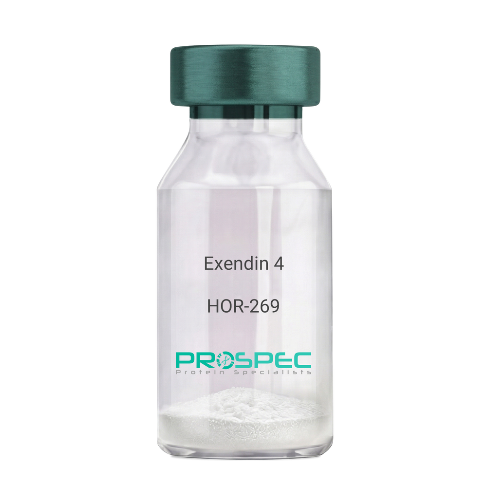
Exendin 4Description:
Exendin-4 Recombinant
Product # :
HOR-269Price :
Quantity :
Shipping Method :
Shipped at Room temp
More Info
- description
- source
- formulation
- purity
- biological activity
- More Info
Description
Exendin-4 Recombinant produced in E.Coli is a non-glycosylated polypeptide chain containing 39 amino acids and having a molecular mass of approximately 4.2kDa. Exendin-4 is purified by proprietary chromatographic techniques.
Source
Escherichia Coli.
Formulation
Lyophilized from a 0.2μm filtered concentrated solution in PBS, pH 7.4.
Purity
Greater than 96.0% as determined by:
(a) Analysis by RP-HPLC.
(b) Analysis by SDS-PAGE.Biological Activity
1. Regulates Glucose levels rapidly
2. Reduces Insulin resistance
3. Reduces Glucagon4. Stimulates beta cell growth production.
More Info
-
Introduction
Exedin-4 Recombinant is a like peptide-1 receptor agonist. The native hormone is produced in the gut of Gila monster Heloderma suspectrum (a type of reptile found in the desert) that stimulates production without causing threateningly low blood sugar, which can occur after using some anti-diabetes products. Recently, researchers used extracted saliva from gila monsters to create an unprecedented breakthrough in Diabetes Type 2 treatment. Unlike other products taken for type 2 diabetes, Exedin-4 has not been linked with weight gain and actually resulted in weight loss, according to the researchers. Exedin-4 enhances glucose-dependant secretion, suppresses inappropriately elevated secretion and slows gastric emptying in vivo. It also promotes B-cell proliferation and neogenesis in vitro and in animal models. Exedin-4 stimulates an increase in acinar cAMP, without stimulating the release of amylase.
-
Physical Appearance
Sterile Filtered White lyophilized (freeze-dried) powder.
-
Stability
Lyophilized Exendin Recombinant although stable at room temperature for 3 weeks, should be stored desiccated below -18°C. Upon reconstitution Exendin-4 should be stored at 4°C between 2-7 days and for future use below -18°C. For long term storage it is recommended to add a carrier protein (0.1% HSA or BSA).Please prevent freeze-thaw cycles.
-
Solubility
It is recommended to reconstitute the lyophilized Exendin Recombinant in sterile 18MΩ-cm H2O not less than 100 µg/ml, which can then be further diluted to other aqueous solutions.
-
Amino Acid Sequence
HGEGTFTSDL SKQMEEEAVR LFIEWLKNGG PSSGAPPPS.
ProSpec's products are furnished for LABORATORY RESEARCH USE ONLY. The product may not be used as drugs, agricultural or pesticidal products, food additives or household chemicals.
- View Data Sheet
Name :
SIGLEC5 HumanDescription:
Sialic Acid Binding Ig Like Lectin 5 Human Recombinant
Sialic Acid Binding Ig Like Lectin 5, Obesity-Binding Protein 2, OB-Binding Protein 2, CD33 Antigen-Like 2, SIGLEC-5, CD33L2, OB-BP2, OBBP2, Sialic Acid-Binding Immunoglobulin-Like Lectin 5, Sialic Acid Binding Ig-Like Lectin 5, Sialic Acid-Binding Ig-Like Lectin 5, CD170 Antigen, CD170, Sialic acid-binding Ig-like lectin 5, Siglec-5, CD33 antigen-like 2, Obesity-binding protein 2, OB-BP2, OB-binding protein 2, CD170.
Product # :
PRO-2432Price :
Quantity :
Shipping Method :
Shipped with Ice Packs
More Info
- description
- source
- formulation
- purity
- More Info
Description
SIGLEC5 Human Recombinant produced in Sf9 Baculovirus cells is a single, glycosylated polypeptide chain containing 667 amino acids (17-441a.a.) and having a molecular mass of 74.2kDa (Molecular size on SDS-PAGE will appear at approximately 70-100kDa). SIGLEC5 is expressed with a 239 amino acid hIgG-His Tag at C-Terminus and purified by proprietary chromatographic techniques.
Source
Sf9, Baculovirus cells.
Formulation
SIGLEC5 protein solution (0.25mg/ml) contains Phosphate Buffered Saline (pH 7.4).
Purity
Greater than 90.0% as determined by SDS-PAGE.
More Info
-
Introduction
Sialic Acid Binding Ig Like Lectin 5, also known as SIGLEC5 is a member of the immunoglobulin superfamily and SIGLEC family. SIGLEC5 contains of 2 Ig-like C2-type domains and 1 Ig-like V-type domain. In addition, SIGLEC5 is expressed by monocytic/myeloid lineage cells. SIGLEC5 is expressed at high levels in peripheral blood leukocytes, spleen, and at lower levels in lymph node, appendix, pancreas, lung, and thymus.
-
Synonyms
Sialic Acid Binding Ig Like Lectin 5, Obesity-Binding Protein 2, OB-Binding Protein 2, CD33 Antigen-Like 2, SIGLEC-5, CD33L2, OB-BP2, OBBP2, Sialic Acid-Binding Immunoglobulin-Like Lectin 5, Sialic Acid Binding Ig-Like Lectin 5, Sialic Acid-Binding Ig-Like Lectin 5, CD170 Antigen, CD170, Sialic acid-binding Ig-like lectin 5, Siglec-5, CD33 antigen-like 2, Obesity-binding protein 2, OB-BP2, OB-binding protein 2, CD170.
-
Physical Appearance
Sterile Filtered colorless solution.
-
Stability
Store at 4°C if entire vial will be used within 2-4 weeks. Store, frozen at -20°C for longer periods of time. For long term storage it is recommended to add a carrier protein (0.1% HSA or BSA).Avoid multiple freeze-thaw cycles.
-
Amino Acid Sequence
ADLEKPVYEL QVQKSVTVQE GLCVLVPCSF SYPWRSWYSS PPLYVYWFRD GEIPYYAEVV ATNNPDRRVK PETQGRFRLL GDVQKKNCSL SIGDARMEDT GSYFFRVERG RDVKYSYQQN KLNLEVTALI EKPDIHFLEP LESGRPTRLS CSLPGSCEAG PPLTFSWTGN ALSPLDPETT RSSELTLTPR PEDHGTNLTC QMKRQGAQVT TERTVQLNVS YAPQTITIFR NGIALEILQN TSYLPVLEGQ ALRLLCDAPS NPPAHLSWFQ GSPALNATPI SNTGILELRR VRSAEEGGFT CRAQHPLGFL QIFLNLSVYS LPQLLGPSCS WEAEGLHCRC SFRARPAPSL CWRLEEKPLE GNSSQGSFKV NSSSAGPWAN SSLILHGGLS SDLKVSCKAW NIYGSQSGSV LLLQGRSNLG TGVVPAALLE PKSCDKTHTC PPCPAPELLG GPSVFLFPPK PKDTLMISRT PEVTCVVVDV SHEDPEVKFN WYVDGVEVHN AKTKPREEQY NSTYRVVSVL TVLHQDWLNG KEYKCKVSNK ALPAPIEKTI SKAKGQPREP QVYTLPPSRD ELTKNQVSLT CLVKGFYPSD IAVEWESNGQ PENNYKTTPP VLDSDGSFFL YSKLTVDKSR WQQGNVFSCS VMHEALHNHY TQKSLSLSPG KHHHHHH.
ProSpec's products are furnished for LABORATORY RESEARCH USE ONLY. The product may not be used as drugs, agricultural or pesticidal products, food additives or household chemicals.
- View Data Sheet
Name :
ATF BovineDescription:
Apo Transferrin Bovine
Serotransferrin, Transferrin, Siderophilin, Beta-1-metal-binding globulin, TF, PRO1557, PRO2086, DKFZp781D0156, Apo Transferrin, ATF.
Product # :
PRO-511Price :
Quantity :
Shipping Method :
Shipped at Room temp
More Info
- description
- source
- formulation
- purity
- More Info
Description
Bovine Apo Transferrin is a glycoprotein of approximately 77kDa.
Source
Bovine Serum.
Formulation
The protein (1mg/ml) was lyophilized with no additives.
Purity
Greater than 95.0% as determined by SDS-PAGE.
More Info
-
Introduction
Transferrin is the iron-transport protein of vertebrate serum and donates iron to cells through interaction with a specific membrane receptor, CD71. Transferrin appears to be indispensable for most cells growing in tissue culture.
It is referred to frequently as a growth factor because, in analogy to other growth factor-receptor interactions, proliferating cells express high numbers of transferrin receptors, and the binding of transferrin to their receptors is needed for cells to initiate and maintain their DNA synthesis. Apart from its role as an iron transport protein transferrin acts as a cytokine and has functions that may not be related to its iron-carrying capacity.
Bovine Transferrin is a crucial component for the cultivation of mammalian cells in- vitro. Bovine Transferrin is Critical for long-term cells growth in-vitro. Bovine Transferrin is used as detoxificant in media by binding contaminating metal ions. Bovine Transferrin is often used as a nutrient in fermentation media for recombinant protein and biopharmaceutical production. Additional common uses of Bovine Transferrin are Molecular weight, Affinity purification of anti-human transferrin antibodies and also as receptor mediated transfection of molecules such as DNA, into cells. -
Synonyms
Serotransferrin, Transferrin, Siderophilin, Beta-1-metal-binding globulin, TF, PRO1557, PRO2086, DKFZp781D0156, Apo Transferrin, ATF.
-
Physical Appearance
Sterile Filtered off-white lyophilized (freeze-dried) powder.
-
Stability
Store the lyophilized Apo Transferrin between 2-8°C, do not freeze.
-
Solubility
It is recommended to reconstitute the lyophilized Apo Transferrin in sterile 18MΩ-cm H2O not less than 1gr/30ml in 20min. at 20-25C.
-
Iron Content
The Iron content was estimated by ICP-OES and was found to be less than 40 ppm.
ProSpec's products are furnished for LABORATORY RESEARCH USE ONLY. The product may not be used as drugs, agricultural or pesticidal products, food additives or household chemicals.
- View Data Sheet
Name :
Thymosin beta 4Description:
Thymosin β4
Thymosin beta-4. TB500, TB-500
Product # :
HOR-275Price :
Quantity :
Shipping Method :
Shipped at Room temp
More Info
- description
- formulation
- purity
- More Info
Description
Thymosin b4 is a 43 amino acid peptide which is regarded as the main intracellular G-actin sequestering peptide. It has a molecular weight of 4963.55 Da, and its molecular formula is: C212H350N56O78S1. Extracellular Thymosin b4 may contribute to physiological processes such as angiogenesis, wound healing, and regulation of inflammation.
Formulation
The protein (1mg/ml) was lyophilized with no additives.
Purity
Greater than 98.0% as determined by RP-HPLC.
More Info
-
Introduction
Thymosin is a hormone secreted from the thymus. Its primary function is to stimulate the production of T cells, which are an important part of the immune system. Thymosin also assists in the development of B cells to plasma cells to produce antibodies. The predominant form of thymosin, thymosin b4, is a member of a highly conserved family of actin monomer-sequestering proteins. b-thymosins are the primary regulators of unpolymerized actin, and are essential for maintaining the small cytoplasmic pool of free G-actin monomers required for rapid filament elongation and allowing for the flux of monomers between the thymosin-bound pool and F-actin.
-
Synonyms
Thymosin beta-4. TB500, TB-500
-
Physical Appearance
Sterile Filtered White lyophilized (freeze-dried) powder.
-
Stability
Lyophilized Thymosin b4 although stable at room temperature for 3 weeks, should be stored desiccated below -18°C. Upon reconstitution T beta 4 should be stored at 4°C between 2-7 days and for future use below -18°C.For long term storage it is recommended to add a carrier protein (0.1% HSA or BSA).Please prevent freeze-thaw cycles.
-
Solubility
It is recommended to reconstitute the lyophilized Thymosin beta-4 in sterile 18MΩ-cm H2O not less than 100 µg/ml, which can then be further diluted to other aqueous solutions.
-
Amino Acid Sequence
Thymosin b4 has an a.a. sequence of Ac-Ser-Asp-Lys-Pro-Asp-Met-Ala-Glu-Ile-Glu-Lys-Phe-Asp-Lys-Ser-Lys-Leu-Lys-Lys-Thr-Glu-Thr-Gln-Glu-Lys-Asn-Pro-Leu-Pro-Ser-Lys-Glu-Thr-Ile-Glu-Gln-Glu-Lys-Gln-Ala-Gly-Glu-Ser-OH.
ProSpec's products are furnished for LABORATORY RESEARCH USE ONLY. The product may not be used as drugs, agricultural or pesticidal products, food additives or household chemicals.
- View Data Sheet
Name :
RSV HumanDescription:
Respiratory Syncytial Virus Human Recombinant
Product # :
RSV-001Price :
Quantity :
Shipping Method :
Shipped with Ice Packs
More Info
- description
- source
- formulation
- purity
- More Info
Description
Recombinant Human Respiratory Syncytial Virus produced in E. coli having a Mw of 44kDa. RSV Human is fused to a 6xHis tag at its C terminal is and purified by proprietary chromatographic technique.
Source
Escherichia Coli.
Formulation
RSV protein solution contains 0.25% sodium azide, 10mM K2CO3 and PBS.
Purity
Protein is >90% pure as determined by 10% PAGE (coomassie staining).
More Info
-
Physical Appearance
Sterile Filtered clear solution.
-
Stability
Store at 4°C if entire vial will be used within 2-4 weeks. Store, frozen at -20°C for longer periods of time. For long term storage it is recommended to add a carrier protein (0.1% HSA or BSA). Avoid multiple freeze-thaw cycles.
-
Amino Acid Sequence
HMALSKVKLNDTLNKDQLLSSSKYTIQRSTGDSIDTPNYDVQKHINKLCGMLLITEDANHKFT GLIGMLYAMSRLGREDTIKILRDAGYHVKANGVDVTTHRQDINGKEMKFEVLTLASLTTEI QINIEIESRKSYKKMLKEMGEVAPEYRHDSPDCGMIILCIAALVITKLAAGDRSGLTAVI RRANNVLKNEMKRYKGLLPKDIANSFYEVFEKHPHFIDVFVHFGIAQSSTRGGSRVEGIFAG LFMNAYGAGQV MLRWGVLAKSVKNIMLGHASVQAEMEQVVEVYEYAQKLGGEAGFYHIL NNPKASLLSLTQFPHFSSVVLGNAAGLGIMGEYRGTPRNQDLYDAAKAYAEQLKENGV
-
Background
Respiratory Syncytial Virus (RSV) remains a formidable pathogen, especially in vulnerable populations such as infants and the elderly, causing significant morbidity and mortality worldwide. The pursuit of effective preventive and therapeutic strategies has led to the exploration of RSV Recombinant Protein as a key player in the molecular battle against this respiratory pathogen. This research aims to unravel the intricacies of RSV Recombinant Protein, shedding light on its structural features, immunogenic potential, and its applications in vaccine development and antiviral therapies. By dissecting the properties of RSV Recombinant Protein, scientists seek to fortify our defenses against RSV and advance the prospects for improved clinical outcomes.
Structural Insights into RSV Recombinant Protein:
RSV Recombinant Protein, engineered to mimic key viral antigens, offers a controlled and standardized tool for studying the virus's structural components. Understanding the protein's three-dimensional structure and conformational characteristics is paramount for elucidating its interactions with the immune system, guiding the design of effective vaccines, and unraveling potential targets for antiviral drugs.
Immunogenic Potential and Vaccine Development:
The quest for an effective RSV vaccine has been challenged by the virus's ability to elude natural immunity. RSV Recombinant Protein, designed to trigger robust immune responses, is a promising candidate for vaccine development. By presenting viral antigens to the immune system in a controlled manner, the recombinant protein aims to induce protective immune responses, particularly neutralizing antibodies, that guard against RSV infection and its associated complications.
Application in Passive Immunization and Therapeutics:
Beyond vaccination, RSV Recombinant Protein holds promise in the realm of passive immunization and antiviral therapies. Monoclonal antibodies derived from the recombinant protein can be employed to provide immediate, targeted immunity against RSV. This approach is particularly relevant for individuals at high risk, such as premature infants or immunocompromised patients, offering a bridge to protection until their own immune responses can be activated.
Challenges and Future Directions:
While RSV Recombinant Protein represents a beacon of hope in the fight against RSV, challenges persist. The virus's ability to mutate and evade immune detection necessitates ongoing research to refine and adapt recombinant strategies. Additionally, considerations of vaccine safety, optimal dosing, and potential side effects demand thorough investigation to ensure the translational success of RSV Recombinant Protein-based interventions.
RSV Recombinant Protein emerges as a key protagonist in the scientific narrative against Respiratory Syncytial Virus. Its structural insights, immunogenic potential, and applications in vaccine development and antiviral therapies mark it as a pivotal tool in our arsenal. As researchers continue to decipher the molecular intricacies of RSV Recombinant Protein, they pave the way for innovative strategies that may revolutionize RSV prevention and treatment, ultimately shaping the landscape of respiratory virus control and global health.
ProSpec's products are furnished for LABORATORY RESEARCH USE ONLY. The product may not be used as drugs, agricultural or pesticidal products, food additives or household chemicals.
- View Data Sheet
Name :
CST3 Mouse, sf9Description:
Cystatin-C Mouse Recombinant, sf9
Cystatin-C, Cystatin-3, Cst3.
Product # :
PRO-2249Price :
Quantity :
Shipping Method :
Shipped with Ice Packs
More Info
- description
- source
- formulation
- purity
- More Info
Description
CST3 produced in Sf9 Insect cells is a single, glycosylated polypeptide chain containing 126 amino acids (21-140a.a.) and having a molecular mass of 14.2kDa. (Molecular size on SDS-PAGE will appear at approximately 13.5-18kDa).CST3 is expressed with a 6 amino acid His tag at C-Terminus and purified by proprietary chromatographic techniques.
Source
Sf9, Insect cells.
Formulation
CST3 protein solution (1mg/ml) contains Phosphate Buffered Saline (pH 7.4) and 10% glycerol.
Purity
Greater than 95.0% as determined by SDS-PAGE.
More Info
-
Introduction
Cystatins are a superfamily of cysteine proteinase inhibitors found in both plants and animals. They comprise a group of proteinase inhibitors, widely distributed in tissues and body fluids, and form tight complexes with cysteine proteases such as cathepsin B, H, L and S. Cystatin C, a secreted molecule of this family, is of interest from biochemical, medicine and evolutionary points of view. Cystatin C, with molecular weight of 13260 Da, is composed of 120 amino acids, lacks carbohydrate and has two disulfide bridges located near the carboxyl terminus. Cystatin C is increased in patients with malignant diseases, and is related to the insufficiency of renal function and appears to be a better marker than creatinine. On the other hand, low levels of cystatin C involve cause the breakdown of the elastic laminae and, subsequently, the atherosclerosis and abdominal aortic aneurysm.
-
Synonyms
Cystatin-C, Cystatin-3, Cst3.
-
Physical Appearance
Sterile filtered colorless solution.
-
Stability
Store at 4°C if entire vial will be used within 2-4 weeks.Store, frozen at -20°C for longer periods of time. For long term storage it is recommended to add a carrier protein (0.1% HSA or BSA).Avoid multiple freeze-thaw cycles.
-
Amino Acid Sequence
ATPKQGPRML GAPEEADANE EGVRRALDFA VSEYNKGSND AYHSRAIQVV RARKQLVAGV NYFLDVEMGR TTCTKSQTNL TDCPFHDQPH LMRKALCSFQ IYSVPWKGTH SLTKFSCKNA HHHHHH.
ProSpec's products are furnished for LABORATORY RESEARCH USE ONLY. The product may not be used as drugs, agricultural or pesticidal products, food additives or household chemicals.
- View Data Sheet
Name :
CST3 Rat, sf9Description:
Cystatin C Rat Recombinant, sf9
Cystatin-C, Cystatin-3.
Product # :
PRO-2311Price :
Quantity :
Shipping Method :
Shipped with Ice Packs
More Info
- description
- source
- formulation
- purity
- More Info
Description
CST3 Rat Recombinant produced in Sf9 Baculovirus cells is a single, glycosylated polypeptide chain containing 128 amino acids (21-140a.a.) and having a molecular mass of 14.3kDa (Molecular size on SDS-PAGE will appear at approximately 13.5-18kDa). CST3 is expressed with an 8 amino acid His tag at C-Terminus and purified by proprietary chromatographic techniques
Source
Sf9, Baculovirus cells.
Formulation
CST3 protein solution (0.5mg/ml) containing Phosphate Buffered Saline (pH 7.4) and 10% glycerol.
Purity
Greater than 95.0% as determined by SDS-PAGE.
More Info
-
Introduction
Cystatins are a superfamily of cysteine proteinase inhibitors found in both plants and animals. They comprise a group of proteinase inhibitors, widely distributed in tissues and body fluids, and form tight complexes with cysteine proteases such as cathepsin B, H, L and S. Cystatin C, a secreted molecule of this family, is of interest from biochemical, medicine and evolutionary points of view. Cystatin C, with molecular weight of 13260 Da, is composed of 120 amino acids, lacks carbohydrate and has two disulfide bridges located near the carboxyl terminus. Cystatin C is increased in patients with malignant diseases, and is related to the insufficiency of renal function and appears to be a better marker than creatinine. On the other hand, low levels of cystatin C involve cause the breakdown of the elastic laminae and, subsequently, the atherosclerosis and abdominal aortic aneurysm.
-
Synonyms
Cystatin-C, Cystatin-3.
-
Physical Appearance
Sterile filtered colorless solution.
-
Stability
Store at 4°C if entire vial will be used within 2-4 weeks.Store, frozen at -20°C for longer periods of time. For long term storage it is recommended to add a carrier protein (0.1% HSA or BSA).Avoid multiple freeze-thaw cycles.
-
Amino Acid Sequence
GTSRPPPRLL GAPQEADASE EGVQRALDFA VSEYNKGSND AYHSRAIQVV RARKQLVAGI NYYLDVEMGR TTCTKSQTNL TNCPFHDQPH LMRKALCSFQ IYSVPWKGTH TLTKSSCKNA LEHHHHHH.
ProSpec's products are furnished for LABORATORY RESEARCH USE ONLY. The product may not be used as drugs, agricultural or pesticidal products, food additives or household chemicals.
- View Data Sheet
Name :
CST6 HumanDescription:
Cystatin E/M Human Recombinant
Cystatin E/M, cystatin 6, Cystatin M, Cystatin-E, Cysteine proteinase inhibitor.
Product # :
PRO-892Price :
Quantity :
Shipping Method :
Shipped with Ice Packs
More Info
- description
- source
- formulation
- purity
- More Info
Description
CST6 Human Recombinant produced in E.coli is a single, non-glycosylated polypeptide chain containing 142 amino acids (29-149) and having a molecular mass of 15.9 kDa.The CST6 is fused to a 21 amino acid His-Tag at N-terminus and purified by proprietary chromatographic techniques.
Source
E.coli.
Formulation
The CST6 solution (0.5mg/ml) contains 20mM Tris-HCl buffer (pH 8.0), 1mM DTT, 0.1M NaCl and 10% glycerol.
Purity
Greater than 95% as determined by SDS-PAGE.
More Info
-
Introduction
CST6 belongs to the type 2 cystatin family. Unlike other family members that act as active cysteine protease inhibitors, CST6 restrain the inhibition of cathepsin B but is not active against cathepsin C. CST6 are secretable proteins that influence osteogenesis and bone resorption, regulation of the hepatocyte growth factor receptors, and the response to systemic inflammation.
-
Synonyms
Cystatin E/M, cystatin 6, Cystatin M, Cystatin-E, Cysteine proteinase inhibitor.
-
Physical Appearance
Sterile Filtered colorless solution.
-
Stability
Store at 4°C if entire vial will be used within 2-4 weeks. Store, frozen at -20°C for longer periods of time. For long term storage it is recommended to add a carrier protein (0.1% HSA or BSA).Avoid multiple freeze-thaw cycles.
-
Amino Acid Sequence
MGSSHHHHHH SSGLVPRGSH MRPQERMVGE LRDLSPDDPQ VQKAAQAAVA SYNMGSNSIY YFRDTHIIKA QSQLVAGIKY FLTMEMGSTD CRKTRVTGDH VDLTTCPLAA GAQQEKLRCD FEVLVVPWQN SSQLLKHNCV QM.
ProSpec's products are furnished for LABORATORY RESEARCH USE ONLY. The product may not be used as drugs, agricultural or pesticidal products, food additives or household chemicals.
- View Data Sheet
Name :
ALCAM (CD166) MouseDescription:
Activated Leukocyte Cell Adhesion Molecule (CD166) Mouse Recombinant
Activated leukocyte cell adhesion molecule, ALCAM, Alcam, CD166 antigen, CD166 antigen isoform1, CD166, Protein DM-GRASP, AI853494 Protein, MuSC, SC1, BEN.
Product # :
PRO-2749Price :
Quantity :
Shipping Method :
Shipped with Ice Packs
More Info
- description
- source
- formulation
- purity
- biological activity
- More Info
Description
ALCAM mouse produced in Sf9 Baculovirus cells is a single, glycosylated polypeptide chain containing 739 amino acids (28-527a.a.) and having a molecular mass of 83.1kDa. ALCAM is expressed with a 239 amino acid hIgG-His-Tag at C-Terminus and purified by proprietary chromatographic techniques.
Source
Sf9, Baculovirus cells.
Formulation
ALCAM protein solution (0.25mg/ml) contains 50% glycerol and Phosphate-Buffered Saline (pH 7.4).
Purity
Greater than 90.0% as determined by SDS-PAGE.
Biological Activity
Measured by its ability to block the adhesion of Jurkat human acute T cell leukemia cells to immobilized Recombinant Human CD6. When cells are added to 1 ug/ml of mouse ALCAM binded to CD6, this effect is more to 50%.
More Info
-
Introduction
CD166 antigen isoform 2 (ALCAM) is a type 1 membrane glycoprotein and belongs to the immunoglobulin superfamily. ALCAM is expressed on thymic epithelium, microvascular endothelium, activated lymphocytes and monocytes, and monocyte-derived dendritic cells. CD166 and CD6 interaction plays a role in T cell development and T cell regulation, as well as in the binding of T cells and B cells to activated leukocytes.
-
Synonyms
Activated leukocyte cell adhesion molecule, ALCAM, Alcam, CD166 antigen, CD166 antigen isoform1, CD166, Protein DM-GRASP, AI853494 Protein, MuSC, SC1, BEN.
-
Physical Appearance
Sterile filtered colorless solution.
-
Stability
Store at 4°C if entire vial will be used within 2-4 weeks.Store, frozen at -20°C for longer periods of time.For long term storage it is recommended to add a carrier protein (0.1% HSA or BSA).Avoid multiple freeze-thaw cycles.
-
Amino Acid Sequence
WYTVNSAYGD TIVMPCRLDV PQNLMFGKWK YEKPDGSPVF IAFRSSTKKS VQYDDVPEYK DRLSLSENYT LSIANAKISD EKRFVCMLVT EDNVFEAPTL VKVFKQPSKP EIVNKAPFLE TDQLKKLGDC ISRDSYPDGN ITWYRNGKVL QPVEGEVAIL FKKEIDPGTQ LYTVTSSLEY KTTRSDIQMP FTCSVTYYGP SGQKTIYSEQ EIFDIYYPTE QVTIQVLPPK NAIKEGDNIT
LQCLGNGNPP PEEFMFYLPG QPEGIRSSNT YTLTDVRRNA TGDYKCSLID KRNMAASTTI TVHYLDLSLN PSGEVTKQIG DTLPVSCTIS ASRNATVVWM KDNIRLRSSP SFSSLHYQDA GNYVCETALQ EVEGLKKRES LTLIVEGKPQ IKMTKKTDPS GLSKTIICHV EGFPKPAIHW TITGSGSVIN QTEESPYING RYYSKIIISP EENVTLTCTA ENQLERTVNS LNVSAISIPE
HDEADDISDE NREKVNDQAK LEPKSCDKTH TCPPCPAPEL LGGPSVFLFP PKPKDTLMIS RTPEVTCVVV DVSHEDPEVK FNWYVDGVEV HNAKTKPREE QYNSTYRVVS VLTVLHQDWL NGKEYKCKVS NKALPAPIEK TISKAKGQPR EPQVYTLPPS RDELTKNQVS LTCLVKGFYP SDIAVEWESN GQPENNYKTT PPVLDSDGSF FLYSKLTVDK SRWQQGNVFS CSVMHEALHN HYTQKSLSLS PGK HHHHHH
ProSpec's products are furnished for LABORATORY RESEARCH USE ONLY. The product may not be used as drugs, agricultural or pesticidal products, food additives or household chemicals.
- View Data Sheet
Name :
MYL9 HumanDescription:
Myosin Light Chain 9 Human Recombinant
Myosin regulatory light polypeptide 9, 20 kDa myosin light chain, LC20, MLC-2C, Myosin RLC, Myosin regulatory light chain 2, smooth muscle isoform, Myosin regulatory light chain 9, Myosin regulatory light chain MRLC1, MYL9, MLC2, MRLC1, MYRL2, MGC3505.
Product # :
PRO-107Price :
Quantity :
Shipping Method :
Shipped with Ice Packs
More Info
- description
- source
- formulation
- purity
- More Info
Description
MYL9 Human Recombinant fused with a20 amino acid His tag at N-terminus produced in E.Coli is a single, non-glycosylated, polypeptide chain containing192 amino acids (1-172 a.a.) and having a molecular mass of 21.9kDa. The MYL9 is purified by proprietary chromatographic techniques.
Source
Escherichia Coli.
Formulation
The MYL9 solution (0.5 mg/ml) contains 20mM Tris-HCl buffer (pH8.0), 0.1M NaCl, 1mM DTT and 10% glycerol.
Purity
Greater than 90.0% as determined by SDS-PAGE.
More Info
-
Introduction
MYL9 is one of the numerous regulatory myosin light chains. Myosin which is a structural component of the muscle consists of 2 heavy chains and 4 light chains. MYL9 is a myosin light chain regulates muscle contraction by modulating the ATPase activity of myosin heads. MYL9 binds calcium and is activated by myosin light chain kinase. Regulatory myosin light chains regulate contraction in smooth muscle and non-muscle cells via phosphorylation by MLCK (myosin light chain kinase). Phosphorylation of regulatory myosin light chains is catalyzed by MLCK in the presence of calcium and calmodulin and it increases the actin-activated myosin ATPase activity, thus regulates the contractile activity.
-
Synonyms
Myosin regulatory light polypeptide 9, 20 kDa myosin light chain, LC20, MLC-2C, Myosin RLC, Myosin regulatory light chain 2, smooth muscle isoform, Myosin regulatory light chain 9, Myosin regulatory light chain MRLC1, MYL9, MLC2, MRLC1, MYRL2, MGC3505.
-
Physical Appearance
Sterile Filtered colorless solution.
-
Stability
Store at 4°C if entire vial will be used within 2-4 weeks. Store, frozen at -20°C for longer periods of time. For long term storage it is recommended to add a carrier protein (0.1% HSA or BSA).Please prevent freeze-thaw cycles.
-
Amino Acid Sequence
MGSSHHHHHH SSGLVPRGSH MSSKRAKAKT TKKRPQRATS NVFAMFDQSQ IQEFKEAFNM IDQNRDGFID KEDLHDMLAS LGKNPTDEYL EGMMSEAPGP INFTMFLTMF GEKLNGTDPE DVIRNAFACF DEEASGFIHE DHLRELLTTM GDRFTDEEVD EMYREAPIDK KGNFNYVEFT RILKHGAKDK DD.
ProSpec's products are furnished for LABORATORY RESEARCH USE ONLY. The product may not be used as drugs, agricultural or pesticidal products, food additives or household chemicals.
- View Data Sheet
Name :
SGK1 HumanDescription:
Serum/Glucocorticoid Regulated Kinase 1 Human Recombinant
Serine/threonine-protein kinase Sgk1, Serum/glucocorticoid-regulated kinase 1, SGK1, SGK.
Product # :
PKA-272Price :
Quantity :
Shipping Method :
Shipped with Ice Packs
More Info
- description
- source
- formulation
- purity
- More Info
Description
SGK1 Human Recombinant produced in E.Coli is a single, non-glycosylated polypeptide chain containing 393 amino acids (60-431 a.a.) and having a molecular mass of 44.5kDa.SGK1 is fused to a 21 amino acid His-tag at N-terminus & purified by proprietary chromatographic techniques.
Source
Escherichia Coli.
Formulation
SGK1 protein solution (0.25mg/ml) containing 20mM Tris-HCl buffer (pH8.0), 30% glycerol, 0.2M NaCl, 2mM DTT and 0.1mM PMSF.
Purity
Greater than 90.0% as determined by SDS-PAGE.
More Info
-
Introduction
Serum-and glucocorticoid-regulated kinase (SGK1) is a serine/threonine protein kinase and a member of the “AGC” subfamily, which includes protein kinases A, G, and C. The SGK1 protein has an imperative role in activating specific potassium, sodium and chloride channels, suggesting a participation in the regulation of processes such as cell survival, neuronal excitability, and renal sodium excretion. SGK1 is activated in vitro by PDK-1 (3-phosphoinositide-dependent protein kinase-1) and in vivo in reaction to signals which activate phosphatidylinositol (PI) 3-kinase.
-
Synonyms
Serine/threonine-protein kinase Sgk1, Serum/glucocorticoid-regulated kinase 1, SGK1, SGK.
-
Physical Appearance
Sterile Filtered colorless solution.
-
Stability
Store at 4°C if entire vial will be used within 2-4 weeks. Store, frozen at -20°C for longer periods of time. For long term storage it is recommended to add a carrier protein (0.1% HSA or BSA).Avoid multiple freeze-thaw cycles.
-
Amino Acid Sequence
MGSSHHHHHH SSGLVPRGSH MISQPQEPEL MNANPSPPPS PSQQINLGPS SNPHAKPSDF HFLKVIGKGS FGKVLLARHK AEEVFYAVKV LQKKAILKKK EEKHIMSERN VLLKNVKHPF LVGLHFSFQT ADKLYFVLDY INGGELFYHL QRERCFLEPR ARFYAAEIAS ALGYLHSLNI VYRDLKPENI LLDSQGHIVL TDFGLCKENI EHNSTTSTFC GTPEYLAPEV LHKQPYDRTV DWWCLGAVLY EMLYGLPPFY SRNTAEMYDN ILNKPLQLKP NITNSARHLL EGLLQKDRTK RLGAKDDFME IKSHVFFSLI NWDDLINKKI TPPFNPNVSG PNDLRHFDPE FTEEPVPNSI GKSPDSVLVT ASVKEAAEAF LGFSYAPPTD SFL.
ProSpec's products are furnished for LABORATORY RESEARCH USE ONLY. The product may not be used as drugs, agricultural or pesticidal products, food additives or household chemicals.
- View Data Sheet
Name :
SIGLEC9 HumanDescription:
Sialic Acid Binding Ig Like Lectin 9 Human Recombinant
Sialic Acid Binding Ig Like Lectin 9, Protein FOAP-9, Siglec-9, CDw329, Sialic Acid Binding Ig-Like Lectin 9, Sialic Acid-Binding Ig-Like Lectin 9, CD329 Antigen, OBBP-LIKE, FOAP-9, CD329.
Product # :
PRO-2447Price :
Quantity :
Shipping Method :
Shipped with Ice Packs
More Info
- description
- source
- formulation
- purity
- More Info
Description
SIGLEC9 Human Recombinant produced in Sf9 Insect cells is a single, glycosylated polypeptide chain containing 573 amino acids (18-348a.a.) and having a molecular mass of 63.3kDa (Molecular size on SDS-PAGE will appear at approximately 70-100kDa).SIGLEC9 is expressed with a 239 amino acids hIgG-His tag at C-Terminus and purified by proprietary chromatographic techniques.
Source
Sf9, Baculovirus cells.
Formulation
SIGLEC9 protein solution (0.5mg/ml) contains Phosphate Buffered Saline (pH 7.4) and 10% glycerol.
Purity
Greater than 95.0% as determined by SDS-PAGE.
More Info
-
Introduction
Sialic Acid Binding Ig Like Lectin 9 (SIGLEC9) is a member of the sialic acid-binding Ig-like lectin family, which is a part of the immunoglobulin superfamily expressed mostly on human blood leukocytes. SIGLEC9 is a Putative adhesion molecule which is expressed in bone marrow, placenta, spleen, and fetal liver. SIGLEC9 is also a part of the recently characterized CD33-related Siglec family of sialic acid binding protein.
-
Synonyms
Sialic Acid Binding Ig Like Lectin 9, Protein FOAP-9, Siglec-9, CDw329, Sialic Acid Binding Ig-Like Lectin 9, Sialic Acid-Binding Ig-Like Lectin 9, CD329 Antigen, OBBP-LIKE, FOAP-9, CD329.
-
Physical Appearance
Sterile Filtered colorless solution.
-
Stability
Store at 4°C if entire vial will be used within 2-4 weeks. Store, frozen at -20°C for longer periods of time. For long term storage it is recommended to add a carrier protein (0.1% HSA or BSA).Avoid multiple freeze-thaw cycles.
-
Amino Acid Sequence
ADPQTSKLLT MQSSVTVQEG LCVHVPCSFS YPSHGWIYPG PVVHGYWFRE GANTDQDAPV ATNNPARAVW EETRDRFHLL GDPHTKNCTL SIRDARRSDA GRYFFRMEKG SIKWNYKHHR LSVNVTALTH RPNILIPGTL ESGCPQNLTC SVPWACEQGT PPMISWIGTS VSPLDPSTTR SSVLTLIPQP QDHGTSLTCQ VTFPGASVTT NKTVHLNVSY PPQNLTMTVF QGDGTVSTVL GNGSSLSLPE GQSLRLVCAV DAVDSNPPAR LSLSWRGLTL CPSQPSNPGV LELPWVHLRD AAEFTCRAQN PLGSQQVYLN VSLQSKATSG VTQGLEPKSC DKTHTCPPCP APELLGGPSV FLFPPKPKDT LMISRTPEVT CVVVDVSHED PEVKFNWYVD GVEVHNAKTK PREEQYNSTY RVVSVLTVLH QDWLNGKEYK CKVSNKALPA PIEKTISKAK GQPREPQVYT LPPSRDELTK NQVSLTCLVK GFYPSDIAVE WESNGQPENN YKTTPPVLDS DGSFFLYSKL TVDKSRWQQG NVFSCSVMHE ALHNHYTQKS LSLSPGKHHH HHH.
ProSpec's products are furnished for LABORATORY RESEARCH USE ONLY. The product may not be used as drugs, agricultural or pesticidal products, food additives or household chemicals.
- View Data Sheet
Name :
Ecotin E.ColiDescription:
Ecotin E.Coli Recombinant
E. coli serine protease inhibitor.
Product # :
ENZ-058Price :
Quantity :
Shipping Method :
Shipped with Ice Packs
More Info
- description
- source
- formulation
- purity
- More Info
Description
Ecotin produced in E.Coli is a single, non-glycosylated polypeptide chain containing 163 amino acids (21-162a.a.) and having a molecular mass of 18.3kDa.Ecotin is fused to a 20 amino acid His-tag at N-terminus & purified by proprietary chromatographic techniques.
Source
Escherichia Coli.
Formulation
The Ecotin protein solution (1mg/ml) is formulated in 20mM Tris-HCl buffer (pH8.0), 1mM DTT, 50mM NaCl and 10% glycerol.
Purity
Greater than 95% as determined by SDS-PAGE.
More Info
-
Introduction
Ecotin inhibits pancreatic serine proteases. Ecotin protein inhibits chymotrypsin, trypsin, elastases, factor X, kallikrein as well as a variety of other proteases. The power of inhibition is not linked to a specific protease specificity.
-
Synonyms
E. coli serine protease inhibitor.
-
Physical Appearance
Sterile Filtered clear solution.
-
Stability
Store at 4°C if entire vial will be used within 2-4 weeks. Store, frozen at -20°C for longer periods of time.Please avoid freeze thaw cycles.
-
Amino Acid Sequence
MGSSHHHHHH SSGLVPRGSH MAESVQPLEK IAPYPQAEKG MKRQVIQLTP QEDESTLKVE LLIGQTLEVD CNLHRLGGKL ENKTLEGWGY DYYVFDKVSS PVSTMMACPD GKKEKKFVTA YLGDAGMLRY NSKLPIVVYT PDNVDVKYRV WKAEEKIDNA VVR
ProSpec's products are furnished for LABORATORY RESEARCH USE ONLY. The product may not be used as drugs, agricultural or pesticidal products, food additives or household chemicals.
- View Data Sheet
Name :
ECSIT HumanDescription:
ECSIT homolog Human Recombinant
ECSIT Homolog (Drosophila), Evolutionarily Conserved Signaling Intermediate In Toll Pathway Mitochondrial, Likely Ortholog Of Mouse Signaling Intermediate In Toll Pathway Evolutionarily Conserved, Protein SITPEC.
Product # :
PRO-1241Price :
Quantity :
Shipping Method :
Shipped with Ice Packs
More Info
- description
- source
- formulation
- purity
- More Info
Description
ECSIT Human Recombinant produced in E. coli is a single polypeptide chain containing 222 amino acids (19-217) and having a molecular mass of 24.6 kDa.ECSIT is fused to a 23 amino acid His-tag at N-terminus & purified by proprietary chromatographic techniques.
Source
E.coli.
Formulation
The ECSIT solution contains 20mM Tris-HCl buffer (pH 8.0), 0.1M NaCl, 1mM DTT and 10% glycerol.
Purity
Greater than 90% as determined by SDS-PAGE.
More Info
-
Introduction
ECSIT homolog (ECSIT) is a ubiquitously expressed protein which has a vital role as an adaptor protein in the cytosolic signal transduction cascade events triggered by Toll receptor activation. ECSIT promotes proteolytic activation of MAP3K1. ECSIT is also involved in the BMP signaling pathway. ECSIT is essential for normal embryonic development. ECSIT was originally classified as a cytoplasmic protein interacting specifically with TNF receptor associated factor (TRAF)-6 in the TLR pathway. ECSIT gene knockdown results in gravely impaired complex I assembly and disrupted mitochondrial function.
-
Synonyms
ECSIT Homolog (Drosophila), Evolutionarily Conserved Signaling Intermediate In Toll Pathway Mitochondrial, Likely Ortholog Of Mouse Signaling Intermediate In Toll Pathway Evolutionarily Conserved, Protein SITPEC.
-
Physical Appearance
Sterile Filtered colorless solution.
-
Stability
Store at 4°C if entire vial will be used within 2-4 weeks. Store, frozen at -20°C for longer periods of time. For long term storage it is recommended to add a carrier protein (0.1% HSA or BSA).Avoid multiple freeze-thaw cycles.
-
Amino Acid Sequence
MGSSHHHHHH SSGLVPRGSH MGSGTCGAAL TGTSISQVPL PKDSTGAADP PQPHIVGIQS PDQQAALARH NPARPVFVEG PFSLWLRNKC VYYHILRADL LPPEEREVEE TPEEWNLYYP MQLDLEYVRS GWDNYEFDIN EVEEGPVFAM CMAGAHDQAT MAKWIQGLQE TNPTLAQIPV VFRLAGSTRE LQTSSAGLEE PPLPEDHQEE DDNLQRQQQG QS
ProSpec's products are furnished for LABORATORY RESEARCH USE ONLY. The product may not be used as drugs, agricultural or pesticidal products, food additives or household chemicals.
- View Data Sheet
Name :
SNURF HumanDescription:
SNRPN Upstream Reading Frame Human Recombinant
SNRPN Upstream Reading Frame Protein.
Product # :
PRO-1849Price :
Quantity :
Shipping Method :
Shipped with Ice Packs
More Info
- description
- source
- formulation
- purity
- More Info
Description
SNURF Human Recombinant produced in E.coli is a single, non-glycosylated polypeptide chain containing 94 amino acids (1-71) and having a molecular mass of 10.8 kDa. SNURF is fused to a 23 amino acid His-tag at N-terminus.
Source
Escherichia Coli.
Formulation
The SNURF solution (0.25mg/ml) contains 20mM Tris-HCl buffer (pH 8.0), 0.15M NaCl, 1mM DTT and 40% glycerol.
Purity
Greater than 90% as determined by SDS-PAGE.
More Info
-
Introduction
SNURF is an extremely basic protein restricted to the nucleus. The evolutionarily reserved open reading frame is located on a bicistronic transcript which has a downstream ORF encoding the small nuclear ribonucleoprotein polypeptide N. The first three exons of the transcript are exploited by the upstream coding region which is known as an imprinting center. The full-length nature of these transcripts is yet to be determined but multiple transcription initiation sites have been identified and large scale alternative splicing takes place in the 5' untranslated region. An alternate exon which substitutes for exon 4 and leads to a truncated, monocistronic transcript was identified. Deletion or alternative splicing produced by a translocation event in the 5' UTR or coding region of this gene results in Prader-Willi syndrome or Angelman syndrome because of parental imprint switch failure.
-
Synonyms
SNRPN Upstream Reading Frame Protein.
-
Physical Appearance
Sterile Filtered clear solution.
-
Stability
Store at 4°C if entire vial will be used within 2-4 weeks. Store, frozen at -20°C for longer periods of time. For long term storage it is recommended to add a carrier protein (0.1% HSA or BSA).Avoid multiple freeze-thaw cycles.
-
Amino Acid Sequence
MGSSHHHHHH SSGLVPRGSH MGSMERARDR LHLRRTTEQH VPEVEVQVKR RRTASLSNQE CQLYPRRSQQ QQVPVVDFQA ELRQAFLAET PRGG
ProSpec's products are furnished for LABORATORY RESEARCH USE ONLY. The product may not be used as drugs, agricultural or pesticidal products, food additives or household chemicals.