Skip to content

High-quality research proteins.

  • 1-800-805-6531

  • Mon - Sat 8.00-18.00, Sun - Closed

  • info@prospecbio.com

  • PO Box 6591 East Brunswick NJ 08816

  • Products
    • Cytokines
    • Growth factors
    • Chemokines
    • Cd antigens
    • Neurotrophins
    • Hormones
    • Enzymes
    • Viral antigens
    • Recombinant proteins
    • Natural proteins
    • Monoclonal antibodies
    • Polyclonal antibodies
    • Thermal Packaging
  • About
    • Company
    • Founder
    • Contribution
  • Customers
    • Eastern Asia
    • Eastern Europe
    • North America
    • South America
    • South East Asia
    • Southern Asia
    • Western Europe
  • Ordering
    • Ordering Method
    • Ordering - Credit Card
    • Ordering - PO
    • Shipping Fees
    • FAQs
  • Services
    • DNA Cloning
    • Protein Expression
    • Monoclonal Antibody
    • Fermentation
    • Technology License
  • Contact us
Log in

Country/region

  • USD $ | Albania
  • USD $ | Andorra
  • USD $ | Argentina
  • USD $ | Armenia
  • USD $ | Australia
  • USD $ | Austria
  • USD $ | Azerbaijan
  • USD $ | Belarus
  • USD $ | Belgium
  • USD $ | Bolivia
  • USD $ | Brazil
  • USD $ | Bulgaria
  • USD $ | Cambodia
  • USD $ | Canada
  • USD $ | Chile
  • USD $ | China
  • USD $ | Colombia
  • USD $ | Costa Rica
  • USD $ | Croatia
  • USD $ | Cyprus
  • USD $ | Czechia
  • USD $ | Denmark
  • USD $ | Estonia
  • USD $ | Finland
  • USD $ | France
  • USD $ | Georgia
  • USD $ | Germany
  • USD $ | Greece
  • USD $ | Hong Kong SAR
  • USD $ | Hungary
  • USD $ | Iceland
  • USD $ | India
  • USD $ | Ireland
  • USD $ | Israel
  • USD $ | Italy
  • USD $ | Japan
  • USD $ | Kazakhstan
  • USD $ | Latvia
  • USD $ | Liechtenstein
  • USD $ | Lithuania
  • USD $ | Madagascar
  • USD $ | Malta
  • USD $ | Mexico
  • USD $ | Moldova
  • USD $ | Monaco
  • USD $ | Netherlands
  • USD $ | New Zealand
  • USD $ | North Macedonia
  • USD $ | Norway
  • USD $ | Panama
  • USD $ | Paraguay
  • USD $ | Peru
  • USD $ | Philippines
  • USD $ | Poland
  • USD $ | Portugal
  • USD $ | Romania
  • USD $ | Singapore
  • USD $ | Slovakia
  • USD $ | Slovenia
  • USD $ | South Africa
  • USD $ | South Korea
  • USD $ | Spain
  • USD $ | Sri Lanka
  • USD $ | Sweden
  • USD $ | Switzerland
  • USD $ | Taiwan
  • USD $ | Thailand
  • USD $ | Türkiye
  • USD $ | Ukraine
  • USD $ | United Kingdom
  • USD $ | United States
  • USD $ | Uruguay
  • USD $ | Vatican City
  • USD $ | Venezuela
  • USD $ | Vietnam
  • Facebook
  • Instagram
logoProSpec - Recombinant Protein Manufacturer for academic and R&D industry
Become a distributor
Account Log in
Wishlist
Cart Cart(0)
  • Products
    +
    • Cytokines
    • Growth factors
    • Chemokines
    • Cd antigens
    • Neurotrophins
    • Hormones
    • Enzymes
    • Viral antigens
    • Recombinant proteins
    • Natural proteins
    • Monoclonal antibodies
    • Polyclonal antibodies
    • Thermal Packaging
  • About
    +
    • Company
    • Founder
    • Contribution
  • Customers
    +
    • Eastern Asia
    • Eastern Europe
    • North America
    • South America
    • South East Asia
    • Southern Asia
    • Western Europe
  • Ordering
    +
    • Ordering Method
    • Ordering - Credit Card
    • Ordering - PO
    • Shipping Fees
    • FAQs
  • Services
    +
    • DNA Cloning
    • Protein Expression
    • Monoclonal Antibody
    • Fermentation
    • Technology License
  • Contact us

Item added to your cart

View cart
View All
  • Cytokines
  • Growth factors
  • Chemokines
  • Cd antigens
  • Neurotrophins
  • Hormones
  • Enzymes
  • Viral antigens
  • Recombinant proteins
  • Natural proteins
  • Monoclonal antibodies
  • Polyclonal antibodies
  • Thermal Packaging
  • Tumor Necrosis Factor

    Tumor Necrosis Factor

    View All
  • 4-1BB

    4-1BB

    View All
  • Adiponectin

    Adiponectin

    View All
  • AIF1

    AIF1

    View All
  • Other Cytokines

    Other Cytokines

    View All
  • AITRL

    AITRL

    View All
  • Angiopoietin

    Angiopoietin

    View All
  • Apolipoprotein

    Apolipoprotein

    View All
  • B-Cell Activating Factor

    B-Cell Activating Factor

    View All
  • Beta Defensin

    Beta Defensin

    View All
  • Bone Morphogenetic Protein

    Bone Morphogenetic Protein

    View All
  • B type Natriuretic Peptide

    B type Natriuretic Peptide

    View All
  • BST

    BST

    View All
  • Betacellulin

    Betacellulin

    View All
  • Cardiotrophin

    Cardiotrophin

    View All
  • Activin

    Activin

    View All
  • Fibroblast Growth Factor

    Fibroblast Growth Factor

    View All
  • Other Growth Factors

    Other Growth Factors

    View All
  • CSF

    CSF

    View All
  • CTGF

    CTGF

    View All
  • IGFBP

    IGFBP

    View All
  • VEGF Protein

    VEGF Protein

    View All
  • EGF

    EGF

    View All
  • Epigen

    Epigen

    View All
  • Erythropoietin

    Erythropoietin

    View All
  • Insulin-Like Growth Factor

    Insulin-Like Growth Factor

    View All
  • Growth Hormone

    Growth Hormone

    View All
  • HDGF

    HDGF

    View All
  • Hepatocyte Growth Factor

    Hepatocyte Growth Factor

    View All
  • Insulin

    Insulin

    View All
  • BCA-1/ BLC (CXCL13)

    BCA-1/ BLC (CXCL13)

    View All
  • BRAK (CXCL14)

    BRAK (CXCL14)

    View All
  • C-10 (CCL6)

    C-10 (CCL6)

    View All
  • Other Chemokines

    Other Chemokines

    View All
  • MEC (CCL28)

    MEC (CCL28)

    View All
  • LD78-beta (CCL3L1)

    LD78-beta (CCL3L1)

    View All
  • CTACK (CCL27)

    CTACK (CCL27)

    View All
  • CXCL16

    CXCL16

    View All
  • CXCL17

    CXCL17

    View All
  • Platelet Factor-4 (CXCL4)

    Platelet Factor-4 (CXCL4)

    View All
  • ENA-78 (CXCL5)

    ENA-78 (CXCL5)

    View All
  • Eotaxin (CCL11,24,26)

    Eotaxin (CCL11,24,26)

    View All
  • Exodus-2 (CCL21)

    Exodus-2 (CCL21)

    View All
  • Fractalkine (CX3CL1)

    Fractalkine (CX3CL1)

    View All
  • CXCL6

    CXCL6

    View All
  • Other CD Antigens

    Other CD Antigens

    View All
  • CD14

    CD14

    View All
  • CD164

    CD164

    View All
  • CD1

    CD1

    View All
  • CD2

    CD2

    View All
  • CD200

    CD200

    View All
  • CD207

    CD207

    View All
  • CD226

    CD226

    View All
  • CD23

    CD23

    View All
  • CD244

    CD244

    View All
  • CD247

    CD247

    View All
  • CD27

    CD27

    View All
  • CD274

    CD274

    View All
  • CD300

    CD300

    View All
  • CD33

    CD33

    View All
  • Other Neurotrophins

    Other Neurotrophins

    View All
  • Beta-NGF

    Beta-NGF

    View All
  • BDNF

    BDNF

    View All
  • CDNF

    CDNF

    View All
  • CNTF

    CNTF

    View All
  • GDNF

    GDNF

    View All
  • Glia Maturation Factor

    Glia Maturation Factor

    View All
  • MANF

    MANF

    View All
  • Midkine

    Midkine

    View All
  • Neuroglobin

    Neuroglobin

    View All
  • Neurotrophic factors

    Neurotrophic factors

    View All
  • Neuregulin

    Neuregulin

    View All
  • Neuropilin

    Neuropilin

    View All
  • Pigment Epithelium-Derived Factor

    Pigment Epithelium-Derived Factor

    View All
  • Pleiotrophin

    Pleiotrophin

    View All
  • Peptide Hormones

    Peptide Hormones

    View All
  • Other Hormones

    Other Hormones

    View All
  • HCG

    HCG

    View All
  • Endothelin

    Endothelin

    View All
  • Exendin

    Exendin

    View All
  • FSH

    FSH

    View All
  • Glucagon

    Glucagon

    View All
  • GHRP

    GHRP

    View All
  • GLP

    GLP

    View All
  • Thyrostimulin

    Thyrostimulin

    View All
  • Inhibin A

    Inhibin A

    View All
  • LHRH

    LHRH

    View All
  • Melanotan

    Melanotan

    View All
  • Procalcitonin

    Procalcitonin

    View All
  • PTH

    PTH

    View All
  • Peroxiredoxin

    Peroxiredoxin

    View All
  • Peptidase

    Peptidase

    View All
  • Synthetase

    Synthetase

    View All
  • Transferase

    Transferase

    View All
  • Hydrolase

    Hydrolase

    View All
  • Dehydrogenase

    Dehydrogenase

    View All
  • ACE2

    ACE2

    View All
  • Esterase

    Esterase

    View All
  • Protein Kinases

    Protein Kinases

    View All
  • Other Enzymes

    Other Enzymes

    View All
  • Phosphatase

    Phosphatase

    View All
  • Deaminase

    Deaminase

    View All
  • Oxygenase

    Oxygenase

    View All
  • Adenylate Kinase

    Adenylate Kinase

    View All
  • Lyase

    Lyase

    View All
  • Chagas

    Chagas

    View All
  • SARS

    SARS

    View All
  • Influenza

    Influenza

    View All
  • ASFV

    ASFV

    View All
  • Astrovirus

    Astrovirus

    View All
  • Borrelia

    Borrelia

    View All
  • Helicobacter Pylori

    Helicobacter Pylori

    View All
  • Chikungunya

    Chikungunya

    View All
  • Chlamydia

    Chlamydia

    View All
  • Cytomegalo

    Cytomegalo

    View All
  • Dengue

    Dengue

    View All
  • EBV

    EBV

    View All
  • Ebola

    Ebola

    View All
  • Feline Leukemia Virus

    Feline Leukemia Virus

    View All
  • Hepatitis A

    Hepatitis A

    View All
  • Other

    Other

    View All
  • Actin

    Actin

    View All
  • ADAM

    ADAM

    View All
  • Complement Component

    Complement Component

    View All
  • Ag85

    Ag85

    View All
  • Eukaryotic Translation Initiation Factor

    Eukaryotic Translation Initiation Factor

    View All
  • Anterior Gradient Protein

    Anterior Gradient Protein

    View All
  • Heat Shock Protein

    Heat Shock Protein

    View All
  • Alpha-2-HS-Glycoprotein

    Alpha-2-HS-Glycoprotein

    View All
  • Hemoglobin

    Hemoglobin

    View All
  • Allergy

    Allergy

    View All
  • Anaplasma

    Anaplasma

    View All
  • Angiogenin

    Angiogenin

    View All
  • Ankyrin Repeat Domain

    Ankyrin Repeat Domain

    View All
  • Annexin

    Annexin

    View All
  • Other Natural Proteins

    Other Natural Proteins

    View All
  • Aprotinin

    Aprotinin

    View All
  • Transferrin

    Transferrin

    View All
  • Avidin

    Avidin

    View All
  • Anti Coagulation Factors

    Anti Coagulation Factors

    View All
  • Natural Albumin

    Natural Albumin

    View All
  • Natural Coagulation Factors

    Natural Coagulation Factors

    View All
  • Fibronectin

    Fibronectin

    View All
  • Thrombin

    Thrombin

    View All
  • Anti Human Enzyme

    Anti Human Enzyme

    View All
  • Other Monoclonal Antibodies

    Other Monoclonal Antibodies

    View All
  • Anti Human Cytokine

    Anti Human Cytokine

    View All
  • Anti Human Heat Shock Protein

    Anti Human Heat Shock Protein

    View All
  • Anti Mouse Lymphocyte

    Anti Mouse Lymphocyte

    View All
  • Anti Human Chemokine

    Anti Human Chemokine

    View All
  • Anti Human Lymphocyte

    Anti Human Lymphocyte

    View All
  • Anti Viral Monoclonal

    Anti Viral Monoclonal

    View All
  • Anti Mouse Cytokine

    Anti Mouse Cytokine

    View All
  • Anti-GST Monoclonal

    Anti-GST Monoclonal

    View All
  • Other Polyclonal Antibodies

    Other Polyclonal Antibodies

    View All
  • Anti Viral

    Anti Viral

    View All
  • Anti-GST Monoclonal

    Anti-GST Monoclonal

    View All
Back

Search results

1000 results found for “papillomavirus”

Name

Description

Product #

Price

Quantity

Shipping Method

  • View Data Sheet

    Name :

    ETS2 Human

    Description:

    V-Ets Avian Erythroblastosis Virus E26 Oncogene 2 Human Recombinant

    Protein C-ets-2, ETS2, V-Ets Avian Erythroblastosis Virus E26 Oncogene 2, V-ets erythroblastosis virus E26 oncogene homolog 2, ETS2IT1.

    Product # :

    PRO-2007

    Price :

    Quantity :

    Shipping Method :

    Ice Icon

    Shipped with Ice Packs

    Add To Cart

    More Info

    • description
    • source
    • formulation
    • purity
    • More Info

    Description

    ETS2 Human Recombinant produced in E.Coli is a single, non-glycosylated polypeptide chain containing 489 amino acids (1-469 a.a) and having a molecular mass of 55.1kDa. ETS2 is fused to a 20 amino acid His-tag at N-terminus & purified by proprietary chromatographic techniques.

    Source

    Escherichia Coli.

    Formulation

    ETS2 protein solution (1mg/ml) containing 20mM Tris-HCl buffer (pH 8.0), 10% glycerol and 0.4M Urea.

    Purity

    Greater than 85.0% as determined by SDS-PAGE.

    More Info

    • Introduction

      V-Ets Avian Erythroblastosis Virus E26 Oncogene 2 (ETS2) is a transcription factor that regulates genes involved in development and apoptosis. ETS2 is also a protooncogene which takes part in regulation of telomerase. ETS2 protein’s pseudogene is located on the X chromosome.

    • Synonyms

      Protein C-ets-2, ETS2, V-Ets Avian Erythroblastosis Virus E26 Oncogene 2, V-ets erythroblastosis virus E26 oncogene homolog 2, ETS2IT1.

    • Physical Appearance

      Sterile Filtered colorless solution.

    • Stability

      Store at 4°C if entire vial will be used within 2-4 weeks. Store, frozen at -20°C for longer periods of time. For long term storage it is recommended to add a carrier protein (0.1% HSA or BSA).Avoid multiple freeze-thaw cycles.

    • Amino Acid Sequence

      MGSSHHHHHH SSGLVPRGSH MNDFGIKNMD QVAPVANSYR GTLKRQPAFD TFDGSLFAVF PSLNEEQTLQ EVPTGLDSIS HDSANCELPL LTPCSKAVMS QALKATFSGF KKEQRRLGIP KNPWLWSEQQ VCQWLLWATN EFSLVNVNLQ RFGMNGQMLC NLGKERFLEL APDFVGDILW EHLEQMIKEN QEKTEDQYEE NSHLTSVPHW INSNTLGFGT EQAPYGMQTQ NYPKGGLLDS MCPASTPSVL SSEQEFQMFP KSRLSSVSVT YCSVSQDFPG SNLNLLTNNS GTPKDHDSPE NGADSFESSD SLLQSWNSQS SLLDVQRVPS FESFEDDCSQ SLCLNKPTMS FKDYIQERSD PVEQGKPVIP AAVLAGFTGS GPIQLWQFLL ELLSDKSCQS FISWTGDGWE FKLADPDEVA RRWGKRKNKP KMNYEKLSRG LRYYYDKNII HKTSGKRYVY RFVCDLQNLL GFTPEELHAI LGVQPDTED.

    ProSpec's products are furnished for LABORATORY RESEARCH USE ONLY. The product may not be used as drugs, agricultural or pesticidal products, food additives or household chemicals.

    Ets2 Human
  • View Data Sheet

    Name :

    PGAM1 Mouse

    Description:

    Phosphoglycerate Mutase 1 Mouse Recombinant

    Phosphoglycerate mutase 1, BPG-dependent PGAM 1, Phosphoglycerate mutase isozyme B, PGAM-B, Pgam1, Pgam-1, 2310050F24Rik.

    Product # :

    ENZ-627

    Price :

    Quantity :

    Shipping Method :

    Ice Icon

    Shipped with Ice Packs

    Add To Cart

    More Info

    • description
    • source
    • formulation
    • purity
    • More Info

    Description

    PGAM1 Mouse Recombinant produced in E.coli is a single, non-glycosylated polypeptide chain containing 278 amino acids (1-254) and having a molecular mass of 31.4kDa.PGAM1 is fused to a 24 amino acid His-tag at N-terminus & purified by proprietary chromatographic techniques.

    Source

    Escherichia Coli.

    Formulation

    The PGAM1 solution (1mg/ml) contains 20mM Tris-HCl buffer (pH8.0), 20% glycerol, 0.1M NaCl and 1mM DTT.

    Purity

    Greater than 95% as determined by SDS-PAGE.

    More Info

    • Introduction

      PGAM1 is part of the phosphoglycerate mutase family. PGAM1 is an essential component of glucose and 2,3-BPGA (2,3-bisphosphoglycerate) metabolism and catalyzes the reversible reaction of 3-phosphoglycerate (3-PGA) to 2-phosphoglycerate (2-PGA) in the glycolytic pathway. PGAM1 is a dimeric enzyme containing, in different tissues, different proportions of a slow-migrating muscle (MM) isozyme, a fast-migrating brain (BB) isozyme, and a hybrid form (MB). PGAM1 mutations lead to muscle phosphoglycerate mutase deficiency, a.k.a. glycogen storage disease X.

    • Synonyms

      Phosphoglycerate mutase 1, BPG-dependent PGAM 1, Phosphoglycerate mutase isozyme B, PGAM-B, Pgam1, Pgam-1, 2310050F24Rik.

    • Physical Appearance

      Sterile Filtered clear colorless solution.

    • Stability

      Store at 4°C if entire vial will be used within 2-4 weeks. Store, frozen at -20°C for longer periods of time. For long term storage it is recommended to add a carrier protein (0.1% HSA or BSA).Avoid multiple freeze-thaw cycles.

    • Amino Acid Sequence

      MGSSHHHHHH SSGLVPRGSH MGSHMAAYKL VLIRHGESAW NLENRFSGWY DADLSPAGHE EAKRGGQALR DAGYEFDICF TSVQKRAIRT LWTVLDAIDQ MWLPVVRTWR LNERHYGGLT GLNKAETAAK HGEAQVKIWR RSYDVPPPPM EPDHPFYSNI SKDRRYADLT EDQLPSCESL KDTIARALPF WNEEIVPQIK EGKRVLIAAH GNSLRGIVKH LEGLSEEAIM ELNLPTGIPI VYELDKNLKP IKPMQFLGDE ETVRKAMEAV AAQGKVKK.

    ProSpec's products are furnished for LABORATORY RESEARCH USE ONLY. The product may not be used as drugs, agricultural or pesticidal products, food additives or household chemicals.

    Pgam1 Mouse
  • View Data Sheet

    Name :

    KIR3DL1 Human

    Description:

    Killer Cell Immunoglobulin-Like Receptor, 3 Domains Long Cytoplasmic Tail 1 Human Recombinant

    Killer cell immunoglobulin-like receptor 3DL1, MHC class I NK cell receptor, Natural killer-associated transcript 3, NKAT-3, p70 natural killer cell receptor clones CL-2/CL-11, HLA-BW4-specific inhibitory NK cell receptor, CD158 antigen-like family member E, CD158e antigen, KIR3DL1, CD158E, NKAT3, NKB1, KIR, NKB1B, CD158E1, MGC119726, MGC119728, MGC126589, MGC126591.

    Product # :

    PRO-427

    Price :

    Quantity :

    Shipping Method :

    Ice Icon

    Shipped with Ice Packs

    Add To Cart

    More Info

    • description
    • source
    • formulation
    • purity
    • More Info

    Description

    Recombinant KIR3DL1 produced in E.Coli is a single, non-glycosylated polypeptide chain containing 132 amino acids and having a molecular mass of 15 kDa.The KIR3DL1 is purified by proprietary chromatographic techniques.

    Source

    Escherichia Coli.

    Formulation

    The protein (1mg/ml) contains 25mM Tris-HCl (pH7.5) and 100mM NaCl.

    Purity

    Greater than 95.0% as determined by(a) Analysis by RP-HPLC.
    (b) Analysis by SDS-PAGE.

    More Info

    • Introduction

      Killer-cell immunoglobulin-like receptors (KIRs), are a family of cell surface glycoproteins found on Natural Killer (NK) Cells, which are important cells of the immune system. They control the killing function of these cells by interacting with MHC class I molecules, which are expressed on all cell types. This interaction allows them to identify virally infected cells or tumor cells that have a distinctive low level of Class I MHC on their surface. The majority of KIRs are inhibitory, which means that their recognition of MHC suppresses the cytotoxic activity of their NK cell. Only a limited number of KIRs have the capacity to activate cells.
      The KIR genes are found in a cluster on chromosome 19q13.4 within the 1 Mb leukocyte receptor complex (LRC). KIR molecules are extremely polymorphic, meaning their gene sequences differ significantly between individuals, so that different individuals have different arrays/repertoires of KIR genes.
      The KIR proteins are categorized by the number of extracellular immunoglobulin domains (2D or 3D) and by whether they have a long (L) or short (S) cytoplasmic domain. KIR proteins with the long cytoplasmic domain transduce inhibitory signals upon ligand binding via an immune tyrosine-based inhibitory motif (ITIM). Whereas KIR proteins with the short cytoplasmic domain lack the ITIM motif and instead associate with the TYRO protein tyrosine kinase binding protein to transduce activating signals.
      The three Ig-domain from of inhibitory killer cell Ig-like receptor 1(KIR3DL1, NKB1, nkat3, p70KIR) is a NK cell receptor for polymorphic HLA-B determinant. KIR3DLl recognizes the Bw4 determinant defined by sequence motifs at positions 77-83 of the HLA-B heavy chain. The cytoplasmic tail of KIR, which contains two immunoreceptor tyrosine-based inhibition motifs (ITIMs), mediates inhibitory signal transduction that prevents killer cell-mediated cytotoxicity. A His-tag fusion protein of KIR3DL1 cytoplasmic tail (361-444aa) was overexpressed as insoluble protein aggregates (inclusion bodies).

    • Synonyms

      Killer cell immunoglobulin-like receptor 3DL1, MHC class I NK cell receptor, Natural killer-associated transcript 3, NKAT-3, p70 natural killer cell receptor clones CL-2/CL-11, HLA-BW4-specific inhibitory NK cell receptor, CD158 antigen-like family member E, CD158e antigen, KIR3DL1, CD158E, NKAT3, NKB1, KIR, NKB1B, CD158E1, MGC119726, MGC119728, MGC126589, MGC126591.

    • Physical Appearance

      Sterile filtered colorless solution.

    • Stability

      Store at 4°C if entire vial will be used within 2-4 weeks. Store, frozen at -20°C for longer periods of time. For long term storage it is recommended to add a carrier protein (0.1% HSA or BSA).Avoid multiple freeze-thaw cycles.

    • Amino Acid Sequence

      MRGSHHHHHH GMASMTGGQQ MGRDLYDDDD KDRWGSTSGT IDKLDIEFHLWCSNKKNAAV MDQEPAGNRT ANSEDSDEQD PEEVTYAQLD HCVFTQRKIT RPSQRPKTPP TDTILYTELP NAKPRSKVVS CP.

    ProSpec's products are furnished for LABORATORY RESEARCH USE ONLY. The product may not be used as drugs, agricultural or pesticidal products, food additives or household chemicals.

    Kir3Dl1 Human
  • View Data Sheet

    Name :

    KLRK1 Human

    Description:

    Killer Cell lectin-Like Receptor Subfamily K, Member 1 Human Recombinant

    CD314, D12S2489E, KLR, NKG2-D, NKG2D, Killer cell lectin-like receptor subfamily K member 1, NK cell receptor D, NKG2-D-activating NK receptor, CD314.

    Product # :

    PRO-1290

    Price :

    Quantity :

    Shipping Method :

    Ice Icon

    Shipped with Ice Packs

    Add To Cart

    More Info

    • description
    • source
    • formulation
    • purity
    • More Info

    Description

    KLRK1 Human Recombinant produced in E.Coli is a single, non-glycosylated polypeptide chain containing 168 amino acids (73-216 a.a.) and having a molecular mass of 19.2kDa. KLRK1 is fused to a 24 amino acid His-tag at N-terminus & purified by proprietary chromatographic techniques.

    Source

    Escherichia Coli.

    Formulation

    KLRK1 protein solution (0.5mg/ml) contains 20mM Tris-HCl buffer (pH8.0), 10% glycerol and 0.4M Urea.

    Purity

    Greater than 90.0% as determined by SDS-PAGE.

    More Info

    • Introduction

      Killer Cell lectin-Like Receptor Subfamily K, Member 1 (KLRK1) is an activating receptor which has recently generated considerable interest. The most fascinating of these, are a couple of closely related proteins known as MICA and MICB. These are cell-surface molecules distantly related to MHC class I proteins, and their genes have elements of heat shock promoters. Thus, MICA and MICB are expressed in the course of cell stress and are up-regulated in tumor cells and during viral infections. This receptor-ligand combination has a crucial role in the immune response to various pathologies.

    • Synonyms

      CD314, D12S2489E, KLR, NKG2-D, NKG2D, Killer cell lectin-like receptor subfamily K member 1, NK cell receptor D, NKG2-D-activating NK receptor, CD314.

    • Physical Appearance

      Sterile Filtered clear solution.

    • Stability

      Store at 4°C if entire vial will be used within 2-4 weeks. Store, frozen at -20°C for longer periods of time. For long term storage it is recommended to add a carrier protein (0.1% HSA or BSA).Avoid multiple freeze-thaw cycles.

    • Amino Acid Sequence

      MGSSHHHHHH SSGLVPRGSH MGSMIWSAVF LNSLFNQEVQ IPLTESYCGP CPKNWICYKN NCYQFFDESK NWYESQASCM SQNASLLKVY SKEDQDLLKL VKSYHWMGLV HIPTNGSWQW EDGSILSPNL LTIIEMQKGD CALYASSFKG YIENCSTPNT YICMQRTV.

    ProSpec's products are furnished for LABORATORY RESEARCH USE ONLY. The product may not be used as drugs, agricultural or pesticidal products, food additives or household chemicals.

    Klrk1 Human
  • View Data Sheet

    Name :

    CINP Human

    Description:

    Cyclin-Dependent Kinase 2 Interacting Protein Human Recombinant

    Cyclin-dependent kinase 2 interacting protein, CDK2-interacting protein, MGC849.

    Product # :

    PKA-269

    Price :

    Quantity :

    Shipping Method :

    Ice Icon

    Shipped with Ice Packs

    Add To Cart

    More Info

    • description
    • source
    • formulation
    • purity
    • More Info

    Description

    CINP Human Recombinant produced in E.coli is a single, non-glycosylated polypeptide chain containing 232 amino acids (1-212) and having a molecular mass of 26.4 kDa.The CINP is fused to a 20 amino acid His-Tag at N-terminus and purified by proprietary chromatographic techniques.

    Source

    Escherichia Coli.

    Formulation

    CINP protein (1mg/ml) is supplied in 20mM Tris-HCL, pH-8, 0.1M NaCl, 1mM DTT and 20% Glycerol.

    Purity

    Greater than 95.0% as determined by SDS-PAGE.

    More Info

    • Introduction

      CINP is a member of the CINP family. CINP cooperates with the components of the replication complex and 2 kinases, CDK2 and CDC7, to provide a working and physical link between CDK2 and CDC7 throughout the firing of the origins of replication.

    • Synonyms

      Cyclin-dependent kinase 2 interacting protein, CDK2-interacting protein, MGC849.

    • Physical Appearance

      CINP is supplied as a sterile filtered clear solution.

    • Stability

      Store at 4°C if entire vial will be used within 2-4 weeks. Store, frozen at -20°C for longer periods of time. For long term storage it is recommended to add a carrier protein (0.1% HSA or BSA).Avoid multiple freeze-thaw cycles.

    • Amino Acid Sequence

      MGSSHHHHHH SSGLVPRGSH MEAKTLGTVT PRKPVLSVSA RKIKDNAADW HNLILKWETL NDAGFTTANN IANLKISLLN KDKIELDSSS PASKENEEKV CLEYNEELEK LCEELQATLD GLTKIQVKME KLSSTTKGIC ELENYHYGEE SKRPPLFHTW PTTHFYEVSH KLLEMYRKEL LLKRTVAKEL AHTGDPDLTL SYLSMWLHQP YVESDSRLHL ESMLLETGHR AL

    ProSpec's products are furnished for LABORATORY RESEARCH USE ONLY. The product may not be used as drugs, agricultural or pesticidal products, food additives or household chemicals.

    Cinp Human
  • View Data Sheet

    Name :

    CXCL1 Human

    Description:

    GRO-Alpha Human Recombinant (CXCL1)

    Growth-regulated protein alpha, CXCL1, Melanoma growth stimulatory activity, MGSA, Neutrophil-activating protein 3, NAP-3, GRO-alpha (1-73), chemokine (C-X-C motif) ligand 1, GRO1, GROa, SCYB1, MGSA-a, MGSA alpha.

    Product # :

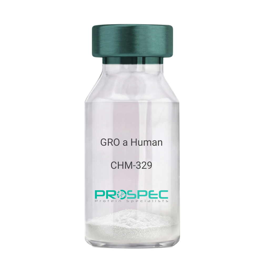
    CHM-329

    Price :

    Quantity :

    Shipping Method :

    Room Temp Icon

    Shipped at Room temp

    Add To Cart

    More Info

    • description
    • source
    • formulation
    • purity
    • biological activity
    • More Info

    Description

    GRO Alpha Human Recombinant produced in E.Coli is a single,non-glycosylated, polypeptide chain containing 73 amino acids and having a molecular mass of 7811 Dalton. The GRO-alpha is purified by proprietary chromatographic techniques.

    Source

    Escherichia Coli.

    Formulation

    Lyophilized from a 0.2µm filtered concentrated (1mg/ml) solution in 20mM PB, pH 7.4, 50mM NaCl.

    Purity

    Greater than 97.0% as determined by:
    (a) Analysis by RP-HPLC.
    (b) Analysis by SDS-PAGE.

    Biological Activity

    Determined by its ability to chemoattract human peripheral blood neutrophils using a concentration range of 10.0-100.0 ng/ml.

    More Info

    • Introduction

      Chemokine (C-X-C motif) ligand 1 (CXCL1) is a small cytokine belonging to the CXC chemokine family that was previously called GRO1 oncogene, Neutrophil-activating protein 3 (NAP-3) and melanoma growth stimulating activity, alpha (MSGA-a). It is secreted by human melanoma cells, has mitogenic properties and is implicated in melanoma pathogenesis. CXCL1 is expressed by macrophages, neutrophilsand epithelial cells, and has neutrophil chemoattractant activity. CXCL1 plays a role in spinal cord development by inhibiting the migration of oligodendrocyte precursors and is involved in the processes of angiogenesis, inflammation, wound healing, and tumorigenesis. This chemokine elicits its effects by signaling through the chemokine receptor CXCR2. The gene for CXCL1 is located on human chromosome 4 amongst genes for other CXC chemokines.

    • Synonyms

      Growth-regulated protein alpha, CXCL1, Melanoma growth stimulatory activity, MGSA, Neutrophil-activating protein 3, NAP-3, GRO-alpha (1-73), chemokine (C-X-C motif) ligand 1, GRO1, GROa, SCYB1, MGSA-a, MGSA alpha.

    • Physical Appearance

      Sterile Filtered White lyophilized (freeze-dried) powder.

    • Stability

      Lyophilized GRO-alpha although stable at room temperature for 3 weeks, should be stored desiccated below -18°C. Upon reconstitution CXCL1 should be stored at 4°C between 2-7 days and for future use below -18°C. For long term storage it is recommended to add a carrier protein (0.1% HSA or BSA).Please prevent freeze-thaw cycles.

    • Solubility

      It is recommended to reconstitute the lyophilized CXCL1 in sterile 18MΩ-cm H2O not less than 100µg/ml, which can then be further diluted to other aqueous solutions.

    • Amino Acid Sequence

      The sequence of the first five N-terminal amino acids was determined and was found to be Ala-Ser-Val-Ala-Thr.

    • Background

      What is the molecular weight/Mw of CXCL1 HUMAN Protein?
      CXCL1 HUMAN Protein has a total Mw of 7.81kDa.

      What is the source or expression system of CXCL1 HUMAN Protein?
      Escherichia Coli.

      What is the Purity of CXCL1 HUMAN Protein?
      CXCL1 HUMAN Protein is >97% pure as determined by SDS-PAGE.

      What is the Biological Activity of CXCL1 HUMAN Protein?
      Determined by its ability to chemoattract human peripheral blood neutrophils using a concentration range of 10.0-100.0 ng/ml.

      What is the amino acid sequence of CXCL1 HUMAN Protein?
      The sequence of the first five N-terminal amino acids was determined and was found to be Ala-Ser-Val-Ala-Thr.

      What applications can CXCL1 HUMAN Protein be used in?
      CXCL1 HUMAN Protein can probably be used in western blot, ELISA and Lateral Flow.

      What is the endotoxin level for CXCL1 HUMAN Protein?
      The endotoxin level is minimal, CXCL1 HUMAN Protein was purified using conventional chromatography techniques.


    ProSpec's products are furnished for LABORATORY RESEARCH USE ONLY. The product may not be used as drugs, agricultural or pesticidal products, food additives or household chemicals.

    Gro A Human
  • View Data Sheet

    Name :

    CRP Paired Antibody

    Description:

    Anti Human C-Reactive Protein Paired Antibody Mouse

    Product # :

    ANT-783

    Price :

    Quantity :

    Shipping Method :

    Ice Icon

    Shipped with Ice Packs

    Add To Cart

    More Info

    • description
    • formulation
    • purity
    • More Info

    Description

    CRP Paired monoclonal antibodies are used to develop rapid test for CRP rapid test. Please note that when ordering for example: 100µg paired antibody, you receive 50µg from each antibody (100µg in total).

    Formulation

    * CRP conjugation antibody in PBS, NaCl and 0.095% NaN3.

    * CRP coating antibody in PBS, NaCl and 0.095% NaN3.

    Purity

    Greater than 95%.

    More Info

    • Physical Appearance

      2 vials of sterile Filtered clear colorless solution.

    • Stability

      For periods up to 1 month CRP Paired Antibody should be stored at 4°C, for longer periods of time, store at -20°C. Prevent freeze thaw cycles.

    • Applications

      Lateral flow immunoassay.

    • Background

      CRP is an important protein that correlates with inflammatory disease and is synthesized by hepatocytes during the acute phase response by IL-1 and TNF Alpha and Beta. CRP levels increase dramatically and serve as a useful marker of inflammation in such conditions as bacterial infection, viral infections, rheumatoid arthritis, transplantation rejection, myocardial infarction, meningitis, septicemia, osteomyelitis and others. CRP is also correlated to Serum Amyloid A levels.

    • Type

      Mouse antibody Monoclonal.

    • Purification Method

      Purified monoclonal IgG1 by protein A chromatography.

    ProSpec's products are furnished for LABORATORY RESEARCH USE ONLY. The product may not be used as drugs, agricultural or pesticidal products, food additives or household chemicals.

    Crp Antibodies Paired
  • View Data Sheet

    Name :

    HCV Cocktail

    Description:

    Hepatitis C Virus Cocktail Recombinant

    Product # :

    HCV-015

    Price :

    Quantity :

    Shipping Method :

    Ice Icon

    Shipped with Ice Packs

    Add To Cart

    More Info

    • description
    • source
    • formulation
    • purity
    • More Info

    Description

    Recombinant HCV cocktail contains the recombinant hepatitis C virus core, NS3, NS4, NS5 proteins.

    Source

    Escherichia Coli.

    Formulation

    HCV Cocktail solution contains 25mM K2CO3 and PBS.

    Purity

    Protein is >95% pure as determined by 10% PAGE (coomassie staining).

    More Info

    • Physical Appearance

      Sterile Filtered clear solution.

    • Stability

      Store at 4°C if entire vial will be used within 2-4 weeks. Store, frozen at -20°C for longer periods of time. For long term storage it is recommended to add a carrier protein (0.1% HSA or BSA). Avoid multiple freeze-thaw cycles.

    • Applications

      ELISA and lateral flow immunoassay.

    • Background

      Hepatitis C virus (HCV) is a major global health concern, with approximately 71 million people infected worldwide. Chronic HCV infection can lead to liver cirrhosis, hepatocellular carcinoma, and other severe liver-related complications. The nonstructural proteins NS3, NS4, and NS5 of HCV play critical roles in viral replication, assembly, and immune evasion, making them attractive targets for antiviral interventions. This research aims to characterize the recombinant forms of HCV NS3, NS4, and NS5 proteins to gain insights into their functional properties and identify potential targets for the development of effective antiviral strategies.

      The primary objective of this study is to express and purify recombinant HCV NS3, NS4, and NS5 proteins using suitable expression systems. Recombinant DNA techniques will be employed to generate expression vectors harboring the respective genes, followed by expression in bacterial, yeast, or mammalian cell-based systems. The recombinant proteins will be purified using affinity chromatography or other appropriate methods, enabling subsequent biochemical and biophysical characterization.

      The second objective is to investigate the roles of NS3, NS4, and NS5 proteins in HCV replication and assembly. In vitro assays will be performed to assess the enzymatic activities of NS3 protease and NS5 RNA-dependent RNA polymerase (RdRp). The effects of potential inhibitors or small molecules on these enzymatic activities will be evaluated. Furthermore, the interactions between NS4 protein and cellular components involved in viral assembly will be explored using protein-protein interaction assays.

      The third objective is to determine the three-dimensional structures of the recombinant NS3, NS4, and NS5 proteins using techniques such as X-ray crystallography or cryo-electron microscopy. Structural information will provide valuable insights into the functional domains and potential binding sites within these proteins. These findings will aid in the design of novel antiviral compounds targeting specific regions of NS3, NS4, and NS5, with the goal of disrupting viral replication and assembly.

      By characterizing the recombinant forms of HCV NS3, NS4, and NS5 proteins, this research aims to enhance our understanding of the molecular mechanisms underlying HCV infection. The insights gained from this study may contribute to the development of novel antiviral strategies targeting HCV, ultimately leading to improved therapeutic options for individuals affected by this persistent and debilitating viral infection.

    ProSpec's products are furnished for LABORATORY RESEARCH USE ONLY. The product may not be used as drugs, agricultural or pesticidal products, food additives or household chemicals.

    Hcv Cocktail
  • View Data Sheet

    Name :

    S.Typhi H

    Description:

    Salmonella Typhi H Antigen Recombinant

    Product # :

    STY-001

    Price :

    Quantity :

    Shipping Method :

    Ice Icon

    Shipped with Ice Packs

    Add To Cart

    More Info

    • description
    • source
    • formulation
    • purity
    • More Info

    Description

    Recombinant Salmonella Typhi H Antigen produced in E.coli is full length flagellin sequence and fused to a 6 His Tag at C-terminus, migrating as a 35kDa band on SDS-PAGE.S. typhi H antigen is a flagellin protein, which strongly reacts to specific IgM and IgG produced in infected patients.

    Source

    Escherichia Coli.

    Formulation

    Sterile Filtered solution containing PBS and 25mM arginine.

    Purity

    Protein is >95% pure as determined by 10% PAGE (coomassie staining).

    More Info

    • Introduction

      Salmonella Typhi is a pathogen causing typhoid fever, affecting over 17 million people with approximately 600,000 deaths annually worldwide. If untreated, typhoid fever cases result in mortality rates ranging from 12-30%.

    • Physical Appearance

      Sterile Filtered solution.

    • Stability

      S.Typhi H although stable at 4°C for 1 week, should be stored below -18°C. Please prevent freeze thaw cycles.

    • Applications

      Immunoassay.

    ProSpec's products are furnished for LABORATORY RESEARCH USE ONLY. The product may not be used as drugs, agricultural or pesticidal products, food additives or household chemicals.

    Styphi H
  • View Data Sheet

    Name :

    HCV NS3 2c

    Description:

    Hepatitis C Virus NS3 Genotype-2c, (1356-1459 a.a.) Recombinant

    Product # :

    HCV-265

    Price :

    Quantity :

    Shipping Method :

    Ice Icon

    Shipped with Ice Packs

    Add To Cart

    More Info

    • description
    • formulation
    • purity
    • More Info

    Description

    The E.coli derived recombinant protein contains the HCV NS3 immunodominant regions, amino acids 1356-1459.

    Formulation

    1.5M urea, 25mM Tris-HCl pH-8, 0.2% Triton-X & 50% Glycerol.

    Purity

    HCV NS3 Genotype-2c protein is >95% pure as determined by 10% PAGE (coomassie staining).

    More Info

    • Introduction

      HCV is a small 50nm, enveloped, single-stranded, positive sense RNAvirus in the family Flaviviridae.
      HCV has a high rate of replication with approximately one trillion particles produced each day in an infected individual. Due to lack of proofreading by the HCV RNA polymerase, the HCV has an exceptionally high mutation rate, a factor that may help it elude the host's immune response. Hepatitis C virus is classified into six genotypes(1-6) with several subtypes within each genotype. The preponderance and distribution of HCV genotypes varies globally. Genotype is clinically important in determining potential response to interferon-based therapy and the required duration of such therapy. Genotypes 1 and 4 are less responsive to interferon-based treatment than are the other genotypes (2, 3, 5 and 6).

    • Stability

      HCV NS3 Genotype-2c although stable at 4°C for 1 week, should be stored below -18°C. Please prevent freeze thaw cycles.

    • Applications

      HCV NS3 Genotype-2c antigen is suitable for ELISA and Western blots, excellent antigen for detection of HCV with minimal specificity problems.

    • Specificity

      Immunoreactive with sera of HCV-infected individuals.

    • Purification Method

      HCV NS3 Genotype-2c protein was purified by proprietary chromatographic technique.

    ProSpec's products are furnished for LABORATORY RESEARCH USE ONLY. The product may not be used as drugs, agricultural or pesticidal products, food additives or household chemicals.

    Hcv Ns3 Genotype 2C 1356 1459
  • View Data Sheet

    Name :

    Chlamydia HSP70 (462-503 a.a.)

    Description:

    Chlamydia Trachomatis HSP70 (462-503 a.a.) Recombinant

    Product # :

    CHT-008

    Price :

    Quantity :

    Shipping Method :

    Ice Icon

    Shipped with Ice Packs

    Add To Cart

    More Info

    • description
    • source
    • formulation
    • purity
    • More Info

    Description

    The E.coli derived recombinant protein contains Chlamydia TrachomatisHSP70 protein epitopes, 462-503 amino acids.

    Source

    Escherichia Coli.

    Formulation

    50mM Tris-HCl, pH 8.0, 8M Urea, 60 mM NaCl and 50% glycerol.

    Purity

    Chlamydia HSP70 protein is >95% pure as determined by 10% PAGE (coomassie staining) and RP-HPLC.

    More Info

    • Introduction

      Chlamydia is a common term for infection with any bacterium belonging to the phylum Chlamydiae. This term derives from the name of the bacterial genus Chlamydiain the family Chlamydiaceae, order Chlamydiales, class and phylum Chlamydiae. There are two genera in Chlamydiaceae: Chlamydia and Chlamydophila. The genus Chlamydia includes three species: C. trachomatis, C. muridarum, and C. suis.

    • Stability

      Chlamydia HSP70 although stable at 4°C for 1 week, should be stored below -18°C. Please prevent freeze thaw cycles.

    • Applications

      Chlamydia HSP70 protein is suitable for use in ELISA. Each laboratory should determine an optimum working titer for use in its particular application. Other applications have not been tested but use in such assays should not necessarily be excluded.

    • Specificity

      Immunoreactive with sera of Chlamydia Trachomatis infected individuals.

    • Purification Method

      Chlamydia HSP70 protein was purified by proprietary chromatographic technique.

    ProSpec's products are furnished for LABORATORY RESEARCH USE ONLY. The product may not be used as drugs, agricultural or pesticidal products, food additives or household chemicals.

    Chlamydia Hsp70 462 503
  • View Data Sheet

    Name :

    HCV Core 22kDa, Biotin

    Description:

    Hepatitis C Virus Core 22kDa, Biotin Recombinant

    Product # :

    HCV-225

    Price :

    Quantity :

    Shipping Method :

    Ice Icon

    Shipped with Ice Packs

    Add To Cart

    More Info

    • description
    • formulation
    • purity
    • More Info

    Description

    The E.coli derived recombinant protein contains the HCV core nucleocapsid immunodominant regions, amino acids 2-192, 22kDa.The Biotin labeled protein is fused with b-galactosidase (114 kDa) at N-terminus.

    Formulation

    20mM Tris-HCl pH 8 and 8M urea.

    Purity

    Protein is >95% pure as determined by 10% PAGE (coomassie staining).

    More Info

    • Introduction

      HCV is a small 50nm, enveloped, single-stranded, positive sense RNAvirus in the family Flaviviridae.
      HCV has a high rate of replication with approximately one trillion particles produced each day in an infected individual. Due to lack of proofreading by the HCV RNA polymerase, the HCV has an exceptionally high mutation rate, a factor that may help it elude the host's immune response. Hepatitis C virus is classified into six genotypes(1-6) with several subtypes within each genotype. The preponderance and distribution of HCV genotypes varies globally. Genotype is clinically important in determining potential response to interferon-based therapy and the required duration of such therapy. Genotypes 1 and 4 are less responsive to interferon-based treatment than are the other genotypes (2, 3, 5 and 6).

    • Stability

      HCV-Core although stable at 4°C for 1 week, should be stored below -18°C. Please prevent freeze thaw cycles.

    • Specificity

      Immunoreactive with sera of HCV-infected individuals.

    • Purification Method

      HCV-Core protein was purified by proprietary chromatographic technique.

    ProSpec's products are furnished for LABORATORY RESEARCH USE ONLY. The product may not be used as drugs, agricultural or pesticidal products, food additives or household chemicals.

    Hcv Core 22Kda Biotin
  • View Data Sheet

    Name :

    HCV Core 22kDa, FLSCN

    Description:

    Hepatitis C Virus Core 22kDa, FLSCN Recombinant

    Product # :

    HCV-261

    Price :

    Quantity :

    Shipping Method :

    Ice Icon

    Shipped with Ice Packs

    Add To Cart

    More Info

    • description
    • formulation
    • purity
    • More Info

    Description

    The E.coli derived recombinant protein contains the HCV core nucleocapsid immunodominant regions, amino acids 2-192. The FLSCN labeled protein is fused with b-galactosidase (114 kDa) at N-terminus.

    Formulation

    20mM Tris-Hcl pH 8, 8M urea and 10mM B-ME.

    Purity

    HCV-Core protein is >95% pure as determined by 10% PAGE (coomassie staining).

    More Info

    • Introduction

      HCV is a small 50nm, enveloped, single-stranded, positive sense RNAvirus in the family Flaviviridae.
      HCV has a high rate of replication with approximately one trillion particles produced each day in an infected individual. Due to lack of proofreading by the HCV RNA polymerase, the HCV has an exceptionally high mutation rate, a factor that may help it elude the host's immune response. Hepatitis C virus is classified into six genotypes(1-6) with several subtypes within each genotype. The preponderance and distribution of HCV genotypes varies globally. Genotype is clinically important in determining potential response to interferon-based therapy and the required duration of such therapy. Genotypes 1 and 4 are less responsive to interferon-based treatment than are the other genotypes (2, 3, 5 and 6).

    • Stability

      HCV-Core although stable at 4°C for 1 week, should be stored below -18°C. Please prevent freeze thaw cycles.

    • Applications

      HCV-Core antigen is suitable for ELISA and Western blots, excellent antigen for detection of HCV with minimal specificity problems.

    • Specificity

      Immunoreactive with sera of HCV-infected individuals.

    • Purification Method

      HCV-Core protein was purified by proprietary chromatographic technique.

    ProSpec's products are furnished for LABORATORY RESEARCH USE ONLY. The product may not be used as drugs, agricultural or pesticidal products, food additives or household chemicals.

    Hcv Core 22Kda Fluorescein
  • View Data Sheet

    Name :

    HIV-1 GAG

    Description:

    HIV-1 GAG Full Length Recombinant

    Product # :

    HIV-010

    Price :

    Quantity :

    Shipping Method :

    Room Temp Icon

    Shipped at Room temp

    Add To Cart

    More Info

    • description
    • source
    • formulation
    • purity
    • More Info

    Description

    HIV-1 GAG Recombinant produced in E.Coli is a single, non-glycosylated polypeptide chain, encoded by the HIV gag gene, HXB2 (790-2292) and having a molecular mass of 55.0kDa. HIV-1 GAG is fused to a His-tag and is purified by proprietary chromatographic technique.

    Source

    Escherichia Coli.

    Formulation

    Lyophilized with 1% glycerol.

    Purity

    Greater than 78.0% as determined by SDS-PAGE.

    More Info

    • Introduction

      Human immunodeficiency virus (HIV) is a retrovirus which may lead to a immune systembegins failure and to opportunistic infections. HIV primarily infects vital cells in the human immune system such as helper T cells (specifically CD4+ T cells), macrophages and dendritic cells.

      The Gag protein is the major structural protein required for virus assembly. It is synthesizedas a polyprotein in the cytosol of an infected cell and contains 4 functional segments. Gag alone is sufficient to produce buddingvirus-like particles (VLP's) due to multimerization of roughly 2000 Gag molecules per virion.The Gag proteins take part throughout the viral life-cycle, including the assembly and release of viral particles, theirsubsequent maturation into infectious virions, and during the events occurring between therelease of capsids into newly infected cells and the integration of proviral DNA.

    • Physical Appearance

      Sterile Filtered White lyophilized (freeze-dried) powder.

    • Stability

      Lyophilized HIV-1 GAG although stable at room temperature for 1 week, should be stored desiccated below -18°C. Upon reconstitution HIV-1 GAG should be stored at 4°C between 2-7 days and for future use below -18°C.For long-term storage it is recommended to add a carrier protein (0.1% HSA or BSA).Please prevent freeze-thaw cycles.

    • Solubility

      It is recommended to reconstitute the lyophilized HIV-1 GAG in sterile 18MΩ-cm H2O not less than 100µg/ml, which can then be further diluted to other aqueous solutions.

    • Applications

      Western Blotting, SDS Page

    ProSpec's products are furnished for LABORATORY RESEARCH USE ONLY. The product may not be used as drugs, agricultural or pesticidal products, food additives or household chemicals.

    Hiv 1 Gag
  • View Data Sheet

    Name :

    IL 12p75 Antibody

    Description:

    Interleukin-12 p75, Rat Anti-Mouse

    Product # :

    ANT-209

    Price :

    Quantity :

    Shipping Method :

    Room Temp Icon

    Shipped at Room temp

    Add To Cart

    More Info

    • formulation
    • More Info

    Formulation

    1mg/ml in PBS (after reconstitution).

    More Info

    • Introduction

      IL-12 is a heterodimeric cytokine that stimulates the production of interferon gamma from T-cells and natural killer cells, and also induces differentiation of Th1 helper cells. It is an initiator of cell-mediated immunity.

    • Solubility

      Reconstitute with sterile H20. Mix gently, wash the sides of the vial and wait 30-60 seconds before use.

    • Immunogen

      r.Murine IL-12 p75.

    • Ig Subclass

      Rat IgG2b.

    • Clone

      NYRmIL-12p75.

    • Applications

      Direct ELISA, Western Blot, Immuneprecipitation.

    • Note

      This antibody recognizes the entire IL-12 molecule and has no reactivity to denatured p40 or p35.

    • Titer

      By direct ELISA, 1:5,000 dilution will yield 0.5 O.D using alkaline phosphatase conjugated rabbit anti-mouse Ig (Jackson Laboratories).

    • Shipping Conditions

      Antibody is shipped lyophilized at ambient temperature.

    • Type

      Rat Anti Mouse Monoclonal.

    • Storage Procedures

      In lyophilized form, for long periods, store at 4oC in a dry environment. After reconstitution, if not intended for use within a month, aliquot and store at -20oC.

    • Purification Method

      Ion exchange.

    ProSpec's products are furnished for LABORATORY RESEARCH USE ONLY. The product may not be used as drugs, agricultural or pesticidal products, food additives or household chemicals.

    Il 12P75 Antibody
  • View Data Sheet

    Name :

    H3N2 Influenza-A Antibody

    Description:

    Influenza-A Hemagglutinin H3N2, Mouse Antibody

    Product # :

    ANT-215

    Price :

    Quantity :

    Shipping Method :

    Ice Icon

    Shipped with Ice Packs

    Add To Cart

    More Info

    • description
    • formulation
    • More Info

    Description

    Hybridoma clones have been derived from hybridization of Sp2/0 myeloma cells with spleen cells of Balb/c mice immunized with Influenza A/Shandong/9/93 H3N2 derived from allantoic fluid of 10 days old embryonated eggs.

    Formulation

    In PBS, pH 7.4 and 0.1 % NaN3.

    More Info

    • Introduction

      H3N2 is a subtype of the influenza A virus. Its name derives from the forms of the two kinds of proteinson the surface of its coat, hemagglutinin(H) and neuraminidase(N). H3N2 exchanges genes for internal proteins with other influenza subtypes. H3N2 has tended to dominate in prevalence over H1N1, H1N2, and influenza B. H3N2 strain descended from H2N2by antigenic shift, in which genes from multiple subtypes re-assorted to form a new virus. Both the H2N2and H3N2 strains contained genesfrom avian influenzaviruses.

    • Physical Appearance

      Sterile Filtered clear solution.

    • Immunogen

      Influenza A hemagglutinin H3N2.

    • Ig Subclass

      mouse IgG1.

    • Clone

      IA-H3N2.

    • Applications

      Influenza A haemagglutinin H3N2 immunodetection in direct or indirect ELISA, and Western Blotting.

    • Shipping Conditions

      Antibody is shipped as in liquid form with ice packs.

    • Type

      Mouse Antibody Monoclonal.

    • Storage Procedures

      Should be stored at 4C.Do not freeze.

    • Purification Method

      Protein-A column.

    ProSpec's products are furnished for LABORATORY RESEARCH USE ONLY. The product may not be used as drugs, agricultural or pesticidal products, food additives or household chemicals.

    Influenza A H3N2 Antibody
  • View Data Sheet

    Name :

    HIV-1 TAT Clade-C

    Description:

    HIV-1 TAT Clade-C Recombinant

    Product # :

    HIV-116

    Price :

    Quantity :

    Shipping Method :

    Ice Icon

    Shipped with Ice Packs

    Add To Cart

    More Info

    • description
    • source
    • formulation
    • purity
    • More Info

    Description

    HIV-1 TAT Recombinant- produced in E.coli is a single, non-glycosylated, polypeptide chain containing 101 amino acids encoded by two exons and having chain having a molecular mass of 21 kDa.

    Source

    Escherichia Coli.

    Formulation

    0.1% glycerol.

    Purity

    Greater than 90.0% as determined by SDS-PAGE.

    More Info

    • Introduction

      Human immunodeficiency virus type-1 (HIV-1) regulatory Tat protein plays an essential role in viral replication and infectivity. In addition, during acute infection, Tat is released extracellularly by infected cells and is taken up by neighboring cells where it transactivates viral replication and increases virus infectivity.
      HIV-1 Tat activates transcription of HIV-1 viral genes by inducing phosphorylation of the C-terminal domain (CTD) of RNA polymerase II (RNAPII). Tat can also disturb cellular metabolism by inhibiting proliferation of antigen-specific T lymphocytes and by inducing cellular apoptosis. Tat-induced apoptosis of T-cells is attributed, in part, to the distortion of microtubules polymerization. LIS1 is a microtubule-associated protein that facilitates microtubule polymerization.

    • Physical Appearance

      Sterile filtered colorless clear solution.

    • Stability

      Store at 4°C if entire vial will be used within 2-4 weeks. Store, frozen at -20°C for longer periods of time. Avoid multiple freeze-thaw cycles.

    ProSpec's products are furnished for LABORATORY RESEARCH USE ONLY. The product may not be used as drugs, agricultural or pesticidal products, food additives or household chemicals.

    Hiv 1 Tat Clade C
  • View Data Sheet

    Name :

    KLK3 Human, HEK

    Description:

    Kallikrein-3 Human Recombinant, HEK

    Prostate-specific antigen, PSA, Gamma-seminoprotein, Seminin, Kallikrein-3, P-30 antigen, Semenogelase, KLK3, APS, hK3, KLK2A1

    Product # :

    ENZ-1149

    Price :

    Quantity :

    Shipping Method :

    Ice Icon

    Shipped with Ice Packs

    Add To Cart

    More Info

    • description
    • source
    • formulation
    • purity
    • biological activity
    • More Info

    Description

    KLK3 Human Recombinant produced in HEK cells is a single, glycosylated, polypeptide chain (18-261 a.a) containing a total of 250 amino acids, having a molecular mass of 27.6kDa.KLK3 is fused to a 6 amino acid His-tag at C-terminus,and is purified by proprietary chromatographic techniques.

    Source

    HEK293 Cells.

    Formulation

    The KLK3 solution (0.25mg/ml) contains 10% Glycerol and Phosphate-Buffered Saline (pH 7.4).

    Purity

    Greater than 95.0% as determined by SDS-PAGE.

    Biological Activity

    Specific activity is  > 250pmol/min/ug. Defined by the amount of enzyme that cleaves 1pmole of Succinyl-ArgPro-Tyr-p-Nitroanilide to Succinyl-Arg-Pro-Tyr and p-Nitroanilide per minute at pH 7.5 at 37˚C.

    More Info

    • Introduction

      Kallikrein-3 or KLK3 is a serine proteases subgroup, enzymes can cleave peptide bonds. KLK3 is found in the prostate and may be hold accountable to the regulation of semen liquefaction via seminogelin hydrolysis. The protein is expressed in low concentrations in men with healthy prostates serum. When elevated quantities are found it may show prostate cancer or different prostate disorders such as prostatitis and benign prostatic hyperplasia.

    • Synonyms

      Prostate-specific antigen, PSA, Gamma-seminoprotein, Seminin, Kallikrein-3, P-30 antigen, Semenogelase, KLK3, APS, hK3, KLK2A1

    • Physical Appearance

      Sterile Filtered colorless solution.

    • Stability

      Store at 4°C if entire vial will be used within 2-4 weeks. Store, frozen at -20°C for longer periods of time. For long term storage it is recommended to add a carrier protein (0.1% HSA or BSA).Avoid multiple freeze-thaw cycles.

    • Amino Acid Sequence

      APLILSRIVG GWECEKHSQP WQVLVASRGR AVCGGVLVHP QWVLTAAHCI RNKSVILLGR HSLFHPEDTG QVFQVSHSFP HPLYDMSLLK NRFLRPGDDS SHDLMLLRLS EPAELTDAVK VMDLPTQEPA LGTTCYASGW GSIEPEEFLT PKKLQCVDLH VISNDVCAQV HPQKVTKFML CAGRWTGGKS TCSGDSGGPL VCNGVLQGIT SWGSEPCALP ERPSLYTKVV HYRKWIKDTI VANPHHHHHH

    ProSpec's products are furnished for LABORATORY RESEARCH USE ONLY. The product may not be used as drugs, agricultural or pesticidal products, food additives or household chemicals.

    Kallikrein 3 Human
  • View Data Sheet

    Name :

    FLAG PAT3G6AT antibody

    Description:

    FLAG peptide Clone PAT3G6AT, Mouse Anti Human

    Product # :

    ANT-736

    Price :

    Quantity :

    Shipping Method :

    Ice Icon

    Shipped with Ice Packs

    Add To Cart

    More Info

    • formulation
    • More Info

    Formulation

    1mg/ml containing PBS, pH-7.4, 10% Glycerol and 0.02% Sodium Azide.

    More Info

    • Introduction

      A fusion tag called FLAG consists of eight amino acids (AspTyrLysAspAspAspAspLys) including an enterokinase-cleavage site. FLAG is specifically designed for immunoaffinity chromatography. It allows elution under non-denaturing conditions. Several antibodies against this peptide have been developed. One antibody denoted as M1 binds the peptide in the presence of bivalent metal cations preferably Ca(+). Elution is effected by chelating agents. Another strategy is competitive elution with excess of free Flag Peptide. The FLAG peptide purifies and detects recombinant fusion proteins. FLAG peptide is useful in Western blotting, immunocytochemistry, immunoprecipitation, flow cytometry, protein purification, and in the study of protein-protein interactions, cell ultrastructure, and protein localization. Flag peptide is a hydrophilic tag which significantly improves the detection and purification of recombinant fusion proteins.

    • Physical Appearance

      Sterile filtered colorless solution.

    • Immunogen

      Anti-human FLAG mAb, is derived from hybridization of mouse F0 myeloma cells with spleen cells from BALB/c mice immunized with a Synthetic peptide (DYKDDDDKC)-KLH.

    • Ig Subclass

      Mouse IgG1 heavy chain and k light chain.

    • Clone

      PAT3G6AT.

    • Applications

      The antibody has been tested by ELISA, Western blot analysis, Flow cytometry and ICC/IF to assure specificity and reactivity. Since application varies, however, each investigation should be titrated by the reagent to obtain optimal results.

    • Type

      Mouse Anti Human Monoclonal.

    • Storage Procedures

      For periods up to 1 month store at 4°C, for longer periods of time, store at -20°C. Prevent freeze thaw cycles.

    • Purification Method

      FLAG antibody was purified from mouse ascitic fluids by protein-A affinity chromatography.

    ProSpec's products are furnished for LABORATORY RESEARCH USE ONLY. The product may not be used as drugs, agricultural or pesticidal products, food additives or household chemicals.

    Flag Antibody
  • View Data Sheet

    Name :

    IFNW1 Human, HEK

    Description:

    Interferon-Omega 1 Human Recombinant, HEK

    IFN omega-1, IFN alpha-II-1, IFNW1.

    Product # :

    CYT-1225

    Price :

    Quantity :

    Shipping Method :

    Ice Icon

    Shipped with Ice Packs

    Add To Cart

    More Info

    • description
    • source
    • formulation
    • purity
    • biological activity
    • More Info

    Description

    IFNW1 Human Recombinant is a single, glycosylated, polypeptide chain (22-195 a.a) containing a total of 180 amino acids and having a molecular mass of 20.9 kDa. IFNW1 is fused to a 6 a.a his-Tag at C-terminus and is purified by proprietary chromatographic techniques.

    Source

    HEK293 Cells.

    Formulation

    The IFNW1 solution (1mg/ml) contains 10% Glycerol and Phosphate-Buffered Saline (pH 7.4).

    Purity

    Greater than 95.0% as determined by SDS-PAGE.

    Biological Activity

    The ED50 is ≤0.07 ng/ml, measured  in a cytotoxicity assay using TF-1 human erythroleukemic cells .

    More Info

    • Synonyms

      IFN omega-1, IFN alpha-II-1, IFNW1.

    • Physical Appearance

      Sterile Filtered colorless solution.

    • Stability

      Store at 4°C if entire vial will be used within 2-4 weeks. Store, frozen at -20°C for longer periods of time. For long term storage it is recommended to add a carrier protein (0.1% HSA or BSA).Avoid multiple freeze-thaw cycles.

    • Amino Acid Sequence

      LGCDLPQNHG LLSRNTLVLL HQMRRISPFL CLKDRRDFRF PQEMVKGSQL QKAHVMSVLH EMLQQIFSLF HTERSSAAWN MTLLDQLHTG LHQQLQHLET CLLQVVGEGE SAGAISSPAL TLRRYFQGIR VYLKEKKYSD CAWEVVRMEI MKSLFLSTNM QERLRSKDRD LGSSHHHHHH.

    • Background

      Interferons, a family of signaling proteins, play a pivotal role in the immune system’s defense against viral infections and other threats. Among these, Interferon W1 (IFNW1), a member of the Type I interferon family, has emerged as a key player in orchestrating antiviral responses and modulating immune reactions. This research embarks on a detailed exploration of the IFNW1 protein, unveiling its structural intricacies, signaling pathways, and its broader implications in immune regulation and disease. By delving into IFNW1, scientists aim to comprehend the nuances of its functions, decipher its interactions within the complex interferon network, and explore its potential applications in therapeutic interventions and beyond.

      Structural Insights into IFNW1:

      IFNW1, like other Type I interferons, exhibits a unique tertiary structure that enables it to interact with specific cell surface receptors. This interaction triggers a cascade of events, leading to the activation of various antiviral genes and immune modulatory pathways. Understanding the structural basis of IFNW1 is crucial for elucidating its binding affinities, biological activities, and its significance in immune responses.

      Signaling Pathways and Antiviral Defense:

      IFNW1 engages with its cognate receptors, initiating Janus kinase (JAK)-Signal Transducer and Activator of Transcription (STAT) signaling pathways. This activation leads to the transcription of interferon-stimulated genes (ISGs) with potent antiviral properties. IFNW1’s ability to induce an antiviral state in infected and neighboring cells is fundamental for restricting viral replication and curtailing the spread of infections. Additionally, IFNW1 plays a role in modulating adaptive immune responses, contributing to the broader immune defense mechanisms.

      IFNW1 in Immunomodulation and Disease:

      Beyond its antiviral functions, IFNW1 is implicated in immunomodulation and disease pathogenesis. Dysregulation of IFNW1 signaling is associated with autoimmune disorders, including lupus and rheumatoid arthritis, highlighting its involvement in immune-related diseases. Moreover, IFNW1 is being explored in cancer immunotherapy, where its ability to modulate the tumor microenvironment and enhance immune surveillance presents opportunities for novel treatment strategies.

      Therapeutic Potential and Future Prospects:

      The unique properties of IFNW1, particularly its role in immune regulation and antiviral defense, position it as a potential therapeutic target. Research efforts are directed towards harnessing its immunomodulatory functions for developing therapies against infectious diseases, autoimmune disorders, and certain cancers. Additionally, understanding IFNW1’s interactions with other components of the immune system opens avenues for innovative approaches in personalized medicine and targeted immunotherapies.

      IFNW1 Protein, as an integral component of the interferon network, stands as a sentinel in the body’s defense against viral invasions and immune dysregulations. Its multifaceted roles in antiviral defense, immune modulation, and disease pathogenesis underscore its significance in biology and medicine. As researchers delve deeper into the intricacies of IFNW1, they pave the way for innovative therapies, immunomodulatory interventions, and a deeper understanding of immune responses. This research not only illuminates the pivotal role of IFNW1 but also holds the promise of transformative advancements in medicine, shaping the future of immunology and disease therapeutics.

      What is the molecular weight/Mw of IFNW1 HUMAN, HEK Protein?
      IFNW1 HUMAN, HEK Protein has a total Mw of 20.9kDa.

      What is the source or expression system of IFNW1 HUMAN, HEK Protein?
      HEK293 Cells.

      What is the Purity of IFNW1 HUMAN, HEK Protein?
      IFNW1 HUMAN, HEK Protein is >95% pure as determined by SDS-PAGE.

      What is the Biological Activity of IFNW1 HUMAN, HEK Protein?
      The ED50 is ≤0.07 ng/ml, measured in a cytotoxicity assay using TF-1 human erythroleukemic cells .

      What is the amino acid sequence of IFNW1 HUMAN, HEK Protein?
      LGCDLPQNHG LLSRNTLVLL HQMRRISPFL CLKDRRDFRF PQEMVKGSQL QKAHVMSVLH EMLQQIFSLF HTERSSAAWN MTLLDQLHTG LHQQLQHLET CLLQVVGEGE SAGAISSPAL TLRRYFQGIR VYLKEKKYSD CAWEVVRMEI MKSLFLSTNM QERLRSKDRD LGSSHHHHHH.

      What applications can IFNW1 HUMAN, HEK Protein be used in?
      IFNW1 HUMAN, HEK Protein can probably be used in western blot, ELISA and Lateral Flow.

      What is the endotoxin level for IFNW1 HUMAN, HEK Protein?
      The endotoxin level is minimal, IFNW1 HUMAN, HEK Protein was purified using conventional chromatography techniques.


    ProSpec's products are furnished for LABORATORY RESEARCH USE ONLY. The product may not be used as drugs, agricultural or pesticidal products, food additives or household chemicals.

    Ifn Omega Human
  • View Data Sheet

    Name :

    DPP4 Human, HEK

    Description:

    Dipeptidyl-Peptidase 4 Human Recombinant, HEK

    CD26, ADABP, ADCP2, DPPIV, TP103, DPP4, Dipeptidyl peptidase 4, Dipeptidyl peptidase IV, DPP IV, T-cell activation antigen CD26, Adenosine deaminase complexing protein 2, CD26 antigen.

    Product # :

    ENZ-1187

    Price :

    Quantity :

    Shipping Method :

    Ice Icon

    Shipped with Ice Packs

    Add To Cart

    More Info

    • description
    • source
    • formulation
    • purity
    • biological activity
    • More Info

    Description

    DPP4 Human Recombinant is a single, glycosylated polypeptide chain containing 977 amino acids (29-766a.a) and having a molecular mass of 112.1kDa (calculated). DPP4 is fused to a 239 amino acid hIgG-Tag at C-terminus and is purified by proprietary chromatographic techniques.

    Source

    HEK293 Cells.

    Formulation

    DPP4 protein solution (0.25mg/ml) contains 10% Glycerol and Phosphate-Buffered Saline (pH 7.4).

    Purity

    Greater than 95.0% as determined by Analysis by SDS-PAGE.

    Biological Activity

    Specific activity is > 15,000 pmol/min/ug in which one unit defined as the amount of enzyme that hydrolyze 1pmole of H-Gly-Pro-AMC.HBr to H-Gly-Pro and AMC per minute at pH 8.0 at 37℃.

     The ED50 range ≤250 ng/ml is measured by its binding ability in a functional ELISA with MERS-CoV Spike S1 Subunit (CAT# sars-052)..

    The ED50 range ≤200 ng/ml is measured by its binding ability in a functional ELISA with MERS-CoV Spike RBD (CAT# sars-054).

    The ED50 range ≤120 ng/ml is measured by its binding ability in a functional ELISA with MERS-CoV Spike (CAT# sars-051).

    More Info

    • Synonyms

      CD26, ADABP, ADCP2, DPPIV, TP103, DPP4, Dipeptidyl peptidase 4, Dipeptidyl peptidase IV, DPP IV, T-cell activation antigen CD26, Adenosine deaminase complexing protein 2, CD26 antigen.

    • Physical Appearance

      Sterile Filtered colorless solution.

    • Stability

      Store at 4°C if entire vial will be used within 2-4 weeks. Store, frozen at -20°C for longer periods of time. For long term storage it is recommended to add a carrier protein (0.1% HSA or BSA).Avoid multiple freeze-thaw cycles.

    • Amino Acid Sequence

      DGSMNKGTDD ATADSRKTYT LTDYLKNTYR LKLYSLRWIS DHEYLYKQEN NILVFNAEYG NSSVFLENST FDEFGHSIND YSISPDGQFI LLEYNYVKQW RHSYTASYDI YDLNKRQLIT EERIPNNTQW VTWSPVGHKL AYVWNNDIYV KIEPNLPSYR ITWTGKEDII YNGITDWVYE EEVFSAYSAL WWSPNGTFLA YAQFNDTEVP LIEYSFYSDE SLQYPKTVRV PYPKAGAVNP TVKFFVVNTD SLSSVTNATS IQITAPASML IGDHYLCDVT WATQERISLQ WLRRIQNYSV MDICDYDESS GRWNCLVARQ HIEMSTTGWV GRFRPSEPHF TLDGNSFYKI ISNEEGYRHI CYFQIDKKDC TFITKGTWEV IGIEALTSDY LYYISNEYKG MPGGRNLYKI QLSDYTKVTC LSCELNPERC QYYSVSFSKE AKYYQLRCSG PGLPLYTLHS SVNDKGLRVL EDNSALDKML QNVQMPSKKL DFIILNETKF WYQMILPPHF DKSKKYPLLL DVYAGPCSQK ADTVFRLNWA TYLASTENII VASFDGRGSG YQGDKIMHAI NRRLGTFEVE DQIEAARQFS KMGFVDNKRI AIWGWSYGGY VTSMVLGSGS GVFKCGIAVA PVSRWEYYDS VYTERYMGLP TPEDNLDHYR NSTVMSRAEN FKQVEYLLIH GTADDNVHFQ QSAQISKALV DVGVDFQAMW YTDEDHGIAS STAHQHIYTH MSHFIKQCFS LPKLLEPKSC DKTHTCPPCP APELLGGPSV FLFPPKPKDT LMISRTPEVT CVVVDVSHED PEVKFNWYVD GVEVHNAKTK PREEQYNSTY RVVSVLTVLH QDWLNGKEYK CKVSNKALPA PIEKTISKAK GQPREPQVYT LPPSRDELTK NQVSLTCLVK GFYPSDIAVE WESNGQPENN YKTTPPVLDS DGSFFLYSKL TVDKSRWQQG NVFSCSVMHE ALHNHYTQKS LSLSPGK.

    • Background

      DPP4 protein, also known as Dipeptidyl peptidase-4 or CD26, is a cell surface protease with diverse functions in cell signaling and metabolism. This research aims to investigate the role of DPP4 protein in various physiological processes and its implications in disease pathogenesis. Understanding the biological significance of DPP4 can provide insights into its potential as a therapeutic target for several disorders.

      Function of DPP4 Protein:

      DPP4 protein is involved in the cleavage and regulation of several peptide hormones and chemokines, influencing their bioactivity and half-life. It is widely expressed in various tissues, including immune cells, endothelial cells, and epithelial cells. DPP4 can modulate immune responses, glucose metabolism, and neuropeptide signaling through enzymatic and non-enzymatic activities.

      Role of DPP4 Protein in Immune Regulation:

      DPP4 protein plays a role in immune cell activation and regulation. It is expressed on the surface of T cells, where it functions as a co-stimulatory molecule. DPP4 engagement on T cells promotes T cell activation, cytokine production, and adhesion to endothelial cells. Additionally, DPP4 can cleave and inactivate certain chemokines, thereby influencing chemotaxis and immune cell recruitment.

      Implications of DPP4 Protein in Metabolic Disorders:

      DPP4 protein is involved in glucose metabolism and insulin regulation. It cleaves incretin hormones, such as glucagon-like peptide-1 (GLP-1) and gastric inhibitory polypeptide (GIP), which play crucial roles in glucose homeostasis. Inhibition of DPP4 activity can enhance the action of these incretin hormones, leading to improved glycemic control. Therefore, DPP4 inhibitors have been developed as antidiabetic drugs.

      Association of DPP4 Protein with Cardiovascular Diseases:

      DPP4 protein has been implicated in the pathogenesis of cardiovascular diseases. Elevated DPP4 levels have been observed in patients with heart failure, atherosclerosis, and hypertension. DPP4 can contribute to endothelial dysfunction, inflammation, and vascular remodeling, which are key factors in the development and progression of cardiovascular disorders. Inhibition of DPP4 activity has shown potential as a therapeutic strategy in preclinical studies.

      Given its involvement in various biological processes and disease pathogenesis, DPP4 protein has emerged as a potential therapeutic target. DPP4 inhibitors, which prevent the enzymatic activity of DPP4, have been developed for the treatment of type 2 diabetes. These inhibitors enhance the action of incretin hormones, leading to improved glycemic control. Additionally, ongoing research aims to explore the therapeutic potential of DPP4 inhibitors in other conditions, such as immune-mediated disorders and cardiovascular diseases.

      Conclusion:

      The investigation of DPP4 protein provides insights into its diverse functions in cell signaling, immune regulation, and metabolism. Understanding the role of DPP4 in disease pathogenesis opens avenues for the development of targeted therapies for conditions such as diabetes, cardiovascular diseases, and immune-mediated disorders. Further research on DPP4 protein and its associated pathways may uncover new therapeutic opportunities and improve patient outcomes.

    ProSpec's products are furnished for LABORATORY RESEARCH USE ONLY. The product may not be used as drugs, agricultural or pesticidal products, food additives or household chemicals.

    Dpp4 Human Hek
  • View Data Sheet

    Name :

    AURKA Human

    Description:

    Aurora Kinase A Human Recombinant

    Serine/threonine-protein kinase 6, Aurora kinase A, Serine/threonine kinase 15, Aurora/IPL1-related kinase 1, Breast tumor-amplified kinase, Aurora-A, Aurora-related kinase 1, hARK1, AURKA, AIK, ARK1, AURA, BTAK, STK15, STK6, STK7, STK15, AURORA2, MGC34538.

    Product # :

    PKA-350

    Price :

    Quantity :

    Shipping Method :

    Ice Icon

    Shipped with Ice Packs

    Add To Cart

    More Info

    • description
    • source
    • formulation
    • purity
    • More Info

    Description

    AURKA Human Recombinant produced in E.Coli is a single, non-glycosylated, polypeptide chain containing 423 amino acids (1-403) and having a molecular mass of 47.9kDa. AURKA is fused to 20 a.a. His-Tag at N-terminus and purified by proprietary chromatographic techniques.

    Source

    Escherichia Coli.

    Formulation

    The AURKA solution containing 20mM Tris-HCl buffer (pH 8.0), 0.5mM DTT, 100mM NaCl, 0.1mM EDTA, 0.1mM EGTA, 0.1mM PMSF and 20% glycerol.

    Purity

    Greater than 95.0% as determined by SDS-PAGE.

    More Info

    • Introduction

      AURKA (Aurora Kinase A) belongs to the mitotic serine/threonine kinases family. AURKA is a cell cycle-regulated kinase which may be involved in microtubule formation and/or stabilization at the spindle pole during chromosome segregation. AURKA is found at the centrosome in interphase cells and at the spindle poles in mitosis. Since the AURKA expression is cell-cycle regulated, it is low in G1/S, it accumulates during G2/M, and it decreases rapidly after. AURKA is involved in important processes during mitosis and meiosis whose proper function is essential for healthy cell proliferation. In addition, AURKA plays an essential role in tumourigenesis and is overexpressed in various types of cancers. AURKA is strongly expressed in the testis, colon, ovarian, prostate, neuroblastoma, breast and cervical cancer cell lines and weakly in skeletal muscle, thymus and spleen. AURKA interacts with its substrates BORA and ARHGEF2, as well as with TACC1 and CPEB1.
      Defects in the AURKA gene cause numerical centrosome aberrations including aneuploidy. AURKA overexpression has been linked to chromosomal instability in colorectal cancer. AURKA expression may have a prognostic significance in ovarian carcinoma.

    • Synonyms

      Serine/threonine-protein kinase 6, Aurora kinase A, Serine/threonine kinase 15, Aurora/IPL1-related kinase 1, Breast tumor-amplified kinase, Aurora-A, Aurora-related kinase 1, hARK1, AURKA, AIK, ARK1, AURA, BTAK, STK15, STK6, STK7, STK15, AURORA2, MGC34538.

    • Physical Appearance

      Sterile filtered colorless solution.

    • Stability

      AURKA although stable at 4°C for 1 week, should be stored below -18°C. For long term storage it is recommended to add a carrier protein (0.1% HSA or BSA).Please prevent freeze thaw cycles.

    • Amino Acid Sequence

      MGSSHHHHHH SSGLVPRGSH MDRSKENCIS GPVKATAPVG GPKRVLVTQQ FPCQNPLPVN SGQAQRVLCP SNSSQRIPLQ AQKLVSSHKP VQNQKQKQLQ ATSVPHPVSR PLNNTQKSKQ PLPSAPENNP EEELASKQKN EESKKRQWAL EDFEIGRPLG KGKFGNVYLA REKQSKFILALKVLFKAQLE KAGVEHQLRR EVEIQSHLRH PNILRLYGYF HDATRVYLIL EYAPLGTVYR ELQKLSKFDE QRTATYITEL ANALSYCHSK RVIHRDIKPE NLLLGSAGEL KIADFGWSVH APSSRRTTLC GTLDYLPPEM IEGRMHDEKV DLWSLGVLCY EFLVGKPPFE ANTYQETYKRISRVEFTFPD FVTEGARDLI SRLLKHNPSQ RPMLREVLEH PWITANSSKP SNCQNKESAS KQS.

    ProSpec's products are furnished for LABORATORY RESEARCH USE ONLY. The product may not be used as drugs, agricultural or pesticidal products, food additives or household chemicals.

    Aurka Human
  • View Data Sheet

    Name :

    PGAM1 Mouse, Active

    Description:

    Phosphoglycerate Mutase 1 Mouse Recombinant, Active

    Phosphoglycerate mutase 1, BPG-dependent PGAM 1, Phosphoglycerate mutase isozyme B, PGAM-B, Pgam1, Pgam-1, 2310050F24Rik.

    Product # :

    ENZ-980

    Price :

    Quantity :

    Shipping Method :

    Ice Icon

    Shipped with Ice Packs

    Add To Cart

    More Info

    • description
    • source
    • formulation
    • purity
    • biological activity
    • More Info

    Description

    PGAM1 Mouse Recombinant produced in E.coli is a single, non-glycosylated polypeptide chain containing 278 amino acids (1-254) and having a molecular mass of 31.4kDa.PGAM1 is fused to a 24 amino acid His-tag at N-terminus & purified by proprietary chromatographic techniques.

    Source

    Escherichia Coli.

    Formulation

    The PGAM1 solution (1mg/ml) contains 20mM Tris-HCl buffer (pH8.0), 20% glycerol, 0.1M NaCl and 1mM DTT.

    Purity

    Greater than 95% as determined by SDS-PAGE.

    Biological Activity

    Specific activity is >150units/mg, in which One unit will convert 1.0 umole of 3-phosphoglycerate to 2-phosphoglcerate per minute at pH 7.6 at 37C.

    More Info

    • Introduction

      PGAM1 is part of the phosphoglycerate mutase family. PGAM1 is an essential component of glucose and 2,3-BPGA (2,3-bisphosphoglycerate) metabolism and catalyzes the reversible reaction of 3-phosphoglycerate (3-PGA) to 2-phosphoglycerate (2-PGA) in the glycolytic pathway. PGAM1 is a dimeric enzyme containing, in different tissues, different proportions of a slow-migrating muscle (MM) isozyme, a fast-migrating brain (BB) isozyme, and a hybrid form (MB). PGAM1 mutations lead to muscle phosphoglycerate mutase deficiency, a.k.a. glycogen storage disease X.

    • Synonyms

      Phosphoglycerate mutase 1, BPG-dependent PGAM 1, Phosphoglycerate mutase isozyme B, PGAM-B, Pgam1, Pgam-1, 2310050F24Rik.

    • Physical Appearance

      Sterile Filtered clear colorless solution.

    • Stability

      Store at 4°C if entire vial will be used within 2-4 weeks. Store, frozen at -20°C for longer periods of time. For long term storage it is recommended to add a carrier protein (0.1% HSA or BSA).Avoid multiple freeze-thaw cycles.

    • Amino Acid Sequence

      MGSSHHHHHH SSGLVPRGSH MGSHMAAYKL VLIRHGESAW NLENRFSGWY DADLSPAGHE EAKRGGQALR DAGYEFDICF TSVQKRAIRT LWTVLDAIDQ MWLPVVRTWR LNERHYGGLT GLNKAETAAK HGEAQVKIWR RSYDVPPPPM EPDHPFYSNI SKDRRYADLT EDQLPSCESL KDTIARALPF WNEEIVPQIK EGKRVLIAAH GNSLRGIVKH LEGLSEEAIM ELNLPTGIPI VYELDKNLKP IKPMQFLGDE ETVRKAMEAV AAQGKVKK.

    ProSpec's products are furnished for LABORATORY RESEARCH USE ONLY. The product may not be used as drugs, agricultural or pesticidal products, food additives or household chemicals.

    Pgam1 Mouse Active
  • View Data Sheet

    Name :

    PI3 Human, Sf9

    Description:

    Peptidase Inhibitor 3 Human Recombinant, Sf9

    Elafin, ESI, SKALP, WAP3, WFDC14.

    Product # :

    PRO-2653

    Price :

    Quantity :

    Shipping Method :

    Ice Icon

    Shipped with Ice Packs

    Add To Cart

    More Info

    • description
    • source
    • formulation
    • purity
    • More Info

    Description

    PI3 Human Recombinant produced in Sf9 Baculovirus cells is a single, glycosylated polypeptide chain containing 101 amino acids (23-117a.a) and having a molecular mass of 10.7kDa.PI3 is fused to an 6 amino acid His-tag at C-terminus & purified by proprietary chromatographic techniques.

    Source

    Sf9, Baculovirus cells.

    Formulation

    The PI3 solution (0.2mg/ml) contains Phosphate-Buffered Saline (pH 7.4) and 10% glycerol.

    Purity

    Greater than 85.0% as determined by SDS-PAGE.

    More Info

    • Introduction

      Peptidase Inhibitor 3, also referred to PI3, is neutrophil and pancreatic elastase-specific inhibitor of skin. The protein may prevent elastase mediated tissue proteolysis. PI3 has shown inhibition of alpha-4-beta-2/CHRNA2-CHRNB2 nicotinic acetylcholine receptor, a weak inhibition on Kv11.1/KCNH2/ERG1 and on the transient receptor potential cation channel subfamily V member 1.

    • Synonyms

      Elafin, ESI, SKALP, WAP3, WFDC14.

    • Physical Appearance

      Sterile Filtered colorless solution.

    • Stability

      Store at 4°C if entire vial will be used within 2-4 weeks. Store, frozen at -20°C for longer periods of time. For long term storage it is recommended to add a carrier protein (0.1% HSA or BSA).Avoid multiple freeze-thaw cycles.

    • Amino Acid Sequence

      AVTGVPVKGQ DTVKGRVPFN GQDPVKGQVS VKGQDKVKAQ EPVKGPVSTK PGSCPIILIR CAMLNPPNRC LKDTDCPGIK KCCEGSCGMA CFVPQHHHHH H

    ProSpec's products are furnished for LABORATORY RESEARCH USE ONLY. The product may not be used as drugs, agricultural or pesticidal products, food additives or household chemicals.

    Elafin Human
  • 1
  • …
  • 25
  • 26
  • 27
  • 28
  • 29
  • …
  • 42
Your browser does not support the video tag.

Products

  • Cytokines
  • Growth Factors
  • Chemokines
  • CD Antigens
  • Neurotrophins
  • Hormones
  • Enzymes
  • Viral Antigens
  • Recombinant Proteins
  • Natural Proteins
  • Monoclonal Antibodies
  • Polyclonal Antibodies
  • Thermal Packaging

About

  • Company
  • Founder
  • Contribution
  • Contact Us
  • My Account

Ordering

  • Ordering Method
  • Ordering - Credit Card
  • Ordering PO
  • Shipping Fees
  • FAQ's

Legal

  • Terms & Conditions
  • Privacy
  • Site Map
  • info@prospecbio.com
  • Facebook
  • Instagram
  • Linkedin Profile

© 2026 ProSpec-Tany TechnoGene Ltd. All Rights Reserved.

  • Choosing a selection results in a full page refresh.
  • Opens in a new window.