Skip to content

High-quality research proteins.

  • 1-800-805-6531

  • Mon - Sat 8.00-18.00, Sun - Closed

  • info@prospecbio.com

  • PO Box 6591 East Brunswick NJ 08816

  • Products
    • Cytokines
    • Growth factors
    • Chemokines
    • Cd antigens
    • Neurotrophins
    • Hormones
    • Enzymes
    • Viral antigens
    • Recombinant proteins
    • Natural proteins
    • Monoclonal antibodies
    • Polyclonal antibodies
    • Thermal Packaging
  • About
    • Company
    • Founder
    • Contribution
  • Customers
    • Eastern Asia
    • Eastern Europe
    • North America
    • South America
    • South East Asia
    • Southern Asia
    • Western Europe
  • Ordering
    • Ordering Method
    • Ordering - Credit Card
    • Ordering - PO
    • Shipping Fees
    • FAQs
  • Services
    • DNA Cloning
    • Protein Expression
    • Monoclonal Antibody
    • Fermentation
    • Technology License
  • Contact us
Log in

Country/region

  • USD $ | Albania
  • USD $ | Andorra
  • USD $ | Argentina
  • USD $ | Armenia
  • USD $ | Australia
  • USD $ | Austria
  • USD $ | Azerbaijan
  • USD $ | Belarus
  • USD $ | Belgium
  • USD $ | Bolivia
  • USD $ | Brazil
  • USD $ | Bulgaria
  • USD $ | Cambodia
  • USD $ | Canada
  • USD $ | Chile
  • USD $ | China
  • USD $ | Colombia
  • USD $ | Costa Rica
  • USD $ | Croatia
  • USD $ | Cyprus
  • USD $ | Czechia
  • USD $ | Denmark
  • USD $ | Estonia
  • USD $ | Finland
  • USD $ | France
  • USD $ | Georgia
  • USD $ | Germany
  • USD $ | Greece
  • USD $ | Hong Kong SAR
  • USD $ | Hungary
  • USD $ | Iceland
  • USD $ | India
  • USD $ | Ireland
  • USD $ | Israel
  • USD $ | Italy
  • USD $ | Japan
  • USD $ | Kazakhstan
  • USD $ | Latvia
  • USD $ | Liechtenstein
  • USD $ | Lithuania
  • USD $ | Madagascar
  • USD $ | Malta
  • USD $ | Mexico
  • USD $ | Moldova
  • USD $ | Monaco
  • USD $ | Netherlands
  • USD $ | New Zealand
  • USD $ | North Macedonia
  • USD $ | Norway
  • USD $ | Panama
  • USD $ | Paraguay
  • USD $ | Peru
  • USD $ | Philippines
  • USD $ | Poland
  • USD $ | Portugal
  • USD $ | Romania
  • USD $ | Singapore
  • USD $ | Slovakia
  • USD $ | Slovenia
  • USD $ | South Africa
  • USD $ | South Korea
  • USD $ | Spain
  • USD $ | Sri Lanka
  • USD $ | Sweden
  • USD $ | Switzerland
  • USD $ | Taiwan
  • USD $ | Thailand
  • USD $ | Türkiye
  • USD $ | Ukraine
  • USD $ | United Kingdom
  • USD $ | United States
  • USD $ | Uruguay
  • USD $ | Vatican City
  • USD $ | Venezuela
  • USD $ | Vietnam
  • Facebook
  • Instagram
logoProSpec - Recombinant Protein Manufacturer for academic and R&D industry
Become a distributor
Account Log in
Wishlist
Cart Cart(0)
  • Products
    +
    • Cytokines
    • Growth factors
    • Chemokines
    • Cd antigens
    • Neurotrophins
    • Hormones
    • Enzymes
    • Viral antigens
    • Recombinant proteins
    • Natural proteins
    • Monoclonal antibodies
    • Polyclonal antibodies
    • Thermal Packaging
  • About
    +
    • Company
    • Founder
    • Contribution
  • Customers
    +
    • Eastern Asia
    • Eastern Europe
    • North America
    • South America
    • South East Asia
    • Southern Asia
    • Western Europe
  • Ordering
    +
    • Ordering Method
    • Ordering - Credit Card
    • Ordering - PO
    • Shipping Fees
    • FAQs
  • Services
    +
    • DNA Cloning
    • Protein Expression
    • Monoclonal Antibody
    • Fermentation
    • Technology License
  • Contact us

Item added to your cart

View cart
View All
  • Cytokines
  • Growth factors
  • Chemokines
  • Cd antigens
  • Neurotrophins
  • Hormones
  • Enzymes
  • Viral antigens
  • Recombinant proteins
  • Natural proteins
  • Monoclonal antibodies
  • Polyclonal antibodies
  • Thermal Packaging
  • Tumor Necrosis Factor

    Tumor Necrosis Factor

    View All
  • 4-1BB

    4-1BB

    View All
  • Adiponectin

    Adiponectin

    View All
  • AIF1

    AIF1

    View All
  • Other Cytokines

    Other Cytokines

    View All
  • AITRL

    AITRL

    View All
  • Angiopoietin

    Angiopoietin

    View All
  • Apolipoprotein

    Apolipoprotein

    View All
  • B-Cell Activating Factor

    B-Cell Activating Factor

    View All
  • Beta Defensin

    Beta Defensin

    View All
  • Bone Morphogenetic Protein

    Bone Morphogenetic Protein

    View All
  • B type Natriuretic Peptide

    B type Natriuretic Peptide

    View All
  • BST

    BST

    View All
  • Betacellulin

    Betacellulin

    View All
  • Cardiotrophin

    Cardiotrophin

    View All
  • Activin

    Activin

    View All
  • Fibroblast Growth Factor

    Fibroblast Growth Factor

    View All
  • Other Growth Factors

    Other Growth Factors

    View All
  • CSF

    CSF

    View All
  • CTGF

    CTGF

    View All
  • IGFBP

    IGFBP

    View All
  • VEGF Protein

    VEGF Protein

    View All
  • EGF

    EGF

    View All
  • Epigen

    Epigen

    View All
  • Erythropoietin

    Erythropoietin

    View All
  • Insulin-Like Growth Factor

    Insulin-Like Growth Factor

    View All
  • Growth Hormone

    Growth Hormone

    View All
  • HDGF

    HDGF

    View All
  • Hepatocyte Growth Factor

    Hepatocyte Growth Factor

    View All
  • Insulin

    Insulin

    View All
  • BCA-1/ BLC (CXCL13)

    BCA-1/ BLC (CXCL13)

    View All
  • BRAK (CXCL14)

    BRAK (CXCL14)

    View All
  • C-10 (CCL6)

    C-10 (CCL6)

    View All
  • Other Chemokines

    Other Chemokines

    View All
  • MEC (CCL28)

    MEC (CCL28)

    View All
  • LD78-beta (CCL3L1)

    LD78-beta (CCL3L1)

    View All
  • CTACK (CCL27)

    CTACK (CCL27)

    View All
  • CXCL16

    CXCL16

    View All
  • CXCL17

    CXCL17

    View All
  • Platelet Factor-4 (CXCL4)

    Platelet Factor-4 (CXCL4)

    View All
  • ENA-78 (CXCL5)

    ENA-78 (CXCL5)

    View All
  • Eotaxin (CCL11,24,26)

    Eotaxin (CCL11,24,26)

    View All
  • Exodus-2 (CCL21)

    Exodus-2 (CCL21)

    View All
  • Fractalkine (CX3CL1)

    Fractalkine (CX3CL1)

    View All
  • CXCL6

    CXCL6

    View All
  • Other CD Antigens

    Other CD Antigens

    View All
  • CD14

    CD14

    View All
  • CD164

    CD164

    View All
  • CD1

    CD1

    View All
  • CD2

    CD2

    View All
  • CD200

    CD200

    View All
  • CD207

    CD207

    View All
  • CD226

    CD226

    View All
  • CD23

    CD23

    View All
  • CD244

    CD244

    View All
  • CD247

    CD247

    View All
  • CD27

    CD27

    View All
  • CD274

    CD274

    View All
  • CD300

    CD300

    View All
  • CD33

    CD33

    View All
  • Other Neurotrophins

    Other Neurotrophins

    View All
  • Beta-NGF

    Beta-NGF

    View All
  • BDNF

    BDNF

    View All
  • CDNF

    CDNF

    View All
  • CNTF

    CNTF

    View All
  • GDNF

    GDNF

    View All
  • Glia Maturation Factor

    Glia Maturation Factor

    View All
  • MANF

    MANF

    View All
  • Midkine

    Midkine

    View All
  • Neuroglobin

    Neuroglobin

    View All
  • Neurotrophic factors

    Neurotrophic factors

    View All
  • Neuregulin

    Neuregulin

    View All
  • Neuropilin

    Neuropilin

    View All
  • Pigment Epithelium-Derived Factor

    Pigment Epithelium-Derived Factor

    View All
  • Pleiotrophin

    Pleiotrophin

    View All
  • Peptide Hormones

    Peptide Hormones

    View All
  • Other Hormones

    Other Hormones

    View All
  • HCG

    HCG

    View All
  • Endothelin

    Endothelin

    View All
  • Exendin

    Exendin

    View All
  • FSH

    FSH

    View All
  • Glucagon

    Glucagon

    View All
  • GHRP

    GHRP

    View All
  • GLP

    GLP

    View All
  • Thyrostimulin

    Thyrostimulin

    View All
  • Inhibin A

    Inhibin A

    View All
  • LHRH

    LHRH

    View All
  • Melanotan

    Melanotan

    View All
  • Procalcitonin

    Procalcitonin

    View All
  • PTH

    PTH

    View All
  • Peroxiredoxin

    Peroxiredoxin

    View All
  • Peptidase

    Peptidase

    View All
  • Synthetase

    Synthetase

    View All
  • Transferase

    Transferase

    View All
  • Hydrolase

    Hydrolase

    View All
  • Dehydrogenase

    Dehydrogenase

    View All
  • ACE2

    ACE2

    View All
  • Esterase

    Esterase

    View All
  • Protein Kinases

    Protein Kinases

    View All
  • Other Enzymes

    Other Enzymes

    View All
  • Phosphatase

    Phosphatase

    View All
  • Deaminase

    Deaminase

    View All
  • Oxygenase

    Oxygenase

    View All
  • Adenylate Kinase

    Adenylate Kinase

    View All
  • Lyase

    Lyase

    View All
  • Chagas

    Chagas

    View All
  • SARS

    SARS

    View All
  • Influenza

    Influenza

    View All
  • ASFV

    ASFV

    View All
  • Astrovirus

    Astrovirus

    View All
  • Borrelia

    Borrelia

    View All
  • Helicobacter Pylori

    Helicobacter Pylori

    View All
  • Chikungunya

    Chikungunya

    View All
  • Chlamydia

    Chlamydia

    View All
  • Cytomegalo

    Cytomegalo

    View All
  • Dengue

    Dengue

    View All
  • EBV

    EBV

    View All
  • Ebola

    Ebola

    View All
  • Feline Leukemia Virus

    Feline Leukemia Virus

    View All
  • Hepatitis A

    Hepatitis A

    View All
  • Other

    Other

    View All
  • Actin

    Actin

    View All
  • ADAM

    ADAM

    View All
  • Complement Component

    Complement Component

    View All
  • Ag85

    Ag85

    View All
  • Eukaryotic Translation Initiation Factor

    Eukaryotic Translation Initiation Factor

    View All
  • Anterior Gradient Protein

    Anterior Gradient Protein

    View All
  • Heat Shock Protein

    Heat Shock Protein

    View All
  • Alpha-2-HS-Glycoprotein

    Alpha-2-HS-Glycoprotein

    View All
  • Hemoglobin

    Hemoglobin

    View All
  • Allergy

    Allergy

    View All
  • Anaplasma

    Anaplasma

    View All
  • Angiogenin

    Angiogenin

    View All
  • Ankyrin Repeat Domain

    Ankyrin Repeat Domain

    View All
  • Annexin

    Annexin

    View All
  • Other Natural Proteins

    Other Natural Proteins

    View All
  • Aprotinin

    Aprotinin

    View All
  • Transferrin

    Transferrin

    View All
  • Avidin

    Avidin

    View All
  • Anti Coagulation Factors

    Anti Coagulation Factors

    View All
  • Natural Albumin

    Natural Albumin

    View All
  • Natural Coagulation Factors

    Natural Coagulation Factors

    View All
  • Fibronectin

    Fibronectin

    View All
  • Thrombin

    Thrombin

    View All
  • Anti Human Enzyme

    Anti Human Enzyme

    View All
  • Other Monoclonal Antibodies

    Other Monoclonal Antibodies

    View All
  • Anti Human Cytokine

    Anti Human Cytokine

    View All
  • Anti Human Heat Shock Protein

    Anti Human Heat Shock Protein

    View All
  • Anti Mouse Lymphocyte

    Anti Mouse Lymphocyte

    View All
  • Anti Human Chemokine

    Anti Human Chemokine

    View All
  • Anti Human Lymphocyte

    Anti Human Lymphocyte

    View All
  • Anti Viral Monoclonal

    Anti Viral Monoclonal

    View All
  • Anti Mouse Cytokine

    Anti Mouse Cytokine

    View All
  • Anti-GST Monoclonal

    Anti-GST Monoclonal

    View All
  • Other Polyclonal Antibodies

    Other Polyclonal Antibodies

    View All
  • Anti Viral

    Anti Viral

    View All
  • Anti-GST Monoclonal

    Anti-GST Monoclonal

    View All
Back

Search results

1000 results found for “hepatitis a”

Name

Description

Product #

Price

Quantity

Shipping Method

  • View Data Sheet

    Name :

    ACE2 (18-740) Human, Biotin

    Description:

    Angiotensin Converting Enzyme 2 (18-740 a.a.), Biotinylated Human Recombinant

    Product # :

    ENZ-1127

    Price :

    Quantity :

    Shipping Method :

    Ice Icon

    Shipped with Ice Packs

    Add To Cart

    More Info

    • description
    • source
    • formulation
    • purity
    • biological activity
    • More Info

    Description

    The HEK293 derived ACE2 Human recombinant biotinylated protein contains the amino acids Gln 18-Ser 740 fused to His-AVI tag at C-terminal having a predicted Mw of 87.2 kDa ( migrates 95-125 kDa under reducing conditions on sds-page due to glycosilation). ACE2 Protein binds to SARS Coronavirus-2 [ CoV-2019 ] Spike receptor binding domain.

    Source

    HEK293 Cells

    Formulation

    ACE2 Human protein solution is supplied in 50mM Tris, pH7.5, 150mM NaCl and 20% glycerol.

    Purity

    ACE-2 Protein is >90% pure as determined SDS-PAGE.

    Biological Activity

    ACE2 activity was measured by its binding ability in a functional ELISA.

    The immobilized Recombinant Human ACE2 protein binds to SARS CoV2 Spike protein Receptor Binding Domain at 2ug per ml.

    More Info

    • Introduction

      ACE-2 (Angiotensin converting enzyme 2) an enzyme bound to cell membranes in various organs such as intestines arteries , lungs, heart & kidney. ACE2 an entry receptor of SARS coronaviruses as well as SARS-CoV-2,.The coronavirus spike (S) glycoprotein is a class I viral fusion antigen located on the external envelope of the virion that takes part in a critical part in viral infection by identifying host cell receptors and facilitating fusion of the viral and cellular membranes. 2 main domains in coronavirus S1 have been recognized, the N-terminal domain and C-terminal domain. One or the other and/or both S1 domains function as a receptor-binding domain. SARS-CoV + MERS-CoV equally use C-domain to attach their receptors.ACE2 is a type I transmembrane antigen with an extracellular N-terminal domain having the catalytic site and an intracellular C-terminal tail. ACE2 obtains a signal peptide, a transmembrane domain, and a single metalloproteinase active site containing an HEXXH zinc-binding domain. ACE-2 plays a role as a mono-carboxypeptidase which degrades Ang I to produce the nonapeptide Ang 1–9 and Ang II to create the heptapeptide Ang 1–7.

    • Physical Appearance

      Sterile Filtered clear solution.

    • Stability

      ACE-2 Human Recombinant Protein is shipped on ice packs. Upon arrival, Store at -20°C. The addition of 0.1% albmin is highly recommended for long term storage use.

    • Purification Method

      Purified by Protein-G chromatographic technique.

    ProSpec's products are furnished for LABORATORY RESEARCH USE ONLY. The product may not be used as drugs, agricultural or pesticidal products, food additives or household chemicals.

    Ace 2 Protein
  • View Data Sheet

    Name :

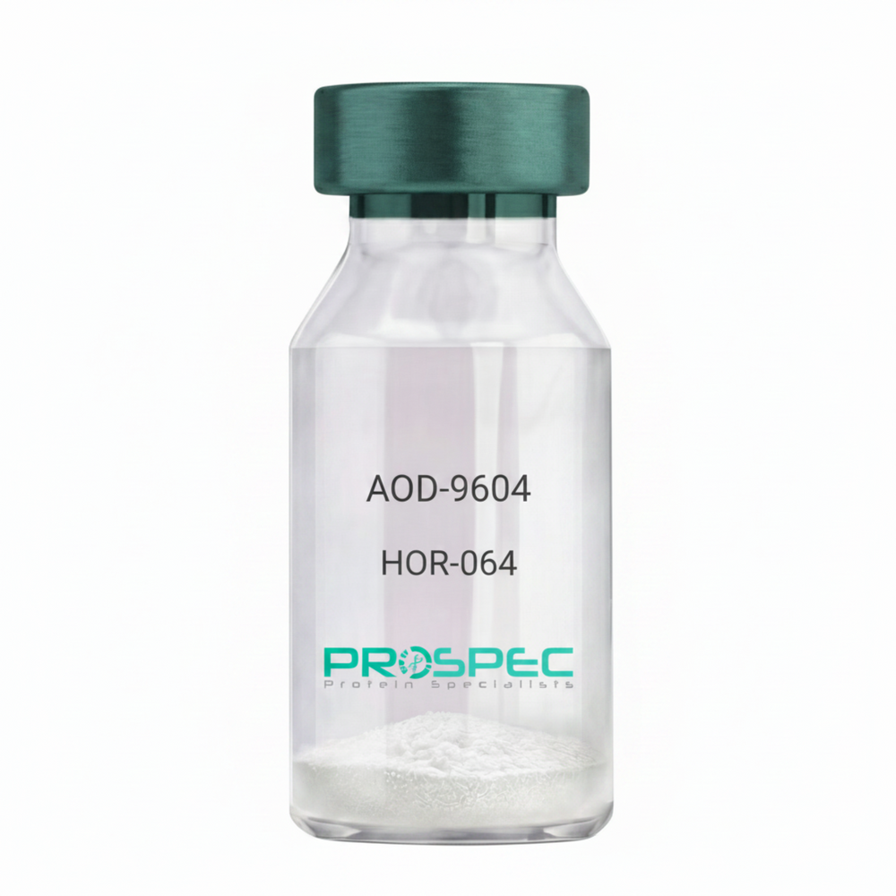
    AOD-9604

    Description:

    AOD-9604

    Product # :

    HOR-064

    Price :

    Quantity :

    Shipping Method :

    Room Temp Icon

    Shipped at Room temp

    Add To Cart

    More Info

    • description
    • formulation
    • purity
    • More Info

    Description

    AOD-9604 is a synthetic single, non-glycosylated polypeptide chain containing 16 amino acids, having a molecular mass of 1815 Dalton and a Molecular formula of C78H123N23O23 S2.

    Formulation

    The protein was lyophilized with no additives.

    Purity

    Greater than 97.0% as determined by analysis by RP-HPLC.

    More Info

    • Physical Appearance

      Sterile Filtered White lyophilized (freeze-dried) powder.

    • Stability

      Lyophilized AOD-9604 although stable at room temperature for 3 weeks, should be stored desiccated below -18°C. Upon reconstitution AOD-9604 should be stored at 4°C between 2-7 days and for future use below -18°C.
      For long term storage it is recommended to add a carrier protein (0.1% HSA or BSA).
      Please prevent freeze-thaw cycles.

    • Solubility

      It is recommended to reconstitute the lyophilized AOD-9604 in sterile 18MΩ-cm H2O not less than 100 µg/ml, which can then be further diluted to other aqueous solutions.

    • Amino Acid Sequence

      H-Tyr-Leu-Arg-Ile-Val-Gln-Cys-Arg-SerVal-Glu-Gly-Ser-Cys-Gly-Phe-OH (Disulfide bond).

    • Background

      AOD-9604 is a synthetic analog of the C-terminal fragment (177–191) AOD-9604 stimulates lipolysis (fat breakdown) without the unwanted growth-promoting or insulin-antagonizing effects of full-length hGH. AOD-9604 is a potent lipolytic tool in animal models and researchers focus on its regenerative properties in joint health and its ability to modulate beta3 adrenergic receptors without the systemic risks of growth hormone.

    ProSpec's products are furnished for LABORATORY RESEARCH USE ONLY. The product may not be used as drugs, agricultural or pesticidal products, food additives or household chemicals.

    AOD-9604
  • View Data Sheet

    Name :

    SAA Feline

    Description:

    Serum Amyloid A (APO-SAA) Feline Recombinant

    Product # :

    CYT-1221

    Price :

    Quantity :

    Shipping Method :

    Room Temp Icon

    Shipped at Room temp

    Add To Cart

    More Info

    • description
    • source
    • formulation
    • purity
    • More Info

    Description

    SAA Feline produced in E.coli is a single, non-glycosylated polypeptide chain (1-111 a.a) containing 121 a.a and having a molecular mass of 13,838 Dalton. SAA s fused with a 10 amino acids His tag at N-Terminus and purified by proprietary chromatographic techniques.

    Source

    Escherichia Coli.

    Formulation

    SAA was lyophilized from 0.01M HCl (pH 2.0).

    Purity

    Greater than 95.0% as determined by SDS-PAGE.

    More Info

    • Physical Appearance

      Sterile Filtered White lyophilized (freeze-dried) powder.

    • Stability

      Lyophilized SAA although stable at room temperature for 3 weeks, should be stored desiccated below -18°C. Upon reconstitution Serum Amyloid A (APO-SAA) should be stored at 4°C between 2-7 days and for future use below -18°C. For long term storage it is recommended to add a carrier protein (0.1% HSA or BSA). Please prevent freeze-thaw cycles.

    • Solubility

      It is recommended to reconstitute the lyophilized SAA in 0.01M HCl (pH 2.0) not less than 100µg/ml, which can then be further diluted to other aqueous solutions.

    • Background

      In response to infection, injury, or inflammation, SAA levels rapidly rise in the bloodstream. Acting as a chemoattractant, SAA recruits immune cells to the site of injury, facilitating the innate immune response. Additionally, SAA modulates the activities of immune cells, influencing cytokine production and phagocytosis. These immunomodulatory functions highlight SAA's significance in shaping the immune landscape during inflammatory events. SAA participates in lipid metabolism by interacting with HDL particles. In the acute-phase response, SAA displaces apolipoprotein A1 from HDL, transforming it into a pro-inflammatory particle. This alteration in HDL composition affects its cardioprotective functions, potentially contributing to atherosclerosis and cardiovascular diseases. Understanding these interactions is crucial for deciphering the intricate relationship between inflammation and cardiovascular health.

    ProSpec's products are furnished for LABORATORY RESEARCH USE ONLY. The product may not be used as drugs, agricultural or pesticidal products, food additives or household chemicals.

    Saa Feline
  • View Data Sheet

    Name :

    DHRS9 Human

    Description:

    Dehydrogenase/Reductase Member 9 Human Recombinant

    Dehydrogenase/reductase SDR family member 9, 3-alpha hydroxysteroid dehydrogenase, 3-alpha-HSD, NADP-dependent retinol dehydrogenase/reductase, RDH-E2, RDHL, Short-chain dehydrogenase/reductase retSDR8, DHRS9, RDH15, RDHTBE, SDR9C4, RETSDR8, 3ALPHA-HSD.

    Product # :

    ENZ-216

    Price :

    Quantity :

    Shipping Method :

    Ice Icon

    Shipped with Ice Packs

    Add To Cart

    More Info

    • description
    • source
    • formulation
    • purity
    • More Info

    Description

    DHRS9 Human Recombinant produced in E.coli is a single, non-glycosylated polypeptide chain containing 327 amino acids (18-319) and having a molecular mass of 35.9kDa.DHRS9 is fused to a 21 amino acid His-tag at N-terminus & purified by proprietary chromatographic techniques.

    Source

    Escherichia Coli.

    Formulation

    The DHRS9 solution (0.5mg/ml) contains 20mM Tris-HCl buffer (pH8.0), 20% glycerol, 0.1M NaCl, 1mM DTT and 0.1mM PMSF.

    Purity

    Greater than 90.0% as determined by SDS-PAGE.

    More Info

    • Introduction

      Dehydrogenase/reductase SDR family member 9 (DHRS9) functions as a homotetramer, which converts both 3-alpha-tetrahydroprogesterone (allopregnanolone) and 3-alpha-androstanediol to dihydroxyprogesterone and is believed to have a part in retinoic acid biosynthesis.

    • Synonyms

      Dehydrogenase/reductase SDR family member 9, 3-alpha hydroxysteroid dehydrogenase, 3-alpha-HSD, NADP-dependent retinol dehydrogenase/reductase, RDH-E2, RDHL, Short-chain dehydrogenase/reductase retSDR8, DHRS9, RDH15, RDHTBE, SDR9C4, RETSDR8, 3ALPHA-HSD.

    • Physical Appearance

      Sterile Filtered colorless solution.

    • Stability

      Store at 4°C if entire vial will be used within 2-4 weeks. Store, frozen at -20°C for longer periods of time. For long term storage it is recommended to add a carrier protein (0.1% HSA or BSA).Avoid multiple freeze-thaw cycles.

    • Amino Acid Sequence

      MGSSHHHHHH SSGLVPRGSH MGSHMRKGKL KIEDITDKYI FITGCDSGFG NLAARTFDKK GFHVIAACLT ESGSTALKAE TSERLRTVLL DVTDPENVKR TAQWVKNQVG EKGLWGLINN AGVPGVLAPT DWLTLEDYRE PIEVNLFGLI SVTLNMLPLV KKAQGRVINV SSVGGRLAIV GGGYTPSKYA VEGFNDSLRR DMKAFGVHVS CIEPGLFKTN LADPVKVIEK KLAIWEQLSP DIKQQYGEGY IEKSLDKLKG NKSYVNMDLS PVVECMDHAL TSLFPKTHYA AGKDAKIFWI PLSHMPAALQ DFLLLKQKAE LANPKAV.

    ProSpec's products are furnished for LABORATORY RESEARCH USE ONLY. The product may not be used as drugs, agricultural or pesticidal products, food additives or household chemicals.

    Dhrs9 Human
  • View Data Sheet

    Name :

    NAT1 Human

    Description:

    N-Acetyltransferase 1 Human Recombinant

    N-acetyltransferase 1 (arylamine N-acetyltransferase), AAC1, Arylamide acetylase 1, Monomorphic arylamine N-acetyltransferase, MNAT, NAT-1.

    Product # :

    ENZ-144

    Price :

    Quantity :

    Shipping Method :

    Ice Icon

    Shipped with Ice Packs

    Add To Cart

    More Info

    • description
    • source
    • formulation
    • purity
    • More Info

    Description

    NAT1 Human Recombinant produced in E.coli is a single, non-glycosylated polypeptide chain containing 310 amino acids (1-290) and having a molecular mass of 36.1 kDa.The NAT1 is fused to a 20 amino acid His-Tag at N-terminus and purified by proprietary chromatographic techniques.

    Source

    Escherichia Coli.

    Formulation

    NAT1 protein 0.5mg/ml is supplied in 20mM Tris-HCl, pH-8, 0.1M NaCl, 2mM DTT and 10% Glycerol.

    Purity

    Greater than 85.0% as determined by SDS-PAGE.

    More Info

    • Introduction

      NAT1 is a member of the arylamine N-acetyltransferase family. NAT1 catalyzes N- or O-acetylation of heterocyclic and arylamine substrates in the detoxification of a large number of drugs. Several alleles triggering high levels of N-acetyltransferase activity were linked to colon and urinary bladder cancers, since NAT1 also bioactivate numerous known carcinogens. NAT1 aids metabolize drugs and other xenobiotics, and takes part in the detoxification of a plethora of hydrazine and arylamine drugs.

    • Synonyms

      N-acetyltransferase 1 (arylamine N-acetyltransferase), AAC1, Arylamide acetylase 1, Monomorphic arylamine N-acetyltransferase, MNAT, NAT-1.

    • Physical Appearance

      NAT1 is supplied as a sterile filtered clear solution.

    • Stability

      Store at 4°C if entire vial will be used within 2-4 weeks. Store, frozen at -20°C for longer periods of time. For long term storage it is recommended to add a carrier protein (0.1% HSA or BSA).Avoid multiple freeze-thaw cycles.

    • Amino Acid Sequence

      MGSSHHHHHH SSGLVPRGSH MDIEAYLERI GYKKSRNKLD LETLTDILQH QIRAVPFENL NIHCGDAMDL GLEAIFDQVV RRNRGGWCLQ VNHLLYWALT TIGFETTMLG GYVYSTPAKK YSTGMIHLLL QVTIDGRNYI VDAGFGRSYQ MWQPLELISG KDQPQVPCIF RLTEENGFWY LDQIRREQYI PNEEFLHSDL LEDSKYRKIY SFTLKPRTIE DFESMNTYLQ TSPASVFTSK SFCSLQTPDG VHCLVGFTLT HRRFNYKDNT DLIEFKTLSE EEIEKVLKNI FNISLQRKLV PKHGDRFFTI

    ProSpec's products are furnished for LABORATORY RESEARCH USE ONLY. The product may not be used as drugs, agricultural or pesticidal products, food additives or household chemicals.

    Nat1 Human
  • View Data Sheet

    Name :

    IDH1 Human

    Description:

    Isocitrate Dehydrogenase-1 Human Recombinant

    Isocitrate dehydrogenase [NADP] cytoplasmic, EC 1.1.1.42, Cytosolic NADP-isocitrate dehydrogenase, Oxalosuccinate decarboxylase, IDH, NADP(+)-specific ICDH, IDP, PICD.

    Product # :

    ENZ-193

    Price :

    Quantity :

    Shipping Method :

    Ice Icon

    Shipped with Ice Packs

    Add To Cart

    More Info

    • description
    • source
    • formulation
    • purity
    • biological activity
    • More Info

    Description

    IDH1 Human Recombinant produced in E.coli is a single, non-glycosylated polypeptide chain containing 434 amino acids (1-414) and having a molecular mass of 48.8 kDa.IDH1 is fused to a 20 amino acid His-tag at N-terminus & purified by proprietary chromatographic techniques.

    Source

    E.coli.

    Formulation

    The IDH1 solution (1mg/ml) contains 20mM Tris-HCl buffer (pH 8.0), 0.1M NaCl,
    1mM DTT, 0.1mM PMSF and 20% glycerol.

    Purity

    Greater than 95% as determined by SDS-PAGE.

    Biological Activity

    The Specific activity is > 0.7 units/ml. One unit will convert 1.0 umole of isocitrate to alpha-ketoglutarate per minute at pH7.5 at 25C.

    More Info

    • Introduction

      Isocitrate Dehydrogenase is an enzyme of the oxidoreductase class that catalyzes the conversion of isocitrate and NAD+ to yield 2-ketoglutarate, carbon dioxide, and NADH. It occurs in cell mitochondria. The enzyme requires Mg2+, Mn2+; it is activated by ADP, citrate, and Ca2+, and inhibited by NADH, NADPH, and ATP. The reaction is the key rate-limiting step of the citric acid (tricarboxylic) cycle.

    • Synonyms

      Isocitrate dehydrogenase [NADP] cytoplasmic, EC 1.1.1.42, Cytosolic NADP-isocitrate dehydrogenase, Oxalosuccinate decarboxylase, IDH, NADP(+)-specific ICDH, IDP, PICD.

    • Physical Appearance

      Sterile Filtered colorless solution.

    • Stability

      Store at 4°C if entire vial will be used within 2-4 weeks. Store, frozen at -20°C for longer periods of time. For long term storage it is recommended to add a carrier protein (0.1% HSA or BSA).Avoid multiple freeze-thaw cycles.

    • Amino Acid Sequence

      MGSSHHHHHH SSGLVPRGSH MSKKISGGSV VEMQGDEMTR IIWELIKEKL IFPYVELDLH SYDLGIENRD ATNDQVTKDA AEAIKKHNVG VKCATITPDE KRVEEFKLKQ MWKSPNGTIR NILGGTVFRE AIICKNIPRL VSGWVKPIII GRHAYGDQYR ATDFVVPGPG KVEITYTPSD GTQKVTYLVH NFEEGGGVAM GMYNQDKSIE DFAHSSFQMA LSKGWPLYLS TKNTILKKYD GRFKDIFQEI YDKQYKSQFE AQKIWYEHRL IDDMVAQAMK SEGGFIWACK NYDGDVQSDS VAQGYGSLGM MTSVLVCPDG KTVEAEAAHG TVTRHYRMYQ KGQETSTNPI ASIFAWTRGL AHRAKLDNNK ELAFFANALE EVSIETIEAG FMTKDLAACI KGLPNVQRSD YLNTFEFMDK LGENLKIKLA QAKL

    ProSpec's products are furnished for LABORATORY RESEARCH USE ONLY. The product may not be used as drugs, agricultural or pesticidal products, food additives or household chemicals.

    Idh1 Human
  • View Data Sheet

    Name :

    EFNA1 Human

    Description:

    Ephrin A1 Human Recombinant

    Ephrin-A1, EPLG1, TNFAIP4, LERK1, EFL1, ECKLG, EPH-related receptor tyrosine kinase ligand 1, Immediate early response protein B61, Tumor necrosis factor alpha-induced protein 4, TNF alpha-induced protein 4, ligand of eph-related kinase 1, tumor necrosis factor, alpha-induced protein 4.

    Product # :

    PRO-971

    Price :

    Quantity :

    Shipping Method :

    Ice Icon

    Shipped with Ice Packs

    Add To Cart

    More Info

    • description
    • source
    • formulation
    • purity
    • More Info

    Description

    EFNA1 Human Recombinant produced in E.coli is a single, non-glycosylated polypeptide chain containing 185 amino acids (19-182) and having a molecular mass of 21.6 kDa.EFNA1 is fused to a 21 amino acid His-tag at N-terminus & purified by proprietary chromatographic techniques.

    Source

    E.coli.

    Formulation

    The EFNA1 solution (1mg/ml) contains 20mM Tris-HCl buffer (pH 8.0), 0.4M Urea and 10% glycerol.

    Purity

    Greater than 90% as determined by SDS-PAGE.

    More Info

    • Introduction

      EFNA1 belongs to the ephrin (EPH) family. The EPH subfamily is the biggest group of receptor protein kinases and they take part in vital nervous system function and development.

    • Synonyms

      Ephrin-A1, EPLG1, TNFAIP4, LERK1, EFL1, ECKLG, EPH-related receptor tyrosine kinase ligand 1, Immediate early response protein B61, Tumor necrosis factor alpha-induced protein 4, TNF alpha-induced protein 4, ligand of eph-related kinase 1, tumor necrosis factor, alpha-induced protein 4.

    • Physical Appearance

      Sterile Filtered colorless solution.

    • Stability

      Store at 4°C if entire vial will be used within 2-4 weeks. Store, frozen at -20°C for longer periods of time. For long term storage it is recommended to add a carrier protein (0.1% HSA or BSA).Avoid multiple freeze-thaw cycles.

    • Amino Acid Sequence

      MGSSHHHHHH SSGLVPRGSH MDRHTVFWNS SNPKFRNEDY TIHVQLNDYV DIICPHYEDH SVADAAMEQY ILYLVEHEEY QLCQPQSKDQ VRWQCNRPSA KHGPEKLSEK FQRFTPFTLG KEFKEGHSYY YISKPIHQHE DRCLRLKVTV SGKITHSPQA HVNPQEKRLA ADDPEVRVLH SIGHS

    ProSpec's products are furnished for LABORATORY RESEARCH USE ONLY. The product may not be used as drugs, agricultural or pesticidal products, food additives or household chemicals.

    Efna1 Human
  • View Data Sheet

    Name :

    BPHL Human

    Description:

    Biphenyl Hydrolase-Like Human Recombinant

    Biphenyl Hydrolase-Like (serine hydrolase), Bph-rp, Breast Epithelial Mucin-Associated Antigen, MCNAA, VACVASE, MGC41865, MGC125930.

    Product # :

    ENZ-055

    Price :

    Quantity :

    Shipping Method :

    Ice Icon

    Shipped with Ice Packs

    Add To Cart

    More Info

    • description
    • source
    • formulation
    • purity
    • More Info

    Description

    BPHL produced in E.Coli is a single, non-glycosylated polypeptide chain containing 275 amino acids (38-291a.a.) and having a molecular mass of 31.1kDa.BPHL is fused to a 21 amino acid His-tag at N-terminus & purified by proprietary chromatographic techniques.

    Source

    Escherichia Coli.

    Formulation

    The BPHL protein solution (0.5mg/1ml) is formulated in 20mM Tris-HCl buffer (pH8.0)
    1mM DTT, 50mM NaCl and 10% glycerol.

    Purity

    Greater than 95% as determined by SDS-PAGE.

    More Info

    • Introduction

      BPHL is a Serine hydrolase that is a part of the AB hydrolase superfamily which catalyzes the hydrolytic activation of amino acid ester prodrugs of nucleoside analogs. BPHL is expressed large quantities in liver and kidney and in minor quantities in heart, intestine and skeletal muscle. BPHL takes part in detoxification processes and is a specific alpha-amino acid ester hydrolase which favors small, hydrophobic, and aromatic side chains and does not have a strict necessity for the leaving group except for favoring a primary alcohol.

    • Synonyms

      Biphenyl Hydrolase-Like (serine hydrolase), Bph-rp, Breast Epithelial Mucin-Associated Antigen, MCNAA, VACVASE, MGC41865, MGC125930.

    • Physical Appearance

      Sterile Filtered clear solution.

    • Stability

      Store at 4°C if entire vial will be used within 2-4 weeks. Store, frozen at -20°C for longer periods of time.Please avoid freeze thaw cycles.

    • Amino Acid Sequence

      MGSSHHHHHH SSGLVPRGSH MSVTSAKVAV NGVQLHYQQT GEGDHAVLLL PGMLGSGETD FGPQLKNLNK KLFTVVAWDP RGYGHSRPPD RDFPADFFER DAKDAVDLMK ALKFKKVSLL GWSDGGITAL IAAAKYPSYI HKMVIWGANA YVTDEDSMIY EGIRDVSKWS ERTRKPLEAL YGYDYFARTC EKWVDGIRQF KHLPDGNICR HLLPRVQCPA LIVHGEKDPL VPRFHADFIH KHVKGSRLHL MPEGKHNLHL RFADEFNKLA EDFLQ

    ProSpec's products are furnished for LABORATORY RESEARCH USE ONLY. The product may not be used as drugs, agricultural or pesticidal products, food additives or household chemicals.

    Bphl Human
  • View Data Sheet

    Name :

    MDH1 Human

    Description:

    Malate Dehydrogenase 1 Human Recombinant

    MDH-s, MDHA, MOR2.

    Product # :

    ENZ-256

    Price :

    Quantity :

    Shipping Method :

    Ice Icon

    Shipped with Ice Packs

    Add To Cart

    More Info

    • description
    • source
    • formulation
    • purity
    • biological activity
    • More Info

    Description

    MDH1 Human Recombinant produced in E.Coli is a single, non-glycosylated polypeptide chain containing 342 amino acids (1-334 a.a.) and having a molecular mass of 37.4 kDa. The MDH1 is fused to an 8 amino acid His tag at C-terminus and purified by conventional chromatography.

    Source

    Escherichia Coli.

    Formulation

    The MDH1 1mg/ml protein solution contains 20mM Tris-HCl pH-8, and 10% glycerol.

    Purity

    Greater than 95.0% as determined by SDS-PAGE.

    Biological Activity

    Specific activity is > 250 units/mg, and is defined as the amount of enzyme that cleaves 1umole of oxalacetate and beta-NADH to L-malate and beta-NAD per minute at pH8.0 at 25°C.

    More Info

    • Introduction

      MDH1 catalyzes the reversible oxidation of malate to oxaloacetate, using the NAD/NADH cofactor system in the citric acid cycle. MDH1 is abundantly found in the cytoplasm and is involved in the malate-aspartate shuttle that functions in the metabolic coordination between cytosol and mitochondria. MDH1 regulates p53-dependent cell-cycle arrest and apoptosis in response to glucose deprivation.

    • Synonyms

      MDH-s, MDHA, MOR2.

    • Physical Appearance

      Sterile Filtered colorless solution.

    • Stability

      Store at 4°C if entire vial will be used within 2-4 weeks. Store, frozen at -20°C for longer periods of time. For long term storage it is recommended to add a carrier protein (0.1% HSA or BSA). Please prevent freeze-thaw cycles.

    • Amino Acid Sequence

      MSEPIRVLVT GAAGQIAYSL LYSIGNGSVF GKDQPIILVL LDITPMMGVL DGVLMELQDC ALPLLKDVIA TDKEDVAFKD LDVAILVGSM PRREGMERKD LLKANVKIFK SQGAALDKYA KKSVKVIVVG NPANTNCLTA SKSAPSIPKE NFSCLTRLDH NRAKAQIALK LGVTANDVKN VIIWGNHSST QYPDVNHAKV KLQGKEVGVY EALKDDSWLK GEFVTTVQQR GAAVIKARKL SSAMSAAKAI CDHVRDIWFG TPEGEFVSMG VISDGNSYGV PDDLLYSFPV VIKNKTWKFV EGLPINDFSR EKMDLTAKEL TEEKESAFEF LSSALEHHHH HH.

    ProSpec's products are furnished for LABORATORY RESEARCH USE ONLY. The product may not be used as drugs, agricultural or pesticidal products, food additives or household chemicals.

    Mdh1 Human
  • View Data Sheet

    Name :

    SPA Long

    Description:

    Staphylococcal Protein-A Long Form Recombinant

    Immunoglobulin G-binding protein A, IgG-binding protein A, Staphylococcal protein A, SPA.

    Product # :

    PRO-1926

    Price :

    Quantity :

    Shipping Method :

    Room Temp Icon

    Shipped at Room temp

    Add To Cart

    More Info

    • description
    • source
    • formulation
    • purity
    • More Info

    Description

    SPA Recombinant Long Form produced in E.Coli is a single non-glycosylated polypeptide chain. SPA Long Form is comprised of 5 IgG-binding domains E-D-A-B-C aligned in series containing 422 amino acids and having a molecular mass of 46.6kDa.

    Source

    Escherichia Coli.

    Formulation

    SPA protein was lyophilized with no additives.

    Purity

    Greater than 97.0% as determined by:
    (a) Analysis by HPLC.
    (b) Analysis by SDS-PAGE.

    More Info

    • Introduction

      Protein A is a cell wall component produced by several strains of Staphylococcus aureus. The recombinant Protein A is genetically engineered protein and holds 5 IgG-binding regions of protein A. Recombinant Protein A functions basically the same as native Protein A and is perfect for purification of polyclonal or monoclonal IgG antibodies. Protein A binds to human IgG1, IgG2 and IgG4, mouse IgG2a, IgG2b and IgG3 and rat IgG2c. Protein A also binds to total IgG from rabbit, pig, dog, cat, and guinea pig.

    • Synonyms

      Immunoglobulin G-binding protein A, IgG-binding protein A, Staphylococcal protein A, SPA.

    • Physical Appearance

      Sterile Filtered White lyophilized (freeze-dried) powder.

    • Stability

      Lyophilized SPA although stable at room temperature for 3 weeks, should be stored desiccated below -18°C. Upon reconstitution SPA should be stored at 4°C between 2-7 days and for future use below -18°C. For long term storage it is recommended to add a carrier protein (0.1% HSA or BSA). Please prevent freeze-thaw cycles.

    • Solubility

      It is recommended to reconstitute the lyophilized SPA in sterile 18MΩ-cm H2O not less than 0.1mg/ml, which can then be further diluted to other aqueous solutions.

    • Amino Acid Sequence

      AQHDEAQQNA FYQVLNMPNL NADQRNGFIQ SLKDDPSQSA NVLGEAQKLN DSQAPKADAQ QNNFNKDQQS AFYEILNMPN LNEAQRNGFI QSLKDDPSQS TNVLGEAKKL NESQAPKADN NFNKEQQNAF YEILNMPNLN EEQRNGFIQS LKDDPSQSAN LLSEAKKLNE SQAPKADNKF NKEQQNAFYE ILHLPNLNEE QRNGFIQSLK DDPSQSANLL AEAKKLNDAQ APKADNKFNK EQQNAFYEIL HLPNLTEEQR NGFIQSLKDD PSVSKEILAE AKKLNDAQAP KEEDNKKPGK EDGNKPGKED GNKPGKEDNK KPGKEDGNKP GKEDNNKPGK EDGNKPGKED NNKPGKEDGN KPGKEDGNKP GKEDGNGVHV VKPGDTVNDI AKANGTTADK IAADNKLADK NMIKPGQELV VD.

    ProSpec's products are furnished for LABORATORY RESEARCH USE ONLY. The product may not be used as drugs, agricultural or pesticidal products, food additives or household chemicals.

    Spa Long
  • View Data Sheet

    Name :

    BCKDHA Human

    Description:

    Branched Chain keto Acid Dehydrogenase E1 Alpha Human Recombinant

    2-oxoisovalerate dehydrogenase subunit alpha mitochondrial, Branched-chain alpha-keto acid dehydrogenase E1 component alpha chain, BCKDE1A, BCKDH E1-alpha, BCKDHA, MSU, MSUD1, OVD1A, FLJ45695.

    Product # :

    ENZ-090

    Price :

    Quantity :

    Shipping Method :

    Ice Icon

    Shipped with Ice Packs

    Add To Cart

    More Info

    • description
    • source
    • formulation
    • purity
    • More Info

    Description

    BCKDHA Human Recombinant fused with a 21 amino acid His tag at N-terminus produced in E.Coli is a single, non-glycosylated, polypeptide chain containing 421 amino acids (46-445 a.a.) and having a molecular mass of 47.8kDa. The BCKDHA is purified by proprietary chromatographic techniques.

    Source

    Escherichia Coli.

    Formulation

    The BCKDHA solution (0.25 mg/ml) contains 20mM Tris-HCl buffer (pH 8.0), 5mM DTT, 30% glycerol and 0.2M NaCl.

    Purity

    Greater than 80.0% as determined by SDS-PAGE.

    More Info

    • Introduction

      Branched-chain ?-keto acid dehydrogenase E1 component ? chain (BCKDHA) is a member of the BCKDHA family. The BCKD (branched-chain alpha-keto acid dehydrogenase) complex is an inner mitochondrial enzyme complex which catalyzes the second major step in the catabolism of the branched-chain amino acids leucine, isoleucine, and valine. This complex consists of 3 catalytic components: a heterotetrameric (alpha2-beta2) branched-chain alpha-keto acid decarboxylase (E1), a dihydrolipoyl transacylase (E2), and a dihydrolipoamide dehydrogenase (E3). Mutations in the BCKDHA gene result in maple syrup urine disease, type IA.

    • Synonyms

      2-oxoisovalerate dehydrogenase subunit alpha mitochondrial, Branched-chain alpha-keto acid dehydrogenase E1 component alpha chain, BCKDE1A, BCKDH E1-alpha, BCKDHA, MSU, MSUD1, OVD1A, FLJ45695.

    • Physical Appearance

      Sterile Filtered colorless solution.

    • Stability

      Store at 4°C if entire vial will be used within 2-4 weeks. Store, frozen at -20°C for longer periods of time. For long term storage it is recommended to add a carrier protein (0.1% HSA or BSA).Avoid multiple freeze-thaw cycles.

    • Amino Acid Sequence

      MGSSHHHHHH SSGLVPRGSH MSSLDDKPQF PGASAEFIDK LEFIQPNVIS GIPIYRVMDR QGQIINPSED PHLPKEKVLK LYKSMTLLNT MDRILYESQR QGRISFYMTN YGEEGTHVGS AAALDNTDLV FGQYREAGVL MYRDYPLELF MAQCYGNISD LGKGRQMPVH YGCKERHFVT ISSPLATQIP QAVGAAYAAK RANANRVVIC YFGEGAASEG DAHAGFNFAA TLECPIIFFC RNNGYAISTP TSEQYRGDGI AARGPGYGIM SIRVDGNDVF AVYNATKEAR RRAVAENQPF LIEAMTYRIG HHSTSDDSSA YRSVDEVNYW DKQDHPISRL RHYLLSQGWW DEEQEKAWRK QSRRKVMEAF EQAERKPKPN PNLLFSDVYQ EMPAQLRKQQ ESLARHLQTY GEHYPLDHFD K.

    ProSpec's products are furnished for LABORATORY RESEARCH USE ONLY. The product may not be used as drugs, agricultural or pesticidal products, food additives or household chemicals.

    Bckdha Human
  • View Data Sheet

    Name :

    IFNG Mouse

    Description:

    IFN-Gamma Mouse Recombinant

    Immune IFN, type II IFN, T cell IFN, MAF, IFNG, IFG, IFI, IFN-gamma.

    Product # :

    CYT-358

    Price :

    Quantity :

    Shipping Method :

    Room Temp Icon

    Shipped at Room temp

    Add To Cart

    More Info

    • description
    • source
    • formulation
    • purity
    • biological activity
    • More Info

    Description

    IFN-gamma Mouse Recombinant produced in E.Coli is a single, non-glycosylated, polypeptide chain containing 134 amino acids and having a molecular mass of 15.6kDa.The IFN-gamma is purified by proprietary chromatographic techniques.

    Source

    Escherichia Coli.

    Formulation

    Lyophilized from a 0.2µm filtered concentrated (1mg/ml) solution in PBS, pH 7.4 and 5% trehalose.

    Purity

    Greater than 95.0% as determined by:
    (a) Analysis by RP-HPLC.
    (b) Analysis by SDS-PAGE.

    Biological Activity

    The specific activity as determined in a viral resistance assay is < 0.1 ng/ml, corresponding to a specific activity of 10,000,000 IU/mg


    More Info

    • Introduction

      IFN-gamma produced by lymphocytes activated by specific antigens or mitogens.
      IFN-gamma, in addition to having antiviral activity, has important immunoregulatory functions, it is a potent activator of macrophages, and has antiproliferative effects on transformed cells and it can potentiate the antiviral and antitumor effects of the type I IFNs.

    • Synonyms

      Immune IFN, type II IFN, T cell IFN, MAF, IFNG, IFG, IFI, IFN-gamma.

    • Physical Appearance

      Sterile Filtered White lyophilized (freeze-dried) powder.

    • Stability

      Lyophilized IFN-gamma although stable at room temperature for 3 weeks, should be stored desiccated below -18°C. Upon reconstitution IFN-gamma should be stored at 4°C between 2-7 days and for future use below -18°C.For long term storage it is recommended to add a carrier protein (0.1% HSA or BSA).Please prevent freeze-thaw cycles.

    • Solubility

      It is recommended to reconstitute the lyophilized IFN-gamma in sterile distilled water or 20mM AcOH at concentrations ranging between 0.1mg-0.5mg/ml, which can then be further diluted to other aqueous solutions.

    • Amino Acid Sequence

      MHGTVIESLE SLNNYFNSSG IDVEEKSLFL DIWRNWQKDG DMKILQSQII SFYLRLFEVL KDNQAISNNI SVIESHLITT FFSNSKAKKD AFMSIAKFEV NNPQVQRQAF NELIRVVHQL LPESSLRKRK RSRC.

    • Background

      What is the molecular weight/Mw of IFNG MOUSE Protein?
      IFNG MOUSE Protein has a total Mw of 15.6kDa.

      What is the source or expression system of IFNG MOUSE Protein?
      Escherichia Coli.

      What is the Purity of IFNG MOUSE Protein?
      IFNG MOUSE Protein is >95% pure as determined by SDS-PAGE.

      What is the Biological Activity of IFNG MOUSE Protein?
      The specific activity as determined in a viral resistance assay is < 0.1 ng/ml, corresponding to a specific activity of 10,000,000 IU/mg


      What is the amino acid sequence of IFNG MOUSE Protein?
      MHGTVIESLE SLNNYFNSSG IDVEEKSLFL DIWRNWQKDG DMKILQSQII SFYLRLFEVL KDNQAISNNI SVIESHLITT FFSNSKAKKD AFMSIAKFEV NNPQVQRQAF NELIRVVHQL LPESSLRKRK RSRC.

      What applications can IFNG MOUSE Protein be used in?
      IFNG MOUSE Protein can probably be used in western blot, ELISA and Lateral Flow.

      What is the endotoxin level for IFNG MOUSE Protein?
      The endotoxin level is minimal, IFNG MOUSE Protein was purified using conventional chromatography techniques.

    ProSpec's products are furnished for LABORATORY RESEARCH USE ONLY. The product may not be used as drugs, agricultural or pesticidal products, food additives or household chemicals.

    Interferon Gamma Mouse
  • View Data Sheet

    Name :

    Prealbumin Human

    Description:

    Transthyretin Human

    TTHY, TTR, ATTR, TBPA, Transthyretin, Prealbumin, PALB, HsT2651TTHY, TTR, ATTR, TBPA, Transthyretin, Prealbumin, PALB, HsT2651.

    Product # :

    PRO-2740

    Price :

    Quantity :

    Shipping Method :

    Room Temp Icon

    Shipped at Room temp

    Add To Cart

    More Info

    • description
    • source
    • formulation
    • purity
    • More Info

    Description

    Human Transthyretin dimer protein produced in Human plasma having a molecular mass of 30kD. Under certain conditions it may be shown as a monomer (15kD) or a tetramer (60kD).

    Source

    Human serum.

    Formulation

    The protein was lyophilized (0.2 µm filtered) from 20mM NH4HCO3.

    Purity

    Greater than 96.0%.

    More Info

    • Introduction

      Prealbumin is a thyroid hormone-binding protein that transports thyroxine from the bloodstream to the brain. Prealbumin is a carrier protein which transports thyroid hormones in the plasma and cerebrospinal fluid, and also transports retinol (vitamin A) in the plasma. Transthyretin consists of a tetramer of identical subunits and is dominantly produced in the liver. Mutations in Prealbumin are related to amyloid deposition, affecting predominantly peripheral nerve and/or the heart. The diseases caused by mutations include amyloidotic polyneuropathy, euthyroid hyperthyroxinaemia, amyloidotic vitreous opacities, cardiomyopathy, oculoleptomeningeal amyloidosis, meningocerebrovascular amyloidosis, and carpal tunnel syndrome. Prealbumin is an indicator of protein-energy malnutrition since it has a circulating half life of 2 days and reacts swiftly to changes in nutritional status.

    • Synonyms

      TTHY, TTR, ATTR, TBPA, Transthyretin, Prealbumin, PALB, HsT2651TTHY, TTR, ATTR, TBPA, Transthyretin, Prealbumin, PALB, HsT2651.

    • Physical Appearance

      Sterile Filtered White lyophilized (freeze-dried) powder.

    • Stability

      Prealbumin Human although stable at room temperature for 3 weeks, should be stored between 2-8°C.

    • Solubility

      It is recommended to reconstitute the lyophilized Prealbumin Human in phosphate buffer pH > 7 containing 0.15M NaCl.

    • Human Virus Test

      Starting material donor has been tested and certified negative for antibodies to HIV-1, HIV-2, HCV, HBSAG, Parvovirus B19, Syphilis and HIV/HBV/HCV (PCR).

    ProSpec's products are furnished for LABORATORY RESEARCH USE ONLY. The product may not be used as drugs, agricultural or pesticidal products, food additives or household chemicals.

    Prealbumin Protein
  • View Data Sheet

    Name :

    XPA Antibody

    Description:

    Xeroderma Pigmentosum Complementation Group A, Mouse Anti Human

    XP1, XPAC, DNA repair protein complementing XP-A cells.

    Product # :

    ANT-504

    Price :

    Quantity :

    Shipping Method :

    Ice Icon

    Shipped with Ice Packs

    Add To Cart

    More Info

    • formulation
    • More Info

    Formulation

    1mg/ml containing PBS, pH-7.4, 10% Glycerol and 0.02% Sodium Azide.

    More Info

    • Introduction

      DNA repair protein complementing XP-A cells, (XPA), is a member of the XPA family. XPA protein takes a part in DNA excision repair. It Inductees repair by binding to damaged sites with different affinities depending on the photoproduct and the transcriptional state of the region. Defects in XPA is the reason of xeroderma pigmentosum complementation group A (XP-A), which is infrequent human autosomal recessive disease which characterized by solar sensitivity, high predisposition for developing cancers on areas exposed to sunlight, also may cause to neurological abnormalities.

    • Synonyms

      XP1, XPAC, DNA repair protein complementing XP-A cells.

    • Physical Appearance

      Sterile filtered colorless solution.

    • Immunogen

      Anti-human XPA mAb, clone PAT71H3, is derived from hybridization of mouse F0 myeloma cells with spleen cells from BALB/c mice immunized with a recombinant human XPA protein 1-273 amino acids purified from E. coli.

    • Ig Subclass

      Mouse IgG1 heavy chain and k light chain.

    • Clone

      PAT71H3AT.

    • Applications

      The antibody has been tested by ELISA, Western blot analysis, ICC/IF to assure specificity and reactivity. Since application varies, however, each investigation should be titrated by the reagent to obtain optimal results.

    • Type

      Mouse Anti Human Monoclonal.

    • Storage Procedures

      For periods up to 1 month store at 4°C, for longer periods of time, store at -20°C. Prevent freeze thaw cycles.

    • Purification Method

      XPA antibody was purified from mouse ascitic fluids by protein-A affinity chromatography.

    ProSpec's products are furnished for LABORATORY RESEARCH USE ONLY. The product may not be used as drugs, agricultural or pesticidal products, food additives or household chemicals.

    Xpa Antibody
  • View Data Sheet

    Name :

    SNCA Human

    Description:

    Alpha Synuclein Human Recombinant

    Alpha-synuclein, Non-A beta component of AD amyloid, Non-A4 component of amyloid precursor, NACP, PD1, PARK1, PARK4, MGC110988, a-Synuclein, SNCA.

    Product # :

    PRO-393

    Price :

    Quantity :

    Shipping Method :

    Ice Icon

    Shipped with Ice Packs

    Add To Cart

    More Info

    • description
    • source
    • formulation
    • purity
    • More Info

    Description

    a-Synuclein Human Recombinant produced in E.Coli is a single, non-glycosylated polypeptide chain of 140 amino acids having a molecular mass of 14.4kDa (Real molecular weight on SDS-PAGE will be shift up). The Recombinant Human a-Synuclein is purified by proprietary chromatographic techniques.

    Source

    Escherichia Coli.

    Formulation

    The protein (1mg/ml) contains 20mM Tris-HCl buffer (pH 7.5), 0.1M NaCl and 1mM MgCl2.

    Purity

    Greater than 95.0% as determined by SDS-PAGE.

    More Info

    • Introduction

      a-Synuclein (amino acids 1-140), an acidic neuronal protein of 140 amino acids, is extremely heat-resistant and is natively unfolded with an extended structure primarily composed of random coils. a-synuclein has been suggested to be implicated in the pathogenesis of Parkinson’s disease and related neurodegenerative disorders, and more recently, to be an important regulatory component of vesicular transport in neuronal cells. Moreover, recent studies have shown that a-synuclein has chaperone activity and that this activity is lost upon removing its C-terminal acidic tail (amino acids 96-140).

    • Synonyms

      Alpha-synuclein, Non-A beta component of AD amyloid, Non-A4 component of amyloid precursor, NACP, PD1, PARK1, PARK4, MGC110988, a-Synuclein, SNCA.

    • Physical Appearance

      Sterile filtered colorless solution.

    • Stability

      Store at 4°C if entire vial will be used within 2-4 weeks. Store, frozen at -20°C for longer periods of time. For long term storage it is recommended to add a carrier protein (0.1% HSA or BSA). Avoid multiple freeze-thaw cycles.

    • Amino Acid Sequence

      MDVFMKGLSK AKEGVVAAAE KTKQGVAEAA GKTKEGVLYV GSKTKEGVVH GVATVAEKTK EQVTNVGGAV VTGVTAVAQK TVEGAGSIAA ATGFVKKDQL GKNEEGAPQE GILEDMPVDP DNEAYEMPSE EGYQDYEPEA.

    ProSpec's products are furnished for LABORATORY RESEARCH USE ONLY. The product may not be used as drugs, agricultural or pesticidal products, food additives or household chemicals.

    Snca Human
  • View Data Sheet

    Name :

    SDF 1a Human, His

    Description:

    Stromal Cell-Derived Factor-1 alpha Human Recombinant, His Tag

    SDF-1, CXCL12, Pre-B cell growth-stimulating factor, PBSF, hIRH, chemokine (C-X-C motif) ligand 12, SDF1, SDF1A, TPAR1, SCYB12, SDF-1a, TLSF-a.

    Product # :

    CHM-241

    Price :

    Quantity :

    Shipping Method :

    Room Temp Icon

    Shipped at Room temp

    Add To Cart

    More Info

    • description
    • source
    • formulation
    • purity
    • More Info

    Description

    Stromal Cell-Derived Factor-1 alpha Human Recombinant produced in E.Coli is a non-glycosylated, Polypeptide chain containing 78 amino acids, having a molecular mass of 9.2 kDa. The SDF-1a is fused to 10 amino acids His-Tag at N-terminus and purified by proprietary chromatographic techniques.

    Source

    Escherichia Coli.

    Formulation

    The protein was filtered (0.4µm) and lyophilized from a concentrated (0.5mg/ml) solution containing 20mM Tris buffer pH-7.5 and 20mM sodium chloride.

    Purity

    Greater than 95.0% as determined SDS-PAGE.

    More Info

    • Introduction

      SDF-1 (stromal cell-derived factor-1) is small cytokine belonging to the chemokine family that is officially designated Chemokine (C-X-C motif) ligand 12 (CXCL12). It is produced in two forms, SDF-1?/CXCL12a and SDF-1?/CXCL12b, by alternate splicing of the same gene. Chemokines are characterized by the presence of four conserved cysteines, which form two disulfide bonds. The CXCL12 proteins belong to the group of CXC chemokines, whose initial pair of cysteines are separated by one intervening amino acid. CXCL12 is strongly chemotactic for lymphocytes and has been implicated as an important cell co-ordinator during development. During embryogenesis it directs the migration of hematopoietic cells from foetal liver to bone marrow. Mice which were knocked-out for CXCL12 gene were lethal before the birth or within just 1 hour of life. As another role, CXCL12a alters also the electrophysiology of neurons. CXCL12 was shown to be expressend in many tissues in mice (including brain, thymus, heart, lung, liver, kidney, spleen and bone marrow).
      The receptor for this chemokine is CXCR4, which was previously called fusin. This CXCL12-CXCR4 interaction used to be considered exclusive (unlike for other chemokines and their receptors), but recently it was suggested that CXCL12 is also bound by CXCR7 receptor.
      The gene for CXCL12 is located on human chromosome 10. In human and mouse both CXCL12 and CXCR4 show high identity of sequence: 99% and 90%, respectively.

    • Synonyms

      SDF-1, CXCL12, Pre-B cell growth-stimulating factor, PBSF, hIRH, chemokine (C-X-C motif) ligand 12, SDF1, SDF1A, TPAR1, SCYB12, SDF-1a, TLSF-a.

    • Physical Appearance

      White lyophilized (freeze-dried) powder.

    • Stability

      Lyophilized SDF1A although stable at room temperature for 3 weeks, should be stored desiccated below -18°C. Upon reconstitution CXCL12 should be stored at 4°C between 2-7 days and for future use below -18°C.For long term storage it is recommended to add a carrier protein (0.1% HSA or BSA).Please prevent freeze-thaw cycles.

    • Solubility

      It is recommended to add deionized water to a working concentration approximately 0.5 mg/ml and let the lyophilized pellet dissolve completely. Product is not sterile! Please filter the product by appropriate sterile filter before using it in the cell culture.

    • Amino Acid Sequence

      MKHHHHHHAS KPVSLSYRCP CRFFESHVAR ANVKHLKILN TPNCALQIVA RLKNNNRQVC IDPKLKWIQE YLEKALNK.

    ProSpec's products are furnished for LABORATORY RESEARCH USE ONLY. The product may not be used as drugs, agricultural or pesticidal products, food additives or household chemicals.

    Sdf 1A Human His
  • View Data Sheet

    Name :

    IFN a 2b Human

    Description:

    IFN-Alpha 2b Human Recombinant

    IFN alpha 2b, IFNA, INFA2, MGC125764, MGC125765.

    Product # :

    CYT-205

    Price :

    Quantity :

    Shipping Method :

    Room Temp Icon

    Shipped at Room temp

    Add To Cart

    More Info

    • description
    • source
    • formulation
    • purity
    • biological activity
    • More Info

    Description

    IFN-a 2b Human Recombinant produced in E.Coli is a single, non-glycosylated, polypeptide chain containing 166 amino acids and having a molecular mass of 19400 Dalton. The difference between IFNA2A and IFNA2B is in the amino acid present at position 23. IFN-alpha 2a has a lysine at that position 23 while IFN-alpha 2b has arginine. The IFN-alpha 2b gene was obtained from human leukocytes. The IFN-a 2b is purified by proprietary chromatographic techniques.

    Source

    Escherichia Coli.

    Formulation

    Lyophilized from a (1 mg/ml) solution in containing 2.3 mg Sodium phosphate dibasic and 0.55 mg sodium phosphate monobasic buffer.

    Purity

    Greater than 98.0% as determined by(a) Analysis by RP-HPLC.
    (b) Analysis by SDS-PAGE.

    Biological Activity

    The specific activity as determined in a viral resistance assay using bovine kidney MDBK cells was found to be 260,000,000 IU/ mg.

    More Info

    • Introduction

      IFN-alpha is produced by macrophages and has antiviral activities. IFN stimulates the production of two enzymes: protein kinase and an oligoadenylate synthetase.

    • Synonyms

      IFN alpha 2b, IFNA, INFA2, MGC125764, MGC125765.

    • Physical Appearance

      Sterile Filtered White lyophilized (freeze-dried) powder.

    • Stability

      Lyophilized IFN although stable at room temperature for 3 weeks, should be stored desiccated below -18°C. Upon reconstitution IFN alpha 2b should be stored at 4°C between 2-7 days and for future use below -18°C.For long term storage it is recommended to add a carrier protein (0.1% HSA or BSA).Please prevent freeze-thaw cycles.

    • Solubility

      It is recommended to reconstitute the lyophilized IFN-alpha 2b in sterile 18MΩ-cm H2O not less than 100µg/ml, which can then be further diluted to other aqueous solutions.

    • Amino Acid Sequence

      MCDLPQTHSL GSRRTLMLLA QMRRISLFSC LKDRHDFGFP QEEFGNQFQK AETIPVLHEM IQQIFNLFST KDSSAAWDET LLDKFYTELY QQLNDLEACV IQGVGVTETP LMKEDSILAV RKYFQRITLY LKEKKYSPCA WEVVRAEIMR SFSLSTNLQE SLRSKE

    • Background

      IFN-alpha forms are produced by monocytes/macrophages, lymphoblastoid cells, fibroblasts, and a number of different cell types following induction by viruses, nucleic acids, glucocorticoid hormones, and low-molecular weight substances (n-butyrate, 5-bromodeoxy uridine). At least 23 different variants of IFN-alpha are known. The individual proteins have molecular masses between 19-26 kDa and consist of proteins with lengths of 156-166 and 172 amino acids. All IFN-alpha subtypes possess a common conserved sequence region between amino acid positions 115-151 while the amino-terminal ends are variable. Many IFN-alpha subtypes differ in their sequences at only one or two positions. Naturally occurring variants also include proteins truncated by 10 amino acids at the carboxy-terminal end. Disulfide bonds are formed between cysteines at positions 1/98 and 29/138. The disulfide bond 29/138 is essential for biological activity while the 1/98 bond can be reduces without affecting biological activity. All IFN-alpha forms contain a potential glycosylation site but most subtypes are not glycosylated. In contrast to IFN-gamma IFN-alpha proteins are stable at pH2.

      There are at least 23 different IFN-alpha genes. They have a length of 1-2 kb and are clustered on human chromosome 9p22. It is not known whether all these genes are expressed following stimulation of the cells. In some cell systems expression of some subtypes (IFN-alpha-1, IFN-alpha-2, IFN-alpha-4) is stronger than those of others. IFN-alpha genes do not contain intron sequences found in many other eukaryotic genes (see also: gene expression). Based upon the structures two types of IFN-alpha genes, designated class 1 and II, are distinguished. They encode proteins of 156-166 and172 amino acids, respectively. Deletions covering 9p22 are observed frequently in cells of lymphoblastoid leukemias. It is not known to date whether this is of significance with respect to IFN expression.

      The gene encoding the IFN-alpha receptor maps to human chromosome 21q22.1. IFN-alpha and IFN-beta are thought to bind to the same IFN binding subunit which is expressed in 100-5000 copies in IFN-alpha sensitive and -resistant cells and is associated with other as yet unidentified proteins. The IFN-omega (Omega IFN) also binds to the IFN-alpha/IFN-beta receptor. Another receptor expressed on B-lymphocytes is identical with CD21. This receptor also binds Epstein-Barr virus through its gp350/220 coat protein.Signal transduction mechanisms elicited after binding of IFN-alpha to its receptors involves tyrosine phosphorylation (see also: PTK; protein tyrosine kinase) of various non-receptor tyrosine kinases belonging to the Janus kinases. Soluble forms of the IFN-alpha receptor, corresponding to truncated forms of the extracellular domain of the cell surface IFN-alpha receptor, have been found in human serum and in normal human urine.

      All known subtypes of IFN-alpha show the same antiviral antiparasitic, antiproliferative activities in suitable bioassays although they may differ in relative activities. Human IFN-alpha is also a potent antiviral substance in murine, porcine, and bovine cell systems. Human IFN-alpha is less active in rodent cells. Site-directed mutagenesis techniques have been used to create some variants of certain subtypes (IFN-alpha-2) that display approximately 100-fold enhanced antiviral activities in mouse cells.

      What is the molecular weight/Mw of IFN A 2B HUMAN Protein?
      IFN A 2B HUMAN Protein has a total Mw of 19.4kDa.

      What is the source or expression system of IFN A 2B HUMAN Protein?
      Escherichia Coli.

      What is the Purity of IFN A 2B HUMAN Protein?
      IFN A 2B HUMAN Protein is >98% pure as determined by SDS-PAGE.

      What is the Biological Activity of IFN A 2B HUMAN Protein?
      The specific activity as determined in a viral resistance assay using bovine kidney MDBK cells was found to be 260,000,000 IU/ mg.

      What is the amino acid sequence of IFN A 2B HUMAN Protein?
      MCDLPQTHSL GSRRTLMLLA QMRRISLFSC LKDRHDFGFP QEEFGNQFQK AETIPVLHEM IQQIFNLFST KDSSAAWDET LLDKFYTELY QQLNDLEACV IQGVGVTETP LMKEDSILAV RKYFQRITLY LKEKKYSPCA WEVVRAEIMR SFSLSTNLQE SLRSKE
      What applications can IFN A 2B HUMAN Protein be used in?
      IFN A 2B HUMAN Protein can probably be used in western blot, ELISA and Lateral Flow.

      What is the endotoxin level for IFN A 2B HUMAN Protein?
      The endotoxin level is minimal, IFN A 2B HUMAN Protein was purified using conventional chromatography techniques.

    • Protein content

      Protein quantitation was carried out by two independent methods1. UV spectroscopy at 280 nm using the absorbency value of 0.924 as the extinction coefficient for a 0.1% (1mg/ml) solution. This value is calculated by the PC GENE computer analysis program of protein sequences (IntelliGenetics). 2. Analysis by RP-HPLC, using a calibrated solution of IFN alpha 2b as a Reference Standard.

    ProSpec's products are furnished for LABORATORY RESEARCH USE ONLY. The product may not be used as drugs, agricultural or pesticidal products, food additives or household chemicals.

    Interferon Alpha 2B Human
  • View Data Sheet

    Name :

    HLA-DRB1 Human

    Description:

    Major Histocompatibility Complex Class II DR Beta 1 Human Recombinant

    DRB1, HLA DRB1, HLA-DR1B, HLA-DRB1, MHC class II antigen DRB1 16, DR-16, DR16, Human Leucocyte AntigenDRB1, MHC Class IIHLA-DR-Beta Cell Surface Glycoprotein, MHC Class IIHLA-DRw10-Beta.

    Product # :

    PRO-1888

    Price :

    Quantity :

    Shipping Method :

    Ice Icon

    Shipped with Ice Packs

    Add To Cart

    More Info

    • description
    • source
    • formulation
    • purity
    • More Info

    Description

    HLA-DRB1 Human Recombinant produced in E.Coli is a single, non-glycosylated polypeptide chain containing 219 amino acids (30-227 a.a) and having a molecular mass of 25.2kDa.HLA-DRB1 is fused to a 21 amino acid His-tag at N-terminus & purified by proprietary chromatographic techniques.

    Source

    Escherichia Coli.

    Formulation

    HLA-DRB1 protein solution (1mg/ml) containing 20mM Tris-HCl buffer (pH 8.0), 0.4M Urea and 10% glycerol.

    Purity

    Greater than 85% as determined by SDS-PAGE.

    More Info

    • Introduction

      Major Histocompatibility Complex Class II DR Beta 1 also known as HLA-DRB1 ia a member of the HLA class II beta chain paralogs. Molecule class II is a heterodimer consisting of an alpha (DRA) and a beta chain (DRB), both anchored in the membrane. HLA-DRB1 takes an essential part in the immune system by presenting peptides derived from extracellular proteins. Class II molecules are expressed in antigen presenting cells (APC: B lymphocytes, dendritic cells, macrophages). Furthermore, the beta chain is approximately 26- 28 kDa. It is encoded by 6 exons. While exon one encodes the leader peptide; exons 2 and 3 encode the two extracellular domains; exon 4 encodes the transmembrane domain; and exon 5 encodes the cytoplasmic tail.

    • Synonyms

      DRB1, HLA DRB1, HLA-DR1B, HLA-DRB1, MHC class II antigen DRB1 16, DR-16, DR16, Human Leucocyte AntigenDRB1, MHC Class IIHLA-DR-Beta Cell Surface Glycoprotein, MHC Class IIHLA-DRw10-Beta.

    • Physical Appearance

      Sterile Filtered clear solution.

    • Stability

      Store at 4°C if entire vial will be used within 2-4 weeks. Store, frozen at -20°C for longer periods of time.For long term storage it is recommended to add a carrier protein (0.1% HSA or BSA).Please avoid freeze thaw cycles.

    • Amino Acid Sequence

      MGSSHHHHHH SSGLVPRGSH MGDTRPRFLW QPKRECHFFN GTERVRFLDR YFYNQEESVR FDSDVGEFRA VTELGRPDAE YWNSQKDILE QARAAVDTYC RHNYGVVESF TVQRRVQPKV TVYPSKTQPL QHHNLLVCSV SGFYPGSIEV RWFLNGQEEK AGMVSTGLIQ NGDWTFQTLV MLETVPRSGE VYTCQVEHPS VTSPLTVEWR ARSESAQSK

    ProSpec's products are furnished for LABORATORY RESEARCH USE ONLY. The product may not be used as drugs, agricultural or pesticidal products, food additives or household chemicals.

    Hla Drb1 Human
  • View Data Sheet

    Name :

    TSTA3 Human

    Description:

    Tissue Specific Transplantation Antigen P35B Human Recombinant

    FX, P35B, SDR4E1, GDP-L-fucose synthase, Protein FX .

    Product # :

    ENZ-259

    Price :

    Quantity :

    Shipping Method :

    Ice Icon

    Shipped with Ice Packs

    Add To Cart

    More Info

    • description
    • source
    • formulation
    • purity
    • More Info

    Description

    TSTA3 Recombinant Human produced in E.Coli is a single, non-glycosylated polypeptide chain containing 341 amino acids (1-321 a.a.) and having a molecular mass of 38 kDa. The TSTA3 is fused to 20 amino acid His-Tag at N-terminus and purified by proprietary chromatographic techniques.

    Source

    Escherichia Coli.

    Formulation

    0.5mg/ml solution containing 20mM Tris pH-8, 2mM DTT, 50mM NaCl & 10% glycerol.

    Purity

    Greater than 90.0% as determined by SDS-PAGE.

    More Info

    • Introduction

      TSTA3 is a NADP(H)-binding protein. TSTA3 catalyzes the two-step epimerase and the reductase reactions in GDP-D-mannose metabolism, converting GDP-4-keto-6-D-deoxymannose to GDP-L-fucose. GDP-L-fucose is the substrate of numerous fucosyltransferases that take part in the expression of several glycoconjugates, including blood group ABH antigens and developmental adhesion antigens.
      Mutations in TSTA3 result in leukocyte adhesion deficiency, type II.

    • Synonyms

      FX, P35B, SDR4E1, GDP-L-fucose synthase, Protein FX .

    • Physical Appearance

      Sterile filtered colorless solution.

    • Stability

      Store at 4°C if entire vial will be used within 2-4 weeks. Store, frozen at -20°C for longer periods of time. For long term storage it is recommended to add a carrier protein (0.1% HSA or BSA). Avoid multiple freeze-thaw cycles.

    • Amino Acid Sequence

      MGSSHHHHHH SSGLVPRGSH MGEPQGSMRI LVTGGSGLVG KAIQKVVADG AGLPGEDWVF VSSKDADLTD TAQTRALFEK VQPTHVIHLA AMVGGLFRNI KYNLDFWRKN VHMNDNVLHS AFEVGARKVV SCLSTCIFPD KTTYPIDETM IHNGPPHNSN FGYSYAKRMI DVQNRAYFQQ YGCTFTAVIP TNVFGPHDNF NIEDGHVLPG LIHKVHLAKS SGSALTVWGT GNPRRQFIYS LDLAQLFIWV LREYNEVEPI ILSVGEEDEV SIKEAAEAVV EAMDFHGEVT FDTTKSDGQF KKTASNSKLR TYLPDFRFTP FKQAVKETCA WFTDNYEQAR K.

    ProSpec's products are furnished for LABORATORY RESEARCH USE ONLY. The product may not be used as drugs, agricultural or pesticidal products, food additives or household chemicals.

    Tsta3 Human
  • View Data Sheet

    Name :

    F8 Protein

    Description:

    Coagulation Factor-VIII Human Recombinant

    Coagulation factor VIII, Procoagulant component, Antihemophilic factor, AHF, F8, F8C, F8B, HEMA, FVIII, DXS1253E, F8 protein.

    Product # :

    PRO-318

    Price :

    Quantity :

    Shipping Method :

    Room Temp Icon

    Shipped at Room temp

    Add To Cart

    More Info

    • description
    • source
    • formulation
    • purity
    • biological activity
    • More Info

    Description

    Antihemophilic Facor Human Recombinant produced in CHO is a glycosylated polypeptide chain having 2332 amino acids. The Factor-VIII is purified by proprietary chromatographic techniques.

    Source

    CHO cells (Chinese Hamster Ovarian Cells).

    Formulation

    Each 250IU vial was lyophilized from a solution containing 8mg Tween-80, 112mM NaCl, 40mg Mannitol, 10mg Trehalose, 1ng VWF and 4.2mM CaCl2.

    Purity

    Greater than 97.0% as determined by SDS-PAGE.

    Biological Activity

    The specific activity was found to be 7,058 IU/mg.

    More Info

    • Introduction

      Coagulation factor VIII participates in the intrinsic pathway of blood coagulation; factor VIII is a cofactor for factor IXa which, in the presence of Ca+2 and phospholipids, converts factor X to the activated form Xa. This gene produces two alternatively spliced transcripts. Transcript variant 1 encodes a large glycoprotein, isoform a, which circulates in plasma and associates with von Willebrand factor in a noncovalent complex. This protein undergoes multiple cleavage events. Transcript variant 2 encodes a putative small protein, isoform b, which consists primarily of the phospholipid binding domain of factor VIIIc. This binding domain is essential for coagulant activity. Defects in this gene results in hemophilia A, a common recessive X-linked coagulation disorder.

    • Synonyms

      Coagulation factor VIII, Procoagulant component, Antihemophilic factor, AHF, F8, F8C, F8B, HEMA, FVIII, DXS1253E, F8 protein.

    • Physical Appearance

      Sterile Filtered White lyophilized (freeze-dried) powder.

    • Stability

      Lyophilized Factor-VIII although stable at room temperature for 3 weeks, should be stored desiccated below -18°C. Upon reconstitution Factor-VIII should be stored at 4°C between 2-7 days and for future use below -18°C.Please prevent freeze-thaw cycles.

    • Solubility

      It is recommended to reconstitute 250IU lyophilized Factor-VIII in 5ml sterile 18M-cm H2O, which can then be further diluted to other aqueous solutions.

    ProSpec's products are furnished for LABORATORY RESEARCH USE ONLY. The product may not be used as drugs, agricultural or pesticidal products, food additives or household chemicals.

    Factor Viii Human Recombinant
  • View Data Sheet

    Name :

    Lassa GP1

    Description:

    Lassa Glycoprotein-1 Recombinant

    Product # :

    LSV-002

    Price :

    Quantity :

    Shipping Method :

    Ice Icon

    Shipped with Ice Packs

    Add To Cart

    More Info

    • description
    • source
    • formulation
    • purity
    • More Info

    Description

    The E.Coli derived Recombinant Lassa Glycoprotein-1 (strain Mouse/Sierra Leone/Josiah/1976) containing 205 amino acids, having an Mw of 30kDa and the Isoelectric point is 6.7. The Lassa GP1 protein is fused to a 6xHis tag at C-terminus and purified by proprietary chromatographic technique.

    Source

    Escherichia Coli.

    Formulation

    Lassa GP1 protein solution contains PBS and 25mM K2CO3

    Purity

    Lassa GP1 is >90% pure as determined by 10% PAGE (coomassie staining).

    More Info

    • Introduction

      Lassa virus, which is a member of the Arenaviridae virus family, causes acute viral hemorrhagic fever. It was first reported in 1969 in the town of Lassa, in Borno State, Nigeria. Natal multimammate mouse is the primary reservoir of Lassa virus, found in sub-Saharan Africa. The virus is transmitted by contact with the feces or urine of animals, contributing to its high rate of incidence. Lassa fever occurs generally in West Africa. Lassa fever results in 300,000 to 500,000 cases annually and causes about 5,000 deaths each year. Outbreaks of this disease have been observed in Nigeria, Liberia, Sierra Leone, Guinea, and the Central African Republic. Protein structure: Lassa virus genome is comprised of two single-stranded RNA molecules defined as small (S) and large (L). Two genes on the S segment encode the nucleoprotein (NP) and two envelope glycoproteins (GP1 and GP2); while, the L segment encodes the viral polymerase (L protein) and RING finger Z matrix protein. GP1 subunit serves a putative role in receptor binding, while the structure of GP2 subunit is consistent with viral transmembrane fusion proteins. NP is a virion protein which binds and protects the viral RNA.

    • Physical Appearance

      Sterile Filtered solution.

    • Stability

      Lassa GP1 protein although stable at 4°C for 1 week, should be stored below -18°C. Please prevent freeze thaw cycles.

    ProSpec's products are furnished for LABORATORY RESEARCH USE ONLY. The product may not be used as drugs, agricultural or pesticidal products, food additives or household chemicals.

    Lassa Gp1
  • View Data Sheet

    Name :

    Heparanase 1 (HPA1), Polyclonal

    Description:

    Heparanase 1 (HPA1), Polyclonal Rabbit Anti-Human

    Product # :

    ANT-155

    Price :

    Quantity :

    Shipping Method :

    Ice Icon

    Shipped with Ice Packs

    Add To Cart

    More Info

    • source
    • formulation
    • More Info

    Source

    Polyclonal rabbit anti-human HPA1 is a Protein G affinity purified polyclonal antibody raised against the 50 kDa-8 kDa Heparanase heterodimer.

    Formulation

    Each vial contains 0.29 mg or 0.57 mg of antibody in 50 or 100 µL, respectively, of 20 mM sodium phosphate; 150 mM NaCl; pH 7.2, containing 0. 01% Thimerosal.

    More Info

    • Introduction

      Heparanase is an endo ß-D-glucuronidase, which degrades heparan sulfate side chains of heparan sulfate proteoglycans (HSPGs) in the extracellular matrix. Heparanase plays an important role in ECM degradation, facilitating the migration and extravasation of tumor cells and inflammatory leukocytes (1,2,3). Upon degradation, heparanase releases growth factors and cytokines that stimulate cell proliferation and chemotaxis (4,5). Heparanase is a heterodimer comprised of a 50 kDa subunit harboring the active site and a 8 kDa subunit. It is produced as a latent 65 kDa precursor and proteolytically processed to its active form (1,6). Heparanase is highly expressed in myeloid leukocytes (i.e. neutrophils) in platelets and in human placenta. Human heparanase was found to be upregulated in various types of primary tumors, correlating in some cases with increased tumor invasiveness and vascularity and with poor prospective survival (7,8).

    • Stability

      Store at 4°C. For extended storage, freeze in working aliquots at -20°C.Avoid repeated freeze-thaw cycles.

    • Applications

      Western blot
      Immunohistochemistry

    • Data Sheet

      To view the FULL VERSION click Heparanase-1 Polyclonal Rabbit :

    • Patent Protected Countries

      Polyclonal and monoclonal Anti-heparanase antibodies and their uses are protected by US. Patents No. 6,177,545; 6,531,129, additional US patent applications and patents and patent applications worldwide.

    • Specificity

      Western blot analysis: The antibody reacts with the 65 kDa precursor as well as the 50 kDa and 8 kDa subunits of human or mouse Heparanase.Immunohistochemistry: The antibody interacts with Heparanase in paraffin sections and blood smears.Recommended dilution range for Western blot analysis: 1:2000.Recommended dilution range for immunohistochemistry: 1:100.

    ProSpec's products are furnished for LABORATORY RESEARCH USE ONLY. The product may not be used as drugs, agricultural or pesticidal products, food additives or household chemicals.

    Heparanase 1 Hpa1 Polyclonal Antibody
  • View Data Sheet

    Name :

    CoV-2 N (329 a.a.)

    Description:

    Coronavirus 2019 Nucleocapsid (329 a.a.), Recombinant

    Product # :

    SARS-044

    Price :

    Quantity :

    Shipping Method :

    Ice Icon

    Shipped with Ice Packs

    Add To Cart

    More Info

    • description
    • source
    • formulation
    • purity
    • More Info

    Description

    The E.Coli derived recombinant protein contains the Coronavirus 2019 C-terminal region 329 a.a. from the Nucleocapsid protein and fused to GST-6xHis tag at N-terminal and having a Mw. of 63.5 kDa.

    Source

    E.Coli.

    Formulation

    CoV-2 Nucleocapsid protein solution is supplied in 50mM Tris-HCl pH 8, 1M Urea, and 50% Glycerol.

    Purity

    CoV-2 Nucleocapsid protein is >95% pure as determined SDS-PAGE.

    More Info

    • Introduction

      A human infecting coronavirus (viral pneumonia) called 2019 novel coronavirus, 2019-nCoV was found in the fish market at the city of Wuhan, Hubei province of China on December 2019.

      The 2019-nCoV shares an 87% identity to the 2 bat-derived severe acute respiratory syndrome 2018 SARS-CoV-2 located in Zhoushan of eastern China. 2019-nCoV has an analogous receptor-BD-structure to that of 2018 SARS-CoV, even though there is a.a. diversity so thus the 2019-nCoV might bind to ACE2 receptor protein (angiotensin-converting enzyme 2) in humans.

      While bats are possibly the host of 2019-nCoV, researchers suspect that animal from the ocean sold at the seafood market was an intermediate host. RSCU analysis proposes that the 2019-nCoV is a recombinant within the viral spike glycoprotein between the bat coronavirus and an unknown coronavirus.

    • Physical Appearance

      Sterile Filtered clear solution.

    • Stability

      CoV-2 Spike Protein is shipped on ice packs. Upon arrival, Store at -20°C.

    • Purification Method

      NTA Sepharose-Affinity Purification.

    ProSpec's products are furnished for LABORATORY RESEARCH USE ONLY. The product may not be used as drugs, agricultural or pesticidal products, food additives or household chemicals.

    product_image.jpg
  • View Data Sheet

    Name :

    Protein A/G

    Description:

    Protein A/G Recombinant

    Product # :

    PRO-646

    Price :

    Quantity :

    Shipping Method :

    Room Temp Icon

    Shipped at Room temp

    Add To Cart

    More Info

    • description
    • source
    • formulation
    • purity
    • More Info

    Description

    The recombinant Protein A/G consists of 5 IgG-binding regions of protein A and 2 of protein G, which corresponds to the Protein A and G domains that are included in the recombinant sequence. Cell wall binding region, cell membrane binding region and albumin binding region have been removed from the recombinant Protein A/G to ensure the maximum specific IgG binding. The Protein A portion is from Staphylococcus aureus segments E, D, A, B and C. The Protein G portion is from Streptococcus segments C1 and C3. The fusion protein has a predicted molecular mass of 47.7kDa and containing 429 amino acids.

    Source

    Escherichia coli.

    Formulation

    Lyophilized white Powder containing no additives.

    Purity

    >97% as determined by SDS-PAGE and RP-HPLC.

    More Info

    • Introduction

      Recombinant Protein A/G fusion protein joins IgG binding domains of both Protein A and Protein G.
      Protein A/G includes four Fc binding domains from Protein A and two from Protein G, yielding a final mass of 50.4 kDa. The binding dependency to pH of Protein A/G lower than Protein A, but has the additive properties of Protein A and G together. Protein A/G binds to all subclasses of human IgG, making it helpful for purifying polyclonal or monoclonal IgG antibodies whose subclasses have not been identifieed. Protein A/G binds to IgA, IgE, IgM and IgD. Protein A/G binds to all subclasses of mouse IgG excluding mouse IgA, IgM or serum albumin. This permits Protein A/G to be used in purification and detection of mouse monoclonal IgG antibodies, with no interference from IgA, IgM and serum albumin. Mouse monoclonal antibodies normally have a stronger affinity to the chimeric Protein A/G than to either Protein A or Protein G. Protein A/G also has been used for purification of macaque IgG.

    • Stability

      After reconstitution, aliquot and store at -20°C. Avoid repeated freeze/thaw cycles.

    • Solubility

      It is recommended to reconstitute the lyophilized Protein-A/G in sterile 18M-cm H2O not less than 0.1mg/ml, which can then be further diluted to other aqueous solutions.

    • Amino Acid Sequence

      NAAQHDEAQQ NAFYQVLNMP NLNADQRNGF IQSLKDDPSQ SANVLGEAQK LNDSQAPKAD AQQNNFNKDQ QSAFYEILNM PNLNEAQRNG FIQSLKDDPS QSTNVLGEAK KLNESQAPKA DNNFNKEQQN AFYEILNMPN LNEEQRNGFI QSLKDDPSQS ANLLSEAKKL NESQAPKADN KFNKEQQNAF YEILHLPNLN EEQRNGFIQS LKDDPSQSAN LLAEAKKLND AQAPKADNKF NKEQQNAFYE ILHLPNLTEE QRNGFIQSLK DDPSVSKEIL AEAKKLNDAQ APKEEDSLEG SGSGTYKLIL NGKTLKGETT TEAVDAATAE KVFKQYANDN GVDGEWTYDD ATKTFTVTEK PEVIDASELT PAVTTYKLVI NGKTLKGETT TKAVDAETAE KAFKQYANDN GVDGVWTYDD ATKTFTVTE.

    ProSpec's products are furnished for LABORATORY RESEARCH USE ONLY. The product may not be used as drugs, agricultural or pesticidal products, food additives or household chemicals.

    Protein A G
  • 1
  • …
  • 25
  • 26
  • 27
  • 28
  • 29
  • …
  • 42
Your browser does not support the video tag.

Products

  • Cytokines
  • Growth Factors
  • Chemokines
  • CD Antigens
  • Neurotrophins
  • Hormones
  • Enzymes
  • Viral Antigens
  • Recombinant Proteins
  • Natural Proteins
  • Monoclonal Antibodies
  • Polyclonal Antibodies
  • Thermal Packaging

About

  • Company
  • Founder
  • Contribution
  • Contact Us
  • My Account

Ordering

  • Ordering Method
  • Ordering - Credit Card
  • Ordering PO
  • Shipping Fees
  • FAQ's

Legal

  • Terms & Conditions
  • Privacy
  • Site Map
  • info@prospecbio.com
  • Facebook
  • Instagram
  • Linkedin Profile

© 2026 ProSpec-Tany TechnoGene Ltd. All Rights Reserved.

  • Choosing a selection results in a full page refresh.
  • Opens in a new window.