Skip to content

High-quality research proteins.

  • 1-800-805-6531

  • Mon - Sat 8.00-18.00, Sun - Closed

  • info@prospecbio.com

  • PO Box 6591 East Brunswick NJ 08816

  • Products
    • Cytokines
    • Growth factors
    • Chemokines
    • Cd antigens
    • Neurotrophins
    • Hormones
    • Enzymes
    • Viral antigens
    • Recombinant proteins
    • Natural proteins
    • Monoclonal antibodies
    • Polyclonal antibodies
    • Thermal Packaging
  • About
    • Company
    • Founder
    • Contribution
  • Customers
    • Eastern Asia
    • Eastern Europe
    • North America
    • South America
    • South East Asia
    • Southern Asia
    • Western Europe
  • Ordering
    • Ordering Method
    • Ordering - Credit Card
    • Ordering - PO
    • Shipping Fees
    • FAQs
  • Services
    • DNA Cloning
    • Protein Expression
    • Monoclonal Antibody
    • Fermentation
    • Technology License
  • Contact us
Log in

Country/region

  • USD $ | Albania
  • USD $ | Andorra
  • USD $ | Argentina
  • USD $ | Armenia
  • USD $ | Australia
  • USD $ | Austria
  • USD $ | Azerbaijan
  • USD $ | Belarus
  • USD $ | Belgium
  • USD $ | Bolivia
  • USD $ | Brazil
  • USD $ | Bulgaria
  • USD $ | Cambodia
  • USD $ | Canada
  • USD $ | Chile
  • USD $ | China
  • USD $ | Colombia
  • USD $ | Costa Rica
  • USD $ | Croatia
  • USD $ | Cyprus
  • USD $ | Czechia
  • USD $ | Denmark
  • USD $ | Estonia
  • USD $ | Finland
  • USD $ | France
  • USD $ | Georgia
  • USD $ | Germany
  • USD $ | Greece
  • USD $ | Hong Kong SAR
  • USD $ | Hungary
  • USD $ | Iceland
  • USD $ | India
  • USD $ | Ireland
  • USD $ | Israel
  • USD $ | Italy
  • USD $ | Japan
  • USD $ | Kazakhstan
  • USD $ | Latvia
  • USD $ | Liechtenstein
  • USD $ | Lithuania
  • USD $ | Madagascar
  • USD $ | Malta
  • USD $ | Mexico
  • USD $ | Moldova
  • USD $ | Monaco
  • USD $ | Netherlands
  • USD $ | New Zealand
  • USD $ | North Macedonia
  • USD $ | Norway
  • USD $ | Panama
  • USD $ | Paraguay
  • USD $ | Peru
  • USD $ | Philippines
  • USD $ | Poland
  • USD $ | Portugal
  • USD $ | Romania
  • USD $ | Singapore
  • USD $ | Slovakia
  • USD $ | Slovenia
  • USD $ | South Africa
  • USD $ | South Korea
  • USD $ | Spain
  • USD $ | Sri Lanka
  • USD $ | Sweden
  • USD $ | Switzerland
  • USD $ | Taiwan
  • USD $ | Thailand
  • USD $ | Türkiye
  • USD $ | Ukraine
  • USD $ | United Kingdom
  • USD $ | United States
  • USD $ | Uruguay
  • USD $ | Vatican City
  • USD $ | Venezuela
  • USD $ | Vietnam
  • Facebook
  • Instagram
logoProSpec - Recombinant Protein Manufacturer for academic and R&D industry
Become a distributor
Account Log in
Wishlist
Cart Cart(0)
  • Products
    +
    • Cytokines
    • Growth factors
    • Chemokines
    • Cd antigens
    • Neurotrophins
    • Hormones
    • Enzymes
    • Viral antigens
    • Recombinant proteins
    • Natural proteins
    • Monoclonal antibodies
    • Polyclonal antibodies
    • Thermal Packaging
  • About
    +
    • Company
    • Founder
    • Contribution
  • Customers
    +
    • Eastern Asia
    • Eastern Europe
    • North America
    • South America
    • South East Asia
    • Southern Asia
    • Western Europe
  • Ordering
    +
    • Ordering Method
    • Ordering - Credit Card
    • Ordering - PO
    • Shipping Fees
    • FAQs
  • Services
    +
    • DNA Cloning
    • Protein Expression
    • Monoclonal Antibody
    • Fermentation
    • Technology License
  • Contact us

Item added to your cart

View cart
View All
  • Cytokines
  • Growth factors
  • Chemokines
  • Cd antigens
  • Neurotrophins
  • Hormones
  • Enzymes
  • Viral antigens
  • Recombinant proteins
  • Natural proteins
  • Monoclonal antibodies
  • Polyclonal antibodies
  • Thermal Packaging
  • Tumor Necrosis Factor

    Tumor Necrosis Factor

    View All
  • 4-1BB

    4-1BB

    View All
  • Adiponectin

    Adiponectin

    View All
  • AIF1

    AIF1

    View All
  • Other Cytokines

    Other Cytokines

    View All
  • AITRL

    AITRL

    View All
  • Angiopoietin

    Angiopoietin

    View All
  • Apolipoprotein

    Apolipoprotein

    View All
  • B-Cell Activating Factor

    B-Cell Activating Factor

    View All
  • Beta Defensin

    Beta Defensin

    View All
  • Bone Morphogenetic Protein

    Bone Morphogenetic Protein

    View All
  • B type Natriuretic Peptide

    B type Natriuretic Peptide

    View All
  • BST

    BST

    View All
  • Betacellulin

    Betacellulin

    View All
  • Cardiotrophin

    Cardiotrophin

    View All
  • Activin

    Activin

    View All
  • Fibroblast Growth Factor

    Fibroblast Growth Factor

    View All
  • Other Growth Factors

    Other Growth Factors

    View All
  • CSF

    CSF

    View All
  • CTGF

    CTGF

    View All
  • IGFBP

    IGFBP

    View All
  • VEGF Protein

    VEGF Protein

    View All
  • EGF

    EGF

    View All
  • Epigen

    Epigen

    View All
  • Erythropoietin

    Erythropoietin

    View All
  • Insulin-Like Growth Factor

    Insulin-Like Growth Factor

    View All
  • Growth Hormone

    Growth Hormone

    View All
  • HDGF

    HDGF

    View All
  • Hepatocyte Growth Factor

    Hepatocyte Growth Factor

    View All
  • Insulin

    Insulin

    View All
  • BCA-1/ BLC (CXCL13)

    BCA-1/ BLC (CXCL13)

    View All
  • BRAK (CXCL14)

    BRAK (CXCL14)

    View All
  • C-10 (CCL6)

    C-10 (CCL6)

    View All
  • Other Chemokines

    Other Chemokines

    View All
  • MEC (CCL28)

    MEC (CCL28)

    View All
  • LD78-beta (CCL3L1)

    LD78-beta (CCL3L1)

    View All
  • CTACK (CCL27)

    CTACK (CCL27)

    View All
  • CXCL16

    CXCL16

    View All
  • CXCL17

    CXCL17

    View All
  • Platelet Factor-4 (CXCL4)

    Platelet Factor-4 (CXCL4)

    View All
  • ENA-78 (CXCL5)

    ENA-78 (CXCL5)

    View All
  • Eotaxin (CCL11,24,26)

    Eotaxin (CCL11,24,26)

    View All
  • Exodus-2 (CCL21)

    Exodus-2 (CCL21)

    View All
  • Fractalkine (CX3CL1)

    Fractalkine (CX3CL1)

    View All
  • CXCL6

    CXCL6

    View All
  • Other CD Antigens

    Other CD Antigens

    View All
  • CD14

    CD14

    View All
  • CD164

    CD164

    View All
  • CD1

    CD1

    View All
  • CD2

    CD2

    View All
  • CD200

    CD200

    View All
  • CD207

    CD207

    View All
  • CD226

    CD226

    View All
  • CD23

    CD23

    View All
  • CD244

    CD244

    View All
  • CD247

    CD247

    View All
  • CD27

    CD27

    View All
  • CD274

    CD274

    View All
  • CD300

    CD300

    View All
  • CD33

    CD33

    View All
  • Other Neurotrophins

    Other Neurotrophins

    View All
  • Beta-NGF

    Beta-NGF

    View All
  • BDNF

    BDNF

    View All
  • CDNF

    CDNF

    View All
  • CNTF

    CNTF

    View All
  • GDNF

    GDNF

    View All
  • Glia Maturation Factor

    Glia Maturation Factor

    View All
  • MANF

    MANF

    View All
  • Midkine

    Midkine

    View All
  • Neuroglobin

    Neuroglobin

    View All
  • Neurotrophic factors

    Neurotrophic factors

    View All
  • Neuregulin

    Neuregulin

    View All
  • Neuropilin

    Neuropilin

    View All
  • Pigment Epithelium-Derived Factor

    Pigment Epithelium-Derived Factor

    View All
  • Pleiotrophin

    Pleiotrophin

    View All
  • Peptide Hormones

    Peptide Hormones

    View All
  • Other Hormones

    Other Hormones

    View All
  • HCG

    HCG

    View All
  • Endothelin

    Endothelin

    View All
  • Exendin

    Exendin

    View All
  • FSH

    FSH

    View All
  • Glucagon

    Glucagon

    View All
  • GHRP

    GHRP

    View All
  • GLP

    GLP

    View All
  • Thyrostimulin

    Thyrostimulin

    View All
  • Inhibin A

    Inhibin A

    View All
  • LHRH

    LHRH

    View All
  • Melanotan

    Melanotan

    View All
  • Procalcitonin

    Procalcitonin

    View All
  • PTH

    PTH

    View All
  • Peroxiredoxin

    Peroxiredoxin

    View All
  • Peptidase

    Peptidase

    View All
  • Synthetase

    Synthetase

    View All
  • Transferase

    Transferase

    View All
  • Hydrolase

    Hydrolase

    View All
  • Dehydrogenase

    Dehydrogenase

    View All
  • ACE2

    ACE2

    View All
  • Esterase

    Esterase

    View All
  • Protein Kinases

    Protein Kinases

    View All
  • Other Enzymes

    Other Enzymes

    View All
  • Phosphatase

    Phosphatase

    View All
  • Deaminase

    Deaminase

    View All
  • Oxygenase

    Oxygenase

    View All
  • Adenylate Kinase

    Adenylate Kinase

    View All
  • Lyase

    Lyase

    View All
  • Chagas

    Chagas

    View All
  • SARS

    SARS

    View All
  • Influenza

    Influenza

    View All
  • ASFV

    ASFV

    View All
  • Astrovirus

    Astrovirus

    View All
  • Borrelia

    Borrelia

    View All
  • Helicobacter Pylori

    Helicobacter Pylori

    View All
  • Chikungunya

    Chikungunya

    View All
  • Chlamydia

    Chlamydia

    View All
  • Cytomegalo

    Cytomegalo

    View All
  • Dengue

    Dengue

    View All
  • EBV

    EBV

    View All
  • Ebola

    Ebola

    View All
  • Feline Leukemia Virus

    Feline Leukemia Virus

    View All
  • Hepatitis A

    Hepatitis A

    View All
  • Other

    Other

    View All
  • Actin

    Actin

    View All
  • ADAM

    ADAM

    View All
  • Complement Component

    Complement Component

    View All
  • Ag85

    Ag85

    View All
  • Eukaryotic Translation Initiation Factor

    Eukaryotic Translation Initiation Factor

    View All
  • Anterior Gradient Protein

    Anterior Gradient Protein

    View All
  • Heat Shock Protein

    Heat Shock Protein

    View All
  • Alpha-2-HS-Glycoprotein

    Alpha-2-HS-Glycoprotein

    View All
  • Hemoglobin

    Hemoglobin

    View All
  • Allergy

    Allergy

    View All
  • Anaplasma

    Anaplasma

    View All
  • Angiogenin

    Angiogenin

    View All
  • Ankyrin Repeat Domain

    Ankyrin Repeat Domain

    View All
  • Annexin

    Annexin

    View All
  • Other Natural Proteins

    Other Natural Proteins

    View All
  • Aprotinin

    Aprotinin

    View All
  • Transferrin

    Transferrin

    View All
  • Avidin

    Avidin

    View All
  • Anti Coagulation Factors

    Anti Coagulation Factors

    View All
  • Natural Albumin

    Natural Albumin

    View All
  • Natural Coagulation Factors

    Natural Coagulation Factors

    View All
  • Fibronectin

    Fibronectin

    View All
  • Thrombin

    Thrombin

    View All
  • Anti Human Enzyme

    Anti Human Enzyme

    View All
  • Other Monoclonal Antibodies

    Other Monoclonal Antibodies

    View All
  • Anti Human Cytokine

    Anti Human Cytokine

    View All
  • Anti Human Heat Shock Protein

    Anti Human Heat Shock Protein

    View All
  • Anti Mouse Lymphocyte

    Anti Mouse Lymphocyte

    View All
  • Anti Human Chemokine

    Anti Human Chemokine

    View All
  • Anti Human Lymphocyte

    Anti Human Lymphocyte

    View All
  • Anti Viral Monoclonal

    Anti Viral Monoclonal

    View All
  • Anti Mouse Cytokine

    Anti Mouse Cytokine

    View All
  • Anti-GST Monoclonal

    Anti-GST Monoclonal

    View All
  • Other Polyclonal Antibodies

    Other Polyclonal Antibodies

    View All
  • Anti Viral

    Anti Viral

    View All
  • Anti-GST Monoclonal

    Anti-GST Monoclonal

    View All
Back

Search results

652 results found for “biglycan”

Name

Description

Product #

Price

Quantity

Shipping Method

  • View Data Sheet

    Name :

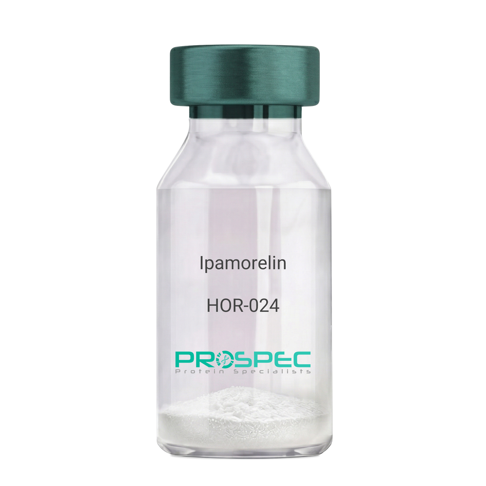
    Ipamorelin

    Description:

    Ipamorelin

    Ipamorelin

    Product # :

    HOR-024

    Price :

    Quantity :

    Shipping Method :

    Room Temp Icon

    Shipped at Room temp

    Add To Cart

    More Info

    • description
    • formulation
    • purity
    • More Info

    Description

    Ipamorelin Synthetic is a single, non-glycosylated polypeptide chain containing 4 amino acids, having a molecular mass of 711.85 Dalton and a Molecular formula of C38H49N9O5.

    Formulation

    The protein was lyophilized with no additives.

    Purity

    Greater than 97.0% as determined by analysis by RP-HPLC.

    More Info

    • Introduction

      Ipamorelin is a peptide selective agonist of the ghrelin/growth hormone secretagogue receptor and a growth hormone secretagogue. Ipamorelin is a pentapeptide that was derived from GHRP1. Ipamorelin significantly increases plasma growth hormone levels in both animals and humans. Like pralmorelin and GHRP-6, ipamorelin does not affect prolactin, FSH, LH or TSH levels. However, unlike GHRP2 and GHRP6, but as growth hormone-releasing hormone (GHRH), ipamorelin does not stimulate the secretion of adrenocorticotropic hormone (ACTH) or cortisol, and is highly selective for inducing the secretion only of GH.

    • Synonyms

      Ipamorelin

    • Physical Appearance

      Sterile Filtered White lyophilized (freeze-dried) powder.

    • Stability

      Lyophilized Ipamorelin although stable at room temperature for 3 weeks, should be stored desiccated below -18°C. Upon reconstitution Ipamorelin should be stored at 4°C between 2-7 days and for future use below -18°C. For long term storage it is recommended to add a carrier protein (0.1% HSA or BSA). Please prevent freeze-thaw cycles.

    • Solubility

      It is recommended to reconstitute the lyophilized Ipamorelin in sterile 18MΩ-cm H2O not less than 100 µg/ml, which can then be further diluted to other aqueous solutions.

    • Amino Acid Sequence

      Aib-His-D-2-Nal-D-Phe-Lys-NH2.

    ProSpec's products are furnished for LABORATORY RESEARCH USE ONLY. The product may not be used as drugs, agricultural or pesticidal products, food additives or household chemicals.

    Ipamorelin
  • View Data Sheet

    Name :

    BCL2 Antibody

    Description:

    B-Cell Leukemia/Lymphoma 2, Mouse Anti Human

    Apoptosis regulator Bcl-2, BCL2, B-cell CLL/lymphoma 2, Bcl-2.

    Product # :

    ANT-022

    Price :

    Quantity :

    Shipping Method :

    Ice Icon

    Shipped with Ice Packs

    Add To Cart

    More Info

    • formulation
    • More Info

    Formulation

    1mg/ml containing PBS, pH-7.4, & 0.1% Sodium Azide.

    More Info

    • Introduction

      BCL2 gene encodes an integral outer mitochondrial membrane protein that blocks the apoptotic death of some cells such as lymphocytes. Constitutive expression of BCL2, such as in the case of translocation of BCL2 to Ig heavy chain locus, is thought to be the cause of follicular lymphoma. Two transcript variants, produced by alternate splicing, differ in their C-terminal ends.

    • Synonyms

      Apoptosis regulator Bcl-2, BCL2, B-cell CLL/lymphoma 2, Bcl-2.

    • Immunogen

      Anti-human BCL2 mAb, is derived from hybridization of mouse FO myeloma cells with spleen cells from BALB/c mice immunized with recombinant human BCL2 amino acids 1-211 purified from E. coli.

    • Ig Subclass

      Mouse IgG2b heavy chain and k light chain.

    • Clone

      PAT1B5AT.

    • Applications

      BCL2 antibody has been tested by ELISA, Western blot, and Immunofluorescence analyses to assure specificity and reactivity. Since applications vary, however, each investigation should be titrated by a reagent to obtain optimal results. Recommended dilution range for Western blot analysis and Immunofluorescence is 1:250 ~ 500.
      Recommended starting dilution is 1:500.

    • Type

      Mouse Anti Human Monoclonal.

    • Storage Procedures

      For periods up to 1 month store at 4°C, for longer periods of time, store at -20°C. Prevent freeze thaw cycles.

    • Purification Method

      BCL2 antibody was purified from mouse ascitic fluids by protein-G affinity chromatography.

    ProSpec's products are furnished for LABORATORY RESEARCH USE ONLY. The product may not be used as drugs, agricultural or pesticidal products, food additives or household chemicals.

    Bcl2 Antibody
  • View Data Sheet

    Name :

    BUB3 Antibody

    Description:

    BUB3, Mouse Anti Human

    BUB3L, hBUB3, Mitotic checkpoint protein BUB3.

    Product # :

    ANT-545

    Price :

    Quantity :

    Shipping Method :

    Ice Icon

    Shipped with Ice Packs

    Add To Cart

    More Info

    • formulation
    • More Info

    Formulation

    1mg/ml containing PBS, pH-7.4, 10% Glycerol and 0.02% Sodium Azide.

    More Info

    • Introduction

      Mitotic checkpoint protein BUB3 (BUB3), is a saved component of the mitotic spindle assembly complex (MCC). The encoded protein has 4 WD repeat domains and has a similar sequence as the yeast BUB3 protein. BUB3 is vital for the kinetochore localization of BUB1 and BUBR1. BUB3 participates in the central spindle checkpoint pathway that operates during early embryogenesis. Furthermore BUB3 has a part in regulating the establishment of correct kinetochore-microtubule attachments.

    • Synonyms

      BUB3L, hBUB3, Mitotic checkpoint protein BUB3.

    • Physical Appearance

      Sterile filtered colorless solution.

    • Immunogen

      Anti-human BUB3 mAb, is derived from hybridization of mouse F0 myeloma cells with spleen cells from BALB/c mice immunized with a recombinant human BUB3 protein 1-328 amino acids purified from E. coli.

    • Ig Subclass

      Mouse IgG2b heavy chain and k light chain.

    • Clone

      PAT2H6AT.

    • Applications

      BUB3 antibody has been tested by ELISA, Western blot analysis to assure specificity and reactivity. Since application varies, however, each investigation should be titrated by the reagent to obtain optimal results. Recommended starting dilution is 1:1000.

    • Type

      Mouse Anti Human Monoclonal.

    • Storage Procedures

      For periods up to 1 month store at 4°C, for longer periods of time, store at -20°C. Prevent freeze thaw cycles.

    • Purification Method

      BUB3 antibody was purified from mouse ascitic fluids by protein-A affinity chromatography.

    ProSpec's products are furnished for LABORATORY RESEARCH USE ONLY. The product may not be used as drugs, agricultural or pesticidal products, food additives or household chemicals.

    Bub3 Antibody
  • View Data Sheet

    Name :

    TBCA Antibody

    Description:

    Tubulin Folding Cofactor A, Mouse Anti Human

    Tubulin-specific chaperone A, Tubulin-folding cofactor A, CFA, TCP1-chaperonin cofactor A, TBCA.

    Product # :

    ANT-362

    Price :

    Quantity :

    Shipping Method :

    Ice Icon

    Shipped with Ice Packs

    Add To Cart

    More Info

    • formulation
    • More Info

    Formulation

    1mg/ml containing PBS, pH-7.4, 10% Glycerol and 0.02% Sodium Azide.

    More Info

    • Introduction

      TBCA is a tubulin-folding protein which is involved in the early step of the tubulin folding pathway. TBCA is one of four proteins (cofactors A, D, E, and C) implicated in the pathway directing to properly folded beta-tubulin from folding intermediates. Cofactors A and D are thought to be a factor in capturing and stabilizing beta-tubulin in a quasi-native confirmation. TBCA is crucial for cell viability, if reduced it causes a decrease in the amount of soluble tubulin, alterations in microtubules and G1 cell cycle arrest. Cofactor E attaches to the cofactor D-tubulin complex, afterward, interaction with cofactor C triggers the release of tubulin polypeptides that are committed to the native state.

    • Synonyms

      Tubulin-specific chaperone A, Tubulin-folding cofactor A, CFA, TCP1-chaperonin cofactor A, TBCA.

    • Immunogen

      Anti-human TBCA mAb is derived from hybridization of mouse F0 myeloma cells with spleen cells from BALB/c mice immunized with recombinant human TBCA amino acids 1-108 purified from E. coli.

    • Ig Subclass

      Mouse IgG2b heavy chain and κ light chain.

    • Clone

      PAT1A5AT.

    • Applications

      TBCA antibody has been tested by ELISA, Western blot, ICC/IF and FACS analysis to assure specificity and reactivity. Since application varies, however, each investigation should be titrated by the reagent to obtain optimal results.

    • Type

      Mouse Anti Human Monoclonal.

    • Storage Procedures

      For periods up to 1 month store at 4°C, for longer periods of time, store at -20°C. Prevent freeze thaw cycles.

    • Purification Method

      TBCA antibody was purified from mouse ascitic fluids by protein-G affinity chromatography.

    ProSpec's products are furnished for LABORATORY RESEARCH USE ONLY. The product may not be used as drugs, agricultural or pesticidal products, food additives or household chemicals.

    Tbca Antibody
  • View Data Sheet

    Name :

    GAS7 Antibody

    Description:

    Growth Arrest-Specific 7 Isoform b, Mouse Anti Human

    GAS7, GAS-7, Growth arrest-specific protein 7, KIAA0394, MGC1348, MLL/GAS7.

    Product # :

    ANT-358

    Price :

    Quantity :

    Shipping Method :

    Ice Icon

    Shipped with Ice Packs

    Add To Cart

    More Info

    • formulation
    • More Info

    Formulation

    1mg/ml containing PBS, pH-7.4, & 0.1% Sodium Azide.

    More Info

    • Introduction

      GAS7 is mainly expressed in terminally differentiated brain cells and predominantly in mature cerebellar Purkinje neurons. GAS7 is involved in neuronal development by promoting maturation and morphological differentiation of cerebellar neurons. Inhibition of GAS7 production in terminally differentiating cultures of embryonic murine cerebellum impedes neurite outgrowth. Over-expression of GAS7 initiats the development of human osteosarcoma.

    • Synonyms

      GAS7, GAS-7, Growth arrest-specific protein 7, KIAA0394, MGC1348, MLL/GAS7.

    • Immunogen

      Anti-human GAS7 mAb is derived from hybridization of mouse F0 myeloma cells with spleen cells from BALB/c mice immunized with recombinant human GAS7 amino acids 1-416 purified from E. coli.

    • Ig Subclass

      Mouse IgG1 heavy chain and κ light chain.

    • Clone

      PAT4H8AT.

    • Applications

      GAS7 antibody has been tested by ELISA and Western blot analysis to assure specificity and reactivity. Since application varies, however, each investigation should be titrated by the reagent to obtain optimal results. Recommended dilution range for Western blot analysis is 1:500 ~ 1000. Recommended starting dilution is 1:500.

    • Type

      Mouse Anti Human Monoclonal.

    • Storage Procedures

      For periods up to 1 month store at 4°C, for longer periods of time, store at -20°C. Prevent freeze thaw cycles.

    • Purification Method

      GAS7 antibody was purified from mouse ascitic fluids by protein-G affinity chromatography.

    ProSpec's products are furnished for LABORATORY RESEARCH USE ONLY. The product may not be used as drugs, agricultural or pesticidal products, food additives or household chemicals.

    Gas7 Antibody
  • View Data Sheet

    Name :

    XPA Antibody

    Description:

    Xeroderma Pigmentosum Complementation Group A, Mouse Anti Human

    XP1, XPAC, DNA repair protein complementing XP-A cells.

    Product # :

    ANT-504

    Price :

    Quantity :

    Shipping Method :

    Ice Icon

    Shipped with Ice Packs

    Add To Cart

    More Info

    • formulation
    • More Info

    Formulation

    1mg/ml containing PBS, pH-7.4, 10% Glycerol and 0.02% Sodium Azide.

    More Info

    • Introduction

      DNA repair protein complementing XP-A cells, (XPA), is a member of the XPA family. XPA protein takes a part in DNA excision repair. It Inductees repair by binding to damaged sites with different affinities depending on the photoproduct and the transcriptional state of the region. Defects in XPA is the reason of xeroderma pigmentosum complementation group A (XP-A), which is infrequent human autosomal recessive disease which characterized by solar sensitivity, high predisposition for developing cancers on areas exposed to sunlight, also may cause to neurological abnormalities.

    • Synonyms

      XP1, XPAC, DNA repair protein complementing XP-A cells.

    • Physical Appearance

      Sterile filtered colorless solution.

    • Immunogen

      Anti-human XPA mAb, clone PAT71H3, is derived from hybridization of mouse F0 myeloma cells with spleen cells from BALB/c mice immunized with a recombinant human XPA protein 1-273 amino acids purified from E. coli.

    • Ig Subclass

      Mouse IgG1 heavy chain and k light chain.

    • Clone

      PAT71H3AT.

    • Applications

      The antibody has been tested by ELISA, Western blot analysis, ICC/IF to assure specificity and reactivity. Since application varies, however, each investigation should be titrated by the reagent to obtain optimal results.

    • Type

      Mouse Anti Human Monoclonal.

    • Storage Procedures

      For periods up to 1 month store at 4°C, for longer periods of time, store at -20°C. Prevent freeze thaw cycles.

    • Purification Method

      XPA antibody was purified from mouse ascitic fluids by protein-A affinity chromatography.

    ProSpec's products are furnished for LABORATORY RESEARCH USE ONLY. The product may not be used as drugs, agricultural or pesticidal products, food additives or household chemicals.

    Xpa Antibody
  • View Data Sheet

    Name :

    HPSE Active

    Description:

    Recombinant Human Heparanase-1 Active

    Product # :

    ENZ-1032

    Price :

    Quantity :

    Shipping Method :

    Ice Icon

    Shipped with Ice Packs

    Add To Cart

    More Info

    • description
    • formulation
    • purity
    • biological activity
    • More Info

    Description

    Heparanase Active Enzyme is produced in CHO cells.The protein is purified by several orthogonal chromatography steps.

    Formulation

    Heparanase Active Enzyme is supplied in20mM Acetate buffer and 750mM NaCl pH 5.4.

    Purity

    Greater than 95.0% as determined by:
    (a) Analysis by RP-HPLC.
    (b) Analysis by SDS-PAGE.

    Biological Activity

    The specific activity of Heparanase Active Enzyme in-house standard is about 0.7 Units (1 unit = 1 μmole of reducing ends of heparan sulfate substrate formed per minute per mg Heparanase Active Enzyme at 37°C). The enzymatic activity of each Heparanase Active Enzyme batch is comparable to the standard as determined by activity assay in which immobilized heparan, released due to heparanase activity, is quantified colorimetrically. Recommended reaction buffer: 20 mM Citrate Phosphate buffer, pH 5.4; 50mM NaCl; 1mM CaCl2.

    More Info

    • Introduction

      Heparanase is an endo β-D-glucuronidase, which degrades heparan sulfate side chains of heparan sulfate proteoglycans (HSPGs) in the extracellular matrix. Heparanase plays an important role in ECM degradation, facilitating the migration and extravasation of tumor cells and inflammatory leukocytes (1,2,3). Upon degradation, heparanase releases growth factors and cytokines that stimulate cell proliferation and chemotaxis (4,5). Heparanase is a heterodimer comprised of a 50 kDa subunit harboring the active site and a 8 kDa subunit. It is produced as a latent 65 kDa precursor and proteolytically processed to its active form (1,6). Heparanase is highly expressed in myeloid leukocytes (i.e. neutrophils) in platelets and in human placenta. Human heparanase was found to be upregulated in various types of primary tumors, correlating in some cases with increased tumor invasiveness and vascularity and with poor prospective survival (7,8).

    • Data Sheet

      To view the FULL VERSION data sheet click Heparanase-1 Active Enzyme

    • Specificity

      Heparanase Active Enzyme is identified by Western blot analysis with polyclonalrabbit anti-HPA1 antibodies as 2 subunits of 8-kDa and 50-kDa.

    • Shipping Conditions

      Heparanase Active Enzyme is shipped frozen on dry ice unless stated otherwise by the customer. Shipping fees to N. America and W. Europe is $400 Shipping fees to Asia, Australia and E. Europe is $500

    • Storage Procedures

      Store at –80ºC, avoid repeated freeze-thaw cycles.

    ProSpec's products are furnished for LABORATORY RESEARCH USE ONLY. The product may not be used as drugs, agricultural or pesticidal products, food additives or household chemicals.

    Heparanase Active
  • View Data Sheet

    Name :

    BAK1 Antibody

    Description:

    Bcl-2 homologous antagonist/killer, Mouse Anti Human

    Bcl-2 homologous antagonist/killer, Apoptosis regulator BAK, Bcl-2-like protein 7, Bcl2-L-7, BAK1, BAK, BCL2L7, CDN1, MGC3887, BAK-LIKE, MGC117255.

    Product # :

    ANT-015

    Price :

    Quantity :

    Shipping Method :

    Ice Icon

    Shipped with Ice Packs

    Add To Cart

    More Info

    • formulation
    • More Info

    Formulation

    1mg/ml containing PBS, pH-7.4, & 0.1% Sodium Azide.

    More Info

    • Introduction

      BAK1 is a member of the BCL2 protein family. The BCL2 family members form oligomers or heterodimers and act as anti- or pro-apoptotic regulators which are involved in a wide variety of cellular activities. The BAK1 gene encodes a receptor-like kinase (RLK) with a putative extracellular domain, a single transmembrane domain, an intracellular-juxtamembrane domain, and a kinase domain. BAK1 localizes to mitochondria, and functions to induce apoptosis. BAK1 interacts with and accelerates the opening of the mitochondrial voltage-dependent anion channel, leading to a loss in membrane potential and the release of cytochrome c. Furthermore, BAK1 interacts with the tumor suppressor P53 after exposure to cell stress. BAK1 expression is linked to the progression of Prostate cancer.

    • Synonyms

      Bcl-2 homologous antagonist/killer, Apoptosis regulator BAK, Bcl-2-like protein 7, Bcl2-L-7, BAK1, BAK, BCL2L7, CDN1, MGC3887, BAK-LIKE, MGC117255.

    • Immunogen

      Anti-human BAK1 mAb, is derived from hybridization of mouse F0 myeloma cells with spleen cells from BALB/c mice immunized with recombinant human BAK1 amino acids 29-187 purified from E. coli.

    • Ig Subclass

      Mouse IgG2a heavy chain and k light chain.

    • Clone

      PAT38E2AT.

    • Applications

      BAK1 antibody has been tested by ELISA, Western blot and Immunofluorescence analysis to assure specificity and reactivity. Since application varies, however, each investigation should be titrated by the reagent to obtain optimal results. Recommended dilution range for Western blot analysis and Immunofluorescence is 1:250 ~ 500.
      Recommended starting dilution is 1:250.

    • Type

      Mouse Anti Human Monoclonal.

    • Storage Procedures

      For periods up to 1 month store at 4°C, for longer periods of time, store at -20°C. Prevent freeze thaw cycles.

    • Purification Method

      BAK1 antibody was purified from mouse ascitic fluids by protein-G affinity chromatography.

    ProSpec's products are furnished for LABORATORY RESEARCH USE ONLY. The product may not be used as drugs, agricultural or pesticidal products, food additives or household chemicals.

    Bak1 Antibody
  • View Data Sheet

    Name :

    BMP-7 Antibody

    Description:

    Bone Morphogenetic Protein-7 Polyclonal Rabbit Anti Human Antibody

    Osteogenic Protein 1, BMP-7.

    Product # :

    ANT-037

    Price :

    Quantity :

    Shipping Method :

    Ice Icon

    Shipped with Ice Packs

    Add To Cart

    More Info

    • formulation
    • purity
    • More Info

    Formulation

    Lyophilized from a sterile filtered (0.2µm) solution containing phosphate buffered saline.

    Purity

    Greater than 98%.

    More Info

    • Introduction

      The bone morphogenetic proteins (BMPs) are a family of secreted signaling molecules that can induce ectopic bone growth. Many BMPs are part of the transforming growth factor-beta (TGFB) superfamily. BMPs were originally identified by an ability of demineralized bone extract to induce endochondral osteogenesis in vivo in an extraskeletal site. Based on its expression early in embryogenesis, the BMP encoded by this gene has a proposed role in early development. In addition, the fact that this BMP is closely related to BMP5 and BMP7 has lead to speculation of possible bone inductive activity.

    • Synonyms

      Osteogenic Protein 1, BMP-7.

    • Stability

      Store at -20°C. For long term storage freezes in working aliquots at -20°C. Repeated freezing and thawing is not recommended.

    • Solubility

      Add 100 ?l of distilled water to create a final concentration of 1 mg/ml.

    • Immunogen

      IgG Anti BMP-7 has been developed in rabbit using highly pure (>98%) recombinant human BMP-7 expressed in plants.

    • Applications

      Western Blot: to detect human BMP-7 by WB analysis this IgG can be used in a dilution of 1:500. For optimal usage we suggest overnight incubation.

    • Antigen Amino Acid Sequence

      FPTI PLSRLFDNAM LRAHRLHQLA FDTYQ EFEEAYIPKEQKYSFLQNPQTSLCFSESIPTPSNREETQQKSNLE LLRISLLLIQSWLEPVQFLRSVFANSLVYGASDSNVYDLL KDLEEGIQTLMGRLE DGSPRTGQIFKQ TYSK DTNSHNDDALLKNYGLLYCFRK DM DKVETFLRIVQCRSVEGSCGF.

    • Type

      Polyclonal Rabbit Antibody.

    • Purification Method

      Purified IgG prepared by affinity chromatography on protein G.

    ProSpec's products are furnished for LABORATORY RESEARCH USE ONLY. The product may not be used as drugs, agricultural or pesticidal products, food additives or household chemicals.

    Bmp 7 Antibody
  • View Data Sheet

    Name :

    PGK2 Human

    Description:

    Phosphoglycerate Kinase 2 Human Recombinant

    Phosphoglycerate kinase 2, Phosphoglycerate kinase testis specific, PGK2, PGKB, PGKPS, dJ417L20.2.

    Product # :

    PKA-017

    Price :

    Quantity :

    Shipping Method :

    Ice Icon

    Shipped with Ice Packs

    Add To Cart

    More Info

    • description
    • source
    • formulation
    • purity
    • More Info

    Description

    PGK2 Human Recombinant produced in E.Coli is a single, non-glycosylated polypeptide chain containing 437 amino acids (1-417 a.a.) and having a molecular mass of 46.9kDa.PGK2 is fused to a 20 amino acid His-tag at N-terminus & purified by proprietary chromatographic techniques.

    Source

    Escherichia Coli.

    Formulation

    PGK2 protein solution (0.5mg/ml) containing 20mM Tris-HCl buffer (pH8.0), 20% glycerol, 1mM DTT and 0.1M NaCl.

    Purity

    Greater than 85.0% as determined by SDS-PAGE.

    More Info

    • Introduction

      PGK2 is a testis-specific form of phosphoglycerate kinase. Originally presumed to be a pseudogene, the PGK2 protein is actually a functional phosphoglycerate kinase which catalyzes the reversible conversion of 1,3-bisphosphoglycerate to 3-phosphoglycerate, during the Embden-Meyerhof-Parnas pathway of glycolysis, in the later stages of spermatogenesis. The PGK2 gene is intronless, results from retrotransposition of the phosphoglycerate kinase 1 gene, and is expressed specifically in the testis.

    • Synonyms

      Phosphoglycerate kinase 2, Phosphoglycerate kinase testis specific, PGK2, PGKB, PGKPS, dJ417L20.2.

    • Physical Appearance

      Sterile Filtered colorless solution.

    • Stability

      Store at 4°C if entire vial will be used within 2-4 weeks. Store, frozen at -20°C for longer periods of time. For long term storage it is recommended to add a carrier protein (0.1% HSA or BSA).Avoid multiple freeze-thaw cycles.

    • Amino Acid Sequence

      MGSSHHHHHH SSGLVPRGSH MSLSKKLTLD KLDVRGKRVI MRVDFNVPMK KNQITNNQRI KASIPSIKYC LDNGAKAVVL MSHLGRPDGV PMPDKYSLAP VAVELKSLLG KDVLFLKDCV GAEVEKACAN PAPGSVILLE NLRFHVEEEG KGQDPSGKKI KAEPDKIEAF RASLSKLGDV YVNDAFGTAH RAHSSMVGVN LPHKASGFLM KKELDYFAKA LENPVRPFLA ILGGAKVADK IQLIKNMLDK VNEMIIGGGM AYTFLKVLNN MEIGASLFDE EGAKIVKDIM AKAQKNGVRI TFPVDFVTGD KFDENAQVGK ATVASGISPG WMGLDCGPES NKNHAQVVAQ ARLIVWNGPL GVFEWDAFAK GTKALMDEIV KATSKGCITV IGGGDTATCC AKWNTEDKVS HVSTGGGASL ELLEGKILPG VEALSNM.

    ProSpec's products are furnished for LABORATORY RESEARCH USE ONLY. The product may not be used as drugs, agricultural or pesticidal products, food additives or household chemicals.

    Pgk2 Human
  • View Data Sheet

    Name :

    IL5 Canine

    Description:

    Interleukin-5 Canine Recombinant

    Interleukin-5, IL-5, EDF, TRF, Eosinophil differentiation factor, T-cell replacing factor.

    Product # :

    CYT-1184

    Price :

    Quantity :

    Shipping Method :

    Ice Icon

    Shipped with Ice Packs

    Add To Cart

    More Info

    • description
    • source
    • formulation
    • purity
    • biological activity
    • More Info

    Description

    IL5 Canine produced in Sf9 Baculovirus cells is a single, glycosylated polypeptide chain containing 119 amino acids (22-134 aa) and having a molecular mass of 13.9kDa.IL5 is fused to a 6 amino acid His tag at C-terminus and purified by proprietary chromatographic techniques.

    Source

    Sf9, Baculovirus cells.

    Formulation

    The IL5 solution (0.25mg/ml) contains 10% Glycerol and Phosphate-Buffered Saline (pH 7.4).

    Purity

    Greater than 90.0% as determined by SDS-PAGE.

    Biological Activity

    Measured in a cell proliferation assay using TF-1 human erythroleukemic cell. The ED50 range ≤ 15 ng/ml.

    More Info

    • Introduction

      The protein encoded by IL5is a cytokine that acts as a growth and differentiation factor for both B cells and eosinophils. IL5is a main regulator of eosinopoiesis, eosinophil maturation and activation. The elevated production of IL5is reported to be related to asthma or hypereosinophilic syndromes. The receptor of this cytokine is a heterodimer, whose beta subunit is shared with the receptors for interleukine 3 (IL3) and colony stimulating factor 2 (CSF2/GM-CSF). Interleukin-5, together with those for interleukin 4 (IL4), interleukin 13 (IL13), and CSF2, form a cytokine gene cluster on chromosome 5. Interleukin-5, IL4, and IL13 are found to be regulated coordinately by long-range regulatory elements spread over 120 kilobases on chromosome 5q31.

    • Synonyms

      Interleukin-5, IL-5, EDF, TRF, Eosinophil differentiation factor, T-cell replacing factor.

    • Physical Appearance

      Sterile Filtered colorless solution.

    • Stability

      Store at 4°C if entire vial will be used within 2-4 weeks. Store, frozen at -20°C for longer periods of time. For long term storage it is recommended to add a carrier protein (0.1% HSA or BSA).Avoid multiple freeze-thaw cycles.

    • Amino Acid Sequence

      VENPMNRLVA ETLTLLSTHR TWLIGDGNLM IPTPENKNHQ LCIKEVFQGI DTLKNQTAHG EAVDKLFQNL SLIKEHIERQ KKRCAGERWR VTKFLDYLQV FLGVINTEWT PESHHHHHH

    ProSpec's products are furnished for LABORATORY RESEARCH USE ONLY. The product may not be used as drugs, agricultural or pesticidal products, food additives or household chemicals.

    Il5 Canine
  • View Data Sheet

    Name :

    IFNG Canine

    Description:

    Interferon-gamma Canine Recombinant

    Immune Interferon, type II interferon, T cell interferon, MAF, IFNG, IFG, IFI, IFN-gamma.

    Product # :

    CYT-404

    Price :

    Quantity :

    Shipping Method :

    Room Temp Icon

    Shipped at Room temp

    Add To Cart

    More Info

    • description
    • source
    • formulation
    • purity
    • biological activity
    • More Info

    Description

    Recombinant IFNG Dog produced in E.coli cells is a non-glycosylated, homodimeric protein containing 143 amino acid chain and having a molecular mass of 16.9kDa. IFNG is purified by proprietary chromatographic techniques.

    Source

    Escherichia Coli.

    Formulation

    The IFNG was lyophilized from a 0.2µm filtered concentrated solution in 25mM Sodium Succinate, pH 5.0, 60mM NaCl, with 0.1% Tween-80.

    Purity

    Greater than 97.0% as determined by:
    (a) Analysis by RP-HPLC.
    (b) Analysis by SDS-PAGE.

    Biological Activity

    The ED50 as determined by an anti-viral assay using A-72 canine fibroma cells infected with vesicular stomatitis virus (VSV) is less than 2.0 ng/ml, corresponding to a specific activity of > 5.0 × 10⁵ IU/mg.

    More Info

    • Introduction

      IFN-gamma produced by lymphocytes activated by specific antigens or mitogens.
      IFN-gamma, in addition to having antiviral activity, has important immunoregulatory functions, it is a potent activator of macrophages, and has antiproliferative effects on transformed cells and it can potentiate the antiviral and antitumor effects of the type I interferons.

    • Synonyms

      Immune Interferon, type II interferon, T cell interferon, MAF, IFNG, IFG, IFI, IFN-gamma.

    • Physical Appearance

      Sterile Filtered White lyophilized (freeze-dried) powder.

    • Stability

      Lyophilized IFNG although stable at room temperature for 3 weeks, should be stored desiccated below -18°C. Upon reconstitution IFNG should be stored at 4°C between 2-7 days and for future use below -18°C.For long term storage it is recommended to add a carrier protein (0.1% HSA or BSA).Please prevent freeze-thaw cycles.

    • Solubility

      It is recommended to reconstitute the lyophilized IFNG in sterile distilled H2O not less than 100µg/ml, which can then be further diluted to other aqueous solutions.

    • Amino Acid Sequence

      QAMFFKEIEN LKEYFNASNP DVSDGGSLFV DILKKWREES DKTIIQSQIV SFYLKLFDNF KDNQIIQRSM DTIKEDMLGK FLNSSTSKRE DFLKLIQIPV NDLQVQRKAI NELIKVMNDL SPRSNLRKRK RSQNLFRGRR ASK

    • Background

      What is the molecular weight/Mw of IFNG CANINE Protein?
      IFNG CANINE Protein has a total Mw of 16.9kDa.

      What is the source or expression system of IFNG CANINE Protein?
      Escherichia Coli.

      What is the Purity of IFNG CANINE Protein?
      IFNG CANINE Protein is >97% pure as determined by SDS-PAGE.

      What is the Biological Activity of IFNG CANINE Protein?
      The ED50 as determined by an anti-viral assay using A-72 canine fibroma cells infected with vesicular stomatitis virus (VSV) is less than 2.0 ng/ml, corresponding to a specific activity of > 5.0 × 10⁵ IU/mg.

      What is the amino acid sequence of IFNG CANINE Protein?
      QAMFFKEIEN LKEYFNASNP DVSDGGSLFV DILKKWREES DKTIIQSQIV SFYLKLFDNF KDNQIIQRSM DTIKEDMLGK FLNSSTSKRE DFLKLIQIPV NDLQVQRKAI NELIKVMNDL SPRSNLRKRK RSQNLFRGRR ASK

      What applications can IFNG CANINE Protein be used in?
      IFNG CANINE Protein can probably be used in western blot, ELISA and Lateral Flow.

      What is the endotoxin level for IFNG CANINE Protein?
      The endotoxin level is minimal, IFNG CANINE Protein was purified using conventional chromatography techniques.

    ProSpec's products are furnished for LABORATORY RESEARCH USE ONLY. The product may not be used as drugs, agricultural or pesticidal products, food additives or household chemicals.

    Ifng Canine
  • View Data Sheet

    Name :

    FCGR3A Human, Sf9

    Description:

    CD16a Human Recombinant, Sf9

    Low affinity immunoglobulin gamma Fc region receptor III-A, CD16a antigen, Fc-gamma, RIII-alpha, Fc-gamma RIII, Fc-gamma RIIIa, FcRIII, FcRIIIa, FcR-10, IgG Fc receptor III-2, CD16a, FCGR3A, FCG3, FCGR3, IGFR3, CD16, FCGRIII.

    Product # :

    PRO-2360

    Price :

    Quantity :

    Shipping Method :

    Ice Icon

    Shipped with Ice Packs

    Add To Cart

    More Info

    • description
    • source
    • formulation
    • purity
    • More Info

    Description

    FCGR3A Human Recombinant produced in Sf9 Baculovirus cells is a single, glycosylated polypeptide chain containing 200 amino acids (18-208 a.a.) and having a molecular mass of 22.8kDa (Molecular size on SDS-PAGE will appear at approximately 28-40kDa).FCGR3A is fused with a 6 amino acids His tag at C-Terminus and purified by proprietary chromatographic techniques.

    Source

    Sf9, Baculovirus cells.

    Formulation

    FCGR3A protein solution (0.25mg/ml) contains Phosphate buffered saline (pH7.4) and 10% glycerol.

    Purity

    Greater than 95.0% as determined by SDS-PAGE.

    More Info

    • Introduction

      Low affinity immunoglobulin gamma Fc region receptor III-A (FCGR3A) is a receptor for the Fc portion of immunoglobulin G, and is involved in the elimination of antigen-antibody complexes from the circulation, as well as other antibody-dependent responses. FCGR3A needs to associate with the gamma subunit of Fc epsilon. The FCGR3A receptor is expressed on natural killer (NK) cells as an integral membrane glycoprotein anchored through a transmembrane peptide, while FCGR3B is expressed on polymorphonuclear neutrophils (PMN) where the receptor is anchored through a phosphatidylinositol (PI) linkage. In addition, FCGR3A is expressed on macrophages, subpopulation of T-cells, immature thymocytes and placental trophoblasts. FCGR3A mediates antibody-dependent cellular cytotoxicity (ADCC) and other antibody-dependent responses, such as phagocytosis. FCGR3A gene mutations are linked with susceptibility to recurrent viral infections, susceptibility to systemic lupus erythematosus, and alloimmune neonatal neutropenia.

    • Synonyms

      Low affinity immunoglobulin gamma Fc region receptor III-A, CD16a antigen, Fc-gamma, RIII-alpha, Fc-gamma RIII, Fc-gamma RIIIa, FcRIII, FcRIIIa, FcR-10, IgG Fc receptor III-2, CD16a, FCGR3A, FCG3, FCGR3, IGFR3, CD16, FCGRIII.

    • Physical Appearance

      Sterile filtered colorless solution.

    • Stability

      Store at 4°C if entire vial will be used within 2-4 weeks. Store, frozen at -20°C for longer periods of time. For long term storage it is recommended to add a carrier protein (0.1% HSA or BSA).Avoid multiple freeze-thaw cycles.

    • Amino Acid Sequence

      ADPMRTEDLP KAVVFLEPQW YRVLEKDSVT LKCQGAYSPE DNSTQWFHNE SLISSQASSY FIDAATVDDS GEYRCQTNLS TLSDPVQLEV HIGWLLLQAP RWVFKEEDPI HLRCHSWKNT ALHKVTYLQN GKGRKYFHHN SDFYIPKATL KDSGSYFCRG LFGSKNVSSE TVNITITQGL AVSTISSFFP PGYQHHHHHH.

    • Background

      CD16A, also known as Fc gamma receptor IIIA (FcγRIIIA), is a key receptor involved in immune responses and antibody-dependent cell-mediated cytotoxicity (ADCC). This research paper aims to explore the structure, function, and therapeutic implications of CD16A, shedding light on its diverse roles in immune regulation and targeted therapies.

      CD16A is a transmembrane protein expressed primarily on natural killer (NK) cells, macrophages, and a subset of activated neutrophils. It interacts with the Fc portion of immunoglobulin G (IgG) antibodies, thereby mediating ADCC. Upon binding to IgG-coated target cells, CD16A triggers intracellular signaling cascades that initiate immune effector functions, leading to target cell destruction.

      The structure of CD16A consists of an extracellular domain responsible for IgG binding, a transmembrane domain, and an intracellular domain involved in signal transduction. Genetic polymorphisms in CD16A have been identified, leading to variations in its affinity for IgG subclasses and influencing immune responses and disease susceptibility.

      The function of CD16A extends beyond its role in ADCC. It also participates in immune cell activation, cytokine production, and regulation of immune responses. CD16A engagement on NK cells can trigger cytotoxicity and cytokine release, contributing to antiviral and antitumor immune responses. Moreover, CD16A-mediated cross-talk between immune cells modulates immune surveillance and inflammation.

      Therapeutically, CD16A has emerged as a potential target for immunotherapy. Monoclonal antibodies designed to enhance CD16A-mediated ADCC have shown promising results in the treatment of cancer. By engaging CD16A on immune effector cells, these antibodies facilitate targeted cell killing and tumor eradication. Additionally, CD16A-based immunotherapies have been explored in infectious diseases and autoimmune disorders.

      The availability of CD16A human recombinant proteins has facilitated extensive research and drug development efforts. Recombinant CD16A proteins serve as valuable tools for studying CD16A interactions, optimizing immunotherapeutic strategies, and evaluating the efficacy of novel therapeutic agents.

    ProSpec's products are furnished for LABORATORY RESEARCH USE ONLY. The product may not be used as drugs, agricultural or pesticidal products, food additives or household chemicals.

    Fcgr3A Human Sf9
  • View Data Sheet

    Name :

    IFNG Equine

    Description:

    Interferon-Gamma Equine Recombinant

    Interferon gamma, IFN-gamma, IFNG.

    Product # :

    CYT-739

    Price :

    Quantity :

    Shipping Method :

    Room Temp Icon

    Shipped at Room temp

    Add To Cart

    More Info

    • description
    • source
    • formulation
    • purity
    • biological activity
    • More Info

    Description

    Recombinant Equine Interferon-gamma produced in E.Coli is a single, non-glycosylated, polypeptide chain containing 143 amino acids and having a molecular mass of 16.7kDa.The IFN-gamma Equine is purified by proprietary chromatographic techniques.

    Source

    Escherichia Coli.

    Formulation

    Lyophilized from a 0.2µm filtered concentrated solution in PBS, pH 7.4 and 5% trehalose.

    Purity

    Greater than 97.0% as determined by:
    (a) Analysis by RP-HPLC.
    (b) Analysis by SDS-PAGE.

    Biological Activity

    The ED50 as determined by an anti-viral assay using human HeLa cells infected with encephalomyocarditis (EMC) virus is less than 10.0 ng/ml, corresponding to a specific activity of > 1.0 × 10⁵ IU/mg.

    More Info

    • Introduction

      IFN-gamma produced by lymphocytes activated by specific antigens or mitogens.
      IFN-gamma, in addition to having antiviral activity, has important immunoregulatory functions, it is a potent activator of macrophages, and has antiproliferative effects on transformed cells and it can potentiate the antiviral and antitumor effects of the type I interferons.

    • Synonyms

      Interferon gamma, IFN-gamma, IFNG.

    • Physical Appearance

      Sterile Filtered White lyophilized (freeze-dried) powder.

    • Stability

      Lyophilized Interferon-gamma although stable at room temperature for 3 weeks, should be stored desiccated below -18°C. Upon reconstitution IFN-gamma should be stored at 4°C between 2-7 days and for future use below -18°C.For long term storage it is recommended to add a carrier protein (0.1% HSA or BSA).Please prevent freeze-thaw cycles.

    • Solubility

      It is recommended to reconstitute the lyophilized Interferon-gamma in sterile 18M-cm H2O not less than 100µg/ml, which can then be further diluted to other aqueous solutions.

    • Amino Acid Sequence

      QAAFFKEIEN LKEYFNASNP DVGDGGPLFL DILKNWKEDS DKKIIQSQIV SFYFKLFENL KDNQVIQKSM DTIKEDLFVK FFNSSTSKLE DFQKLIQIPV NDLKVQRKAI SELIKVMNDL SPKANLRKRK RSQNPFRGRR ALQ.

    • Background

      What is the molecular weight/Mw of IFNG EQUINE Protein?
      IFNG EQUINE Protein has a total Mw of 16.7kDa.

      What is the source or expression system of IFNG EQUINE Protein?
      Escherichia Coli.

      What is the Purity of IFNG EQUINE Protein?
      IFNG EQUINE Protein is >97% pure as determined by SDS-PAGE.

      What is the Biological Activity of IFNG EQUINE Protein?
      The ED50 as determined by an anti-viral assay using human HeLa cells infected with encephalomyocarditis (EMC) virus is less than 10.0 ng/ml, corresponding to a specific activity of > 1.0 × 10⁵ IU/mg.

      What is the amino acid sequence of IFNG EQUINE Protein?
      QAAFFKEIEN LKEYFNASNP DVGDGGPLFL DILKNWKEDS DKKIIQSQIV SFYFKLFENL KDNQVIQKSM DTIKEDLFVK FFNSSTSKLE DFQKLIQIPV NDLKVQRKAI SELIKVMNDL SPKANLRKRK RSQNPFRGRR ALQ.
      What applications can IFNG EQUINE Protein be used in?
      IFNG EQUINE Protein can probably be used in western blot, ELISA and Lateral Flow.

      What is the endotoxin level for IFNG EQUINE Protein?
      The endotoxin level is minimal, IFNG EQUINE Protein was purified using conventional chromatography techniques.


    ProSpec's products are furnished for LABORATORY RESEARCH USE ONLY. The product may not be used as drugs, agricultural or pesticidal products, food additives or household chemicals.

    Interferon Gamma Equine
  • View Data Sheet

    Name :

    CXCL8 Canine, HEK

    Description:

    Interleukin-8, HEK Canine Recombinant

    Interleukin-8, IL-8, C-X-C motif chemokine 8, IL8, CXCL8

    Product # :

    CHM-045

    Price :

    Quantity :

    Shipping Method :

    Ice Icon

    Shipped with Ice Packs

    Add To Cart

    More Info

    • description
    • source
    • formulation
    • purity
    • More Info

    Description

    IL8 Canine, HEK Recombinant produced in HEK293 Cells is a single, glycosylated polypeptide chain containing 80 amino acids (28-101 a.a) and having a molecular mass of 9.4 kDa.IL8 is expressed with a 6 amino acid His tag at C-Terminus and purified by proprietary chromatographic techniques.

    Source

    HEK293 Cells.

    Formulation

    The IL8 solution (0.25mg/1ml) contains phosphate buffered saline (pH7.4) and 20% glycerol.

    Purity

    Greater than 90.0% as determined by SDS-PAGE.

    More Info

    • Introduction

      Interleukin-8 (IL-8) is a chemokine produced by macrophages and other cell types such as epithelial cells. IL8 is also synthesized by endothelial cells, which store IL-8 in their storage vesicles, the Weibel-Palade bodies.When first encountering an antigen, the primary cells to encounter it are the macrophages who phagocytose the particle. Upon processing, they release chemokines to signal other immune cells to come in to the site of inflammation. IL-8 is one such chemokine. It serves as a chemical signal that attracts neutrophils at the site of inflammation, and therefore is also known as Neutrophil Chemotactic Factor.

    • Synonyms

      Interleukin-8, IL-8, C-X-C motif chemokine 8, IL8, CXCL8

    • Physical Appearance

      Sterile filtered colorless solution.

    • Stability

      Store at 4°C if entire vial will be used within 2-4 weeks.Store, frozen at -20°C for longer periods of time.For long term storage it is recommended to add a carrier protein (0.1% HSA or BSA).Avoid multiple freeze-thaw cycles.

    • Amino Acid Sequence

      VSSELRCQCI KTHSTPFHPK YIKELRVIDS GPHCENSEII VKLFNGNEVC LDPKEKWVQK VVQIFLKKAE KQDPHHHHHH

    • Background

      What is the molecular weight/Mw of CXCL8 CANINE, HEK Protein?
      CXCL8 CANINE, HEK Protein has a total Mw of 9.4kDa.

      What is the source or expression system of CXCL8 CANINE, HEK Protein?
      HEK293 Cells.

      What is the Purity of CXCL8 CANINE, HEK Protein?
      CXCL8 CANINE, HEK Protein is >90% pure as determined by SDS-PAGE.

      What is the Biological Activity of CXCL8 CANINE, HEK Protein?
      The biological functionality of CXCL8 CANINE, HEK Protein will be determined in the future.

      What is the amino acid sequence of CXCL8 CANINE, HEK Protein?
      VSSELRCQCI KTHSTPFHPK YIKELRVIDS GPHCENSEII VKLFNGNEVC LDPKEKWVQK VVQIFLKKAE KQDPHHHHHH

      What applications can CXCL8 CANINE, HEK Protein be used in?
      CXCL8 CANINE, HEK Protein can probably be used in western blot, ELISA and Lateral Flow.

      What is the endotoxin level for CXCL8 CANINE, HEK Protein?
      The endotoxin level is minimal, CXCL8 CANINE, HEK Protein was purified using conventional chromatography techniques.

    ProSpec's products are furnished for LABORATORY RESEARCH USE ONLY. The product may not be used as drugs, agricultural or pesticidal products, food additives or household chemicals.

    Il8 Canine
  • View Data Sheet

    Name :

    IL21 Canine

    Description:

    Interleukin-21 Canine Recombinant

    Interleukin-21.

    Product # :

    CYT-1181

    Price :

    Quantity :

    Shipping Method :

    Ice Icon

    Shipped with Ice Packs

    Add To Cart

    More Info

    • description
    • source
    • formulation
    • purity
    • biological activity
    • More Info

    Description

    IL21 Canine Recombinant produced in E.Coli is a single, non-glycosylated polypeptide chain containing 130 amino acids (18-146a.a.) and having a molecular mass of 15kDa.

    Source

    Escherichia Coli.

    Formulation

    IL21 protein solution (0.5mg/ml) containing Phosphate-Buffered Saline (pH7.4).

    Purity

    Greater than 95.0% as determined by SDS-PAGE.

    Biological Activity

    The activity is determined by the IFN-g ELISA in a using NK-92 human natural killer cells. The ED50 range ≤ 2ng/ml.

    More Info

    • Introduction

      Interleukin-21 isoform 1 or IL21, categorized as a T helper cytokine. IL21 is part of the regulation of immune responses, haematopoiesis and cell-mediated responses. IL21 takes a main part in haematopoiesis and immune responses regulation, therefore can affect lymphocyte development. Activation of T cells leads to production of the IL21 which has an important role in the viral infections persistence.

    • Synonyms

      Interleukin-21.

    • Physical Appearance

      Sterile Filtered colorless solution.

    • Stability

      Store at 4°C if entire vial will be used within 2-4 weeks. Store, frozen at -20°C for longer periods of time. For long term storage it is recommended to add a carrier protein (0.1% HSA or BSA).Avoid multiple freeze-thaw cycles.

    • Amino Acid Sequence

      MHKSSFQEQD LLLIRMRQLI DIVDQLKNYV NDLDPESLPA PEDVKRHCER SAFSCFQKVQ LKAANTGGNE QIINVLTKQL KRKLPPTNAG RRQKHRPACP SCDSYEKAPP KEFLERLKSL IQKMIHQHLS

    ProSpec's products are furnished for LABORATORY RESEARCH USE ONLY. The product may not be used as drugs, agricultural or pesticidal products, food additives or household chemicals.

    Il21 Canine
  • View Data Sheet

    Name :

    CXCL8 Canine

    Description:

    Interleukin-8 Canine Recombinant

    Interleukin-8, IL-8, C-X-C motif chemokine 8, IL8, CXCL8.

    Product # :

    CHM-009

    Price :

    Quantity :

    Shipping Method :

    Room Temp Icon

    Shipped at Room temp

    Add To Cart

    More Info

    • description
    • source
    • formulation
    • purity
    • biological activity
    • More Info

    Description

    Interleukin-8 Canine Recombinant produced in E.Coli is a single, non-glycosylated polypeptide chain containing 79 amino acids and having a molecular mass of 9.1kDa. The IL8 is purified by proprietary chromatographic techniques.

    Source

    Escherichia Coli.

    Formulation

    Lyophilized from a 0.2µm filtered concentrated solution in 1×PBS, pH 7.4.

    Purity

    Greater than 95.0% as determined by:
    (a) Analysis by RP-HPLC.
    (b) Analysis by SDS-PAGE.

    Biological Activity

    The biological activity determined by a chemotaxis bioassay using human CXCR2 transfected murine BaF3 cells is in a concentration range of 0.15-0.75 ng/ml.

    More Info

    • Introduction

      Interleukin-8 (IL-8) is a chemokine produced by macrophages and other cell types such as epithelial cells. It is also synthesized by endothelial cells, which store IL-8 in their storage vesicles, the Weibel-Palade bodies.When first encountering an antigen, the primary cells to encounter it are the macrophages who phagocytose the particle. Upon processing, they release chemokines to signal other immune cells to come in to the site of inflammation. IL-8 is one such chemokine. It serves as a chemical signal that attracts neutrophils at the site of inflammation, and therefore is also known as Neutrophil Chemotactic Factor.

    • Synonyms

      Interleukin-8, IL-8, C-X-C motif chemokine 8, IL8, CXCL8.

    • Physical Appearance

      Sterile Filtered White lyophilized (freeze-dried) powder.

    • Stability

      Lyophilized IL-8 although stable at room temperature for 3 weeks, should be stored desiccated below -18°C. Upon reconstitution IL8 should be stored at 4°C between 2-7 days and for future use below -18°C. For long term storage it is recommended to add a carrier protein (0.1% HSA or BSA). Please prevent freeze-thaw cycles.

    • Solubility

      It is recommended to reconstitute the lyophilized IL-8 in sterile 18M-cm H2O not less than 100µg/ml, which can then be further diluted to other aqueous solutions.

    • Amino Acid Sequence

      AVLSRVSSEL RCQCIKTHST PFHPKYIKEL RVIDSGPHCE NSEIIVKLFN GNEVCLDPKE KWVQKVVQIF LKKAEKQDP.

    • Background

      What is the molecular weight/Mw of CXCL8 CANINE Protein?
      CXCL8 CANINE Protein has a total Mw of 9.1kDa.

      What is the source or expression system of CXCL8 CANINE Protein?
      Escherichia Coli.

      What is the Purity of CXCL8 CANINE Protein?
      CXCL8 CANINE Protein is >95% pure as determined by SDS-PAGE.

      What is the Biological Activity of CXCL8 CANINE Protein?
      The biological activity determined by a chemotaxis bioassay using human CXCR2 transfected murine BaF3 cells is in a concentration range of 0.15-0.75 ng/ml.

      What is the amino acid sequence of CXCL8 CANINE Protein?
      AVLSRVSSEL RCQCIKTHST PFHPKYIKEL RVIDSGPHCE NSEIIVKLFN GNEVCLDPKE KWVQKVVQIF LKKAEKQDP.

      What applications can CXCL8 CANINE Protein be used in?
      CXCL8 CANINE Protein can probably be used in western blot, ELISA and Lateral Flow.

      What is the endotoxin level for CXCL8 CANINE Protein?
      The endotoxin level is minimal, CXCL8 CANINE Protein was purified using conventional chromatography techniques.

    ProSpec's products are furnished for LABORATORY RESEARCH USE ONLY. The product may not be used as drugs, agricultural or pesticidal products, food additives or household chemicals.

    Il 8 Canine
  • View Data Sheet

    Name :

    PPP1CA Antibody

    Description:

    Protein Phosphatase 1 Catalytic Subunit Alpha, Mouse Anti Human

    Serine/threonine-protein phosphatase PP1-alpha catalytic subunit, PP-1A, PPP1CA, PPP1A, protein phosphatase 1 catalytic subunit alpha isozyme, MGC1674, MGC15877, PP1alpha.

    Product # :

    ANT-415

    Price :

    Quantity :

    Shipping Method :

    Ice Icon

    Shipped with Ice Packs

    Add To Cart

    More Info

    • formulation
    • More Info

    Formulation

    1mg/ml containing PBS, pH-7.4, & 0.1% Sodium Azide.

    More Info

    • Introduction

      PPP1CA is one of the 3 catalytic subunits of protein phosphatase 1 (PP1). PP1 is a serine/threonine specific protein phosphatase which is involved in the regulation of a variety of cellular processes, such as cell division, glycogen metabolism, muscle contractility, protein synthesis, and HIV-1 viral transcription. Increased PP1 activity is detected in the end stage of heart failure. Studies in both human and mice suggest that PP1 is a significant regulator of cardiac function. Mouse studies also propose that PP1 functions as a suppressor of learning and memory.

    • Synonyms

      Serine/threonine-protein phosphatase PP1-alpha catalytic subunit, PP-1A, PPP1CA, PPP1A, protein phosphatase 1 catalytic subunit alpha isozyme, MGC1674, MGC15877, PP1alpha.

    • Physical Appearance

      Sterile Filtered clear solution.

    • Immunogen

      Anti-human PPP1CA mAb, is derived from hybridization of mouse FO myeloma cells with spleen cells from BALB/c mice immunized with recombinant human PPP1CA amino acids 30-299 purified from E. coli.

    • Ig Subclass

      Mouse IgG2a heavy chain and κ light chain.

    • Clone

      P4G3AT.

    • Applications

      PPP1CA antibody has been tested by ELISA and Western blot analysis to assure specificity and reactivity. Since application varies, however, each investigation should be titrated by the reagent to obtain optimal results. Recommended dilution range for Western blot analysis is 1:500 ~ 2,000. Recommended starting dilution is 1:1,000.

    • Type

      Mouse Anti Human Monoclonal.

    • Storage Procedures

      For periods up to 1 month store at 4°C, for longer periods of time, store at -20°C. Prevent freeze thaw cycles.

    • Purification Method

      PPP1CA antibody was purified from mouse ascitic fluids by protein-G affinity chromatography.

    ProSpec's products are furnished for LABORATORY RESEARCH USE ONLY. The product may not be used as drugs, agricultural or pesticidal products, food additives or household chemicals.

    Ppp1Ca Antibody
  • View Data Sheet

    Name :

    Borrelia p100

    Description:

    Borrelia Burgdorferi p100 Recombinant

    Product # :

    BOR-003

    Price :

    Quantity :

    Shipping Method :

    Ice Icon

    Shipped with Ice Packs

    Add To Cart

    More Info

    • description
    • source
    • formulation
    • purity
    • More Info

    Description

    Recombinant Borrelia Burgdorferi p100 (p100/p83) produced in SF9 is a glycosylated, polypeptide chain having a calculated molecular mass of 77,813 Dalton. Borrelia p100 is expressed with a -6x His tag at N-terminus and purified by proprietary chromatographic techniques.

    Source

    Sf9 insect cells.

    Formulation

    Borrelia p100 is supplied in 20mM HEPES buffer pH-8.0, 200mM NaCl and 20% glycerol.

    Purity

    Greater than 95% as determined by SDS-PAGE.

    More Info

    • Introduction

      Borrelia belongs to a genus of bacteria of the spirochete phylum. Borrelia causes borreliosis, which is a zoonotic, vector-borne disease transmitted mainly by ticks and some by lice, depending on the species. Of the 36 known species of Borrelia, 12 are distinguished to cause Lyme disease or borreliosis and are transmitted by ticks. The main Borrelia species causing Lyme disease are Borrelia burgdorferi, Borrelia afzelii, and Borrelia garinii. The Borrelia genus members have a linear chromosome which is about 900 kbp in length as well as an excess of both linear and circular plasmids in the 5-220 kbp size range. The plasmids are atypical, as compared to most bacterial plasmids, since they contain many paralogous sequences, a large number of pseudogenes and, in some cases, essential genes. Moreover, a number of the plasmids have features suggesting that they are prophages.

    • Physical Appearance

      Sterile Filtered clear solution.

    • Stability

      Store at 4°C if entire vial will be used within 2-4 weeks. Store, frozen at -20°C for longer periods of time. Avoid multiple freeze-thaw cycles.

    • Applications

      Western blot with Lyme positive plasma.

    ProSpec's products are furnished for LABORATORY RESEARCH USE ONLY. The product may not be used as drugs, agricultural or pesticidal products, food additives or household chemicals.

    Borrelia P100
  • View Data Sheet

    Name :

    JMJD7 Human

    Description:

    Jumonji Domain Containing 7 Human Recombinant

    Jumonji Domain Containing 7, Jumonji Domain-Containing Protein 7, JmjC Domain-Containing Protein 7.

    Product # :

    PRO-1762

    Price :

    Quantity :

    Shipping Method :

    Ice Icon

    Shipped with Ice Packs

    Add To Cart

    More Info

    • description
    • source
    • formulation
    • purity
    • More Info

    Description

    JMJD7 Human Recombinant produced in E.Coli is a single, non-glycosylated polypeptide chain containing 339 amino acids (1-316 a.a) and having a molecular mass of 38.3kDa.JMJD7 is fused to a 23 amino acid His-tag at N-terminus & purified by proprietary chromatographic techniques.

    Source

    Escherichia Coli.

    Formulation

    JMJD7 protein solution (1mg/ml) containing 20mM Tris-HCl buffer (pH8.0), 10% glycerol and 0.1M NaCl.

    Purity

    Greater than 90.0% as determined by SDS-PAGE.

    More Info

    • Introduction

      JmjC domain-containing protein 7 (JMJD7) is a member of the JMJD family. JMJD7 encodes highly conserved proteins with a JmjC domain, which are part of the cupin metalloenzyme superfamily. JMJD7 was first thought to be an alternatively spliced isoform of PLA2G4B. It is derived from JMJD7 which is located upstream of PLA2G4B. Most tissues also express read-through transcripts from JMJD7 into the downstream PLA2G4B gene, some of which may encode fusion proteins combining the N-terminus of JMJD7 with PLA2G4B protein.

    • Synonyms

      Jumonji Domain Containing 7, Jumonji Domain-Containing Protein 7, JmjC Domain-Containing Protein 7.

    • Physical Appearance

      Sterile Filtered clear solution.

    • Stability

      Store at 4°C if entire vial will be used within 2-4 weeks. Store, frozen at -20°C for longer periods of time.For long term storage it is recommended to add a carrier protein (0.1% HSA or BSA).Please avoid freeze thaw cycles.

    • Amino Acid Sequence

      MGSSHHHHHH SSGLVPRGSH MGSMAEAALE AVRSELREFP AAARELCVPL AVPYLDKPPT PLHFYRDWVC PNRPCIIRNA LQHWPALQKW SLPYFRATVG STEVSVAVTP DGYADAVRGD RFMMPAERRL PLSFVLDVLE GRAQHPGVLY VQKQCSNLPS ELPQLLPDLE SHVPWASEAL GKMPDAVNFW LGEAAAVTSL HKDHYENLYC VVSGEKHFLF HPPSDRPFIP YELYTPATYQ LTEEGTFKVV DEEAMEKVPW IPLDPLAPDL ARYPSYSQAQ ALRCTVRAGE MLYLPALWFH HVQQSQGCIA VNFWYDMEYD LKYSYFQLLD SLTKASGLD

    ProSpec's products are furnished for LABORATORY RESEARCH USE ONLY. The product may not be used as drugs, agricultural or pesticidal products, food additives or household chemicals.

    Jmjd7 Human
  • View Data Sheet

    Name :

    TARDBP Human

    Description:

    TAR DNA Binding Protein Human Recombinant

    ALS10, TDP43, TAR DNA-binding protein 43, TDP-43, TARDBP.

    Product # :

    PRO-1410

    Price :

    Quantity :

    Shipping Method :

    Ice Icon

    Shipped with Ice Packs

    Add To Cart

    More Info

    • description
    • source
    • formulation
    • purity
    • More Info

    Description

    TARDBP Human Recombinant produced in E.Coli is a single, non-glycosylated polypeptide chain containing 296 amino acids (1-260 a.a) and having a molecular mass of 33.6kDa. TARDBP is fused to a 36 amino acid His-tag at N-terminus & purified by proprietary chromatographic techniques.

    Source

    E.coli.

    Formulation

    TARDBP protein solution (0.5mg/ml) containing 20mM Tris-HCl buffer (pH8.0), 10% glycerol and 0.1M NaCl.

    Purity

    Greater than 90% as determined by SDS-PAGE.

    More Info

    • Introduction

      TARDBP binds both DNA and RNA and have numerous roles in transcriptional repression, pre-mRNA splicing and translational regulation. TARDBP was initially identified as a transcriptional repressor that binds to chromosomally integrated TAR DNA and represses HIV-1 transcription. TARDBP has also been identified in individuals diagnosed with chronic traumatic ncephalopathy, a condition which frequently mimics ALS and that has been associated with athletes who have experienced multiple concussions and other types of head injury. TARDBP may also be involved in microRNA biogenesis, apoptosis and cell division.

    • Synonyms

      ALS10, TDP43, TAR DNA-binding protein 43, TDP-43, TARDBP.

    • Physical Appearance

      Sterile Filtered colorless solution.

    • Stability

      Store at 4°C if entire vial will be used within 2-4 weeks. Store, frozen at -20°C for longer periods of time. For long term storage it is recommended to add a carrier protein (0.1% HSA or BSA).Avoid multiple freeze-thaw cycles.

    • Amino Acid Sequence

      MRGSHHHHHH GMASMTGGQQ MGRDLYDDDD KDRWGSMSEY IRVTEDENDE PIEIPSEDDG TVLLSTVTAQ FPGACGLRYR NPVSQCMRGV RLVEGILHAP DAGWGNLVYV VNYPKDNKRK MDETDASSAV KVKRAVQKTS DLIVLGLPWK TTEQDLKEYF STFGEVLMVQ VKKDLKTGHS KGFGFVRFTE YETQVKVMSQ RHMIDGRWCD CKLPNSKQSQ DEPLRSRKVF VGRCTEDMTE DELREFFSQY GDVMDVFIPK PFRAFAFVTF ADDQIAQSLC GEDLIIKGIS VHISNA.

    ProSpec's products are furnished for LABORATORY RESEARCH USE ONLY. The product may not be used as drugs, agricultural or pesticidal products, food additives or household chemicals.

    Tardbp Human
  • View Data Sheet

    Name :

    IGFBP7 Human

    Description:

    Insulin-Like Growth Factor Binding Protein-7 Human Recombinant

    Insulin-like growth factor-binding protein 7, IBP-7, IGF-binding protein 7, IGFBP-7, IGFBP-rP1, MAC25 protein, PGI2-stimulating factor, Prostacyclin-stimulating factor, Tumor-derived adhesion factor, TAF, IGFBP7, MAC25, PSF, AGM, FSTL2, RAMSVPS, IGFBP-7v, IGFBPRP1.

    Product # :

    CYT-788

    Price :

    Quantity :

    Shipping Method :

    Room Temp Icon

    Shipped at Room temp

    Add To Cart

    More Info

    • description
    • source
    • formulation
    • purity
    • More Info

    Description

    Recombinant Human IGFBP7 produced in E.coli cells is a non-glycosylated, homodimeric protein containing 2x256 amino acid chains and having a molecular mass of 26.4kDa. The IGFBP-7 is purified by proprietary chromatographic techniques.

    Source

    Escherichia Coli.

    Formulation

    The IGFBP7 was lyophilized from a 0.2µm filtered concentrated solution in 20mM Tris-HCl, pH 8.5 and 150mM NaCl.

    Purity

    Greater than 97.0% as determined by:
    (a) Analysis by RP-HPLC.
    (b) Analysis by SDS-PAGE.

    More Info

    • Introduction

      Insulin-like Growth Factor-Binding Protein 7 (IGFBP7) is a member of the IGFBP family. IGFBP family members are all cysteine rich proteins with conserved cysteine and have an IGFBP domain, a Kazal-like domain and an Ig-like C2-type domain. IGFBP7 is expressed in a broad range of normal human tissues and it mostly shows reduced expression in cancer cell lines of prostate, breast, colon, and lung origin. IGFBP7 has a role in skeletal myogenesis by binding to IGF in a manner which inhibits IGF induced differentiation of skeletal myoblasts, without disturbing IGF induced proliferation. Moreover, IGFBP7 suppresses growth and colony formation of prostate and breast cancer cell lines via an IGF independent mechanism, which triggers a delay in the G1 phase of the cell cycle, and increased apoptosis.

    • Synonyms

      Insulin-like growth factor-binding protein 7, IBP-7, IGF-binding protein 7, IGFBP-7, IGFBP-rP1, MAC25 protein, PGI2-stimulating factor, Prostacyclin-stimulating factor, Tumor-derived adhesion factor, TAF, IGFBP7, MAC25, PSF, AGM, FSTL2, RAMSVPS, IGFBP-7v, IGFBPRP1.

    • Physical Appearance

      Sterile Filtered White lyophilized (freeze-dried) powder.

    • Stability

      Lyophilized IGFBP7 although stable at room temperature for 3 weeks, should be stored desiccated below -18°C. Upon reconstitution IGFBP-7 should be stored at 4°C between 2-7 days and for future use below -18°C. For long term storage it is recommended to add a carrier protein (0.1% HSA or BSA).Please prevent freeze-thaw cycles.

    • Solubility

      It is recommended to reconstitute the lyophilized IGFBP-7 in sterile 20mM AcOH (acetic Acid) not less than 100µg/ml, which can then be further diluted to other aqueous solutions.

    • Amino Acid Sequence

      SSSDTCGPCE PASCPPLPPL GCLLGETRDA CGCCPMCARG EGEPCGGGGA GRGYCAPGME CVKSRKRRKG KAGAAAGGPG VSGVCVCKSR YPVCGSDGTT YPSGCQLRAA SQRAESRGEK AITQVSKGTC EQGPSIVTPP KDIWNVTGAQ VYLSCEVIGI PTPVLIWNKV KRGHYGVQRT ELLPGDRDNL AIQTRGGPEK HEVTGWVLVS PLSKEDAGEY ECHASNSQGQ ASASAKITVV DALHEIPVKK GEGAEL.

    • Background

      What is the molecular weight/Mw of IGFBP7 HUMAN Protein?
      IGFBP7 HUMAN Protein has a total Mw of 26.4kDa.

      What is the source or expression system of IGFBP7 HUMAN Protein?
      Escherichia Coli.

      What is the Purity of IGFBP7 HUMAN Protein?
      IGFBP7 HUMAN Protein is >97% pure as determined by SDS-PAGE.

      What is the Biological Activity of IGFBP7 HUMAN Protein?
      The biological functionality of IGFBP7 HUMAN Protein will be determined in the future.

      What is the amino acid sequence of IGFBP7 HUMAN Protein?
      SSSDTCGPCE PASCPPLPPL GCLLGETRDA CGCCPMCARG EGEPCGGGGA GRGYCAPGME CVKSRKRRKG KAGAAAGGPG VSGVCVCKSR YPVCGSDGTT YPSGCQLRAA SQRAESRGEK AITQVSKGTC EQGPSIVTPP KDIWNVTGAQ VYLSCEVIGI PTPVLIWNKV KRGHYGVQRT ELLPGDRDNL AIQTRGGPEK HEVTGWVLVS PLSKEDAGEY ECHASNSQGQ ASASAKITVV DALHEIPVKK GEGAEL.

      What applications can IGFBP7 HUMAN Protein be used in?
      IGFBP7 HUMAN Protein can probably be used in western blot, ELISA and Lateral Flow.

      What is the endotoxin level for IGFBP7 HUMAN Protein?
      The endotoxin level is minimal, IGFBP7 HUMAN Protein was purified using conventional chromatography techniques.


    ProSpec's products are furnished for LABORATORY RESEARCH USE ONLY. The product may not be used as drugs, agricultural or pesticidal products, food additives or household chemicals.

    Igfbp7 Human
  • View Data Sheet

    Name :

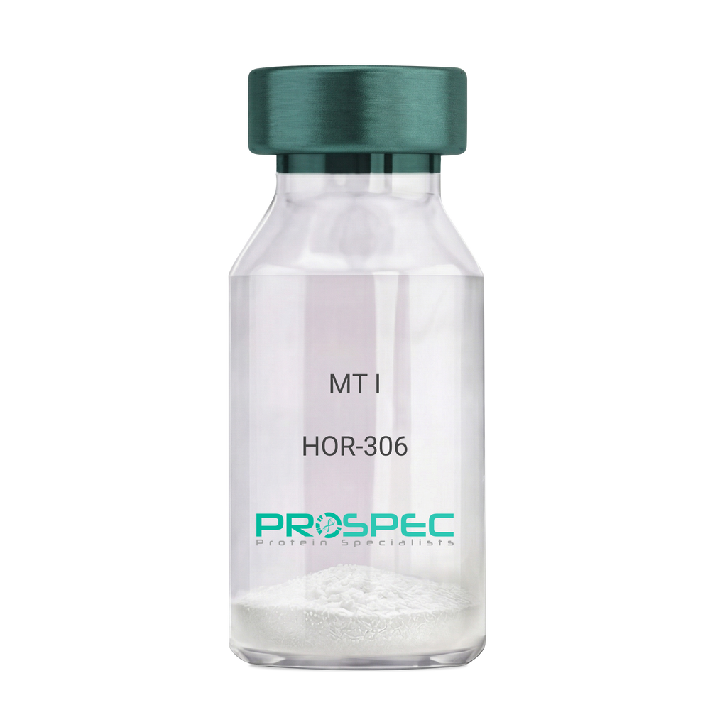
    MT I

    Description:

    Melanotan-I

    Melanotan-I, MT-I, Melanotan-1.

    Product # :

    HOR-306

    Price :

    Quantity :

    Shipping Method :

    Room Temp Icon

    Shipped at Room temp

    Add To Cart

    More Info

    • description
    • formulation
    • purity
    • More Info

    Description

    Melanotan-I has the amino acid sequence of Ser-Tyr-Ser-Nle-Glu-His-D-Phe-Arg-Trp-Gly-Lys-Pro-Val and a molecular weight of 1647.4 Dalton.

    Formulation

    The protein (1mg/ml) was lyophilized with no additives.

    Purity

    Greater than 98.0% as determined by RP-HPLC.

    More Info

    • Synonyms

      Melanotan-I, MT-I, Melanotan-1.

    • Physical Appearance

      Sterile Filtered off-White lyophilized (freeze-dried) powder.

    • Stability

      Lyophilized Melanotan-I although stable at room temperature for 3 weeks, should be stored desiccated below -18°C. Upon reconstitution MT-I should be stored at 4°C between 2-7 days and for future use below -18°C.For long term storage it is recommended to add a carrier protein (0.1% HSA or BSA).Please prevent freeze-thaw cycles.

    • Solubility

      It is recommended to reconstitute the lyophilized Melanotan-I in sterile 1% acetic acid not less than 100 µg/ml, which can then be further diluted to other aqueous solutions.

    ProSpec's products are furnished for LABORATORY RESEARCH USE ONLY. The product may not be used as drugs, agricultural or pesticidal products, food additives or household chemicals.

    Melanotan I
  • View Data Sheet

    Name :

    CJC-1295

    Description:

    CJC-1295

    Product # :

    HOR-066

    Price :

    Quantity :

    Shipping Method :

    Room Temp Icon

    Shipped at Room temp

    Add To Cart

    More Info

    • description
    • formulation
    • purity
    • More Info

    Description

    CJC-1295 is a synthetic single, non-glycosylated polypeptide chain containing 29 amino acids, having a molecular mass of 3367.89 Dalton and a Molecular formula of C152H252N44O42.

    Formulation

    The protein was lyophilized with no additives.

    Purity

    Greater than 97.0% as determined by analysis by RP-HPLC.

    More Info

    • Physical Appearance

      Sterile Filtered White lyophilized (freeze-dried) powder.

    • Stability

      Lyophilized CJC-1295 although stable at room temperature for 3 weeks, should be stored desiccated below -18°C. Upon reconstitution CJC-1295 should be stored at 4°C between 2-7 days and for future use below -18°C.
      For long term storage it is recommended to add a carrier protein (0.1% HSA or BSA).
      Please prevent freeze-thaw cycles.

    • Solubility

      It is recommended to reconstitute the lyophilized CJC-1295 in sterile 18MΩ-cm H2O not less than 100 µg/ml, which can then be further diluted to other aqueous solutions.

    • Amino Acid Sequence

      H-Tyr-D-Ala-Asp-Ala-IlePhe-Thr-Gln-Ser-Tyr-Arg-Lys-Val-Leu-Ala-Gln-Leu-Ser-Ala-Arg-LysLeu-Leu-Gln-Asp-Ile-Leu-Ser-Arg-NH2.

    • Background

      CJC-1295 is a synthetic growth hormone-releasing hormone (GHRH) analog which regulates the GH–IGF-1 axis, aging biology, metabolism and peptide pharmacology. CJC-1295 identifies serum protein biomarkers downstream of GH/IGF-1 activation. CJC-1295 plays a role as a bridge compound between modified peptide therapeutics, native hypothalamic peptides and long-acting biologic hormone regulators.

    ProSpec's products are furnished for LABORATORY RESEARCH USE ONLY. The product may not be used as drugs, agricultural or pesticidal products, food additives or household chemicals.

    CJC-1295
  • 1
  • …
  • 25
  • 26
  • 27
  • 28
Your browser does not support the video tag.

Products

  • Cytokines
  • Growth Factors
  • Chemokines
  • CD Antigens
  • Neurotrophins
  • Hormones
  • Enzymes
  • Viral Antigens
  • Recombinant Proteins
  • Natural Proteins
  • Monoclonal Antibodies
  • Polyclonal Antibodies
  • Thermal Packaging

About

  • Company
  • Founder
  • Contribution
  • Contact Us
  • My Account

Ordering

  • Ordering Method
  • Ordering - Credit Card
  • Ordering PO
  • Shipping Fees
  • FAQ's

Legal

  • Terms & Conditions
  • Privacy
  • Site Map
  • info@prospecbio.com
  • Facebook
  • Instagram
  • Linkedin Profile

© 2026 ProSpec-Tany TechnoGene Ltd. All Rights Reserved.

  • Choosing a selection results in a full page refresh.
  • Opens in a new window.