Skip to content

High-quality research proteins.

  • 1-800-805-6531

  • Mon - Sat 8.00-18.00, Sun - Closed

  • info@prospecbio.com

  • PO Box 6591 East Brunswick NJ 08816

  • Products
    • Cytokines
    • Growth factors
    • Chemokines
    • Cd antigens
    • Neurotrophins
    • Hormones
    • Enzymes
    • Viral antigens
    • Recombinant proteins
    • Natural proteins
    • Monoclonal antibodies
    • Polyclonal antibodies
    • Thermal Packaging
  • About
    • Company
    • Founder
    • Contribution
  • Customers
    • Eastern Asia
    • Eastern Europe
    • North America
    • South America
    • South East Asia
    • Southern Asia
    • Western Europe
  • Ordering
    • Ordering Method
    • Ordering - Credit Card
    • Ordering - PO
    • Shipping Fees
    • FAQs
  • Services
    • DNA Cloning
    • Protein Expression
    • Monoclonal Antibody
    • Fermentation
    • Technology License
  • Contact us
Log in

Country/region

  • USD $ | Albania
  • USD $ | Andorra
  • USD $ | Argentina
  • USD $ | Armenia
  • USD $ | Australia
  • USD $ | Austria
  • USD $ | Azerbaijan
  • USD $ | Belarus
  • USD $ | Belgium
  • USD $ | Bolivia
  • USD $ | Brazil
  • USD $ | Bulgaria
  • USD $ | Cambodia
  • USD $ | Canada
  • USD $ | Chile
  • USD $ | China
  • USD $ | Colombia
  • USD $ | Costa Rica
  • USD $ | Croatia
  • USD $ | Cyprus
  • USD $ | Czechia
  • USD $ | Denmark
  • USD $ | Estonia
  • USD $ | Finland
  • USD $ | France
  • USD $ | Georgia
  • USD $ | Germany
  • USD $ | Greece
  • USD $ | Hong Kong SAR
  • USD $ | Hungary
  • USD $ | Iceland
  • USD $ | India
  • USD $ | Ireland
  • USD $ | Israel
  • USD $ | Italy
  • USD $ | Japan
  • USD $ | Kazakhstan
  • USD $ | Latvia
  • USD $ | Liechtenstein
  • USD $ | Lithuania
  • USD $ | Madagascar
  • USD $ | Malta
  • USD $ | Mexico
  • USD $ | Moldova
  • USD $ | Monaco
  • USD $ | Netherlands
  • USD $ | New Zealand
  • USD $ | North Macedonia
  • USD $ | Norway
  • USD $ | Panama
  • USD $ | Paraguay
  • USD $ | Peru
  • USD $ | Philippines
  • USD $ | Poland
  • USD $ | Portugal
  • USD $ | Romania
  • USD $ | Singapore
  • USD $ | Slovakia
  • USD $ | Slovenia
  • USD $ | South Africa
  • USD $ | South Korea
  • USD $ | Spain
  • USD $ | Sri Lanka
  • USD $ | Sweden
  • USD $ | Switzerland
  • USD $ | Taiwan
  • USD $ | Thailand
  • USD $ | Türkiye
  • USD $ | Ukraine
  • USD $ | United Kingdom
  • USD $ | United States
  • USD $ | Uruguay
  • USD $ | Vatican City
  • USD $ | Venezuela
  • USD $ | Vietnam
  • Facebook
  • Instagram
logoProSpec - Recombinant Protein Manufacturer for academic and R&D industry
Become a distributor
Account Log in
Wishlist
Cart Cart(0)
  • Products
    +
    • Cytokines
    • Growth factors
    • Chemokines
    • Cd antigens
    • Neurotrophins
    • Hormones
    • Enzymes
    • Viral antigens
    • Recombinant proteins
    • Natural proteins
    • Monoclonal antibodies
    • Polyclonal antibodies
    • Thermal Packaging
  • About
    +
    • Company
    • Founder
    • Contribution
  • Customers
    +
    • Eastern Asia
    • Eastern Europe
    • North America
    • South America
    • South East Asia
    • Southern Asia
    • Western Europe
  • Ordering
    +
    • Ordering Method
    • Ordering - Credit Card
    • Ordering - PO
    • Shipping Fees
    • FAQs
  • Services
    +
    • DNA Cloning
    • Protein Expression
    • Monoclonal Antibody
    • Fermentation
    • Technology License
  • Contact us

Item added to your cart

View cart
View All
  • Cytokines
  • Growth factors
  • Chemokines
  • Cd antigens
  • Neurotrophins
  • Hormones
  • Enzymes
  • Viral antigens
  • Recombinant proteins
  • Natural proteins
  • Monoclonal antibodies
  • Polyclonal antibodies
  • Thermal Packaging
  • Tumor Necrosis Factor

    Tumor Necrosis Factor

    View All
  • 4-1BB

    4-1BB

    View All
  • Adiponectin

    Adiponectin

    View All
  • AIF1

    AIF1

    View All
  • Other Cytokines

    Other Cytokines

    View All
  • AITRL

    AITRL

    View All
  • Angiopoietin

    Angiopoietin

    View All
  • Apolipoprotein

    Apolipoprotein

    View All
  • B-Cell Activating Factor

    B-Cell Activating Factor

    View All
  • Beta Defensin

    Beta Defensin

    View All
  • Bone Morphogenetic Protein

    Bone Morphogenetic Protein

    View All
  • B type Natriuretic Peptide

    B type Natriuretic Peptide

    View All
  • BST

    BST

    View All
  • Betacellulin

    Betacellulin

    View All
  • Cardiotrophin

    Cardiotrophin

    View All
  • Activin

    Activin

    View All
  • Fibroblast Growth Factor

    Fibroblast Growth Factor

    View All
  • Other Growth Factors

    Other Growth Factors

    View All
  • CSF

    CSF

    View All
  • CTGF

    CTGF

    View All
  • IGFBP

    IGFBP

    View All
  • VEGF Protein

    VEGF Protein

    View All
  • EGF

    EGF

    View All
  • Epigen

    Epigen

    View All
  • Erythropoietin

    Erythropoietin

    View All
  • Insulin-Like Growth Factor

    Insulin-Like Growth Factor

    View All
  • Growth Hormone

    Growth Hormone

    View All
  • HDGF

    HDGF

    View All
  • Hepatocyte Growth Factor

    Hepatocyte Growth Factor

    View All
  • Insulin

    Insulin

    View All
  • BCA-1/ BLC (CXCL13)

    BCA-1/ BLC (CXCL13)

    View All
  • BRAK (CXCL14)

    BRAK (CXCL14)

    View All
  • C-10 (CCL6)

    C-10 (CCL6)

    View All
  • Other Chemokines

    Other Chemokines

    View All
  • MEC (CCL28)

    MEC (CCL28)

    View All
  • LD78-beta (CCL3L1)

    LD78-beta (CCL3L1)

    View All
  • CTACK (CCL27)

    CTACK (CCL27)

    View All
  • CXCL16

    CXCL16

    View All
  • CXCL17

    CXCL17

    View All
  • Platelet Factor-4 (CXCL4)

    Platelet Factor-4 (CXCL4)

    View All
  • ENA-78 (CXCL5)

    ENA-78 (CXCL5)

    View All
  • Eotaxin (CCL11,24,26)

    Eotaxin (CCL11,24,26)

    View All
  • Exodus-2 (CCL21)

    Exodus-2 (CCL21)

    View All
  • Fractalkine (CX3CL1)

    Fractalkine (CX3CL1)

    View All
  • CXCL6

    CXCL6

    View All
  • Other CD Antigens

    Other CD Antigens

    View All
  • CD14

    CD14

    View All
  • CD164

    CD164

    View All
  • CD1

    CD1

    View All
  • CD2

    CD2

    View All
  • CD200

    CD200

    View All
  • CD207

    CD207

    View All
  • CD226

    CD226

    View All
  • CD23

    CD23

    View All
  • CD244

    CD244

    View All
  • CD247

    CD247

    View All
  • CD27

    CD27

    View All
  • CD274

    CD274

    View All
  • CD300

    CD300

    View All
  • CD33

    CD33

    View All
  • Other Neurotrophins

    Other Neurotrophins

    View All
  • Beta-NGF

    Beta-NGF

    View All
  • BDNF

    BDNF

    View All
  • CDNF

    CDNF

    View All
  • CNTF

    CNTF

    View All
  • GDNF

    GDNF

    View All
  • Glia Maturation Factor

    Glia Maturation Factor

    View All
  • MANF

    MANF

    View All
  • Midkine

    Midkine

    View All
  • Neuroglobin

    Neuroglobin

    View All
  • Neurotrophic factors

    Neurotrophic factors

    View All
  • Neuregulin

    Neuregulin

    View All
  • Neuropilin

    Neuropilin

    View All
  • Pigment Epithelium-Derived Factor

    Pigment Epithelium-Derived Factor

    View All
  • Pleiotrophin

    Pleiotrophin

    View All
  • Peptide Hormones

    Peptide Hormones

    View All
  • Other Hormones

    Other Hormones

    View All
  • HCG

    HCG

    View All
  • Endothelin

    Endothelin

    View All
  • Exendin

    Exendin

    View All
  • FSH

    FSH

    View All
  • Glucagon

    Glucagon

    View All
  • GHRP

    GHRP

    View All
  • GLP

    GLP

    View All
  • Thyrostimulin

    Thyrostimulin

    View All
  • Inhibin A

    Inhibin A

    View All
  • LHRH

    LHRH

    View All
  • Melanotan

    Melanotan

    View All
  • Procalcitonin

    Procalcitonin

    View All
  • PTH

    PTH

    View All
  • Peroxiredoxin

    Peroxiredoxin

    View All
  • Peptidase

    Peptidase

    View All
  • Synthetase

    Synthetase

    View All
  • Transferase

    Transferase

    View All
  • Hydrolase

    Hydrolase

    View All
  • Dehydrogenase

    Dehydrogenase

    View All
  • ACE2

    ACE2

    View All
  • Esterase

    Esterase

    View All
  • Protein Kinases

    Protein Kinases

    View All
  • Other Enzymes

    Other Enzymes

    View All
  • Phosphatase

    Phosphatase

    View All
  • Deaminase

    Deaminase

    View All
  • Oxygenase

    Oxygenase

    View All
  • Adenylate Kinase

    Adenylate Kinase

    View All
  • Lyase

    Lyase

    View All
  • Chagas

    Chagas

    View All
  • SARS

    SARS

    View All
  • Influenza

    Influenza

    View All
  • ASFV

    ASFV

    View All
  • Astrovirus

    Astrovirus

    View All
  • Borrelia

    Borrelia

    View All
  • Helicobacter Pylori

    Helicobacter Pylori

    View All
  • Chikungunya

    Chikungunya

    View All
  • Chlamydia

    Chlamydia

    View All
  • Cytomegalo

    Cytomegalo

    View All
  • Dengue

    Dengue

    View All
  • EBV

    EBV

    View All
  • Ebola

    Ebola

    View All
  • Feline Leukemia Virus

    Feline Leukemia Virus

    View All
  • Hepatitis A

    Hepatitis A

    View All
  • Other

    Other

    View All
  • Actin

    Actin

    View All
  • ADAM

    ADAM

    View All
  • Complement Component

    Complement Component

    View All
  • Ag85

    Ag85

    View All
  • Eukaryotic Translation Initiation Factor

    Eukaryotic Translation Initiation Factor

    View All
  • Anterior Gradient Protein

    Anterior Gradient Protein

    View All
  • Heat Shock Protein

    Heat Shock Protein

    View All
  • Alpha-2-HS-Glycoprotein

    Alpha-2-HS-Glycoprotein

    View All
  • Hemoglobin

    Hemoglobin

    View All
  • Allergy

    Allergy

    View All
  • Anaplasma

    Anaplasma

    View All
  • Angiogenin

    Angiogenin

    View All
  • Ankyrin Repeat Domain

    Ankyrin Repeat Domain

    View All
  • Annexin

    Annexin

    View All
  • Other Natural Proteins

    Other Natural Proteins

    View All
  • Aprotinin

    Aprotinin

    View All
  • Transferrin

    Transferrin

    View All
  • Avidin

    Avidin

    View All
  • Anti Coagulation Factors

    Anti Coagulation Factors

    View All
  • Natural Albumin

    Natural Albumin

    View All
  • Natural Coagulation Factors

    Natural Coagulation Factors

    View All
  • Fibronectin

    Fibronectin

    View All
  • Thrombin

    Thrombin

    View All
  • Anti Human Enzyme

    Anti Human Enzyme

    View All
  • Other Monoclonal Antibodies

    Other Monoclonal Antibodies

    View All
  • Anti Human Cytokine

    Anti Human Cytokine

    View All
  • Anti Human Heat Shock Protein

    Anti Human Heat Shock Protein

    View All
  • Anti Mouse Lymphocyte

    Anti Mouse Lymphocyte

    View All
  • Anti Human Chemokine

    Anti Human Chemokine

    View All
  • Anti Human Lymphocyte

    Anti Human Lymphocyte

    View All
  • Anti Viral Monoclonal

    Anti Viral Monoclonal

    View All
  • Anti Mouse Cytokine

    Anti Mouse Cytokine

    View All
  • Anti-GST Monoclonal

    Anti-GST Monoclonal

    View All
  • Other Polyclonal Antibodies

    Other Polyclonal Antibodies

    View All
  • Anti Viral

    Anti Viral

    View All
  • Anti-GST Monoclonal

    Anti-GST Monoclonal

    View All
Back

Search results

970 results found for “Guanine Nucleotide Binding Protein”

Name

Description

Product #

Price

Quantity

Shipping Method

  • View Data Sheet

    Name :

    sRANKL (158-316) Mouse

    Description:

    Soluble RANK Ligand (158-316 a.a) Mouse Recombinant

    Soluble Receptor Activator of NFkB Ligand, TNFSF11, TRANCE, TNF-related activation-induced cytokine, OPGL, ODF, Osteoclast differentiation factor, Tumor necrosis factor ligand superfamily member 11, Receptor activator of nuclear factor kappa B ligand, RANKL, Osteoprotegerin ligand, CD254 antigen, sRANKL, sOdf.

    Product # :

    CYT-958

    Price :

    Quantity :

    Shipping Method :

    Ice Icon

    Shipped with Ice Packs

    Add To Cart

    More Info

    • description
    • source
    • formulation
    • purity
    • biological activity
    • More Info

    Description

    sRANKL Mouse Recombinant produced in E.Coli is a single, non-glycosylated polypeptide chain containing 160 amino acids (158-316 a.a.) and having a molecular mass of 17.9kDa. sRANKL is purified by proprietary chromatographic techniques.

    Source

    Escherichia Coli.

    Formulation

    sRANKL protein solution (1mg/ml) containing Tris-Hcl buffer pH-8.5 and 0.1M NaCl.

    Purity

    Greater than 90% as determined by SDS-PAGE.

    Biological Activity

    The ED50, as measured by its ability to induce osteoclast differentiation of RAW 264.7 mouse monocyte/macrophage cells, is less than 2ng/ml.

    More Info

    • Introduction

      RANKL binds to tnfrsf11b/opg and to tnfrsf11a/rank. Osteoclast differentiation and activation factor. augments the ability of dendritic cells to stimulate naive t-cell proliferation. May be an important regulator of interactions between t-cells and dendritic cells and may play a role in the regulation of the t-cell-dependent immune response. sRANKL may also play an important role in enhanced bone-resorption in humoral hypercalcemia of malignancy.

    • Synonyms

      Soluble Receptor Activator of NFkB Ligand, TNFSF11, TRANCE, TNF-related activation-induced cytokine, OPGL, ODF, Osteoclast differentiation factor, Tumor necrosis factor ligand superfamily member 11, Receptor activator of nuclear factor kappa B ligand, RANKL, Osteoprotegerin ligand, CD254 antigen, sRANKL, sOdf.

    • Physical Appearance

      Sterile Filtered colorless solution.

    • Stability

      Store at 4°C if entire vial will be used within 2-4 weeks. Store, frozen at -20°C for longer periods of time. For long term storage it is recommended to add a carrier protein (0.1% HSA or BSA). Avoid multiple freeze-thaw cycles.

    • Amino Acid Sequence

      MKPEAQPFAH LTINAASIPS GSHKVTLSSW YHDRGWAKIS NMTLSNGKLR VNQDGFYYLY ANICFRHHET SGSVPTDYLQ LMVYVVKTSI KIPSSHNLMK GGSTKNWSGN SEFHFYSINV GGFFKLRAGE EISIQVSNPS LLDPDQDATY FGAFKVQDID

    ProSpec's products are furnished for LABORATORY RESEARCH USE ONLY. The product may not be used as drugs, agricultural or pesticidal products, food additives or household chemicals.

    Srankl 158 316 Mouse
  • View Data Sheet

    Name :

    PLAUR Human

    Description:

    PLAUR Human Recombinant

    PLAUR, Monocyte Activation Antigen Mo3, U-PAR, UPAR, U-Plasminogen Activator Receptor Form 2, CD87 Antigen , CD87, URKR, MO3.

    Product # :

    PRO-2340

    Price :

    Quantity :

    Shipping Method :

    Ice Icon

    Shipped with Ice Packs

    Add To Cart

    More Info

    • description
    • source
    • formulation
    • purity
    • More Info

    Description

    PLAUR produced in Sf9 Baculovirus cells is a single, glycosylated polypeptide chain containing 291 amino acids (23-305 a.a.) and having a molecular mass of 32.5kDa (Molecular size on SDS-PAGE will appear at approximately 40-57 kDa).PLAUR is fused to a 8 amino acid His-tag at C-terminus & purified by proprietary chromatographic techniques.

    Source

    Sf9, Baculovirus cells.

    Formulation

    PLAUR protein solution (0.5mg/ml) contains Phosphate Buffered Saline (pH 7.4) and 10% glycerol.

    Purity

    Greater than 95.0% as determined by SDS-PAGE.

    More Info

    • Introduction

      PLAUR is one of 2 activators which convert the extracellular zymogen plasminogen to plasmin, a serine protease involved in a various normal and pathological processes that require cell migration and/or tissue destruction. PLAUR protein is synthesized and released from cells as a single-chain proenzyme with narrow enzymatic activity and is converted to an active two-chain disulfide-linked active enzyme by plasmin and other specific proteinases.

    • Synonyms

      PLAUR, Monocyte Activation Antigen Mo3, U-PAR, UPAR, U-Plasminogen Activator Receptor Form 2, CD87 Antigen , CD87, URKR, MO3.

    • Physical Appearance

      Sterile Filtered colorless solution.

    • Stability

      Store at 4°C if entire vial will be used within 2-4 weeks. Store, frozen at -20°C for longer periods of time. For long term storage it is recommended to add a carrier protein (0.1% HSA or BSA). Avoid multiple freeze-thaw cycles.

    • Amino Acid Sequence

      LRCMQCKTNG DCRVEECALG QDLCRTTIVR LWEEGEELEL VEKSCTHSEK TNRTLSYRTG LKITSLTEVV CGLDLCNQGN SGRAVTYSRS RYLECISCGS SDMSCERGRH QSLQCRSPEE QCLDVVTHWI QEGEEGRPKD DRHLRGCGYL PGCPGSNGFH NNDTFHFLKC CNTTKCNEGP ILELENLPQN GRQCYSCKGN STHGCSSEET FLIDCRGPMN QCLVATGTHE PKNQSYMVRG CATASMCQHA HLGDAFSMNH IDVSCCTKSG CNHPDLDVQY RSGLEHHHHH H.

    ProSpec's products are furnished for LABORATORY RESEARCH USE ONLY. The product may not be used as drugs, agricultural or pesticidal products, food additives or household chemicals.

    Plaur Human
  • View Data Sheet

    Name :

    RNF114 Human

    Description:

    Ring Finger Protein 114 Human Recombinant

    E3 ubiquitin-protein ligase RNF114, RING finger protein 114, Zinc finger protein 228, Zinc finger protein 313, RNF114, ZNF228, ZNF313, PSORS12.

    Product # :

    PRO-1901

    Price :

    Quantity :

    Shipping Method :

    Ice Icon

    Shipped with Ice Packs

    Add To Cart

    More Info

    • description
    • source
    • formulation
    • purity
    • More Info

    Description

    RNF114 Human Recombinant produced in E.Coli is a single, non-glycosylated polypeptide chain containing 251 amino acids (1-228 a.a) and having a molecular mass of 28.1kDa.RNF114 is fused to a 23 amino acid His-tag at N-terminus & purified by proprietary chromatographic techniques.

    Source

    Escherichia Coli.

    Formulation

    RNF114 protein solution (1mg/ml) containing 20mM Tris-HCl buffer (pH 8.0), 0.4M UREA and 10% glycerol.

    Purity

    Greater than 90.0% as determined by SDS-PAGE.

    More Info

    • Introduction

      Ring Finger Protein 114 (RNF114) is expressed in various tissues, including skin, CD4 lymphocytes and dendritic cells. RNF114, which contains 1 RING-type zinc finger, takes part in spermatogenesis.

    • Synonyms

      E3 ubiquitin-protein ligase RNF114, RING finger protein 114, Zinc finger protein 228, Zinc finger protein 313, RNF114, ZNF228, ZNF313, PSORS12.

    • Physical Appearance

      Sterile Filtered colorless solution.

    • Stability

      Store at 4°C if entire vial will be used within 2-4 weeks. Store, frozen at -20°C for longer periods of time. For long term storage it is recommended to add a carrier protein (0.1% HSA or BSA).Avoid multiple freeze-thaw cycles.

    • Amino Acid Sequence

      MGSSHHHHHH SSGLVPRGSH MGSMAAQQRD CGGAAQLAGP AAEADPLGRF TCPVCLEVYE KPVQVPCGHV FCSACLQECL KPKKPVCGVC RSALAPGVRA VELERQIEST ETSCHGCRKN FFLSKIRSHV ATCSKYQNYI MEGVKATIKD ASLQPRNVPN RYTFPCPYCP EKNFDQEGLV EHCKLFHSTD TKSVVCPICA SMPWGDPNYR SANFREHIQR RHRFSYDTFV DYDVDEEDMM NQVLQRSIID Q.

    ProSpec's products are furnished for LABORATORY RESEARCH USE ONLY. The product may not be used as drugs, agricultural or pesticidal products, food additives or household chemicals.

    Rnf114 Human
  • View Data Sheet

    Name :

    Borrelia Garinii DbpA

    Description:

    Borrelia Garinii Decorin Binding Protein A Recombinant

    Product # :

    BOR-025

    Price :

    Quantity :

    Shipping Method :

    Ice Icon

    Shipped with Ice Packs

    Add To Cart

    More Info

    • description
    • source
    • formulation
    • purity
    • More Info

    Description

    Recombinant Borrelia Garinii Decorin Binding Protein A produced in E.coli is a non-glycosylated, polypeptide chain having a calculated molecular mass of 19kDa. Borrelia Garinii DbpA is expressed with a -10x His tag at N-terminus and purified by proprietary chromatographic techniques.

    Source

    Escherichia Coli.

    Formulation

    Borrelia Garinii DbpA is supplied in 20mM HEPES buffer pH-7.6, 250mM NaCl and 20% glycerol.

    Purity

    Greater than 80.0% as determined by SDS-PAGE.

    More Info

    • Introduction

      Borrelia is a part of the genus of bacteria of the spirochete phylum. Borrelia causes borreliosis, which is a zoonotic, vector-borne disease transmitted mainly by ticks and some by lice, depending on the species. Of the 36 known species of Borrelia, 12 are distinguished to cause Lyme disease or borreliosis and are transmitted by ticks. The main Borrelia species causing Lyme disease are Borrelia burgdorferi, Borrelia afzelii, and Borrelia garinii. Borreliella garinii DbpA demonstrates the typical lipid anchor and decorin/glycoaminoglycon binding properties of a DbpA protein.

    • Physical Appearance

      Sterile Filtered clear solution.

    • Stability

      Store at 4°C if entire vial will be used within 2-4 weeks. Store, frozen at -20°C for longer periods of time. Avoid multiple freeze-thaw cycles.

    • Immunological Functions

      1. Binds IgG- and IgM-type human antibodies.2. Immunodot test with Lyme disease positive/negative plasma.

    • Applications

      Western blot with Lyme positive plasma.

    ProSpec's products are furnished for LABORATORY RESEARCH USE ONLY. The product may not be used as drugs, agricultural or pesticidal products, food additives or household chemicals.

    Borrelia Garinii Dbpa
  • View Data Sheet

    Name :

    RASSF3 Human

    Description:

    Ras Association Domain-Containing Protein 3 Human Recombinant

    Ras association domain-containing protein 3, RASSF3.

    Product # :

    PRO-1378

    Price :

    Quantity :

    Shipping Method :

    Ice Icon

    Shipped with Ice Packs

    Add To Cart

    More Info

    • description
    • source
    • formulation
    • purity
    • More Info

    Description

    RASSF3 Human Recombinant produced in E.Coli is a single, non-glycosylated polypeptide chain containing 261 amino acids (1-238a.a) and having a molecular mass of 30kDa. RASSF3 is fused to a 23 amino acid His-tag at N-terminus & purified by proprietary chromatographic techniques.

    Source

    E.coli.

    Formulation

    RASSF3 protein solution (1mg/ml) containing 20mM Tris-HCl buffer (pH 8.0), 0.4M Urea and 10% glycerol.

    Purity

    Greater than 85% as determined by SDS-PAGE.

    More Info

    • Introduction

      Ras association domain-containing protein 3 (RASSF3) is a ubiquitously expressed protein found in normal and cancerous tissues. RASSF3 is a member of RASSF family whose members contain a highly conserved Ras association domain (Ral GDS/AF-6 or RA) and function as Ras effectors/tumor suppressors. RASSF3 contains an N-terminal RA domain and a coiled-coil SARAH domain and may have 3 isoforms (RASSF3A, RASSF3B and RASSF3C) due to alternative splicing. RASSF3B and RASSF3C are shorter than RASSF3A and do not contain the RA and SARAH domain.

    • Synonyms

      Ras association domain-containing protein 3, RASSF3.

    • Physical Appearance

      Sterile Filtered colorless solution.

    • Stability

      Store at 4°C if entire vial will be used within 2-4 weeks. Store, frozen at -20°C for longer periods of time. For long term storage it is recommended to add a carrier protein (0.1% HSA or BSA).Avoid multiple freeze-thaw cycles.

    • Amino Acid Sequence

      MGSSHHHHHH SSGLVPRGSH MGSMSSGYSS LEEDAEDFFF TARTSFFRRA PQGKPRSGQQ DVEKEKETHS YLSKEEIKEK VHKYNLAVTD KLKMTLNSNG IYTGFIKVQM ELCKPPQTSP NSGKLSPSSN GCMNTLHISS TNTVGEVIEA LLKKFLVTES PAKFALYKRC HREDQVYACK LSDREHPLYL RLVAGPRTDT LSFVLREHEI GEWEAFSLPE LQNFLRILDK EEDEQLQNLK RRYTAYRQKL EEALREVWKP D.

    ProSpec's products are furnished for LABORATORY RESEARCH USE ONLY. The product may not be used as drugs, agricultural or pesticidal products, food additives or household chemicals.

    Rassf3 Human
  • View Data Sheet

    Name :

    HB-EGF Human

    Description:

    HB-EGF Human Recombinant

    HBEGF, DTR, DTS, HEGFL, HB-EGF, Diphtheria toxin receptor, DT-R, DTSF.

    Product # :

    CYT-119

    Price :

    Quantity :

    Shipping Method :

    Room Temp Icon

    Shipped at Room temp

    Add To Cart

    More Info

    • description
    • source
    • formulation
    • purity
    • biological activity
    • More Info

    Description

    HB-EGF Human Recombinant produced in E.Coli is a single, non-glycosylated, polypeptide chain containing 87 amino acids and having a molecular mass of 9.9kDa. The HB-EGF is purified by proprietary chromatographic techniques.

    Source

    Escherichia Coli.

    Formulation

    The protein was lyophilized from a concentrated (1mg/ml) solution containing 10mM sodium phosphate pH-7.5.

    Purity

    Greater than 95.0% as determined by SDS-PAGE.

    Biological Activity

    The ED50 was determined by the ability to induce proliferation of 3T3 cells and is 0.13-0.2ng/ml. This corresponds to an expected specific activity of 7.7 x 10⁶units/mg.

    More Info

    • Introduction

      HB-EGF is an EGF related growth factor which signals via the EGF receptor, and stimulates the proliferation of SMC (smooth muscle cells), fibroblasts, epithelial cells and keratinocytes. HB-EGF is expressed in various cell types and tissues, including vascular endothelial cells and SMC, macrophages, skeletal muscle, keratinocytes and particular tumor cells. HB-EGF’s ability to explicitly bind HPR sulfate proteoglycans is dissimilar from other EGF-like molecules, and might be related to the enhanced mitogenic activity, relative to EGF, that HB-EGF exerts on smooth muscle cells.

    • Synonyms

      HBEGF, DTR, DTS, HEGFL, HB-EGF, Diphtheria toxin receptor, DT-R, DTSF.

    • Physical Appearance

      Sterile Filtered White lyophilized (freeze-dried) powder.

    • Stability

      Lyophilized Human HB-EGF Recombinant although stable at room temperature for 3 weeks, should be stored desiccated below -18°C. Upon reconstitution HB-EGF should be stored at 4°C between 2-7 days and for future use below -18°C.For long term storage it is recommended to add a carrier protein (0.1% HSA or BSA).Please prevent freeze-thaw cycles.

    • Solubility

      It is recommended to reconstitute the lyophilized Human HB-EGF in sterile 18M-cm H2O not less than 100 µg/ml, which can then be further diluted to other aqueous solutions.

    • Amino Acid Sequence

      MDLQEADLDL LRVTLSSKPQ ALATPNKEEH GKRKKKGKGL GKKRDPCLRK YKDFCIHGEC

      KYVKELRAPS CICHPGYHGE RCHGLSL.

    • Background

      What is the molecular weight/Mw of HB-EGF Protein?
      HB-EGF Protein has a total Mw of 9.9kDa.

      What is the source or expression system of HB-EGF Protein?
      Escherichia Coli.

      What is the Purity of HB-EGF Protein?
      HB-EGF Protein is >95% pure as determined by SDS-PAGE.

      What is the Biological Activity of HB-EGF Protein?
      The ED50 was determined by the ability to induce proliferation of 3T3 cells and is 0.13-0.2ng/ml. This corresponds to an expected specific activity of 7.7 x 10⁶units/mg.

      What is the amino acid sequence of HB-EGF Protein?
      MDLQEADLDL LRVTLSSKPQ ALATPNKEEH GKRKKKGKGL GKKRDPCLRK YKDFCIHGEC
      KYVKELRAPS CICHPGYHGE RCHGLSL.

      What applications can HB-EGF Protein be used in?
      HB-EGF Protein can probably be used in western blot, ELISA and Lateral Flow.

      What is the endotoxin level for HB-EGF Protein?
      The endotoxin level is minimal, HB-EGF Protein was purified using conventional chromatography techniques.

    ProSpec's products are furnished for LABORATORY RESEARCH USE ONLY. The product may not be used as drugs, agricultural or pesticidal products, food additives or household chemicals.

    Hb Egf Human
  • View Data Sheet

    Name :

    ARL1 Human

    Description:

    ADP-Ribosylation Factor-Like 1 Human Recombinant

    ARFL1.

    Product # :

    PRO-508

    Price :

    Quantity :

    Shipping Method :

    Ice Icon

    Shipped with Ice Packs

    Add To Cart

    More Info

    • description
    • source
    • formulation
    • purity
    • More Info

    Description

    ARL1 Recombinant Human produced in E.Coli is a single, non-glycosylated polypeptide chain containing 201 amino acids (1-181 a.a.) and having a molecular mass of 22.5 kDa. The ARL1 is fused to a 20 amino acid His-Tag at N-terminus and purified by proprietary chromatographic techniques.

    Source

    Escherichia Coli.

    Formulation

    The ARL1 solution (0.25mg/ml) containing 20mM Tris-HCl pH-8, 2mM DTT, 100mM NaCl and 40% glycerol.

    Purity

    Greater than 95.0% as determined by SDS-PAGE.

    More Info

    • Introduction

      ARL1 is part of the ARL (ADP-ribosylation factor-like) family of proteins, which are structurally associated to ADP-ribosylation factors (ARFs). ARFs, described as activators of cholera toxin (CT) ADP-ribosyltransferase activity, control intracellular vesicular membrane trafficking, and stimulate a phospholipase D (PLD) isoform.
      ARL1 is a weak stimulator of PLD and CT in a phospholipid dependent manner.
      ARL1 is a GTP-binding protein that has low efficiency as allosteric activator of the cholera toxin catalytic subunit, an ADP-ribosyltransferase. ARL1 is involved in the Golgi apparatus.

    • Synonyms

      ARFL1.

    • Physical Appearance

      Sterile filtered colorless solution.

    • Stability

      Store at 4°C if entire vial will be used within 2-4 weeks. Store, frozen at -20°C for longer periods of time. For long term storage it is recommended to add a carrier protein (0.1% HSA or BSA).Avoid multiple freeze-thaw cycles.

    • Amino Acid Sequence

      MGSSHHHHHH SSGLVPRGSH MGGFFSSIFS SLFGTREMRI LILGLDGAGK TTILYRLQVG EVVTTIPTIG FNVETVTYKN LKFQVWDLGG QTSIRPYWRC YYSNTDAVIY VVDSCDRDRI GISKSELVAM LEEEELRKAI LVVFANKQDM EQAMTSSEMA NSLGLPALKD RKWQIFKTSA TKGTGLDEAM EWLVETLKSR Q.

    ProSpec's products are furnished for LABORATORY RESEARCH USE ONLY. The product may not be used as drugs, agricultural or pesticidal products, food additives or household chemicals.

    Arl1 Human
  • View Data Sheet

    Name :

    CTBP1 Human

    Description:

    C-Terminal Binding Protein 1 Human Recombinant

    EC 1.1.1, BARS, MGC104684, CTBP1, C-terminal-binding protein 1, CTBP.

    Product # :

    PRO-796

    Price :

    Quantity :

    Shipping Method :

    Ice Icon

    Shipped with Ice Packs

    Add To Cart

    More Info

    • description
    • source
    • formulation
    • purity
    • More Info

    Description

    CTBP1 produced in E.Coli is a single, non-glycosylated polypeptide chain containing 440 amino acids (1-440 a.a.) and having a molecular mass of 47.5kDa.CTBP1 is purified by proprietary chromatographic techniques.

    Source

    Escherichia Coli.

    Formulation

    The CTBP1 protein solution contains 20mM Tris-HCl, pH-8, 100mM NaCl and 10% glycerol.

    Purity

    Greater than 90.0% as determined by SDS-PAGE.

    More Info

    • Introduction

      CTBP1 participates in controlling the equilibrium between tubular and stacked structures in the Golgi complex. CTBP1 functions in brown adipose tissue differentiation. CTBP1 has dehydrogenase activity. CTBP1 binds to the C-terminus of adenovirus E1A proteins. CTBP1 phosphoprotein is a transcriptional repressor and that takes part during cellular proliferation. CTBP1 & CTBP2 can dimerize and interact with a polycomb group protein complex which is involved in regulation of gene expression during development.

    • Synonyms

      EC 1.1.1, BARS, MGC104684, CTBP1, C-terminal-binding protein 1, CTBP.

    • Physical Appearance

      Sterile filtered colorless solution.

    • Stability

      Store at 4°C if entire vial will be used within 2-4 weeks. Store, frozen at -20°C for longer periods of time. For long term storage it is recommended to add a carrier protein (0.1% HSA or BSA). Avoid multiple freeze-thaw cycles.

    • Amino Acid Sequence

      MGSSHLLNKG LPLGVRPPIM NGPLHPRPLV ALLDGRDCTV EMPILKDVAT VAFCDAQSTQ EIHEKVLNEA VGALMYHTIT LTREDLEKFK ALRIIVRIGS GFDNIDIKSA GDLGIAVCNV PAASVEETAD STLCHILNLY RRATWLHQAL REGTRVQSVE QIREVASGAA RIRGETLGII GLGRVGQAVA LRAKAFGFNV LFYDPYLSDG VERALGLQRV STLQDLLFHS DCVTLHCGLN EHNHHLINDF TVKQMRQGAF LVNTARGGLV DEKALAQALK EGRIRGAALD VHESEPFSFS QGPLKDAPNL ICTPHAAWYS EQASIEMREE AAREIRRAIT GRIPDSLKNC VNKDHLTAAT HWASMDPAVV HPELNGAAYR YPPGVVGVAP TGIPAAVEGI VPSAMSLSHG LPPVAHPPHA PSPGQTVKPE ADRDHASDQL.

    ProSpec's products are furnished for LABORATORY RESEARCH USE ONLY. The product may not be used as drugs, agricultural or pesticidal products, food additives or household chemicals.

    Ctbp1 Human
  • View Data Sheet

    Name :

    CXCL1 Human

    Description:

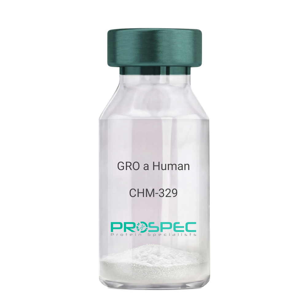
    GRO-Alpha Human Recombinant (CXCL1)

    Growth-regulated protein alpha, CXCL1, Melanoma growth stimulatory activity, MGSA, Neutrophil-activating protein 3, NAP-3, GRO-alpha (1-73), chemokine (C-X-C motif) ligand 1, GRO1, GROa, SCYB1, MGSA-a, MGSA alpha.

    Product # :

    CHM-329

    Price :

    Quantity :

    Shipping Method :

    Room Temp Icon

    Shipped at Room temp

    Add To Cart

    More Info

    • description
    • source
    • formulation
    • purity
    • biological activity
    • More Info

    Description

    GRO Alpha Human Recombinant produced in E.Coli is a single,non-glycosylated, polypeptide chain containing 73 amino acids and having a molecular mass of 7811 Dalton. The GRO-alpha is purified by proprietary chromatographic techniques.

    Source

    Escherichia Coli.

    Formulation

    Lyophilized from a 0.2µm filtered concentrated (1mg/ml) solution in 20mM PB, pH 7.4, 50mM NaCl.

    Purity

    Greater than 97.0% as determined by:
    (a) Analysis by RP-HPLC.
    (b) Analysis by SDS-PAGE.

    Biological Activity

    Determined by its ability to chemoattract human peripheral blood neutrophils using a concentration range of 10.0-100.0 ng/ml.

    More Info

    • Introduction

      Chemokine (C-X-C motif) ligand 1 (CXCL1) is a small cytokine belonging to the CXC chemokine family that was previously called GRO1 oncogene, Neutrophil-activating protein 3 (NAP-3) and melanoma growth stimulating activity, alpha (MSGA-a). It is secreted by human melanoma cells, has mitogenic properties and is implicated in melanoma pathogenesis. CXCL1 is expressed by macrophages, neutrophilsand epithelial cells, and has neutrophil chemoattractant activity. CXCL1 plays a role in spinal cord development by inhibiting the migration of oligodendrocyte precursors and is involved in the processes of angiogenesis, inflammation, wound healing, and tumorigenesis. This chemokine elicits its effects by signaling through the chemokine receptor CXCR2. The gene for CXCL1 is located on human chromosome 4 amongst genes for other CXC chemokines.

    • Synonyms

      Growth-regulated protein alpha, CXCL1, Melanoma growth stimulatory activity, MGSA, Neutrophil-activating protein 3, NAP-3, GRO-alpha (1-73), chemokine (C-X-C motif) ligand 1, GRO1, GROa, SCYB1, MGSA-a, MGSA alpha.

    • Physical Appearance

      Sterile Filtered White lyophilized (freeze-dried) powder.

    • Stability

      Lyophilized GRO-alpha although stable at room temperature for 3 weeks, should be stored desiccated below -18°C. Upon reconstitution CXCL1 should be stored at 4°C between 2-7 days and for future use below -18°C. For long term storage it is recommended to add a carrier protein (0.1% HSA or BSA).Please prevent freeze-thaw cycles.

    • Solubility

      It is recommended to reconstitute the lyophilized CXCL1 in sterile 18MΩ-cm H2O not less than 100µg/ml, which can then be further diluted to other aqueous solutions.

    • Amino Acid Sequence

      The sequence of the first five N-terminal amino acids was determined and was found to be Ala-Ser-Val-Ala-Thr.

    • Background

      What is the molecular weight/Mw of CXCL1 HUMAN Protein?
      CXCL1 HUMAN Protein has a total Mw of 7.81kDa.

      What is the source or expression system of CXCL1 HUMAN Protein?
      Escherichia Coli.

      What is the Purity of CXCL1 HUMAN Protein?
      CXCL1 HUMAN Protein is >97% pure as determined by SDS-PAGE.

      What is the Biological Activity of CXCL1 HUMAN Protein?
      Determined by its ability to chemoattract human peripheral blood neutrophils using a concentration range of 10.0-100.0 ng/ml.

      What is the amino acid sequence of CXCL1 HUMAN Protein?
      The sequence of the first five N-terminal amino acids was determined and was found to be Ala-Ser-Val-Ala-Thr.

      What applications can CXCL1 HUMAN Protein be used in?
      CXCL1 HUMAN Protein can probably be used in western blot, ELISA and Lateral Flow.

      What is the endotoxin level for CXCL1 HUMAN Protein?
      The endotoxin level is minimal, CXCL1 HUMAN Protein was purified using conventional chromatography techniques.


    ProSpec's products are furnished for LABORATORY RESEARCH USE ONLY. The product may not be used as drugs, agricultural or pesticidal products, food additives or household chemicals.

    Gro A Human
  • View Data Sheet

    Name :

    MAPKSP1 antibody

    Description:

    MAPK Scaffold Protein 1, Mouse Anti Human

    MAPBP, MP1, MAPK Scaffold Protein 1, Interacting Protein 1, MEK binding partner 1, Mitogen-Activated Protein Kinase kinase 1.

    Product # :

    ANT-734

    Price :

    Quantity :

    Shipping Method :

    Ice Icon

    Shipped with Ice Packs

    Add To Cart

    More Info

    • formulation
    • More Info

    Formulation

    1mg/ml containing PBS, pH-7.4, 10% Glycerol and 0.02% Sodium Azide.

    More Info

    • Introduction

      MAPKSP1 is a scaffold protein which operates in the extracellular signal-regulated kinase (ERK) cascade. MAPKSP1 protein is restricted to late endosomes by the mitogen-activated protein-binding protein-interacting protein, and binds specifically to MAP kinase kinase MAP2K1/MEK1, MAP kinase MAPK3/ERK1, and MAP kinase MAPK1/ERK2.

    • Synonyms

      MAPBP, MP1, MAPK Scaffold Protein 1, Interacting Protein 1, MEK binding partner 1, Mitogen-Activated Protein Kinase kinase 1.

    • Physical Appearance

      Sterile filtered colorless solution.

    • Immunogen

      Anti-human MAPKSP1 mAb, is derived from hybridization of mouse F0 myeloma cells with spleen cells from BALB/c mice immunized with a recombinant human MAPKSP1 protein 1-124 amino acids purified from E. coli.

    • Ig Subclass

      Mouse IgG2b heavy chain and k light chain.

    • Clone

      PAT1F10AT.

    • Applications

      The antibody has been tested by ELISA, Western blot analysis, Flow cytometry and ICC/IF to assure specificity and reactivity. Since application varies, however, each investigation should be titrated by the reagent to obtain optimal results.

    • Type

      Mouse Anti Human Monoclonal.

    • Storage Procedures

      For periods up to 1 month store at 4°C, for longer periods of time, store at -20°C. Prevent freeze thaw cycles.

    • Purification Method

      MAPKSP1 antibody was purified from mouse ascitic fluids by protein-A affinity chromatography.

    ProSpec's products are furnished for LABORATORY RESEARCH USE ONLY. The product may not be used as drugs, agricultural or pesticidal products, food additives or household chemicals.

    Mapksp1 Antibody 2
  • View Data Sheet

    Name :

    GRAP2 Human

    Description:

    GRB2-Related Adaptor Protein 2 Human Recombinant

    GRB2-related adaptor protein 2, GADS, GRBLG, GRID, GRBX, Grf40, Mona, Adapter protein GRID, Growth factor receptor-binding protein, Hematopoietic cell-associated adapter protein GRPL, GRB2L, P38, GRB-2-like protein, SH3-SH2-SH3 adapter Mona, GRAP-2, GRB2-related protein with insert domain, Growth Factor Receptor-bound protein 2-related adaptor protein 2, SH3-SH2-SH3 adaptor molecule, Protein GADS.

    Product # :

    PRO-081

    Price :

    Quantity :

    Shipping Method :

    Ice Icon

    Shipped with Ice Packs

    Add To Cart

    More Info

    • description
    • source
    • formulation
    • purity
    • More Info

    Description

    GRAP2 produced in E.Coli is a single, non-glycosylated polypeptide chain containing 350 amino acids (1-330.a.a) and having a molecular mass of 40.0kDa. GRAP2 is fused to a 20 amino acid His-tag at N-terminus & purified by proprietary chromatographic techniques.

    Source

    Escherichia Coli.

    Formulation

    The GRAP2 protein solution (0.5mg/ml) is formulated in 20 mM Tris-HCl buffer (pH8.0), 0.2M NaCl, 5mM DTT, 1mM EDTA and 30% glycerol.

    Purity

    Greater than 90% as determined by SDS-PAGE.

    More Info

    • Introduction

      GRAP2 belongs to the GRB2/Sem5/Drk family. GRAP2 is an adaptor-like protein has a part in leukocyte-specific protein-tyrosine kinase signaling. Similar to related family member, GRB2-related adaptor protein contains an SH2 domain flanked by two SH3 domains and it interacts with other proteins, for example GRB2-associated binding protein 1 (GAB1) and the SLP-76 leukocyte protein (LCP2), through its SH3 domains.

    • Synonyms

      GRB2-related adaptor protein 2, GADS, GRBLG, GRID, GRBX, Grf40, Mona, Adapter protein GRID, Growth factor receptor-binding protein, Hematopoietic cell-associated adapter protein GRPL, GRB2L, P38, GRB-2-like protein, SH3-SH2-SH3 adapter Mona, GRAP-2, GRB2-related protein with insert domain, Growth Factor Receptor-bound protein 2-related adaptor protein 2, SH3-SH2-SH3 adaptor molecule, Protein GADS.

    • Physical Appearance

      Sterile Filtered clear solution.

    • Stability

      Store at 4°C if entire vial will be used within 2-4 weeks. Store, frozen at -20°C for longer periods of time.For long term storage it is recommended to add a carrier protein (0.1% HSA or BSA).Avoid multiple freeze-thaw cycles.

    • Amino Acid Sequence

      MGSSHHHHHH SSGLVPRGSH MEAVAKFDFT ASGEDELSFH TGDVLKILSN QEEWFKAELG SQEGYVPKNF IDIQFPKWFH EGLSRHQAEN LLMGKEVGFF IIRASQSSPG DFSISVRHED DVQHFKVMRD NKGNYFLWTE KFPSLNKLVD YYRTNSISRQ KQIFLRDRTR EDQGHRGNSL DRRSQGGPHL SGAVGEEIRP SMNRKLSDHP PTLPLQQHQH QPQPPQYAPA PQQLQQPPQQ RYLQHHHFHQ ERRGGSLDIN DGHCGTGLGS EMNAALMHRR HTDPVQLQAA GRVRWARALY DFEALEDDEL GFHSGEVVEV LDSSNPSWWT GRLHNKLGLF PANYVAPMTR.

    ProSpec's products are furnished for LABORATORY RESEARCH USE ONLY. The product may not be used as drugs, agricultural or pesticidal products, food additives or household chemicals.

    Grap2 Human
  • View Data Sheet

    Name :

    GSTP1 Antibody

    Description:

    Glutathione S-Transferase pi 1, Mouse Anti Human

    Glutathione S-transferase P 1, Gst P1, GST YF-YF, GST class-pi, GST-piB, Preadipocyte growth factor.

    Product # :

    ANT-729

    Price :

    Quantity :

    Shipping Method :

    Ice Icon

    Shipped with Ice Packs

    Add To Cart

    More Info

    • formulation
    • More Info

    Formulation

    1mg/ml containing PBS, pH-7.4, 10% Glycerol and 0.02% Sodium Azide.

    More Info

    • Introduction

      GSTP1 is a polymorphic gene encoding active, functionally different GSTP1 variant proteins that are believed to function in xenobiotic metabolism and have a part in susceptibility to cancer, and other diseases. GSTP1 is a glutathione S-transferase belonging to the pi class. GST family enzymes play a significant role in detoxification by catalyzing the conjugation of many hydrophobic and electrophilic compounds with reduced glutathione. Based upon the biochemical, immunologic and structural properties of the soluble GSTs they are grouped into 4 main classes: alpha, mu, pi, and theta. The GSTP1 enzyme acts by catalyzing the reaction of glutathione with an acceptor molecule to form a Sulfur-substituted glutathione. The reactions employing glutathione contribute the transformation of a broad range of electrophiles, including reactive products of lipid, protein, carcinogens, therapeutic drugs, environmental toxins, and products of oxidative stress.
      The GSTP1 inactivation through CpG hypermethylation is frequent in pituitary adenomas and may be a factor in aggressive pituitary tumor behavior. GSTP1 is may be a transcriptional target of the p53 tumor suppressor gene. Single-nucleotide polymorphism in GSTP1 is linked to modified protein binding, which influence GSTP1's contribution to carcinogen and drug metabolism, and possibly disease pathogenesis and/or drug response. GST-pi might have central roles in proliferation of androgen-independent human prostate cancer cells.

    • Synonyms

      Glutathione S-transferase P 1, Gst P1, GST YF-YF, GST class-pi, GST-piB, Preadipocyte growth factor.

    • Physical Appearance

      Sterile filtered colorless solution.

    • Immunogen

      Anti-human GSTP1 mAb, is derived from hybridization of mouse F0 myeloma cells with spleen cells from BALB/c mice immunized with recombinant human GSTP1 amino acids 1-210 purified from E. coli.

    • Ig Subclass

      Mouse IgG1 heavy chain and κ light chain.

    • Clone

      PAT12C10AT

    • Applications

      GSTP1 antibody has been tested by ELISA, Western blot analysis, ICC/IF and Flow cytometry to assure specificity and reactivity. Since application varies, however, each investigation should be titrated by the reagent to obtain optimal results.

    • Type

      Mouse Anti Human Monoclonal.

    • Storage Procedures

      For periods up to 1 month store at 4°C, for longer periods of time, store at -20°C. Prevent freeze thaw cycles.

    • Purification Method

      GSTP1 antibody was purified from mouse ascitic fluids by protein-A affinity chromatography.

    ProSpec's products are furnished for LABORATORY RESEARCH USE ONLY. The product may not be used as drugs, agricultural or pesticidal products, food additives or household chemicals.

    Gstp1 Antibody
  • View Data Sheet

    Name :

    AITRL Human, T7

    Description:

    AITRL Human Recombinant, T7 Tag

    Osteostat, TNFSF18, Activation-induced TNFR member Ligand, GITRL,TL6, AITRL, Glucocorticoid-induced TNF-related ligand, hGITRL, Tumor necrosis factor ligand superfamily member 18, MGC138237.

    Product # :

    CYT-807

    Price :

    Quantity :

    Shipping Method :

    Ice Icon

    Shipped with Ice Packs

    Add To Cart

    More Info

    • description
    • source
    • formulation
    • purity
    • More Info

    Description

    AITRL Human Recombinant produced in E.Coli is a single, non-glycosylated polypeptide chain containing 134 amino acids (81-199 a.a) and having a molecular mass of 15kDa.AITRL is fused to a 15 amino acid T7-tag at N-terminus & purified by proprietary chromatographic techniques.

    Source

    Escherichia Coli.

    Formulation

    AITRL protein solution (0.25mg/ml) contains 20mM Tris-HCl buffer (pH 8.0), 10% glycerol and 0.4M Urea.

    Purity

    Greater than 90% as determined by SDS-PAGE.

    More Info

    • Introduction

      Osteostat is the cytokine that binds to TNFRSF18/AITR/GITR and is important for interactions between activated T-lymphocytes and endothelial cells and may modulate T-lymphocyte survival in peripheral tissues. Osteostat is expressed at high levels in the small intestine, ovary, testis, kidney and endothelial cells after stimulation by lipopolysaccharides.
      Osteostat protein is detectable in human microvascular EC and is highly up-regulated by IFN-alpha and IFN-beta. Osteostat inhibit differentiation of osteoclasts from monocytic precursor cells. Osteostat suppresses the early stage of osteoclastogenesis via inhibition of macrophage colony-stimulating factorinduced receptor activator of NF-kappaB (RANK) expression in the osteoclast precursor cells. Osteostat does not inhibit lipopolysaccharide-induced RANK expression in monocytes and dendritic cells, or activation-induced RANK expression in T cells. Osteostat is a novel regulator of osteoclast generation and substantiate the major role played by the endothelium in bone physiology.

    • Synonyms

      Osteostat, TNFSF18, Activation-induced TNFR member Ligand, GITRL,TL6, AITRL, Glucocorticoid-induced TNF-related ligand, hGITRL, Tumor necrosis factor ligand superfamily member 18, MGC138237.

    • Physical Appearance

      Sterile Filtered colorless solution.

    • Stability

      Store at 4°C if entire vial will be used within 2-4 weeks. Store, frozen at -20°C for longer periods of time. For long term storage it is recommended to add a carrier protein (0.1% HSA or BSA).Avoid multiple freeze-thaw cycles.

    • Amino Acid Sequence

      MASMTGGQQM GRGSHMAKFG PLPSKWQMAS SEPPCVNKVS DWKLEILQNG LYLIYGQVAP NANYNDVAPF EVRLYKNKDM IQTLTNKSKI QNVGGTYELH VGDTIDLIFN SEHQVLKNNT YWGIILLANP QFIS.

    • Background

      What is the molecular weight/Mw of AITRL Protein?
      AITRL Protein has a total Mw of 15kDa.

      What is the source or expression system of AITRL Protein?
      Escherichia Coli.

      What is the Purity of AITRL Protein?
      AITRL Protein is >90% pure as determined by SDS-PAGE.

      What is the Biological Activity of AITRL Protein?
      The biological functionality of AITRL Protein will be determined in the future.

      What is the amino acid sequence of AITRL Protein?
      MASMTGGQQM GRGSHMAKFG PLPSKWQMAS SEPPCVNKVS DWKLEILQNG LYLIYGQVAP NANYNDVAPF EVRLYKNKDM IQTLTNKSKI QNVGGTYELH VGDTIDLIFN SEHQVLKNNT YWGIILLANP QFIS.

      What applications can AITRL Protein be used in?
      AITRL Protein can probably be used in western blot, ELISA and Lateral Flow.

      What is the endotoxin level for AITRL Protein?
      The endotoxin level is minimal, AITRL Protein was purified using conventional chromatography techniques.

    ProSpec's products are furnished for LABORATORY RESEARCH USE ONLY. The product may not be used as drugs, agricultural or pesticidal products, food additives or household chemicals.

    Aitrl Human T7
  • View Data Sheet

    Name :

    SNCG Mouse

    Description:

    Gamma-Synuclein Mouse Recombinant

    Gamma-synuclein, Persyn.

    Product # :

    PRO-2490

    Price :

    Quantity :

    Shipping Method :

    Ice Icon

    Shipped with Ice Packs

    Add To Cart

    More Info

    • description
    • source
    • formulation
    • purity
    • More Info

    Description

    SNCG Mouse Recombinant produced in E.Coli is a single, non-glycosylated polypeptide chain containing 146 amino acids (1-123 a.a) and having a molecular mass of 15.5kDa.SNCG is fused to a 23 amino acid His-tag at N-terminus & purified by proprietary chromatographic techniques.

    Source

    Escherichia Coli.

    Formulation

    SNCG protein solution (1mg/ml) containing 20mM Tris-HCl(pH8.0) and 0.1M NaCl.

    Purity

    Greater than 95.0% as determined by SDS-PAGE.

    More Info

    • Introduction

      g-synuclein(Originally known as a breast cancer specific gene product, BCSG1) is an acidic neuronal protein of 127 amino acids. Gamma-Synuclein is a member of the Synuclein protein family, which is believed to be involved in the pathogenesis of neurodegenerative diseases. High levels of Gamma-Synuclein have been found in advanced breast carcinomas suggesting a correlation between overexpression of SNCG and breast tumor development. Synuclein-Gamma is found mostly in the peripheral nervous system (in primary sensory neurons, sympathetic neurons, and motor neurons) and retina. SNCG is also identified in the brain, ovarian tumors, and in the olfactory epithelium. SNCG expression in breast tumors is a marker for tumor progression. A modification in the expression of gamma-synuclein has been detected in the retina of Alzheimer's patients.

    • Synonyms

      Gamma-synuclein, Persyn.

    • Physical Appearance

      Sterile filtered colorless solution.

    • Stability

      Store at 4°C if entire vial will be used within 2-4 weeks. Store, frozen at -20°C for longer periods of time.For long term storage it is recommended to add a carrier protein (0.1% HSA or BSA). Avoid multiple freeze-thaw cycles.

    • Amino Acid Sequence

      MGSSHHHHHH SSGLVPRGSH MGSMDVFKKG FSIAKEGVVG AVEKTKQGVT EAAEKTKEGV MYVGTKTKEN VVQSVTSVAE KTKEQANAVS AVVSSVNTV ANKTVEEAEN IVVTTGVVRK EDLEPPAQDQ EAKEQEENEE AKSGED.

    ProSpec's products are furnished for LABORATORY RESEARCH USE ONLY. The product may not be used as drugs, agricultural or pesticidal products, food additives or household chemicals.

    Sncg Mouse
  • View Data Sheet

    Name :

    KIR2DL4 Human

    Description:

    Killer Cell Immunoglobulin-Like Receptor, 2 Domains Long Cytoplasmic Tail 4 Human Recombinant

    Killer Cell Immunoglobulin Like Receptor, Two Ig Domains And Long Cytoplasmic Tail 4,Killer Cell Immunoglobulin-Like Receptor, Two Domains, Long Cytoplasmic Tail 4,Killer Cell Inhibitory Receptor 103AS, MHC Class I NK Cell Receptor KIR103AS,CD158 Antigen-Like Family Member D, KIR-103AS, KIR103AS, CD158D, G9P, Killer Cell Immunoglobulin-Like Receptor 2DL4, CD158d Antigen, KIR-2DL4, KIR103, KIR2DL4.  

    Product # :

    PRO-2460

    Price :

    Quantity :

    Shipping Method :

    Ice Icon

    Shipped with Ice Packs

    Add To Cart

    More Info

    • description
    • source
    • formulation
    • purity
    • More Info

    Description

    KIR2DL4 Human Recombinant produced in Sf9 Insect cells is a single, glycosylated polypeptide chain containing 458 amino acids (24-242 a.a.) and having a molecular mass of 51kDa (Molecular size on SDS-PAGE will appear at approximately 50-70kDa). KIR2DL4 is expressed with a 239 amino acids hIgG-His tag at C-Terminus and purified by proprietary chromatographic techniques.

    Source

    Sf9, Baculovirus cells.

    Formulation

    KIR2DL4 protein solution (0.25mg/ml) contains Phosphate buffered saline (pH7.4) and 10% glycerol.

    Purity

    Greater than 85.0% as determined by SDS-PAGE.

    More Info

    • Introduction

      Killer cell immunoglobulin-like receptor 2DL4, also known as KIR2DL4 is part of the killer cell Ig-like receptor (KIR) family. KIR proteins with the long cytoplasmic domain transduce inhibitory signals through an immune tyrosine-based inhibitory motif (ITIM), whereas KIR proteins which contain the short cytoplasmic domain, lack the ITIM motif and as a substitute associate with the TYRO protein tyrosine kinase binding protein to transduce activating signals. KIR2DL4 stimulates NK cells to produce IFN-gamma and stimulation with IL-2 upregulates cell surface expression on CD56dimcells and leads to the inhibition of the cytolytic NK cell function.

    • Synonyms

      Killer Cell Immunoglobulin Like Receptor, Two Ig Domains And Long Cytoplasmic Tail 4,Killer Cell Immunoglobulin-Like Receptor, Two Domains, Long Cytoplasmic Tail 4,Killer Cell Inhibitory Receptor 103AS, MHC Class I NK Cell Receptor KIR103AS,CD158 Antigen-Like Family Member D, KIR-103AS, KIR103AS, CD158D, G9P, Killer Cell Immunoglobulin-Like Receptor 2DL4, CD158d Antigen, KIR-2DL4, KIR103, KIR2DL4.

    • Physical Appearance

      Sterile Filtered colorless solution.

    • Stability

      Store at 4°C if entire vial will be used within 2-4 weeks. Store, frozen at -20°C for longer periods of time. For long term storage it is recommended to add a carrier protein (0.1% HSA or BSA).Avoid multiple freeze-thaw cycles.

    • Amino Acid Sequence

      HVGGQDKPFC SAWPSAVVPQ GGHVTLRCHY RRGFNIFTLY KKDGVPVPEL YNRIFWNSFL ISPVTPAHAG TYRCRGFHPH SPTEWSAPSN PLVIMVTGLY EKPSLTARPG PTVRAGENVT LSCSSQSSFD IYHLSREGEA HELRLPAVPS INGTFQADFP LGPATHGETY RCFGSFHGSP
      YEWSDPSDPL PVSVTGNPSS SWPSPTEPSF KTGIARHLHL EPKSCDKTHT CPPCPAPELL GGPSVFLFPP KPKDTLMISR TPEVTCVVVD VSHEDPEVKF NWYVDGVEVH NAKTKPREEQ YNSTYRVVSV LTVLHQDWLN GKEYKCKVSN KALPAPIEKT ISKAKGQPRE PQVYTLPPSR
      DELTKNQVSL TCLVKGFYPS DIAVEWESNG QPENNYKTTP PVLDSDGSFF LYSKLTVDKS RWQQGNVFSC SVMHEALHNH YTQKSLSLSP GKHHHHHH

    ProSpec's products are furnished for LABORATORY RESEARCH USE ONLY. The product may not be used as drugs, agricultural or pesticidal products, food additives or household chemicals.

    Kir2Dl4 Human
  • View Data Sheet

    Name :

    FBLIM1 Human

    Description:

    Filamin Binding LIM Protein 1 Human Recombinant

    Filamin binding LIM protein 1, CAL, FBLP-1, FBLP1, RP11-169K16.5, Migfilin, Mitogen-inducible 2-interacting protein, MIG2-interacting protein.

    Product # :

    PRO-1473

    Price :

    Quantity :

    Shipping Method :

    Ice Icon

    Shipped with Ice Packs

    Add To Cart

    More Info

    • description
    • source
    • formulation
    • purity
    • More Info

    Description

    FBLIM1 Human Recombinant produced in E.coli is a single, non-glycosylated polypeptide chain containing 396 amino acids (1-373) and having a molecular mass of 43.1 kDa. FBLIM1 is fused to a 23 amino acid His-tag at N-terminus & purified by proprietary chromatographic techniques.

    Source

    Escherichia Coli.

    Formulation

    The FBLIM1 solution (0.25mg/ml) contains 20mM Tris-HCl buffer (pH 8.0), 0.15M NaCl, 10% glycerol and 1mM DTT.

    Purity

    Greater than 85.0% as determined by SDS-PAGE.

    More Info

    • Introduction

      Filamin binding LIM protein 1 (FBLIM1) plays a role as an anchoring site for cell-ECM adhesion proteins and filamin-containing actin filaments. FBLIM1 is involved in cell shape spreading and motility. FBLIM1 participates in the regulation of filamin-mediated cross-linking and stabilization of actin filaments. FBLIM1 promotes stimulation of integrins and regulates integrin-mediated cell-cell adhesion.

    • Synonyms

      Filamin binding LIM protein 1, CAL, FBLP-1, FBLP1, RP11-169K16.5, Migfilin, Mitogen-inducible 2-interacting protein, MIG2-interacting protein.

    • Physical Appearance

      Sterile Filtered colorless solution.

    • Stability

      Store at 4°C if entire vial will be used within 2-4 weeks. Store, frozen at -20°C for longer periods of time. For long term storage it is recommended to add a carrier protein (0.1% HSA or BSA).Avoid multiple freeze-thaw cycles.

    • Amino Acid Sequence

      MGSSHHHHHH SSGLVPRGSH MGSMASKPEK RVASSVFITL APPRRDVAVA EEVRQAVCEA RRGRPWEAPA PMKTPEAGLA GRPSPWTTPG RAAATVPAAP MQLFNGGCPP PPPVLDGEDV LPDLDLLPPP PPPPPVLLPS EEEAPAPMGA SLIADLEQLH LSPPPPPPQA PAEGPSVQPG PLRPMEEELP PPPAEPVEKG ASTDICAFCH KTVSPRELAV EAMKRQYHAQ CFTCRTCRRQ LAGQSFYQKD GRPLCEPCYQ DTLERCGKCG EVVRDHIIRA LGQAFHPSCF TCVTCARCIG DESFALGSQN EVYCLDDFYR KFAPVCSICE NPIIPRDGKD AFKIECMGRN FHENCYRCED CRILLSVEPT DQGCYPLNNH LFCKPCHVKR SAAGCC.

    ProSpec's products are furnished for LABORATORY RESEARCH USE ONLY. The product may not be used as drugs, agricultural or pesticidal products, food additives or household chemicals.

    Fblim1 Human
  • View Data Sheet

    Name :

    AMBP Human

    Description:

    Microglobulin Alpha-1 Protein Human

    Alpha-1 Microglobulin, A1M.

    Product # :

    PRO-407

    Price :

    Quantity :

    Shipping Method :

    Room Temp Icon

    Shipped at Room temp

    Add To Cart

    More Info

    • description
    • source
    • formulation
    • purity
    • More Info

    Description

    Alpha 1-microglobulin (A1M) is an immunomodulatory protein with a broad spectrum of possible clinical applications and seems a promising marker for evaluation of tubular function.

    Source

    Purified from the urine of patients with chronic renal tubular proteinuria.

    Formulation

    Lyophilized from 0.02M NH4HCO3. May contain traces of buffer salts.

    Purity

    Greater than 96.0%.

    More Info

    • Introduction

      Alpha 1-microglobulin (A1M) is a lipocalin superfamily member (kernal lipocalins). A1M is a low molecular weight protein component of plasma. A1M is distributed in plasma and extravascular compartments of all organs. Alpha-1 Microglobulin is found in mammals, birds, amphibians and fish. The primary sites of A1M synthesis are the liver and the kidney. Around the opening of the lipocalin pocket three lysyl residues are situated; those residues carry yellow-brown modification derived from the binding and degradation of heme and kynurenin (a tryptophan metabolite). A1-Microglobulin’s reductase and dehydrogenase have broad biological substrate specificity properties due to its’ free cysteine side-chain which is located in a flexible loop. Alpha-1-microglobulin is glycosylated by three separate carbohydrate chains: two complex carbohydrates which are N-linked to asparagines at residues 17 and 96, and the other simple carbohydrate which is O-linked to threonine at position 5. The carbohydrates comprise 22% of the total molecular mass of the protein. The glycosylation varies between species. A1M exists in two forms- a free form and complexed to other macromolecules: in humans- complexed to immunoglobulin A (IgA), in rat- complexed to alpha-1-inhibitor-3. Free A1M is exceptionally heterogeneous in charge (therefore also known as protein HC), and is found tightly linked to a chromophore. The free Alpha-1-microglobulin is a monomeric protein composed of one 188 residue polypeptide and contains three cysteines, two of which (residues 75 and 173) form a conserved intra-molecular disulphide link. The chromophoric group is covalently bound to the free cysteine residue at position 34. A1M binds retinol as a major ligand, but this is probably distinct from its covalent chromophore. Half of all human plasma A1M (approximately 0.03mg/ml) forms a 1:1 complex with about 5% of plasma immunoglobulin A. The resulting macromolecular complexes’ molecular weight is 200000, and a plasma concentration of 0.3mg/ml. The complex can exhibit both antibody activity and affect many of the biological actions of free Alpha-1-microglobulin. Alpha-1-microglobulin was first discovered in pathological human urine. It was suggested that A1M might be involved in tissue defense against reactive oxygen species, oxidation by heme and kynurenin. Evidence also suggests that A1M functions in the regulation of the immune system. Other functions include: inhibition of stimulation of cultured lymphocytes by protein antigens; induction of cell division of lymphocytes, a mitogenic effect that can either be enhanced or inhibited by the action of other plasma components; inhibition of neutrophil granulocyte migration in vitro; and inhibition of chemotaxis.
      Other functions include inhibition of stimulation of cultured lymphocytes by protein antigens; induction of cell division of lymphocytes, a mitogenic effect that can either be enhanced or inhibited by the action of other plasma components; inhibition of neutrophil granulocyte migration in vitro; and inhibition of chemotaxis.

    • Synonyms

      Alpha-1 Microglobulin, A1M.

    • Physical Appearance

      Sterile Filtered Off-White lyophilized (freeze-dried) powder.

    • Stability

      Human A1M although stable at room temperature for 3 weeks, should be stored between 2-8°C.

    • Solubility

      Use phosphate buffer, pH>7.0 containing 0.15M NaCl, is recommended.

    • Human Virus Test

      Starting material tested and certified negative for HIV I & II antibodies, Hepatitis B surface antigen, and Hepatitis C antibodies.

    ProSpec's products are furnished for LABORATORY RESEARCH USE ONLY. The product may not be used as drugs, agricultural or pesticidal products, food additives or household chemicals.

    Microglobulin Alpha 1 Human
  • View Data Sheet

    Name :

    S100A8 Human, His

    Description:

    S100 Calcium Binding Protein A8 Human Recombinant, His Tag

    Calgranulin A, MRP8, CAGA, CGLA, CFAG, Protein S100-A8, S100 calcium-binding protein A8, Migration inhibitory factor-related protein 8, MRP-8, p8, Cystic fibrosis antigen, Leukocyte L1 complex light chain, Calprotectin L1L subunit, Urinary stone protein band A, S100A8, MIF, NIF, L1Ag, CP-10, MA387, 60B8AG.

    Product # :

    PRO-150

    Price :

    Quantity :

    Shipping Method :

    Room Temp Icon

    Shipped at Room temp

    Add To Cart

    More Info

    • description
    • source
    • formulation
    • More Info

    Description

    The Recombinant Human S100A8 produced in E.coli has a molecular mass of 12.08kDa containing 103 amino acid residues of the human S100A8 and fused to a 10 a.a. His tag at N-terminus.

    Source

    Escherichia Coli.

    Formulation

    S100A8 was filtered (0.4µm) and lyophilized in 0.5mg/ml in 20mM Tris and 100mM NaCl, pH 7.5.

    More Info

    • Introduction

      S100A8 is a part of the S100 family of proteins containing 2 EF-hand calcium-binding motifs. S100 proteins are localized in the cytoplasm and/or nucleus of a broad range of cells, and participate in the regulation of cellular processes such as cell cycle progression and differentiation. S100A8 plays a role in the inhibition of casein kinase and as a cytokine. S100A8 altered expression is related with cystic fibrosis disease. S100A8 is a calcium-binding protein that has antimicrobial activity against bacteria and fungi.S100A8 is crucial for resistance towards invasion by pathogenic bacteria. S100A8 up-regulates transcription of genes that are under the control of NF-kappa-B. S100A8 plays a role in the development of endotoxic shock in response to bacterial lipopolysaccharide. S100A8 endorses tubulin polymerization and promotes phagocyte migration and infiltration of granulocytes at sites of wounding. S100A8 takes part as a pro-inflammatory mediator in acute and chronic inflammation and up-regulates the release of IL8 and cell-surface expression of ICAM1.

    • Synonyms

      Calgranulin A, MRP8, CAGA, CGLA, CFAG, Protein S100-A8, S100 calcium-binding protein A8, Migration inhibitory factor-related protein 8, MRP-8, p8, Cystic fibrosis antigen, Leukocyte L1 complex light chain, Calprotectin L1L subunit, Urinary stone protein band A, S100A8, MIF, NIF, L1Ag, CP-10, MA387, 60B8AG.

    • Stability

      Store lyophilized protein at -20°C. Aliquot the product after reconstitution to avoid repeated freezing/thawing cycles. Reconstituted protein can be stored at 4°C for a limited period of time; it does not show any change after two weeks at 4°C.

    • Solubility

      It is recommended to add deionized water to prepare a working stock solution of approximately 0.5 mg/ml and let the lyophilized pellet dissolve completely. Product is not sterile! Please filter the product by an appropriate sterile filter before using it in the cell culture.

    • Amino Acid Sequence

      MKHHHHHHAS MLTELEKALN SIIDVYHKYS LIKGNFHAVY RDDLKKLLET ECPQYIRKKG ADVWFKELDI NTDGAVNFQEMLTELEKALN SIIDVYHKYS LIKGNFHAVY RDDLKKLLET ECPQYIRKKG ADVWFKELDI NTDGAVNFQE FLILVIKMGV AAHKKSHEES HKE.

    ProSpec's products are furnished for LABORATORY RESEARCH USE ONLY. The product may not be used as drugs, agricultural or pesticidal products, food additives or household chemicals.

    S100A8 Human His
  • View Data Sheet

    Name :

    SNAPIN Human

    Description:

    SNAP Associated Protein Human Recombinant

    SNAPAP, SNARE-associated protein Snapin, Synaptosomal-associated protein 25-binding protein, SNAP-associated protein, SNAPIN, SNAP25BP.

    Product # :

    PRO-663

    Price :

    Quantity :

    Shipping Method :

    Ice Icon

    Shipped with Ice Packs

    Add To Cart

    More Info

    • description
    • source
    • formulation
    • purity
    • More Info

    Description

    SNAPIN Human Recombinant produced in E.Coli is a single, non-glycosylated polypeptide chain containing 156 amino acids (1-136) and having a molecular mass of 17 kDa. SNAPIN is fused to 20 amino acid His Tag at N-terminus and purified by standard chromatography techniques.

    Source

    Escherichia Coli.

    Formulation

    The protein solution contains 20mM Tris-HCl pH-8, 5mM DTT, 2mM EDTA, 0.2M NaCl, and 40% glycerol.

    Purity

    Greater than 90.0% as determined by SDS-PAGE.

    More Info

    • Introduction

      SNAPIN is involved in the neurotransmitter release process through its modulation of the sequential interactions between the SNAREs and synaptotagmin. SNAPIN is part of the SNARE complex of proteins that is needed for synaptic vesicle docking and fusion. SNAPAP is enriched in neurons and exclusively located on synaptic vesicle membrane protein. SNAPIN is also an important factor of the BLOC1 multisubunit protein complex. BLOC1 is required for normal biogenesis of specialized organelles of the endosomal-lysosomal system, such as melanosomes and platelet dense granules.

    • Synonyms

      SNAPAP, SNARE-associated protein Snapin, Synaptosomal-associated protein 25-binding protein, SNAP-associated protein, SNAPIN, SNAP25BP.

    • Physical Appearance

      Sterile Filtered colorless solution.

    • Stability

      Store at 4°C if entire vial will be used within 2-4 weeks. Store, frozen at -20°C for longer periods of time. For long term storage it is recommended to add a carrier protein (0.1% HSA or BSA).Avoid multiple freeze-thaw cycles.

    • Amino Acid Sequence

      MGSSHHHHHH SSGLVPRGSH MAGAGSAAVS GAGTPVAGPT GRDLFAEGLL EFLRPAVQQL DSHVHAVRES QVELREQIDN LATELCRINEDQKVALDLDP YVKKLLNARR RVVLVNNILQ NAQERLRRLN HSVAKETARR RAMLDSGIYP PGSPGK.

    ProSpec's products are furnished for LABORATORY RESEARCH USE ONLY. The product may not be used as drugs, agricultural or pesticidal products, food additives or household chemicals.

    Snapin Human
  • View Data Sheet

    Name :

    CANX Human

    Description:

    Calnexin Human Recombinant

    Calnexin, Major histocompatibility complex class I antigen-binding protein p88, p90, IP90, CANX, CNX, FLJ26570.

    Product # :

    PRO-725

    Price :

    Quantity :

    Shipping Method :

    Ice Icon

    Shipped with Ice Packs

    Add To Cart

    More Info

    • description
    • source
    • formulation
    • purity
    • More Info

    Description

    CANX Human Recombinant produced in E.Coli is a single, non-glycosylated, polypeptide chain containing 462 amino acids (21-481 a.a.) and having a molecular mass of 52.5kDa.The CANX is purified by proprietary chromatographic techniques.

    Source

    Escherichia Coli.

    Formulation

    The CANX protein solution contains 20mM Tris-HCl buffer (pH8.0), 1mM DTT and 20% glycerol.

    Purity

    Greater than 90.0% as determined by SDS-PAGE.

    More Info

    • Introduction

      Calnexin (CANX) belongs to the calnexin family of molecular chaperones. Calnexin is a calcium-binding, ER-associated protein that interacts briefly with newly synthesized N-linked glycoproteins, facilitating protein folding and assembly. Calnexin may also have a key role in the quality control of protein folding by retaining incorrectly folded protein subunits within the ER for degradation. Calnexin grants long-term protection of wild-type Shaker protein from ER-associated degradation. Polypeptide substrate recognition by Calnexin requires specific conformations of the Calnexin protein. Calnexin dwindles with aging and might contribute to a cytoprotection in an array of human age-related diseases.

    • Synonyms

      Calnexin, Major histocompatibility complex class I antigen-binding protein p88, p90, IP90, CANX, CNX, FLJ26570.

    • Physical Appearance

      Sterile Filtered colorless solution.

    • Stability

      Store at 4°C if entire vial will be used within 2-4 weeks. Store, frozen at -20°C for longer periods of time. For long term storage it is recommended to add a carrier protein (0.1% HSA or BSA).Avoid multiple freeze-thaw cycles.

    • Amino Acid Sequence

      MHDGHDDDVI DIEDDLDDVI EEVEDSKPDT TAPPSSPKVT YKAPVPTGEV YFADSFDRGT LSGWILSKAK KDDTDDEIAK YDGKWEVEEM KESKLPGDKG LVLMSRAKHH AISAKLNKPF LFDTKPLIVQ YEVNFQNGIE CGGAYVKLLS KTPELNLDQF HDKTPYTIMF GPDKCGEDYK LHFIFRHKNP KTGIYEEKHA KRPDADLKTY FTDKKTHLYT LILNPDNSFE ILVDQSVVNS GNLLNDMTPP VNPSREIEDP EDRKPEDWDE RPKIPDPEAV KPDDWDEDAP AKIPDEEATK PEGWLDDEPE YVPDPDAEKP EDWDEDMDGE WEAPQIANPR CESAPGCGVW QRPVIDNPNY KGKWKPPMID NPSYQGIWKP RKIPNPDFFE DLEPFRMTPF SAIGLELWSM TSDIFFDNFI ICADRRIVDD WANDGWGLKK AADGAAEPGV VGQMIEAAEE RP.

    ProSpec's products are furnished for LABORATORY RESEARCH USE ONLY. The product may not be used as drugs, agricultural or pesticidal products, food additives or household chemicals.

    Canx Human
  • View Data Sheet

    Name :

    SPA-Cys

    Description:

    Staphylococcal Protein-A Cys Recombinant

    Immunoglobulin G-binding protein A, IgG-binding protein A, Staphylococcal protein A, SPA.

    Product # :

    PRO-1922

    Price :

    Quantity :

    Shipping Method :

    Room Temp Icon

    Shipped at Room temp

    Add To Cart

    More Info

    • description
    • source
    • formulation
    • purity
    • More Info

    Description

    SPA-Cys Recombinant produced in E.Coli is a single non-glycosylated polypeptide chain with a Cys on C-terminus. SPA-Cys is comprised of 5 IgG-binding domains E-D-A-B-C aligned in series containing 297 amino acids and having a molecular mass of 33.5kDa containing little or no carbohydrate. Cell wall binding region, cell membrane binding region and albumin binding region were removed to ensure the highest specific IgG binding.

    Source

    Escherichia Coli.

    Formulation

    SPA protein was lyophilized with no additives.

    Purity

    Greater than 98.0% as determined by: (a) Analysis by HPLC.(b) Analysis by SDS-PAGE.

    More Info

    • Introduction

      Protein A is a cell wall component produced by several strains of Staphylococcus aureus. The recombinant Protein A is genetically engineered protein and holds 5 IgG-binding regions of protein A. Recombinant Protein A functions basically the same as native Protein A and is perfect for purification of polyclonal or monoclonal IgG antibodies. Protein A binds to human IgG1, IgG2 and IgG4, mouse IgG2a, IgG2b and IgG3 and rat IgG2c. Protein A also binds to total IgG from rabbit, pig, dog, cat, and guinea pig.

    • Synonyms

      Immunoglobulin G-binding protein A, IgG-binding protein A, Staphylococcal protein A, SPA.

    • Physical Appearance

      Sterile Filtered White lyophilized (freeze-dried) powder.

    • Stability

      Lyophilized SPA although stable at room temperature for 3 weeks, should be stored desiccated below -18°C. Upon reconstitution SPA should be stored at 4°C between 2-7 days and for future use below -18°C. For long term storage it is recommended to add a carrier protein (0.1% HSA or BSA). Please prevent freeze-thaw cycles.

    • Solubility

      It is recommended to reconstitute the lyophilized SPA in sterile 18MΩ-cm H2O not less than 0.1mg/ml, which can then be further diluted to other aqueous solutions.

    • Amino Acid Sequence

      NAAQHDEAQQ NAFYQVLNMP NLNADQRNGF IQSLKDDPSQ SANVLGEAQK LNDSQAPKAD AQQNNFNKDQ QSAFYEILNM PNLNEAQRNG FIQSLKDDPS QSTNVLGEAK KLNESQAPKA DNNFNKEQQN AFYEILNMPN LNEEQRNGFI QSLKDDPSQS ANLLSEAKKL NESQAPKADN KFNKEQQNAF YEILHLPNLN EEQRNGFIQS LKDDPSQSAN LLAEAKKLND AQAPKADNKF NKEQQNAFYE ILHLPNLTEE QRNGFIQSLK DDPSVSKEIL AEAKKLNDAQ APKEEDC

    ProSpec's products are furnished for LABORATORY RESEARCH USE ONLY. The product may not be used as drugs, agricultural or pesticidal products, food additives or household chemicals.

    Spa Cys
  • View Data Sheet

    Name :

    DNAJB2 Human

    Description:

    DnaJ (Hsp40) homolog, subfamily B, member 2 Human Recombinant

    DnaJ homolog subfamily B member 2, DnaJ protein homolog 1, Heat shock 40 kDa protein 3, Heat shock protein J1, HSJ-1, DNAJB2, HSJ1, HSPF3.

    Product # :

    HSP-035

    Price :

    Quantity :

    Shipping Method :

    Ice Icon

    Shipped with Ice Packs

    Add To Cart

    More Info

    • description
    • source
    • formulation
    • purity
    • More Info

    Description

    DNAJB2 Human Recombinant fused with a 23 amino acid His tag at N-terminus produced in E.Coli is a single, non-glycosylated, polypeptide chain containing 300 amino acids (1-277 a.a.) and having a molecular mass of 33kDa. The DNAJB2 is purified by proprietary chromatographic techniques.

    Source

    Escherichia Coli.

    Formulation

    The DNAJB2 solution (1 mg/ml) contains 20mM Tris-HCl buffer(pH 8.0), 10% glycerol, 2mM DTT and 0.1M NaCl.

    Purity

    Greater than 90.0% as determined by SDS-PAGE.

    More Info

    • Introduction

      DnaJB2 is a member of the DnaJ family. The DnaJ family is one of the largest of all the chaperone families which has evolved with diverse cellular localization and functions. DnaJB2 are important mediators of proteolysis which are involved in the regulation of protein degradation, exocytosis and endocytosis. The DnaJ proteins play a significant role in the HSP70 chaperone machine by interacting with HSP70 to stimulate ATP hydrolysis. DnaJB2 is expressed almost solely in the brain, with the highest levels in the frontal cortex and hippocampus.

    • Synonyms

      DnaJ homolog subfamily B member 2, DnaJ protein homolog 1, Heat shock 40 kDa protein 3, Heat shock protein J1, HSJ-1, DNAJB2, HSJ1, HSPF3.

    • Physical Appearance

      Sterile Filtered colorless solution.

    • Stability

      Store at 4°C if entire vial will be used within 2-4 weeks. Store, frozen at -20°C for longer periods of time. For long term storage it is recommended to add a carrier protein (0.1% HSA or BSA).Avoid multiple freeze-thaw cycles.

    • Amino Acid Sequence

      MGSSHHHHHH SSGLVPRGSH MGSMASYYEI LDVPRSASAD DIKKAYRRKA LQWHPDKNPD NKEFAEKKFK EVAEAYEVLS DKHKREIYDR YGREGLTGTG TGPSRAEAGS GGPGFTFTFR SPEEVFREFF GSGDPFAELF DDLGPFSELQ NRGSRHSGPF FTFSSSFPGH SDFSSSSFSF SPGAGAFRSV STSTTFVQGR RITTRRIMEN GQERVEVEED GQLKSVTING VPDDLALGLE LSRREQQPSV TSRSGGTQVQ QTPASCPLDS DLSEDEDLQL AMAYSLSEME AAGKKPADVF.

    ProSpec's products are furnished for LABORATORY RESEARCH USE ONLY. The product may not be used as drugs, agricultural or pesticidal products, food additives or household chemicals.

    Dnajb2 Human
  • View Data Sheet

    Name :

    BATF Human

    Description:

    Basic Leucine Zipper Transcription Factor Human Recombinant

    Basic leucine zipper transcriptional factor ATF-like, B-cell-activating transcription factor, B-ATF, SF-HT-activated gene 2 protein, SFA-2, BATF, SFA2, BATF1.

    Product # :

    PRO-119

    Price :

    Quantity :

    Shipping Method :

    Ice Icon

    Shipped with Ice Packs

    Add To Cart

    More Info

    • description
    • source
    • formulation
    • purity
    • More Info

    Description

    BATF Human Recombinant fused with a 20 amino acid His tag at N-terminus produced in E.Coli is a single, non-glycosylated, polypeptide chain containing 145 amino acids (1-125 a.a.) and having a molecular mass of 16.2kDa. The BATF is purified by proprietary chromatographic techniques.

    Source

    Escherichia Coli.

    Formulation

    The BATF solution (1 mg/ml) contains 20mM Tris-HCl buffer (pH8.0), 0.2M NaCl and 40% glycerol.

    Purity

    BATF purity was found to be greater than 90.0% as determined by SDS-PAGE.

    More Info

    • Introduction

      BATF is a nuclear basic leucine zipper protein which is a member of the AP-1/ATF superfamily of transcription factors. BATF is intensely expressed in mature T and B lymphocytes, and is up-regulated after transformation by human T-cell leukemia virus type I. BATF acts as a tissue-specific modulator of the AP-1 transcription complex in human cells. Furthermore, BATF connects with IFP35 which is a leucine zipper protein that translocates to the nucleus following IFN treatment.

    • Synonyms

      Basic leucine zipper transcriptional factor ATF-like, B-cell-activating transcription factor, B-ATF, SF-HT-activated gene 2 protein, SFA-2, BATF, SFA2, BATF1.

    • Physical Appearance

      BATF is supplied as a sterile filtered colorless solution.

    • Stability

      Store at 4°C if entire vial will be used within 2-4 weeks. Store, frozen at -20°C for longer periods of time. For long term storage it is recommended to add a carrier protein (0.1% HSA or BSA).Avoid multiple freeze-thaw cycles.

    • Amino Acid Sequence

      MGSSHHHHHH SSGLVPRGSH MPHSSDSSDS SFSRSPPPGK QDSSDDVRRV QRREKNRIAA QKSRQRQTQK ADTLHLESED LEKQNAALRK EIKQLTEELK YFTSVLNSHE PLCSVLAAST PSPPEVVYSA HAFHQPHVSS PRFQP.

    ProSpec's products are furnished for LABORATORY RESEARCH USE ONLY. The product may not be used as drugs, agricultural or pesticidal products, food additives or household chemicals.

    Batf Human
  • View Data Sheet

    Name :

    SNTA1 Antibody

    Description:

    Syntrophin, Alpha 1, Mouse Anti Human

    Alpha-1-syntrophin, 59 kDa dystrophin-associated protein A1 acidic component 1, Pro-TGF-alpha cytoplasmic domain-interacting protein 1, TACIP1, Syntrophin-1, SNTA1, SNT1, LQT12, dJ1187J4.5.

    Product # :

    ANT-471

    Price :

    Quantity :

    Shipping Method :

    Ice Icon

    Shipped with Ice Packs

    Add To Cart

    More Info

    • formulation
    • More Info

    Formulation

    1mg/ml containing PBS, pH-7.4, 10% Glycerol and 0.02% Sodium Azide.

    More Info

    • Introduction

      SNTA1 is a member of the syntrophin gene family. SNTA1 is a peripheral membrane protein found linked with dystrophin and dystrophin-related proteins. Dystrophin is a large, rod-like cytoskeletal protein located at the inner surface of muscle fibers. Dystrophin is absent in Duchenne Muscular Dystrophy patients, however it is present in reduced amounts in Becker Muscular Dystrophy patients. Syntrophins are cytoplasmic peripheral membrane scaffold proteins and components of the dystrophin-associated protein complex. The N-terminal PDZ domain of SNTA1 interacts with the C-terminus of the pore-forming alpha subunit (SCN5A) of the cardiac sodium channel Nav1.5. In addition, SNTA1 associates cardiac sodium channels with the nitric oxide synthase-PMCA4b (plasma membrane Ca-ATPase subtype 4b) complex in cardiomyocytes. The SNTA1 gene is a predisposition locus for Long-QT syndrome (LQT) - an inherited disorder associated with sudden cardiac death from arrhythmia - and sudden infant death syndrome (SIDS). SNTA1 also associates with dystrophin and dystrophin-related proteins at the neuromuscular junction and modifies intracellular calcium ion levels in muscle tissue.

    • Synonyms

      Alpha-1-syntrophin, 59 kDa dystrophin-associated protein A1 acidic component 1, Pro-TGF-alpha cytoplasmic domain-interacting protein 1, TACIP1, Syntrophin-1, SNTA1, SNT1, LQT12, dJ1187J4.5.

    • Physical Appearance

      Sterile filtered colorless solution.

    • Immunogen

      Anti-human SNTA1 mAb, clone PAT1E1A, is derived from hybridization of mouse F0 myeloma cells with spleen cells from BALB/c mice immunized with a recombinant human SNTA1 protein 1-505 amino acids purified from E. coli.

    • Ig Subclass

      Mouse IgG2b heavy chain and Kappa light chain.

    • Clone

      PAT1E1A.

    • Applications

      The antibody has been tested by ELISA, Western blot analysis to assure specificity and reactivity. Since application varies, however, each investigation should be titrated by the reagent to obtain optimal results. Recommended starting dilution is 1:1000.

    • Type

      Mouse Anti Human Monoclonal.

    • Storage Procedures

      For periods up to 1 month store at 4°C, for longer periods of time, store at -20°C. Prevent freeze thaw cycles.

    ProSpec's products are furnished for LABORATORY RESEARCH USE ONLY. The product may not be used as drugs, agricultural or pesticidal products, food additives or household chemicals.

    Snta1 Antibody
  • 1
  • …
  • 25
  • 26
  • 27
  • 28
  • 29
  • …
  • 41
Your browser does not support the video tag.

Products

  • Cytokines
  • Growth Factors
  • Chemokines
  • CD Antigens
  • Neurotrophins
  • Hormones
  • Enzymes
  • Viral Antigens
  • Recombinant Proteins
  • Natural Proteins
  • Monoclonal Antibodies
  • Polyclonal Antibodies
  • Thermal Packaging

About

  • Company
  • Founder
  • Contribution
  • Contact Us
  • My Account

Ordering

  • Ordering Method
  • Ordering - Credit Card
  • Ordering PO
  • Shipping Fees
  • FAQ's

Legal

  • Terms & Conditions
  • Privacy
  • Site Map
  • info@prospecbio.com
  • Facebook
  • Instagram
  • Linkedin Profile

© 2026 ProSpec-Tany TechnoGene Ltd. All Rights Reserved.

  • Choosing a selection results in a full page refresh.
  • Opens in a new window.