Skip to content

High-quality research proteins.

  • 1-800-805-6531

  • Mon - Sat 8.00-18.00, Sun - Closed

  • info@prospecbio.com

  • PO Box 6591 East Brunswick NJ 08816

  • Products
    • Cytokines
    • Growth factors
    • Chemokines
    • Cd antigens
    • Neurotrophins
    • Hormones
    • Enzymes
    • Viral antigens
    • Recombinant proteins
    • Natural proteins
    • Monoclonal antibodies
    • Polyclonal antibodies
    • Thermal Packaging
  • About
    • Company
    • Founder
    • Contribution
  • Customers
    • Eastern Asia
    • Eastern Europe
    • North America
    • South America
    • South East Asia
    • Southern Asia
    • Western Europe
  • Ordering
    • Ordering Method
    • Ordering - Credit Card
    • Ordering - PO
    • Shipping Fees
    • FAQs
  • Services
    • DNA Cloning
    • Protein Expression
    • Monoclonal Antibody
    • Fermentation
    • Technology License
  • Contact us
Log in

Country/region

  • USD $ | Albania
  • USD $ | Andorra
  • USD $ | Argentina
  • USD $ | Armenia
  • USD $ | Australia
  • USD $ | Austria
  • USD $ | Azerbaijan
  • USD $ | Belarus
  • USD $ | Belgium
  • USD $ | Bolivia
  • USD $ | Brazil
  • USD $ | Bulgaria
  • USD $ | Cambodia
  • USD $ | Canada
  • USD $ | Chile
  • USD $ | China
  • USD $ | Colombia
  • USD $ | Costa Rica
  • USD $ | Croatia
  • USD $ | Cyprus
  • USD $ | Czechia
  • USD $ | Denmark
  • USD $ | Estonia
  • USD $ | Finland
  • USD $ | France
  • USD $ | Georgia
  • USD $ | Germany
  • USD $ | Greece
  • USD $ | Hong Kong SAR
  • USD $ | Hungary
  • USD $ | Iceland
  • USD $ | India
  • USD $ | Ireland
  • USD $ | Israel
  • USD $ | Italy
  • USD $ | Japan
  • USD $ | Kazakhstan
  • USD $ | Latvia
  • USD $ | Liechtenstein
  • USD $ | Lithuania
  • USD $ | Madagascar
  • USD $ | Malta
  • USD $ | Mexico
  • USD $ | Moldova
  • USD $ | Monaco
  • USD $ | Netherlands
  • USD $ | New Zealand
  • USD $ | North Macedonia
  • USD $ | Norway
  • USD $ | Panama
  • USD $ | Paraguay
  • USD $ | Peru
  • USD $ | Philippines
  • USD $ | Poland
  • USD $ | Portugal
  • USD $ | Romania
  • USD $ | Singapore
  • USD $ | Slovakia
  • USD $ | Slovenia
  • USD $ | South Africa
  • USD $ | South Korea
  • USD $ | Spain
  • USD $ | Sri Lanka
  • USD $ | Sweden
  • USD $ | Switzerland
  • USD $ | Taiwan
  • USD $ | Thailand
  • USD $ | Türkiye
  • USD $ | Ukraine
  • USD $ | United Kingdom
  • USD $ | United States
  • USD $ | Uruguay
  • USD $ | Vatican City
  • USD $ | Venezuela
  • USD $ | Vietnam
  • Facebook
  • Instagram
logoProSpec - Recombinant Protein Manufacturer for academic and R&D industry
Become a distributor
Account Log in
Wishlist
Cart Cart(0)
  • Products
    +
    • Cytokines
    • Growth factors
    • Chemokines
    • Cd antigens
    • Neurotrophins
    • Hormones
    • Enzymes
    • Viral antigens
    • Recombinant proteins
    • Natural proteins
    • Monoclonal antibodies
    • Polyclonal antibodies
    • Thermal Packaging
  • About
    +
    • Company
    • Founder
    • Contribution
  • Customers
    +
    • Eastern Asia
    • Eastern Europe
    • North America
    • South America
    • South East Asia
    • Southern Asia
    • Western Europe
  • Ordering
    +
    • Ordering Method
    • Ordering - Credit Card
    • Ordering - PO
    • Shipping Fees
    • FAQs
  • Services
    +
    • DNA Cloning
    • Protein Expression
    • Monoclonal Antibody
    • Fermentation
    • Technology License
  • Contact us

Item added to your cart

View cart
View All
  • Cytokines
  • Growth factors
  • Chemokines
  • Cd antigens
  • Neurotrophins
  • Hormones
  • Enzymes
  • Viral antigens
  • Recombinant proteins
  • Natural proteins
  • Monoclonal antibodies
  • Polyclonal antibodies
  • Thermal Packaging
  • Tumor Necrosis Factor

    Tumor Necrosis Factor

    View All
  • 4-1BB

    4-1BB

    View All
  • Adiponectin

    Adiponectin

    View All
  • AIF1

    AIF1

    View All
  • Other Cytokines

    Other Cytokines

    View All
  • AITRL

    AITRL

    View All
  • Angiopoietin

    Angiopoietin

    View All
  • Apolipoprotein

    Apolipoprotein

    View All
  • B-Cell Activating Factor

    B-Cell Activating Factor

    View All
  • Beta Defensin

    Beta Defensin

    View All
  • Bone Morphogenetic Protein

    Bone Morphogenetic Protein

    View All
  • B type Natriuretic Peptide

    B type Natriuretic Peptide

    View All
  • BST

    BST

    View All
  • Betacellulin

    Betacellulin

    View All
  • Cardiotrophin

    Cardiotrophin

    View All
  • Activin

    Activin

    View All
  • Fibroblast Growth Factor

    Fibroblast Growth Factor

    View All
  • Other Growth Factors

    Other Growth Factors

    View All
  • CSF

    CSF

    View All
  • CTGF

    CTGF

    View All
  • IGFBP

    IGFBP

    View All
  • VEGF Protein

    VEGF Protein

    View All
  • EGF

    EGF

    View All
  • Epigen

    Epigen

    View All
  • Erythropoietin

    Erythropoietin

    View All
  • Insulin-Like Growth Factor

    Insulin-Like Growth Factor

    View All
  • Growth Hormone

    Growth Hormone

    View All
  • HDGF

    HDGF

    View All
  • Hepatocyte Growth Factor

    Hepatocyte Growth Factor

    View All
  • Insulin

    Insulin

    View All
  • BCA-1/ BLC (CXCL13)

    BCA-1/ BLC (CXCL13)

    View All
  • BRAK (CXCL14)

    BRAK (CXCL14)

    View All
  • C-10 (CCL6)

    C-10 (CCL6)

    View All
  • Other Chemokines

    Other Chemokines

    View All
  • MEC (CCL28)

    MEC (CCL28)

    View All
  • LD78-beta (CCL3L1)

    LD78-beta (CCL3L1)

    View All
  • CTACK (CCL27)

    CTACK (CCL27)

    View All
  • CXCL16

    CXCL16

    View All
  • CXCL17

    CXCL17

    View All
  • Platelet Factor-4 (CXCL4)

    Platelet Factor-4 (CXCL4)

    View All
  • ENA-78 (CXCL5)

    ENA-78 (CXCL5)

    View All
  • Eotaxin (CCL11,24,26)

    Eotaxin (CCL11,24,26)

    View All
  • Exodus-2 (CCL21)

    Exodus-2 (CCL21)

    View All
  • Fractalkine (CX3CL1)

    Fractalkine (CX3CL1)

    View All
  • CXCL6

    CXCL6

    View All
  • Other CD Antigens

    Other CD Antigens

    View All
  • CD14

    CD14

    View All
  • CD164

    CD164

    View All
  • CD1

    CD1

    View All
  • CD2

    CD2

    View All
  • CD200

    CD200

    View All
  • CD207

    CD207

    View All
  • CD226

    CD226

    View All
  • CD23

    CD23

    View All
  • CD244

    CD244

    View All
  • CD247

    CD247

    View All
  • CD27

    CD27

    View All
  • CD274

    CD274

    View All
  • CD300

    CD300

    View All
  • CD33

    CD33

    View All
  • Other Neurotrophins

    Other Neurotrophins

    View All
  • Beta-NGF

    Beta-NGF

    View All
  • BDNF

    BDNF

    View All
  • CDNF

    CDNF

    View All
  • CNTF

    CNTF

    View All
  • GDNF

    GDNF

    View All
  • Glia Maturation Factor

    Glia Maturation Factor

    View All
  • MANF

    MANF

    View All
  • Midkine

    Midkine

    View All
  • Neuroglobin

    Neuroglobin

    View All
  • Neurotrophic factors

    Neurotrophic factors

    View All
  • Neuregulin

    Neuregulin

    View All
  • Neuropilin

    Neuropilin

    View All
  • Pigment Epithelium-Derived Factor

    Pigment Epithelium-Derived Factor

    View All
  • Pleiotrophin

    Pleiotrophin

    View All
  • Peptide Hormones

    Peptide Hormones

    View All
  • Other Hormones

    Other Hormones

    View All
  • HCG

    HCG

    View All
  • Endothelin

    Endothelin

    View All
  • Exendin

    Exendin

    View All
  • FSH

    FSH

    View All
  • Glucagon

    Glucagon

    View All
  • GHRP

    GHRP

    View All
  • GLP

    GLP

    View All
  • Thyrostimulin

    Thyrostimulin

    View All
  • Inhibin A

    Inhibin A

    View All
  • LHRH

    LHRH

    View All
  • Melanotan

    Melanotan

    View All
  • Procalcitonin

    Procalcitonin

    View All
  • PTH

    PTH

    View All
  • Peroxiredoxin

    Peroxiredoxin

    View All
  • Peptidase

    Peptidase

    View All
  • Synthetase

    Synthetase

    View All
  • Transferase

    Transferase

    View All
  • Hydrolase

    Hydrolase

    View All
  • Dehydrogenase

    Dehydrogenase

    View All
  • ACE2

    ACE2

    View All
  • Esterase

    Esterase

    View All
  • Protein Kinases

    Protein Kinases

    View All
  • Other Enzymes

    Other Enzymes

    View All
  • Phosphatase

    Phosphatase

    View All
  • Deaminase

    Deaminase

    View All
  • Oxygenase

    Oxygenase

    View All
  • Adenylate Kinase

    Adenylate Kinase

    View All
  • Lyase

    Lyase

    View All
  • Chagas

    Chagas

    View All
  • SARS

    SARS

    View All
  • Influenza

    Influenza

    View All
  • ASFV

    ASFV

    View All
  • Astrovirus

    Astrovirus

    View All
  • Borrelia

    Borrelia

    View All
  • Helicobacter Pylori

    Helicobacter Pylori

    View All
  • Chikungunya

    Chikungunya

    View All
  • Chlamydia

    Chlamydia

    View All
  • Cytomegalo

    Cytomegalo

    View All
  • Dengue

    Dengue

    View All
  • EBV

    EBV

    View All
  • Ebola

    Ebola

    View All
  • Feline Leukemia Virus

    Feline Leukemia Virus

    View All
  • Hepatitis A

    Hepatitis A

    View All
  • Other

    Other

    View All
  • Actin

    Actin

    View All
  • ADAM

    ADAM

    View All
  • Complement Component

    Complement Component

    View All
  • Ag85

    Ag85

    View All
  • Eukaryotic Translation Initiation Factor

    Eukaryotic Translation Initiation Factor

    View All
  • Anterior Gradient Protein

    Anterior Gradient Protein

    View All
  • Heat Shock Protein

    Heat Shock Protein

    View All
  • Alpha-2-HS-Glycoprotein

    Alpha-2-HS-Glycoprotein

    View All
  • Hemoglobin

    Hemoglobin

    View All
  • Allergy

    Allergy

    View All
  • Anaplasma

    Anaplasma

    View All
  • Angiogenin

    Angiogenin

    View All
  • Ankyrin Repeat Domain

    Ankyrin Repeat Domain

    View All
  • Annexin

    Annexin

    View All
  • Other Natural Proteins

    Other Natural Proteins

    View All
  • Aprotinin

    Aprotinin

    View All
  • Transferrin

    Transferrin

    View All
  • Avidin

    Avidin

    View All
  • Anti Coagulation Factors

    Anti Coagulation Factors

    View All
  • Natural Albumin

    Natural Albumin

    View All
  • Natural Coagulation Factors

    Natural Coagulation Factors

    View All
  • Fibronectin

    Fibronectin

    View All
  • Thrombin

    Thrombin

    View All
  • Anti Human Enzyme

    Anti Human Enzyme

    View All
  • Other Monoclonal Antibodies

    Other Monoclonal Antibodies

    View All
  • Anti Human Cytokine

    Anti Human Cytokine

    View All
  • Anti Human Heat Shock Protein

    Anti Human Heat Shock Protein

    View All
  • Anti Mouse Lymphocyte

    Anti Mouse Lymphocyte

    View All
  • Anti Human Chemokine

    Anti Human Chemokine

    View All
  • Anti Human Lymphocyte

    Anti Human Lymphocyte

    View All
  • Anti Viral Monoclonal

    Anti Viral Monoclonal

    View All
  • Anti Mouse Cytokine

    Anti Mouse Cytokine

    View All
  • Anti-GST Monoclonal

    Anti-GST Monoclonal

    View All
  • Other Polyclonal Antibodies

    Other Polyclonal Antibodies

    View All
  • Anti Viral

    Anti Viral

    View All
  • Anti-GST Monoclonal

    Anti-GST Monoclonal

    View All
Back

Search results

675 results found for “Signal Recognition Particle”

Name

Description

Product #

Price

Quantity

Shipping Method

  • View Data Sheet

    Name :

    CCL8 Human

    Description:

    Monocyte Chemotactic Protein-2 Human Recombinant (CCL8)

    Small inducible cytokine A8, CCL8, Monocyte chemotactic protein 2, MCP-2, Monocyte chemoattractant protein 2, HC14, chemokine (C-C motif) ligand 8, MCP2, SCYA8, SCYA10.

    Product # :

    CHM-316

    Price :

    Quantity :

    Shipping Method :

    Room Temp Icon

    Shipped at Room temp

    Add To Cart

    More Info

    • description
    • source
    • formulation
    • purity
    • biological activity
    • More Info
    • Citations

    Description

    Monocyte Chemotactic Protein-2 Human Recombinant produced in E.Coli is a non-glycosylated, Polypeptide chain containing 76 amino acids and having a molecular mass of 8904 Dalton. The MCP2 is purified by proprietary chromatographic techniques.

    Source

    Escherichia Coli.

    Formulation

    The protein was lyophilized from a concentrated (1mg/ml) sterile solution containing no additives.

    Purity

    Greater than 95.0% as determined by:
    (a) Analysis by RP-HPLC.
    (b) Analysis by SDS-PAGE.

    Biological Activity

    The biological activity was determined by measuring the dose dependent mobilization of intracellular calcium (calcium flux) with human THP-1 cells. Significant calcium mobilization is observed with 500ng/mL of recombinant human MCP-2. Human MCP-2 also induces dose dependent chemotaxis of human THP-1 cells with an ED50=30-100 ng/mL  corresponding to a Specific Activity of 10,000-33,334IU/mg.

    Citations

    More Info

    • Introduction

      Chemokine (C-C motif) ligand 8 (CCL8) is a small cytokine belonging to the CC chemokine family that was once called monocyte chemotactic protein-2 (MCP-2). The CCL8 protein is produced as a precursor containing 109 amino acids, which is cleaved to produce mature CCL8 containing 75 amino acids. The gene for CCL8 is encoded by 3 exons and is located within a large cluster of CC chemokines on chromosome 17q11.2 in humans. MCP-2 is chemotactic for and activates a many different immune cells, including mast cells, eosinophils and basophils, (that are implicated in allergic responses), and monocytes, T cells, and NK cells that are involved in the inflammatory response. CCL8 elicits its effects by binding to several different cell surface receptors called chemokine receptors. These receptors include CCR1, CCR2B and CCR5.

    • Synonyms

      Small inducible cytokine A8, CCL8, Monocyte chemotactic protein 2, MCP-2, Monocyte chemoattractant protein 2, HC14, chemokine (C-C motif) ligand 8, MCP2, SCYA8, SCYA10.

    • Physical Appearance

      Sterile Filtered White lyophilized (freeze-dried) powder.

    • Stability

      Lyophilized MCP2 although stable at room temperature for 3 weeks, should be stored desiccated below -18°C. Upon reconstitution CCL8should be stored at 4°C between 2-7 days and for future use below -18°C. For long term storage it is recommended to add a carrier protein (0.1% HSA or BSA).Please prevent freeze-thaw cycles.

    • Solubility

      It is recommended to reconstitute the lyophilized Monocyte Chemotactic Protein-2in sterile 18MΩ-cm H2O not less than 100µg/ml, which can then be further diluted to other aqueous solutions.

    • Amino Acid Sequence

      The sequence of the first five N-terminal amino acids was determined and was found to be Gln-Pro-Asp-Ser-Val.

    • Background

      What is the molecular weight/Mw of CCL8 HUMAN Protein?
      CCL8 HUMAN Protein has a total Mw of 8.9kDa.

      What is the source or expression system of CCL8 HUMAN Protein?
      Escherichia Coli.

      What is the Purity of CCL8 HUMAN Protein?
      CCL8 HUMAN Protein is >95% pure as determined by SDS-PAGE.

      What is the Biological Activity of CCL8 HUMAN Protein?
      The biological activity was determined by measuring the dose dependent mobilization of intracellular calcium (calcium flux) with human THP-1 cells. Significant calcium mobilization is observed with 500ng/mL of recombinant human MCP-2. Human MCP-2 also induces dose dependent chemotaxis of human THP-1 cells with an ED50=30-100 ng/mL corresponding to a Specific Activity of 10,000-33,334IU/mg.

      What is the amino acid sequence of CCL8 HUMAN Protein?
      The sequence of the first five N-terminal amino acids was determined and was found to be Gln-Pro-Asp-Ser-Val.

      What applications can CCL8 HUMAN Protein be used in?
      CCL8 HUMAN Protein can probably be used in western blot, ELISA and Lateral Flow.

      What is the endotoxin level for CCL8 HUMAN Protein?
      The endotoxin level is minimal, CCL8 HUMAN Protein was purified using conventional chromatography techniques.

      Who supplies recombinant CCL8 HUMAN that is in stock and available to order?
      ProSpec has the protein in stock and available for immediate delivery.

      Where can I order bulk mg quantities of CCL8 HUMAN?
      ProSpec supplies bulk mg quantities at affordable prices that meets research and academic budgets.

    ProSpec's products are furnished for LABORATORY RESEARCH USE ONLY. The product may not be used as drugs, agricultural or pesticidal products, food additives or household chemicals.

    Mcp 2 Human
  • View Data Sheet

    Name :

    Leptin Super Antagonist Human

    Description:

    Leptin Super Antagonist Human Recombinant

    Product # :

    CYT-1238

    Price :

    Quantity :

    Shipping Method :

    Room Temp Icon

    Shipped at Room temp

    Add To Cart

    More Info

    • description
    • source
    • formulation
    • purity
    • biological activity
    • More Info
    • Citations

    Description

    Super Leptin Antagonist Human Recombinant is a single polypeptide chain containing 146 amino acids. Super Human Leptin Antagonist was mutated, resulting in D23L/L39A/D40A/F41A super human leptin antagonist that was purified by proprietary chromatographic techniques.

    Source

    Escherichia coli.

    Formulation

    The protein was lyophilized from a concentrated (1mg/ml) solution with 0.003mM NaHCO3.

    Purity

    Greater than 98.0% as determined by:

    (a) Gel filtration analysis.

    (b) Analysis by SDS-PAGE.

    Biological Activity

    ProSpec’s super human leptin antagonist is capable of inhibiting leptin-induced proliferation of BAF/3 cells stably transfected with the long form of human leptin.

    Citations

    More Info

    • Physical Appearance

      White lyophilized (freeze-dried) powder.

    • Stability

      Lyophilized Super Leptin Antagonist although stable at room temperature for several weeks, should be stored desiccated below -18°C. Upon reconstitution of super human leptin antagonist at > 0.1 mg/ml and up to 2 mg/ml and filter sterilization super human leptin antagonist can be stored at 4C or even room temperature for several weeks making it suitable for long term infusion studies using osmotic pumps. At lower concentration addition of a carrier protein (0.1% HSA or BSA) is suggested. Please prevent freeze-thaw cycles.

    • Solubility

      It is recommended to reconstitute the lyophilized Super Leptin Antagonist in sterile 0.4% NaHCO3 adjusted to pH 8-9, not less than 100µg/ml, which can then be further diluted to other aqueous solutions.

    • Amino Acid Sequence

      The sequence of the first five N-terminal amino acids was determined and was found to be Ala-Val-Pro-Ile-Gln.

    • Background

      Leptin is a hormone which takes part in regulating body weight, metabolism and reproductive function. Leptin is a~16 kDa protein which is encoded by the obese gene. leptin is expressed predominantly by adipocytes, which fits with the idea that body weight is sensed as the total mass of fat in the body. Smaller amounts of leptin are also secreted by cellsin the epithelium of the stomach and in the placenta. Leptin receptors are highly expressed in areas of the hypothalamus which regulates body weight, as well as in T lymphocytes and vascular endothelial cells.

    ProSpec's products are furnished for LABORATORY RESEARCH USE ONLY. The product may not be used as drugs, agricultural or pesticidal products, food additives or household chemicals.

    Leptin Antagonist Super Human
  • View Data Sheet

    Name :

    NAP 2 Antibody

    Description:

    Natural anti-proliferative protein, Mouse-Anti Human

    Platelet basic protein, PBP, Small inducible cytokine B7, CXCL7, Leukocyte-derived growth factor, LDGF, Macrophage-derived growth factor, MDGF, pro-platelet basic protein (chemokine (C-X-C motif) ligand 7), TC1, TC2, TGB, TGB1, B-TG1, CTAP3, NAP-2, SCYB7, THBGB, LA-PF4, THBGB1, Beta-TG, CTAPIII, CTAP-III.

    Product # :

    ANT-170

    Price :

    Quantity :

    Shipping Method :

    Room Temp Icon

    Shipped at Room temp

    Add To Cart

    More Info

    • formulation
    • More Info
    • Citations

    Formulation

    1mg/ml in PBS (after reconstitution).

    Citations

    More Info

    • Introduction

      Chemokine (C-X-C motif) ligand (CXCL7) is a small cytokine belonging to the CXC chemokine family. It is a protein that is released in large amounts from platelets following their activation. It stimulates various processes including mitogenesis, synthesis of extracellular matrix, glucose metabolism and synthesis of plasminogen activator.

    • Synonyms

      Platelet basic protein, PBP, Small inducible cytokine B7, CXCL7, Leukocyte-derived growth factor, LDGF, Macrophage-derived growth factor, MDGF, pro-platelet basic protein (chemokine (C-X-C motif) ligand 7), TC1, TC2, TGB, TGB1, B-TG1, CTAP3, NAP-2, SCYB7, THBGB, LA-PF4, THBGB1, Beta-TG, CTAPIII, CTAP-III.

    • Solubility

      Reconstitute with sterile H20. Mix gently, wash the sides of the vial and wait 30-60 seconds before use.

    • Immunogen

      r.Human NAP-2.

    • Ig Subclass

      Mouse IgG1.

    • Clone

      NYRhNAP-2.

    • Applications

      Direct ELISA, Western Blot, Immuneprecipitation.

    • Note

      This antibody will bind very well to protein A in a buffer (PBS) containing high salt concentration (3M Nacl).

    • Titer

      In direct ELISA, using alkaline phosphatase goat anti-mouse Ig (Jackson Laboratories) 1:10,000 dilution will yield 0.5 O.D within 10 minutes.

    • Shipping Conditions

      Antibody is shipped lyophilized at ambient temperature.

    • Type

      Mouse Anti Human Monoclonal.

    • Storage Procedures

      In lyophilized form, for long periods, store at 4oC in a dry environment. After reconstitution, if not intended for use within a month, aliquot and store at -20oC.

    • Purification Method

      ion exchange.

    ProSpec's products are furnished for LABORATORY RESEARCH USE ONLY. The product may not be used as drugs, agricultural or pesticidal products, food additives or household chemicals.

    Nap2 Antibody
  • View Data Sheet

    Name :

    CDCP1 Human

    Description:

    CUB Domain Containing Protein 1 Human Recombinant

    CUB Domain Containing Protein 1, Subtractive Immunization M Plus HEp3-Associated 135 KDa Protein, Transmembrane And Associated With Src Kinases, Membrane Glycoprotein Gp140, SIMA135, TRASK, CD318 Antigen, CD318, CUB domain-containing protein 1, Membrane glycoprotein gp140, Subtractive immunization M plus HEp3-associated 135 kDa protein, Transmembrane and associated with src kinases.

    Product # :

    PRO-2264

    Price :

    Quantity :

    Shipping Method :

    Ice Icon

    Shipped with Ice Packs

    Add To Cart

    More Info

    • description
    • source
    • formulation
    • purity
    • More Info
    • Citations

    Description

    CDCP1 produced in Sf9 Baculovirus cells is a single, glycosylated polypeptide chain containing 646 amino acids (30-667a.a.) and having a molecular mass of 72.8kDa (Molecular size on SDS-PAGE will appear at approximately 70-100kDa). CDCP1 is expressed with an 8 amino acid His tag at C-Terminus and purified by proprietary chromatographic techniques.

    Source

    Sf9, Baculovirus cells.

    Formulation

    CDCP1 protein solution (0.25mg/ml) contains Phosphate Buffered Saline (pH 7.4) and 10% glycerol.

    Purity

    Greater than 90.0% as determined by SDS-PAGE.

    Citations

    More Info

    • Introduction

      CUB Domain Containing Protein 1, also known as CDCP1 is a transmembrane protein which contains three extracellular CUB domains. CDCP1 is implicated in cell adhesion and cell matrix association. In addition, CDCP1 takes part in the regulation of anchorage versus migration or proliferation versus differentiation through its phosphorylation. CDCP1 is also a novel marker for leukemia diagnosis as well as for immature hematopoietic stem cell subsets. Furthermore, the extracellular region of human CDCP1 shares amino acid identity sequence with the mouse CDCP1.

    • Synonyms

      CUB Domain Containing Protein 1, Subtractive Immunization M Plus HEp3-Associated 135 KDa Protein, Transmembrane And Associated With Src Kinases, Membrane Glycoprotein Gp140, SIMA135, TRASK, CD318 Antigen, CD318, CUB domain-containing protein 1, Membrane glycoprotein gp140, Subtractive immunization M plus HEp3-associated 135 kDa protein, Transmembrane and associated with src kinases.

    • Physical Appearance

      Sterile Filtered colorless solution.

    • Stability

      Store at 4°C if entire vial will be used within 2-4 weeks. Store, frozen at -20°C for longer periods of time. For long term storage it is recommended to add a carrier protein (0.1% HSA or BSA).Avoid multiple freeze-thaw cycles.

    • Amino Acid Sequence

      FEIALPRESN ITVLIKLGTP TLLAKPCYIV ISKRHITMLS IKSGERIVFT FSCQSPENHF VIEIQKNIDC MSGPCPFGEV QLQPSTSLLP TLNRTFIWDV KAHKSIGLEL QFSIPRLRQI GPGESCPDGV THSISGRIDA TVVRIGTFCS NGTVSRIKMQ EGVKMALHLP WFHPRNVSGF SIANRSSIKR LCIIESVFEG EGSATLMSAN YPEGFPEDEL MTWQFVVPAH LRASVSFLNF NLSNCERKEE RVEYYIPGST TNPEVFKLED KQPGNMAGNF NLSLQGCDQD AQSPGILRLQ FQVLVQHPQN ESNKIYVVDL SNERAMSLTI EPRPVKQSRK FVPGCFVCLE SRTCSSNLTL TSGSKHKISF LCDDLTRLWM NVEKTISCTD HRYCQRKSYS LQVPSDILHL PVELHDFSWK LLVPKDRLSL VLVPAQKLQQ HTHEKPCNTS FSYLVASAIP SQDLYFGSFC PGGSIKQIQV KQNISVTLRT FAPSFQQEAS RQGLTVSFIP YFKEEGVFTV TPDTKSKVYL RTPNWDRGLP SLTSVSWNIS VPRDQVACLT FFKERSGVVC QTGRAFMIIQ EQRTRAEEIF SLDEDVLPKP SFHHHSFWVN ISNCSPTSGK QLDLLFSVTL TPRTVDLTLE HHHHHH.

    ProSpec's products are furnished for LABORATORY RESEARCH USE ONLY. The product may not be used as drugs, agricultural or pesticidal products, food additives or household chemicals.

    Cdcp1 Human
  • View Data Sheet

    Name :

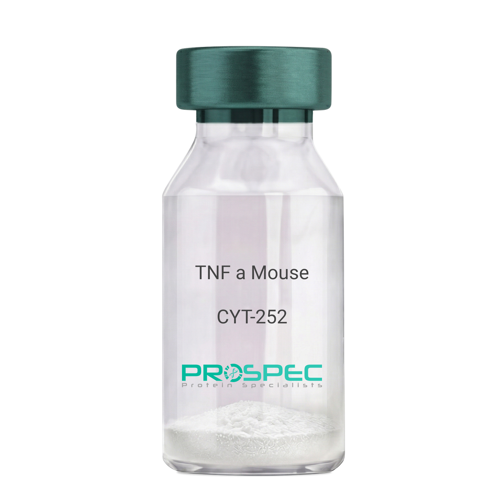
    TNF a Mouse

    Description:

    Tumor Necrosis Factor-Alpha Mouse Recombinant

    TNF-alpha, Tumor necrosis factor ligand superfamily member 2, TNF-a, Cachectin, DIF, TNFA, TNFSF2.

    Product # :

    CYT-252

    Price :

    Quantity :

    Shipping Method :

    Room Temp Icon

    Shipped at Room temp

    Add To Cart

    More Info

    • description
    • source
    • formulation
    • purity
    • biological activity
    • More Info
    • Citations

    Description

    Tumor Necrosis Factor-a Mouse Recombinant produced in E. coli is a single, non glycosylated, polypeptide chain containing 157 amino acids and having a molecular mass of 17301.32 Dalton. The TNF-alpha is purified by standard chromatographic techniques.

    Source

    Escherichia Coli.

    Formulation

    Lyophilized from a 0.2µm filtered concentrated solution in PBS, pH 7.2.

    Purity

    Greater than 97.0% as determined by:
    (a) Analysis by RP-HPLC.
    (c) Analysis by SDS-PAGE.

    Biological Activity

    The ED50 as determined by the cytolysis of murine L929 cells in the presence of Actinomycin D is < 0.1ng/ml, corresponding to a Specific Activity of 10,000,000 Units/mg.

    Citations

    More Info

    • Introduction

      Tumor necrosis factor is a cytokine involved in systemic inflammation and is a member of a group of cytokines that all stimulate the acute phase reaction. TNF is mainly secreted by macrophages.
      TNF causes apoptotic cell death, cellular proliferation, differentiation, inflammation, tumorigenesis and viral replication, TNF is also involved in lipid metabolism, and coagulation. TNF's primary role is in the regulation of immune cells.
      Dysregulation and, in particular, overproduction of TNF have been implicated in a variety of human diseases- autoimmune diseases, insulin resistance, and cancer.

    • Synonyms

      TNF-alpha, Tumor necrosis factor ligand superfamily member 2, TNF-a, Cachectin, DIF, TNFA, TNFSF2.

    • Physical Appearance

      Sterile Filtered White lyophilized (freeze-dried) powder.

    • Stability

      Lyophilized Tumor Necrosis Factor-a although stable at room temperature for 3 weeks, should be stored desiccated below -18°C. Upon reconstitution TNF-a should be stored at 4°C between 2-7 days and for future use below -18°C.For long term storage it is recommended to add a carrier protein (0.1% HSA or BSA).Please prevent freeze-thaw cycles.

    • Solubility

      It is recommended to reconstitute the lyophilized Tumor Necrosis Factor-alpha in sterile 18M-cm H2O not less than 100µg/ml, which can then be further diluted to other aqueous solutions.

    • Amino Acid Sequence

      MLRSSSQNSS DKPVAHVVAN HQVEEQLEWL SQRANALLAN GMDLKDNQLV VPADGLYLVY SQVLFKGQGC PDYVLLTHTV SRFAISYQEK VNLLSAVKSP CPKDTPEGAE LKPWYEPIYL GGVFQLEKGD QLSAEVNLPK YLDFAESGQV YFGVIAL

    • Background

      Tumor Necrosis Factor-alpha (TNF-α) is a pro-inflammatory cytokine that plays a critical role in the regulation of immune responses, inflammation, and cell survival. It is primarily produced by activated macrophages, but can also be secreted by other immune cells, such as T cells, natural killer cells, and mast cells. TNF-α is involved in a wide range of physiological and pathological processes, including the defense against infections, the development of autoimmune diseases, and the progression of cancer.

      TNF-α exerts its effects by binding to two distinct cell surface receptors, TNF receptor 1 (TNFR1) and TNF receptor 2 (TNFR2). Upon binding to its receptors, TNF-α activates multiple signaling pathways, including the nuclear factor-kappa B (NF-κB) pathway, the mitogen-activated protein kinase (MAPK) pathway, and the apoptotic pathway. These signaling pathways regulate various cellular processes, such as inflammation, cell proliferation, differentiation, and apoptosis.

      In the context of infections, TNF-α plays a crucial role in the body's defense against pathogens. It promotes the recruitment and activation of immune cells, enhances the production of other pro-inflammatory cytokines, and stimulates the expression of adhesion molecules on endothelial cells, facilitating the migration of immune cells to the site of infection. TNF-α also helps to induce fever, which is an important component of the body's immune response to infections.

      However, excessive or prolonged production of TNF-α can contribute to the development of chronic inflammatory diseases, such as rheumatoid arthritis, inflammatory bowel disease, and psoriasis. In these conditions, elevated levels of TNF-α promote the infiltration of immune cells into the affected tissues, leading to tissue damage and the perpetuation of inflammation. The central role of TNF-α in the pathogenesis of these diseases has led to the development of anti-TNF-α therapies, which have revolutionized the treatment of chronic inflammatory diseases. These therapies include monoclonal antibodies, such as infliximab and adalimumab, and soluble TNF receptor fusion proteins, such as etanercept. Anti-TNF-α therapies have been shown to be effective in reducing inflammation, improving symptoms, and slowing disease progression in patients with chronic inflammatory diseases.

      In the context of cancer, TNF-α has complex and context-dependent effects on tumor development and progression. On one hand, TNF-α can promote anti-tumor immunity by activating immune cells and stimulating the production of other pro-inflammatory cytokines. On the other hand, chronic inflammation driven by TNF-α can promote tumor growth, angiogenesis, and metastasis. Therefore, the role of TNF-α in cancer is still an area of active research, and the development of TNF-α-targeted therapies for cancer remains a challenge.

      In conclusion, TNF-α is a pro-inflammatory cytokine that plays a critical role in the regulation of immune responses, inflammation, and cell survival. Its involvement in various physiological and pathological processes has made it an important target for the development of therapies for chronic inflammatory diseases and cancer. Anti-TNF-α therapies have revolutionized the treatment of chronic inflammatory diseases, but the complex role of TNF-α in cancer remains an area of ongoing research. Understanding the precise mechanisms by which TNF-α contributes to disease pathogenesis will be crucial for the development of more effective and targeted therapies.

    • Protein content

      Protein quantitation was carried out by two independent methods1. UV spectroscopy at 280 nm using the absorbency value of 1.24 as the extinction coefficient for a 0.1% (1mg/ml) solution. This value is calculated by the PC GENE computer analysis program of protein sequences (IntelliGenetics). 2. Analysis by RP-HPLC, using a calibrated solution of TNF-a as a Reference Standard.

    ProSpec's products are furnished for LABORATORY RESEARCH USE ONLY. The product may not be used as drugs, agricultural or pesticidal products, food additives or household chemicals.

    Tnf Alpha Mouse
  • View Data Sheet

    Name :

    TNFSF12 Human

    Description:

    TNF Ligand Superfamily Member 12 Human Recombinant

    TWEAK, TNF-related weak inducer of apoptosis, TNFSF12, DR3LG, Apo3-Ligand, APO3L, TNFRSF12A, Tumor necrosis factor ligand superfamily member 12, MGC20669, MGC129581.

    Product # :

    CYT-699

    Price :

    Quantity :

    Shipping Method :

    Room Temp Icon

    Shipped at Room temp

    Add To Cart

    More Info

    • description
    • source
    • formulation
    • purity
    • biological activity
    • More Info
    • Citations

    Description

    TNFSF12 Human Recombinant (94-249 a.a.) produced in E.Coli is a single, non-glycosylated, polypeptide chain containing 156 amino acids and having a total molecular mass of 18kDa. The TNFSF12 is fused with an 8 amino acids his tag at N-terminal (M-HHHHHH-R, total 164 a.a.) and purified by proprietary chromatographic techniques.

    Source

    Escherichia coli.

    Formulation

    Lyophilized from a 0.2μm filtered solution in PBS.

    Purity

    Greater than 95% as determined by SDS-PAGE.

    Biological Activity

    The ED50, as measured in a proliferation assay using HUVECs, is less than 8ng/ml.

    Citations

    More Info

    • Introduction

      TNFSF12 is a cytokine that is part of the TNF ligand family. TNFSF12 is a ligand for the FN14/TWEAKR receptor. TNFSF12 has overlapping signaling functions with TNF, but displays a much wider tissue distribution. TNFSF12 induces apoptosis through multiple pathways of cell death in a cell type-specific manner. TNFSF12 promotes proliferation and migration of endothelial cells, and therefore acts as a regulator of angiogenesis. TNFSF12 is expressed in adult heart, pancreas, skeletal muscle, small intestine, spleen and peripheral blood lymphocytes. TWEAK h induces NFkB and chemokine secretion. TNFSF12 exerts an apoptotic activity in HT-29 human adenocarcinoma cells whilst cultured in the presence of IFN-?. TNFSF12 promotes proliferation and migration of endothelial cells.

    • Synonyms

      TWEAK, TNF-related weak inducer of apoptosis, TNFSF12, DR3LG, Apo3-Ligand, APO3L, TNFRSF12A, Tumor necrosis factor ligand superfamily member 12, MGC20669, MGC129581.

    • Physical Appearance

      Filtered White lyophilized (freeze-dried) powder.

    • Stability

      Lyophilized TNFSF12 although stable at room temperature for 3 weeks, should be stored desiccated below -18°C. Upon reconstitution TWEAK should be stored at 4°C between 2-7 days and for future use below -18°C. For long term storage it is recommended to add a carrier protein (0.1% HSA or BSA). Please prevent freeze-thaw cycles.

    • Solubility

      It is recommended to reconstitute the lyophilized TNFSF12 in sterile 18MΩ-cm H2O not less than 100µg/ml, which can then be further diluted to other aqueous solutions.

    • Amino Acid Sequence

      MHHHHHHRSA PKGRKTRARR AIAAHYEVHP RPGQDGAQAG VDGTVSGWEE ARINSSSPLR YNRQIGEFIV TRAGLYYLYC QVHFDEGKAV YLKLDLLVDG VLALRCLEEF SATAASSLGP QLRLCQVSGL LALRPGSSLR IRTLPWAHLK AAPFLTYFGL FQVH.

    ProSpec's products are furnished for LABORATORY RESEARCH USE ONLY. The product may not be used as drugs, agricultural or pesticidal products, food additives or household chemicals.

    Tnfsf12 Human
  • View Data Sheet

    Name :

    OSM Human, His

    Description:

    Oncostatin-M Human Recombinant, His Tag

    OSM, MGC20461.

    Product # :

    CYT-060

    Price :

    Quantity :

    Shipping Method :

    Ice Icon

    Shipped with Ice Packs

    Add To Cart

    More Info

    • description
    • source
    • formulation
    • purity
    • More Info
    • Citations

    Description

    OSM Human Recombinant produced in E.coli is a single, non-glycosylated polypeptide chain containing 230 amino acids (26-234) and having a molecular mass of 25.9 kDa.The OSM is fused to a 21 amino acid His-Tag at N-terminus and purified by proprietary chromatographic techniques.

    Source

    Escherichia Coli.

    Formulation

    OSM protein (0.5mg/ml) is supplied in 20mM Tris-HCl, pH-8, 1mM DTT and 20% Glycerol.

    Purity

    Greater than 95.0% as determined by SDS-PAGE.

    Citations

    More Info

    • Introduction

      POU2AF1 is a lymphocyte specific transcription coactivator protein. POU2AF1 cooperates only with the Oct1/2 proteins using sub domains in the POU domain of the Oct1/2 proteins, increasing their transcriptional efficiency. Even though POU2AF1 have no basic ability to bind DNA, it links firmly with the octomer motif in the presence of Oct1 and Oct2. POU2AF1 is expressed at maximum levels in spleen and peripheral blood leukocytes.

    • Synonyms

      OSM, MGC20461.

    • Physical Appearance

      Sterile Filtered colorless solution.

    • Stability

      Store at 4°C if entire vial will be used within 2-4 weeks. Store, frozen at -20°C for longer periods of time. For long term storage it is recommended to add a carrier protein (0.1% HSA or BSA).Avoid multiple freeze-thaw cycles.

    • Amino Acid Sequence

      MGSSHHHHHH SSGLVPRGSH MAAIGSCSKE YRVLLGQLQK QTDLMQDTSR LLDPYIRIQG LDVPKLREHC RERPGAFPSE ETLRGLGRRG FLQTLNATLG CVLHRLADLE QRLPKAQDLE RSGLNIEDLE KLQMARPNIL GLRNNIYCMA QLLDNSDTAE PTKAGRGASQ PPTPTPASDA FQRKLEGCRF LHGYHRFMHS VGRVFSKWGE SPNRSRRHSP HQALRKGVRR.

    ProSpec's products are furnished for LABORATORY RESEARCH USE ONLY. The product may not be used as drugs, agricultural or pesticidal products, food additives or household chemicals.

    Osm Human His
  • View Data Sheet

    Name :

    CXCL7 Human

    Description:

    Neutrophil Activating Protein-2 Human Recombinant (CXCL7)

    Platelet basic protein, PBP, Small inducible cytokine B7, CXCL7, Leukocyte-derived growth factor, LDGF, Macrophage-derived growth factor, MDGF, pro-platelet basic protein (chemokine (C-X-C motif) ligand 7), TC1, TC2, TGB, TGB1, B-TG1, CTAP3, NAP-2, SCYB7, THBGB, LA-PF4, THBGB1, Beta-TG, CTAPIII, CTAP-III.

    Product # :

    CHM-274

    Price :

    Quantity :

    Shipping Method :

    Room Temp Icon

    Shipped at Room temp

    Add To Cart

    More Info

    • description
    • source
    • formulation
    • purity
    • biological activity
    • More Info
    • sds-page
    • Citations

    Description

    Neutrophil Activating Protein-2 Human Recombinant produced in E.Coli is a non-glycosylated, Polypeptide chain containing 70 amino acids and having a molecular mass of 7609 Dalton. The NAP-2 is purified by proprietary chromatographic techniques.

    Source

    Escherichia Coli.

    Formulation

    The CXCL7 protein was lyophilized from a concentrated (1mg/ml) sterile solution containing no additives.

    Purity

    Greater than 97% as determined by:
    (a) Analysis by RP-HPLC.
    (b) Analysis by SDS-PAGE.

    Biological Activity

    The specific activity as determined by the ability of NAP2 to chemoattract human neurotrophils using a concentration of 1-10ng/ml corresponding to a Specific Activity of 100,000-1,000,000IU/mg.

    sds-page

    CXCL7 Human SDS-PAGE - Product image 1

    Citations

    More Info

    • Introduction

      Chemokine (C-X-C motif) ligand (CXCL7) is a small cytokine belonging to the CXC chemokine family. It is a protein that is released in large amounts from platelets following their activation. It stimulates various processes including mitogenesis, synthesis of extracellular matrix, glucose metabolism and synthesis of plasminogen activator.

    • Synonyms

      Platelet basic protein, PBP, Small inducible cytokine B7, CXCL7, Leukocyte-derived growth factor, LDGF, Macrophage-derived growth factor, MDGF, pro-platelet basic protein (chemokine (C-X-C motif) ligand 7), TC1, TC2, TGB, TGB1, B-TG1, CTAP3, NAP-2, SCYB7, THBGB, LA-PF4, THBGB1, Beta-TG, CTAPIII, CTAP-III.

    • Physical Appearance

      Sterile Filtered White lyophilized (freeze-dried) powder.

    • Stability

      Lyophilized NAP-2 although stable at room temperature for 3 weeks, should be stored desiccated below -18°C. Upon reconstitution CXCL7should be stored at 4°C between 2-7 days and for future use below -18°C. For long term storage it is recommended to add a carrier protein (0.1% HSA or BSA).Please prevent freeze-thaw cycles.

    • Solubility

      It is recommended to reconstitute the lyophilized Neutrophil Activating Protein-2in sterile 18MΩ-cm H2O not less than 100µg/ml, which can then be further diluted to other aqueous solutions.

    • Amino Acid Sequence

      The sequence of the first five N-terminal amino acids was determined and was found to be Ala-Glu-Leu-Arg-Cys.

    • Background

      What is the molecular weight/Mw of CXCL7 HUMAN Protein?
      CXCL7 HUMAN Protein has a total Mw of 7.6kDa.

      What is the source or expression system of CXCL7 HUMAN Protein?
      Escherichia Coli.

      What is the Purity of CXCL7 HUMAN Protein?
      CXCL7 HUMAN Protein is > 97% pure as determined by SDS-PAGE.

      What is the Biological Activity of CXCL7 HUMAN Protein?
      The specific activity as determined by the ability of NAP2 to chemoattract human neurotrophils using a concentration of 1-10ng/ml corresponding to a Specific Activity of 100,000-1,000,000IU/mg.

      What is the amino acid sequence of CXCL7 HUMAN Protein?
      The sequence of the first five N-terminal amino acids was determined and was found to be Ala-Glu-Leu-Arg-Cys.

      What applications can CXCL7 HUMAN Protein be used in?
      CXCL7 HUMAN Protein can probably be used in western blot, ELISA and Lateral Flow.

      What is the endotoxin level for CXCL7 HUMAN Protein?
      The endotoxin level is minimal, CXCL7 HUMAN Protein was purified using conventional chromatography techniques.

      Who supplies recombinant CXCL7 HUMAN that is in stock and available to order?
      ProSpec has the protein in stock and available for immediate delivery.

      Where can I order bulk mg quantities of CXCL7 HUMAN?
      ProSpec supplies bulk mg quantities at affordable prices that meets research and academic budgets.


    ProSpec's products are furnished for LABORATORY RESEARCH USE ONLY. The product may not be used as drugs, agricultural or pesticidal products, food additives or household chemicals.

    Nap2 Human
  • View Data Sheet

    Name :

    CTLA 4 Human

    Description:

    Cytotoxic T-Lymphocyte Associated Antigen-4 Human Recombinant

    GSE, CD152, IDDM12, CELIAC3, CTLA-4.

    Product # :

    CYT-366

    Price :

    Quantity :

    Shipping Method :

    Ice Icon

    Shipped with Ice Packs

    Add To Cart

    More Info

    • description
    • source
    • formulation
    • purity
    • More Info
    • sds-page
    • Citations

    Description

    CTLA 4 Human Recombinant produced in E. coli is a single polypeptide chain containing 149 amino acids (36-161) and having a molecular mass of 15.9 kDa.CTLA 4 is fused to a 23 amino acid His-tag at N-terminus & purified by proprietary chromatographic techniques.

    Source

    Escherichia Coli.

    Formulation

    The CTLA 4 solution (1mg/1ml) contains 20mM Tris-HCl buffer (pH 8.0), 0.4M urea and 10% glycerol.

    Purity

    Greater than 90% as determined by SDS-PAGE.

    sds-page

    CTLA4-sds-page - Product image 1

    Citations

    More Info

    • Introduction

      CTLA-4 is a member of the immunoglobulin superfamily and encodes a protein which transmits an inhibitory signal to T cells. The protein contains a V domain, a transmembrane domain, and a cytoplasmic tail. Alternate transcriptional splice variants, encoding different isoforms, have been characterized. The membrane-bound isoform functions as a homodimer interconnected by a disulfide bond, while the soluble isoform functions as a monomer. Mutations in this gene have been associated with insulin-dependent diabetes mellitus, Graves disease, Hashimoto thyroiditis, celiac disease, systemic lupus erythematosus, thyroid-associated orbitopathy, and other autoimmune diseases.

    • Synonyms

      GSE, CD152, IDDM12, CELIAC3, CTLA-4.

    • Physical Appearance

      Sterile filtered colorless solution.

    • Stability

      Store at 4°C if entire vial will be used within 2-4 weeks. Store, frozen at -20°C for longer periods of time. For long term storage it is recommended to add a carrier protein (0.1% HSA or BSA).Avoid multiple freeze-thaw cycles.

    • Amino Acid Sequence

      MGSSHHHHHH SSGLVPRGSH MGSKAMHVAQ PAVVLASSRG IASFVCEYAS PGKATEVRVT VLRQADSQVT EVCAATYMMG NELTFLDDSI CTGTSSGNQV NLTIQGLRAM DTGLYICKVE LMYPPPYYLG IGNGTQIYVI DPEPCPDSD.

    • Background

      Title: Cytotoxic T-Lymphocyte Associated Antigen-4 Human Recombinant: A Potential Immunotherapeutic Target

      Abstract:


      Cytotoxic T-lymphocyte associated antigen-4 (CTLA-4) is a key immune checkpoint receptor that plays a crucial role in regulating T-cell responses. This research paper provides an in-depth analysis of human recombinant CTLA-4, focusing on its production, characterization, and potential applications in immunotherapy. The paper discusses the significance of CTLA-4 in immune regulation, tumor immunity, and autoimmune diseases. Furthermore, it explores ongoing research and clinical trials investigating the therapeutic potential of recombinant CTLA-4 in various pathological conditions. The information presented in this paper aims to enhance our understanding of human recombinant CTLA-4 and its utility as a research tool and a potential immunotherapeutic agent.

      Introduction:


      Cytotoxic T-lymphocyte associated antigen-4 (CTLA-4) is a cell surface receptor primarily expressed on T-cells. It functions as a negative regulator of T-cell activation, dampening immune responses to prevent excessive inflammation. Human recombinant CTLA-4, produced through genetic engineering techniques, provides researchers with a valuable tool to study its biological functions and explore its therapeutic potential.

      Production and Characterization:


      Recombinant CTLA-4 is typically generated using expression systems such as mammalian cells or bacteria. The protein is then purified and characterized to ensure its structural integrity and functional activity. Quality control measures are implemented to confirm the specificity and bioactivity of the recombinant CTLA-4.

      Role in Immune Regulation:


      CTLA-4 plays a critical role in immune regulation by downregulating T-cell activation and suppressing immune responses. It competes with the co-stimulatory receptor CD28 for binding to its ligands, CD80 and CD86, on antigen-presenting cells. This interaction inhibits T-cell activation and promotes immune tolerance. Recombinant CTLA-4 serves as a valuable tool for studying immune checkpoint mechanisms and their impact on immune responses.

      Therapeutic Implications:


      The blockade of CTLA-4 has emerged as a promising immunotherapeutic strategy in cancer treatment. Monoclonal antibodies targeting CTLA-4, such as ipilimumab, have shown significant clinical efficacy in enhancing anti-tumor immune responses. Recombinant CTLA-4-based therapies, including fusion proteins and engineered T-cell receptors, are being explored as potential immunotherapeutic interventions. Additionally, CTLA-4 plays a role in autoimmune diseases, making it a potential target for the development of novel treatments.

      Conclusion:


      Human recombinant CTLA-4 is a valuable research tool and a potential immunotherapeutic target. Its production, characterization, and applications in immune regulation contribute to our understanding of T-cell biology and the development of novel immunotherapies. Continued research and clinical trials investigating the therapeutic potential of recombinant CTLA-4 offer promising prospects for improving outcomes in cancer and autoimmune diseases.

      What is the molecular weight/Mw of CTLA4 Protein?
      CTLA4 Protein has a total Mw of 15.9kDa.

      What is the source or expression system of CTLA4 Protein?
      Escherichia Coli.

      What is the Purity of CTLA4 Protein?
      CTLA4 Protein is >90% pure as determined by SDS-PAGE.

      What is the Biological Activity of CTLA4 Protein?
      The biological functionality of CTLA4 Protein will be determined in the future.

      What is the amino acid sequence of CTLA4 Protein?
      MGSSHHHHHH SSGLVPRGSH MGSKAMHVAQ PAVVLASSRG IASFVCEYAS PGKATEVRVT VLRQADSQVT EVCAATYMMG NELTFLDDSI CTGTSSGNQV NLTIQGLRAM DTGLYICKVE LMYPPPYYLG IGNGTQIYVI DPEPCPDSD.

      What applications can CTLA4 Protein be used in?
      CTLA4 Protein can probably be used in western blot, ELISA and Lateral Flow.

      What is the endotoxin level for CTLA4 Protein?
      The endotoxin level is minimal, CTLA4 Protein was purified using conventional chromatography techniques.

    ProSpec's products are furnished for LABORATORY RESEARCH USE ONLY. The product may not be used as drugs, agricultural or pesticidal products, food additives or household chemicals.

    Ctla 4 Human
  • View Data Sheet

    Name :

    B9D2 Human

    Description:

    B9 Protein Domain 2 Human Recombinant

    B9 Protein Domain 2, B9D2, B9 domain-containing protein 2, MKS1-related protein 2, MKSR2, ICIS-1, MKS10.

    Product # :

    PRO-2051

    Price :

    Quantity :

    Shipping Method :

    Ice Icon

    Shipped with Ice Packs

    Add To Cart

    More Info

    • description
    • source
    • formulation
    • purity
    • More Info
    • Citations

    Description

    B9D2 Human Recombinant produced in E.coli is a single, non-glycosylated polypeptide chain containing 198 amino acids (1-175) and having a molecular mass of 21.6 kDa.B9D2 is fused to a 23 amino acid His-tag at N-terminus & purified by proprietary chromatographic techniques.

    Source

    E.coli.

    Formulation

    The B9D2 solution (0.25mg/ml) contains 20mM Tris-HCl buffer (pH 8.0), 0.2M NaCl, 50% glycerol, 2mM DTT and 1mM EDTA.

    Purity

    Greater than 85% as determined by SDS-PAGE.

    Citations

    More Info

    • Introduction

      B9 Protein Domain 2 (B9D2) is a component of the tectonic-like complex which acts as a barrier that prevents diffusion of transmembrane proteins between the cilia and plasma membranes. B9D2 contains a B9 domain protein, which is found solely in ciliated organisms. B9D2 is upregulated throughout mucociliary differentiation.

    • Synonyms

      B9 Protein Domain 2, B9D2, B9 domain-containing protein 2, MKS1-related protein 2, MKSR2, ICIS-1, MKS10.

    • Physical Appearance

      Sterile Filtered colorless solution.

    • Stability

      Store at 4°C if entire vial will be used within 2-4 weeks. Store, frozen at -20°C for longer periods of time. For long term storage it is recommended to add a carrier protein (0.1% HSA or BSA).Avoid multiple freeze-thaw cycles.

    • Amino Acid Sequence

      MGSSHHHHHH SSGLVPRGSH MGSMAEVHVI GQIIGASGFS ESSLFCKWGI HTGAAWKLLS GVREGQTQVD TPQIGDMAYW SHPIDLHFAT KGLQGWPRLH FQVWSQDSFG RCQLAGYGFC HVPSSPGTHQ LACPTWRPLG SWREQLARAF VGGGPQLLHG DTIYSGADRY RLHTAAGGTV HLEIGLLLRN FDRYGVEC.

    ProSpec's products are furnished for LABORATORY RESEARCH USE ONLY. The product may not be used as drugs, agricultural or pesticidal products, food additives or household chemicals.

    B9D2 Human
  • View Data Sheet

    Name :

    HB-EGF Human, His

    Description:

    Proheparin-Binding EGF-like Growth Factor Human Recombinant, His Tag

    Proheparin-binding EGF-like growth factor, HBEGF, DTR, DTS, HEGFL, HB-EGF, Heparin-binding EGF-like growth factor, Diphtheria toxin receptor, DT-R, DTSF.

    Product # :

    CYT-761

    Price :

    Quantity :

    Shipping Method :

    Ice Icon

    Shipped with Ice Packs

    Add To Cart

    More Info

    • description
    • source
    • formulation
    • purity
    • More Info
    • Citations

    Description

    HB-EGF His Human Recombinant produced in E.coli is a single, non-glycosylated polypeptide chain containing 109 amino acids (63-148) and having a molecular mass of 12.1kDa.HB-EGF His is fused to a 23 amino acid His-tag at N-terminus & purified by proprietary chromatographic techniques.

    Source

    Escherichia Coli.

    Formulation

    The HB-EGF His solution (0. 5mg/ml) contains 20mM Tris-HCl buffer (pH 8.0), 0.2M NaCl, 2mM DTT and 50% glycerol.

    Purity

    Greater than 85% as determined by SDS-PAGE.

    Citations

    More Info

    • Introduction

      HB-EGF is an EGF related growth factor which signals via the EGF receptor, and stimulates the proliferation of SMC (smooth muscle cells), fibroblasts, epithelial cells and keratinocytes. HB-EGF is expressed in various cell types and tissues, including vascular endothelial cells and SMC, macrophages, skeletal muscle, keratinocytes and particular tumor cells. HB-EGF’s ability to explicitly bind heparin and heparin sulfate proteoglycans is dissimilar from other EGF-like molecules, and might be related to the enhanced mitogenic activity, relative to EGF, that HB-EGF exerts on smooth muscle cells.

    • Synonyms

      Proheparin-binding EGF-like growth factor, HBEGF, DTR, DTS, HEGFL, HB-EGF, Heparin-binding EGF-like growth factor, Diphtheria toxin receptor, DT-R, DTSF.

    • Physical Appearance

      Sterile Filtered clear solution.

    • Stability

      Store at 4°C if entire vial will be used within 2-4 weeks. Store, frozen at -20°C for longer periods of time. For long term storage it is recommended to add a carrier protein (0.1% HSA or BSA).Avoid multiple freeze-thaw cycles.

    • Amino Acid Sequence

      MGSSHHHHHH SSGLVPRGSH MGSDLQEADL DLLRVTLSSK PQALATPNKE EHGKRKKKGK GLGKKRDPCL RKYKDFCIHG ECKYVKELRA PSCICHPGYH GERCHGLSL.

    • Background

      What is the molecular weight/Mw of HB-EGF Protein?
      HB-EGF Protein has a total Mw of 12.1kDa.

      What is the source or expression system of HB-EGF Protein?
      Escherichia Coli.

      What is the Purity of HB-EGF Protein?
      HB-EGF Protein is >85% pure as determined by SDS-PAGE.

      What is the Biological Activity of HB-EGF Protein?
      The biological functionality of HB-EGF Protein will be determined in the future.

      What is the amino acid sequence of HB-EGF Protein?
      MGSSHHHHHH SSGLVPRGSH MGSDLQEADL DLLRVTLSSK PQALATPNKE EHGKRKKKGK GLGKKRDPCL RKYKDFCIHG ECKYVKELRA PSCICHPGYH GERCHGLSL.

      What applications can HB-EGF Protein be used in?
      HB-EGF Protein can probably be used in western blot, ELISA and Lateral Flow.

      What is the endotoxin level for HB-EGF Protein?
      The endotoxin level is minimal, HB-EGF Protein was purified using conventional chromatography techniques.

    ProSpec's products are furnished for LABORATORY RESEARCH USE ONLY. The product may not be used as drugs, agricultural or pesticidal products, food additives or household chemicals.

    Hb Egf Human His
  • View Data Sheet

    Name :

    SMAD3 Human

    Description:

    Mothers Against Decapentaplegic Homolog 3 Human Recombinant

    MADH3, JV15-2, HSPC193, HsT17436, MGC60396, DKFZp586N0721, DKFZp686J10186, SMAD3, Mothers against decapentaplegic homolog 3, Mothers against DPP homolog 3, MAD homolog 3, Mad3, hMAD-3, SMAD family member 3, SMAD 3, hSMAD3.

    Product # :

    PRO-751

    Price :

    Quantity :

    Shipping Method :

    Ice Icon

    Shipped with Ice Packs

    Add To Cart

    More Info

    • description
    • source
    • formulation
    • purity
    • More Info
    • Citations

    Description

    SMAD3 Human Recombinant produced in E.Coli is a single, non-glycosylated polypeptide chain containing 445 amino acids (1-425) and having a molecular mass of 50.2 kDa. SMAD3 is fused to a 20 amino acid His-Tag at N-Terminus and purified by standard chromatography techniques.

    Source

    Escherichia Coli.

    Formulation

    The SMAD3 protein solution contains 20mM Tris-HCl pH-8, 0.1M NaCl, 0.1mM DTT and 20% glycerol.

    Purity

    Greater than 90.0% as determined by SDS-PAGE.

    Citations

    More Info

    • Introduction

      SMAD3 is part of the SMAD family of proteins that mediate signal transduction by the TGF-beta/activin/BMP-2/4 cytokine superfamily from receptor Ser/Thr protein kinases at the cell surface to the nucleus. SMAD3 has a role as a transcriptional modulator activated by TGF-Beta and is involved in the regulation of carcinogenesis. SMAD3 mutations are related with the pathogenesis of human osteoarthritis. SMAD3 expression take part in tumor suppression in the premature stages of gastric carcinogenesis. SMAD3 is involved in nuclear translocation of beta-catenin.

    • Synonyms

      MADH3, JV15-2, HSPC193, HsT17436, MGC60396, DKFZp586N0721, DKFZp686J10186, SMAD3, Mothers against decapentaplegic homolog 3, Mothers against DPP homolog 3, MAD homolog 3, Mad3, hMAD-3, SMAD family member 3, SMAD 3, hSMAD3.

    • Physical Appearance

      Sterile Filtered colorless solution.

    • Stability

      Store at 4°C if entire vial will be used within 2-4 weeks. Store, frozen at -20°C for longer periods of time. For long term storage it is recommended to add a carrier protein (0.1% HSA or BSA).Avoid multiple freeze-thaw cycles.

    • Amino Acid Sequence

      MGSSHHHHHH SSGLVPRGSH MSSILPFTPP IVKRLLGWKK GEQNGQEEKW CEKAVKSLVK KLKKTGQLDE LEKAITTQNV NTKCITIPRS LDGRLQVSHR KGLPHVIYCR LWRWPDLHSH HELRAMELCE FAFNMKKDEV CVNPYHYQRV ETPVLPPVLV PRHTEIPAEF PPLDDYSHSI PENTNFPAGI EPQSNIPETP PPGYLSEDGE TSDHQMNHSM DAGSPNLSPN PMSPAHNNLD LQPVTYCEPA FWCSISYYEL NQRVGETFHA SQPSMTVDGF TDPSNSERFC LGLLSNVNRN AAVELTRRHI GRGVRLYYIG GEVFAECLSD SAIFVQSPNC NQRYGWHPAT VCKIPPGCNL KIFNNQEFAA LLAQSVNQGF EAVYQLTRMC TIRMSFVKGW GAEYRRQTVT STPCWIELHL NGPLQWLDKV LTQMGSPSIR CSSVS.

    ProSpec's products are furnished for LABORATORY RESEARCH USE ONLY. The product may not be used as drugs, agricultural or pesticidal products, food additives or household chemicals.

    Smad3 Human
  • View Data Sheet

    Name :

    FGF 2 Human, Plant

    Description:

    Fibroblast Growth Factor-Basic Human Recombinant, Plant

    Prostatropin, HBGH-2, HBGF-2, FGF-2, FGF-b, Fibroblast growth factor 2, Basic fibroblast growth factor, Heparin-binding growth factor 2.

    Product # :

    CYT-123

    Price :

    Quantity :

    Shipping Method :

    Room Temp Icon

    Shipped at Room temp

    Add To Cart

    More Info

    • description
    • source
    • formulation
    • purity
    • biological activity
    • More Info
    • Citations

    Description

    FGF-2 Human Recombinant produced in rice is a single, non-glycosylated polypeptide chain containing 146 amino acids and having a molecular mass of ~17kDa.The FGF-b protein is purified by proprietary chromatographic techniques.

    Source

    Rice Grain (Oryza Sativa).

    Formulation

    FGF-b was lyophilized from a concentrated solution without any additives.

    Purity

    Greater than 95.0% as determined by SDS-PAGE.

    Biological Activity

    The ED50, as calculated by the dose-dependent proliferation of Balb/c 3T3 cells expressing FGF receptors is <1 ng/ml, corresponding to a specific activity of >1 x10⁶ Units/mg.

    Citations

    More Info

    • Introduction

      Basic fibroblast growth factor is a member of the fibroblast growth factor (FGF) family. FGF family members possess broad mitogenic and cell survival activities, and are involved in a variety of biological processes, including embryonic development, cell growth, morphogenesis, tissue repair, tumor growth and invasion. This protein functions as a modifier of endothelial cell migration and proliferation, as well as an angiogenic factor. It acts as a mitogen for a variety of mesoderm- and neuroectoderm-derived cells in vitro, thus is thought to be involved in organogenesis. Three alternatively spliced variants encoding different isoforms have been described. The heparin-binding growth factors are angiogenic agents in vivo and are potent mitogens for a variety of cell types in vitro. There are differences in the tissue distribution and concentration of these 2 growth factors.

    • Synonyms

      Prostatropin, HBGH-2, HBGF-2, FGF-2, FGF-b, Fibroblast growth factor 2, Basic fibroblast growth factor, Heparin-binding growth factor 2.

    • Physical Appearance

      Sterile Filtered White lyophilized (freeze-dried) powder.

    • Stability

      Lyophilized Fibroblast Growth Factor-2 although stable at room temperature for 3 weeks, should be stored desiccated below -18°C. Upon reconstitution FGF-b should be stored at 4°C between 2-7 days and for future use below -18°C.For long term storage it is recommended to add a carrier protein (0.1% HSA or BSA).Please prevent freeze-thaw cycles.

    • Solubility

      It is recommended to reconstitute the lyophilized Fibroblast Growth Factor Basic in sterile 18MΩ-cm H2O at 100µg/ml, which can then be further diluted to other aqueous solutions.

    • Background

      What is the molecular weight/Mw of FGF 2 HUMAN, PLANT Protein?
      FGF 2 HUMAN, PLANT Protein has a total Mw of 17kDa.

      What is the source or expression system of FGF 2 HUMAN, PLANT Protein?
      Rice Grain (Oryza Sativa).

      What is the Purity of FGF 2 HUMAN, PLANT Protein?
      FGF 2 HUMAN, PLANT Protein is >95% pure as determined by SDS-PAGE.

      What is the Biological Activity of FGF 2 HUMAN, PLANT Protein?
      The ED50, as calculated by the dose-dependent proliferation of Balb/c 3T3 cells expressing FGF receptors is <1 ng/ml, corresponding to a specific activity of >1 x10⁶ Units/mg.

      What is the amino acid sequence of FGF 2 HUMAN, PLANT Protein?
      FGF 2 HUMAN, PLANT Protein is composed from 146 amino acids.

      What applications can FGF 2 HUMAN, PLANT Protein be used in?
      FGF 2 HUMAN, PLANT Protein can probably be used in western blot, ELISA and Lateral Flow.

      What is the endotoxin level for FGF 2 HUMAN, PLANT Protein?
      The endotoxin level is minimal, FGF 2 HUMAN, PLANT Protein was purified using conventional chromatography techniques.

    ProSpec's products are furnished for LABORATORY RESEARCH USE ONLY. The product may not be used as drugs, agricultural or pesticidal products, food additives or household chemicals.

    Bfgf Human Plant
  • View Data Sheet

    Name :

    AURKB Human

    Description:

    Aurora Kinase B Human Recombinant

    Serine/threonine-protein kinase 12, Aurora kinase B, Serine/threonine-protein kinase aurora-B, Aurora- and Ipl1-like midbody-associated protein 1, Aurora/IPL1-related kinase 2, Aurora-related kinase 2, AIM-1, ARK-2, STK-1, AURKB, AIK2, AIM1, ARK2, STK12, AurB, IPL1, STK5, aurkb-sv1, aurkb-sv2.

    Product # :

    PKA-355

    Price :

    Quantity :

    Shipping Method :

    Ice Icon

    Shipped with Ice Packs

    Add To Cart

    More Info

    • description
    • source
    • formulation
    • purity
    • More Info
    • Citations

    Description

    AURKB Human Recombinant produced in E.Coli is a single, non-glycosylated, polypeptide chain containing 364 amino acids (1-344) and having a molecular mass of 41.4kDa. AURKB is fused to 20 a.a. His-Tag at N-terminus and purified by proprietary chromatographic techniques.

    Source

    Escherichia Coli.

    Formulation

    The AURKB solution containing 20mM Tris-HCl buffer (pH8.0), 0.5mM DTT, 20% glycerol, 0.1mM EDTA, 0.1mM EGTA, 0.1M NaCl and 0.1mM PMSF.

    Purity

    Greater than 90.0% as determined by SDS-PAGE.

    Citations

    More Info

    • Introduction

      Aurora Kinase B (AURKB) belongs to a family of mitotic serine/threonine kinases. AURKB connects with chromosomes for the period of prophase prior to relocalizing to the spindle at anaphase. AURKB localizes to microtubules near kinetochores, specifically to the specialized microtubules called K-fibers. AURKB controls chromosome segregation through the control of microtubule-kinetochore attachment and cytokinesis. AURKB is required for kinetochore localization of BUB1 and SGOL1. AURKB expression during the G2/M phase transition is firmly coordinated with histone H3 phosphorylation, while overexpression is seen in many kinds of cancers. AURKB phosphorylates 'Ser-10' and 'Ser-28' of histone H3 during mitosis. AURKB is a component of the CPC (chromosomal passenger complex), which is a complex that acts as a key regulator of mitosis.
      High level expression of AURKB is seen in the thymus, which is also expressed in the spleen, lung, testis, colon, placenta and fetal liver. AURKB is expressed during S and G2/M phase and expression is up-regulated in cancer cells during M phase.

    • Synonyms

      Serine/threonine-protein kinase 12, Aurora kinase B, Serine/threonine-protein kinase aurora-B, Aurora- and Ipl1-like midbody-associated protein 1, Aurora/IPL1-related kinase 2, Aurora-related kinase 2, AIM-1, ARK-2, STK-1, AURKB, AIK2, AIM1, ARK2, STK12, AurB, IPL1, STK5, aurkb-sv1, aurkb-sv2.

    • Physical Appearance

      Sterile filtered colorless solution.

    • Stability

      AURKB although stable at 4°C for 1 week, should be stored below -18°C. For long term storage it is recommended to add a carrier protein (0.1% HSA or BSA).Please prevent freeze thaw cycles.

    • Amino Acid Sequence

      MGSSHHHHHH SSGLVPRGSH MAQKENSYPW PYGRQTAPSG LSTLPQRVLR KEPVTPSALV LMSRSNVQPT AAPGQKVMEN SSGTPDILTR HFTIDDFEIG RPLGKGKFGN VYLAREKKSH FIVALKVLFK SQIEKEGVEH QLRREIEIQA HLHHPNILRL YNYFYDRRRI YLILEYAPRG ELYKELQKSC TFDEQRTATI MEELADALMY CHGKKVIHRD IKPENLLLGL KGELKIADFG WSVHAPSLRR KTMCGTLDYL PPEMIEGRMH NEKVDLWCIG VLCYELLVGN PPFESASHNE TYRRIVKVDL KFPASVPMGA QDLISKLLRH NPSERLPLAQ VSAHPWVRAN SRRVLPPSAL QSVA.

    ProSpec's products are furnished for LABORATORY RESEARCH USE ONLY. The product may not be used as drugs, agricultural or pesticidal products, food additives or household chemicals.

    Aurkb Human
  • View Data Sheet

    Name :

    CCN1 Human

    Description:

    Cysteine-Rich Angiogenic Inducer 61 Human Recombinant

    CYR61, Protein CYR61, Cysteine-rich angiogenic inducer 61, IGF-binding protein 10, IGFBP-10, IBP-10, Protein GIG1, CCN family member 1, CCN1, GIG1, IGFBP10.

    Product # :

    CYT-164

    Price :

    Quantity :

    Shipping Method :

    Room Temp Icon

    Shipped at Room temp

    Add To Cart

    More Info

    • description
    • source
    • formulation
    • purity
    • biological activity
    • More Info
    • Citations

    Description

    CYR61 Human Recombinant produced in E.Coli is a single, non-glycosylated, polypeptide chain containing 357 amino acids and having a molecular mass of 39.5kDa.The CYR61 is purified by proprietary chromatographic techniques.

    Source

    Escherichia Coli.

    Formulation

    Lyophilized from a 0.2m filtered concentrated solution in PBS, pH 7.4.

    Purity

    Greater than 95.0% as determined by SDS-PAGE.

    Biological Activity

    The ED50 was determined by the proliferation of mouse 3T3 cells is < 2.0 ug/ml, corresponding to a specific activity of > 500 units/mg.

    Citations

    More Info

    • Introduction

      CYR61 is a growth factor-inducible, immediate-early gene that has multifaceted activities in various cancers. CYR61 is a secreted, cysteine-rich, binding protein which is encoded by a growth factor-inducible immediate-early gene. Acting as an extracellular, matrix-associated signaling molecule, CYR61 promotes the adhesion of endothelial cells through interaction with integrin and enhances growth factor-induced DNA synthesis in the same cell type.

    • Synonyms

      CYR61, Protein CYR61, Cysteine-rich angiogenic inducer 61, IGF-binding protein 10, IGFBP-10, IBP-10, Protein GIG1, CCN family member 1, CCN1, GIG1, IGFBP10.

    • Physical Appearance

      Sterile Filtered White lyophilized (freeze-dried) powder.

    • Stability

      Lyophilized CYR61 Human although stable at room temperature for 3 weeks, should be stored desiccated below -18°C. Upon reconstitution CYR61 should be stored at 4°C between 2-7 days and for future use below -18°C.Please prevent freeze-thaw cycles.

    • Solubility

      It is recommended to reconstitute the lyophilized CYR61 in sterile 18M-cm H2O not less than 100µg/ml, which can then be further diluted to other aqueous solutions.

    • Amino Acid Sequence

      TCPAACHCPL EAPKCAPGVG LVRDGCGCCK VCAKQLNEDC SKTQPCDHTK GLECNFGASS TALKGICRAQ SEGRPCEYNS RIYQNGESFQ PNCKHQCTCI DGAVGCIPLC PQELSLPNLG CPNPRLVKVT GQCCEEWVCD EDSIKDPMED QDGLLGKELG FDASEVELTR NNELIAVGKG SSLKRLPVFG MEPRILYNPL QGQKCIVQTT SWSQCSKTCG TGISTRVTND NPECRLVKET RICEVRPCGQ PVYSSLKKGK KCSKTKKSPE PVRFTYAGCL SVKKYRPKYC GSCVDGRCCT
      PQLTRTVKMR FRCEDGETFS KNVMMIQSCK CNYNCPHANE AAFPFYRLFN DIHKFRD

    • Background

      Title: Cysteine-Rich Angiogenic Inducer 61 Human Recombinant: A Potential Regulator of Angiogenesis

      Abstract:


      Cysteine-rich angiogenic inducer 61 (CYR61) is an important extracellular matrix-associated protein that plays a significant role in angiogenesis and cell adhesion. This research paper provides a comprehensive analysis of human recombinant CYR61, focusing on its production, characterization, and potential applications in regulating angiogenesis. The paper discusses the significance of CYR61 in physiological and pathological angiogenesis, including wound healing, tumor development, and cardiovascular diseases. Furthermore, it explores ongoing research and clinical trials investigating the therapeutic potential of recombinant CYR61 in various pathological conditions. The information presented in this paper aims to enhance our understanding of human recombinant CYR61 and its utility as a research tool and a potential regulator of angiogenesis.

      Introduction:


      Cysteine-rich angiogenic inducer 61 (CYR61) is an extracellular matrix-associated protein that plays a crucial role in angiogenesis, the formation of new blood vessels from pre-existing ones. Human recombinant CYR61, produced through genetic engineering techniques, provides researchers with a valuable tool to study its biological functions and explore its therapeutic potential.

      Production and Characterization:


      Recombinant CYR61 is typically generated using expression systems such as mammalian cells or bacteria. The protein is then purified and characterized to ensure its structural integrity and functional activity. Quality control measures are implemented to confirm the specificity and bioactivity of the recombinant CYR61.

      Role in Angiogenesis:


      CYR61 is involved in various aspects of angiogenesis, including endothelial cell proliferation, migration, and tube formation. It interacts with integrins and other cell surface receptors to modulate signaling pathways involved in angiogenic processes. Recombinant CYR61 serves as a valuable tool for studying the mechanisms underlying angiogenesis and exploring its potential as a therapeutic target.

      Therapeutic Implications:


      The dysregulation of angiogenesis is associated with several pathological conditions, including cancer, cardiovascular diseases, and chronic wounds. Recombinant CYR61 has shown promise as a potential regulator of angiogenesis and a therapeutic agent. It can be used to promote or inhibit angiogenesis, depending on the specific context. Ongoing research and clinical trials are investigating the therapeutic applications of recombinant CYR61 in various diseases, including cancer and ischemic disorders.

      Conclusion:


      Human recombinant CYR61 is a valuable research tool and a potential regulator of angiogenesis. Its production, characterization, and applications in modulating angiogenic processes contribute to our understanding of angiogenesis and the development of novel therapeutic interventions. Continued research and clinical trials exploring the therapeutic potential of recombinant CYR61 offer promising prospects for improving outcomes in cancer, cardiovascular diseases, and wound healing.

      What is the molecular weight/Mw of CCN1 Protein?
      CCN1 Protein has a total Mw of 39.5kDa.

      What is the source or expression system of CCN1 Protein?
      Escherichia Coli.

      What is the Purity of CCN1 Protein?
      CCN1 Protein is >95% pure as determined by SDS-PAGE.

      What is the Biological Activity of CCN1 Protein?
      The ED50 was determined by the proliferation of mouse 3T3 cells is < 2.0 ug/ml, corresponding to a specific activity of > 500 units/mg.

      What is the amino acid sequence of CCN1 Protein?
      TCPAACHCPL EAPKCAPGVG LVRDGCGCCK VCAKQLNEDC SKTQPCDHTK GLECNFGASS TALKGICRAQ SEGRPCEYNS RIYQNGESFQ PNCKHQCTCI DGAVGCIPLC PQELSLPNLG CPNPRLVKVT GQCCEEWVCD EDSIKDPMED QDGLLGKELG FDASEVELTR NNELIAVGKG SSLKRLPVFG MEPRILYNPL QGQKCIVQTT SWSQCSKTCG TGISTRVTND NPECRLVKET RICEVRPCGQ PVYSSLKKGK KCSKTKKSPE PVRFTYAGCL SVKKYRPKYC GSCVDGRCCT
      PQLTRTVKMR FRCEDGETFS KNVMMIQSCK CNYNCPHANE AAFPFYRLFN DIHKFRD

      What applications can CCN1 Protein be used in?
      CCN1 Protein can probably be used in western blot, ELISA and Lateral Flow.

      What is the endotoxin level for CCN1 Protein?
      The endotoxin level is minimal, CCN1 Protein was purified using conventional chromatography techniques.

    ProSpec's products are furnished for LABORATORY RESEARCH USE ONLY. The product may not be used as drugs, agricultural or pesticidal products, food additives or household chemicals.

    Cyr61 Human
  • View Data Sheet

    Name :

    CoV-2 S1 (319-537)

    Description:

    Coronavirus 2019 Spike Glycoprotein-S1 Receptor Binding Domain (319-537 a.a), Recombinant

    Product # :

    SARS-038

    Price :

    Quantity :

    Shipping Method :

    Room Temp Icon

    Shipped at Room temp

    Add To Cart

    More Info

    • description
    • source
    • formulation
    • purity
    • More Info
    • Citations

    Description

    The HEK293 derived recombinant protein contains the Coronavirus 2019 Spike Glycoprotein S1 Receptor Binding Domain [ RBD ], Wuhan-Hu-1 strain, amino acids 319-537, having a Mw of 26.5kDa. Cov-2 RBD is fused to His tag at C-terminal

    Source

    HEK293 Cells.

    Formulation

    Lyophilized from a 0.2μm filtered concentrated solution in PBS pH-7.4 and 10% treaholse.

    Purity

    Protein is >90% pure as determined SDS-PAGE.

    Citations

    More Info

    • Introduction

      A human infecting coronavirus (viral pneumonia) called 2019 novel coronavirus, 2019-nCoV was found in the fish market at the city of Wuhan, Hubei province of China on December 2019.

      The 2019-nCoV shares an 87% identity to the 2 bat-derived severe acute respiratory syndrome 2018 SARS-CoV-2 located in Zhoushan of eastern China. 2019-nCoV has an analogous receptor-BD-structure to that of 2018 SARS-CoV, even though there is a.a. diversity so thus the 2019-nCoV might bind to ACE2 receptor protein (angiotensin-converting enzyme 2) in humans.

      While bats are possibly the host of 2019-nCoV, researchers suspect that animal from the ocean sold at the seafood market was an intermediate host. RSCU analysis proposes that the 2019-nCoV is a recombinant within the viral spike glycoprotein between the bat coronavirus and an unknown coronavirus.

    • Physical Appearance

      Sterile Filtered White lyophilized (freeze-dried) powder.

    • Stability

      Lyophilized Cov-2 RBD protein although stable at room temperature for 3 weeks, should be stored desiccated below -18°C. Upon reconstitution CoV2 RBD protein should be stored at 4°C between 2-7 days and for future use below -18°C.For long term storage it is recommended to add a carrier protein (0.1% HSA or BSA).Please prevent freeze-thaw cycles.

    • Solubility

      It is recommended to reconstitute the lyophilized CoV-2 S1 protein in sterile 18M-cm H2O at a conc. of 0.5mg/ml and not less than 0.1mg/ml, which can then be further diluted to other aqueous solutions

    • Purification Method

      Purified by Metal-Afinity chromatographic technique.

    ProSpec's products are furnished for LABORATORY RESEARCH USE ONLY. The product may not be used as drugs, agricultural or pesticidal products, food additives or household chemicals.

    product_image.jpg
  • View Data Sheet

    Name :

    RELM b Human

    Description:

    RELM-Beta Human Recombinant

    Resistin-like beta, RELM beta, Cysteine-rich secreted protein FIZZ2, Colon and small intestine-specific cysteine-rich protein, Cysteine-rich secreted protein A12-alpha-like 1, Colon carcinoma-related gene protein, RELM-b, XCP2, HXCP2.

    Product # :

    CYT-780

    Price :

    Quantity :

    Shipping Method :

    Room Temp Icon

    Shipped at Room temp

    Add To Cart

    More Info

    • description
    • source
    • formulation
    • purity
    • More Info
    • Citations

    Description

    RELM-b Human Recombinant produced in E.Coli is a disulfide-linked homodimer, non-glycosylated, polypeptide chain containing 2 x 89 amino acids and having a total molecular mass of 19kDa. RELM-b is purified by proprietary chromatographic techniques.

    Source

    Escherichia Coli.

    Formulation

    RELM-b was lyophilized from a solution containing 0.1% Trifluoroacetic Acid (TFA).

    Purity

    Greater than 90.0% as determined by SDS-PAGE.

    Citations

    More Info

    • Introduction

      RELM-beta (Resistin-Like Molecule-beta) is a member of a recently identified family of secreted proteins containing a conserved cystein-rich C-terminus. The RELM family consists of resistin (also called FIZZ3), RELM-alfa (FIZZ1), RELM-beta (FIZZ2) and RELM-gamma. Only resisistin and RELM-beta were found in humans whereas all four RELM family members were identified in rodents.
      RELM-beta appears to be produced as a homodimer exclusively by intestinal goblet cells and can be found in high quantities in stool. Remarkably, stool of germ-free mice displaying sterile intestinal tract does not contain RELM-beta until bacterial colonization takes place after pathogen-free mice entered natural environment. Some, but not all, colon carcinoma cell lines secrete RELM-beta into the cell culture supernatant.
      The physiological function of RELM-beta is not known. High doses of recombinant RELM-beta showed hyperglycemic effects including lowered glucose disposal and increased hepatic glucose production in mice.

    • Synonyms

      Resistin-like beta, RELM beta, Cysteine-rich secreted protein FIZZ2, Colon and small intestine-specific cysteine-rich protein, Cysteine-rich secreted protein A12-alpha-like 1, Colon carcinoma-related gene protein, RELM-b, XCP2, HXCP2.

    • Physical Appearance

      Sterile Filtered White lyophilized (freeze-dried) powder.

    • Stability

      Lyophilized RELM-b although stable at room temperature for 3 weeks, should be stored desiccated below -18°C. Upon reconstitution RELM-b Human Recombinant should be stored at 4°C between 2-7 days and for future use below -18°C.For long term storage it is recommended to add a carrier protein (0.1% HSA or BSA).Please prevent freeze-thaw cycles.

    • Solubility

      It is recommended to reconstitute the lyophilized RELM-b in sterile 0.1% TFA not less than 100µg/ml, which can then be further diluted to other aqueous solutions.

    • Amino Acid Sequence

      MQCSLDSVMD KKIKDVLNSL EYSPSPISKK LSCASVKSQG RPSSCPAGMA VTGCACGYGC GSWDVQLETT CHCQCSVVDW TTARCCHLT.

    ProSpec's products are furnished for LABORATORY RESEARCH USE ONLY. The product may not be used as drugs, agricultural or pesticidal products, food additives or household chemicals.

    Relm B Human
  • View Data Sheet

    Name :

    BASP1 Human

    Description:

    Brain Abundant Membrane Attached Signal Protein 1 Human Recombinant

    CAP-23, CAP23, NAP-22, NAP22, Brain acid soluble protein 1, BASP1, BASP1 Human, 22 kDa neuronal tissue-enriched acidic protein, Neuronal axonal membrane protein NAP-22.

    Product # :

    PRO-1355

    Price :

    Quantity :

    Shipping Method :

    Ice Icon

    Shipped with Ice Packs

    Add To Cart

    More Info

    • description
    • source
    • formulation
    • purity
    • More Info
    • Citations

    Description

    BASP1 Human Recombinant produced in E.coli is a single, non-glycosylated polypeptide chain containing 250 amino acids (1-227) and having a molecular mass of 25 kDa (Molecular size on SDS-PAGE will appear higher). BASP1 is fused to a 23 amino acid His-tag at N-terminus & purified by proprietary chromatographic techniques.

    Source

    Escherichia Coli.

    Formulation

    The BASP1 solution (0.25mg/ml) contains 20mM Tris-HCl buffer (pH 8.0), 0.2M NaCl and 20% glycerol.

    Purity

    Greater than 95.0% as determined by SDS-PAGE.

    Citations

    More Info

    • Introduction

      Brain Abundant Membrane Attached Signal Protein 1 (BASP1) is a membrane bound protein with numerous transient phosphorylation positions and PEST motifs. Preservation of proteins with PEST sequences amongst diverse species supports their functional significance. PEST sequences take place in proteins with high turnover rates. Immunological attributes of this protein are species specific. BASP1 undergoes N-terminal myristoylation.

    • Synonyms

      CAP-23, CAP23, NAP-22, NAP22, Brain acid soluble protein 1, BASP1, BASP1 Human, 22 kDa neuronal tissue-enriched acidic protein, Neuronal axonal membrane protein NAP-22.

    • Physical Appearance

      Sterile Filtered colorless solution.

    • Stability

      Store at 4°C if entire vial will be used within 2-4 weeks. Store, frozen at -20°C for longer periods of time. For long term storage it is recommended to add a carrier protein (0.1% HSA or BSA).Avoid multiple freeze-thaw cycles.

    • Amino Acid Sequence

      MGSSHHHHHH SSGLVPRGSH MGSMGGKLSK KKKGYNVNDE KAKEKDKKAE GAATEEEGTP KESEPQAAAE PAEAKEGKEK PDQDAEGKAE EKEGEKDAAA AKEEAPKAEP EKTEGAAEAK AEPPKAPEQE QAAPGPAAGG EAPKAAEAAA APAESAAPAA GEEPSKEEGE PKKTEAPAAP AAQETKSDGA PASDSKPGSS EAAPSSKETP AATEAPSSTP KAQGPAASAE EPKPVEAPAA NSDQTVTVKE.

    ProSpec's products are furnished for LABORATORY RESEARCH USE ONLY. The product may not be used as drugs, agricultural or pesticidal products, food additives or household chemicals.

    Basp1 Human
  • View Data Sheet

    Name :

    CD3G Human, Sf9

    Description:

    CD3G Human Recombinant, Sf9

    T-cell surface glycoprotein CD3 gamma chain, CD3-GAMMA, T3G, CD3G

    Product # :

    PRO-2412

    Price :

    Quantity :

    Shipping Method :

    Ice Icon

    Shipped with Ice Packs

    Add To Cart

    More Info

    • description
    • source
    • formulation
    • purity
    • More Info
    • Citations

    Description

    CD3G Human Recombinant produced in Sf9 Insect cells is a single, glycosylated polypeptide chain containing 103 amino acids (23-116 a.a.) and having a molecular mass of 11.8kDa.CD3G is expressed with a 6 amino acids His tag at C-Terminus and purified by proprietary chromatographic techniques.

    Source

    Sf9, Insect cells.

    Formulation

    CD3G protein solution (0.5mg/ml) contains phosphate buffered saline (pH7.4) and 10% glycerol.

    Purity

    Greater than 85.0% as determined by SDS-PAGE.

    Citations

    More Info

    • Introduction

      CD3-gamma polypeptide is encoded by CD3 Gamma (CD3G) which together with CD3-epsilon, -delta and -zeta, and the T-cell receptor alpha/beta and gamma/delta heterodimers, configures the T-cell receptor-CD3 complex. The T-cell receptor-CD3 complex takes part in coupling antigen recognition to numerous intracellular signal-transduction pathways. The genes encoding the epsilon, gamma and delta polypeptides are located in the same cluster on chromosome 11. Deficiencies in this gene are associated with T cell immunodeficiency.

    • Synonyms

      T-cell surface glycoprotein CD3 gamma chain, CD3-GAMMA, T3G, CD3G

    • Physical Appearance

      Sterile Filtered clear solution.

    • Stability

      Store at 4°C if entire vial will be used within 2-4 weeks. Store, frozen at -20°C for longer periods of time. For long term storage it is recommended to add a carrier protein (0.1% HSA or BSA).Avoid multiple freeze-thaw cycles.

    • Amino Acid Sequence

      ADPQSIKGNH LVKVYDYQED GSVLLTCDAE AKNITWFKDG KMIGFLTEDK KKWNLGSNAK DPRGMYQCKG SQNKSKPLQV YYRMCQNCIE LNAATISHHH HHH.

    ProSpec's products are furnished for LABORATORY RESEARCH USE ONLY. The product may not be used as drugs, agricultural or pesticidal products, food additives or household chemicals.

    Cd3G Human 2
  • View Data Sheet

    Name :

    TIMP2 Human

    Description:

    Tissue Inhibitor of Metalloprotease 2 Human Recombinant

    Fibroblast growth factor 17, FGF-17, FGF17, FGF-13, HH20.

    Product # :

    ENZ-782

    Price :

    Quantity :

    Shipping Method :

    Room Temp Icon

    Shipped at Room temp

    Add To Cart

    More Info

    • description
    • source
    • formulation
    • purity
    • biological activity
    • More Info
    • Citations

    Description

    TIMP2 Human Recombinant produced in E.Coli is a single, non-glycosylated, polypeptide chain containing 194 amino acids and having a molecular mass of 21.8kDa.

    Source

    Escherichia Coli.

    Formulation

    TIMP2 protein was lyophilized from a 0.2 µm filtered concentrated solution in PBS, pH 7.4, with 3 % trehalose.

    Purity

    Greater than 95.0% as determined by: (a) Analysis by RP-HPLC.(b) Analysis by SDS-PAGE.

    Biological Activity

    Fully biologically active when compared to standard. The biologically active as determined by its ability to inhibit human MMP-2 cleavage of a fluorogenic peptide substrate Mca-PLGL-Dpa-AR-NH2.

    Citations

    More Info

    • Introduction

      TIMP2 belongs to the TIMP gene family. The proteins encoded by this gene family are natural inhibitors of the matrix metalloproteinases, a group of peptidases that take part in degradation of the extracellular matrix. Besides having an inhibitory role against metalloproteinases, the encoded protein has a exclusive part among TIMP family members in its capability to directly suppress the proliferation of endothelial cells. Consequently, the encoded protein is crucial to the conservation of tissue homeostasis by suppressing the production of quiescent tissues as an answer to angiogenic factors, and by inhibiting protease activity in tissues undergoing renovation of the extracellular matrix.

    • Synonyms

      Fibroblast growth factor 17, FGF-17, FGF17, FGF-13, HH20.

    • Physical Appearance

      Sterile Filtered White lyophilized (freeze-dried) powder.

    • Stability

      Lyophilized TIMP2 although stable at room temperature for 3 weeks, should be stored desiccated below -18°C. Upon reconstitution TIMP2 should be stored at 4°C between 2-7 days and for future use below -18°C. For long term storage it is recommended to add a carrier protein (0.1% HSA or BSA). Please prevent freeze-thaw cycles.

    • Solubility

      It is recommended to reconstitute the lyophilized TIMP2 in sterile 18MΩ-cm H2O not less than 100µg/ml, which can then be further diluted to other aqueous solutions.

    • Amino Acid Sequence

      CSCSPVHPQQ AFCNADVVIR AKAVSEKEVD SGNDIYGNPI KRIQYEIKQI KMFKGPEKDI EFIYTAPSSA VCGVSLDVGG KKEYLIAGKA EGDGKMHITL CDFIVPWDTL STTQKKSLNH RYQMGCECKI TRCPMIPCYI SSPDECLWMD WVTEKNINGH QAKFFACIKR SDGSCAWYRG AAPPKQEFLD IEDP

    ProSpec's products are furnished for LABORATORY RESEARCH USE ONLY. The product may not be used as drugs, agricultural or pesticidal products, food additives or household chemicals.

    Timp 2 Human
  • View Data Sheet

    Name :

    HB-EGF Rat

    Description:

    Proheparin-Binding EGF-like Growth Factor Rat Recombinant

    Proheparin-binding EGF-like growth factor, Heparin-binding EGF-like growth factor, HB-EGF, HBEGF, Dtr, Hegfl, GFHB.

    Product # :

    CYT-170

    Price :

    Quantity :

    Shipping Method :

    Room Temp Icon

    Shipped at Room temp

    Add To Cart

    More Info

    • description
    • source
    • formulation
    • purity
    • biological activity
    • More Info
    • Citations

    Description

    HB-EGF Rat Recombinant produced in E.Coli is a single, non-glycosylated, polypeptide chain containing 86 amino acids and having a molecular mass of 9.7kDa.The HB-EGF is purified by proprietary chromatographic techniques.

    Source

    Escherichia Coli.

    Formulation

    The protein was filtered (0.2µm) and lyophilized from a concentrated solution containing PBS, 300mM NaCl, pH 7.4 and 5% trehalose.

    Purity

    Greater than 95.0% as determined by:
    (a) Analysis by RP-HPLC.
    (b) Analysis by SDS-PAGE.

    Biological Activity

    The ED50 as determined by a cell proliferation assay using balb/c 3T3 cells is < 1.0 ng/ml, corresponding to a specific activity of > 1.0×10⁶ units/mg.

    Citations

    More Info

    • Introduction

      HB-EGF is an EGF related growth factor which signals via the EGF receptor, and stimulates the proliferation of SMC (smooth muscle cells), fibroblasts, epithelial cells and keratinocytes. HB-EGF is expressed in various cell types and tissues, including vascular endothelial cells and SMC, macrophages, skeletal muscle, keratinocytes and particular tumor cells. HB-EGF’s ability to explicitly bind heparin and heparin sulfate proteoglycans is dissimilar from other EGF-like molecules, and might be related to the enhanced mitogenic activity, relative to EGF, that HB-EGF exerts on smooth muscle cells.

    • Synonyms

      Proheparin-binding EGF-like growth factor, Heparin-binding EGF-like growth factor, HB-EGF, HBEGF, Dtr, Hegfl, GFHB.

    • Physical Appearance

      Sterile Filtered White lyophilized (freeze-dried) powder.

    • Stability

      Lyophilized Rat HB-EGF Recombinant although stable at room temperature for 3 weeks, should be stored desiccated below -18°C. Upon reconstitution HB-EGF should be stored at 4°C between 2-7 days and for future use below -18°C.For long term storage it is recommended to add a carrier protein (0.1% HSA or BSA).Please prevent freeze-thaw cycles.

    • Solubility

      It is recommended to reconstitute the lyophilized Rat HB-EGF in sterile 18M-cm H2O not less than 100 µg/ml, which can then be further diluted to other aqueous solutions.

    • Amino Acid Sequence

      DLEGTDLDLF KVAFSSKPQA LATPGKEKNG KKKRKGKGLG KKRDPCLKKY KDYCIHGECR YLKELRIPSC HCLPGYHGQR CHGLTL.

    • Background

      What is the molecular weight/Mw of HB-EGF Protein?
      HB-EGF Protein has a total Mw of 9.7kDa.

      What is the source or expression system of HB-EGF Protein?
      Escherichia Coli.

      What is the Purity of HB-EGF Protein?
      HB-EGF Protein is >95% pure as determined by SDS-PAGE.

      What is the Biological Activity of HB-EGF Protein?
      The ED50 as determined by a cell proliferation assay using balb/c 3T3 cells is < 1.0 ng/ml, corresponding to a specific activity of > 1.0×10⁶ units/mg.

      What is the amino acid sequence of HB-EGF Protein?
      DLEGTDLDLF KVAFSSKPQA LATPGKEKNG KKKRKGKGLG KKRDPCLKKY KDYCIHGECR YLKELRIPSC HCLPGYHGQR CHGLTL.

      What applications can HB-EGF Protein be used in?
      HB-EGF Protein can probably be used in western blot, ELISA and Lateral Flow.

      What is the endotoxin level for HB-EGF Protein?
      The endotoxin level is minimal, HB-EGF Protein was purified using conventional chromatography techniques.

    ProSpec's products are furnished for LABORATORY RESEARCH USE ONLY. The product may not be used as drugs, agricultural or pesticidal products, food additives or household chemicals.

    Hb Egf Rat
  • View Data Sheet

    Name :

    BCL2A1 Human

    Description:

    BCL2-Related Protein A1 Human Recombinant

    BCL2-related protein A1, Hemopoietic-specific early response protein, hematopoietic BCL2-related protein A1, Bcl-2-like protein 5, Protein BFL-1, BCL2L5, Protein GRS, HBPA1, ACC-1, ACC-2.

    Product # :

    PRO-1102

    Price :

    Quantity :

    Shipping Method :

    Ice Icon

    Shipped with Ice Packs

    Add To Cart

    More Info

    • description
    • source
    • formulation
    • purity
    • More Info
    • Citations

    Description

    BCL2A1 Human Recombinant produced in E. coli is a single polypeptide chain containing 176 amino acids (1-152) and having a molecular mass of 20.2kDa.BCL2A1 is fused to a 24 amino acid His-tag at N-terminus & purified by conventional chromatography.

    Source

    E.coli.

    Formulation

    The BCL2A1 solution (0.5mg/1ml) contains 20mM Tris-HCl buffer (pH 8.0) and 10% glycerol.

    Purity

    Greater than 95% as determined by SDS-PAGE.

    Citations

    More Info

    • Introduction

      BCL2A1 delays apoptosis induced by IL-3 deficiency. BCL2A1 takes part in the response of hemopoietic cells to external signals and in preserving endothelial survival during infection.

    • Synonyms

      BCL2-related protein A1, Hemopoietic-specific early response protein, hematopoietic BCL2-related protein A1, Bcl-2-like protein 5, Protein BFL-1, BCL2L5, Protein GRS, HBPA1, ACC-1, ACC-2.

    • Physical Appearance

      Sterile Filtered colorless solution.

    • Stability

      Store at 4°C if entire vial will be used within 2-4 weeks. Store, frozen at -20°C for longer periods of time. For long term storage it is recommended to add a carrier protein (0.1% HSA or BSA).Avoid multiple freeze-thaw cycles.

    • Amino Acid Sequence

      MGSSHHHHHH SSGLVPRGSH MGSHMTDCEF GYIYRLAQDY LQYVLQIPQP GSGPSKTSRV LQKVAFSVQK EVEKNLKSCL DNVNVVSVDT ARTLFNQVME KEFEDDIINW GRIVTIFAFE GILIKKLLRQ QIAPDVDTYK EISYFVAEFI MNNTGEWIRQ NGGWENGFVK KFEPKS.

    ProSpec's products are furnished for LABORATORY RESEARCH USE ONLY. The product may not be used as drugs, agricultural or pesticidal products, food additives or household chemicals.

    Bcl2A1 Human
  • View Data Sheet

    Name :

    IL1A Mouse

    Description:

    Interleukin-1 alpha Mouse Recombinant

    Hematopoietin-1, Lymphocyte-activating factor (LAF), Endogenous Pyrogen (EP), Leukocyte Endogenous Mediator (LEM), Mononuclear Cell Factor (MCF), IL-1 alpha,IL1, IL-1A, IL1F1.

    Product # :

    CYT-523

    Price :

    Quantity :

    Shipping Method :

    Room Temp Icon

    Shipped at Room temp

    Add To Cart

    More Info

    • description
    • source
    • formulation
    • purity
    • biological activity
    • More Info
    • Citations

    Description

    Interleukin-1A Mouse Recombinant produced in E.Coli is single, a non-glycosylated, Polypeptide chain containing 156 amino acids and having a molecular mass of 17.9 kDa. The IL-1A is purified by proprietary chromatographic techniques.

    Source

    Escherichia Coli.

    Formulation

    The protein was lyophilized from a concentrated (1mg/ml) sterile solution containing 1xPBS, pH=7.4.

    Purity

    Greater than 97.0% as determined by:
    (a) Analysis by RP-HPLC.
    (b) Analysis by SDS-PAGE.

    Biological Activity

    The ED50 as determined by the dose-dependant stimulation of murine D10S cells is < 0.002 ng/ml.

    Citations

    More Info

    • Introduction

      Interleukin-1 alpha is a proinflammatory cytokine produced by a wide variety of cell types, including macrophages, osteoblasts, monocytes and hepatocytes. Circulating levels of are normally low and only rise after stimulation by agents such as those produced byinflammation, infection or microbial endotoxins. IL-1 alpha possesses a wide variety of biological activities and exerts its effects by binding to specific cell surface receptors.

    • Synonyms

      Hematopoietin-1, Lymphocyte-activating factor (LAF), Endogenous Pyrogen (EP), Leukocyte Endogenous Mediator (LEM), Mononuclear Cell Factor (MCF), IL-1 alpha,IL1, IL-1A, IL1F1.

    • Physical Appearance

      Sterile Filtered White lyophilized (freeze-dried) powder.

    • Stability

      Lyophilized Interleukin-1a although stable at room temperature for 3 weeks, should be stored desiccated below -18°C. Upon reconstitution IL1A should be stored at 4°C between 2-7 days and for future use below -18°C.For long term storage it is recommended to add a carrier protein (0.1% HSA or BSA).Please prevent freeze-thaw cycles.

    • Solubility

      It is recommended to reconstitute the lyophilized Interleukin 1a in sterile 18MΩ-cm H2O not less than 100µg/ml, which can then be further diluted to other aqueous solutions.

    • Amino Acid Sequence

      SAPYTYQSDL RYKLMKLVRQ KFVMNDSLNQ TIYQDVDKHY LSTTWLNDLQ QEVKFDMYAY SSGGDDSKYP VTLKISDSQL FVSAQGEDQP VLLKELPETP KLITGSETDL IFFWKSINSK NYFTSAAYPE LFIATKEQSR VHLARGLPSM TDFQIS.

    • Background

      What is the molecular weight/Mw of IL1A MOUSE Protein?
      IL1A MOUSE Protein has a total Mw of 17.9kDa.

      What is the source or expression system of IL1A MOUSE Protein?
      Escherichia Coli.

      What is the Purity of IL1A MOUSE Protein?
      IL1A MOUSE Protein is >95% pure as determined by SDS-PAGE.

      What is the Biological Activity of IL1A MOUSE Protein?
      The biological functionality of IL1A MOUSE Protein will be determined in the future.

      What is the amino acid sequence of IL1A MOUSE Protein?
      SAPYTYQSDL RYKLMKLVRQ KFVMNDSLNQ TIYQDVDKHY LSTTWLNDLQ QEVKFDMYAY SSGGDDSKYP VTLKISDSQL FVSAQGEDQP VLLKELPETP KLITGSETDL IFFWKSINSK NYFTSAAYPE LFIATKEQSR VHLARGLPSM TDFQIS.
      What applications can IL1A MOUSE Protein be used in?
      IL1A MOUSE Protein can probably be used in western blot, ELISA and Lateral Flow.

      What is the endotoxin level for IL1A MOUSE Protein?
      The endotoxin level is minimal, IL1A MOUSE Protein was purified using conventional chromatography techniques.


    ProSpec's products are furnished for LABORATORY RESEARCH USE ONLY. The product may not be used as drugs, agricultural or pesticidal products, food additives or household chemicals.

    Il 1 Alpha Mouse
  • View Data Sheet

    Name :

    FGF6 Human

    Description:

    Fibroblast Growth Factor-6 Human Recombinant

    Fibroblast Growth Factor 6, Heparin Secretory-Transforming Protein 2, Heparin-Binding Growth Factor 6, HBGF-6, HSTF-2, FGF-6, HST-2, HST2, HSTF2, FGF6.

    Product # :

    CYT-979

    Price :

    Quantity :

    Shipping Method :

    Room Temp Icon

    Shipped at Room temp

    Add To Cart

    More Info

    • description
    • source
    • formulation
    • purity
    • More Info
    • Citations

    Description

    FGF6 Human Recombinant produced in E.coli is a single, non-glycosylated polypeptide chain having containing 169 amino acids and having a molecular mass of 18.9kDa.The FGF-6 is purified by proprietary chromatographic techniques.

    Source

    Escherichia Coli.

    Formulation

    FGF-6 protein was lyophilized from a 0.2µm filtered solution in 10mM sodium phosphate and 50mM sodium chloride pH 7.5.

    Purity

    Greater than 95.0% as determined by SDS-PAGE.

    Citations

    More Info

    • Introduction

      Fibroblast Growth Factor-6 (FGF6) belongs to the fibroblast growth factor (FGF) family. FGF family members possess extensive mitogenic and cell survival functions, and are involved in various biological processes, including embryonic development, cell growth, morphogenesis, tissue repair, tumor growth and invasion. The FGF6 gene displayes oncogenic transforming activity when transfected into mammalian cells. The mouse homolog of the FGF6 gene displays a restricted expression profile predominantly in the myogenic lineage, suggesting a role in muscle regeneration or differentiation.

    • Synonyms

      Fibroblast Growth Factor 6, Heparin Secretory-Transforming Protein 2, Heparin-Binding Growth Factor 6, HBGF-6, HSTF-2, FGF-6, HST-2, HST2, HSTF2, FGF6.

    • Physical Appearance

      Sterile Filtered White lyophilized (freeze-dried) powder.

    • Stability

      Lyophilized FGF6 although stable at room temperature for 3 weeks, should be stored desiccated below -18°C. Upon reconstitution FGF-6 should be stored at 4°C between 2-7 days and for future use below -18°C. For long term storage it is recommended to add a carrier protein (0.1% HSA or BSA). Please prevent freeze-thaw cycles.

    • Solubility

      It is recommended to reconstitute the lyophilized FGF6 in sterile 18M-cm H2O not less than 100µg/ml, which can then be further diluted to other aqueous solutions.

    • Amino Acid Sequence

      MGTRANNTLL DSRGWGTLLS RSRAGLAGEI AGVNWESGYL VGIKRQRRLY CNVGIGFHLQ VLPDGRISGT HEENPYSLLE ISTVERGVVS LFGVRSALFV AMNSKGRLYA TPSFQEECKF RETLLPNNYN AYESDLYQGT YIALSKYGRV KRGSKVSPIM TVTHFLPRI.

    • Background

      What is the molecular weight/Mw of FGF6 Protein?
      FGF6 Protein has a total Mw of 18.9kDa.

      What is the source or expression system of FGF6 Protein?
      Escherichia Coli.

      What is the Purity of FGF6 Protein?
      FGF6 Protein is >95% pure as determined by SDS-PAGE.

      What is the Biological Activity of FGF6 Protein?
      The biological functionality of FGF6 Protein will be determined in the future.

      What is the amino acid sequence of FGF6 Protein?
      MGTRANNTLL DSRGWGTLLS RSRAGLAGEI AGVNWESGYL VGIKRQRRLY CNVGIGFHLQ VLPDGRISGT HEENPYSLLE ISTVERGVVS LFGVRSALFV AMNSKGRLYA TPSFQEECKF RETLLPNNYN AYESDLYQGT YIALSKYGRV KRGSKVSPIM TVTHFLPRI.

      What applications can FGF6 Protein be used in?
      FGF6 Protein can probably be used in western blot, ELISA and Lateral Flow.

      What is the endotoxin level for FGF6 Protein?
      The endotoxin level is minimal, FGF6 Protein was purified using conventional chromatography techniques.

    ProSpec's products are furnished for LABORATORY RESEARCH USE ONLY. The product may not be used as drugs, agricultural or pesticidal products, food additives or household chemicals.

    Fgf6 Human
  • 1
  • …
  • 24
  • 25
  • 26
  • 27
  • 28
  • 29
Your browser does not support the video tag.

Products

  • Cytokines
  • Growth Factors
  • Chemokines
  • CD Antigens
  • Neurotrophins
  • Hormones
  • Enzymes
  • Viral Antigens
  • Recombinant Proteins
  • Natural Proteins
  • Monoclonal Antibodies
  • Polyclonal Antibodies
  • Thermal Packaging

About

  • Company
  • Founder
  • Contribution
  • Contact Us
  • My Account

Ordering

  • Ordering Method
  • Ordering - Credit Card
  • Ordering PO
  • Shipping Fees
  • FAQ's

Legal

  • Terms & Conditions
  • Privacy
  • Site Map
  • info@prospecbio.com
  • Facebook
  • Instagram
  • Linkedin Profile

© 2026 ProSpec-Tany TechnoGene Ltd. All Rights Reserved.

  • Choosing a selection results in a full page refresh.
  • Opens in a new window.