Skip to content

High-quality research proteins.

  • 1-800-805-6531

  • Mon - Sat 8.00-18.00, Sun - Closed

  • info@prospecbio.com

  • PO Box 6591 East Brunswick NJ 08816

  • Products
    • Cytokines
    • Growth factors
    • Chemokines
    • Cd antigens
    • Neurotrophins
    • Hormones
    • Enzymes
    • Viral antigens
    • Recombinant proteins
    • Natural proteins
    • Monoclonal antibodies
    • Polyclonal antibodies
    • Thermal Packaging
  • About
    • Company
    • Founder
    • Contribution
  • Customers
    • Eastern Asia
    • Eastern Europe
    • North America
    • South America
    • South East Asia
    • Southern Asia
    • Western Europe
  • Ordering
    • Ordering Method
    • Ordering - Credit Card
    • Ordering - PO
    • Shipping Fees
    • FAQs
  • Services
    • DNA Cloning
    • Protein Expression
    • Monoclonal Antibody
    • Fermentation
    • Technology License
  • Contact us
Log in

Country/region

  • USD $ | Albania
  • USD $ | Andorra
  • USD $ | Argentina
  • USD $ | Armenia
  • USD $ | Australia
  • USD $ | Austria
  • USD $ | Azerbaijan
  • USD $ | Belarus
  • USD $ | Belgium
  • USD $ | Bolivia
  • USD $ | Brazil
  • USD $ | Bulgaria
  • USD $ | Cambodia
  • USD $ | Canada
  • USD $ | Chile
  • USD $ | China
  • USD $ | Colombia
  • USD $ | Costa Rica
  • USD $ | Croatia
  • USD $ | Cyprus
  • USD $ | Czechia
  • USD $ | Denmark
  • USD $ | Estonia
  • USD $ | Finland
  • USD $ | France
  • USD $ | Georgia
  • USD $ | Germany
  • USD $ | Greece
  • USD $ | Hong Kong SAR
  • USD $ | Hungary
  • USD $ | Iceland
  • USD $ | India
  • USD $ | Ireland
  • USD $ | Israel
  • USD $ | Italy
  • USD $ | Japan
  • USD $ | Kazakhstan
  • USD $ | Latvia
  • USD $ | Liechtenstein
  • USD $ | Lithuania
  • USD $ | Madagascar
  • USD $ | Malta
  • USD $ | Mexico
  • USD $ | Moldova
  • USD $ | Monaco
  • USD $ | Netherlands
  • USD $ | New Zealand
  • USD $ | North Macedonia
  • USD $ | Norway
  • USD $ | Panama
  • USD $ | Paraguay
  • USD $ | Peru
  • USD $ | Philippines
  • USD $ | Poland
  • USD $ | Portugal
  • USD $ | Romania
  • USD $ | Singapore
  • USD $ | Slovakia
  • USD $ | Slovenia
  • USD $ | South Africa
  • USD $ | South Korea
  • USD $ | Spain
  • USD $ | Sri Lanka
  • USD $ | Sweden
  • USD $ | Switzerland
  • USD $ | Taiwan
  • USD $ | Thailand
  • USD $ | Türkiye
  • USD $ | Ukraine
  • USD $ | United Kingdom
  • USD $ | United States
  • USD $ | Uruguay
  • USD $ | Vatican City
  • USD $ | Venezuela
  • USD $ | Vietnam
  • Facebook
  • Instagram
logoProSpec - Recombinant Protein Manufacturer for academic and R&D industry
Become a distributor
Account Log in
Wishlist
Cart Cart(0)
  • Products
    +
    • Cytokines
    • Growth factors
    • Chemokines
    • Cd antigens
    • Neurotrophins
    • Hormones
    • Enzymes
    • Viral antigens
    • Recombinant proteins
    • Natural proteins
    • Monoclonal antibodies
    • Polyclonal antibodies
    • Thermal Packaging
  • About
    +
    • Company
    • Founder
    • Contribution
  • Customers
    +
    • Eastern Asia
    • Eastern Europe
    • North America
    • South America
    • South East Asia
    • Southern Asia
    • Western Europe
  • Ordering
    +
    • Ordering Method
    • Ordering - Credit Card
    • Ordering - PO
    • Shipping Fees
    • FAQs
  • Services
    +
    • DNA Cloning
    • Protein Expression
    • Monoclonal Antibody
    • Fermentation
    • Technology License
  • Contact us

Item added to your cart

View cart
View All
  • Cytokines
  • Growth factors
  • Chemokines
  • Cd antigens
  • Neurotrophins
  • Hormones
  • Enzymes
  • Viral antigens
  • Recombinant proteins
  • Natural proteins
  • Monoclonal antibodies
  • Polyclonal antibodies
  • Thermal Packaging
  • Tumor Necrosis Factor

    Tumor Necrosis Factor

    View All
  • 4-1BB

    4-1BB

    View All
  • Adiponectin

    Adiponectin

    View All
  • AIF1

    AIF1

    View All
  • Other Cytokines

    Other Cytokines

    View All
  • AITRL

    AITRL

    View All
  • Angiopoietin

    Angiopoietin

    View All
  • Apolipoprotein

    Apolipoprotein

    View All
  • B-Cell Activating Factor

    B-Cell Activating Factor

    View All
  • Beta Defensin

    Beta Defensin

    View All
  • Bone Morphogenetic Protein

    Bone Morphogenetic Protein

    View All
  • B type Natriuretic Peptide

    B type Natriuretic Peptide

    View All
  • BST

    BST

    View All
  • Betacellulin

    Betacellulin

    View All
  • Cardiotrophin

    Cardiotrophin

    View All
  • Activin

    Activin

    View All
  • Fibroblast Growth Factor

    Fibroblast Growth Factor

    View All
  • Other Growth Factors

    Other Growth Factors

    View All
  • CSF

    CSF

    View All
  • CTGF

    CTGF

    View All
  • IGFBP

    IGFBP

    View All
  • VEGF Protein

    VEGF Protein

    View All
  • EGF

    EGF

    View All
  • Epigen

    Epigen

    View All
  • Erythropoietin

    Erythropoietin

    View All
  • Insulin-Like Growth Factor

    Insulin-Like Growth Factor

    View All
  • Growth Hormone

    Growth Hormone

    View All
  • HDGF

    HDGF

    View All
  • Hepatocyte Growth Factor

    Hepatocyte Growth Factor

    View All
  • Insulin

    Insulin

    View All
  • BCA-1/ BLC (CXCL13)

    BCA-1/ BLC (CXCL13)

    View All
  • BRAK (CXCL14)

    BRAK (CXCL14)

    View All
  • C-10 (CCL6)

    C-10 (CCL6)

    View All
  • Other Chemokines

    Other Chemokines

    View All
  • MEC (CCL28)

    MEC (CCL28)

    View All
  • LD78-beta (CCL3L1)

    LD78-beta (CCL3L1)

    View All
  • CTACK (CCL27)

    CTACK (CCL27)

    View All
  • CXCL16

    CXCL16

    View All
  • CXCL17

    CXCL17

    View All
  • Platelet Factor-4 (CXCL4)

    Platelet Factor-4 (CXCL4)

    View All
  • ENA-78 (CXCL5)

    ENA-78 (CXCL5)

    View All
  • Eotaxin (CCL11,24,26)

    Eotaxin (CCL11,24,26)

    View All
  • Exodus-2 (CCL21)

    Exodus-2 (CCL21)

    View All
  • Fractalkine (CX3CL1)

    Fractalkine (CX3CL1)

    View All
  • CXCL6

    CXCL6

    View All
  • Other CD Antigens

    Other CD Antigens

    View All
  • CD14

    CD14

    View All
  • CD164

    CD164

    View All
  • CD1

    CD1

    View All
  • CD2

    CD2

    View All
  • CD200

    CD200

    View All
  • CD207

    CD207

    View All
  • CD226

    CD226

    View All
  • CD23

    CD23

    View All
  • CD244

    CD244

    View All
  • CD247

    CD247

    View All
  • CD27

    CD27

    View All
  • CD274

    CD274

    View All
  • CD300

    CD300

    View All
  • CD33

    CD33

    View All
  • Other Neurotrophins

    Other Neurotrophins

    View All
  • Beta-NGF

    Beta-NGF

    View All
  • BDNF

    BDNF

    View All
  • CDNF

    CDNF

    View All
  • CNTF

    CNTF

    View All
  • GDNF

    GDNF

    View All
  • Glia Maturation Factor

    Glia Maturation Factor

    View All
  • MANF

    MANF

    View All
  • Midkine

    Midkine

    View All
  • Neuroglobin

    Neuroglobin

    View All
  • Neurotrophic factors

    Neurotrophic factors

    View All
  • Neuregulin

    Neuregulin

    View All
  • Neuropilin

    Neuropilin

    View All
  • Pigment Epithelium-Derived Factor

    Pigment Epithelium-Derived Factor

    View All
  • Pleiotrophin

    Pleiotrophin

    View All
  • Peptide Hormones

    Peptide Hormones

    View All
  • Other Hormones

    Other Hormones

    View All
  • HCG

    HCG

    View All
  • Endothelin

    Endothelin

    View All
  • Exendin

    Exendin

    View All
  • FSH

    FSH

    View All
  • Glucagon

    Glucagon

    View All
  • GHRP

    GHRP

    View All
  • GLP

    GLP

    View All
  • Thyrostimulin

    Thyrostimulin

    View All
  • Inhibin A

    Inhibin A

    View All
  • LHRH

    LHRH

    View All
  • Melanotan

    Melanotan

    View All
  • Procalcitonin

    Procalcitonin

    View All
  • PTH

    PTH

    View All
  • Peroxiredoxin

    Peroxiredoxin

    View All
  • Peptidase

    Peptidase

    View All
  • Synthetase

    Synthetase

    View All
  • Transferase

    Transferase

    View All
  • Hydrolase

    Hydrolase

    View All
  • Dehydrogenase

    Dehydrogenase

    View All
  • ACE2

    ACE2

    View All
  • Esterase

    Esterase

    View All
  • Protein Kinases

    Protein Kinases

    View All
  • Other Enzymes

    Other Enzymes

    View All
  • Phosphatase

    Phosphatase

    View All
  • Deaminase

    Deaminase

    View All
  • Oxygenase

    Oxygenase

    View All
  • Adenylate Kinase

    Adenylate Kinase

    View All
  • Lyase

    Lyase

    View All
  • Chagas

    Chagas

    View All
  • SARS

    SARS

    View All
  • Influenza

    Influenza

    View All
  • ASFV

    ASFV

    View All
  • Astrovirus

    Astrovirus

    View All
  • Borrelia

    Borrelia

    View All
  • Helicobacter Pylori

    Helicobacter Pylori

    View All
  • Chikungunya

    Chikungunya

    View All
  • Chlamydia

    Chlamydia

    View All
  • Cytomegalo

    Cytomegalo

    View All
  • Dengue

    Dengue

    View All
  • EBV

    EBV

    View All
  • Ebola

    Ebola

    View All
  • Feline Leukemia Virus

    Feline Leukemia Virus

    View All
  • Hepatitis A

    Hepatitis A

    View All
  • Other

    Other

    View All
  • Actin

    Actin

    View All
  • ADAM

    ADAM

    View All
  • Complement Component

    Complement Component

    View All
  • Ag85

    Ag85

    View All
  • Eukaryotic Translation Initiation Factor

    Eukaryotic Translation Initiation Factor

    View All
  • Anterior Gradient Protein

    Anterior Gradient Protein

    View All
  • Heat Shock Protein

    Heat Shock Protein

    View All
  • Alpha-2-HS-Glycoprotein

    Alpha-2-HS-Glycoprotein

    View All
  • Hemoglobin

    Hemoglobin

    View All
  • Allergy

    Allergy

    View All
  • Anaplasma

    Anaplasma

    View All
  • Angiogenin

    Angiogenin

    View All
  • Ankyrin Repeat Domain

    Ankyrin Repeat Domain

    View All
  • Annexin

    Annexin

    View All
  • Other Natural Proteins

    Other Natural Proteins

    View All
  • Aprotinin

    Aprotinin

    View All
  • Transferrin

    Transferrin

    View All
  • Avidin

    Avidin

    View All
  • Anti Coagulation Factors

    Anti Coagulation Factors

    View All
  • Natural Albumin

    Natural Albumin

    View All
  • Natural Coagulation Factors

    Natural Coagulation Factors

    View All
  • Fibronectin

    Fibronectin

    View All
  • Thrombin

    Thrombin

    View All
  • Anti Human Enzyme

    Anti Human Enzyme

    View All
  • Other Monoclonal Antibodies

    Other Monoclonal Antibodies

    View All
  • Anti Human Cytokine

    Anti Human Cytokine

    View All
  • Anti Human Heat Shock Protein

    Anti Human Heat Shock Protein

    View All
  • Anti Mouse Lymphocyte

    Anti Mouse Lymphocyte

    View All
  • Anti Human Chemokine

    Anti Human Chemokine

    View All
  • Anti Human Lymphocyte

    Anti Human Lymphocyte

    View All
  • Anti Viral Monoclonal

    Anti Viral Monoclonal

    View All
  • Anti Mouse Cytokine

    Anti Mouse Cytokine

    View All
  • Anti-GST Monoclonal

    Anti-GST Monoclonal

    View All
  • Other Polyclonal Antibodies

    Other Polyclonal Antibodies

    View All
  • Anti Viral

    Anti Viral

    View All
  • Anti-GST Monoclonal

    Anti-GST Monoclonal

    View All
Back

Search results

638 results found for “mip (ccl3,4,9,15)”

Name

Description

Product #

Price

Quantity

Shipping Method

  • View Data Sheet

    Name :

    RAC2 Antibody

    Description:

    Ras-Related C3 Botulinum Toxin Substrate 2, Mouse Anti Human

    Ras-related C3 botulinum toxin substrate 2, p21-Rac2, Small G protein, GX, RAC2, HSPC022, EN-7.

    Product # :

    ANT-067

    Price :

    Quantity :

    Shipping Method :

    Ice Icon

    Shipped with Ice Packs

    Add To Cart

    More Info

    • formulation
    • More Info
    • Citations

    Formulation

    1mg/ml containing PBS, pH-7.4, & 0.1% Sodium Azide.

    Citations

    More Info

    • Introduction

      RAC2 is a small signaling G protein/GTPase which is part of the RAS superfamily of small GTP-binding proteins which regulate cell growth, actin cytoskeletal organization, cell proliferation, survival, cell cycle progression, gene transcription rgulation and the activation of protein kinases. RAC2 cycles between an active gtp-bound and inactive gdp-bound state. In active state, RAC2 binds to a variety of effector proteins to regulate cellular responses, such as secretory processes, phagocytose of apoptotic cells and epithelial cell polarization. RAC2 is involved in the regulation of the nadph oxidase.

    • Synonyms

      Ras-related C3 botulinum toxin substrate 2, p21-Rac2, Small G protein, GX, RAC2, HSPC022, EN-7.

    • Immunogen

      Anti-human RAC2, is derived from hybridization of mouse FO myeloma cells with spleen cells from BALB/c mice immunized with recombinant human RAC2 amino acids 1-189 purified from E. coli.

    • Ig Subclass

      Mouse IgG2b heavy chain and ? light chain.

    • Clone

      PAT2G11.

    • Applications

      The antibody has been tested by ELISA, Western blot analysis to assure specificity and reactivity. Since application varies, however, each investigation should be titrated by the reagent to obtain optimal results. Recommended dilution range for Western blot analysis is 1:1000.

    • Type

      Mouse Anti Human Monoclonal.

    • Storage Procedures

      For periods up to 1 month store at 4°C, for longer periods of time, store at -20°C. Prevent freeze thaw cycles.

    • Purification Method

      Rac2 antibody was purified from mouse ascitic fluids by protein-G affinity chromatography.

    ProSpec's products are furnished for LABORATORY RESEARCH USE ONLY. The product may not be used as drugs, agricultural or pesticidal products, food additives or household chemicals.

    Rac2 Antibody
  • View Data Sheet

    Name :

    FGFR1 Human, (22-285)

    Description:

    Fibroblast Growth Factor Receptor-1 Human Recombinant, (22-285 a.a.)

    FGFR-1, bFGF-R, C-FGR, CD331, fms-related tyrosine kinase 2, Pfeiffer syndrome, CEK, FLG, FLT2, KAL2, BFGFR, FGFBR, HBGFR, FGFR1/FGFR1OP2 FUSION GENE, FGFR1/ZNF198 FUSION GENE, FLG FGFR1/BCR FUSION GENE, FLG protein, FMS-LIKE GENE, N-sam tyrosine kinase, basic fibroblast growth factor receptor 1.

    Product # :

    PKA-114

    Price :

    Quantity :

    Shipping Method :

    Ice Icon

    Shipped with Ice Packs

    Add To Cart

    More Info

    • description
    • source
    • formulation
    • purity
    • More Info
    • Citations

    Description

    FGFR1 Human Recombinant produced in Sf9 Baculovirus cells is a single, glycosylated polypeptide chain containing 272 amino acids (22-285) and having a molecular mass of 30.4kDa. (Molecular size on SDS-PAGE will appear at approximately 40-57kDa). FGFR1 is fused to a 8 amino acid His-tag at C-terminus & purified by proprietary chromatographic techniques.

    Source

    Sf9, Baculovirus cells.

    Formulation

    The FGFR1 solution (0.25mg/1ml) contains phosphate buffered Saline (pH7.4), and 10% glycerol.

    Purity

    Greater than 90.0% as determined by SDS-PAGE.

    Citations

    More Info

    • Introduction

      Fibroblast Growth Factors (FGFs) comprise a family of at least 18 structurally related proteins that are involved in a multitude of physiological and pathological cellular processes, including cell growth, differentiation, angiogenesis, wound healing and tumorigenesis. The biological activities of the FGFs are mediated by a family of type I transmembrane tyrosine kinases which undergo dimerization and autophosphorylation after ligand binding. Multiple forms of FGFR-1 to -3 are generated by alternative splicing of the mRNAs. A frequent splicing event involving FGFR-1 and -2 results in receptors containing all three Ig domains, referred to as the alpha isoform, or only IgII and IgIII, referred to as the ? isoform. Only the alpha isoform has been identified for FGFR-3 and FGFR-4. Additional splicing events for FGFR-1 to -3, involving the C-terminal half of the IgIII domain encoded by two mutually exclusive alternative exons, generate FGF receptors with alternative IgIII domains (IIIb and IIIc). An IIIa isoform which is a secreted FGF binding protein containing only the N-terminal half of the IgIII domain plus some intron sequences has also been reported for FGFR-1. Mutations in FGFR-1 to -3 have been found in patients with birth defects involving craniosynostosis.

    • Synonyms

      FGFR-1, bFGF-R, C-FGR, CD331, fms-related tyrosine kinase 2, Pfeiffer syndrome, CEK, FLG, FLT2, KAL2, BFGFR, FGFBR, HBGFR, FGFR1/FGFR1OP2 FUSION GENE, FGFR1/ZNF198 FUSION GENE, FLG FGFR1/BCR FUSION GENE, FLG protein, FMS-LIKE GENE, N-sam tyrosine kinase, basic fibroblast growth factor receptor 1.

    • Physical Appearance

      Sterile Filtered colorless solution.

    • Stability

      Store at 4°C if entire vial will be used within 2-4 weeks.Store, frozen at -20°C for longer periods of time. For long term storage it is recommended to add a carrier protein (0.1% HSA or BSA).Avoid multiple freeze-thaw cycles.

    • Amino Acid Sequence

      RPSPTLPEQD ALPSSEDDDD DDDSSSEEKE TDNTKPNPVA PYWTSPEKME KKLHAVPAAK TVKFKCPSSG TPNPTLRWLK NGKEFKPDHRIGGYKVRYAT WSIIMDSVVP SDKGNYTCIV ENEYGSINHT YQLDVVERSP HRPILQAGLP ANKTVALGSN VEFMCKVYSD PQPHIQWLKH IEVNGSKIGP DNLPYVQILK TAGVNTTDKE MEVLHLRNVS FEDAGEYTCL AGNSIGLSHH SAWLTVLEAL EERPAVMTSP LYLELEHHHH HH.

    ProSpec's products are furnished for LABORATORY RESEARCH USE ONLY. The product may not be used as drugs, agricultural or pesticidal products, food additives or household chemicals.

    Fgfr1 Protein
  • View Data Sheet

    Name :

    UBE2G2 Human

    Description:

    Ubiquitin-Conjugating Enzyme E2G2 Human Recombinant

    Ubiquitin-conjugating enzyme E2 G2, E2 ubiquitin-conjugating enzyme G2, Ubiquitin carrier protein G2, Ubiquitin-protein ligase G2, UBE2G2, Ubiquitin-Conjugating Enzyme E2G2, Ubiquitin-conjugating enzyme E2 G2 isoform 1, UBC7.

    Product # :

    ENZ-884

    Price :

    Quantity :

    Shipping Method :

    Ice Icon

    Shipped with Ice Packs

    Add To Cart

    More Info

    • description
    • source
    • formulation
    • purity
    • More Info
    • Citations

    Description

    UBE2G2 Human Recombinant produced in E.Coli is a single, non-glycosylated polypeptide chain containing 188 amino acids (1-165a.a.) and having a molecular mass of 21kDa.UBE2G2 is fused to a 23 amino acid His-tag at N-terminus & purified by proprietary chromatographic techniques.

    Source

    Escherichia Coli.

    Formulation

    UBE2G2 protein solution (1mg/ml) containing Phosphate buffered saline (pH7.4), 10% glycerol and 1mM DTT.

    Purity

    Greater than 95.0% as determined by SDS-PAGE.

    Citations

    More Info

    • Introduction

      Ubiquitin-Conjugating Enzyme E2G2 (UBE2G2) is a protein coding gene which is a part of the E2 ubiquitin-conjugating enzyme family. UBE2G2 accepts ubiquitin from the E1 complex and catalyzes its covalent attachment to various proteins. UBE2G2 is ubiquitously expressed mainly in adult muscle. UBE2G2 also takes part in endoplasmic reticulum-associated degradation (ERAD).

    • Synonyms

      Ubiquitin-conjugating enzyme E2 G2, E2 ubiquitin-conjugating enzyme G2, Ubiquitin carrier protein G2, Ubiquitin-protein ligase G2, UBE2G2, Ubiquitin-Conjugating Enzyme E2G2, Ubiquitin-conjugating enzyme E2 G2 isoform 1, UBC7.

    • Physical Appearance

      Sterile Filtered clear solution.

    • Stability

      Store at 4°C if entire vial will be used within 2-4 weeks. Store, frozen at -20°C for longer periods of time. For long term storage it is recommended to add a carrier protein (0.1% HSA or BSA).Avoid multiple freeze-thaw cycles.

    • Amino Acid Sequence

      MGSSHHHHHH SSGLVPRGSH MGSMAGTALK RLMAEYKQLT LNPPEGIVAG PMNEENFFEW EALIMGPEDT CFEFGVFPAI LSFPLDYPLS PPKMRFTCEM FHPNIYPDGR VCISILHAPG DDPMGYESSA ERWSPVQSVE KILLSVVSML AEPNDESGAN VDASKMWRDD REQFYKIAKQ IVQKSLGL.

    ProSpec's products are furnished for LABORATORY RESEARCH USE ONLY. The product may not be used as drugs, agricultural or pesticidal products, food additives or household chemicals.

    Ube2G2 Human
  • View Data Sheet

    Name :

    TNFRSF10B Human

    Description:

    TNF Ligand Receptor Superfamily Member 10B Human Recombinant

    Tumor necrosis factor receptor superfamily member 10B, Death receptor 5, TNF-related apoptosis-inducing ligand receptor 2, TRAIL receptor 2, TRAIL-R2, CD262, TNFRSF10B, DR5, KILLER, TRAILR2, TRICK2, ZTNFR9, TRICKB, TRICK2A, TRICK2B, KILLER/DR5.

    Product # :

    CYT-069

    Price :

    Quantity :

    Shipping Method :

    Room Temp Icon

    Shipped at Room temp

    Add To Cart

    More Info

    • description
    • source
    • formulation
    • purity
    • biological activity
    • More Info
    • Citations

    Description

    TNFRSF10B Human Recombinant produced in E.Coli is a single, non-glycosylated, polypeptide chain containing 132 amino acids and having a molecular mass of 14.8kDa.The TNFRSF10B is purified by proprietary chromatographic techniques.

    Source

    Escherichia Coli.

    Formulation

    Lyophilized from a 0.2µm filtered concentrated solution in PBS, pH7.4.

    Purity

    Greater than 95.0% as determined by SDS-PAGE.

    Biological Activity

    The TNFRSF10B reduced the production of LPS-induced TNF by its ability to neutralize endogenous TRAIL in fresh human PBMC. In this assay, endogenous TRAIL is induced during a 24 hour exposure to LPS (10ng/mL) but in the presence of TNFRSF10B, TRAIL-induced TNF is suppressed.

    Citations

    More Info

    • Introduction

      TRAIL Receptor-1 (DR4) and TRAIL Receptor-2(DR5) are members of the TNFR superfamily of transmembrane proteins and contain a cytoplasmic "death domain", which is capable of activating the cell's apoptotic machinery. These receptors are activated by binding to either membrane anchored or soluble TRAIL/Apo2L.

    • Synonyms

      Tumor necrosis factor receptor superfamily member 10B, Death receptor 5, TNF-related apoptosis-inducing ligand receptor 2, TRAIL receptor 2, TRAIL-R2, CD262, TNFRSF10B, DR5, KILLER, TRAILR2, TRICK2, ZTNFR9, TRICKB, TRICK2A, TRICK2B, KILLER/DR5.

    • Physical Appearance

      Sterile Filtered White lyophilized (freeze-dried) powder.

    • Stability

      Lyophilized TNFRSF10B although stable at room temperature for 3 weeks, should be stored desiccated below -18°C. Upon reconstitution TNFRSF10B should be stored at 4°C between 2-7 days and for future use below -18°C. For long term storage it is recommended to add a carrier protein (0.1% HSA or BSA).Please prevent freeze-thaw cycles.

    • Solubility

      It is recommended to reconstitute the lyophilized TNFRSF10B in sterile 18M-cm H2O not less than 100µg/ml, which can then be further diluted to other aqueous solutions.

    • Amino Acid Sequence

      ESALITQQD LAPQQRVAPQ QKRSSPSEGL CPPGHHISED GRDCISCKYG QDYSTHWNDL LFCLRCTRCD SGEVELSPCT TTRNTVCQCE EGTFREEDSP EMCRKCRTGC PRGMVKVGDC TPWSDIECVH KES.

    ProSpec's products are furnished for LABORATORY RESEARCH USE ONLY. The product may not be used as drugs, agricultural or pesticidal products, food additives or household chemicals.

    Tnfrsf10B Human
  • View Data Sheet

    Name :

    SDHAF1 Human

    Description:

    Succinate Dehydrogenase Complex Assembly Factor 1 Human Recombinant

    Succinate Dehydrogenase Complex Assembly Factor 1, LYR Motif-Containing Protein 8, LYR Motif Containing 8, SDH Assembly Factor 1, LYRM8, Succinate Dehydrogenase Assembly Factor 1, Mitochondrial, Succinate dehydrogenase assembly factor 1, mitochondrial.

    Product # :

    PRO-2123

    Price :

    Quantity :

    Shipping Method :

    Ice Icon

    Shipped with Ice Packs

    Add To Cart

    More Info

    • description
    • source
    • formulation
    • purity
    • More Info
    • Citations

    Description

    SDHAF1 Human Recombinant produced in E.Coli is a single, non-glycosylated polypeptide chain containing 138 amino acids (1-115 a.a) and having a molecular mass of 15.2kDa. SDHAF1 is fused to a 23 amino acid His-tag at N-terminus & purified by proprietary chromatographic techniques.

    Source

    Escherichia Coli.

    Formulation

    SDHAF1 protein solution (0.25mg/ml) containing Phosphate buffered saline (pH7.4), 30% glycerol, 2mM DTT and 0.1mM PMSF.

    Purity

    Greater than 85.0% as determined by SDS-PAGE.

    Citations

    More Info

    • Introduction

      Succinate dehydrogenase assembly factor 1 (SDHAF1) is a member of the complex I LYR family. SDHAF1 plays an important role in succinate dehydrogenase complex (SDH) assembly, a complex which is involved in complex II of the mitochondrial electron transport chain. SDHAF1 functions through taking part in mitochondrial biosynthesis of iron-sulfur centers for complex II.

    • Synonyms

      Succinate Dehydrogenase Complex Assembly Factor 1, LYR Motif-Containing Protein 8, LYR Motif Containing 8, SDH Assembly Factor 1, LYRM8, Succinate Dehydrogenase Assembly Factor 1, Mitochondrial, Succinate dehydrogenase assembly factor 1, mitochondrial.

    • Physical Appearance

      Sterile Filtered colorless solution.

    • Stability

      Store at 4°C if entire vial will be used within 2-4 weeks. Store, frozen at -20°C for longer periods of time. For long term storage it is recommended to add a carrier protein (0.1% HSA or BSA).Avoid multiple freeze-thaw cycles.

    • Amino Acid Sequence

      MGSSHHHHHH SSGLVPRGSH MGSMSRHSRL QRQVLSLYRD LLRAGRGKPG AEARVRAEFR QHAGLPRSDV LRIEYLYRRG RRQLQLLRSG HATAMGAFVR PRAPTGEPGG VGSQPDDGDS PRNPHDSTGA PETRPDGR.

    ProSpec's products are furnished for LABORATORY RESEARCH USE ONLY. The product may not be used as drugs, agricultural or pesticidal products, food additives or household chemicals.

    Sdhaf1 Human
  • View Data Sheet

    Name :

    ACTB Antibody

    Description:

    Actin Beta, Mouse Anti Human

    Actin Beta, PS1TP5-Binding Protein 1, Beta Cytoskeletal Actin, Actin, Cytoplasmic 1, Beta-Actin, PS1TP5BP1, BRWS1, ACTB.

    Product # :

    ANT-687

    Price :

    Quantity :

    Shipping Method :

    Ice Icon

    Shipped with Ice Packs

    Add To Cart

    More Info

    • formulation
    • More Info
    • Citations

    Formulation

    1mg/ml containing PBS, pH-7.4, 10% Glycerol and 0.02% Sodium Azide.

    Citations

    More Info
    • Introduction

      Actin Beta (ACTB) is one of 6 different actin isoforms which have been identified in humans. ACTB is one of the 2 nonmuscle cytoskeletal actins. Actins are highly conserved proteins which are involved in cell motility, structure and integrity. Alpha actins are a key component of the contractile apparatus.

    • Synonyms

      Actin Beta, PS1TP5-Binding Protein 1, Beta Cytoskeletal Actin, Actin, Cytoplasmic 1, Beta-Actin, PS1TP5BP1, BRWS1, ACTB.

    • Physical Appearance

      Sterile filtered colorless solution.

    • Immunogen

      Anti-human ACTB mAb, is derived from hybridization of mouse F0 myeloma cells with spleen cells from BALB/c mice immunized with recombinant human ACTB amino acids 1-375 purified from E. coli.

    • Ig Subclass

      Mouse IgG1 heavy chain and κ light chain.

    • Clone

      PAT4G6AT.

    • Applications

      ACTB antibody has been tested by ELISA, Western blot analysis and ICC/IF to assure specificity and reactivity. Since application varies, however, each investigation should be titrated by the reagent to obtain optimal results.

    • Type

      Mouse Anti Human Monoclonal.

    • Storage Procedures

      For periods up to 1 month store at 4°C, for longer periods of time, store at -20°C. Prevent freeze thaw cycles.

    • Purification Method

      ACTB antibody was purified from mouse ascitic fluids by protein-A affinity chromatography.

    ProSpec's products are furnished for LABORATORY RESEARCH USE ONLY. The product may not be used as drugs, agricultural or pesticidal products, food additives or household chemicals.

    Actb Antibody
  • View Data Sheet

    Name :

    VEGF Rat

    Description:

    Vascular Endothelial Growth Factor Rat Recombinant

    Vascular endothelial growth factor A, VEGF-A, Vascular permeability factor, VPF, VEGF, MGC70609.

    Product # :

    CYT-392

    Price :

    Quantity :

    Shipping Method :

    Room Temp Icon

    Shipped at Room temp

    Add To Cart

    More Info

    • description
    • source
    • formulation
    • purity
    • biological activity
    • More Info
    • Citations

    Description

    Vascular Endothelial Growth Factor Rat Recombinant produced in E.Coli is a double, non-glycosylated, polypeptide chain containing 165 amino acids and having a molecular mass of 38,750 Dalton. The VEGF is purified by proprietary chromatographic techniques.

    Source

    Escherichia Coli.

    Formulation

    The protein was lyophilized from 10mM NaP, pH 7.5.

    Purity

    Greater than 97.0% as determined by:
    (a) Analysis by RP-HPLC.
    (b) Analysis by SDS-PAGE.

    Biological Activity

    Determined by the dose-dependent stimulation of the proliferation of human umbilical vein endothelial cells (HUVEC) using a concentration range of 4.0-8.0 ng/ml.

    Citations

    More Info

    • Introduction

      Vascular endothelial growth factor is an important signaling protein involved in both vasculogenesis and angiogenesis. As its name implies, VEGF activity has been mostly studied on cells of the vascular endothelium, although it does have effects on a number of other cell types (e.g. stimulation monocyte/ macrophagemigration, neurons, cancer cells, kidney epithelial cells ).VEGF mediates increased vascular permeability, induces angiogenesis, vasculogenesis and endothelial cell growth, promotes cell migration, and inhibits apoptosis. In vitro, VEGF has been shown to stimulate endothelial cell mitogenesisand cell migration. VEGF is also a vasodilator and increases microvascular permeability and was originally referred to as vascular permeability factor.
      Elevated levels of this protein are linked to POEMS syndrome, also known as Crow-Fukase syndrome. Mutations in this gene have been associated with proliferative and nonproliferative diabetic retinopathy.

    • Synonyms

      Vascular endothelial growth factor A, VEGF-A, Vascular permeability factor, VPF, VEGF, MGC70609.

    • Physical Appearance

      Sterile Filtered White lyophilized (freeze-dried) powder.

    • Stability

      Lyophilized Vascular Endothelial Growth Factor although stable at room temperature for 3 weeks, should be stored desiccated below -18°C. Upon reconstitution VEGF should be stored at 4°C between 2-7 days and for future use below -18°C.For long term storage it is recommended to add a carrier protein (0.1% HSA or BSA).Please prevent freeze-thaw cycles.

    • Solubility

      It is recommended to reconstitute the lyophilized Vascular Endothelial Growth Factor in sterile 18MΩ-cm H2O not less than 100µg/ml, which can then be further diluted to other aqueous solutions.

    • Amino Acid Sequence

      MAPTTEGEQK AHEVVKFMDV YQRSYCRPIE TLVDIFQEYP DEIEYIFKPS CVPLMRCAGC CNDEALECVP TSESNVTMQI MRIKPHQSQH IGEMSFLQHS RCECRPKKDR TKPEKHCEPC SERRKHLFVQ DPQTCKCSCK NTDSRCKARQ LELNERTCRC DKPRR.

    ProSpec's products are furnished for LABORATORY RESEARCH USE ONLY. The product may not be used as drugs, agricultural or pesticidal products, food additives or household chemicals.

    Vegf Rat
  • View Data Sheet

    Name :

    ACOT11 Antibody

    Description:

    Acyl-Coenzyme A Thioesterase 11, Mouse Anti Human

    Acyl-coenzyme A thioesterase 11, Acyl-CoA thioesterase 11, Acyl-CoA thioester hydrolase 11, Brown fat-inducible thioesterase, BFIT, Adipose-associated thioesterase, ACOT11, BFIT, KIAA0707, THEA, BFIT1, BFIT2, THEM1, STARD14.

    Product # :

    ANT-424

    Price :

    Quantity :

    Shipping Method :

    Ice Icon

    Shipped with Ice Packs

    Add To Cart

    More Info

    • formulation
    • More Info
    • Citations

    Formulation

    1mg/ml containing PBS, pH-7.4, 0.02% Sodium Azide & 10% Glycerol.

    Citations

    More Info

    • Introduction

      ACOT11 is a protein with acyl-CoA thioesterase activity towards medium (C12) and long-chain (C18) fatty acyl-CoA substrates. Expression of a similar murine protein in brown adipose tissue is induced by cold exposure and repressed by warmth. Expression of the mouse ACOT11 has been linked to obesity, with higher expression found in obesity-resistant mice compared with obesity-prone mice.

    • Synonyms

      Acyl-coenzyme A thioesterase 11, Acyl-CoA thioesterase 11, Acyl-CoA thioester hydrolase 11, Brown fat-inducible thioesterase, BFIT, Adipose-associated thioesterase, ACOT11, BFIT, KIAA0707, THEA, BFIT1, BFIT2, THEM1, STARD14.

    • Immunogen

      Anti-human ACOT11 mAb, is derived from hybridization of mouse F0 myeloma cells with spleen cells from BALB/c mice immunized with recombinant human ACOT11 amino acids 19-250 purified from E. coli.

    • Ig Subclass

      Mouse IgG2b heavy chain and κ light chain.

    • Clone

      PJ4B2AT.

    • Applications

      ACOT11 antibody has been tested by ELISA, Western blot and immunohistochemistry analysis to assure specificity and reactivity. Since application varies, however, each investigation should be titrated by the reagent to obtain optimal results. Recommended dilution range for Western blot is 1:500 ~ 1:2,000 and immunohistochemistry analysis is 1:50~100. Recommended starting dilution for Western blot is 1:1,000 and Immunohistochemistry is 1:100.

    • Type

      Mouse Anti Human Monoclonal.

    • Storage Procedures

      For periods up to 1 month store at 4°C, for longer periods of time, store at -20°C. Prevent freeze thaw cycles.

    • Purification Method

      ACOT11 antibody was purified from mouse ascitic fluids by protein-G affinity chromatography.

    ProSpec's products are furnished for LABORATORY RESEARCH USE ONLY. The product may not be used as drugs, agricultural or pesticidal products, food additives or household chemicals.

    Acot11 Antibody
  • View Data Sheet

    Name :

    SCGB1A1 Rat

    Description:

    Uteroglobin Rat Recombinant

    Uteroglobin, Clara cell 17 kDa protein, Clara cell phospholipid-binding protein, CCPBP, Clara cells 10 kDa secretory protein, CC10, PCB-binding protein, Secretoglobin family 1A member 1, Scgb1a1, Ugb, Utg, UG, CC16, CCSP, PCB-BP.

    Product # :

    CYT-820

    Price :

    Quantity :

    Shipping Method :

    Room Temp Icon

    Shipped at Room temp

    Add To Cart

    More Info

    • description
    • source
    • formulation
    • purity
    • biological activity
    • More Info
    • Citations

    Description

    SCGB1A1 Rat Recombinant produced in E.Coli is a homodimeric non-glycosylated polypeptide chains consisting of two 77 amino acids and having a molecular mass of 17.0kDa.

    Source

    Escherichia Coli.

    Formulation

    SCGB1A1 protein was lyophilized from a 0.2 µm filtered concentrated solution in PBS, pH 7.4.

    Purity

    Greater than 97.0% as determined by:
    (a) Analysis by RP-HPLC.
    (b) Analysis by SDS-PAGE.

    Biological Activity

    Fully biologically active when compared to standard. The ED50 as determined by the ability of the immobilized protein to support the adhesion of the A549 human lung carcinoma cells is less than 5.0 ?g/ml, corresponding to a specific activity of >200IU/mg.

    Citations

    More Info

    • Introduction

      Uteroglobin (SCGB1A1) which belongs to the Secretoglobin (SCGBs) superfamily, is a multifunctional protein that exerts anti-inflammatory and anti-tumorigenic effects by binding small hydrophobic molecules such as phospholipids and prostaglandins. Uteroglobin is involved in numerous functions including anti-inflammation, inhibition of phospholipase A2 and the sequestering of hydrophobic ligands. SCGB1A1 is expressed by Clara cells, the non-ciliated, non-mucous secretory cells predominant in lung bronchioles, and by other epithelia which communicate with the external environment. On top of sequestering pro-inflammatory mediators and carcinogens, Uteroglobin is implicated in the inhibition of cell migration and invasion, platelet aggregation, and T cell differentiation. SCGB1A1 gene defects are associated with a susceptibility to asthma.

    • Synonyms

      Uteroglobin, Clara cell 17 kDa protein, Clara cell phospholipid-binding protein, CCPBP, Clara cells 10 kDa secretory protein, CC10, PCB-binding protein, Secretoglobin family 1A member 1, Scgb1a1, Ugb, Utg, UG, CC16, CCSP, PCB-BP.

    • Physical Appearance

      Sterile Filtered White lyophilized (freeze-dried) powder.

    • Stability

      Lyophilized SCGB1A1 although stable at room temperature for 3 weeks, should be stored desiccated below -18°C. Upon reconstitution SCGB1A1 should be stored at 4°C between 2-7 days and for future use below -18°C. For long term storage it is recommended to add a carrier protein (0.1% HSA or BSA). Please prevent freeze-thaw cycles.

    • Solubility

      It is recommended to reconstitute the lyophilized SCGB1A1 in sterile 18MΩ-cm H2O not less than 100µg/ml, which can then be further diluted to other aqueous solutions.

    • Amino Acid Sequence

      SSDICPGFLQ VLEALLLGSE SNYEAALKPF NPASDLQNAG TQLKRLVDTL PQETRINIVK LTEKILTSPL CEQDLRV

    ProSpec's products are furnished for LABORATORY RESEARCH USE ONLY. The product may not be used as drugs, agricultural or pesticidal products, food additives or household chemicals.

    Scgb1A1 Rat
  • View Data Sheet

    Name :

    KRAS 2A Human

    Description:

    Kirsten Rat Sarcoma Viral Oncogene, Isoform 2A Human Recombinant

    GTPase KRas, K-Ras 2, Ki-Ras, c-K-ras, c-Ki-ras, KRAS, KRAS2, RASK2, C-K-RAS, CFC2, K-RAS2A, K-RAS2B, K-RAS4A, K-RAS4B, KI-RAS, KRAS1, NS, NS3, Kirsten rat sarcoma viral oncogene homolog.

    Product # :

    PRO-1446

    Price :

    Quantity :

    Shipping Method :

    Ice Icon

    Shipped with Ice Packs

    Add To Cart

    More Info

    • description
    • source
    • formulation
    • purity
    • More Info
    • Citations

    Description

    KRAS 2A Human Recombinant produced in E.Coli is a single, non-glycosylated polypeptide chain containing 210 amino acids (1-186 a.a) and having a molecular mass of 23.8kDa.KRAS 2A is fused to a 24 amino acid His-tag at N-terminus & purified by proprietary chromatographic techniques.

    Source

    Escherichia Coli.

    Formulation

    KRAS 2A protein solution (0.5mg/ml) containing 20mM Tris-HCl buffer (pH8.0), 20% glycerol, 0.1M NaCl and 1mM DTT.

    Purity

    Greater than 90.0% as determined by SDS-PAGE.

    Citations

    More Info

    • Introduction

      Kirsten Rat Sarcoma Viral Oncogene, Isoform 2A (KRAS-2A) belongs to the small GTPase superfamily. The KRAS-2A protein is involved in a variety of malignancies, including lung adenocarcinoma, mucinous adenoma, ductal carcinoma of the pancreas and colorectal carcinoma. Ras family members impact cell growth and differentiation events in a subcellular membrane compartmentalization-based signaling system under normal conditions. Oncogenic Ras can deregulate processes which control both cell proliferation and apoptosis.

    • Synonyms

      GTPase KRas, K-Ras 2, Ki-Ras, c-K-ras, c-Ki-ras, KRAS, KRAS2, RASK2, C-K-RAS, CFC2, K-RAS2A, K-RAS2B, K-RAS4A, K-RAS4B, KI-RAS, KRAS1, NS, NS3, Kirsten rat sarcoma viral oncogene homolog.

    • Physical Appearance

      Sterile Filtered colorless solution.

    • Stability

      Store at 4°C if entire vial will be used within 2-4 weeks. Store, frozen at -20°C for longer periods of time. For long term storage it is recommended to add a carrier protein (0.1% HSA or BSA).Avoid multiple freeze-thaw cycles.

    • Amino Acid Sequence

      MGSSHHHHHH SSGLVPRGSH MGSHMTEYKL VVVGAGGVGK SALTIQLIQN HFVDEYDPTI EDSYRKQVVI DGETCLLDIL DTAGQEEYSA MRDQYMRTGE GFLCVFAINN TKSFEDIHHY REQIKRVKDS EDVPMVLVGN KCDLPSRTVD TKQAQDLARS YGIPFIETSA KTRQRVEDAF YTLVREIRQY RLKKISKEEK TPGCVKIKKC.

    ProSpec's products are furnished for LABORATORY RESEARCH USE ONLY. The product may not be used as drugs, agricultural or pesticidal products, food additives or household chemicals.

    Kras 2A Human
  • View Data Sheet

    Name :

    ACAT1 Antibody

    Description:

    Acetyl-Coenzyme A acetyltransferase 1, Mouse Anti Human

    Acetyl-CoA acetyltransferase, mitochondrial, EC 2.3.1.9, Acetoacetyl-CoA thiolase, T2, ACAT1, ACAT, MAT, THIL.

    Product # :

    ANT-101

    Price :

    Quantity :

    Shipping Method :

    Ice Icon

    Shipped with Ice Packs

    Add To Cart

    More Info

    • formulation
    • More Info
    • Citations

    Formulation

    1mg/ml containing PBS, pH-7.4, 10% Glycerol and 0.01% Sodium Azide.

    Citations

    More Info

    • Introduction

      Acetoacetyl-CoA thiolase (ACAT1) is an enzyme member of the membrane-bound acyltransferase family and Sterol o-acyltransferase subfamily. The ACAT1 enzyme catalyzes the reversible formation of acetoacetyl-CoA from 2 molecules of acetyl-CoA. ACAT1 plays a part in lipoprotein compilation and dietary cholesterol absorption. Added to its acyltransferase activity, ACAT1 acts as a ligase.

    • Synonyms

      Acetyl-CoA acetyltransferase, mitochondrial, EC 2.3.1.9, Acetoacetyl-CoA thiolase, T2, ACAT1, ACAT, MAT, THIL.

    • Physical Appearance

      Sterile Filtered colorless solution.

    • Immunogen

      Anti-human ACAT1 mAb, clone PAT2C5A, is derived from hybridization of mouse F0 myeloma cells with spleen cells from BALB/c mice immunized with a recombinant human ACAT1 protein.

    • Ig Subclass

      Mouse IgG1 heavy chain and Kappa light chain.

    • Clone

      PAT2C5A.

    • Applications

      The antibody has been tested by ELISA, Western blot analysis to assure specificity and reactivity. Since application varies, however, each investigation should be titrated by the reagent to obtain optimal results. Recommended starting dilution is 1:1000.

    • Type

      Mouse Anti Human Monoclonal.

    • Storage Procedures

      For periods up to 1 month store at 4°C, for longer periods of time, store at -20°C. Prevent freeze thaw cycles.

    ProSpec's products are furnished for LABORATORY RESEARCH USE ONLY. The product may not be used as drugs, agricultural or pesticidal products, food additives or household chemicals.

    Acat1 Antibody
  • View Data Sheet

    Name :

    NAGK Human

    Description:

    N-Acetylglucosamine Kinase Human Recombinant

    N-acetylglucosamine kinase, GNK, GlcNAc kinase, N-acetyl-D-glucosamine kinase, HSA242910, EC 2.7.1.59.

    Product # :

    PKA-027

    Price :

    Quantity :

    Shipping Method :

    Ice Icon

    Shipped with Ice Packs

    Add To Cart

    More Info

    • description
    • source
    • formulation
    • purity
    • More Info
    • Citations

    Description

    NAGK Human Recombinant produced in E. coli is a single polypeptide chain containing 367 amino acids (1-344) and having a molecular mass of 39.8kDa.NAGK is fused to a 23 amino acid His-tag at N-terminus & purified by proprietary chromatographic techniques.

    Source

    E.coli.

    Formulation

    The NAGK solution (1mg/ml) contains 20mM Tris-HCl buffer (pH 8.0), 200mM NaCl, 2mM DTT and 20% glycerol.

    Purity

    Greater than 95% as determined by SDS-PAGE.

    Citations

    More Info

    • Introduction

      NAGK is a member of the eukaryotic-type N-acetylglucosamine kinase family. NAGK converts endogenous N-acetylglucosamine (GlcNAc), a key component of complex carbohydrates, from lysosomal degradation or alimentary sources into GlcNAc 6-phosphate. NAGK is a prominent salvage enzyme of amino sugar metabolism in mammals. NAGK cooperates with STK16 and LNX1 and has ManNAc kinase activity.

    • Synonyms

      N-acetylglucosamine kinase, GNK, GlcNAc kinase, N-acetyl-D-glucosamine kinase, HSA242910, EC 2.7.1.59.

    • Physical Appearance

      Sterile Filtered colorless solution.

    • Stability

      Store at 4°C if entire vial will be used within 2-4 weeks. Store, frozen at -20°C for longer periods of time. For long term storage it is recommended to add a carrier protein (0.1% HSA or BSA).Avoid multiple freeze-thaw cycles.

    • Amino Acid Sequence

      MGSSHHHHHH SSGLVPRGSH MGSMAAIYGG VEGGGTRSEV LLVSEDGKIL AEADGLSTNH WLIGTDKCVE RINEMVNRAK RKAGVDPLVP LRSLGLSLSG GDQEDAGRIL IEELRDRFPY LSESYLITTD AAGSIATATP DGGVVLISGT GSNCRLINPD GSESGCGGWG HMMGDEGSAY WIAHQAVKIV FDSIDNLEAA PHDIGYVKQA MFHYFQVPDR LGILTHLYRD FDKCRFAGFC RKIAEGAQQG DPLSRYIFRK AGEMLGRHIV AVLPEIDPVL FQGKIGLPIL CVGSVWKSWE LLKEGFLLAL TQGREIQAQN FFSSFTLMKL RHSSALGGAS LGARHIGHLL PMDYSANAIA FYSYTFS

    ProSpec's products are furnished for LABORATORY RESEARCH USE ONLY. The product may not be used as drugs, agricultural or pesticidal products, food additives or household chemicals.

    Nagk Human
  • View Data Sheet

    Name :

    Dengue Envelope-2 & 4

    Description:

    Dengue Virus Subtype 2 & 4 fused Envelope 52kDa Recombinant

    Product # :

    DEN-026

    Price :

    Quantity :

    Shipping Method :

    Ice Icon

    Shipped with Ice Packs

    Add To Cart

    More Info

    • description
    • source
    • formulation
    • purity
    • More Info
    • Citations

    Description

    The E.coli derived recombinant 52kDa protein is genetically engineered peptide which is derived from Dengue Type-2 and 4 to be expressed as a fused envelope, each part in this fusion contains 170 a.a (positions 46-217),it is used in ELISA assay. This fusion protein is connected to a 6xHis Tag. Dengue Type-2 and 4 is purified by proprietary chromatographic technique.

    Source

    Escherichia Coli.

    Formulation

    The protein solution contains Phosphate buffered saline, pH-7.4 and 0.05% sodium azide.

    Purity

    Protein is >95% pure as determined by 12% PAGE (coomassie staining).

    Citations

    More Info

    • Introduction

      Caused by one of four closely related virus serotypes of the genus Flavivirus, family Flaviviridae, each serotype is sufficiently different that there is no cross-protection and epidemics caused by multiple serotypes (hyperendemicity) can occur. In cell culture experiments and mice Morpholino antisense oligos have shown specific activity against Dengue virus.

    • Physical Appearance

      Sterile filtered colorless solution.

    • Stability

      Dengue Envelope-2 & 4 Recombinant although stable at 4°C for 1 week, should be stored below -18°C. Please prevent freeze thaw cycles.

    ProSpec's products are furnished for LABORATORY RESEARCH USE ONLY. The product may not be used as drugs, agricultural or pesticidal products, food additives or household chemicals.

    Dengue Envelope 2 4
  • View Data Sheet

    Name :

    TNF a Rat

    Description:

    Tumor Necrosis Factor-Alpha Rat Recombinant

    TNF-alpha, Tumor necrosis factor ligand superfamily member 2, TNF-a, Cachectin, DIF, TNFA, TNFSF2.

    Product # :

    CYT-393

    Price :

    Quantity :

    Shipping Method :

    Room Temp Icon

    Shipped at Room temp

    Add To Cart

    More Info

    • description
    • source
    • formulation
    • purity
    • biological activity
    • More Info
    • Citations

    Description

    Tumor Necrosis Factor-a Rat Recombinant produced in E.Coli is a single, non-glycosylated, polypeptide chain containing 157 amino acids and having a molecular mass of 17339.44 Dalton. The TNF-alpha is purified by standard chromatographic techniques.

    Source

    Escherichia Coli.

    Formulation

    The concentrated protein solution (1mg/ml) was lyophilized from 20mM phosphate buffer and 0.1M NaCl.

    Purity

    Greater than 95.0% as determined by:
    (a) Analysis by RP-HPLC.
    (b) Analysis by SDS-PAGE.

    Biological Activity

    The ED50 as determined by the cytolysis of murine L929 cells in the presence of Actinomycin D is < 0.05ng/ml, corresponding to a Specific Activity of 20,000,000 IU/mg.

    Citations

    More Info

    • Introduction

      Tumor necrosis factor is a cytokine involved in systemic inflammation and is a member of a group of cytokines that all stimulate the acute phase reaction. TNF is mainly secreted by macrophages.
      TNF causes apoptotic cell death, cellular proliferation, differentiation, inflammation, tumorigenesis and viral replication, TNF is also involved in lipid metabolism, and coagulation. TNF's primary role is in the regulation of immune cells.
      Dysregulation and, in particular, overproduction of TNF have been implicated in a variety of human diseases- autoimmune diseases, and cancer.

    • Synonyms

      TNF-alpha, Tumor necrosis factor ligand superfamily member 2, TNF-a, Cachectin, DIF, TNFA, TNFSF2.

    • Physical Appearance

      Sterile Filtered White lyophilized (freeze-dried) powder.

    • Stability

      Lyophilized Tumor Necrosis Factor-a although stable at room temperature for 3 weeks, should be stored desiccated below -18°C. Upon reconstitution TNF-a should be stored at 4°C between 2-7 days and for future use below -18°C.For long term storage it is recommended to add a carrier protein (0.1% HSA or BSA).Please prevent freeze-thaw cycles.

    • Solubility

      It is recommended to reconstitute the lyophilized Tumor Necrosis Factor-alpha in sterile 18M-cm H2O not less than 100µg/ml, which can then be further diluted to other aqueous solutions.

    • Amino Acid Sequence

      MLRSSSQNSS DKPVVHVVAN HQAEEQLEWL SQRANALLAN GMDLKDNQLV VPADGLYLIY SQVLFKGQGC PDYVLLTHTV SRFATSYQEK VSLLSAIKSP CPKDTPEGAE LKPWYEPMYL GGVSQLEKGD LLSAEVNLPK YLDITESGQV YFGVIAL.

    ProSpec's products are furnished for LABORATORY RESEARCH USE ONLY. The product may not be used as drugs, agricultural or pesticidal products, food additives or household chemicals.

    Tnf Alpha Rat
  • View Data Sheet

    Name :

    TGFA Human

    Description:

    Transforming Growth Factor-Alpha Human Recombinant

    Transforming Growth Factor Alpha, Protransforming Growth Factor Alpha, TGF-Alpha, TGFA.

    Product # :

    CYT-871

    Price :

    Quantity :

    Shipping Method :

    Room Temp Icon

    Shipped at Room temp

    Add To Cart

    More Info

    • description
    • source
    • formulation
    • purity
    • biological activity
    • More Info
    • Citations

    Description

    TGFA Human Recombinant (40-89) produced in E.Coli is a single, non-glycosylated, polypeptide chain containing 50 amino acids and having a molecular mass of 5.6kDa. The TGFA is purified by proprietary chromatographic techniques.

    Source

    Escherichia Coli.

    Formulation

    Lyophilized from a 0.2µm filtered solution in 0.1% TFA.

    Purity

    Greater than 95.0% as determined by SDS-PAGE.

    Biological Activity

    The ED50, as measured in a proliferation assay using mouse BALB/c 3T3 cells, is 0.395ng/ml.

    Citations

    More Info

    • Introduction

      Transforming Growth Factor-Alpha (TGF-alpha) belongs to the EGF family of cytokines. TGFA soluble form is discharged from the membrane by proteolytic cleavage. Membrane-bound proTGF-alpha is biologically active and has a role in cell-cell adhesion or in the stimulation of adjacent cells. TGFA expression is common in transformed cells. Additionally, TGFA is expressed in normal tissues during embryogenesis and in adult cells/tissues, including the pituitary, keratinocytes, and macrophages.

    • Synonyms

      Transforming Growth Factor Alpha, Protransforming Growth Factor Alpha, TGF-Alpha, TGFA.

    • Physical Appearance

      Sterile Filtered White lyophilized (freeze-dried) powder.

    • Stability

      Lyophilized TGFA although stable at room temperature for 3 weeks, should be stored desiccated below -18°C. Upon reconstitution TGFA should be stored at 4°C between 2-7 days and for future use below -18°C.For long term storage it is recommended to add a carrier protein (0.1% HSA or BSA).Please prevent freeze-thaw cycles.

    • Solubility

      It is recommended to reconstitute the lyophilized TGFA in sterile 18M-cm H2O not less than 100µg/ml, which can then be further diluted to other aqueous solutions.

    • Amino Acid Sequence

      VVSHFNDCPD SHTQFCFHGT CRFLVQEDKP ACVCHSGYVG ARCEHADLLA.

    • Background

      Title: Transforming Growth Factor-Alpha Human Recombinant, Yeast: A Versatile Biopharmaceutical for Therapeutic Applications

      Abstract:


      Transforming Growth Factor-Alpha (TGF-α) is a potent growth factor involved in numerous physiological processes, including cell proliferation, differentiation, and tissue repair. The development of TGF-α human recombinant using yeast expression systems has provided a valuable biopharmaceutical tool for therapeutic applications. This research paper explores the production process, characteristics, and potential therapeutic applications of TGF-α human recombinant derived from yeast, highlighting its versatility and clinical significance.

      Introduction:


      TGF-α is a crucial growth factor that regulates cellular functions and plays a vital role in tissue development and repair. Harnessing the therapeutic potential of TGF-α has been limited by challenges in its production and stability. However, the development of TGF-α human recombinant using yeast expression systems has overcome these limitations, making it an attractive biopharmaceutical for therapeutic interventions.

      Production Process and Characteristics:


      TGF-α human recombinant derived from yeast is produced through recombinant DNA technology, utilizing yeast cells as expression hosts. Yeast expression systems offer several advantages, including high expression yields, cost-effectiveness, and the ability to produce correctly folded and biologically active TGF-α. The resulting TGF-α human recombinant closely resembles native TGF-α in terms of structure and function, allowing for effective therapeutic intervention.

      Therapeutic Applications:


      TGF-α human recombinant derived from yeast has shown promise in various therapeutic applications. It has been investigated for its wound-healing properties, where it promotes tissue regeneration and accelerates the healing process. Additionally, TGF-α has been explored in tissue engineering and regenerative medicine, playing a crucial role in stimulating cell proliferation and tissue development. Furthermore, TGF-α has been studied in the context of cancer research, as it is involved in tumor growth and angiogenesis, making it a potential target for anticancer therapies.

      Advantages and Challenges:


      The use of yeast expression systems for producing TGF-α human recombinant offers several advantages, including scalability, cost-effectiveness, and the ability to produce bioactive protein. However, challenges remain, such as optimizing production processes, purification methods, and ensuring product consistency and stability. Further research is needed to address these challenges and maximize the clinical potential of TGF-α human recombinant derived from yeast.

      Conclusion:


      TGF-α human recombinant derived from yeast represents a versatile biopharmaceutical tool with significant therapeutic potential. Its production using yeast expression systems offers advantages in terms of scalability, cost-effectiveness, and bioactivity. The therapeutic applications of TGF-α human recombinant extend to wound healing, tissue engineering, and cancer research. Continued research and development efforts are crucial to optimizing production processes, overcoming challenges, and fully exploiting the clinical benefits of TGF-α human recombinant as a therapeutic agent.

    ProSpec's products are furnished for LABORATORY RESEARCH USE ONLY. The product may not be used as drugs, agricultural or pesticidal products, food additives or household chemicals.

    Tgfa Human
  • View Data Sheet

    Name :

    VAMP2 Antibody

    Description:

    Synaptobrevin-2, Mouse Anti Human

    Vesicle-associated membrane protein 2, SYB2, VAMP-2, Synaptobrevin-2, VAMP2, FLJ11460.

    Product # :

    ANT-334

    Price :

    Quantity :

    Shipping Method :

    Ice Icon

    Shipped with Ice Packs

    Add To Cart

    More Info

    • formulation
    • More Info
    • Citations

    Formulation

    1mg/ml containing PBS, pH-7.4, & 0.1% Sodium Azide.

    Citations

    More Info

    • Introduction

      Synaptobrevin 2 which is an 18 kDa integral membrane protein localized to the cytoplasmic surface of synaptic vesicle, consists of a proline-rich N-terminal region, a highly conserved hydrophilic domain, followed by a transmembrane anchor and a C-terminal. Synaptobrevin 2 is predominantly expressed in Langerhans islets and glomerular cells. The N-terminal domain of the protein (residues 1-89) forms a specific SNARE complex with the target membrane-associated t- or Q-SNAREs syntaxin 1 and SNAP-25.

    • Synonyms

      Vesicle-associated membrane protein 2, SYB2, VAMP-2, Synaptobrevin-2, VAMP2, FLJ11460.

    • Immunogen

      Anti-human VAMP2 mAb is derived from hybridization of mouse SP2/O myeloma cells with spleen cells from BALB/c mice immunized with recombinant human VAMP2 amino acids 1-89 purified from E. coli.

    • Ig Subclass

      Mouse IgG1 heavy chain and κ light chain.

    • Clone

      P3E5AT.

    • Applications

      VAMP2 antibody has been tested by ELISA and Western blot analysis to assure specificity and reactivity. Since application varies, however, each investigation should be titrated by the reagent to obtain optimal results. Recommended dilution range for Western blot analysis is 1:1,000 ~ 3,000. Recommended starting dilution is 1:2,000.

    • Type

      Mouse Anti Human Monoclonal.

    • Storage Procedures

      For periods up to 1 month store at 4°C, for longer periods of time, store at -20°C. Prevent freeze thaw cycles.

    • Purification Method

      VAMP2 antibody was purified from mouse ascitic fluids by protein-G affinity chromatography.

    ProSpec's products are furnished for LABORATORY RESEARCH USE ONLY. The product may not be used as drugs, agricultural or pesticidal products, food additives or household chemicals.

    Vamp2 Antibody
  • View Data Sheet

    Name :

    WHSC1L1 Antibody

    Description:

    Histone-lysine N-methyltransferase NSD3, Mouse Anti Human

    Histone-lysine N-methyltransferase NSD3, Nuclear SET domain-containing protein 3, Wolf-Hirschhorn syndrome candidate 1-like protein 1, Protein whistle, WHSC1-like 1 isoform 9 with methyltransferase activity to lysine, WHSC1-like protein 1, WHSC1L1, NSD3, DC28, pp14328, FLJ20353, MGC126766, MGC142029, DKFZp667H044.

    Product # :

    ANT-412

    Price :

    Quantity :

    Shipping Method :

    Ice Icon

    Shipped with Ice Packs

    Add To Cart

    More Info

    • formulation
    • More Info
    • Citations

    Formulation

    1mg/ml containing PBS, pH-7.4, & 0.1% Sodium Azide.

    Citations

    More Info

    • Introduction

      WHSC1L1 gene is related to the Wolf-Hirschhorn syndrome candidate-1 gene and encodes a protein with PWWP (proline-tryptophan-tryptophan-proline) domains. The function of the WHSC1L1 protein has not been determined. WHSC1L1 is highly expressed in brain, heart and skeletal muscle; however it is expressed at lower level in the liver and the lung.

    • Synonyms

      Histone-lysine N-methyltransferase NSD3, Nuclear SET domain-containing protein 3, Wolf-Hirschhorn syndrome candidate 1-like protein 1, Protein whistle, WHSC1-like 1 isoform 9 with methyltransferase activity to lysine, WHSC1-like protein 1, WHSC1L1, NSD3, DC28, pp14328, FLJ20353, MGC126766, MGC142029, DKFZp667H044.

    • Immunogen

      Anti-human WHSC1L1 mAb, is derived from hybridization of mouse F0 myeloma cells with spleen cells from BALB/c mice immunized with recombinant human WHSC1L1 amino acids 383-660 purified from E. coli.

    • Ig Subclass

      Mouse IgG2b heavy chain and κ light chain.

    • Clone

      P2E9AT.

    • Applications

      WHSC1L1 antibody has been tested by ELISA and Western blot analysis to assure specificity and reactivity. Since application varies, however, each investigation should be titrated by the reagent to obtain optimal results. Recommended dilution range for Western blot analysis is 1:1,000 ~ 3,000. Recommended starting dilution is 1:2,000.

    • Type

      Mouse Anti Human Monoclonal.

    • Storage Procedures

      For periods up to 1 month store at 4°C, for longer periods of time, store at -20°C. Prevent freeze thaw cycles.

    • Purification Method

      WHSC1L1 antibody was purified from mouse ascitic fluids by protein-G affinity chromatography.

    ProSpec's products are furnished for LABORATORY RESEARCH USE ONLY. The product may not be used as drugs, agricultural or pesticidal products, food additives or household chemicals.

    Whsc1L1 Antibody
  • View Data Sheet

    Name :

    OSTF1 Antibody

    Description:

    Osteoclast Stimulating Factor-1, Mouse Anti Human

    SH3P2, OSF, OSTF-1, Osteoclast-stimulating factor 1, OSTF1, FLJ20559, bA235O14.1.

    Product # :

    ANT-056

    Price :

    Quantity :

    Shipping Method :

    Ice Icon

    Shipped with Ice Packs

    Add To Cart

    More Info

    • formulation
    • More Info
    • Citations

    Formulation

    1mg/ml containing PBS, pH-7.4, 10% Glycerol and 0.02% Sodium Azide.

    Citations

    More Info

    • Introduction

      OSTF1 is an intracellular protein produced by osteoclasts that induces bone resorption, via signaling cascade which results in the secretion of factors enhancing osteoclast formation and activity.

    • Synonyms

      SH3P2, OSF, OSTF-1, Osteoclast-stimulating factor 1, OSTF1, FLJ20559, bA235O14.1.

    • Immunogen

      Anti-human OSTF1 mAb, is derived from hybridization of mouse FO myeloma cells with spleen cells from BALB/c mice immunized with recombinant human OSTF1 amino acids 1-217 purified from E. coli.

    • Ig Subclass

      Mouse IgG2a heavy chain and k light chain.

    • Clone

      PAT9G4AT.

    • Applications

      OSTF1 antibody has been tested by ELISA, Western blot analysis, Flow cytometry and ICC/IF to assure specificity and reactivity. Since application varies, however, each investigation should be titrated by the reagent to obtain optimal results.

    • Type

      Mouse Anti Human Monoclonal.

    • Storage Procedures

      For periods up to 1 month store at 4°C, for longer periods of time, store at -20°C. Prevent freeze thaw cycles.

    • Purification Method

      OSTF1 antibody was purified from mouse ascitic fluids by protein-G affinity chromatography.

    ProSpec's products are furnished for LABORATORY RESEARCH USE ONLY. The product may not be used as drugs, agricultural or pesticidal products, food additives or household chemicals.

    Ostf1 Antibody
  • View Data Sheet

    Name :

    ATXN3 Human

    Description:

    Ataxin-3 Human Recombinant

    Ataxin-3, Machado-Joseph disease protein 1, Spinocerebellar ataxia type 3 protein, ATXN3, ATX3, MJD, MJD1, SCA3, AT3, JOS.

    Product # :

    PRO-708

    Price :

    Quantity :

    Shipping Method :

    Ice Icon

    Shipped with Ice Packs

    Add To Cart

    More Info

    • description
    • source
    • formulation
    • purity
    • More Info
    • Citations

    Description

    ATXN3 produced in E.Coli is a single, non-glycosylated polypeptide chain containing 370 amino acids (1-370 a.a.) and having a molecular mass of 42.4kDa.ATXN3 is purified by proprietary chromatographic techniques.

    Source

    Escherichia Coli.

    Formulation

    The ATXN3 protein solution contains 20mM Tris-HCl buffer (pH 7.5), 2mM DTT, 50mM NaCl and 10% glycerol.

    Purity

    Greater than 90.0% as determined by SDS-PAGE.

    Citations

    More Info

    • Introduction

      Ataxin 3 is otherwise known as Machado-Joseph disease protein 1. Machado–Joseph disease is a hereditary autosomal dominant neurodegenerative disorder. ATXN3 contains trinucleotide CAG repeats in the coding region, and the expansion of these repeats from the normal 13-36 to 68-79 causes the Machado-Joseph disease. ATXN3 is a poly-ubiquitin-binding protein whose cellular turnover is regulated by its catalytic activity.
      In addition, ATXN3 is a proteasome-associated factor which mediates the degradation of ubiquitinated proteins. ATXN3 folds reversibly using a single intermediate; partial destabilization of ATXN3 by chemical denaturation causes the formation of fibrillar aggregates by the non-pathological variant.
      Ataxin-3 interacts with the major histone acetyltransferases cAMP-response-element binding protein (CREB)-binding protein, p300, and p300/CREB-binding protein-associated factor and hinders transcription by these coactivators.

    • Synonyms

      Ataxin-3, Machado-Joseph disease protein 1, Spinocerebellar ataxia type 3 protein, ATXN3, ATX3, MJD, MJD1, SCA3, AT3, JOS.

    • Physical Appearance

      Sterile filtered colorless solution.

    • Stability

      Store at 4°C if entire vial will be used within 2-4 weeks.Store, frozen at -20°C for longer periods of time. For long term storage it is recommended to add a carrier protein (0.1% HSA or BSA).Avoid multiple freeze-thaw cycles.

    • Amino Acid Sequence

      MESIFHEKQE GSLCAQHCLN NLLQGEYFSP VELSSIAHQL DEEERMRMAE GGVTSEDYRT FLQQPSGNMD DSGFFSIQVI SNALKVWGLELILFNSPEYQ RLRIDPINER SFICNYKEHW FTVRKLGKQW FNLNSLLTGP ELISDTYLAL FLAQLQQEGY SIFVVKGDLP DCEADQLLQM IRVQQMHRPK LIGEELAQLK EQRVHKTDLE RVLEANDGSG MLDEDEEDLQ RALALSRQEI DMEDEEADLR RAIQLSMQGS SRNISQDMTQ TSGTNLTSEE LRKRREAYFE KQQQKQQQQQ QQQQQQQQQQ QQQQGDLSGQ SSHPCERPAT SSGALGSDLG DAMSEEDMLQ AAVTMSLETV RNDLKTEGKK.

    ProSpec's products are furnished for LABORATORY RESEARCH USE ONLY. The product may not be used as drugs, agricultural or pesticidal products, food additives or household chemicals.

    Atxn3 Human
  • View Data Sheet

    Name :

    NCS1 Antibody

    Description:

    Neuronal Calcium Sensor 1, Mouse Anti Human

    Frequenin homolog, FLUP, FREQ, Neuronal calcium sensor 1, Frequenin-like protein, Frequenin-like ubiquitous protein, NCS-1, NCS1, DKFZp761L1223.

    Product # :

    ANT-673

    Price :

    Quantity :

    Shipping Method :

    Ice Icon

    Shipped with Ice Packs

    Add To Cart

    More Info

    • formulation
    • More Info
    • Citations

    Formulation

    1mg/ml containing PBS, pH-7.4, 10% Glycerol and 0.02% Sodium Azide.

    Citations

    More Info

    • Introduction

      NCS1 is part of the neuronal calcium sensor gene family, which encode calcium-binding proteins expressed primarily in neurons. NCS1 regulates G protein-coupled receptor phosphorylation in a calcium-dependent manner and can substitute for calmodulin. NCS1 is related with secretory granules and modulates synaptic transmission and synaptic plasticity. NCS1 regulates GRK1 and substitutes for calmodulin. NCS1 stimulates PI4KB kinase activity and participates in long-term synaptic plasticity through its interaction with PICK1. NCS1 takes part in neuron differentiation through inhibition of the activity of N-type voltage-gated calcium channel.

    • Synonyms

      Frequenin homolog, FLUP, FREQ, Neuronal calcium sensor 1, Frequenin-like protein, Frequenin-like ubiquitous protein, NCS-1, NCS1, DKFZp761L1223.

    • Physical Appearance

      Sterile filtered colorless solution.

    • Immunogen

      Anti-human NCS1 mAb, is derived from hybridization of mouse F0 myeloma cells with spleen cells from BALB/c mice immunized with a recombinant human NCS1 protein 1-190 amino acids purified from E. coli.

    • Ig Subclass

      Mouse IgG1 heavy chain and k light chain.

    • Clone

      PAT11B8AT.

    • Applications

      The antibody has been tested by ELISA, Western blot analysis and Flow cytometry to assure specificity and reactivity. Since application varies, however, each investigation should be titrated by the reagent to obtain optimal results.

    • Type

      Mouse Anti Human Monoclonal.

    • Storage Procedures

      For periods up to 1 month store at 4°C, for longer periods of time, store at -20°C. Prevent freeze thaw cycles.

    • Purification Method

      NCS1 antibody was purified from mouse ascitic fluids by protein-A affinity chromatography.

    ProSpec's products are furnished for LABORATORY RESEARCH USE ONLY. The product may not be used as drugs, agricultural or pesticidal products, food additives or household chemicals.

    Ncs1 Antibody
  • View Data Sheet

    Name :

    VEGF (121 a.a.) Human

    Description:

    Vascular Endothelial Growth Factor (121 a.a.) Human Recombinant

    Vascular endothelial growth factor A, VEGF-A, Vascular permeability factor, VPF, VEGF, MGC70609.

    Product # :

    CYT-343

    Price :

    Quantity :

    Shipping Method :

    Room Temp Icon

    Shipped at Room temp

    Add To Cart

    More Info

    • description
    • source
    • formulation
    • purity
    • biological activity
    • More Info
    • Citations

    Description

    Vascular Endothelial Growth Factor-121 Human Recombinant produced in E.Coli is a non-glycosylated, polypeptide double chain containing 2x121 amino acids and having a molecular mass of 28.4kDa. VEGF121 circulates more freely than other VEGF forms, which bind more tightly with vascular heparin sulfates.

    Source

    Escherichia Coli.

    Formulation

    The protein was lyophilized from a concentrated (1mg/ml) solution with no additives.

    Purity

    Greater than 98.0% as determined by:
    (a) Analysis by RP-HPLC.
    (b) Analysis by SDS-PAGE.

    Biological Activity

    VEGF-121 has full biological activity when compared to standards. The activity is determined by the dose-dependent proliferation of HUVECs and is typically 1-6ng/ml corresponding to a specific activity of 166,667-1,000,000U/mg.

    Citations

    More Info

    • Introduction

      Vascular endothelial growth factor is an important signaling protein involved in both vasculogenesis and angiogenesis. As its name implies, VEGF activity has been mostly studied on cells of the vascular endothelium, although it does have effects on a number of other cell types (e.g. stimulation monocyte/ macrophagemigration, neurons, cancer cells, kidney epithelial cells ).VEGF mediates increased vascular permeability, induces angiogenesis, vasculogenesis and endothelial cell growth, promotes cell migration, and inhibits apoptosis. In vitro, VEGF has been shown to stimulate endothelial cell mitogenesisand cell migration. VEGF is also a vasodilator and increases microvascular permeability and was originally referred to as vascular permeability factor.
      Elevated levels of this protein are linked to POEMS syndrome, also known as Crow-Fukase syndrome. Mutations in this gene have been associated with proliferative and nonproliferative diabetic retinopathy.

    • Synonyms

      Vascular endothelial growth factor A, VEGF-A, Vascular permeability factor, VPF, VEGF, MGC70609.

    • Physical Appearance

      Sterile Filtered White lyophilized (freeze-dried) powder.

    • Stability

      Lyophilized Vascular Endothelial Growth Factor 121 although stable at room temperature for 3 weeks, should be stored desiccated below -18°C. Upon reconstitution VEGF-121 should be stored at 4°C between 2-7 days and for future use below -18°C.For long term storage it is recommended to add a carrier protein (0.1% HSA or BSA).Please prevent freeze-thaw cycles.

    • Solubility

      It is recommended to reconstitute the lyophilized Vascular Endothelial Growth Factor -121 in sterile 18MΩ-cm H2O not less than 100µg/ml, which can then be further diluted to other aqueous solutions.

    • Amino Acid Sequence

      APMAEGGGQN HHEVVKFMDV YQRSYCHPIE TLVDIFQEYP DEIEYIFKPS CVPLMRCGGC CNDEGLECVP TEESNITMQI MRIKPHQGQH IGEMSFLQHN KCECRPKKDR ARQENCDKPR R

    ProSpec's products are furnished for LABORATORY RESEARCH USE ONLY. The product may not be used as drugs, agricultural or pesticidal products, food additives or household chemicals.

    Vegf121 Human
  • View Data Sheet

    Name :

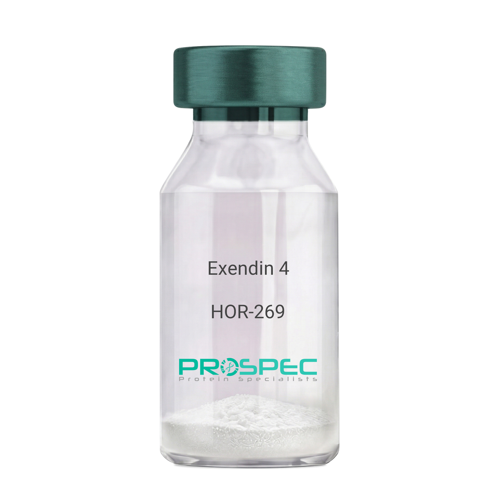
    Exendin 4

    Description:

    Exendin-4 Recombinant

    Product # :

    HOR-269

    Price :

    Quantity :

    Shipping Method :

    Room Temp Icon

    Shipped at Room temp

    Add To Cart

    More Info

    • description
    • source
    • formulation
    • purity
    • biological activity
    • More Info
    • Citations

    Description

    Exendin-4 Recombinant produced in E.Coli is a non-glycosylated polypeptide chain containing 39 amino acids and having a molecular mass of approximately 4.2kDa. Exendin-4 is purified by proprietary chromatographic techniques.

    Source

    Escherichia Coli.

    Formulation

    Lyophilized from a 0.2μm filtered concentrated solution in PBS, pH 7.4.

    Purity

    Greater than 96.0% as determined by:
    (a) Analysis by RP-HPLC.
    (b) Analysis by SDS-PAGE.

    Biological Activity

    1. Regulates Glucose levels rapidly
    2. Reduces Insulin resistance 
    3. Reduces Glucagon

    4. Stimulates beta cell growth production.

    Citations

    More Info

    • Introduction

      Exedin-4 Recombinant is a like peptide-1 receptor agonist. The native hormone is produced in the gut of Gila monster Heloderma suspectrum (a type of reptile found in the desert) that stimulates production without causing threateningly low blood sugar, which can occur after using some anti-diabetes products. Recently, researchers used extracted saliva from gila monsters to create an unprecedented breakthrough in Diabetes Type 2 treatment. Unlike other products taken for type 2 diabetes, Exedin-4 has not been linked with weight gain and actually resulted in weight loss, according to the researchers. Exedin-4 enhances glucose-dependant secretion, suppresses inappropriately elevated secretion and slows gastric emptying in vivo. It also promotes B-cell proliferation and neogenesis in vitro and in animal models. Exedin-4 stimulates an increase in acinar cAMP, without stimulating the release of amylase.

    • Physical Appearance

      Sterile Filtered White lyophilized (freeze-dried) powder.

    • Stability

      Lyophilized Exendin Recombinant although stable at room temperature for 3 weeks, should be stored desiccated below -18°C. Upon reconstitution Exendin-4 should be stored at 4°C between 2-7 days and for future use below -18°C. For long term storage it is recommended to add a carrier protein (0.1% HSA or BSA).Please prevent freeze-thaw cycles.

    • Solubility

      It is recommended to reconstitute the lyophilized Exendin Recombinant in sterile 18MΩ-cm H2O not less than 100 µg/ml, which can then be further diluted to other aqueous solutions.

    • Amino Acid Sequence

      HGEGTFTSDL SKQMEEEAVR LFIEWLKNGG PSSGAPPPS.

    • Background

      What is the molecular weight/Mw of EXENDIN 4 Protein?
      EXENDIN 4 Protein has a total Mw of 4.2kDa.

      What is the source or expression system of EXENDIN 4 Protein?
      Escherichia Coli.

      What is the Purity of EXENDIN 4 Protein?
      EXENDIN 4 Protein is > 96% pure as determined by SDS-PAGE.

      What is the Biological Activity of EXENDIN 4 Protein?
      1. Regulates Glucose levels rapidly
      2. Reduces Insulin resistance
      3. Reduces Glucagon
      4. Stimulates beta cell growth production.

      What is the amino acid sequence of EXENDIN 4 Protein?
      HGEGTFTSDL SKQMEEEAVR LFIEWLKNGG PSSGAPPPS

      What applications can EXENDIN 4 Protein be used in?
      EXENDIN 4 Protein can probably be used in western blot, ELISA and Lateral Flow.

      What is the endotoxin level for EXENDIN 4 Protein?
      The endotoxin level is minimal, EXENDIN 4 Protein was purified using conventional chromatography techniques.

      Who supplies recombinant EXENDIN 4 that is in stock and available to order?
      ProSpec has the protein in stock and available for immediate delivery.

      Where can I order bulk mg quantities of EXENDIN 4?
      ProSpec supplies bulk mg quantities at affordable prices that meets research and academic budgets.

    ProSpec's products are furnished for LABORATORY RESEARCH USE ONLY. The product may not be used as drugs, agricultural or pesticidal products, food additives or household chemicals.

    Exendin 4
  • View Data Sheet

    Name :

    FGF 9 Human

    Description:

    Fibroblast Growth Factor-9 Human Recombinant

    GAF (Glia-activating factor), HBGF-9, MGC119914, MGC119915, FGF-9.

    Product # :

    CYT-415

    Price :

    Quantity :

    Shipping Method :

    Room Temp Icon

    Shipped at Room temp

    Add To Cart

    More Info

    • description
    • source
    • formulation
    • purity
    • biological activity
    • More Info
    • Citations

    Description

    Fibroblast Growth Factor-9 Human Recombinant produced in E.coli is a single, non-glycosylated, polypeptide chain containing 207 amino acids and having a molecular mass of 23.4 kDa. The FGF-9 is purified by proprietary chromatographic techniques.

    Source

    Escherichia coli.

    Formulation

    The sterile protein powder is lyophilized from 1mg/ml solution containing 1xPBS.

    Purity

    Greater than 95.0% as determined by RP-HPLC and SDS-PAGE.

    Biological Activity

    The ED50, calculated by the dose-dependant proliferation of BAF3 cells expressing FGF receptors (measured by 3H-thymidine uptake) is <0.5ng/ml, corresponding to a specific activity of 2,000,000 Units/mg.

    Citations

    More Info

    • Introduction

      The human FGF-9 cDNA encodes a 208 amino acid residue protein that contains a single, potential N-linked glycosylation site. The native protein is glycosylated and is efficiently secreted after synthesis, although FGF -9 lacks a typical secretion signal. Rat and mouse FGF-9 show a very high homology to human FGF-9. The transcripts for FGF-9 have been found in brain and in kidney tissue. Fibroblast Growth Factor-9 is a member of the fibroblast growth factor (FGF) family. FGF family members possess broad mitogenic and cell survival activities, and are involved in a variety of biological processes, including embryonic development, cell growth, morphogenesis, tissue repair, tumor growth and invasion. FGF9 was isolated as a secreted factor that exhibits a growth-stimulating effect on cultured glial cells. In nervous system, this protein is produced mainly by neurons and may be important for glial cell development. Expression of the mouse homolog of this gene was found to be dependent on Sonic hedgehog (Shh) signaling. Mice lacking the homolog gene displayed a male-to-female sex reversal phenotype, which suggested a role in testicular embryogenesis Fibroblast Growth Factor 9 may have a role in glial cell growth and differentiation during development, gliosis during repair and regeneration of brain tissue after damage, differentiation and survival of neuronal cells, and growth stimulation of glial tumors.

    • Synonyms

      GAF (Glia-activating factor), HBGF-9, MGC119914, MGC119915, FGF-9.

    • Physical Appearance

      Sterile Filtered white lyophilized powder.

    • Stability

      Lyophilized Fibroblast Growth Factor 9 Human Recombinant although stable at room temperature for 3 weeks, should be stored desiccated below -18°C. Upon reconstitution FGF-9 should be stored at 4°C between 2-7 days and for future use below -18°C.For long term storage it is recommended to add a carrier protein (0.1% HSA or BSA).Please prevent freeze-thaw cycles.

    • Solubility

      It is recommended to reconstitute the lyophilized Fibroblast Growth Factor-9 Human Recombinant sterile 18M-cm H2O not less than 100µg/ml, which can then be further diluted to other aqueous solutions.

    • Amino Acid Sequence

      APLGEVGNYF GVQDAVPFGN VPVLPVDSPV LLSDHLGQSE AGGLPRGPAV
      TDLDHLKGIL RRRQLYCRTG FHLEIFPNGT IQGTRKDHSR FGILEFISIA
      VGLVSIRGVD SGLYLGMNEK GELYGSEKLT QECVFREQFE ENWYNTYSSN
      LYKHVDTGRR YYVALNKDGT PREGTRTKRH QKFTHFLPRP VDPDKVPELY
      KDILSQS.

    • Background

      What is the molecular weight/Mw of FGF9 Protein?
      FGF9 Protein has a total Mw of 23.4kDa.

      What is the source or expression system of FGF9 Protein?
      Escherichia Coli.

      What is the Purity of FGF9 Protein?
      FGF9 Protein is >95% pure as determined by SDS-PAGE.

      What is the Biological Activity of FGF9 Protein?
      The ED50, calculated by the dose-dependant proliferation of BAF3 cells expressing FGF receptors (measured by 3H-thymidine uptake) is <0.5ng/ml, corresponding to a specific activity of 2,000,000 Units/mg.

      What is the amino acid sequence of FGF9 Protein?
      APLGEVGNYF GVQDAVPFGN VPVLPVDSPV LLSDHLGQSE AGGLPRGPAV
      TDLDHLKGIL RRRQLYCRTG FHLEIFPNGT IQGTRKDHSR FGILEFISIA
      VGLVSIRGVD SGLYLGMNEK GELYGSEKLT QECVFREQFE ENWYNTYSSN
      LYKHVDTGRR YYVALNKDGT PREGTRTKRH QKFTHFLPRP VDPDKVPELY
      KDILSQS.

      What applications can FGF9 Protein be used in?
      FGF9 Protein can probably be used in western blot, ELISA and Lateral Flow.

      What is the endotoxin level for FGF9 Protein?
      The endotoxin level is minimal, FGF9 Protein was purified using conventional chromatography techniques.

    ProSpec's products are furnished for LABORATORY RESEARCH USE ONLY. The product may not be used as drugs, agricultural or pesticidal products, food additives or household chemicals.

    Fgf9 Human
  • View Data Sheet

    Name :

    CASQ2 Antibody

    Description:

    Calsequestrin-2, Mouse Anti Human

    PDIB2, CASQ2, Calsequestrin-2, Calsequestrin cardiac muscle isoform, FLJ26321, FLJ93514.

    Product # :

    ANT-045

    Price :

    Quantity :

    Shipping Method :

    Ice Icon

    Shipped with Ice Packs

    Add To Cart

    More Info

    • formulation
    • More Info
    • Citations

    Formulation

    1mg/ml containing PBS, pH-7.4, & 0.02% Sodium Azide and 10% Glycerol.

    Citations

    More Info

    • Introduction

      CASQ2 belongs to the calsequestrin family and is localized to the sarcoplasmic reticulum in cardiac and slow skeletal muscle cells. CASQ2 is a calcium binding protein that stores calcium for muscle function. The discharge of calcium bound to CASQ2 through a calcium release channel activates muscle contraction. CASQ2 binds 40 to 50 moles of calcium. CASQ2 mutations result in stress-induced polymorphic ventricular tachycardia, also called catecholaminergic polymorphic ventricular tachycardia 2 which is known fir its bidirectional ventricular tachycardia that causes cardiac arrest.

    • Synonyms

      PDIB2, CASQ2, Calsequestrin-2, Calsequestrin cardiac muscle isoform, FLJ26321, FLJ93514.

    • Immunogen

      Anti-human CASQ2 mAb, is derived from hybridization of mouse FO myeloma cells with spleen cells from BALB/c mice immunized with recombinant human CASQ2 amino acids 20-399 purified from E. coli.

    • Ig Subclass

      Mouse IgG1 heavy chain and k light chain.

    • Clone

      PAT4E10AT.

    • Applications

      CASQ2 antibody has been tested by ELISA and Western blot analysis to assure specificity and reactivity. Since application varies, however, each investigation should be titrated by the reagent to obtain optimal results. Recommended dilution range for Western blot analysis is 1:1000.
      Recommended starting dilution is 1:1000.

    • Type

      Mouse Anti Human Monoclonal.

    • Storage Procedures

      For periods up to 1 month store at 4°C, for longer periods of time, store at -20°C. Prevent freeze thaw cycles.

    • Purification Method

      CASQ2 antibody was purified from mouse ascitic fluids by protein-G affinity chromatography.

    ProSpec's products are furnished for LABORATORY RESEARCH USE ONLY. The product may not be used as drugs, agricultural or pesticidal products, food additives or household chemicals.

    Casq2 Antibody
  • 1
  • …
  • 23
  • 24
  • 25
  • 26
  • 27
Your browser does not support the video tag.

Products

  • Cytokines
  • Growth Factors
  • Chemokines
  • CD Antigens
  • Neurotrophins
  • Hormones
  • Enzymes
  • Viral Antigens
  • Recombinant Proteins
  • Natural Proteins
  • Monoclonal Antibodies
  • Polyclonal Antibodies
  • Thermal Packaging

About

  • Company
  • Founder
  • Contribution
  • Contact Us
  • My Account

Ordering

  • Ordering Method
  • Ordering - Credit Card
  • Ordering PO
  • Shipping Fees
  • FAQ's

Legal

  • Terms & Conditions
  • Privacy
  • Site Map
  • info@prospecbio.com
  • Facebook
  • Instagram
  • Linkedin Profile

© 2026 ProSpec-Tany TechnoGene Ltd. All Rights Reserved.

  • Choosing a selection results in a full page refresh.
  • Opens in a new window.