Search results
1000 results found for “beta lactamase”
Name
Description
Product #
Price
Quantity
Shipping Method
- View Data Sheet
Name :
Alkaline Phosphatase BovineDescription:
Alkaline Phosphatase Bovine Intestinal
EC 3.1.3.1, IAP, AP, ALPI, Intestinal-type alkaline phosphatase, Intestinal alkaline phosphatase.
Product # :
ENZ-322Price :
Quantity :
Shipping Method :
Shipped with Ice Packs
More Info
- description
- source
- formulation
- purity
- biological activity
- More Info
- Citations
Description
The Alkaline Phosphatase is purified by affinity chromatography, which results in an enzyme of high specific activity and purity. Alkaline Phosphatase is Dimeric protein having a molecular weight of 140 kDa, one Zn++ ion is tightly bound to each subunit, and another less tightly bound is involved in the catalytic reaction. Mg++ stimulates the catalysis. The binding site for Mg++ is different to that of Zn++, but will be occupied by excess Zn++ followed by loss of enzyme activity.
Source
Calf Intestine.
Formulation
50% glycerol, 5mM MgCl2, 0.1mM ZnCl2 and 5mM TRIS, pH 7.0.
Purity
95% pure by Gel Filtration.
Biological Activity
>1500 U/mg (pH 9.6), 25°C and 0.025M glycin, 10% glycerol as buffer.
Citations
More Info
-
Synonyms
EC 3.1.3.1, IAP, AP, ALPI, Intestinal-type alkaline phosphatase, Intestinal alkaline phosphatase.
-
Physical Appearance
Sterile Filtered clear colorless solution.
-
Stability
AP should be stored at 4°C. Please Do-Not freeze.
ProSpec's products are furnished for LABORATORY RESEARCH USE ONLY. The product may not be used as drugs, agricultural or pesticidal products, food additives or household chemicals.
- View Data Sheet
Name :
BDH2 HumanDescription:
3-Hydroxybutyrate Dehydrogenase, Type 2 Human Recombinant
3-hydroxybutyrate dehydrogenase type 2, FLJ13261, PRO20933, SDR15C1, UCPA-OR, UNQ6308, dehydrogenase/reductase (SDR family) member 6, Oxidoreductase UCPA, DHRS6, R-beta-hydroxybutyrate dehydrogenase, EFA6R, EC 1.1.1.30.
Product # :
ENZ-060Price :
Quantity :
Shipping Method :
Shipped with Ice Packs
More Info
- description
- source
- formulation
- purity
- More Info
- Citations
Description
BDH2 produced in E.Coli is a single, non-glycosylated polypeptide chain containing 265 amino acids (1-245a.a.) and having a molecular mass of 28.8kDa.BDH2 is fused to a 20 amino acid His-tag at N-terminus & purified by proprietary chromatographic techniques.
Source
Escherichia Coli.
Formulation
The BDH2 protein solution (1mg/1ml) is formulated in 20mM Tris-HCl buffer (pH8.0) 0.1M NaCl and 10% glycerol.
Purity
Greater than 95% as determined by SDS-PAGE.
Citations
More Info
-
Introduction
BDH2 is a member of the short-chain dehydrogenases/reductases (SDR) family. BDH2 protein has a significant part in the peripheral utilization of 3-hydroxybutyrate. BDH2 can convert high levels of circulating 3-hydroxybutyrate into acetoacetate due to cytoplasmic localization in high ratio of oxidized NAD+, the NAD+ dependence and the kinetic parameters.
-
Synonyms
3-hydroxybutyrate dehydrogenase type 2, FLJ13261, PRO20933, SDR15C1, UCPA-OR, UNQ6308, dehydrogenase/reductase (SDR family) member 6, Oxidoreductase UCPA, DHRS6, R-beta-hydroxybutyrate dehydrogenase, EFA6R, EC 1.1.1.30.
-
Physical Appearance
Sterile Filtered clear solution.
-
Stability
Store at 4°C if entire vial will be used within 2-4 weeks. Store, frozen at -20°C for longer periods of time.Please avoid freeze thaw cycles.
-
Amino Acid Sequence
MGSSHHHHHH SSGLVPRGSH MGRLDGKVII LTAAAQGIGQ AAALAFAREG AKVIATDINE SKLQELEKYP GIQTRVLDVT KKKQIDQFAN EVERLDVLFN VAGFVHHGTV LDCEEKDWDF SMNLNVRSMY LMIKAFLPKM LAQKSGNIIN MSSVASSVKG VVNRCVYSTT KAAVIGLTKS VAADFIQQGI RCNCVCPGTV DTPSLQERIQ ARGNPEEARN DFLKRQKTGR FATAEEIAML CVYLASDESA YVTGNPVIID GGWSL
ProSpec's products are furnished for LABORATORY RESEARCH USE ONLY. The product may not be used as drugs, agricultural or pesticidal products, food additives or household chemicals.
- View Data Sheet
Name :
P4HB HumanDescription:
Prolyl 4-Hydroxylase Beta Human Recombinant
P4Hbeta, PDI, PDIA1, PHD, PO4DB, PO4HB, ERBA2L.
Product # :
ENZ-252Price :
Quantity :
Shipping Method :
Shipped with Ice Packs
More Info
- description
- source
- formulation
- purity
- More Info
- Citations
Description
P4HB Human Recombinant produced in E.Coli is a single, non-glycosylated polypeptide chain containing 512 amino acids (18-508 a.a.) and having a molecular mass of 57.5 kDa. The P4HB is fused to a 21 amino acid His Tag and purified by conventional chromatography.
Source
Escherichia Coli.
Formulation
The P4HB 1mg/ml protein solution contains 20mM Tris-HCl pH-8, and 10% glycerol.
Purity
Greater than 90.0% as determined by SDS-PAGE.
Citations
More Info
-
Introduction
P4HB is a multifunctional and highly abundant enzyme that is part of the protein disulfide isomerase family. When present as a tetramer consisting of two alpha and two beta subunits, P4HB has a role in hydroxylation of prolyl residues in preprocollagen. P4HB is a disulfide isomerase containing two thioredoxin domains that catalyze the formation, breakage and rearrangement of disulfide bonds.
-
Synonyms
P4Hbeta, PDI, PDIA1, PHD, PO4DB, PO4HB, ERBA2L.
-
Physical Appearance
Sterile Filtered colorless solution.
-
Stability
Store at 4°C if entire vial will be used within 2-4 weeks. Store, frozen at -20°C for longer periods of time. For long term storage it is recommended to add a carrier protein (0.1% HSA or BSA). Avoid multiple freeze-thaw cycles.
-
Amino Acid Sequence
MGSSHHHHHH SSGLVPRGSH MDAPEEEDHV LVLRKSNFAE ALAAHKYLLV EFYAPWCGHC KALAPEYAKA AGKLKAEGSE IRLAKVDATE ESDLAQQYGV RGYPTIKFFR NGDTASPKEY TAGREADDIV NWLKKRTGPA ATTLPDGAAA ESLVESSEVA VIGFFKDVES DSAKQFLQAA
EAIDDIPFGI TSNSDVFSKY QLDKDGVVLF KKFDEGRNNF EGEVTKENLL DFIKHNQLPL VIEFTEQTAP KIFGGEIKTH ILLFLPKSVS DYDGKLSNFK TAAESFKGKI LFIFIDSDHT DNQRILEFFG LKKEECPAVR LITLEEEMTK YKPESEELTA ERITEFCHRF LEGKIKPHLM
SQELPEDWDK QPVKVLVGKN FEDVAFDEKK NVFVEFYAPW CGHCKQLAPI WDKLGETYKD HENIVIAKMD STANEVEAVK VHSFPTLKFF PASADRTVID YNGERTLDGF KKFLESGGQD GAGDDDDLED LEEAEEPDME EDDDQKAVKD EL.
ProSpec's products are furnished for LABORATORY RESEARCH USE ONLY. The product may not be used as drugs, agricultural or pesticidal products, food additives or household chemicals.
- View Data Sheet
Name :
Anaplasma p44Description:
Anaplasma phagocytophilum p44 Recombinant
Product # :
PRO-2566Price :
Quantity :
Shipping Method :
Shipped with Ice Packs
More Info
- description
- source
- formulation
- purity
- More Info
- Citations
Description
Recombinant Anaplasma p44 produced in E.Coli is a single, non-glycosylated polypeptide chain having a molecular mass of 49kDa. Anaplasma p44 is expressed with a -10x His tag at N-terminus and purified by proprietary chromatographic techniques.
Source
Escherichia Coli.
Formulation
Anaplasma p44 is supplied at a 20mM HEPES buffer pH-8.0, 250mM NaCl and 6M Urea.
Purity
Greater than 95% as determined by SDS-PAGE.
Citations
More Info
-
Introduction
Anaplasma p44 which belongs to the outer membrane antigen superfamily (OMP1/Msp2/p44), is a serodiagnostic antigen for HGA. Anaplasma p44 allows the bacterium to adhere to the host cell and prevents host immune surveillance.
-
Physical Appearance
Sterile Filtered solution.
-
Stability
Store at 4°C if entire vial will be used within 2-4 weeks. Store, frozen at -20°C for longer periods of time. Avoid multiple freeze-thaw cycles.
-
Immunological Functions
1. Binds IgG- and IgM-type human antibodies. 2. Immunodot test with positive/negative samples.
ProSpec's products are furnished for LABORATORY RESEARCH USE ONLY. The product may not be used as drugs, agricultural or pesticidal products, food additives or household chemicals.
- View Data Sheet
Name :
PMSGDescription:
Pregnant Mare Serum Gonadotropin
Product # :
HOR-272Price :
Quantity :
Shipping Method :
Shipped at Room temp
More Info
- description
- source
- formulation
- More Info
- Citations
Description
PMSG is a complex glycoprotein obtained from the serum of pregnant mares. This 43-63 kda protein is capable of supplementing and being substituted for the follicle stimulating and interstitial cell-stimulating hormone of the anterior pituitary gland in both the male and female. Thus PMSG-Intervet stimulates development of the ovarian follicle in the female.
Source
Serum of pregnant mares.
Formulation
The PMSG was lyophilized with no additives.
Citations
More Info
-
Introduction
PMSG Hormone is a well know used hormone together with progestogen to increase ovulation just before to artificial insemination. PMSG hormone is a placental glycoprotein produced from the serum of pregnant mares. PMSG comprises of an alfa subunit and a beta subunit. PMSG hormone is secreted from endometrial cups within the pregnant mare uterus aging from 40 to 130 days into their maturation, and once extracted, it can been used to promote artificially estrus in female animals. These assemblies produce PMSG hormone to induce mare's ovarian and repsouctive structures. PMSG can induce the growth of follicles by ovaries and results in ovulatation. PMSG hormone has an about a 4 day half-life of bioactivity in species other than horses. The extended biological activity can cause ovarian stimulation and ovulation. However, PMSG use alone often causes cystic ovarian disease because of the unrestrained ovarian stimulation and due to the sugar molecules which decrease clearance of the hormone. PMSG is more likely to be used than other pituitary hormones due to the extended circulatory half-life. PMSG solely exhibits luteinizing hormone like activity, however in other animal classes it has FSH & LH like activity.
-
Physical Appearance
Sterile Filtered White lyophilized (freeze-dried) powder.
-
Stability
Lyophilized PMSG although stable at room temperature for 3 weeks, should be stored between 2-8°C.
-
Solubility
It is recommended to reconstitute the lyophilized PMSG in sterile 18M-cm H2O at a concentration of 1000 IU/ml, which can then be further diluted to other aqueous solutions.
ProSpec's products are furnished for LABORATORY RESEARCH USE ONLY. The product may not be used as drugs, agricultural or pesticidal products, food additives or household chemicals.
- View Data Sheet
Name :
PITPNB HumanDescription:
Phosphatidylinositol Transfer Protein Beta Human Recombinant
Phosphatidylinositol transfer protein beta isoform, PI-TP-beta, PtdIns transfer protein beta, PtdInsTP beta, PITPNB, VIB1B, PtdInsTP.
Product # :
PRO-003Price :
Quantity :
Shipping Method :
Shipped with Ice Packs
More Info
- description
- source
- formulation
- purity
- More Info
- Citations
Description
PITPNB Human Recombinant fused with a 20 amino acid His tag at N-terminus produced in E.Coli is a single, non-glycosylated, polypeptide chain containing 291 amino acids (1-271 a.a.) and having a molecular mass of 33.7kDa. The PITPNB is purified by proprietary chromatographic techniques.
Source
Escherichia Coli.
Formulation
The PITPNB solution (1 mg/ml) contains 20mM Tris-HCl buffer (pH 8.0), 1mM DTT and 10% glycerol.
Purity
Greater than 95.0% as determined by SDS-PAGE.
Citations
More Info
-
Introduction
Phosphatidylinositol transfer protein beta isoform (PITPNB) is found in the cytoplasm, where it catalyzes the transfer of phosphatidylinositol (PI) and phosphatidylcholine (PC) between membranes. PITPNB mobilizes PI from the endoplasmic reticulum and regulates its release from stored vesicles in the Golgi network. PITPNB is extensively expressed in various tissues.
-
Synonyms
Phosphatidylinositol transfer protein beta isoform, PI-TP-beta, PtdIns transfer protein beta, PtdInsTP beta, PITPNB, VIB1B, PtdInsTP.
-
Physical Appearance
Sterile Filtered colorless solution.
-
Stability
Store at 4°C if entire vial will be used within 2-4 weeks. Store, frozen at -20°C for longer periods of time. For long term storage it is recommended to add a carrier protein (0.1% HSA or BSA).Avoid multiple freeze-thaw cycles.
-
Amino Acid Sequence
MGSSHHHHHH SSGLVPRGSH MVLIKEFRVV LPCSVQEYQV GQLYSVAEAS KNETGGGEGI EVLKNEPYEK DGEKGQYTHK IYHLKSKVPA FVRMIAPEGS LVFHEKAWNA YPYCRTIVTN EYMKDDFFIK IETWHKPDLG TLENVHGLDP NTWKTVEIVH IDIADRSQVE PADYKADEDP ALFQSVKTKR GPLGPNWKKE LANSPDCPQM CAYKLVTIKF KWWGLQSKVE NFIQKQEKRI FTNFHRQLFC WIDKWIDLTM EDIRRMEDET QKELETMRKR GSVRGTSAAD V.
ProSpec's products are furnished for LABORATORY RESEARCH USE ONLY. The product may not be used as drugs, agricultural or pesticidal products, food additives or household chemicals.
- View Data Sheet
Name :
Chlamydia W5-W6Description:
Chlamydia Trachomatis W5-W6 Recombinant
Product # :
CHT-005Price :
Quantity :
Shipping Method :
Shipped with Ice Packs
More Info
- description
- source
- formulation
- purity
- More Info
- Citations
Description
The E.coli derived recombinant 6xHis fusion at C-terminus protein contains Chlamydia Trachomatis MOMP protein epitopes, 252-398 amino acids.
Source
Escherichia Coli.
Formulation
20mM Tris-HCl, pH 7.2, 50% glycerol and 1.5M urea (1mg/1ml).
Purity
Protein is >90% pure as determined by SDS-PAGE.
Citations
More Info
-
Introduction
Chlamydia is a common term for infection with any bacterium belonging to the phylum Chlamydiae. This term derives from the name of the bacterial genus Chlamydiain the family Chlamydiaceae, order Chlamydiales, class and phylum Chlamydiae. There are two genera in Chlamydiaceae: Chlamydia and Chlamydophila. The genus Chlamydia includes three species: C. trachomatis, C. muridarum, and C. suis.
-
Stability
Chlamydia W5-W6 protein although stable at 4°C for 1 week, should be stored below -18°C. Please prevent freeze thaw cycles.
-
Applications
Elisa, WB and Flow-Through.
-
Specificity
Immunoreactive with sera of Chlamydia Trachomatis infected individuals with minimum specificity problems.
-
Purification Method
Chlamydia W5-W6 protein was purified by proprietary chromatographic technique.
ProSpec's products are furnished for LABORATORY RESEARCH USE ONLY. The product may not be used as drugs, agricultural or pesticidal products, food additives or household chemicals.
- View Data Sheet
Name :
MMP 3 HumanDescription:
Matrix Metalloproteinase-3 Human Recombinant
CHDS6, MMP-3, SL-1, STMY, STMY1, STR1, Stromelysin-1, Matrix metalloproteinase-3, Transin-1, MMP3.
Product # :
ENZ-774Price :
Quantity :
Shipping Method :
Shipped with Ice Packs
More Info
- description
- source
- formulation
- purity
- More Info
- Citations
Description
MMP 3 Human Recombinant produced in E.coli is a single, non-glycosylated polypeptide chain containing 401 amino acids (100-477a.a) and having a molecular mass of 45.2kDa. MMP 3 is fused to a 23 amino acid His-tag at N-terminus & purified by proprietary chromatographic techniques.
Source
Escherichia Coli.
Formulation
The MMP 3 solution (0.25mg/ml) contains 20mM Tris-HCl buffer (pH 8.0), 0.15M NaCl, and 10% glycerol.
Purity
Greater than 90% as determined by SDS-PAGE.
Citations
More Info
-
Introduction
MMP-3 enzyme is also known as Stromelysin-1or as Transin-1 which hydrolyzes natural collagen at physiological pH and temperature. It dissolves the intervertebral nucleus pulposus and annulus fibrosus of Herniated Lumbar Intervertebral Disk . MMP-3 hydrolyzes components of the extracellular matrix like proteoglycan, laminin, fibronectin, gelatin and collagen types III, IV and IX. It also activates pro-MMP-9 and pro-MMP-8 and superactivates plasmin activated MMP-1. MMP-3 is secreted as a latent proenzyme and is activated by a variety of proteinases, e.g. plasmin, trypsin, chymotrypsin, cathepsin G or human neutrophil elastase. MMP-3 was found to be capable of activating the precursor of IL1-beta.
-
Synonyms
CHDS6, MMP-3, SL-1, STMY, STMY1, STR1, Stromelysin-1, Matrix metalloproteinase-3, Transin-1, MMP3.
-
Physical Appearance
Sterile Filtered colorless solution.
-
Stability
Store at 4°C if entire vial will be used within 2-4 weeks. Store, frozen at -20°C for longer periods of time. For long term storage it is recommended to add a carrier protein (0.1% HSA or BSA). Avoid multiple freeze-thaw cycles.
-
Amino Acid Sequence
MGSSHHHHHH SSGLVPRGSH MGSFRTFPGI PKWRKTHLTY RIVNYTPDLP KDAVDSAVEK ALKVWEEVTP LTFSRLYEGE ADIMISFAVR EHGDFYPFDG PGNVLAHAYA PGPGINGDAH FDDDEQWTKD TTGTNLFLVA AHEIGHSLGL FHSANTEALM YPLYHSLTDL TRFRLSQDDI NGIQSLYGPP PDSPETPLVP TEPVPPEPGT PANCDPALSF DAVSTLRGEI LIFKDRHFWR KSLRKLEPEL HLISSFWPSL PSGVDAAYEV TSKDLVFIFK GNQFWAIRGN EVRAGYPRGI HTLGFPPTVR KIDAAISDKE KNKTYFFVED KYWRFDEKRN SMEPGFPKQI AEDFPGIDSK IDAVFEEFGF FYFFTGSSQL EFDPNAKKVT HTLKSNSWLN C.
ProSpec's products are furnished for LABORATORY RESEARCH USE ONLY. The product may not be used as drugs, agricultural or pesticidal products, food additives or household chemicals.
- View Data Sheet
Name :
Thymosin beta 4Description:
Thymosin β4
Thymosin beta-4. TB500, TB-500
Product # :
HOR-275Price :
Quantity :
Shipping Method :
Shipped at Room temp
More Info
- description
- formulation
- purity
- More Info
- Citations
Description
Thymosin b4 is a 43 amino acid peptide which is regarded as the main intracellular G-actin sequestering peptide. It has a molecular weight of 4963.55 Da, and its molecular formula is: C212H350N56O78S1. Extracellular Thymosin b4 may contribute to physiological processes such as angiogenesis, wound healing, and regulation of inflammation.
Formulation
The protein (1mg/ml) was lyophilized with no additives.
Purity
Greater than 98.0% as determined by RP-HPLC.
Citations
More Info
-
Introduction
Thymosin is a hormone secreted from the thymus. Its primary function is to stimulate the production of T cells, which are an important part of the immune system. Thymosin also assists in the development of B cells to plasma cells to produce antibodies. The predominant form of thymosin, thymosin b4, is a member of a highly conserved family of actin monomer-sequestering proteins. b-thymosins are the primary regulators of unpolymerized actin, and are essential for maintaining the small cytoplasmic pool of free G-actin monomers required for rapid filament elongation and allowing for the flux of monomers between the thymosin-bound pool and F-actin.
-
Synonyms
Thymosin beta-4. TB500, TB-500
-
Physical Appearance
Sterile Filtered White lyophilized (freeze-dried) powder.
-
Stability
Lyophilized Thymosin b4 although stable at room temperature for 3 weeks, should be stored desiccated below -18°C. Upon reconstitution T beta 4 should be stored at 4°C between 2-7 days and for future use below -18°C.For long term storage it is recommended to add a carrier protein (0.1% HSA or BSA).Please prevent freeze-thaw cycles.
-
Solubility
It is recommended to reconstitute the lyophilized Thymosin beta-4 in sterile 18MΩ-cm H2O not less than 100 µg/ml, which can then be further diluted to other aqueous solutions.
-
Amino Acid Sequence
Thymosin b4 has an a.a. sequence of Ac-Ser-Asp-Lys-Pro-Asp-Met-Ala-Glu-Ile-Glu-Lys-Phe-Asp-Lys-Ser-Lys-Leu-Lys-Lys-Thr-Glu-Thr-Gln-Glu-Lys-Asn-Pro-Leu-Pro-Ser-Lys-Glu-Thr-Ile-Glu-Gln-Glu-Lys-Gln-Ala-Gly-Glu-Ser-OH.
-
Background
What is the molecular weight/Mw of THYMOSIN BETA 4 Protein?
THYMOSIN BETA 4 Protein has a total Mw of 4.96kDa.
What is the Purity of THYMOSIN BETA 4 Protein?
THYMOSIN BETA 4 Protein is >98% pure as determined by SDS-PAGE.
What is the Biological Activity of THYMOSIN BETA 4 Protein?
The biological functionality of THYMOSIN BETA 4 Protein will be determined in the future.
What is the amino acid sequence of THYMOSIN BETA 4 Protein?
Thymosin b4 has an a.a. sequence of Ac-Ser-Asp-Lys-Pro-Asp-Met-Ala-Glu-Ile-Glu-Lys-Phe-Asp-Lys-Ser-Lys-Leu-Lys-Lys-Thr-Glu-Thr-Gln-Glu-Lys-Asn-Pro-Leu-Pro-Ser-Lys-Glu-Thr-Ile-Glu-Gln-Glu-Lys-Gln-Ala-Gly-Glu-Ser-OH.
What applications can THYMOSIN BETA 4 Protein be used in?
THYMOSIN BETA 4 Protein can probably be used in western blot, ELISA and Lateral Flow.
What is the endotoxin level for THYMOSIN BETA 4 Protein?
The endotoxin level is minimal, THYMOSIN BETA 4 Protein was purified using conventional chromatography techniques.
Who supplies THYMOSIN BETA 4 that is in stock and available to order?
ProSpec has the protein in stock and available for immediate delivery.
Where can I order bulk mg quantities of THYMOSIN BETA 4?
ProSpec supplies bulk mg quantities at affordable prices that meets research and academic budgets.
ProSpec's products are furnished for LABORATORY RESEARCH USE ONLY. The product may not be used as drugs, agricultural or pesticidal products, food additives or household chemicals.
- View Data Sheet
Name :
TGFB3 (207 a.a.) HumanDescription:
Transforming Growth Factor-Beta 3 (207 a.a.) Human Recombinant
Transforming Growth Factor-beta3, TGFB3, ARVD, FLJ16571, TGF-beta3.
Product # :
CYT-319Price :
Quantity :
Shipping Method :
Shipped with Ice Packs
More Info
- description
- source
- formulation
- More Info
- Citations
Description
TGFB3 Human Recombinant protein encoding amino acids 644-850 and total molecular mass of 50 kDa (Including GST Tag).
Source
Escherichia Coli.
Formulation
100µl purified human TGF Beta 3protein at 100µg/ml in 50mM Tris-Acetate, pH-7.5, 1mM EDTA and 20% Glycerol.
Citations
More Info
-
Introduction
Transforming growth factor betas (TGF Betas) mediate many cell-cell interactions that occur during embryonic development. Three TGF Betas have been identified in mammals. TGF Beta 1, TGF Beta 2 and TGF Beta 3 are each synthesized as precursor proteins that are very similar in that each is cleaved to yield a 112 amino acid polypeptide that remains associated with the latent portion of the molecule.
-
Synonyms
Transforming Growth Factor-beta3, TGFB3, ARVD, FLJ16571, TGF-beta3.
-
Physical Appearance
Sterile Filtered clear solution.
-
Stability
TGF-beta 3 Human Recombinant although stable at 4°C for 1 week, should be stored at -20°C. For long term storage it is recommended to add a carrier protein (0.1% HSA or BSA).Please avoid freeze-thaw cycles.
-
Applications
(a) ELISA.
(b) Western Blotting.
(c) Inhibition Assays.
ProSpec's products are furnished for LABORATORY RESEARCH USE ONLY. The product may not be used as drugs, agricultural or pesticidal products, food additives or household chemicals.
- View Data Sheet
Name :
ATP1B2 Human, Sf9Description:
ATPase Transporting Beta 2 Human Recombinant, Sf9
ATP1B2, AMOG, Sodium/Potassium-Transporting ATPase Beta-2 Chain, Sodium/Potassium-Dependent ATPase Beta-2 Subunit, Na, K-ATPase Beta-2 Polypeptide, Adhesion Molecule On Glia, ATPase Na+/K+ Transporting Subunit Beta 2, Sodium-Potassium ATPase Subunit Beta 2 (Non-Catalytic), Sodium/Potassium-Transporting ATPase Subunit Beta-2, Sodium/Potassium-Dependent ATPase Subunit Beta-2, ATPase, Na+/K+ Transporting, Beta 2 Polypeptide, Sodium Pump Subunit Beta-2, Adhesion Molecule In Glia.
Product # :
PRO-2397Price :
Quantity :
Shipping Method :
Shipped with Ice Packs
More Info
- description
- source
- formulation
- purity
- More Info
- Citations
Description
ATP1B2 Human Recombinant produced in Sf9 Baculovirus cells is a single, glycosylated polypeptide chain containing 232 amino acids (68-290a.a.) and having a molecular mass of 26.4kDa (Molecular size on SDS-PAGE will appear at approximately 28-40 kDa). ATP1B2 is expressed with a 9 amino acid His tag at C-Terminus and purified by proprietary chromatographic techniques.
Source
Sf9, Baculovirus cells.
Formulation
ATP1B2 protein solution (0.5mg/ml) contains Phosphate Buffered Saline (pH 7.4) and 10% glycerol.
Purity
Greater than 90.0% as determined by SDS-PAGE.
Citations
More Info
-
Introduction
ATPaseTransporting Beta 2 (ATP1B2) is the non-catalytic component of the active enzyme, which catalyzes the hydrolysis of ATP coupled with the exchange of Na+ and K+ ions across the plasma membrane. The precise function of the beta-2 subunit is not known. The ATP1B2 protein is composed of 3 subunits: alpha (catalytic), beta and gamma.
-
Synonyms
ATP1B2, AMOG, Sodium/Potassium-Transporting ATPase Beta-2 Chain, Sodium/Potassium-Dependent ATPase Beta-2 Subunit, Na, K-ATPase Beta-2 Polypeptide, Adhesion Molecule On Glia, ATPase Na+/K+ Transporting Subunit Beta 2, Sodium-Potassium ATPase Subunit Beta 2 (Non-Catalytic), Sodium/Potassium-Transporting ATPase Subunit Beta-2, Sodium/Potassium-Dependent ATPase Subunit Beta-2, ATPase, Na+/K+ Transporting, Beta 2 Polypeptide, Sodium Pump Subunit Beta-2, Adhesion Molecule In Glia.
-
Physical Appearance
Sterile Filtered colorless solution.
-
Stability
Store at 4°C if entire vial will be used within 2-4 weeks. Store, frozen at -20°C for longer periods of time. For long term storage it is recommended to add a carrier protein (0.1% HSA or BSA).Avoid multiple freeze-thaw cycles.
-
Amino Acid Sequence
ADPDHTPKYQ DRLATPGLMI RPKTENLDVI VNVSDTESWD QHVQKLNKFL EPYNDSIQAQ KNDVCRPGRY YEQPDNGVLN YPKRACQFNR TQLGNCSGIG DSTHYGYSTG QPCVFIKMNR VINFYAGANQ SMNVTCAGKR DEDAENLGNF VMFPANGNID LMYFPYYGKK FHVNYTQPLV AVKFLNVTPN VEVNVECRIN AANIATDDER DKFAGRVAFK LRINKTHHHH HH.
ProSpec's products are furnished for LABORATORY RESEARCH USE ONLY. The product may not be used as drugs, agricultural or pesticidal products, food additives or household chemicals.
- View Data Sheet
Name :
Borrelia p45Description:
Borrelia Burgdorferi p45 Recombinant
Product # :
BOR-018Price :
Quantity :
Shipping Method :
Shipped with Ice Packs
More Info
- description
- source
- formulation
- purity
- More Info
- Citations
Description
Recombinant Borrelia Burgdorferi p45 produced in SF9 is a glycosylated, polypeptide chain having a calculated molecular mass of 45,259.3 Dalton. Borrelia p45 is expressed with a 10xHis tag at N-terminus and purified by proprietary chromatographic techniques.
Source
Sf9 insect cells.
Formulation
Borrelia p45 is supplied in 20mM HEPES buffer pH-7.6, 250mM NaCl and 20% glycerol.
Purity
Greater than 80.0% as determined by SDS-PAGE.
Citations
More Info
-
Introduction
Borrelia belongs to a genus of bacteria of the spirochete phylum. Borrelia causes borreliosis, which is a zoonotic, vector-borne disease transmitted mainly by ticks and some by lice, depending on the species. Of the 36 known species of Borrelia, 12 are distinguished to cause Lyme disease or borreliosis and are transmitted by ticks. The main Borrelia species causing Lyme disease are Borrelia burgdorferi, Borrelia afzelii, and Borrelia garinii. The Borrelia genus members have a linear chromosome which is about 900 kbp in length as well as an excess of both linear and circular plasmids in the 5-220 kbp size range. The plasmids are atypical, as compared to most bacterial plasmids, since they contain many paralogous sequences, a large number of pseudogenes and, in some cases, essential genes. Moreover, a number of the plasmids have features suggesting that they are prophages.
-
Physical Appearance
Sterile Filtered clear solution.
-
Stability
Store at 4°C if entire vial will be used within 2-4 weeks. Store, frozen at -20°C for longer periods of time. Avoid multiple freeze-thaw cycles.
-
Immunological Functions
1.Immunodot test with Lyme disease positive/negative plasma, suitable for LTT (lymphocyte transformation test). 2.T cell stimulation assays. 3.ELISPOT (enzyme-linked immunospot).
ProSpec's products are furnished for LABORATORY RESEARCH USE ONLY. The product may not be used as drugs, agricultural or pesticidal products, food additives or household chemicals.
- View Data Sheet
Name :
LAT HumanDescription:
Linker for Activation of T Cells Human Recombinant
Linker for activation of T cells, LAT1, 36 kDa phospho-tyrosine adapter protein, p36-38, pp36, linker for activation of T cells transmembrane adaptor, linker for activation of T-cells family member 1.
Product # :
PRO-948Price :
Quantity :
Shipping Method :
Shipped with Ice Packs
More Info
- description
- source
- formulation
- purity
- More Info
- Citations
Description
LAT Human Recombinant produced in E.coli is a single, non-glycosylated polypeptide chain containing 227 amino acids (28-233) and having a molecular mass of 24.4 kDa (Molecular weight on SDS-PAGE will appear higher).LAT is fused to a 21 amino acid His-tag at N-terminus & purified by proprietary chromatographic techniques.
Source
E.coli.
Formulation
The LAT solution (1mg/ml) contains 20mM Tris-HCl buffer (pH 8.0) and 1mM DTT.
Purity
Greater than 95% as determined by SDS-PAGE.
Citations
More Info
-
Introduction
LAT is a vital membrane protein that is phosphorylated by ZAP-70/Syk protein tyrosine kinases succeeding activation of the T-cell antigen receptor (TCR) signal transduction pathway. LAT is expressed in T lymphocytes in interstitial spaces, platelets and megakaryocytes.
-
Synonyms
Linker for activation of T cells, LAT1, 36 kDa phospho-tyrosine adapter protein, p36-38, pp36, linker for activation of T cells transmembrane adaptor, linker for activation of T-cells family member 1.
-
Physical Appearance
Sterile Filtered colorless solution.
-
Stability
Store at 4°C if entire vial will be used within 2-4 weeks. Store, frozen at -20°C for longer periods of time. For long term storage it is recommended to add a carrier protein (0.1% HSA or BSA).Avoid multiple freeze-thaw cycles.
-
Amino Acid Sequence
MGSSHHHHHH SSGLVPRGSH MHCHRLPGSY DSTSSDSLYP RGIQFKRPHT VAPWPPAYPP VTSYPPLSQP DLLPIPRSPQ PLGGSHRTPS SRRDSDGANS VASYENEEPA CEDADEDEDD YHNPGYLVVL PDSTPATSTA APSAPALSTP GIRDSAFSME SIDDYVNVPE SGESAEASLD GSREYVNVSQ ELHPGAAKTE PAALSSQEAE EVEEEGAPDY ENLQELN
ProSpec's products are furnished for LABORATORY RESEARCH USE ONLY. The product may not be used as drugs, agricultural or pesticidal products, food additives or household chemicals.
- View Data Sheet
Name :
Chymotrypsin PorcineDescription:
Alpha Chymotrypsin Porcine
a-chymotrypsin, alpha chymotrypsin.
Product # :
ENZ-1195Price :
Quantity :
Shipping Method :
Shipped at Room temp
More Info
- description
- source
- formulation
- biological activity
- More Info
- Citations
Description
Chymotrypsin purified from porcine pancreas, CAS: 9004-07-3, EC: 3.4.21.1 having a molecular mass of ~25kDa.
Source
Porcine Pancreas.
Formulation
The protein (1mg/ml) was lyophilized with no additives.
Biological Activity
Greater than 1500 USP U/mg.
Citations
More Info
-
Synonyms
a-chymotrypsin, alpha chymotrypsin.
-
Physical Appearance
Sterile Filtered White lyophilized (freeze-dried) powder.
-
Stability
Lyophilized Chymotrypsin although stable at room temperature for 3 weeks, should be stored desiccated below -18°C. Upon reconstitution Chymotrypsin should be stored at 4°C between 2-7 days and for future use below -18°C. For long term storage it is recommended to add a carrier protein (0.1% HSA or BSA). Please prevent freeze-thaw cycles.
-
Solubility
It is recommended to reconstitute the lyophilized Chymotrypsin in sterile 18MΩ-cm H2O not less than 100µg/ml, which can then be further diluted to other aqueous solutions.
-
Background
Porcine chymotrypsin, derived from the pancreas of pigs, stands as a cornerstone in enzymology, serving as a paradigmatic model for understanding proteolytic mechanisms and substrate specificity. Renowned for its catalytic prowess and structural intricacies, porcine chymotrypsin has garnered significant attention from researchers across various scientific disciplines.
The fascination with porcine chymotrypsin stems from its ability to cleave peptide bonds selectively after large hydrophobic amino acids, such as tryptophan, tyrosine, and phenylalanine.
ProSpec's products are furnished for LABORATORY RESEARCH USE ONLY. The product may not be used as drugs, agricultural or pesticidal products, food additives or household chemicals.
- View Data Sheet
Name :
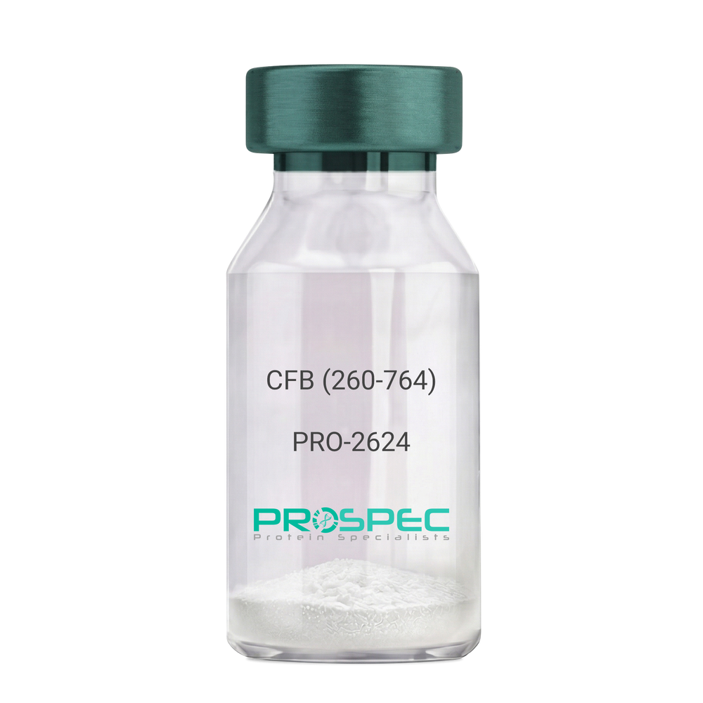
CFB (260-764) Human, Sf9Description:
Complement Factor B (260-764 a.a.) Human Recombinant, Sf9
CFB, C3/C5 convertase, Glycine-rich beta glycoprotein, GBG, PBF2, Properdin factor B, BF, BFD, AHUS4, ARMD14, CFAB, CFBD, FB, FBI12, GBG, H2-Bf.
Product # :
PRO-2624Price :
Quantity :
Shipping Method :
Shipped with Ice Packs
More Info
- description
- source
- formulation
- purity
- More Info
- Citations
Description
CFB Human Recombinant produced in Sf9 Baculovirus cells is a single, glycosylated polypeptide chain containing 514 amino acids (260-764 a.a) and having a molecular mass of 58.1kDa.CFB is fused to an 9 amino acid His-tag at C-terminus & purified by proprietary chromatographic techniques.
Source
Sf9, Baculovirus cells.
Formulation
CFB protein solution (0.5mg/ml) containing Phosphate Buffered Saline (pH 7.4) and 20% glycerol.
Purity
Greater than 90.0% as determined by SDS-PAGE.
Citations
More Info
-
Introduction
Factor B is a protein, build from a single chain that circulates in the blood. Complement factor D cleaves and activates factor B, thus creating a catalytic subunit Bb and noncatalytic chain Ba. Subunit Bb is a serine protease that can bind to C3b and forms alternative pathway C3 convertase. Subunit Ba acts as an inhibitor for proliferation of lymphocytes, when Bb is part of the lymphocytes’ proliferation. The gene that codes for this protein is located on chromosome six.
-
Synonyms
CFB, C3/C5 convertase, Glycine-rich beta glycoprotein, GBG, PBF2, Properdin factor B, BF, BFD, AHUS4, ARMD14, CFAB, CFBD, FB, FBI12, GBG, H2-Bf.
-
Physical Appearance
Sterile Filtered colorless solution.
-
Stability
Store at 4°C if entire vial will be used within 2-4 weeks. Store, frozen at -20°C for longer periods of time. For long term storage it is recommended to add a carrier protein (0.1% HSA or BSA).Avoid multiple freeze-thaw cycles.
-
Amino Acid Sequence
ADPKIVLDPS GSMNIYLVLD GSDSIGASNF TGAKKCLVNL IEKVASYGVK PRYGLVTYAT
YPKIWVKVSE ADSSNADWVT KQLNEINYED HKLKSGTNTK KALQAVYSMM SWPDDVPPEG
WNRTRHVIIL MTDGLHNMGG DPITVIDEIR DLLYIGKDRK NPREDYLDVY VFGVGPLVNQ
VNINALASKK DNEQHVFKVK DMENLEDVFY QMIDESQSLS LCGMVWEHRK GTDYHKQPWQ
AKISVIRPSK GHESCMGAVV SEYFVLTAAH CFTVDDKEHS IKVSVGGEKR DLEIEVVLFH
PNYNINGKKE AGIPEFYDYD VALIKLKNKL KYGQTIRPIC LPCTEGTTRA LRLPPTTTCQ
QQKEELLPAQ DIKALFVSEE EKKLTRKEVY IKNGDKKGSC ERDAQYAPGY DKVKDISEVV
TPRFLCTGGV SPYADPNTCR GDSGGPLIVH KRSRFIQVGV ISWGVVDVCK NQKRQKQVPA HARDFHINLF QVLPWLKEKL QDEDLGFLHH HHHH
ProSpec's products are furnished for LABORATORY RESEARCH USE ONLY. The product may not be used as drugs, agricultural or pesticidal products, food additives or household chemicals.
- View Data Sheet
Name :
UNG E.Coli ActiveDescription:
Recombinant E.Coli Uracil DNA Glycosylase, Active
UDG, b2580, JW2564, EC 3.2.2.27, DGU, UNG15, HIGM5, Uracil-DNA Glycosylase 1, EC 3.2.2, HIGM4, UNG2.
Product # :
ENZ-1182Price :
Quantity :
Shipping Method :
Shipped with Ice Packs
More Info
- description
- source
- formulation
- purity
- More Info
- Citations
Description
UNG E.Coli Recombinant produced in E.Coli is a single, non-glycosylated polypeptide UNG is purified by proprietary chromatographic techniques.
Source
Escherichia Coli.
Formulation
UNG protein solution (5U/ul) containing 10mM Tris-HCl (25℃, pH 7.4), 50mM KCl, 0.1 mM EDTA, 1mM DTT, 0.1mg/ml BSA & 50% glycerol.
Purity
Greater than 97.0% as determined by SDS-PAGE.
Citations
More Info
-
Introduction
Uracil DNA glycosylase (UDG), or uracil-DNA glycosylase 1, is a crucial enzyme found in all life forms, involved in repairing damaged DNA by specifically removing uracil bases that are misincorporated into DNA during replication or deaminated cytosine. In various organisms, UDG goes by different names, such as b2580, JW2564, EC 3.2.2.27, DGU, UNG15, HIGM5, EC 3.2.2, HIGM4, and UNG2. Here, we delve into the E. coli UDG, examining its structure, function, and applications in molecular biology.
Structure: The crystal structure of E. coli UDG has been extensively studied, revealing that it belongs to the uracil DNA glycosylase (UDG) superfamily. The E. coli UDG monomer has 229 amino acids with a molecular weight of 25 kDa. The protein has a beta-sheet-rich structure with an alpha-helix on one side and a groove on the other side that binds to DNA. The active site of E. coli UDG contains a conserved glutamic acid residue that acts as a catalytic base to facilitate the hydrolysis of the N-glycosidic bond between uracil and the sugar phosphate backbone.
Function: E. coli UDG plays a critical role in maintaining the integrity of the genome by preventing the accumulation of mutations that can arise from the incorporation of uracil into DNA. Uracil in DNA can occur spontaneously from the deamination of cytosine or can be incorporated during DNA synthesis when dUTP is used instead of dTTP. Unrepaired uracil bases can lead to DNA damage and genomic instability, possibly resulting in cell death or disease. E. coli UDG specifically recognizes and removes uracil bases from DNA, creating an abasic site that is further processed by other repair enzymes.
-
Synonyms
UDG, b2580, JW2564, EC 3.2.2.27, DGU, UNG15, HIGM5, Uracil-DNA Glycosylase 1, EC 3.2.2, HIGM4, UNG2.
-
Physical Appearance
Sterile Filtered clear solution.
-
Stability
Store at 4°C if entire vial will be used within 2-4 weeks. Store, frozen at -20°C for longer periods of time. For long term storage it is recommended to add a carrier protein (0.1% HSA or BSA).Avoid multiple freeze-thaw cycles.
-
Applications
Treatment of 0.1μg of uracil containing DNA with 1U UDG for 10 min. at 37℃ renders the DNA incapable of being copied by DNA polymerase. The enzyme can be 95% heat killed by incubation at 95℃ for 10 minutes. Since UDG remains partially active following heat treatment at 95℃, it is recommended that uracil glycosylase inhibitor be added to prevent degradation of product DNA. Alternatively, reaction products can be immediately extracted with phenol/chloroform
-
Unit Definition
1 unit is defined as the amount of enzyme that catalyzes the release of 60pmol of uracil/minute from double-stranded, uracil-containing DNA. Activity is measured by release of [3H]-uracil in a 50µl reaction containing 0.2µg DNA (104-105 cpm/µg) in 30 min. at 37°C.
ProSpec's products are furnished for LABORATORY RESEARCH USE ONLY. The product may not be used as drugs, agricultural or pesticidal products, food additives or household chemicals.
- View Data Sheet
Name :
LGALS1 MouseDescription:
Galectin-1 Mouse Recombinant
Galectin-1, Gal-1, 14 kDa lectin, Beta-galactoside-binding lectin L-14-I, Galaptin, Lactose-binding lectin 1, Lectin galactoside-binding soluble 1, S-Lac lectin 1, Lgals1, Gbp, L14, Galbp, L-14.5, Lect14, AA410090.
Product # :
CYT-186Price :
Quantity :
Shipping Method :
Shipped with Ice Packs
More Info
- description
- source
- formulation
- purity
- More Info
- SDS-PAGE
- Citations
Description
LGALS1 mouse Recombinant produced E. coli is a single polypeptide chain containing 159 amino acids (1-135) and having a molecular mass of 17kDa.LGALS1 is fused to a 24 amino acid His-tag at N-terminus & purified by proprietary chromatographic techniques.
Source
Escherichia Coli.
Formulation
The LGALS1 solution (1mg/ml) contains 20mM Tris-HCl buffer (pH 8.0), 0.15M NaCl and 10% glycerol.
Purity
Greater than 95.0% as determined by SDS-PAGE.
SDS-PAGE
Citations
More Info
-
Introduction
The galectins are a family of beta-galactoside-binding proteins implicated in modulating cell-cell and cell-matrix interactions. Galectin-1 is an autocrine negative growth factor that regulates cell proliferation. Galectin-1 regulates cell apoptosis and cell differentiation. Galectin-1 binds CD45, CD3 and CD4 & inhibits CD45 protein phosphatase activity and therefore the dephosphorylation of lyn kinase. Galectin-1 and its ligands are one of the master regulators of immune responses as T-cell homeostasis and survival, T-cell immune disorders, inflammation and allergies as well as host–pathogen interactions. Galectin-1 expression or overexpression in tumors and/or the tissue surrounding them must be considered as a sign of the malignant tumor progression that is often related to the long-range dissemination of tumoral cells (metastasis), to their dissemination into the surrounding normal tissue, and to tumor immune-escape. Galectin-1 in its oxidized form plays a number of important roles in the regeneration of the central nervous system after injury. The targeted overexpression (or delivery) of Galectin-1 should be considered as a method of choice for the treatment of some kinds of inflammation-related diseases, neurodegenerative pathologies and muscular dystrophies. In contrast, the targeted inhibition of Galectin-1 expression is what should be developed for therapeutic applications against cancer progression. Galectin-1 is thus a promising molecular target for the development of new and original therapeutic tools. There is 88% homology between the human and mouse galectin-1.
-
Synonyms
Galectin-1, Gal-1, 14 kDa lectin, Beta-galactoside-binding lectin L-14-I, Galaptin, Lactose-binding lectin 1, Lectin galactoside-binding soluble 1, S-Lac lectin 1, Lgals1, Gbp, L14, Galbp, L-14.5, Lect14, AA410090.
-
Physical Appearance
Sterile Filtered colorless solution.
-
Stability
Store at 4°C if entire vial will be used within 2-4 weeks. Store, frozen at -20°C for longer periods of time. For long term storage it is recommended to add a carrier protein (0.1% HSA or BSA).Avoid multiple freeze-thaw cycles.
-
Amino Acid Sequence
MGSSHHHHHH SSGLVPRGSH MGSHMACGLV ASNLNLKPGE CLKVRGEVAS DAKSFVLNLG KDSNNLCLHF NPRFNAHGDA NTIVCNTKED GTWGTEHREP AFPFQPGSIT EVCITFDQAD LTIKLPDGHE FKFPNRLNME AINYMAADGD FKIKCVAFE.
-
Background
What is the molecular weight/Mw of LGALS1 MOUSE Protein?
LGALS1 MOUSE Protein has a total Mw of 17kDa.
What is the source or expression system of LGALS1 MOUSE Protein?
Escherichia Coli.
What is the Purity of LGALS1 MOUSE Protein?
LGALS1 MOUSE Protein is >95% pure as determined by SDS-PAGE.
What is the Biological Activity of LGALS1 MOUSE Protein?
The biological functionality of LGALS1 MOUSE Protein will be determined in the future.
What is the amino acid sequence of LGALS1 MOUSE Protein?
MGSSHHHHHH SSGLVPRGSH MGSHMACGLV ASNLNLKPGE CLKVRGEVAS DAKSFVLNLG KDSNNLCLHF NPRFNAHGDA NTIVCNTKED GTWGTEHREP AFPFQPGSIT EVCITFDQAD LTIKLPDGHE FKFPNRLNME AINYMAADGD FKIKCVAFE.
What applications can LGALS1 MOUSE Protein be used in?
LGALS1 MOUSE Protein can probably be used in western blot, ELISA and Lateral Flow.
What is the endotoxin level for LGALS1 MOUSE Protein?
The endotoxin level is minimal, LGALS1 MOUSE Protein was purified using conventional chromatography techniques.
ProSpec's products are furnished for LABORATORY RESEARCH USE ONLY. The product may not be used as drugs, agricultural or pesticidal products, food additives or household chemicals.
- View Data Sheet
Name :
IL6 Human, HisDescription:
Interleukin-6 Human Recombinant, His Tag
IFN-b2, B cell differentiation factor, BCDF, BSF-2, HPGF, HSF, MGI-2, B-cell stimulatory factor 2, Interferon beta-2, Hybridoma growth factor, CTL differentiation factor, CDF, IL-6, HGF.
Product # :
CYT-484Price :
Quantity :
Shipping Method :
Shipped with Ice Packs
More Info
- description
- source
- formulation
- purity
- More Info
- SDS-PAGE
- Citations
Description
IL-6 Human Recombinant produced in E.Coli migrates to 25kDa and is fused to a 6 amino acid his tag at its C-terminus. IL-6 is purified by proprietary chromatographic technique.
Source
Escherichia Coli.
Formulation
Interleukin-6 His-Tag protein is supplied in Phosphate buffered saline and 25mM K2CO3
Purity
Protein is >95% pure as determined by 10% PAGE (coomassie staining).
SDS-PAGE
Citations
More Info
-
Introduction
Interleukin-6 is a potent pro-inflammatory cytokine primarily produced by activated T cells and an assortment of other cells including endothelial cells and macrophages. IL-6 affects B and T lymphocytes and has been shown to have a role in host defense, acute phase reactions, immune responses and hematopoiesis.
-
Synonyms
IFN-b2, B cell differentiation factor, BCDF, BSF-2, HPGF, HSF, MGI-2, B-cell stimulatory factor 2, Interferon beta-2, Hybridoma growth factor, CTL differentiation factor, CDF, IL-6, HGF.
-
Physical Appearance
Sterile Filtered clear solution.
-
Stability
Store at 4°C if entire vial will be used within 2-4 weeks. Store, frozen at -20°C for longer periods of time. For long term storage it is recommended to add a carrier protein (0.1% HSA or BSA).Avoid multiple freeze-thaw cycles.
-
Background
Research Paper on Interleukin-6 Human Recombinant, His Tag
Abstract:
Interleukin-6 (IL-6) Human Recombinant, tagged with His, serves as a cornerstone in unraveling the intricacies of immune modulation. This research paper delves into its molecular attributes and implications within immunological studies. Through an exploration of its functions, synonyms like DIF, TNFA, and TNFSF2, and potential applications, we gain insights into its pivotal role in shaping immune responses.
Introduction:
IL-6 Human Recombinant, bearing a His tag, holds a pivotal position in immunology research. This paper aims to comprehensively elucidate its molecular characteristics and its contribution to our understanding of immune mechanisms.
Molecular Structure and Insights:
Analyzing the molecular architecture of IL-6 Human Recombinant, His Tag, we unearth its pivotal role in immune signaling. Its interactions and functions contribute to orchestrating immune responses.
Navigating Immune Dynamics:
The well-established role of IL-6 in immune cell activation and inflammation forms a foundation. IL-6 Human Recombinant, His Tag, enables a deeper exploration of these immune processes, enriching our comprehension of cytokine-mediated functions.
Synonyms and Network Connections:
Understanding synonyms linked with IL-6, such as DIF, TNFA, and TNFSF2, enhances our grasp of immune signaling networks. IL-6 Human Recombinant, His Tag, plays a vital role in unraveling the intricacies of these interconnected pathways.
Potential Applications in Research and Beyond:
IL-6 Human Recombinant, His Tag, extends beyond research realms, holding promise in elucidating immune-related diseases. Its significance stretches to potential therapeutic interventions and diagnostic applications.
Clinical Implications and Future Prospects:
The clinical relevance of IL-6 Human Recombinant, His Tag, is underscored by its role in diseases marked by dysregulated IL-6 signaling. Exploring its therapeutic potential paves the way for innovative strategies in disease management.
Conclusion:
Within the landscape of immunology, IL-6 Human Recombinant, His Tag, stands as a vital tool. Its molecular insights, fundamental functions, and potential implications position it as a cornerstone in advancing our understanding of immune regulation.
What is the molecular weight/Mw of IL 6 HUMAN, HIS Protein?
IL 6 HUMAN, HIS Protein has a total Mw of 25kDa.
What is the source or expression system of IL 6 HUMAN, HIS Protein?
Escherichia Coli.
What is the Purity of IL 6 HUMAN, HIS Protein?
IL 6 HUMAN, HIS Protein is >95% pure as determined by SDS-PAGE.
What is the Biological Activity of IL 6 HUMAN, HIS Protein?
The biological functionality of IL 6 HUMAN, HIS Protein will be determined in the future.
What is the amino acid sequence of IL 6 HUMAN, HIS Protein?
IL 6 HUMAN, HIS Protein is composed from 6 amino acids.
What applications can IL 6 HUMAN, HIS Protein be used in?
IL 6 HUMAN, HIS Protein can probably be used in western blot, ELISA and Lateral Flow.
What is the endotoxin level for IL 6 HUMAN, HIS Protein?
The endotoxin level is minimal, IL 6 HUMAN, HIS Protein was purified using conventional chromatography techniques.
Who supplies recombinant IL 6 HUMAN, HIS that is in stock and available to order?
ProSpec has the protein in stock and available for immediate delivery.
Where can I order bulk mg quantities of IL 6 HUMAN, HIS?
ProSpec supplies bulk mg quantities at affordable prices that meets research and academic budgets.
ProSpec's products are furnished for LABORATORY RESEARCH USE ONLY. The product may not be used as drugs, agricultural or pesticidal products, food additives or household chemicals.
- View Data Sheet
Name :
CD84 HumanDescription:
CD84 Human Recombinant
CD84 Molecule, CD84 Antigen (Leukocyte Antigen), Cell Surface Antigen MAX.3, Leukocyte Differentiation Antigen CD84, Signaling Lymphocytic Activation Molecule 5, Leukocyte Antigen CD84, SLAM Family Member 5, SLAMF5, Hly9-Beta, LY9B, HCD84, MCD84.
Product # :
PRO-1229Price :
Quantity :
Shipping Method :
Shipped with Ice Packs
More Info
- description
- source
- formulation
- purity
- More Info
- Citations
Description
CD84 Human Recombinant produced in E. coli is a single polypeptide chain containing 229 amino acids (22-225) and having a molecular mass of 25.4 kDa.CD84 is fused to a 25 amino acid His-tag at N-terminus & purified by proprietary chromatographic techniques.
Source
E.coli.
Formulation
The CD84 solution (1mg/1ml) contains 20mM Tris-HCl buffer (pH 8.0), 0.4M urea and 10% glycerol.
Purity
Greater than 80% as determined by SDS-PAGE.
Citations
More Info
-
Introduction
CD84 belongs to the signaling lymphocyte activation molecule (SLAM) family which forms a subset of the greater CD2 cell-surface receptor Ig superfamily. CD84, a membrane glycoprotein, is a homophilic adhesion molecule found in various immune cells types and takes part in regulating receptor-mediated signaling in those cells.
-
Synonyms
CD84 Molecule, CD84 Antigen (Leukocyte Antigen), Cell Surface Antigen MAX.3, Leukocyte Differentiation Antigen CD84, Signaling Lymphocytic Activation Molecule 5, Leukocyte Antigen CD84, SLAM Family Member 5, SLAMF5, Hly9-Beta, LY9B, HCD84, MCD84.
-
Physical Appearance
Sterile Filtered colorless solution.
-
Stability
Store at 4°C if entire vial will be used within 2-4 weeks. Store, frozen at -20°C for longer periods of time. For long term storage it is recommended to add a carrier protein (0.1% HSA or BSA).Avoid multiple freeze-thaw cycles.
-
Amino Acid Sequence
MGSSHHHHHH SSGLVPRGSH MGSHMKDSEI FTVNGILGES VTFPVNIQEP RQVKIIAWTS KTSVAYVTPG DSETAPVVTV THRNYYERIH ALGPNYNLVI SDLRMEDAGD YKADINTQAD PYTTTKRYNL QIYRRLGKPK ITQSLMASVN STCNVTLTCS VEKEEKNVTY NWSPLGEEGN VLQIFQTPED QELTYTCTAQ NPVSNNSDSI SARQLCADIA MGFRTHHTG.
ProSpec's products are furnished for LABORATORY RESEARCH USE ONLY. The product may not be used as drugs, agricultural or pesticidal products, food additives or household chemicals.
- View Data Sheet
Name :
ENO2 ProteinDescription:
Enolase-2 Human
Gamma-enolase, EC 4.2.1.11, 2-phospho-D-glycerate hydro-lyase, Neural enolase, Neuron-specific enolase, NSE, Enolase 2, ENO2.
Product # :
ENZ-371Price :
Quantity :
Shipping Method :
Shipped with Ice Packs
More Info
- description
- source
- formulation
- purity
- More Info
- Citations
Description
Human Neurone Specific Enolase produced in Human CNS having a molecular mass of 45kDa.
Source
Human CNS.
Formulation
The protein solution is in 10mM NaH2PO4 buffer pH 7.4 containing 150mM NaCl and 5mM MgSO4.
Purity
Greater than 96.0%.
Citations
More Info
-
Introduction
Neuron-specificenolase also caled NSE is a glycolytic isoenzyme which is situated in central and peripheral neurons and neuroendocrine cells. Enolase-2 is released into the CSF when neural tissue is injured. Neoplasms derived from neural or neuroendocrine tissue release Enolase-2 into the blood. Enolase-2 is a useful substance that has been detected in patients with certain tumors, such as neuroblastoma, small cell lung cancer, medullary thyroid cancer, carcinoid tumors, pancreatic endocrine tumors, and melanoma. ENO2 is 1 of the 3 enolase isoenzymes found in mammals. ENO2 isoenzyme, is found in mature neurons and cells of neuronal origin. An exchange from alpha enolase to gamma enolase occurs in neural tissue during development in rats and primates.
-
Synonyms
Gamma-enolase, EC 4.2.1.11, 2-phospho-D-glycerate hydro-lyase, Neural enolase, Neuron-specific enolase, NSE, Enolase 2, ENO2.
-
Physical Appearance
Sterile Filtered clear solution.
-
Stability
Human NSE although stable at 4°C for 1 week, should be stored at -18°C.
ProSpec's products are furnished for LABORATORY RESEARCH USE ONLY. The product may not be used as drugs, agricultural or pesticidal products, food additives or household chemicals.
- View Data Sheet
Name :
ACTB AntibodyDescription:
Actin Beta, Mouse Anti Human
Actin Beta, PS1TP5-Binding Protein 1, Beta Cytoskeletal Actin, Actin, Cytoplasmic 1, Beta-Actin, PS1TP5BP1, BRWS1, ACTB.
Product # :
ANT-687Price :
Quantity :
Shipping Method :
Shipped with Ice Packs
More Info
- formulation
- More Info
- Citations
Formulation
1mg/ml containing PBS, pH-7.4, 10% Glycerol and 0.02% Sodium Azide.
Citations
More Info
-
Introduction
Actin Beta (ACTB) is one of 6 different actin isoforms which have been identified in humans. ACTB is one of the 2 nonmuscle cytoskeletal actins. Actins are highly conserved proteins which are involved in cell motility, structure and integrity. Alpha actins are a key component of the contractile apparatus.
-
Synonyms
Actin Beta, PS1TP5-Binding Protein 1, Beta Cytoskeletal Actin, Actin, Cytoplasmic 1, Beta-Actin, PS1TP5BP1, BRWS1, ACTB.
-
Physical Appearance
Sterile filtered colorless solution.
-
Immunogen
Anti-human ACTB mAb, is derived from hybridization of mouse F0 myeloma cells with spleen cells from BALB/c mice immunized with recombinant human ACTB amino acids 1-375 purified from E. coli.
-
Ig Subclass
Mouse IgG1 heavy chain and κ light chain.
-
Clone
PAT4G6AT.
-
Applications
ACTB antibody has been tested by ELISA, Western blot analysis and ICC/IF to assure specificity and reactivity. Since application varies, however, each investigation should be titrated by the reagent to obtain optimal results.
-
Type
Mouse Anti Human Monoclonal.
-
Storage Procedures
For periods up to 1 month store at 4°C, for longer periods of time, store at -20°C. Prevent freeze thaw cycles.
-
Purification Method
ACTB antibody was purified from mouse ascitic fluids by protein-A affinity chromatography.
ProSpec's products are furnished for LABORATORY RESEARCH USE ONLY. The product may not be used as drugs, agricultural or pesticidal products, food additives or household chemicals.
- View Data Sheet
Name :
CoV-2 Envelope (1-75)Description:
Coronavirus 2019 Envelope (1-75 a.a.), Recombinant
Product # :
SARS-037Price :
Quantity :
Shipping Method :
Shipped at Room temp
More Info
- description
- source
- formulation
- purity
- More Info
- Citations
Description
The E.coli derived recombinant protein contains the Coronavirus 2019 CoV-2 Envelope, Wuhan-Hu-1 strain, amino acids 1-75 fused to GST-tag at N-terminal & His-tag at C-terminal having a calculated Mw of 36.8 kDa and migrating between 33-35 umder reducing condition.
Source
E.Coli
Formulation
CoV-2 Envelope protein is supplied in 20mM Tris, 5mM EDTA & 0.5M Arginine, pH-8 and 10% sucrose.
Purity
Protein is >90% pure as determined SDS-PAGE.
Citations
More Info
-
Introduction
A human infecting coronavirus (viral pneumonia) called 2019 novel coronavirus, 2019-nCoV was found in the fish market at the city of Wuhan, Hubei province of China on December 2019.
The 2019-nCoV shares an 87% identity to the 2 bat-derived severe acute respiratory syndrome 2018 SARS-CoV-2 located in Zhoushan of eastern China. 2019-nCoV has an analogous receptor-BD-structure to that of 2018 SARS-CoV, even though there is a.a. diversity so thus the 2019-nCoV might bind to ACE2 receptor protein (angiotensin-converting enzyme 2) in humans.
While bats are possibly the host of 2019-nCoV, researchers suspect that animal from the ocean sold at the seafood market was an intermediate host. RSCU analysis proposes that the 2019-nCoV is a recombinant within the viral spike glycoprotein between the bat coronavirus and an unknown coronavirus.
-
Physical Appearance
Sterile Filtered White lyophilized (freeze-dried) powder.
-
Stability
Lyophilized Cov-2 Envelope protein although stable at room temperature for 3 weeks, should be stored desiccated below -18°C. Upon reconstitution CoV2 Envelope protein should be stored at 4°C between 2-7 days and for future use below -18°C.For long term storage it is recommended to add a carrier protein (0.1% HSA or BSA).Please prevent freeze-thaw cycles.
-
Solubility
It is recommended to add deionized water to prepare a working stock solution of approximately 0.5mg/ml and let the lyophilized pellet dissolve completely.
-
Purification Method
Purified by Metal-Afinity chromatographic technique.
ProSpec's products are furnished for LABORATORY RESEARCH USE ONLY. The product may not be used as drugs, agricultural or pesticidal products, food additives or household chemicals.
- View Data Sheet
Name :
CXCL7 95 a.a HumanDescription:
Neutrophil Activating Protein-2 (CXCL7) Human Recombinant, 95 a.a.
Platelet basic protein, PBP, Small inducible cytokine B7, CXCL7, Leukocyte-derived growth factor, LDGF, Macrophage-derived growth factor, MDGF, pro-platelet basic protein (chemokine (C-X-C motif) ligand 7), TC1, TC2, TGB, TGB1, B-TG1, CTAP3, NAP-2, SCYB7, THBGB, LA-PF4, THBGB1, Beta-TG, CTAPIII, CTAP-III.
Product # :
CHM-277Price :
Quantity :
Shipping Method :
Shipped with Ice Packs
More Info
- description
- source
- formulation
- purity
- More Info
- Citations
Description
NAP 2 Human Recombinant produced in E.coli is a single, non-glycosylated polypeptide chain containing 95 amino acids (35-128) and having a molecular mass of 10.3 kDa.The NAP 2 is purified by proprietary chromatographic techniques.
Source
Escherichia Coli.
Formulation
NAP 2 protein 1mg/ml is supplied in 20mM Tris-HCL, pH-7.5, 1mM DTT and 10% Glycerol.
Purity
Greater than 90.0% as determined by SDS-PAGE.
Citations
More Info
-
Introduction
Chemokine (C-X-C motif) ligand (CXCL7) is a small cytokine belonging to the CXC chemokine family. It is a protein that is released in large amounts from platelets following their activation. It stimulates various processes including mitogenesis, synthesis of extracellular matrix, glucose metabolism and synthesis of plasminogen activator.
-
Synonyms
Platelet basic protein, PBP, Small inducible cytokine B7, CXCL7, Leukocyte-derived growth factor, LDGF, Macrophage-derived growth factor, MDGF, pro-platelet basic protein (chemokine (C-X-C motif) ligand 7), TC1, TC2, TGB, TGB1, B-TG1, CTAP3, NAP-2, SCYB7, THBGB, LA-PF4, THBGB1, Beta-TG, CTAPIII, CTAP-III.
-
Physical Appearance
NAP 2 is supplied as a sterile filtered clear solution.
-
Stability
Store at 4°C if entire vial will be used within 2-4 weeks. Store, frozen at -20°C for longer periods of time. For long term storage it is recommended to add a carrier protein (0.1% HSA or BSA).Avoid multiple freeze-thaw cycles.
-
Amino Acid Sequence
MSSTKGQTKR NLAKGKEESL DSDLYAELRC MCIKTTSGIH PKNIQSLEVI GKGTHCNQVE VIATLKDGRK ICLDPDAPRI KKIVQKKLAG DESAD
-
Background
What is the molecular weight/Mw of CXCL7 95 A.A HUMAN Protein?
CXCL7 95 A.A HUMAN Protein has a total Mw of 10.3kDa.
What is the source or expression system of CXCL7 95 A.A HUMAN Protein?
Escherichia Coli.
What is the Purity of CXCL7 95 A.A HUMAN Protein?
CXCL7 95 A.A HUMAN Protein is > 90% pure as determined by SDS-PAGE.
What is the Biological Activity of CXCL7 95 A.A HUMAN Protein?
The biological functionality of CXCL7 95 A.A HUMAN Protein will be determined in the future.
What is the amino acid sequence of CXCL7 95 A.A HUMAN Protein?
MSSTKGQTKR NLAKGKEESL DSDLYAELRC MCIKTTSGIH PKNIQSLEVI GKGTHCNQVE VIATLKDGRK ICLDPDAPRI KKIVQKKLAG DESAD
What applications can CXCL7 95 A.A HUMAN Protein be used in?
CXCL7 95 A.A HUMAN Protein can probably be used in western blot, ELISA and Lateral Flow.
What is the endotoxin level for CXCL7 95 A.A HUMAN Protein?
The endotoxin level is minimal, CXCL7 95 A.A HUMAN Protein was purified using conventional chromatography techniques.
ProSpec's products are furnished for LABORATORY RESEARCH USE ONLY. The product may not be used as drugs, agricultural or pesticidal products, food additives or household chemicals.
- View Data Sheet
Name :
GNB2L1 HumanDescription:
Guanine Nucleotide Binding Protein beta polypeptide 2-like 1 Human Recombinant
Guanine nucleotide binding protein (G protein) beta polypeptide 2-like 1, RACK1, Receptor for Activated C Kinase 1, Gnb2-rs1, H12.3, Cell proliferation-inducing gene 21 protein, Guanine nucleotide-binding protein subunit beta-like protein 12.3, Human lung cancer oncogene 7 protein, Receptor of activated protein kinase C 1, HLC-7, PIG21, proliferation-inducing gene 21, protein homologous to chicken B complex protein guanine nucleotide binding.
Product # :
PRO-1023Price :
Quantity :
Shipping Method :
Shipped with Ice Packs
More Info
- description
- source
- formulation
- purity
- More Info
- Citations
Description
GNB2L1 Human Recombinant produced in E.coli is a single, non-glycosylated polypeptide chain containing 337 amino acids (1-317 and having a molecular mass of 37.2kDa.GNB2L1 is fused to a 20 amino acid His-tag at N-terminus & purified by proprietary chromatographic techniques.
Source
E.coli.
Formulation
The GNB2L1 solution (0.25mg/ml) contains 20mM Tris-HCl buffer (pH 8.0), 200mM NaCl, 5mM DTT and 50% glycerol.
Purity
Greater than 95% as determined by SDS-PAGE.
Citations
More Info
-
Introduction
GNB2L1 was first known as an anchoring protein for protein kinase C beta (PKCbeta), which it stabilizes in the active state and anchors to membranes or functional sites. But, there is now confirmation that GNB2L1 has an essential part of in critical biological responses. GNB2L1 binds specifically to the active form of PKCbeta isoforms and cooperates with numerous other vital signaling proteins such as the androgen receptor, the Src kinase family, integrin beta1, integrin beta2, integrin beta3 and integrin beta5, beta-spectrin and dynamin, RasGAP, IGF-1r, Epstein-Barr virus trans-activator protein BZLF1, p73 and pRB. GNB2L1 is highly expressed in tissues of higher mammals and humans including spleen, liver and brain.
-
Synonyms
Guanine nucleotide binding protein (G protein) beta polypeptide 2-like 1, RACK1, Receptor for Activated C Kinase 1, Gnb2-rs1, H12.3, Cell proliferation-inducing gene 21 protein, Guanine nucleotide-binding protein subunit beta-like protein 12.3, Human lung cancer oncogene 7 protein, Receptor of activated protein kinase C 1, HLC-7, PIG21, proliferation-inducing gene 21, protein homologous to chicken B complex protein guanine nucleotide binding.
-
Physical Appearance
Sterile Filtered colorless solution.
-
Stability
Store at 4°C if entire vial will be used within 2-4 weeks. Store, frozen at -20°C for longer periods of time. For long term storage it is recommended to add a carrier protein (0.1% HSA or BSA).Avoid multiple freeze-thaw cycles.
-
Amino Acid Sequence
MGSSHHHHHH SSGLVPRGSH MTEQMTLRGT LKGHNGWVTQ IATTPQFPDM ILSASRDKTI IMWKLTRDET NYGIPQRALR GHSHFVSDVV ISSDGQFALS GSWDGTLRLW DLTTGTTTRR FVGHTKDVLS VAFSSDNRQI VSGSRDKTIK LWNTLGVCKY TVQDESHSEW VSCVRFSPNS SNPIIVSCGW DKLVKVWNLA NCKLKTNHIG HTGYLNTVTV SPDGSLCASG GKDGQAMLWD LNEGKHLYTL DGGDIINALC FSPNRYWLCA ATGPSIKIWD LEGKIIVDEL KQEVISTSSK AEPPQCTSLA WSADGQTLFA GYTDNLVRVW QVTIGTR
ProSpec's products are furnished for LABORATORY RESEARCH USE ONLY. The product may not be used as drugs, agricultural or pesticidal products, food additives or household chemicals.