Skip to content

High-quality research proteins.

  • 1-800-805-6531

  • Mon - Sat 8.00-18.00, Sun - Closed

  • info@prospecbio.com

  • PO Box 6591 East Brunswick NJ 08816

  • Products
    • Cytokines
    • Growth factors
    • Chemokines
    • Cd antigens
    • Neurotrophins
    • Hormones
    • Enzymes
    • Viral antigens
    • Recombinant proteins
    • Natural proteins
    • Monoclonal antibodies
    • Polyclonal antibodies
    • Thermal Packaging
  • About
    • Company
    • Founder
    • Contribution
  • Customers
    • Eastern Asia
    • Eastern Europe
    • North America
    • South America
    • South East Asia
    • Southern Asia
    • Western Europe
  • Ordering
    • Ordering Method
    • Ordering - Credit Card
    • Ordering - PO
    • Shipping Fees
    • FAQs
  • Services
    • DNA Cloning
    • Protein Expression
    • Monoclonal Antibody
    • Fermentation
    • Technology License
  • Contact us
Log in

Country/region

  • USD $ | Albania
  • USD $ | Andorra
  • USD $ | Argentina
  • USD $ | Armenia
  • USD $ | Australia
  • USD $ | Austria
  • USD $ | Azerbaijan
  • USD $ | Belarus
  • USD $ | Belgium
  • USD $ | Bolivia
  • USD $ | Brazil
  • USD $ | Bulgaria
  • USD $ | Cambodia
  • USD $ | Canada
  • USD $ | Chile
  • USD $ | China
  • USD $ | Colombia
  • USD $ | Costa Rica
  • USD $ | Croatia
  • USD $ | Cyprus
  • USD $ | Czechia
  • USD $ | Denmark
  • USD $ | Estonia
  • USD $ | Finland
  • USD $ | France
  • USD $ | Georgia
  • USD $ | Germany
  • USD $ | Greece
  • USD $ | Hong Kong SAR
  • USD $ | Hungary
  • USD $ | Iceland
  • USD $ | India
  • USD $ | Ireland
  • USD $ | Israel
  • USD $ | Italy
  • USD $ | Japan
  • USD $ | Kazakhstan
  • USD $ | Latvia
  • USD $ | Liechtenstein
  • USD $ | Lithuania
  • USD $ | Madagascar
  • USD $ | Malta
  • USD $ | Mexico
  • USD $ | Moldova
  • USD $ | Monaco
  • USD $ | Netherlands
  • USD $ | New Zealand
  • USD $ | North Macedonia
  • USD $ | Norway
  • USD $ | Panama
  • USD $ | Paraguay
  • USD $ | Peru
  • USD $ | Philippines
  • USD $ | Poland
  • USD $ | Portugal
  • USD $ | Romania
  • USD $ | Singapore
  • USD $ | Slovakia
  • USD $ | Slovenia
  • USD $ | South Africa
  • USD $ | South Korea
  • USD $ | Spain
  • USD $ | Sri Lanka
  • USD $ | Sweden
  • USD $ | Switzerland
  • USD $ | Taiwan
  • USD $ | Thailand
  • USD $ | Türkiye
  • USD $ | Ukraine
  • USD $ | United Kingdom
  • USD $ | United States
  • USD $ | Uruguay
  • USD $ | Vatican City
  • USD $ | Venezuela
  • USD $ | Vietnam
  • Facebook
  • Instagram
logoProSpec - Recombinant Protein Manufacturer for academic and R&D industry
Become a distributor
Account Log in
Wishlist
Cart Cart(0)
  • Products
    +
    • Cytokines
    • Growth factors
    • Chemokines
    • Cd antigens
    • Neurotrophins
    • Hormones
    • Enzymes
    • Viral antigens
    • Recombinant proteins
    • Natural proteins
    • Monoclonal antibodies
    • Polyclonal antibodies
    • Thermal Packaging
  • About
    +
    • Company
    • Founder
    • Contribution
  • Customers
    +
    • Eastern Asia
    • Eastern Europe
    • North America
    • South America
    • South East Asia
    • Southern Asia
    • Western Europe
  • Ordering
    +
    • Ordering Method
    • Ordering - Credit Card
    • Ordering - PO
    • Shipping Fees
    • FAQs
  • Services
    +
    • DNA Cloning
    • Protein Expression
    • Monoclonal Antibody
    • Fermentation
    • Technology License
  • Contact us

Item added to your cart

View cart
View All
  • Cytokines
  • Growth factors
  • Chemokines
  • Cd antigens
  • Neurotrophins
  • Hormones
  • Enzymes
  • Viral antigens
  • Recombinant proteins
  • Natural proteins
  • Monoclonal antibodies
  • Polyclonal antibodies
  • Thermal Packaging
  • Tumor Necrosis Factor

    Tumor Necrosis Factor

    View All
  • 4-1BB

    4-1BB

    View All
  • Adiponectin

    Adiponectin

    View All
  • AIF1

    AIF1

    View All
  • Other Cytokines

    Other Cytokines

    View All
  • AITRL

    AITRL

    View All
  • Angiopoietin

    Angiopoietin

    View All
  • Apolipoprotein

    Apolipoprotein

    View All
  • B-Cell Activating Factor

    B-Cell Activating Factor

    View All
  • Beta Defensin

    Beta Defensin

    View All
  • Bone Morphogenetic Protein

    Bone Morphogenetic Protein

    View All
  • B type Natriuretic Peptide

    B type Natriuretic Peptide

    View All
  • BST

    BST

    View All
  • Betacellulin

    Betacellulin

    View All
  • Cardiotrophin

    Cardiotrophin

    View All
  • Activin

    Activin

    View All
  • Fibroblast Growth Factor

    Fibroblast Growth Factor

    View All
  • Other Growth Factors

    Other Growth Factors

    View All
  • CSF

    CSF

    View All
  • CTGF

    CTGF

    View All
  • IGFBP

    IGFBP

    View All
  • VEGF Protein

    VEGF Protein

    View All
  • EGF

    EGF

    View All
  • Epigen

    Epigen

    View All
  • Erythropoietin

    Erythropoietin

    View All
  • Insulin-Like Growth Factor

    Insulin-Like Growth Factor

    View All
  • Growth Hormone

    Growth Hormone

    View All
  • HDGF

    HDGF

    View All
  • Hepatocyte Growth Factor

    Hepatocyte Growth Factor

    View All
  • Insulin

    Insulin

    View All
  • BCA-1/ BLC (CXCL13)

    BCA-1/ BLC (CXCL13)

    View All
  • BRAK (CXCL14)

    BRAK (CXCL14)

    View All
  • C-10 (CCL6)

    C-10 (CCL6)

    View All
  • Other Chemokines

    Other Chemokines

    View All
  • MEC (CCL28)

    MEC (CCL28)

    View All
  • LD78-beta (CCL3L1)

    LD78-beta (CCL3L1)

    View All
  • CTACK (CCL27)

    CTACK (CCL27)

    View All
  • CXCL16

    CXCL16

    View All
  • CXCL17

    CXCL17

    View All
  • Platelet Factor-4 (CXCL4)

    Platelet Factor-4 (CXCL4)

    View All
  • ENA-78 (CXCL5)

    ENA-78 (CXCL5)

    View All
  • Eotaxin (CCL11,24,26)

    Eotaxin (CCL11,24,26)

    View All
  • Exodus-2 (CCL21)

    Exodus-2 (CCL21)

    View All
  • Fractalkine (CX3CL1)

    Fractalkine (CX3CL1)

    View All
  • CXCL6

    CXCL6

    View All
  • Other CD Antigens

    Other CD Antigens

    View All
  • CD14

    CD14

    View All
  • CD164

    CD164

    View All
  • CD1

    CD1

    View All
  • CD2

    CD2

    View All
  • CD200

    CD200

    View All
  • CD207

    CD207

    View All
  • CD226

    CD226

    View All
  • CD23

    CD23

    View All
  • CD244

    CD244

    View All
  • CD247

    CD247

    View All
  • CD27

    CD27

    View All
  • CD274

    CD274

    View All
  • CD300

    CD300

    View All
  • CD33

    CD33

    View All
  • Other Neurotrophins

    Other Neurotrophins

    View All
  • Beta-NGF

    Beta-NGF

    View All
  • BDNF

    BDNF

    View All
  • CDNF

    CDNF

    View All
  • CNTF

    CNTF

    View All
  • GDNF

    GDNF

    View All
  • Glia Maturation Factor

    Glia Maturation Factor

    View All
  • MANF

    MANF

    View All
  • Midkine

    Midkine

    View All
  • Neuroglobin

    Neuroglobin

    View All
  • Neurotrophic factors

    Neurotrophic factors

    View All
  • Neuregulin

    Neuregulin

    View All
  • Neuropilin

    Neuropilin

    View All
  • Pigment Epithelium-Derived Factor

    Pigment Epithelium-Derived Factor

    View All
  • Pleiotrophin

    Pleiotrophin

    View All
  • Peptide Hormones

    Peptide Hormones

    View All
  • Other Hormones

    Other Hormones

    View All
  • HCG

    HCG

    View All
  • Endothelin

    Endothelin

    View All
  • Exendin

    Exendin

    View All
  • FSH

    FSH

    View All
  • Glucagon

    Glucagon

    View All
  • GHRP

    GHRP

    View All
  • GLP

    GLP

    View All
  • Thyrostimulin

    Thyrostimulin

    View All
  • Inhibin A

    Inhibin A

    View All
  • LHRH

    LHRH

    View All
  • Melanotan

    Melanotan

    View All
  • Procalcitonin

    Procalcitonin

    View All
  • PTH

    PTH

    View All
  • Peroxiredoxin

    Peroxiredoxin

    View All
  • Peptidase

    Peptidase

    View All
  • Synthetase

    Synthetase

    View All
  • Transferase

    Transferase

    View All
  • Hydrolase

    Hydrolase

    View All
  • Dehydrogenase

    Dehydrogenase

    View All
  • ACE2

    ACE2

    View All
  • Esterase

    Esterase

    View All
  • Protein Kinases

    Protein Kinases

    View All
  • Other Enzymes

    Other Enzymes

    View All
  • Phosphatase

    Phosphatase

    View All
  • Deaminase

    Deaminase

    View All
  • Oxygenase

    Oxygenase

    View All
  • Adenylate Kinase

    Adenylate Kinase

    View All
  • Lyase

    Lyase

    View All
  • Chagas

    Chagas

    View All
  • SARS

    SARS

    View All
  • Influenza

    Influenza

    View All
  • ASFV

    ASFV

    View All
  • Astrovirus

    Astrovirus

    View All
  • Borrelia

    Borrelia

    View All
  • Helicobacter Pylori

    Helicobacter Pylori

    View All
  • Chikungunya

    Chikungunya

    View All
  • Chlamydia

    Chlamydia

    View All
  • Cytomegalo

    Cytomegalo

    View All
  • Dengue

    Dengue

    View All
  • EBV

    EBV

    View All
  • Ebola

    Ebola

    View All
  • Feline Leukemia Virus

    Feline Leukemia Virus

    View All
  • Hepatitis A

    Hepatitis A

    View All
  • Other

    Other

    View All
  • Actin

    Actin

    View All
  • ADAM

    ADAM

    View All
  • Complement Component

    Complement Component

    View All
  • Ag85

    Ag85

    View All
  • Eukaryotic Translation Initiation Factor

    Eukaryotic Translation Initiation Factor

    View All
  • Anterior Gradient Protein

    Anterior Gradient Protein

    View All
  • Heat Shock Protein

    Heat Shock Protein

    View All
  • Alpha-2-HS-Glycoprotein

    Alpha-2-HS-Glycoprotein

    View All
  • Hemoglobin

    Hemoglobin

    View All
  • Allergy

    Allergy

    View All
  • Anaplasma

    Anaplasma

    View All
  • Angiogenin

    Angiogenin

    View All
  • Ankyrin Repeat Domain

    Ankyrin Repeat Domain

    View All
  • Annexin

    Annexin

    View All
  • Other Natural Proteins

    Other Natural Proteins

    View All
  • Aprotinin

    Aprotinin

    View All
  • Transferrin

    Transferrin

    View All
  • Avidin

    Avidin

    View All
  • Anti Coagulation Factors

    Anti Coagulation Factors

    View All
  • Natural Albumin

    Natural Albumin

    View All
  • Natural Coagulation Factors

    Natural Coagulation Factors

    View All
  • Fibronectin

    Fibronectin

    View All
  • Thrombin

    Thrombin

    View All
  • Anti Human Enzyme

    Anti Human Enzyme

    View All
  • Other Monoclonal Antibodies

    Other Monoclonal Antibodies

    View All
  • Anti Human Cytokine

    Anti Human Cytokine

    View All
  • Anti Human Heat Shock Protein

    Anti Human Heat Shock Protein

    View All
  • Anti Mouse Lymphocyte

    Anti Mouse Lymphocyte

    View All
  • Anti Human Chemokine

    Anti Human Chemokine

    View All
  • Anti Human Lymphocyte

    Anti Human Lymphocyte

    View All
  • Anti Viral Monoclonal

    Anti Viral Monoclonal

    View All
  • Anti Mouse Cytokine

    Anti Mouse Cytokine

    View All
  • Anti-GST Monoclonal

    Anti-GST Monoclonal

    View All
  • Other Polyclonal Antibodies

    Other Polyclonal Antibodies

    View All
  • Anti Viral

    Anti Viral

    View All
  • Anti-GST Monoclonal

    Anti-GST Monoclonal

    View All
Back

Search results

1000 results found for “Papillomavirus”

Name

Description

Product #

Price

Quantity

Shipping Method

  • View Data Sheet

    Name :

    HIV-1 TAT Clade-C

    Description:

    HIV-1 TAT Clade-C Recombinant

    Product # :

    HIV-116

    Price :

    Quantity :

    Shipping Method :

    Ice Icon

    Shipped with Ice Packs

    Add To Cart

    More Info

    • description
    • source
    • formulation
    • purity
    • More Info

    Description

    HIV-1 TAT Recombinant- produced in E.coli is a single, non-glycosylated, polypeptide chain containing 101 amino acids encoded by two exons and having chain having a molecular mass of 21 kDa.

    Source

    Escherichia Coli.

    Formulation

    0.1% glycerol.

    Purity

    Greater than 90.0% as determined by SDS-PAGE.

    More Info

    • Introduction

      Human immunodeficiency virus type-1 (HIV-1) regulatory Tat protein plays an essential role in viral replication and infectivity. In addition, during acute infection, Tat is released extracellularly by infected cells and is taken up by neighboring cells where it transactivates viral replication and increases virus infectivity.
      HIV-1 Tat activates transcription of HIV-1 viral genes by inducing phosphorylation of the C-terminal domain (CTD) of RNA polymerase II (RNAPII). Tat can also disturb cellular metabolism by inhibiting proliferation of antigen-specific T lymphocytes and by inducing cellular apoptosis. Tat-induced apoptosis of T-cells is attributed, in part, to the distortion of microtubules polymerization. LIS1 is a microtubule-associated protein that facilitates microtubule polymerization.

    • Physical Appearance

      Sterile filtered colorless clear solution.

    • Stability

      Store at 4°C if entire vial will be used within 2-4 weeks. Store, frozen at -20°C for longer periods of time. Avoid multiple freeze-thaw cycles.

    ProSpec's products are furnished for LABORATORY RESEARCH USE ONLY. The product may not be used as drugs, agricultural or pesticidal products, food additives or household chemicals.

    Hiv 1 Tat Clade C
  • View Data Sheet

    Name :

    SARS Mosaic S(C)

    Description:

    SARS-Associated Coronavirus Spike Mosaic S(C) Recombinant

    Product # :

    SARS-257

    Price :

    Quantity :

    Shipping Method :

    Ice Icon

    Shipped with Ice Packs

    Add To Cart

    More Info

    • description
    • formulation
    • purity
    • More Info

    Description

    The E.Coli derived 37 kDa recombinant mosaic protein contains the C-terminal t section of the Spike protein 1051-1076, 1121-1154, 1162-1190 amino acids immunodominant regions.

    Formulation

    25mM Tris-HCl, 0.4% sarcosyl, 0.25% Triton –100 and 50% glycerol.

    Purity

    SARS Mosaic Protein is >95% pure as determined by 10% PAGE (coomassie staining).

    More Info

    • Introduction

      SARS Coronavirus is an enveloped virus containing three outer structural proteins, namely the membrane (M), envelope (E), and spike (S) proteins. Spike (S)-glycoprotein of the virus interacts with a cellular receptor and mediates membrane fusion to allow viral entry into susceptible target cells. Accordingly, S-protein plays an important role in virus infection cycle and is the primary target of neutralizing antibodies.

    • Stability

      SARS Mosaic protein is shipped at ambient temperature.Upon arrival, store at -20C.

    • Applications

      SARS Mosaic antigen is suitable for ELISA and Western blots, excellent antigen for detection of SARS with minimal specificity problems.

    • Specificity

      SARS Mosaic protein is Immunoreactive with sera of SARS-infected individuals.

    • Purification Method

      SARS Mosaic protein was purified by proprietary chromatographic technique.

    ProSpec's products are furnished for LABORATORY RESEARCH USE ONLY. The product may not be used as drugs, agricultural or pesticidal products, food additives or household chemicals.

    product_image.jpg
  • View Data Sheet

    Name :

    PGAM2 Antibody

    Description:

    Phosphoglycerate Mutase 2, Mouse Anti Human

    Phosphoglycerate mutase 2, BPG-dependent PGAM 2, Muscle-specific phosphoglycerate mutase, Phosphoglycerate mutase isozyme M, PGAM-M, PGAM2, PGAMM, GSD10.

    Product # :

    ANT-622

    Price :

    Quantity :

    Shipping Method :

    Ice Icon

    Shipped with Ice Packs

    Add To Cart

    More Info

    • formulation
    • More Info

    Formulation

    1mg/ml containing PBS, pH-7.4, 10% Glycerol and 0.02% Sodium Azide.

    More Info

    • Introduction

      Phosphoglycerate mutase 2 (PGAM2) is a member of the phosphoglycerate mutase family. PGAM is a dimeric enzyme which contains in separate tissues, different proportions of a slow-migrating muscle (MM) isozyme, a fast-migrating brain (BB) isozyme, and a hybrid form (MB). PGAM (Phosphoglycerate mutase) catalyzes the reversible reaction of 3-phosphoglycerate (3-PGA) to 2-phosphoglycerate (2-PGA) in the glycolytic pathway. PGAM2 gene mutations cause muscle phosphoglycerate mutase efficiency, otherwise known as glycogen storage disease X.

    • Synonyms

      Phosphoglycerate mutase 2, BPG-dependent PGAM 2, Muscle-specific phosphoglycerate mutase, Phosphoglycerate mutase isozyme M, PGAM-M, PGAM2, PGAMM, GSD10.

    • Physical Appearance

      Sterile filtered colorless solution.

    • Immunogen

      Anti-human PGAM2 mAb, is derived from hybridization of mouse F0 myeloma cells with spleen cells from BALB/c mice immunized with a recombinant human PGAM2 protein 1-253 amino acids purified from E. coli.

    • Ig Subclass

      Mouse IgG2b heavy chain and k light chain.

    • Clone

      PAT5A7AT.

    • Applications

      The antibody has been tested by ELISA, Western blot analysis, Flow cytometry and ICC/IF to assure specificity and reactivity. Since application varies, however, each investigation should be titrated by the reagent to obtain optimal results.

    • Type

      Mouse Anti Human Monoclonal.

    • Storage Procedures

      For periods up to 1 month store at 4°C, for longer periods of time, store at -20°C. Prevent freeze thaw cycles.

    • Purification Method

      PGAM2 antibody was purified from mouse ascitic fluids by protein-A affinity chromatography.

    ProSpec's products are furnished for LABORATORY RESEARCH USE ONLY. The product may not be used as drugs, agricultural or pesticidal products, food additives or household chemicals.

    Pgam2 Antibody
  • View Data Sheet

    Name :

    VAMP3 Antibody

    Description:

    Synaptobrevin-3, Mouse Anti Human

    VAMP3, VAMP-3, Cellubrevin, Vesicle-Associated Membrane Protein 3, Synaptobrevin-3, CEB, SYB3.

    Product # :

    ANT-352

    Price :

    Quantity :

    Shipping Method :

    Ice Icon

    Shipped with Ice Packs

    Add To Cart

    More Info

    • formulation
    • More Info

    Formulation

    1mg/ml containing PBS, pH-7.4, & 0.1% Sodium Azide.

    More Info

    • Introduction

      VAMP3 is present in recycling endosomes and endosome-derived vesicles. VAMP3 has been implicated in recycling of transferrin receptors to the plasma membrane, secretion of alpha-granules in platelets, recycling of T-cell receptors to the immunological synapses, and membrane trafficking during cell migration. VAMP-3 is present in human platelets and necessary for granule secretion. Synaptobrevins are the main components of a protein complex involved in the docking and/or fusion of synaptic vesicles with the presynaptic membrane. VAMP3 high homology to other VAMPs in its broad tissue distribution and subcellular localization is shown to be the human equivalent of the rodent cellubrevin. In platelets the protein resides on a compartment that is not mobilized to the plasma membrane on calcium or thrombin stimulation.

    • Synonyms

      VAMP3, VAMP-3, Cellubrevin, Vesicle-Associated Membrane Protein 3, Synaptobrevin-3, CEB, SYB3.

    • Immunogen

      Anti-human VAMP3 mAb is derived from hybridization of mouse F0 myeloma cells with spleen cells from BALB/c mice immunized with recombinant human VAMP3 amino acids 1-77 purified from E. coli.

    • Ig Subclass

      Mouse IgG2b heavy chain and κ light chain.

    • Clone

      Pk2A2AT.

    • Applications

      VAMP3 antibody has been tested by ELISA and Western blot analysis to assure specificity and reactivity. Since application varies, however, each investigation should be titrated by the reagent to obtain optimal results. Recommended dilution range for Western blot analysis is 1:1,000 ~ 2,000. Recommended starting dilution is 1:1,000.

    • Type

      Mouse Anti Human Monoclonal.

    • Storage Procedures

      For periods up to 1 month store at 4°C, for longer periods of time, store at -20°C. Prevent freeze thaw cycles.

    • Purification Method

      VAMP3 antibody was purified from mouse ascitic fluids by protein-G affinity chromatography.

    ProSpec's products are furnished for LABORATORY RESEARCH USE ONLY. The product may not be used as drugs, agricultural or pesticidal products, food additives or household chemicals.

    Vamp3 Antibody
  • View Data Sheet

    Name :

    FLAG PAT3G6AT antibody

    Description:

    FLAG peptide Clone PAT3G6AT, Mouse Anti Human

    Product # :

    ANT-736

    Price :

    Quantity :

    Shipping Method :

    Ice Icon

    Shipped with Ice Packs

    Add To Cart

    More Info

    • formulation
    • More Info

    Formulation

    1mg/ml containing PBS, pH-7.4, 10% Glycerol and 0.02% Sodium Azide.

    More Info

    • Introduction

      A fusion tag called FLAG consists of eight amino acids (AspTyrLysAspAspAspAspLys) including an enterokinase-cleavage site. FLAG is specifically designed for immunoaffinity chromatography. It allows elution under non-denaturing conditions. Several antibodies against this peptide have been developed. One antibody denoted as M1 binds the peptide in the presence of bivalent metal cations preferably Ca(+). Elution is effected by chelating agents. Another strategy is competitive elution with excess of free Flag Peptide. The FLAG peptide purifies and detects recombinant fusion proteins. FLAG peptide is useful in Western blotting, immunocytochemistry, immunoprecipitation, flow cytometry, protein purification, and in the study of protein-protein interactions, cell ultrastructure, and protein localization. Flag peptide is a hydrophilic tag which significantly improves the detection and purification of recombinant fusion proteins.

    • Physical Appearance

      Sterile filtered colorless solution.

    • Immunogen

      Anti-human FLAG mAb, is derived from hybridization of mouse F0 myeloma cells with spleen cells from BALB/c mice immunized with a Synthetic peptide (DYKDDDDKC)-KLH.

    • Ig Subclass

      Mouse IgG1 heavy chain and k light chain.

    • Clone

      PAT3G6AT.

    • Applications

      The antibody has been tested by ELISA, Western blot analysis, Flow cytometry and ICC/IF to assure specificity and reactivity. Since application varies, however, each investigation should be titrated by the reagent to obtain optimal results.

    • Type

      Mouse Anti Human Monoclonal.

    • Storage Procedures

      For periods up to 1 month store at 4°C, for longer periods of time, store at -20°C. Prevent freeze thaw cycles.

    • Purification Method

      FLAG antibody was purified from mouse ascitic fluids by protein-A affinity chromatography.

    ProSpec's products are furnished for LABORATORY RESEARCH USE ONLY. The product may not be used as drugs, agricultural or pesticidal products, food additives or household chemicals.

    Flag Antibody
  • View Data Sheet

    Name :

    HCV NS5 Genotype-5

    Description:

    Hepatitis C Virus NS5 Genotype-5 Recombinant

    Product # :

    HCV-223

    Price :

    Quantity :

    Shipping Method :

    Ice Icon

    Shipped with Ice Packs

    Add To Cart

    More Info

    • description
    • formulation
    • purity
    • More Info

    Description

    The E.coli derived recombinant protein contains the HCV NS5 Genotype 5 immunodominant regions, amino acids 2212-2313.

    Formulation

    1.5M urea, 25 mM Tris-HCl, pH-8, 0.2% Triton-X & 50% Glycerol.

    Purity

    HCV NS5 Genotype-5 protein is >95% pure as determined by 10% PAGE (coomassie staining).

    More Info

    • Introduction

      HCV is a small 50nm, enveloped, single-stranded, positive sense RNAvirus in the family Flaviviridae.
      HCV has a high rate of replication with approximately one trillion particles produced each day in an infected individual. Due to lack of proofreading by the HCV RNA polymerase, the HCV has an exceptionally high mutation rate, a factor that may help it elude the host's immune response. Hepatitis C virus is classified into six genotypes(1-6) with several subtypes within each genotype. The preponderance and distribution of HCV genotypes varies globally. Genotype is clinically important in determining potential response to interferon-based therapy and the required duration of such therapy. Genotypes 1 and 4 are less responsive to interferon-based treatment than are the other genotypes (2, 3, 5 and 6).

    • Stability

      HCV NS5 Genotype-5 although stable at 4°C for 1 week, should be stored below -18°C. Please prevent freeze thaw cycles.

    • Applications

      HCV NS5 Genotype-5 antigen is suitable for ELISA and Western blots, excellent antigen for detection of HCV with minimal specificity problems.

    • Specificity

      Immunoreactive with sera of HCV-infected individuals.

    • Purification Method

      HCV NS5 Genotype-5 protein was purified by proprietary chromatographic technique.

    ProSpec's products are furnished for LABORATORY RESEARCH USE ONLY. The product may not be used as drugs, agricultural or pesticidal products, food additives or household chemicals.

    Hcv Ns5 Genotype 5
  • View Data Sheet

    Name :

    B.Microti p41

    Description:

    Babesia Microti p41 Recombinant

    Product # :

    PRO-2567

    Price :

    Quantity :

    Shipping Method :

    Ice Icon

    Shipped with Ice Packs

    Add To Cart

    More Info

    • description
    • source
    • formulation
    • purity
    • More Info

    Description

    Recombinant Babesia Microti p41 produced in SF9 is a glycosylated, polypeptide chain having a calculated molecular mass of 38kDa. B.Microti p41 is expressed with a 6xHis tag at N-terminus and purified by proprietary chromatographic techniques.

    Source

    Sf9 insect cells.

    Formulation

    B.Microti p41 is supplied in 20mM HEPES buffer pH-7.6, 250mM NaCl and 20% glycerol.

    Purity

    Greater than 80.0% as determined by SDS-PAGE.

    More Info

    • Introduction

      Babesiosis is a disease caused by apicomplexan parasites of the Babesia genus. The Babesia microti life cycle involves 2 hosts, which include a rodent, mainly the white-footed mouse (Peromyscus leucopus) and a tick in the Ixodes genus. During a blood meal, a Babesia-infected tick introduces sporozoites into the mouse host. Sporozoites pass into the erythrocytes and undergo asexual reproduction (budding). In the blood, some parasites differentiate into male and female gametes, though these cannot be distinguished by light microscopy. The definitive host is the tick. Once ingested by an proper tick, gametes unite and undergo a sporogonic cycle resulting in sporozoites. Transovarial transmission (aka vertical or hereditary transmission) has been detected for "large" Babesia species but not for the "small" Babesia, such as B. microti. Humans enter the cycle when bitten by the infected ticks. Thus during a blood meal, a Babesia-infected tick introduces sporozoites into the human host. Sporozoites then enter erythrocytes and undergo asexual replication (budding). Multiplication of the blood-stage parasites is responsible for the clinical manifestations of the disease. Humans typically are dead-end hosts. However, human-to-human transmission is well acknowledged to occur via contaminated blood transfusions.

    • Physical Appearance

      Sterile Filtered clear solution.

    • Stability

      Store at 4°C if entire vial will be used within 2-4 weeks. Store, frozen at -20°C for longer periods of time. Avoid multiple freeze-thaw cycles.

    ProSpec's products are furnished for LABORATORY RESEARCH USE ONLY. The product may not be used as drugs, agricultural or pesticidal products, food additives or household chemicals.

    Babesia Microti P41
  • View Data Sheet

    Name :

    SARS MERS Spike S1

    Description:

    Mouse Anti SARS MERS Spike S1

    Product # :

    ANT-766

    Price :

    Quantity :

    Shipping Method :

    Ice Icon

    Shipped with Ice Packs

    Add To Cart

    More Info

    • description
    • formulation
    • purity
    • More Info

    Description

    SARS MERS Spike-S1 antibody is specific for the S1 domain of the spike protein of the MERS virus.

    Formulation

    1mg/ml in PBS and 0.1% NaN3.

    Purity

    Greater than 90% as determined by SDS-PAGE gel.

    More Info

    • Introduction

      SARS Coronavirus is an enveloped virus containing three outer structural proteins, namely the membrane (M), envelope (E), and spike (S) proteins. Spike (S)-glycoprotein of the virus interacts with a cellular receptor and mediates membrane fusion to allow viral entry into susceptible target cells. Accordingly, S-protein plays an important role in virus infection cycle and is the primary target of neutralizing antibodies.It has recently been shown that SARS is caused by a human coronavirus. Human coronaviruses are the major cause of upper respiratory tract illness in humans, such as the common cold. Coronaviruses are positive-stranded RNA viruses, featuring the largest viral RNA genomes known to date (27-31 kb). The first step in coronavirus infection is binding of the viral spike protein, a 139-kDa protein, to certain receptors on host cells. The spike protein is the main surface antigen of the coronavirus. The most prominent protein in the culture supernatants infected with SARS virus is a 46 kDa nucleocapsid protein. This suggests that the nucleocapsid protein is a major immunogen that may be useful for early diagnostics.

    • Physical Appearance

      Sterile Filtered liquid formulation.

    • Stability

      Store at 4°C, stable for 2 weeks. For long-term storage, store at -20°C.

    • Applications

      ELISA.

    • Isotype

      IgG1.

    • Type

      Mouse antibody Monoclonal.

    • Purification Method

      Protein A affinity purified.

    ProSpec's products are furnished for LABORATORY RESEARCH USE ONLY. The product may not be used as drugs, agricultural or pesticidal products, food additives or household chemicals.

    Cov Mers Antibody
  • View Data Sheet

    Name :

    SARS Spike (14-1195)

    Description:

    SARS Associated Coronavirus Spike (14-1195 a.a.) Recombinant

    Spike glycoprotein, S glycoprotein, Peplomer protein, E2 glycoprotein precursor, Severe acute repiratory Syndrome-related Coronavirus, SARS, SRAS-CoV, SARS-CoV1, E2.

    Product # :

    SARS-048

    Price :

    Quantity :

    Shipping Method :

    Ice Icon

    Shipped with Ice Packs

    Add To Cart

    More Info

    • description
    • source
    • formulation
    • purity
    • biological activity
    • More Info

    Description

    SARS Spike produced in Sf9 Baculovirus cells is a single, glycosylated polypeptide chain containing 1188 amino acids (14-1195 aa) and having a molecular mass of 131.9kDa.SARS Spike is fused to a 6 amino acid His tag at C-terminus and purified by proprietary chromatographic techniques.

    Source

    Sf9, Baculovirus cells.

    Formulation

    The SARS Spike (14-1195) solution (0.25mg/ml) contains Phosphate-Buffered Saline (pH 7.4) and 10% Glycerol.

    Purity

    Greater than 85.0% as determined by SDS-PAGE.

    Biological Activity

    Measured by its binding ability in a functional ELISA with Human ACE-2 (CAT# enz-1159).

    More Info

    • Introduction

      SARS Coronavirus is an enveloped virus containing 3 outer structural proteins, namely the membrane (M), envelope (E), and spike (S) proteins. Spike (S)-glycoprotein of the virus interacts with a cellular receptor and mediates membrane fusion to allow viral entry into susceptible target cells. Accordingly, S-protein takes part in virus infection cycle and is the primary target of neutralizing antibodies.

    • Synonyms

      Spike glycoprotein, S glycoprotein, Peplomer protein, E2 glycoprotein precursor, Severe acute repiratory Syndrome-related Coronavirus, SARS, SRAS-CoV, SARS-CoV1, E2.

    • Physical Appearance

      Sterile Filtered colorless solution.

    • Stability

      Store at 4°C if entire vial will be used within 2-4 weeks. Store, frozen at -20°C for longer periods of time. For long term storage it is recommended to add a carrier protein (0.1% HSA or BSA). Avoid multiple freeze-thaw cycles.

    • Amino Acid Sequence

      SDLDRCTTFD DVQAPNYTQH TSSMRGVYYP DEIFRSDTLY LTQDLFLPFY SNVTGFHTIN HTFGNPVIPF KDGIYFAATE KSNVVRGWVF GSTMNNKSQS VIIINNSTNV VIRACNFELC DNPFFAVSKP MGTQTHTMIF DNAFNCTFEY ISDAFSLDVS EKSGNFKHLR EFVFKNKDGF LYVYKGYQPI DVVRDLPSGF NTLKPIFKLP LGINITNFRA ILTAFSPAQD IWGTSAAAYF VGYLKPTTFM LKYDENGTIT DAVDCSQNPL AELKCSVKSF EIDKGIYQTS NFRVVPSGDV VRFPNITNLC PFGEVFNATK FPSVYAWERK KISNCVADYS VLYNSTFFST FKCYGVSATK LNDLCFSNVY ADSFVVKGDD VRQIAPGQTG VIADYNYKLP DDFMGCVLAW NTRNIDATST GNYNYKYRYL RHGKLRPFER DISNVPFSPD GKPCTPPALN CYWPLNDYGF YTTTGIGYQP YRVVVLSFEL LNAPATVCGP KLSTDLIKNQ CVNFNFNGLT GTGVLTPSSK RFQPFQQFGR DVSDFTDSVR DPKTSEILDI SPCAFGGVSV ITPGTNASSE VAVLYQDVNC TDVSTAIHAD QLTPAWRIYS TGNNVFQTQA GCLIGAEHVD TSYECDIPIG AGICASYHTV SLLRSTSQKS IVAYTMSLGA DSSIAYSNNT IAIPTNFSIS ITTEVMPVSM AKTSVDCNMY ICGDSTECAN LLLQYGSFCT QLNRALSGIA AEQDRNTREV FAQVKQMYKT PTLKYFGGFN FSQILPDPLK PTKRSFIEDL LFNKVTLADA GFMKQYGECL GDINARDLIC AQKFNGLTVL PPLLTDDMIA AYTAALVSGT ATAGWTFGAG AALQIPFAMQ MAYRFNGIGV TQNVLYENQK QIANQFNKAI SQIQESLTTT STALGKLQDV VNQNAQALNT LVKQLSSNFG AISSVLNDIL SRLDKVEAEV QIDRLITGRL QSLQTYVTQQ LIRAAEIRAS ANLAATKMSE CVLGQSKRVD FCGKGYHLMS FPQAAPHGVV FLHVTYVPSQ ERNFTTAPAI CHEGKAYFPR EGVFVFNGTS WFITQRNFFS PQIITTDNTF VSGNCDVVIG IINNTVYDPL QPELDSFKEE LDKYFKNHTS PDVDLGDISG INASVVNIQK EIDRLNEVAK NLNESLIDLQ ELGKYEQYIK WPHHHHHH

    ProSpec's products are furnished for LABORATORY RESEARCH USE ONLY. The product may not be used as drugs, agricultural or pesticidal products, food additives or household chemicals.

    product_image.jpg
  • View Data Sheet

    Name :

    HCV 8th Generation

    Description:

    Hepatitis C Virus 8th Generation Recombinant

    Product # :

    HCV-273

    Price :

    Quantity :

    Shipping Method :

    Ice Icon

    Shipped with Ice Packs

    Add To Cart

    More Info

    • description
    • source
    • formulation
    • purity
    • More Info

    Description

    The E.Coli derived HCV Eighth generation antigen recombinant mosaic fusion protein contains multiple gene including env – core - NS3 - NS4 – NS5 and covers genotype I, II and III. The protein size is about 80kDa with GST tag at N-terminal

    Source

    Escherichia Coli.

    Formulation

    HCV 8th generation protein 1mg/ml solution contains PBS, 25mM K2CO3 and 3M urea

    Purity

    Protein is >95% pure as determined by 12% PAGE (coomassie staining).

    More Info

    • Introduction

      HCV is a small 50nm, enveloped, single-stranded, positive sense RNAvirus belongs to the Flaviviridae family. HCV has a high rate of replication with approximately one trillion particles produced each day in an infected individual. Due to lack of proofreading by the HCV RNA polymerase, the HCV has an exceptionally high mutation rate, a factor that may help it elude the host's immune response. Hepatitis C virus is classified into six genotypes (1-6) with several subtypes within each genotype. The preponderance and distribution of HCV genotypes varies globally. Genotype is clinically important in determining potential response to interferon-based therapy and the required duration of such therapy. Genotypes 1 and 4 are less responsive to interferon-based treatment than are the other genotypes (2, 3, 5 and 6). Hepatitis C Virus 8th Generationis a mosaic fusion protein which contains multiple genecassettes including env – core - NS3 - NS4 – NS5 and covers genotype I, II and III.

    • Physical Appearance

      Sterile Filtered clear solution.

    • Stability

      Store at 4°C if entire vial will be used within 2-4 weeks. Store, frozen at -20°C for longer periods of time. For long term storage it is recommended to add a carrier protein (0.1% HSA or BSA).Avoid multiple freeze-thaw cycles.

    ProSpec's products are furnished for LABORATORY RESEARCH USE ONLY. The product may not be used as drugs, agricultural or pesticidal products, food additives or household chemicals.

    Hcv Antigen
  • View Data Sheet

    Name :

    HCV Core Genotype-2a

    Description:

    Hepatitis C Virus Core Genotype-2a Recombinant

    Product # :

    HCV-214

    Price :

    Quantity :

    Shipping Method :

    Ice Icon

    Shipped with Ice Packs

    Add To Cart

    More Info

    • description
    • formulation
    • purity
    • More Info

    Description

    The E.coli derived recombinant protein contains the HCV core nucleocapsid immunodominant regions, amino acids 2-119. The protein is fused to a GST tag at N-Terminus.

    Formulation

    25mM Tris-HCl, pH 8.0, 1.5M Urea, 0.2% Triton-X and 50% glycerol.

    Purity

    HCV Core Genotype-2a protein is >95% pure as determined by 10% PAGE (coomassie staining).

    More Info

    • Introduction

      HCV is a small 50nm, enveloped, single-stranded, positive sense RNAvirus in the family Flaviviridae.
      HCV has a high rate of replication with approximately one trillion particles produced each day in an infected individual. Due to lack of proofreading by the HCV RNA polymerase, the HCV has an exceptionally high mutation rate, a factor that may help it elude the host's immune response. Hepatitis C virus is classified into six genotypes(1-6) with several subtypes within each genotype. The preponderance and distribution of HCV genotypes varies globally. Genotype is clinically important in determining potential response to interferon-based therapy and the required duration of such therapy. Genotypes 1 and 4 are less responsive to interferon-based treatment than are the other genotypes (2, 3, 5 and 6).

    • Stability

      HCV Core Genotype-2a although stable at 4°C for 1 week, should be stored below -18°C. Please prevent freeze thaw cycles.

    • Specificity

      Immunoreactive with sera of HCV-infected individuals.

    • Purification Method

      HCV Core Genotype-2a protein was purified by proprietary chromatographic technique.

    ProSpec's products are furnished for LABORATORY RESEARCH USE ONLY. The product may not be used as drugs, agricultural or pesticidal products, food additives or household chemicals.

    Hcv Core Genotype 2A
  • View Data Sheet

    Name :

    HCV Core Genotype-6a

    Description:

    Hepatitis C Virus Core Genotype-6a Recombinant

    Product # :

    HCV-218

    Price :

    Quantity :

    Shipping Method :

    Ice Icon

    Shipped with Ice Packs

    Add To Cart

    More Info

    • description
    • formulation
    • purity
    • More Info

    Description

    The E.coli derived recombinant protein fused to a His tag contains the HCV core nucleocapsid immunodominant regions, amino acids 2-119.

    Formulation

    50mM Tris-HCl, pH-8, 60mM NaCl, 10mM glutathione, 0.25% sarkosil & 50% glycerol.

    Purity

    HCV Core Genotype-6a protein is >95% pure as determined by 10% PAGE (coomassie staining).

    More Info

    • Introduction

      HCV is a small 50nm, enveloped, single-stranded, positive sense RNAvirus in the family Flaviviridae.
      HCV has a high rate of replication with approximately one trillion particles produced each day in an infected individual. Due to lack of proofreading by the HCV RNA polymerase, the HCV has an exceptionally high mutation rate, a factor that may help it elude the host's immune response. Hepatitis C virus is classified into six genotypes(1-6) with several subtypes within each genotype. The preponderance and distribution of HCV genotypes varies globally. Genotype is clinically important in determining potential response to interferon-based therapy and the required duration of such therapy. Genotypes 1 and 4 are less responsive to interferon-based treatment than are the other genotypes (2, 3, 5 and 6).

    • Stability

      HCV Core Genotype-6a although stable at 4°C for 1 week, should be stored below -18°C. Please prevent freeze thaw cycles.

    • Applications

      HCV Core Genotype-6a antigen is suitable for ELISA and Western blots, excellent antigen for detection of HCV with minimal specificity problems.

    • Specificity

      Immunoreactive with sera of HCV-infected individuals.

    • Purification Method

      HCV Core Genotype-6a protein was purified by proprietary chromatographic technique.

    ProSpec's products are furnished for LABORATORY RESEARCH USE ONLY. The product may not be used as drugs, agricultural or pesticidal products, food additives or household chemicals.

    Hcv Core Genotype 6A
  • View Data Sheet

    Name :

    HCV NS3 Genotype-3

    Description:

    Hepatitis C Virus NS3 Genotype-3, (1356-1459 a.a.) Recombinant

    Product # :

    HCV-251

    Price :

    Quantity :

    Shipping Method :

    Ice Icon

    Shipped with Ice Packs

    Add To Cart

    More Info

    • description
    • formulation
    • purity
    • More Info

    Description

    The E.coli derived recombinant protein contains the HCV NS3 immunodominant regions, amino acids 1359-1456. The protein is fused to a GST tag at N-Terminus.

    Formulation

    1.5M urea, 25mM Tris-HCl pH-8, 0.2% Triton-X & 50% Glycerol.

    Purity

    HCV NS3 Genotype-3 protein is >95% pure as determined by 10% PAGE (coomassie staining).

    More Info

    • Introduction

      HCV is a small 50nm, enveloped, single-stranded, positive sense RNAvirus in the family Flaviviridae.
      HCV has a high rate of replication with approximately one trillion particles produced each day in an infected individual. Due to lack of proofreading by the HCV RNA polymerase, the HCV has an exceptionally high mutation rate, a factor that may help it elude the host's immune response. Hepatitis C virus is classified into six genotypes(1-6) with several subtypes within each genotype. The preponderance and distribution of HCV genotypes varies globally. Genotype is clinically important in determining potential response to interferon-based therapy and the required duration of such therapy. Genotypes 1 and 4 are less responsive to interferon-based treatment than are the other genotypes (2, 3, 5 and 6).

    • Stability

      HCV NS3 Genotype-3 although stable at 4°C for 1 week, should be stored below -18°C. Please prevent freeze thaw cycles.

    • Applications

      HCV NS3 Genotype-3 antigen is suitable for ELISA and Western blots, excellent antigen for detection of HCV with minimal specificity problems.

    • Specificity

      Immunoreactive with sera of HCV-infected individuals.

    • Purification Method

      HCV NS3 Genotype-3 protein was purified by proprietary chromatographic technique.

    ProSpec's products are furnished for LABORATORY RESEARCH USE ONLY. The product may not be used as drugs, agricultural or pesticidal products, food additives or household chemicals.

    Hcv Ns3 Genotype 3
  • View Data Sheet

    Name :

    HCV NS4 Mosaic Genotype-1

    Description:

    Hepatitis C Virus NS4 Mosaic Genotype-1 Recombinant

    Product # :

    HCV-257

    Price :

    Quantity :

    Shipping Method :

    Ice Icon

    Shipped with Ice Packs

    Add To Cart

    More Info

    • description
    • formulation
    • purity
    • More Info

    Description

    The E.coli derived recombinant protein contains the HCV NS4 immunodominant regions, amino acids 1691-1710, 1712-1733 & 1921-1940, fused with a GST tag at N-terminus.

    Formulation

    1.5M urea, 25mM Tris-HCl, pH 8.0, 0.2% Triton-X & 50% glycerol.

    Purity

    Protein is >95% pure as determined by 10% PAGE (coomassie staining).

    More Info

    • Introduction

      HCV is a small 50nm, enveloped, single-stranded, positive sense RNAvirus in the family Flaviviridae.
      HCV has a high rate of replication with approximately one trillion particles produced each day in an infected individual. Due to lack of proofreading by the HCV RNA polymerase, the HCV has an exceptionally high mutation rate, a factor that may help it elude the host's immune response. Hepatitis C virus is classified into six genotypes(1-6) with several subtypes within each genotype. The preponderance and distribution of HCV genotypes varies globally. Genotype is clinically important in determining potential response to interferon-based therapy and the required duration of such therapy. Genotypes 1 and 4 are less responsive to interferon-based treatment than are the other genotypes (2, 3, 5 and 6).

    • Stability

      HCV NS4 Mosaic Genotype-1 although stable at 4°C for 1 week, should be stored below -18°C. Please prevent freeze thaw cycles.

    • Applications

      ELISA, Western blots and Flow-through.

    • Specificity

      Immunoreactive with sera of HCV-infected individuals.

    • Purification Method

      Purified by proprietary chromatographic technique.

    ProSpec's products are furnished for LABORATORY RESEARCH USE ONLY. The product may not be used as drugs, agricultural or pesticidal products, food additives or household chemicals.

    Hcv Ns4 Mosaic Genotype 1
  • View Data Sheet

    Name :

    HCV NS4 Mosaic Genotype-2

    Description:

    Hepatitis C Virus NS4 Mosaic Genotype-2 Recombinant

    Product # :

    HCV-258

    Price :

    Quantity :

    Shipping Method :

    Ice Icon

    Shipped with Ice Packs

    Add To Cart

    More Info

    • description
    • formulation
    • purity
    • More Info

    Description

    The E.coli derived recombinant protein contains the HCV NS4 immunodominant regions, amino acids 1691-1710, 1712-1733 & 1921-1940, fused with a GST tag at N-terminus.

    Formulation

    1.5M urea, 25mM Tris-HCl, pH 8.0, 0.2% Triton-X & 50% glycerol.

    Purity

    Protein is >95% pure as determined by 10% PAGE (coomassie staining).

    More Info

    • Introduction

      HCV is a small 50nm, enveloped, single-stranded, positive sense RNAvirus in the family Flaviviridae.
      HCV has a high rate of replication with approximately one trillion particles produced each day in an infected individual. Due to lack of proofreading by the HCV RNA polymerase, the HCV has an exceptionally high mutation rate, a factor that may help it elude the host's immune response. Hepatitis C virus is classified into six genotypes(1-6) with several subtypes within each genotype. The preponderance and distribution of HCV genotypes varies globally. Genotype is clinically important in determining potential response to interferon-based therapy and the required duration of such therapy. Genotypes 1 and 4 are less responsive to interferon-based treatment than are the other genotypes (2, 3, 5 and 6).

    • Stability

      HCV NS4 Mosaic Genotype-2 although stable at 4°C for 1 week, should be stored below -18°C. Please prevent freeze thaw cycles.

    • Applications

      Antigen in ELISA and Western blots, excellent antigen for detection of HCV with minimal specificity problems.

    • Specificity

      Immunoreactive with sera of HCV-infected individuals.

    • Purification Method

      Purified by proprietary chromatographic technique

    ProSpec's products are furnished for LABORATORY RESEARCH USE ONLY. The product may not be used as drugs, agricultural or pesticidal products, food additives or household chemicals.

    Hcv Ns4 Mosaic Genotype 2
  • View Data Sheet

    Name :

    HCV NS5 Genotype-1b

    Description:

    Hepatitis C Virus NS5 Genotype-1b Recombinant

    Product # :

    HCV-228

    Price :

    Quantity :

    Shipping Method :

    Ice Icon

    Shipped with Ice Packs

    Add To Cart

    More Info

    • description
    • source
    • formulation
    • purity
    • More Info

    Description

    The E.coli derived recombinant protein contains the HCV NS5 Genotype1b immunodominant regions, amino acids 2212-2313. The protein is fused to a GST tag at N-terminus.

    Source

    Escherichia Coli.

    Formulation

    1.5M urea, 25mM Tris-HCl pH 8.0, 0.2% Triton-X and 50% Glycerol.

    Purity

    HCV NS5 Genotype-1b protein is >95% pure as determined by SDS- PAGE (coomassie staining).

    More Info

    • Introduction

      HCV is a small 50nm, enveloped, single-stranded, positive sense RNAvirus in the family Flaviviridae.
      HCV has a high rate of replication with approximately one trillion particles produced each day in an infected individual. Due to lack of proofreading by the HCV RNA polymerase, the HCV has an exceptionally high mutation rate, a factor that may help it elude the host's immune response. Hepatitis C virus is classified into six genotypes(1-6) with several subtypes within each genotype. The preponderance and distribution of HCV genotypes varies globally. Genotype is clinically important in determining potential response to interferon-based therapy and the required duration of such therapy. Genotypes 1 and 4 are less responsive to interferon-based treatment than are the other genotypes (2, 3, 5 and 6).

    • Physical Appearance

      Sterile Filtered clear solution.

    • Stability

      HCV NS5 Genotype-1b although stable at 4°C for 1 week, should be stored below -18°C. Please prevent freeze thaw cycles.

    • Applications

      HCV NS5 Genotype-1b antigen is suitable for ELISA and Western blots, excellent antigen for detection of HCV with minimal specificity problems.

    • Specificity

      Immunoreactive with sera of HCV-infected individuals.

    • Purification Method

      HCV NS5 Genotype-1b protein was purified by proprietary chromatographic technique.

    ProSpec's products are furnished for LABORATORY RESEARCH USE ONLY. The product may not be used as drugs, agricultural or pesticidal products, food additives or household chemicals.

    Hcv Ns5 Genotype 1B
  • View Data Sheet

    Name :

    HCV NS5 Genotype-3b

    Description:

    Hepatitis C Virus NS5 Genotype-3b Recombinant

    Product # :

    HCV-233

    Price :

    Quantity :

    Shipping Method :

    Ice Icon

    Shipped with Ice Packs

    Add To Cart

    More Info

    • description
    • source
    • formulation
    • purity
    • More Info

    Description

    The E.coli derived recombinant protein contains the HCV NS5 Genotype 3b immunodominant regions, amino acids 2212-2313. The protein is fused to a GST-tag at N-terminus.

    Source

    Escherichia Coli.

    Formulation

    1.5M urea, 25mM Tris-HCl, pH-8, 0.2% Triton-X & 50% Glycerol.

    Purity

    HCV NS5 Genotype-3b protein is >95% pure as determined by 10% PAGE (coomassie staining).

    More Info

    • Introduction

      HCV is a small 50nm, enveloped, single-stranded, positive sense RNAvirus in the family Flaviviridae.
      HCV has a high rate of replication with approximately one trillion particles produced each day in an infected individual. Due to lack of proofreading by the HCV RNA polymerase, the HCV has an exceptionally high mutation rate, a factor that may help it elude the host's immune response. Hepatitis C virus is classified into six genotypes(1-6) with several subtypes within each genotype. The preponderance and distribution of HCV genotypes varies globally. Genotype is clinically important in determining potential response to interferon-based therapy and the required duration of such therapy. Genotypes 1 and 4 are less responsive to interferon-based treatment than are the other genotypes (2, 3, 5 and 6).

    • Stability

      HCV NS5 Genotype-3b although stable at 4°C for 1 week, should be stored below -18°C. Please prevent freeze thaw cycles.

    • Applications

      HCV NS5 Genotype-3b antigen is suitable for ELISA and Western blots, excellent antigen for detection of HCV with minimal specificity problems.

    • Specificity

      Immunoreactive with sera of HCV-infected individuals.

    • Purification Method

      HCV NS5 Genotype-3b protein was purified by proprietary chromatographic technique.

    ProSpec's products are furnished for LABORATORY RESEARCH USE ONLY. The product may not be used as drugs, agricultural or pesticidal products, food additives or household chemicals.

    Hcv Ns5 Genotype 3B
  • View Data Sheet

    Name :

    HCV NS5 Genotype-4

    Description:

    Hepatitis C Virus NS5 Genotype-4 Recombinant

    Product # :

    HCV-222

    Price :

    Quantity :

    Shipping Method :

    Ice Icon

    Shipped with Ice Packs

    Add To Cart

    More Info

    • description
    • formulation
    • purity
    • More Info

    Description

    The E.coli derived recombinant protein contains the HCV NS5 Genotype 4 immunodominant regions, amino acids 2212-2313. The protein is fused with GST tag at N-terminus.

    Formulation

    1.5M urea, 25mM Tris-HCl, pH-8, 0.2% Triton-X & 50% Glycerol.

    Purity

    HCV NS5 Genotype-4 protein is >95% pure as determined by 10% PAGE (coomassie staining).

    More Info

    • Introduction

      HCV is a small 50nm, enveloped, single-stranded, positive sense RNAvirus in the family Flaviviridae.
      HCV has a high rate of replication with approximately one trillion particles produced each day in an infected individual. Due to lack of proofreading by the HCV RNA polymerase, the HCV has an exceptionally high mutation rate, a factor that may help it elude the host's immune response. Hepatitis C virus is classified into six genotypes(1-6) with several subtypes within each genotype. The preponderance and distribution of HCV genotypes varies globally. Genotype is clinically important in determining potential response to interferon-based therapy and the required duration of such therapy. Genotypes 1 and 4 are less responsive to interferon-based treatment than are the other genotypes (2, 3, 5 and 6).

    • Stability

      HCV NS5 Genotype-4 although stable at 4°C for 1 week, should be stored below -18°C. Please prevent freeze thaw cycles.

    • Applications

      HCV NS5 Genotype-4 antigen is suitable for ELISA and Western blots, excellent antigen for detection of HCV with minimal specificity problems.

    • Specificity

      Immunoreactive with sera of HCV-infected individuals.

    • Purification Method

      HCV NS5 Genotype-4 protein was purified by proprietary chromatographic technique.

    ProSpec's products are furnished for LABORATORY RESEARCH USE ONLY. The product may not be used as drugs, agricultural or pesticidal products, food additives or household chemicals.

    Hcv Ns5 Genotype 4
  • View Data Sheet

    Name :

    M.Pneumoniae P116

    Description:

    Mycoplasma Pneumoniae P116 Recombinant

    Product # :

    PRO-513

    Price :

    Quantity :

    Shipping Method :

    Ice Icon

    Shipped with Ice Packs

    Add To Cart

    More Info

    • description
    • source
    • formulation
    • purity
    • More Info

    Description

    The Recombinant Mycoplasma Pneumoniae P116 was expressed in E. coli having an Mw of 116 kDa. P116 is fused to a His-Tag.

    Source

    E.Coli

    Formulation

    Mycoplasma Pneumoniae P116 is formulated in 1xPBS, pH 7.4 and 25mM arginine.

    Purity

    Greater than 95% as determined by 10% PAGE (Coomassie staining).

    More Info

    • Introduction

      Mycoplasma pneumonia is part of the atypical pneumonia subtype which is caused by the bacteria M. pneumoniae. Mycoplasma pneumonia affects individuals younger than 40. It makes up 15 - 50% of all pneumonia cases in adults and especially in school-aged children. People at great risk for mycoplasma pneumonia comprise of those living or working in busy areas such as schools and homeless shelters, although many people who contract mycoplasma pneumonia have no identifiable risk factor. P1, P30, and P116 of mycoplasma pneumonia membrane proteins have been recognized as adhensive factors, P1 is considered as a main adhension protein of the organism colonization.

    • Physical Appearance

      Streile filtered colorless solution.

    • Stability

      Upon arrival, Store at -20°C.Please prevent freeze-thaw cycles.

    • Applications

      Immunoassay.

    • Purification Method

      The recombinant fusion protein was purified by GSH affinity chromatography technique.

    ProSpec's products are furnished for LABORATORY RESEARCH USE ONLY. The product may not be used as drugs, agricultural or pesticidal products, food additives or household chemicals.

    Mpneumoniae P116
  • View Data Sheet

    Name :

    SARS CoV-2 IgM S1

    Description:

    Recombinant Anti Human SARS CoV-2 IgM Spike S1

    Product # :

    ANT-761

    Price :

    Quantity :

    Shipping Method :

    Ice Icon

    Shipped with Ice Packs

    Add To Cart

    More Info

    • description
    • source
    • formulation
    • More Info

    Description

    Recombinant Anti Human SARS CoV-2 IgM Kappa Spike S1 is a recombinant monoclonal antibody derived from HEK293 cells which recognizes the SARS-CoV and SARS-CoV-2 Spike glycoprotein. CoV-2 IgM S1 Antibody binds to both SARS-CoV and SARS-CoV-2 with high affinity at amino acids 318-510 (RBD, Receptor Binding Domain) in the S1 subunit of the Spike protein.

    Source

    HEK293 Cells

    Formulation

    1mg/ml in PBS and 0.02% Proclin 300.

    More Info

    • Introduction

      SARS Coronavirus is an enveloped virus containing three outer structural proteins, namely the membrane (M), envelope (E), and spike (S) proteins. Spike (S)-glycoprotein of the virus interacts with a cellular receptor and mediates membrane fusion to allow viral entry into susceptible target cells. Accordingly, S-protein plays an important role in virus infection cycle and is the primary target of neutralizing antibodies.It has recently been shown that SARS is caused by a human coronavirus. Human coronaviruses are the major cause of upper respiratory tract illness in humans, such as the common cold. Coronaviruses are positive-stranded RNA viruses, featuring the largest viral RNA genomes known to date (27-31 kb). The first step in coronavirus infection is binding of the viral spike protein, a 139-kDa protein, to certain receptors on host cells. The spike protein is the main surface antigen of the coronavirus. The most prominent protein in the culture supernatants infected with SARS virus is a 46 kDa nucleocapsid protein. This suggests that the nucleocapsid protein is a major immunogen that may be useful for early diagnostics.

    • Physical Appearance

      Sterile Filtered liquid formulation.

    • Stability

      Store at 4°C, stable for 2 weeks. For long-term storage, store at -20°C.

    • Immunogen

      The native monoclonal antibody was generated by sequencing peripheral blood lymphocytes of a patient exposed to the SARS-CoV. Mouse Anti Human SARS CoV-2 IgM Kappa Spike S1 Reformatted using the variable domain sequences of the original Human IgG1 format.

    • Applications

      ELISA

    • Type

      Mouse antibody Monoclonal.

    • Purification Method

      Affinity purified via recombinant lectin column.

    ProSpec's products are furnished for LABORATORY RESEARCH USE ONLY. The product may not be used as drugs, agricultural or pesticidal products, food additives or household chemicals.

    Cov2 Antibody
  • View Data Sheet

    Name :

    HBsAgA Antibody

    Description:

    Hepatitis B surface Antigen A, Polyclonal Goat Antibody

    Product # :

    ANT-163

    Price :

    Quantity :

    Shipping Method :

    Ice Icon

    Shipped with Ice Packs

    Add To Cart

    More Info

    • description
    • formulation
    • More Info

    Description

    Affinity-purified Goat antibodies against the Native Ad/AyHBsAg antigens.1mg/ml in PBS, was passed through a 0.2um Filter into a 50ml sterile conic Tube.

    Formulation

    (1ml) PBS.

    More Info

    • Introduction

      Hepatitis B is one of a few known non-retroviralviruses which employ reverse transcriptionas a part of its replication process. (HIV, a completely unrelated virus, also uses reverse transcription, but it is a retrovirus.) HBV invades the cell by binding to surface receptor and become internalized. The viral core particles then migrate to the hepatocyte nucleus and the partially double-stranded, relaxed circular genomes (RC-DNA) are repaired to form a covalently closed circular DNA (cccDNA), which is the template for viral genomic and sub-genomic RNAs by cellular RNA polymerase II. Of these, the pregenomic RNA (pgRNA is selectively packaged into progeny capsids and is then reverse-transcribed into new RC-DNA. The core can either bud into the endoplasmic reticulum to be enveloped or exported from the cell or recycled back into the genome for conversion to cccDNA.

    • Physical Appearance

      Sterile Filtered solution.

    • Host

      Rabbit.

    • Specificity

      Immunoreactive with all HBsAg A antigens. Direct ELISA shows signal above cut-off at antibody levels of 100ng/test and above.

    • Type

      Polyclonal Goat Antibody.

    • Storage Procedures

      Store at -20°C.

    ProSpec's products are furnished for LABORATORY RESEARCH USE ONLY. The product may not be used as drugs, agricultural or pesticidal products, food additives or household chemicals.

    HBsAgA Antibody
  • View Data Sheet

    Name :

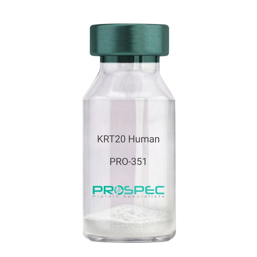
    KRT20 Human

    Description:

    Cytokeratin 20 Human Recombinant

    Keratin type I cytoskeletal 20, Cytokeratin-20, CK-20, Keratin-20, K20, Protein IT, KRT20, CD20, CK20, KRT21, MGC35423.

    Product # :

    PRO-351

    Price :

    Quantity :

    Shipping Method :

    Room Temp Icon

    Shipped at Room temp

    Add To Cart

    More Info

    • description
    • source
    • formulation
    • purity
    • More Info

    Description

    Cytokeratin 20 Human Recombinant produced in E.Coli is a single,non-glycosylated polypeptide chain having a molecular mass of 48,553 Dalton. The KRT20 is purified by proprietary chromatographic techniques.

    Source

    Escherichia Coli.

    Formulation

    The protein (1mg/1ml) was lyophilized after from a sterile solution containing 30mM Tris-HCL pH-8, 9.5M urea, 2mM DDT, 2mM EDTA and 10mM methylammonium chloride.

    Purity

    Greater than 95.0% as determined by SDS-PAGE.

    More Info

    • Introduction

      KRT20 is a member of the keratin family. The keratins are intermediate filament proteins responsible for the structural integrity of epithelial cells and are subdivided into cytokeratins and hair keratins. The type I cytokeratins consist of acidic proteins which are arranged in pairs of heterotypic keratin chains. This cytokeratin is a major cellular protein of mature enterocytes and goblet cells and is specifically expressed in the gastric and intestinal mucosa. The type I cytokeratin genes are clustered in a region of chromosome 17q12-q21.

    • Synonyms

      Keratin type I cytoskeletal 20, Cytokeratin-20, CK-20, Keratin-20, K20, Protein IT, KRT20, CD20, CK20, KRT21, MGC35423.

    • Physical Appearance

      Sterile Filtered White lyophilized (freeze-dried) powder.

    • Stability

      Lyophilized KRT20 although stable at room temperature for 3 weeks, should be stored desiccated below -18°C. Upon reconstitution KRT20 should be stored at 4°C between 2-7 days and for future use below -18°C.For long term storage it is recommended to add a carrier protein (0.1% HSA or BSA).Please prevent freeze-thaw cycles.

    • Solubility

      It is recommended to reconstitute the lyophilized KRT20 in sterile 18MΩ-cm H2O not less than 100µg/ml, which can then be further diluted to other aqueous solutions.

    • Reconstitution to filaments

      Performed by mixing equimolar amounts of cytokeratins of type I and type II at concentrations of approx. 0.5 mg/ml, both dissolved in 9.5 M urea buffer (see above). Protofilaments and filament complexes are obtained by dialyzing the resulting polypeptide solution stepwise to a concentration of 4 M urea and then to low salt condition (50 mM NaCI, 2 mM dithiothreitol, 10 mM Tris-HCI, pH 7.4). For immunization purposes, the solution can be further dialyzed against PBS (phosphate buffered saline, e.g. Dulbecco's PBS).

    ProSpec's products are furnished for LABORATORY RESEARCH USE ONLY. The product may not be used as drugs, agricultural or pesticidal products, food additives or household chemicals.

    Krt20 Human
  • View Data Sheet

    Name :

    CoV-2 S1 (16-685), Fc

    Description:

    Coronavirus 2019 Spike Glycoprotein-S1 (16-685 a.a.), Fc Recombinant

    Product # :

    SARS-027

    Price :

    Quantity :

    Shipping Method :

    Room Temp Icon

    Shipped at Room temp

    Add To Cart

    More Info

    • description
    • source
    • formulation
    • purity
    • More Info

    Description

    The HEK293 derived recombinant protein contains the Coronavirus 2019 CoV-2 Spike Glycoprotein S1, Wuhan-Hu-1 strain, amino acids 16-685 fused to Fc tag at C-terminal.

    Source

    HEK293 Cells.

    Formulation

    CoV-2 S1 protein is supplied in sodium chloride, glycine, arginine pH-7.5 & 5% trehalose.

    Purity

    Protein is >95% pure as determined SDS-PAGE.

    More Info

    • Introduction

      A human infecting coronavirus (viral pneumonia) called 2019 novel coronavirus, 2019-nCoV was found in the fish market at the city of Wuhan, Hubei province of China on December 2019.

      The 2019-nCoV shares an 87% identity to the 2 bat-derived severe acute respiratory syndrome 2018 SARS-CoV-2 located in Zhoushan of eastern China. 2019-nCoV has an analogous receptor-BD-structure to that of 2018 SARS-CoV, even though there is a.a. diversity so thus the 2019-nCoV might bind to ACE2 receptor protein (angiotensin-converting enzyme 2) in humans.

      While bats are possibly the host of 2019-nCoV, researchers suspect that animal from the ocean sold at the seafood market was an intermediate host. RSCU analysis proposes that the 2019-nCoV is a recombinant within the viral spike glycoprotein between the bat coronavirus and an unknown coronavirus.

    • Physical Appearance

      Sterile Filtered White lyophilized (freeze-dried) powder.

    • Stability

      Lyophilized Cov-2 Spike S1 Glycoprotein although stable at room temperature for 3 weeks, should be stored desiccated below -18°C. Upon reconstitution CoV2 Spike protein should be stored at 4°C between 2-7 days and for future use below -18°C.For long term storage it is recommended to add a carrier protein (0.1% HSA or BSA).Please prevent freeze-thaw cycles.

    • Purification Method

      Purified by Protein-G chromatographic technique.

    ProSpec's products are furnished for LABORATORY RESEARCH USE ONLY. The product may not be used as drugs, agricultural or pesticidal products, food additives or household chemicals.

    product_image.jpg
  • View Data Sheet

    Name :

    SARS Spike (306-527)

    Description:

    SARS Spike Receptor Binding Domain(306-527 a.a.), Recombinant

    Product # :

    SARS-032

    Price :

    Quantity :

    Shipping Method :

    Room Temp Icon

    Shipped at Room temp

    Add To Cart

    More Info

    • description
    • source
    • formulation
    • purity
    • More Info

    Description

    The HEK293 derived recombinant protein contains the SARS Coronavirus spike S glycoprotein Receptor Binding Domain, amino acids 306-527 fused to His tag at C-terminal.

    Source

    HEK293

    Formulation

    SARS Spike S glycoprotein RBD is lyophilized from 1x PBS pH-7.4 + 5% trehalose.

    Purity

    Protein is >90% pure as determined SDS-PAGE.

    More Info

    • Introduction

      SARS Coronavirus is an enveloped virus containing three outer structural proteins, namely the membrane (M), envelope (E), and spike (S) proteins. Spike (S)-glycoprotein of the virus interacts with a cellular receptor and mediates membrane fusion to allow viral entry into susceptible target cells. Accordingly, S-protein plays an important role in virus infection cycle and is the primary target of neutralizing antibodies.

    • Physical Appearance

      Lyophilized freezed dried powder.

    • Stability

      SARS Spike S1 although stable at room temperature for 3 weeks, should be stored desiccated below -18°C. Upon reconstitution SARS Spike protein should be stored at 4°C between 2-7 days and for future use below -18°C.For long term storage it is recommended to add a carrier protein (0.1% HSA or BSA).Please prevent freeze-thaw cycles.

    • Purification Method

      Purified by immobilized metal affinity chromatographic technique.

    ProSpec's products are furnished for LABORATORY RESEARCH USE ONLY. The product may not be used as drugs, agricultural or pesticidal products, food additives or household chemicals.

    product_image.jpg
  • 1
  • …
  • 23
  • 24
  • 25
  • 26
  • 27
  • …
  • 42
Your browser does not support the video tag.

Products

  • Cytokines
  • Growth Factors
  • Chemokines
  • CD Antigens
  • Neurotrophins
  • Hormones
  • Enzymes
  • Viral Antigens
  • Recombinant Proteins
  • Natural Proteins
  • Monoclonal Antibodies
  • Polyclonal Antibodies
  • Thermal Packaging

About

  • Company
  • Founder
  • Contribution
  • Contact Us
  • My Account

Ordering

  • Ordering Method
  • Ordering - Credit Card
  • Ordering PO
  • Shipping Fees
  • FAQ's

Legal

  • Terms & Conditions
  • Privacy
  • Site Map
  • info@prospecbio.com
  • Facebook
  • Instagram
  • Linkedin Profile

© 2026 ProSpec-Tany TechnoGene Ltd. All Rights Reserved.

  • Choosing a selection results in a full page refresh.
  • Opens in a new window.