Search results
884 results found for “Density Lipoprotein”
Name
Description
Product #
Price
Quantity
Shipping Method
- View Data Sheet
Name :
Colloidal GoldDescription:
Colloidal Gold 40nM 2OD
Product # :
PRO-2830Price :
Quantity :
Shipping Method :
Shipped with Ice Packs
More Info
- description
- formulation
- More Info
Description
Gold nanoparticles have one-of-a-kind properties turn them to ideal for detecting even the smallest amounts of biological molecules. These particles can be loaded with drugs or other therapeutic agents and then targeted to specific cells/tissues. Further to their diagnostic applications, gold nanoparticles may also take part in drug delivery and cancer therapy. Colloidal Gold is purified by proprietary chromatographic techniques.
Formulation
Colloidal Gold solution (6.7mg/ml) contains 50mM sodium citrate.
More Info
-
Physical Appearance
Sterile Filtered red solution.
-
Stability
Colloidal Gold should be stored at 4°C, DO NOT FREEZE.
-
Background
Colloidal gold is liquid form colloid nanoparticles of gold in a water. The colloidal gold color is dependent of the particle size, below 100 nm gives a is deep purple red color due to the spherical nanoparticles. Colloid gold is crucial for research due to its unique properties such as electronic charge, Mw and optical properties. Colloid gold is used in biomedical devices, electron microscopy and nanotechnology.
ProSpec's products are furnished for LABORATORY RESEARCH USE ONLY. The product may not be used as drugs, agricultural or pesticidal products, food additives or household chemicals.
- View Data Sheet
Name :
Protein-G HisDescription:
Protein G His Tag Recombinant
Product # :
PRO-1237Price :
Quantity :
Shipping Method :
Shipped at Room temp
More Info
- description
- source
- formulation
- purity
- More Info
Description
Protein G His Tag Recombinant produced in E.Coli is a 201 amino acids protein which contains amino acid 190-384 of the Streptococcus sp with a C-terminal 6-His tag, and having a molecular mass of 21.6kDa. But it migrates with an apparent molecular mass of 32kDa in SDS-PAGE.The Protein G His Tag is purified by proprietary chromatographic techniques.
Source
Escherichia Coli.
Formulation
Lyophilized with no additives.
Purity
Greater than 95.0% as determined by SDS-PAGE and HPLC analyses.
More Info
-
Physical Appearance
Sterile Filtered White lyophilized (freeze-dried) powder.
-
Stability
Lyophilized Protein G although stable at room temperature for 3 weeks, should be stored desiccated below -18°C. Upon reconstitution Protein G should be stored at 4°C between 2-7 days and for future use below -18°C.Please prevent freeze-thaw cycles.
-
Solubility
It is recommended to reconstitute the lyophilized Protein G in sterile 18M-cm H2O not less than 100µg/ml, which can then be further diluted to other aqueous solutions.
-
Applications
Protein G binds to the constant region of many species of immunoglobulin G. It can be used to detect, quantify and purify IgG antibodies and antibody/antigen complexes. Recombinant Protein G contains only IgG binding domains. The albumin-binding domain as well as cell wall and cell membrane binding domains have been removed to ensure the maximum specific IgG binding capacity.
-
Specificity
1. Binds with greater affinity to most mammalian immunoglobulins than Protein A, including human IgG3 and rat IgG2a.2. Does not bind to human IgM, IgD and IgA.
ProSpec's products are furnished for LABORATORY RESEARCH USE ONLY. The product may not be used as drugs, agricultural or pesticidal products, food additives or household chemicals.
- View Data Sheet
Name :
GDF7 HumanDescription:
Growth and Differentiation factor 7 Human Recombinant
Growth Differentiation Factor 7, GDF-7, Growth/Differentiation Factor 7, BMP12, GDF7.
Product # :
CYT-870Price :
Quantity :
Shipping Method :
Shipped at Room temp
More Info
- description
- source
- formulation
- purity
- biological activity
- More Info
Description
GDF7 Human Recombinant (322-450) produced in E.Coli is a disulfide-linked homodimeric, non-glycosylated, polypeptide chain containing 129 amino acids and having a molecular mass of 28kDa.The GDF-7 is purified by proprietary chromatographic techniques.
Source
Escherichia Coli.
Formulation
Lyophilized from a 0.2µm filtered solution in HCl.
Purity
Greater than 95.0% as determined by SDS-PAGE.
Biological Activity
The ED50, as determined by inducing alkaline phosphatase production by mouse ATDC5 cells, is less than 1.25µg/ml.More Info
-
Introduction
Growth Differentiation Factor-7 (GDF-7) belongs to the BMP family of TGF-b superfamily proteins. GDF7 elicits its bioactivity via a heterodimeric receptor complex comprised of a type 1 (BMPR-IB) and a type II (BMPR-II or Activin RII) serine/threonine kinase receptor. GDF7 signaling results in the phosphorylation and activation of Smad proteins. GDF-7 is also involved in tendon and ligament formation and repair. In addition, GDF7 regulates bone formation, mesenchymal stem cell differentiation, neuronal differentiation, and axon guidance.
-
Synonyms
Growth Differentiation Factor 7, GDF-7, Growth/Differentiation Factor 7, BMP12, GDF7.
-
Physical Appearance
Sterile Filtered White lyophilized (freeze-dried) powder.
-
Stability
Lyophilized GDF7 although stable at room temperature for 3 weeks, should be stored desiccated below -18°C. Upon reconstitution GDF-7 should be stored at 4°C between 2-7 days and for future use below -18°C.For long term storage it is recommended to add a carrier protein (0.1% HSA or BSA).Please prevent freeze-thaw cycles.
-
Solubility
It is recommended to reconstitute the lyophilized GDF-7 in sterile 18M-cm H2O not less than 100µg/ml, which can then be further diluted to other aqueous solutions.
-
Amino Acid Sequence
TALAGTRTAQ GSGGGAGRGH GRRGRSRCSR KPLHVDFKEL GWDDWIIAPL DYEAYHCEGL CDFPLRSHLE PTNHAIIQTL LNSMAPDAAP ASCCVPARLS PISILYIDAA NNVVYKQYED MVVEACGCR.
-
Background
What is the molecular weight/Mw of GDF7 Protein?
GDF7 Protein has a total Mw of 28kDa.
What is the source or expression system of GDF7 Protein?
Escherichia Coli.
What is the Purity of GDF7 Protein?
GDF7 Protein is >95% pure as determined by SDS-PAGE.
What is the Biological Activity of GDF7 Protein?
The ED50, as determined by inducing alkaline phosphatase production by mouse ATDC5 cells, is less than 1.25µg/ml.
What is the amino acid sequence of GDF7 Protein?
TALAGTRTAQ GSGGGAGRGH GRRGRSRCSR KPLHVDFKEL GWDDWIIAPL DYEAYHCEGL CDFPLRSHLE PTNHAIIQTL LNSMAPDAAP ASCCVPARLS PISILYIDAA NNVVYKQYED MVVEACGCR.
What applications can GDF7 Protein be used in?
GDF7 Protein can probably be used in western blot, ELISA and Lateral Flow.
What is the endotoxin level for GDF7 Protein?
The endotoxin level is minimal, GDF7 Protein was purified using conventional chromatography techniques.
ProSpec's products are furnished for LABORATORY RESEARCH USE ONLY. The product may not be used as drugs, agricultural or pesticidal products, food additives or household chemicals.
- View Data Sheet
Name :
ARF4 HumanDescription:
ADP-Ribosylation Factor 4 Human Recombinant
ADP-ribosylation factor 4, ARF4, ARF2.
Product # :
PRO-066Price :
Quantity :
Shipping Method :
Shipped with Ice Packs
More Info
- description
- source
- formulation
- purity
- More Info
Description
ARF4 Human Recombinant fused with a 24 amino acid His tag at N-terminus produced in E.Coli is a single, non-glycosylated, polypeptide chain containing 204 amino acids (1-180 a.a.) and having a molecular mass of 23kDa. The ARF4 is purified by proprietary chromatographic techniques.
Source
Escherichia Coli.
Formulation
The ARF4 solution (0.5 mg/ml) in 20mM Tris-HCl buffer (pH8.0), 0.2M NaCl, 5mM DTT and 20% glycerol.
Purity
Greater than 95.0% as determined by SDS-PAGE.
More Info
-
Introduction
ARF4 belongs to the ARF gene family whose members encode small guanine nucleotide-binding proteins which stimulate the ADP-ribosyltransferase activity of cholera toxin and have a role in vesicular trafficking and as activators of phospholipase D. The ARF proteins include five ARF proteins and eleven ARF-like proteins and constitute one family of the RAS superfamily. They are classified as class I, class II and class III; the ARF4 gene is a class II member. The members of each class share a common gene organization. The ARF4 gene spans approximately 12kb and contains 6 exons and 5 introns. The ARF4 gene is the most divergent member of the human ARFs.
-
Synonyms
ADP-ribosylation factor 4, ARF4, ARF2.
-
Physical Appearance
Sterile Filtered colorless solution.
-
Stability
Store at 4°C if entire vial will be used within 2-4 weeks.Store, frozen at -20°C for longer periods of time. For long term storage it is recommended to add a carrier protein (0.1% HSA or BSA).Avoid multiple freeze-thaw cycles.
-
Amino Acid Sequence
MGSSHHHHHH SSGLVPRGSH MGSHMGLTIS SLFSRLFGKK QMRILMVGLD AAGKTTILYK LKLGEIVTTI PTIGFNVETV EYKNICFTVW DVGGQDRIRP LWKHYFQNTQ GLIFVVDSND RERIQEVADE LQKMLLVDEL RDAVLLLFAN KQDLPNAMAI SEMTDKLGLQ SLRNRTWYVQ ATCATQGTGL YEGLDWLSNE LSKR.
ProSpec's products are furnished for LABORATORY RESEARCH USE ONLY. The product may not be used as drugs, agricultural or pesticidal products, food additives or household chemicals.
- View Data Sheet
Name :
Follistatin HumanDescription:
Follistatin Human Recombinant
FST, FS
Product # :
CYT-232Price :
Quantity :
Shipping Method :
Shipped at Room temp
More Info
- description
- source
- formulation
- purity
- biological activity
- More Info
Description
Follistatin Human Recombinant produced in E.Coli is a single, non-glycosylated polypeptide chain containing 288 amino acids and having a total molecular mass of 31.5kDa.The FST is purified by proprietary chromatographic techniques.
Source
Escherichia Coli.
Formulation
Lyophilized from a concentrated (1mg/ml) solution containing no additives.
Purity
Greater than 95.0% as determined by:
(a) Analysis by RP-HPLC.
(b) Analysis by SDS-PAGE.Biological Activity
The activity is determined by the ability to neutralize ACTV inhibitory effect of mouse MPC-11 cells. The expected ED50 is 100-400ng/ml, corresponding to a Specific Activity of 2,500-10,000units/mg in the presence of 7.5ng/ml ACTV A.
More Info
-
Introduction
Follistatin is a single-chain gonadal protein that specifically inhibits follicle-stimulating hormone release. The single FST gene encodes two isoforms, FST317 and FST344 containing 317 and 344 amino acids respectively, resulting from alternative splicing of the precursor mRNA. In a study in which 37 candidate genes were tested for linkage and association with polycystic ovary syndrome (PCOS) or hyperandrogenemia in 150 families, evidence was found for linkage between PCOS and follistatin. Follistatin functions as an ACTV antagonist. specific inhibitor of the biosynthesis and secretion of pituitary follicle stimulating hormone (fsh).
-
Synonyms
FST, FS
-
Physical Appearance
Sterile Filtered White lyophilized (freeze-dried) powder.
-
Stability
Lyophilized Follistatin although stable at room temperature for 3 weeks, should be stored desiccated below -18°C. Upon reconstitution FST should be stored at 4°C between 2-7 days and for future use below -18°C.For long term storage it is recommended to add a carrier protein (0.1% HSA or BSA).Please prevent freeze-thaw cycles.
-
Solubility
It is recommended to reconstitute the lyophilized Follistatin in sterile 18MΩ-cm H2O not less than 100µg/ml, which can then be further diluted to other aqueous solutions.
-
Amino Acid Sequence
The sequence of the first five N-terminal amino acids was determined and was found to be Gly-Asn-Cys-Trp-Leu.
-
Background
What is the molecular weight/Mw of FOLLISTATIN HUMAN Protein?
FOLLISTATIN HUMAN Protein has a total Mw of 31.5kDa.
What is the source or expression system of FOLLISTATIN HUMAN Protein?
Escherichia Coli.
What is the Purity of FOLLISTATIN HUMAN Protein?
FOLLISTATIN HUMAN Protein is >95% pure as determined by SDS-PAGE.
What is the Biological Activity of FOLLISTATIN HUMAN Protein?
The activity is determined by the ability to neutralize ACTV inhibitory effect of mouse MPC-11 cells. The expected ED50 is 100-400ng/ml, corresponding to a Specific Activity of 2,500-10,000units/mg in the presence of 7.5ng/ml ACTV A.
What is the amino acid sequence of FOLLISTATIN HUMAN Protein?
The sequence of the first five N-terminal amino acids was determined and was found to be Gly-Asn-Cys-Trp-Leu.
What applications can FOLLISTATIN HUMAN Protein be used in?
FOLLISTATIN HUMAN Protein can probably be used in western blot, ELISA and Lateral Flow.
What is the endotoxin level for FOLLISTATIN HUMAN Protein?
The endotoxin level is minimal, FOLLISTATIN HUMAN Protein was purified using conventional chromatography techniques.
ProSpec's products are furnished for LABORATORY RESEARCH USE ONLY. The product may not be used as drugs, agricultural or pesticidal products, food additives or household chemicals.
- View Data Sheet
Name :
T.pallidum p17Description:
Treponema pallidum p17 Recombinant
Product # :
TRP-241Price :
Quantity :
Shipping Method :
Shipped with Ice Packs
More Info
- description
- source
- formulation
- purity
- More Info
Description
The E.Coli derived recombinant protein contains the T. Pallidum p17 immunodominant regions. The protein contains beta- galactosidase (114 kDa) fused at the N- terminus.
Source
Escherichia Coli.
Formulation
8M urea, 20mM Tris-HCl pH-8 and10mM B-ME.
Purity
Treponema Pallidum protein protein is >90% pure as determined by 10% PAGE (coomassie staining).
More Info
-
Introduction
Treponema pallidum is a gram-negative spirochaete bacterium and is considered to be metabolically crippled. There are at least four known subspecies: T. pallidum pallidum, T. pallidum pertenue, T. pallidum carateum and T. pallidum endemicum. The helical structure of T. pallidum pallidum allows it to move in a corkscrew motion through viscous mediums such as mucus. Treponema pallidum sub sp. pallidum has one of the smallest bacterial genomes at 1.14 million base pairs (Mb) and has limited metabolic capabilities, reflecting its adaptation through genome reduction to the rich environment of mammalian tissue.
-
Stability
Treponema Pallidum protein although stable at 4°C for 1 week, should be stored below -18°C. Please prevent freeze thaw cycles.
-
Applications
Treponema Pallidum protein is suitable for ELISA and Western blots, excellent antigen for detection of Trp. Pallidum with minimal specificity problems.
-
Specificity
Immunoreactive with sera of Trp. Pallidum infected individuals.
-
Purification Method
Treponema Pallidum protein was purified by proprietary chromatographic technique.
ProSpec's products are furnished for LABORATORY RESEARCH USE ONLY. The product may not be used as drugs, agricultural or pesticidal products, food additives or household chemicals.
- View Data Sheet
Name :
EnfuvirtideDescription:
Enfuvirtide (T-20)
Product # :
PRO-376Price :
Quantity :
Shipping Method :
Shipped at Room temp
More Info
- description
- formulation
- purity
- More Info
Description
Enfuvirtide has an amino acid sequence Ac-Tyr-Thr-Ser-Leu-Ile-His-Ser-Leu-Ile-Glu-Glu-Ser-Gln-Asn-Gln-Gln-Glu-Lys-Asn-Glu-Gln-Glu-Leu-Leu-Glu-Leu-Asp-Lys-Trp-Ala-Ser-Leu-Trp-Asn-Trp-Phe-NH2, molecular formula of C204H301N51O64 and a molecular weight of 4488.9 Dalton.
Formulation
The protein (1mg/ml) was lyophilized with no additives.
Purity
Greater than 98.0% as determined by RP-HPLC.
More Info
-
Introduction
Enfuvirtide also called human immunodeficiency fusion inhibitor blocks HIV’s ability to infect healthy CD4 cells. It can reduce the amount of HIV in the blood and increase the number of CD4 cells. Enfuvirtide operates by disrupting the HIV-1 molecular machinery at the final phase of fusion with the target cell, preventing uninfected cells from becoming infected. Enfuvirtide mimics components of the HIV-1 fusion apparatus and displace them, preventing normal fusion. HIV attaches to the host CD4+ cell receptor using the viral protein GP120; upon binding, GP120 deforms allowing the viral protein GP41 to insert itself into the host cell's plasma membrane. Entry inhibitors bind to GP41 preventing the formation of an entry pore for the capsid of the virus, keeping it out of the cell.
-
Physical Appearance
Sterile Filtered White lyophilized (freeze-dried) powder.
-
Stability
Lyophilized Enfuvirtide although stable at room temperature for 3 weeks, should be stored desiccated below -18°C. Upon reconstitution Enfuvirtide should be stored at 4°C between 2-7 days and for future use below -18°C.For long term storage it is recommended to add a carrier protein (0.1% HSA or BSA).Please prevent freeze-thaw cycles.
-
Solubility
Enfuvirtide lyophilized powder is soluble in water at a concentration of 1mg/ml.
ProSpec's products are furnished for LABORATORY RESEARCH USE ONLY. The product may not be used as drugs, agricultural or pesticidal products, food additives or household chemicals.
- View Data Sheet
Name :
Leptin OvineDescription:
Leptin Ovine Recombinant
OB Protein, Obesity Protein, OBS, Obesity factor.
Product # :
CYT-239Price :
Quantity :
Shipping Method :
Shipped at Room temp
More Info
- description
- source
- formulation
- purity
- biological activity
- More Info
Description
Leptin Ovine Recombinant produced in E.Coli is a single, non-glycosylated, polypeptide chain containing 146 amino acids and having a molecular mass of 16 kDa.The Leptin is purified by proprietary chromatographic techniques.
Source
Escherichia Coli.
Formulation
The protein was lyophilized from a concentrated (1mg/ml) solution with 0.0045mM NaHCO3.
Purity
Greater than 95.0% as determined by:
(a) Analysis by SEC-HPLC.
(b) Analysis by SDS-PAGE.Biological Activity
Biological active as evidenced by inducing proliferation of BAF/3 cells stably transfected with the long form of human leptin receptor.More Info
-
Introduction
A 16-kDa peptide hormone secreted from white adipocytes and implicated in the regulation of food intake and energy balance. Leptin provides the key afferent signal from fat cells in the feedback system that controls body fat stores.
-
Synonyms
OB Protein, Obesity Protein, OBS, Obesity factor.
-
Physical Appearance
Sterile Filtered White lyophilized (freeze-dried) powder.
-
Stability
Lyophilized Leptin although stable at room temperature for 3 weeks, should be stored desiccated below -18°C. Upon reconstitution Leptin should be stored at 4°C between 2-7 days and for future use below -18°C.For long term storage it is recommended to add a carrier protein (0.1% HSA or BSA).Please prevent freeze-thaw cycles.
-
Solubility
It is recommended to reconstitute the lyophilized Leptin in sterile 18MΩ-cm H2O not less than 100µg/ml, which can then be further diluted to other aqueous solutions.
-
Amino Acid Sequence
The sequence of the first five N-terminal amino acids was determined and was found to be Ala-Val-Pro-Ile-Arg.
-
Protein content
Protein quantitation was carried out by two independent methods1. UV spectroscopy at 280 nm using the absorbency value of 0.2 as the extinction coefficient for a 0.1% (1mg/ml) solution. This value is calculated by the PC GENE computer analysis program of protein sequences (IntelliGenetics). 2. Analysis by RP-HPLC,using calibrated solution of Leptin Ovine as a Reference Standard.
ProSpec's products are furnished for LABORATORY RESEARCH USE ONLY. The product may not be used as drugs, agricultural or pesticidal products, food additives or household chemicals.
- View Data Sheet
Name :
FGF23 HumanDescription:
Fibroblast Growth Factor-23 Human Recombinant
Tumor-derived hypophosphatemia-inducing factor, HYPF, ADHR, HPDR2, PHPTC, FGF23, FGF-23, Fibroblast Growth Factor-23.
Product # :
CYT-020Price :
Quantity :
Shipping Method :
Shipped at Room temp
More Info
- description
- source
- formulation
- purity
- biological activity
- More Info
Description
Fibroblast Growth Factor-23 Human Recombinant produced in E.Coli is a single, non-glycosylated polypeptide chain containing a total of 228 amino acids and having a molecular mass of 22.5kDa. The FGF-23 is and purified by chromatographic techniques.
Source
Escherichia Coli.
Formulation
The FGF-23 protein (0.5mg/ml) was lyophilized from a 0.2µm filtered concentrated solution in PBS, pH 7.4.
Purity
Greater than 95.0% as determined by SDS-PAGE.
Biological Activity
The biological activity of FGF-23 was measured in a cell proliferation assay using NIH/3T3 mouse embryonic fibroblasts. The ED50 for this effect is typically 0.05-0.5µg/ml in the presence of 5µg/ml of Recombinant Mouse Klotho and 10 µg/ml of HPR.
More Info
-
Introduction
FGF-23 is a member of the fibroblast growth factor (FGF) family. FGF family members possess broad mitogenic and cell survival activities and are involved in a variety of biological processes including embryonic development, cell growth, morphogenesis, tissue repair, tumor growth and invasion. FGF-23 inhibits renal tubular phosphate transport. The FGF-23 gene was identified by its mutations associated with autosomal dominant hypophosphatemic rickets (ADHR), an inherited phosphate wasting disorder. Abnormally high level expression of FGF-23 was found in oncogenic hypophosphatemic osteomalacia (OHO), a phenotypically similar disease caused by abnormal phosphate metabolism. FGF-23 mutations have also been shown to cause familial tumoral calcinosis with hyperphosphatemia.
-
Synonyms
Tumor-derived hypophosphatemia-inducing factor, HYPF, ADHR, HPDR2, PHPTC, FGF23, FGF-23, Fibroblast Growth Factor-23.
-
Physical Appearance
Sterile Filtered white lyophilized powder.
-
Stability
Lyophilized FGF-23 although stable at room temperature for 3 weeks, should be stored desiccated below -18°C. Upon reconstitution FGF-23 should be stored at 4°C between 2-7 days and for future use below -18°C.For long term storage it is recommended to add a carrier protein (0.1% HSA or BSA).Please prevent freeze-thaw cycles.
-
Solubility
It is recommended to reconstitute the lyophilized FGF-23 in sterile 18M-cm H2O not less than 100µg/ml, which can then be further diluted to other aqueous solutions.
-
Amino Acid Sequence
MYPNASPLLG SSWGGLIHLY TATARNSYHL QIHKNGHVDG APHQTIYSAL MIRSEDAGFV VITGVMSRRY LCMDFRGNIF GSHYFDPENC RFQHQTLENG YDVYHSPQYH FLVSLGRAKR AFLPGMNPPP YSQFLSRRNE IPLIHFNTPI PRRHTRSAED DSERDPLNVL KPRARMTPAP ASCSQELPSA EDNSPMASDP LGVVRGGRVN THAGGTGPEG CRPFAKFI.
-
Background
Before it was discovered in 2000, there was a hypothesis that a similar type of protein existed that performed many of the functions we see in FGF23. This was originally referred to as phosphatonin. Various effects were described and noted by researchers including inhibition of production and inhibition of secretion of parathyroid hormone. Derived from the bone Fibroblast Growth Factor 23 is a phosphaturic hormone. It increases phosphate excretion when acting on the kidney and also suppresses the biosynthesis of 1,25(OH)2D3.
Mechanism
Despite various research studies into the topic, many of the mechanisms for the regulations of FGF23 production remain a mystery to the scientific community. While we know that mutations in PHEX, ENPP1 and DMP1 result in the increased expression of FGF23, it is unclear why this occurs. This also means that currently, it is not possible to regulate the production of FGF23 either. We also do not know how signals from FGF23 regulate vitamin D metabolism. However, understanding these types of mechanisms could offer information needed to provide better treatment for deranged bone and mineral metabolism.Interactions
Molecular interactions involving FGF-23, vitamin D and klotho do provide the solution needed to regulate phosphate levels within the body. Furthermore, an interaction between Vitamin D and FGF3 can have an impact on renal phosphate balance. As well as this, when in the presence of klotho, FGF3 does actually increase bioactivity and begins to change systemic phosphate homeostasis.Function
Based on research it seems that the main function for FGF23 is the regulation of phosphate concentration in plasma. It seems to be secreted from the osteocytes due to elevated levels . When acting upon the kidneys, the hormone reduces the expression of NPT2. This is a sodium-phosphate cotransporter found in the proximal tube.
As such, it appears as though FGF23 is able to reduce the reabsorption, all the while maxing the excretion of phosphate. It has also been suggested that the hormone is able to suppress 1-alpha-hydroxylase. If this is the case, it can limit its potential to activate vitamin D and thus impair the ability for calcium absorption.Structure
FGF243 is located on the chromosome 12. It is composed of three exons. The crystal structure of FGF23 is completely different from the common conformation typically adopted by paracrine-acting FGFs. Instead, there is a conformation of the HPR region between beta strands 10 and 12. As well as this, there is a cleft between the other HPR-binding region, the beta1-beta12 loop and this one. This comes before a direct interaction between HPR sulfate and FGF23s backbone atoms. Due to this, endocrine function is benefitted and HPR-binding affinity is reduced for the lipangs.
Certain mutations that cause the protein to be completely resistant to proteolytic cleavage does trigger a surge in activity of the protein and the renal phosphate loss typically found in certain human diseases including hypophosphatemic rickets.
Studies have also revealed that FGF23 is overproduced in certain tumors including phosphaturic mesenchymal tumors. Furthermore a reduced level of activity for this protein is believed to lead to higher phosphate levels and familial tumor calcinosis clinical syndrome.What is the molecular weight/Mw of FGF23 HUMAN Protein?
FGF23 HUMAN Protein has a total Mw of 22.5kDa.
What is the source or expression system of FGF23 HUMAN Protein?
Escherichia Coli.
What is the Purity of FGF23 HUMAN Protein?
FGF23 HUMAN Protein is >95X% pure as determined by SDS-PAGE.
What is the Biological Activity of FGF23 HUMAN Protein?
The biological activity of FGF-23 was measured in a cell proliferation assay using NIH/3T3 mouse embryonic fibroblasts. The ED50 for this effect is typically 0.05-0.5µg/ml in the presence of 5µg/ml of Recombinant Mouse Klotho and 10 µg/ml of HPR.
What is the amino acid sequence of FGF23 HUMAN Protein?
MYPNASPLLG SSWGGLIHLY TATARNSYHL QIHKNGHVDG APHQTIYSAL MIRSEDAGFV VITGVMSRRY LCMDFRGNIF GSHYFDPENC RFQHQTLENG YDVYHSPQYH FLVSLGRAKR AFLPGMNPPP YSQFLSRRNE IPLIHFNTPI PRRHTRSAED DSERDPLNVL KPRARMTPAP ASCSQELPSA EDNSPMASDP LGVVRGGRVN THAGGTGPEG CRPFAKFI.
What applications can FGF23 HUMAN Protein be used in?
FGF23 HUMAN Protein can probably be used in western blot, ELISA and Lateral Flow.
What is the endotoxin level for FGF23 HUMAN Protein?
The endotoxin level is minimal, FGF23 HUMAN Protein was purified using conventional chromatography techniques.
ProSpec's products are furnished for LABORATORY RESEARCH USE ONLY. The product may not be used as drugs, agricultural or pesticidal products, food additives or household chemicals.
- View Data Sheet
Name :
PPM1A HumanDescription:
Protein Phosphatase 1A Alpha Isoform Human Recombinant
Protein phosphatase 1A, EC 3.1.3.16, Protein phosphatase 2C isoform alpha, PP2C-alpha, IA, PPM1A, PP2CA, MGC9201.
Product # :
PKA-222Price :
Quantity :
Shipping Method :
Shipped with Ice Packs
More Info
- description
- source
- formulation
- purity
- More Info
Description
PPM1A Human Recombinant produced in E.Coli is a single, non-glycosylated polypeptide chain containing 418 amino acids (1-382a.a) and having a molecular mass of 46.6kDa. PPM1A is fused to a 36 amino acid His-tag at N-terminus & purified by proprietary chromatographic techniques.
Source
Escherichia Coli.
Formulation
PPM1A protein solution (1mg/ml) contains 10mM Tris-HCl, pH7.5, 50mM NaCl, 2mM DTT, 1mM MnCl2 and 20% glycerol.
Purity
Greater than 95.0% as determined by SDS-PAGE.
More Info
-
Introduction
Protein Phosphatase 2C alpha is a member of the PP2C family of Ser/Thr protein phosphatases. PP2C family members are known to be negative regulators of cell stress response pathways. This phosphatase dephosphorylates, and negatively regulates the activities of, MAP kinases and MAP kinase kinases. It has been shown to inhibit the activation of p38 and JNK kinase cascades induced by environmental stresses. This phosphatase can also dephosphorylate cyclin-dependent kinases, and thus may be involved in cell cycle control. Overexpression of this phosphatase is reported to activate the expression of the tumor suppressor gene TP53/p53, which leads to G2/M cell cycle arrest and apoptosis. Three alternatively spliced transcript variants encoding two distinct isoforms have been described.
Protein phosphatase 2C(PP2C?) is a Mn2+- or Mg2+-dependent protein serine/threonine phosphatase that is essential for regulating cellular stress response in eukaryotes. -
Synonyms
Protein phosphatase 1A, EC 3.1.3.16, Protein phosphatase 2C isoform alpha, PP2C-alpha, IA, PPM1A, PP2CA, MGC9201.
-
Physical Appearance
Sterile filtered colorless solution.
-
Stability
Store at 4°C if entire vial will be used within 2-4 weeks. Store, frozen at -20°C for longer periods of time. For long term storage it is recommended to add a carrier protein (0.1% HSA or BSA). Avoid multiple freeze-thaw cycles.
-
Amino Acid Sequence
MRGSHHHHHH GMASMTGGQQ MGRDLYDDDD KDRWILMGAF LDKPKMEKHN AQGQGNGLRY GLSSMQGWRV EMEDAHTAVI GLPSGLESWS FFAVYDGHAG SQVAKYCCEH LLDHITNNQD FKGSAGAPSV ENVKNGIRTG FLEIDEHMRV MSEKKHGADR SGSTAVGVLI SPQHTYFINC GDSRGLLCRN RKVHFFTQDH KPSNPLEKER IQNAGGSVMI QRVNGSLAVS RALGDFDYKC VHGKGPTEQL VSPEPEVHDI ERSEEDDQFI ILACDGIWDV MGNEELCDFV RSRLEVTDDL EKVCNEVVDT CLYKGSRDNM SVILICFPNA PKVSPEAVKK EAELDKYLEC RVEEIIKKQG EGVPDLVHVM RTLASENIPS LPPGGELASK RNVIEAVYNR LNPYKNDDTD STSTDDMW.
-
Unit Definition
One unit will hydrolyze 1 nanomole of p-nitrophenyl phosphatate (pNPP) per minute at pH 7.5 at 37°C.
-
Specific Activity
>1,400 U/mg.
ProSpec's products are furnished for LABORATORY RESEARCH USE ONLY. The product may not be used as drugs, agricultural or pesticidal products, food additives or household chemicals.
- View Data Sheet
Name :
EGF Rat ProteinDescription:
Epidermal Growth Factor Rat
Urogastrone, URG, EGF.
Product # :
CYT-556Price :
Quantity :
Shipping Method :
Shipped at Room temp
More Info
- description
- source
- formulation
- purity
- More Info
Description
Epidermal Growth Factor Rat purified from submandibular gland is a single, glycosylated, polypeptide chain having a molecular mass of 6.15 kDa.The EGF is purified by proprietary chromatographic techniques.
Source
Adult Male Rat Submandibular Glands.
Formulation
The protein was lyophilized from a concentrated (1mg/ml) solution containing 0.01M sodium acetate buffer.
Purity
Greater than 99.0% as determined by(a) Analysis by RP-HPLC.
(b) Analysis by SDS-PAGE.More Info
-
Introduction
Epidermal growth factor has a profound effect on the differentiation of specific cells in vivo and is a potent mitogenic factor for a variety of cultured cells of both ectodermal and mesodermal origin. The EGF precursor is believed to exist as a membrane-bound molecule which is proteolytically cleaved to generate the 53-amino acid peptide hormone that stimulates cells to divide.
EGF stimulates the growth of various epidermal and epithelial tissues in vivo and in vitro and of some fibroblasts in cell culture. -
Synonyms
Urogastrone, URG, EGF.
-
Physical Appearance
Sterile Filtered White lyophilized (freeze-dried) powder.
-
Stability
Lyophilized Epidermal Growth Factor Recombinant although stable at room temperature for 3 weeks, should be stored desiccated below -18°C. Upon reconstitution EGF should be stored at 4°C between 2-7 days and for future use below -18°C.For long term storage it is recommended to add a carrier protein (0.1% HSA or BSA).Please prevent freeze-thaw cycles.
-
Solubility
It is recommended to reconstitute the lyophilized Epidermal Growth Factor in sterile 18MΩ-cm H2O not less than 100µg/ml, which can then be further diluted to other aqueous solutions.
-
Background
Illuminating Epidermal Growth Factor Rat Recombinant: Deciphering Cellular Signaling and Therapeutic Potential
Abstract:
This research paper delves into the enigmatic realm of Epidermal Growth Factor Rat Recombinant (EGF-RR), unraveling its intricate molecular attributes, signaling cascades, and therapeutic prospects. By employing cutting-edge methodologies encompassing protein expression, receptor binding assays, and bioinformatics analyses, this study sheds light on the multifaceted interplay between EGF-RR and cellular responses, offering novel avenues for therapeutic interventions.
Introduction:
Epidermal Growth Factor (EGF) is pivotal in cellular regulation. This paper navigates the complexities of Epidermal Growth Factor Rat Recombinant (EGF-RR), focusing on its unique molecular properties and potential therapeutic applications.
Protein Expression and Purification:
The study embarks on precise gene optimization to enhance EGF-RR expression. Purification techniques like affinity chromatography yield purified EGF-RR, primed for subsequent analyses.
Receptor Binding Assays and Ligand Interaction:
Employing advanced receptor binding assays, the paper deciphers EGF-RR's engagement with its cognate receptor. Quantitative assessments uncover binding kinetics, shedding light on the intricacies of EGF-RR's molecular interaction.
Cellular Signaling Pathways and Responses:
In vitro cellular assays unveil the signaling cascades ignited by EGF-RR. Through quantitative phosphoproteomic profiling, the study unravels phosphorylation events triggered by EGF-RR, delineating its role in cellular proliferation, migration, and differentiation.
Bioinformatics Insights and Structural Modeling:
Bioinformatics tools facilitate molecular dynamics simulations, offering insights into EGF-RR's receptor interactions and downstream signaling pathways. Structural modeling captures EGF-RR's conformational changes during signaling cascades.
Therapeutic Implications and Future Prospects:
EGF-RR's intricate signaling dynamics open avenues for therapeutic exploration. Harnessing its potential in wound healing, tissue regeneration, and cancer modulation emerges as a promising avenue for precision medicine.
Challenges and Future Directions:
Challenges, including context-specific responses, beckon further investigation. Future research should delve into cross-talk between signaling pathways and EGF-RR's contributions to diverse disease contexts.
Conclusion:
A fusion of advanced methodologies and visionary insights unveils Epidermal Growth Factor Rat Recombinant as an intriguing subject. Its molecular intricacies and complex cellular interplay ignite prospects for therapeutic breakthroughs, ushering in a new era of precision medicine.
What is the molecular weight/Mw of EGF RAT Protein?
EGF RAT Protein has a total Mw of 6.15kDa.
What is the source or expression system of EGF RAT Protein?
Adult Male Rat Submandibular Glands.
What is the Purity of EGF RAT Protein?
EGF RAT Protein is >99% pure as determined by SDS-PAGE.
What is the Biological Activity of EGF RAT Protein?
The biological functionality of EGF RAT Protein will be determined in the future.
What is the amino acid sequence of EGF RAT Protein?
EGF RAT Protein is composed from 53 amino acids.
What applications can EGF RAT Protein be used in?
EGF RAT Protein can probably be used in western blot, ELISA and Lateral Flow.
What is the endotoxin level for EGF RAT Protein?
The endotoxin level is minimal, EGF RAT Protein was purified using conventional chromatography techniques.
ProSpec's products are furnished for LABORATORY RESEARCH USE ONLY. The product may not be used as drugs, agricultural or pesticidal products, food additives or household chemicals.
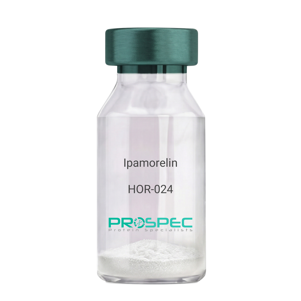
- View Data Sheet
Name :
IpamorelinDescription:
Ipamorelin
Ipamorelin
Product # :
HOR-024Price :
Quantity :
Shipping Method :
Shipped at Room temp
More Info
- description
- formulation
- purity
- More Info
Description
Ipamorelin Synthetic is a single, non-glycosylated polypeptide chain containing 4 amino acids, having a molecular mass of 711.85 Dalton and a Molecular formula of C38H49N9O5.
Formulation
The protein was lyophilized with no additives.
Purity
Greater than 97.0% as determined by analysis by RP-HPLC.
More Info
-
Introduction
Ipamorelin is a peptide selective agonist of the ghrelin/growth hormone secretagogue receptor and a growth hormone secretagogue. Ipamorelin is a pentapeptide that was derived from GHRP1. Ipamorelin significantly increases plasma growth hormone levels in both animals and humans. Like pralmorelin and GHRP-6, ipamorelin does not affect prolactin, FSH, LH or TSH levels. However, unlike GHRP2 and GHRP6, but as growth hormone-releasing hormone (GHRH), ipamorelin does not stimulate the secretion of adrenocorticotropic hormone (ACTH) or cortisol, and is highly selective for inducing the secretion only of GH.
-
Synonyms
Ipamorelin
-
Physical Appearance
Sterile Filtered White lyophilized (freeze-dried) powder.
-
Stability
Lyophilized Ipamorelin although stable at room temperature for 3 weeks, should be stored desiccated below -18°C. Upon reconstitution Ipamorelin should be stored at 4°C between 2-7 days and for future use below -18°C. For long term storage it is recommended to add a carrier protein (0.1% HSA or BSA). Please prevent freeze-thaw cycles.
-
Solubility
It is recommended to reconstitute the lyophilized Ipamorelin in sterile 18MΩ-cm H2O not less than 100 µg/ml, which can then be further diluted to other aqueous solutions.
-
Amino Acid Sequence
Aib-His-D-2-Nal-D-Phe-Lys-NH2.
ProSpec's products are furnished for LABORATORY RESEARCH USE ONLY. The product may not be used as drugs, agricultural or pesticidal products, food additives or household chemicals.
- View Data Sheet
Name :
Ebola Sudan ProteinDescription:
Ebola Sudan Recombinant Protein
Product # :
EVD-079Price :
Quantity :
Shipping Method :
Shipped with Ice Packs
More Info
- description
- source
- formulation
- purity
- More Info
Description
Nucleoprotein (NP) of Ebola virus has strong antigenicity in immune reactions. C-terminal of EBO-S nucleoprotein was expressed and purified from E. coli, it migrated at 15kDa on SDS-PAGE and pI is 4.52. Ebola Sudan was purified by a proprietary chromatographic technique.
Source
Escherichia Coli.
Formulation
Ebola Sudan protein solution is supplied in Phosphate buffer and 0.02% sodium azide.
Purity
>95% pure as determined by 12% SDS-PAGE (Coomassie blue stain).
More Info
-
Introduction
Ebolavirus (EVD) belongs to the Filoviridae family of proteins which is comprised of a single-strand, non-infectious RNA genome. EVD genome is about 19,000 base pairs long and covers 7 genes in the order 3'-UTR-NP-VP35-VP40-GP-VP30-VP24-L-5'-UTR. There are 4 different ebolaviruses such as: Zaire (EBO-Z), Sudan (EBO-S), Cote d’Ivoire (EBO-CI) and Reston (EBO-R) that differ in amino acid sequence and location of where the gene overlaps. Similar to filoviruses and ebolavirions, EVD is a filamentous element that could appear in 3 forms: shepherd's crook, U shape or a 6 shape. Ebolaviruses may be coiled, toroid, or branched. Most ebolavirions are 80nm wideand 14,000nm long.
-
Stability
Store at 4°C if entire vial will be used within 2-4 weeks. Store, frozen at -20°C for longer periods of time. For long term storage it is recommended to add a carrier protein (0.1% HSA or BSA).Avoid multiple freeze-thaw cycles.
-
Applications
Immunoassay.
ProSpec's products are furnished for LABORATORY RESEARCH USE ONLY. The product may not be used as drugs, agricultural or pesticidal products, food additives or household chemicals.
- View Data Sheet
Name :
Chitinase ProteinDescription:
Chitinase Clostridium Paraputrificum Recombinant
Product # :
ENZ-031Price :
Quantity :
Shipping Method :
Shipped at Room temp
More Info
- description
- source
- formulation
- purity
- More Info
Description
Chitinase Clostridium Paraputrificum Recombinant fused with a His tag at N-terminus produced in E.Coli is a single, non-glycosylated, polypeptide chain containing 582 amino acids and having a molecular mass of 64.2kDa. The Chitinase is purified by proprietary chromatographic techniques.
Source
Escherichia Coli.
Formulation
Chitinase lyophilized from a 0.2µm filtered concentrated solution in PBS.
Purity
Greater than 95.0% as determined by SDS-PAGE.
More Info
-
Introduction
Chitinase is a digestive enzyme which breaks down glycosidic bonds in chitin. Due to chitin being a component of the cell walls of fungi and exoskeletal elements of some animals (including worms and arthropods), chitinases are usually found in organisms that either need to remake their own chitin or to dissolve and digest the chitin of fungi or animals. Chitinivorous organisms include many bacteria genuses such as Aeromonas, Bacillus, Vibrio, among others, which may be pathogenic or detritivorous. Chitinase expression is mediated by the NPR1 gene and the salicylic acid pathway, both of which are involved in resisting fungal and insect attack. Human chitinases appear in gastric juices. They are likely to be digestive chitinases, for catabolic activity. Chitinase activity is identified systemically in humans, in the blood, and possibly cartilage. Chitinase has been related to allergies, asthma in particular has been linked to enhanced chitinase expression levels, also dust mites and mold spores which are both chitin covered.
-
Physical Appearance
Sterile Filtered White lyophilized (freeze-dried) powder.
-
Stability
Lyophilized Chitinase although stable at room temperature for 3 weeks, should be stored desiccated below -18°C. Upon reconstitution Chitinase should be stored at 4°C between 2-7 days and for future use below -18°C.For long term storage it is recommended to add a carrier protein (0.1% HSA or BSA).Please prevent freeze-thaw cycles.
-
Solubility
It is recommended to reconstitute the lyophilized Chitinase in sterile 18MΩ-cm H2O not less than 100µg/ml, which can then be further diluted to other aqueous solutions.
-
Amino Acid Sequence
MRGSGSHHHH HHMYYGDWSI WGGQGNFYPK DIPADKLTHL NFAFMDFNSS GELIYCDKDA AIGHPLGNLG VTYGDVNGGI LNAFQVLKSE NPNLKIGVSL GGWSKSGDFS TIAATPSIRA KFVENVMKFI KYTNMDFVDI DWEYPGDYRE PDKTDNINDE GTPNASAGDK ENYILLLQDL KEALNKQGKE LGKVYELSVA LPAGVSKIEK GIDVDKLFNI VDFANIMTYD MAGAWSTTSG HQTALYTNPN APEEYKGLSV DESVKYYISQ GAEREKIVVG AAYYTRGWEQ VSDKGTDPNN PGLFGEAAVV NKDADLSPTP GALNEAPMKN GEGGRAGGVW GYNALDKLKS KYTGLKEYWD DSAKAPYLYN SETGAFFTYD NIRSIQEKAK YVKENNLGGI IGWMASQDAT TNSTKRDELT TATKESLFGK EDLPKYEIKY TENDITCTVT PVKQSWGSGG VLKMSITNNE KLDESGEVLS TVETSAKTVK NMKVYIKTDG IAITGSQYPA GPVTKEGDYY VIDFGKISDG KLMKAGITFT FDLNLDKAIE DTNNIISIEV SQRMYQTSPE FNRQTIWENT NS.
ProSpec's products are furnished for LABORATORY RESEARCH USE ONLY. The product may not be used as drugs, agricultural or pesticidal products, food additives or household chemicals.
- View Data Sheet
Name :
SIRPA HumanDescription:
Signal-Regulatory Protein Alpha Human Recombinant
Signal-Regulatory Protein Alpha, SHPS1, CD172 Antigen-Like Family Member A, Inhibitory Receptor SHPS-1, Macrophage Fusion Receptor, PTPNS1, SIRP, P84, BIT, MFR, Brain-Immunoglobulin-Like Molecule With Tyrosine-Based Activation Motifs, Brain Ig-Like Molecule With Tyrosine-Based Activation Motifs, Protein Tyrosine Phosphatase, Non-Receptor Type Substrate 1, Tyrosine-Protein Phosphatase Non-Receptor Type Substrate 1, Tyrosine Phosphatase SHP Substrate 1, Signal-Regulatory Protein Alpha-1 , ignal-Regulatory Protein Alpha-2, Signal-Regulatory Protein Alpha-3, SHP Substrate 1, CD172a Antigen, Myd-1 Antigen, MyD-1 Antigen, Sirp-Alpha-1, Sirp-Alpha-2, Sirp-Alpha-3, CD172A, SHPS-1, MYD-1, MYD1, Tyrosine-protein phosphatase non-receptor type substrate 1.
Product # :
PRO-2080Price :
Quantity :
Shipping Method :
Shipped with Ice Packs
More Info
- description
- source
- formulation
- purity
- More Info
Description
SIRPA Human Recombinant produced in E.Coli is a single, non-glycosylated polypeptide chain containing 370 amino acids (27-373 a.a) and having a molecular mass of 40.4kDa. SIRPA is fused to a 23 amino acid His-tag at N-terminus & purified by proprietary chromatographic techniques.
Source
Escherichia Coli.
Formulation
SIRPA protein solution (0.5mg/ml) containing PBS buffer (pH 7.4), 10% glycerol and 1mM DTT.
Purity
Greater than 90.0% as determined by SDS-PAGE.
More Info
-
Introduction
Signal-Regulatory Protein Alpha, SIRPA belongs to the signal-regulatory-protein (SIRP) family, as well as theimmunoglobulin super family. The members of the SIRP family are receptor-type transmembrane glycoproteins which areinvolved in the negative regulation of receptor tyrosine kinase-coupled signaling processes. SIRPA can bephosphorylated by tyrosine kinases. The phospho-tyrosine residues of this PTP have been shown to recruit SH2domain containing tyrosine phosphatases (PTP), and perform as substrates of PTPs. SIRPA take part insignal transduction mediated by a variety of growth factor receptors. CD47 has been shown to be a ligand for SIRPA.
-
Synonyms
Signal-Regulatory Protein Alpha, SHPS1, CD172 Antigen-Like Family Member A, Inhibitory Receptor SHPS-1, Macrophage Fusion Receptor, PTPNS1, SIRP, P84, BIT, MFR, Brain-Immunoglobulin-Like Molecule With Tyrosine-Based Activation Motifs, Brain Ig-Like Molecule With Tyrosine-Based Activation Motifs, Protein Tyrosine Phosphatase, Non-Receptor Type Substrate 1, Tyrosine-Protein Phosphatase Non-Receptor Type Substrate 1, Tyrosine Phosphatase SHP Substrate 1, Signal-Regulatory Protein Alpha-1 , ignal-Regulatory Protein Alpha-2, Signal-Regulatory Protein Alpha-3, SHP Substrate 1, CD172a Antigen, Myd-1 Antigen, MyD-1 Antigen, Sirp-Alpha-1, Sirp-Alpha-2, Sirp-Alpha-3, CD172A, SHPS-1, MYD-1, MYD1, Tyrosine-protein phosphatase non-receptor type substrate 1.
-
Physical Appearance
Sterile Filtered colorless solution.
-
Stability
Store at 4°C if entire vial will be used within 2-4 weeks. Store, frozen at -20°C for longer periods of time. For long term storage it is recommended to add a carrier protein (0.1% HSA or BSA).Avoid multiple freeze-thaw cycles.
-
Amino Acid Sequence
MGSSHHHHHH SSGLVPRGSH MGSGVAGEEE LQVIQPDKSV LVAAGETATL RCTATSLIPV GPIQWFRGAG PGRELIYNQK EGHFPRVTTV SDLTKRNNMD FSIRIGNITP ADAGTYYCVK FRKGSPDDVE FKSGAGTELS VRAKPSAPVV SGPAARATPQ HTVSFTCESH GFSPRDITLK WFKNGNELSD FQTNVDPVGE SVSYSIHSTA KVVLTREDVH SQVICEVAHV TLQGDPLRGT ANLSETIRVP PTLEVTQQPV RAENQVNVTC QVRKFYPQRL QLTWLENGNV SRTETASTVT ENKDGTYNWM SWLLVNVSAH RDDVKLTCQV EHDGQPAVSK SHDLKVSAHP KEQGSNTAAE NTGSNERNIY.
ProSpec's products are furnished for LABORATORY RESEARCH USE ONLY. The product may not be used as drugs, agricultural or pesticidal products, food additives or household chemicals.
- View Data Sheet
Name :
IFI30 HumanDescription:
IFN Gamma-Inducible protein 30 Human Recombinant
IFI30, Gamma-IFN-Inducible Lysosomal Thiol Reductase, IFN Gamma-Inducible Protein 30 Preproprotein, Gamma-IFN-Inducible Protein IP-30, Legumaturain, GILT, IP30, IFI-30, MGC32056, EC 1.8.
Product # :
CYT-183Price :
Quantity :
Shipping Method :
Shipped with Ice Packs
More Info
- description
- source
- formulation
- purity
- More Info
- SDS-PAGE
Description
IFI30 Human Recombinant produced in E. coli is a single polypeptide chain containing 199 amino acids (58-232) and having a molecular mass of 22.5 kDa. IFI30 is fused to a 24 amino acid His-tag at N-terminus & purified by proprietary chromatographic techniques.
Source
E.coli.
Formulation
The IFI30 solution (1mg/1ml) contains 20mM Tris-HCl buffer (pH 8.0), 0.1M NaCl, 1mM DTT and 10% glycerol.
Purity
Greater than 90% as determined by SDS-PAGE.
SDS-PAGE
More Info
-
Introduction
IFNI30 inducible lysosomal thiol reductase (IFI30), is a part of the GILT family. IFI30 is a lysosomal thiol reductase which at low pH is capable of decreasing protein’s disulfide bonds. IFI30 is expressed constitutively in antigen-presenting cells and induced by gamma-IFN in other cell types. Also, IFI30 plays an important role in MHC class II-restricted antigen processing. IFI30 facilitates the generation of MHC class II-restricted epitopes from disulfide bond-containing antigen by the endocytic reduction of disulfide bonds and Also facilitates MHC class I-restricted recognition of exogenous antigens containing disulfide bonds by CD8+ T-cells or cross-presentation.
-
Synonyms
IFI30, Gamma-IFN-Inducible Lysosomal Thiol Reductase, IFN Gamma-Inducible Protein 30 Preproprotein, Gamma-IFN-Inducible Protein IP-30, Legumaturain, GILT, IP30, IFI-30, MGC32056, EC 1.8.
-
Physical Appearance
Sterile Filtered colorless solution.
-
Stability
Store at 4°C if entire vial will be used within 2-4 weeks. Store, frozen at -20°C for longer periods of time. For long term storage it is recommended to add a carrier protein (0.1% HSA or BSA).Avoid multiple freeze-thaw cycles.
-
Amino Acid Sequence
MGSSHHHHHH SSGLVPRGSH MGSMNAPLVN VTLYYEALCG GCRAFLIREL FPTWLLVMEI LNVTLVPYGN AQEQNVSGRW EFKCQHGEEE CKFNKVEACV LDELDMELAF LTIVCMEEFE DMERSLPLCL QLYAPGLSPD TIMECAMGDR GMQLMHANAQ RTDALQPPHE YVPWVTVNGK PLEDQTQLLT LVCQLYQGK.
-
Background
What is the molecular weight/Mw of IFI30 HUMAN Protein?
IFI30 HUMAN Protein has a total Mw of 22.5kDa.
What is the source or expression system of IFI30 HUMAN Protein?
Escherichia Coli.
What is the Purity of IFI30 HUMAN Protein?
IFI30 HUMAN Protein is >90% pure as determined by SDS-PAGE.
What is the Biological Activity of IFI30 HUMAN Protein?
The biological functionality of IFI30 HUMAN Protein will be determined in the future.
What is the amino acid sequence of IFI30 HUMAN Protein?
MGSSHHHHHH SSGLVPRGSH MGSMNAPLVN VTLYYEALCG GCRAFLIREL FPTWLLVMEI LNVTLVPYGN AQEQNVSGRW EFKCQHGEEE CKFNKVEACV LDELDMELAF LTIVCMEEFE DMERSLPLCL QLYAPGLSPD TIMECAMGDR GMQLMHANAQ RTDALQPPHE YVPWVTVNGK PLEDQTQLLT LVCQLYQGK.
What applications can IFI30 HUMAN Protein be used in?
IFI30 HUMAN Protein can probably be used in western blot, ELISA and Lateral Flow.
What is the endotoxin level for IFI30 HUMAN Protein?
The endotoxin level is minimal, IFI30 HUMAN Protein was purified using conventional chromatography techniques.
ProSpec's products are furnished for LABORATORY RESEARCH USE ONLY. The product may not be used as drugs, agricultural or pesticidal products, food additives or household chemicals.
- View Data Sheet
Name :
TNFAIP8 HumanDescription:
Tumor Necrosis Factor, Alpha-Induced Protein 8 Human Recombinant
GG2-1; MDC-3.13, SCC-S2, SCCS2, Tumor necrosis factor alpha-induced protein 8, TNF alpha-induced protein 8, Head and neck tumor and metastasis-related protein, NF-kappa-B-inducible DED-containing protein, NDED, TNF-induced protein GG2-1, TNFAIP8.
Product # :
CYT-759Price :
Quantity :
Shipping Method :
Shipped with Ice Packs
More Info
- description
- source
- formulation
- purity
- More Info
Description
TNFAIP8 Human Recombinant produced in E.Coli is a single, non-glycosylated polypeptide chain containing 221 amino acids (1-198a.a.) and having a molecular mass of 25kDa. TNFAIP8 is fused to a 23 amino acid His-tag at N-terminus & purified by proprietary chromatographic techniques.
Source
Escherichia Coli.
Formulation
TNFAIP8 protein solution (0.25mg/ml) contains 20mM Tris-HCl buffer (pH 8.0), 0.15M NaCl, 10% glycerol and 1mM DTT.
Purity
Greater than 90% as determined by SDS-PAGE.
More Info
-
Introduction
TNFAIP8 which is a part of the TNFAIP8 family acts as a negative mediator of apoptosis and takes part in tumor progression. TNFAIP8 suppresses the TNF-mediated apoptosis by inhibiting caspase-8 activity but not the processing of procaspase-8, resulting in inhibition of BID cleavage and activation of caspase-3.
-
Synonyms
GG2-1; MDC-3.13, SCC-S2, SCCS2, Tumor necrosis factor alpha-induced protein 8, TNF alpha-induced protein 8, Head and neck tumor and metastasis-related protein, NF-kappa-B-inducible DED-containing protein, NDED, TNF-induced protein GG2-1, TNFAIP8.
-
Physical Appearance
Sterile filtered colorless solution.
-
Stability
Store at 4°C if entire vial will be used within 2-4 weeks. Store, frozen at -20°C for longer periods of time. For long term storage it is recommended to add a carrier protein (0.1% HSA or BSA).Avoid multiple freeze-thaw cycles.
-
Amino Acid Sequence
MGSSHHHHHH SSGLVPRGSH MGSMHSEAEE SKEVATDVFN SKNLAVQAQK KILGKMVSKS IATTLIDDTS SEVLDELYRV TREYTQNKKE AEKIIKNLIK TVIKLAILYR NNQFNQDELA LMEKFKKKVH QLAMTVVSFH QVDYTFDRNV LSRLLNECRE MLHQIIQRHL TAKSHGRVNN VFDHFSDCEF LAALYNPFGN FKPHLQKLCD GINKMLDEEN I.
ProSpec's products are furnished for LABORATORY RESEARCH USE ONLY. The product may not be used as drugs, agricultural or pesticidal products, food additives or household chemicals.
- View Data Sheet
Name :
GDF15 MouseDescription:
Growth and Differentiation factor 15 Mouse Recombinant
Growth/differentiation factor 15, GDF-15.
Product # :
CYT-857Price :
Quantity :
Shipping Method :
Shipped with Ice Packs
More Info
- description
- source
- formulation
- purity
- More Info
- sds-page
Description
GDF15 Mouse Recombinant produced in E.Coli is a single, non-glycosylated polypeptide chain containing 138 amino acids (189-303 a.a) and having a molecular mass of 14.9kDa. GDF15 is fused to a 23 amino acid His-tag at N-terminus & purified by proprietary chromatographic techniques.
Source
Escherichia Coli.
Formulation
GDF15 protein solution (1.0mg/ml) containing 20mM Phosphate buffer (pH 8.0) and 10% glycerol.
Purity
Greater than 90.0% as determined by SDS-PAGE.
sds-page
More Info
-
Introduction
GDF15 is part of the TGF-Beta superfamily which is involved in regulating inflammatory and apoptotic pathways in injured tissues and throughout disease processes. GDF15 is most abundant in the liver. Its expression in liver can be considerably up-regulated in during injury of organs such as liver, kidney, heart and lung. GDF-15 promotes proliferation or growth arrest and differentiation due to differences in cellular differentiation. GDF15 prevents apoptosis in cerebellar granule neurons by activating Akt and inhibiting endogenously active ERK. GDF15 is a novel autocrine/endocrine factor that antagonizes the hypertrophic response and loss of ventricular performance.
-
Synonyms
Growth/differentiation factor 15, GDF-15.
-
Physical Appearance
Sterile Filtered colorless solution.
-
Stability
Store at 4°C if entire vial will be used within 2-4 weeks. Store, frozen at -20°C for longer periods of time. For long term storage it is recommended to add a carrier protein (0.1% HSA or BSA).Avoid multiple freeze-thaw cycles.
-
Amino Acid Sequence
MGSSHHHHHH SSGLVPRGSH MGSSAHAHPR DSCPLGPGRC CHLETVQATL EDLGWSDWVL SPRQLQLSMC VGECPHLYRS ANTHAQIKAR LHGLQPDKVP APCCVPSSYT PVVLMHRTDS GVSLQTYDDL VARGCHCA.
-
Background
What is the molecular weight/Mw of GDF15 MOUSE Protein?
GDF15 MOUSE Protein has a total Mw of 14.9kDa.
What is the source or expression system of GDF15 MOUSE Protein?
Escherichia Coli.
What is the Purity of GDF15 MOUSE Protein?
GDF15 MOUSE Protein is >90% pure as determined by SDS-PAGE.
What is the Biological Activity of GDF15 MOUSE Protein?
The biological functionality of GDF15 MOUSE Protein will be determined in the future.
What is the amino acid sequence of GDF15 MOUSE Protein?
MGSSHHHHHH SSGLVPRGSH MGSSAHAHPR DSCPLGPGRC CHLETVQATL EDLGWSDWVL SPRQLQLSMC VGECPHLYRS ANTHAQIKAR LHGLQPDKVP APCCVPSSYT PVVLMHRTDS GVSLQTYDDL VARGCHCA.
What applications can GDF15 MOUSE Protein be used in?
GDF15 MOUSE Protein can probably be used in western blot, ELISA and Lateral Flow.
What is the endotoxin level for GDF15 MOUSE Protein?
The endotoxin level is minimal, GDF15 MOUSE Protein was purified using conventional chromatography techniques.
ProSpec's products are furnished for LABORATORY RESEARCH USE ONLY. The product may not be used as drugs, agricultural or pesticidal products, food additives or household chemicals.
- View Data Sheet
Description:
Hepatocyte Growth Factor Human Recombinant, HEK
Scatter Factor (SF), Hepatopoietin (HPTA), HGF, HGFB, F-TCF.
Product # :
CYT-090Price :
Quantity :
Shipping Method :
Shipped at Room temp
More Info
- description
- source
- formulation
- purity
- biological activity
- More Info
Description
HGF Human Recombinant produced in HEK cells is a glycosylated disulfide-linked heterodimer, containing 697 a.a. (Gln-32 to Ser-728) having a total molecular weight of 80kDa. The HGF is purified by proprietary chromatographic techniques.
Source
HEK.
Formulation
The HGF was lyophilized from a solution (1mg/ml) containing 10mM Sodium phosphate, 150mM NaCl, 0.01% Tween 80 and 100mM L-Arginine, pH 6.5.
Purity
Greater than 90.0% as determined by SDS-PAGE.
Biological Activity
The activity was measured in a cell proliferation assay using 4MBr-5 rhesus monkey epithelial cells (ATCC CCL-208). The EC50 for this effect is typically 20-40ng/ml.
More Info
-
Introduction
Hepatocyte Growth Factor (HGF) is a multifunctional growth factor which regulates both cell growth and cell motility. It exerts a strong mitogenic effect on hepatocytes and primary epithelial cells. HGF synergizes with Interleukin-3 and GM-CSF to stimulate colony formation of hematopoietic progenitor cells in vitro and may, therefore, also modulate hematopoiesis.
-
Synonyms
Scatter Factor (SF), Hepatopoietin (HPTA), HGF, HGFB, F-TCF.
-
Physical Appearance
Sterile Filtered White lyophilized (freeze-dried) powder.
-
Stability
Lyophilized HGF although stable at room temperature for 3 weeks, should be stored desiccated below -18°C. Upon reconstitution HGF should be stored at 4°C between 2-7 days and for future use below -18°C.For long term storage it is recommended to add a carrier protein (0.1% HSA or BSA).Please prevent freeze-thaw cycles.
-
Solubility
It is recommended to reconstitute the lyophilized Hepatocyte Growth Factor in sterile 18MΩ-cm H2O not less than 100µg/ml, which can then be further diluted to other aqueous solutions.
-
Amino Acid Sequence
32-QRKRRNTIHE FKKSAKTTLI KIDPALKIKT KKVNTADQCA NRCTRNKGLP FTCKAFVFDK ARKQCLWFPF NSMSSGVKKE FGHEFDLYEN KDYIRNCIIG KGRSYKGTVS ITKSGIKCQP WSSMIPHEHS FLPSSYRGKD LQENYCRNPR GEEGGPWCFT SNPEVRYEVC DIPQCSEVEC MTCNGESYRG LMDHTESGKI CQRWDHQTPH RHKFLPERYP DKGFDDNYCR NPDGQPRPWC YTLDPHTRWE YCAIKTCADN TMNDTDVPLE TTECIQGQGE GYRGTVNTIW NGIPCQRWDS QYPHEHDMTP ENFKCKDLRE NYCRNPDGSE SPWCFTTDPN IRVGYCSQIP NCDMSHGQDC YRGNGKNYMG NLSQTRSGLT CSMWDKNMED LHRHIFWEPD ASKLNENYCR NPDDDAHGPW CYTGNPLIPW DYCPISRCEG DTTPTIVNLD HPVISCAKTK QLRVVNGIPT RTNIGWMVSL RYRNKHICGG SLIKESWVLT ARQCFPSRDL KDYEAWLGIH DVHGRGDEKC KQVLNVSQLV YGPEGSDLVL MKLARPAVLD DFVSTIDLPN YGCTIPEKTS CSVYGWGYTG LINYDGLLRV AHLYIMGNEK CSQHHRGKVT LNESEICAGA EKIGSGPCEG DYGGPLVCEQ HKMRMVLGVI VPGRGCAIPN RPGIFVRVAY YAKWIHKIIL TYKVPQS-728
-
Background
What is the molecular weight/Mw of HEPATOCYTE GROWTH FACTOR HUMAN,HEK Protein?
HEPATOCYTE GROWTH FACTOR HUMAN,HEK Protein has a total Mw of 80kDa.
What is the source or expression system of HEPATOCYTE GROWTH FACTOR HUMAN,HEK Protein?
HEK.
What is the Purity of HEPATOCYTE GROWTH FACTOR HUMAN,HEK Protein?
HEPATOCYTE GROWTH FACTOR HUMAN,HEK Protein is >90% pure as determined by SDS-PAGE.
What is the Biological Activity of HEPATOCYTE GROWTH FACTOR HUMAN,HEK Protein?
The activity was measured in a cell proliferation assay using 4MBr-5 rhesus monkey epithelial cells (ATCC CCL-208). The EC50 for this effect is typically 20-40ng/ml.
What is the amino acid sequence of HEPATOCYTE GROWTH FACTOR HUMAN,HEK Protein?
32-QRKRRNTIHE FKKSAKTTLI KIDPALKIKT KKVNTADQCA NRCTRNKGLP FTCKAFVFDK ARKQCLWFPF NSMSSGVKKE FGHEFDLYEN KDYIRNCIIG KGRSYKGTVS ITKSGIKCQP WSSMIPHEHS FLPSSYRGKD LQENYCRNPR GEEGGPWCFT SNPEVRYEVC DIPQCSEVEC MTCNGESYRG LMDHTESGKI CQRWDHQTPH RHKFLPERYP DKGFDDNYCR NPDGQPRPWC YTLDPHTRWE YCAIKTCADN TMNDTDVPLE TTECIQGQGE GYRGTVNTIW NGIPCQRWDS QYPHEHDMTP ENFKCKDLRE NYCRNPDGSE SPWCFTTDPN IRVGYCSQIP NCDMSHGQDC YRGNGKNYMG NLSQTRSGLT CSMWDKNMED LHRHIFWEPD ASKLNENYCR NPDDDAHGPW CYTGNPLIPW DYCPISRCEG DTTPTIVNLD HPVISCAKTK QLRVVNGIPT RTNIGWMVSL RYRNKHICGG SLIKESWVLT ARQCFPSRDL KDYEAWLGIH DVHGRGDEKC KQVLNVSQLV YGPEGSDLVL MKLARPAVLD DFVSTIDLPN YGCTIPEKTS CSVYGWGYTG LINYDGLLRV AHLYIMGNEK CSQHHRGKVT LNESEICAGA EKIGSGPCEG DYGGPLVCEQ HKMRMVLGVI VPGRGCAIPN RPGIFVRVAY YAKWIHKIIL TYKVPQS-728
What applications can HEPATOCYTE GROWTH FACTOR HUMAN,HEK Protein be used in?
HEPATOCYTE GROWTH FACTOR HUMAN,HEK Protein can probably be used in western blot, ELISA and Lateral Flow.
What is the endotoxin level for HEPATOCYTE GROWTH FACTOR HUMAN,HEK Protein?
The endotoxin level is minimal, HEPATOCYTE GROWTH FACTOR HUMAN,HEK Protein was purified using conventional chromatography techniques.
ProSpec's products are furnished for LABORATORY RESEARCH USE ONLY. The product may not be used as drugs, agricultural or pesticidal products, food additives or household chemicals.
- View Data Sheet
Name :
HN1L HumanDescription:
Hematological and Neurological Expressed 1-Like Human Recombinant
C16orf34, L11, Hematological and neurological expressed 1-like protein, HN1-like protein.
Product # :
PRO-027Price :
Quantity :
Shipping Method :
Shipped with Ice Packs
More Info
- description
- source
- formulation
- purity
- More Info
Description
HN1L Human Recombinant produced in E.Coli is a single, non-glycosylated polypeptide chain containing 213 amino acids (1-190 a.a.) and having a molecular mass of 22.5kDa.HN1L is fused to a 23 amino acid His-tag at N-terminus & purified by proprietary chromatographic techniques.
Source
Escherichia Coli.
Formulation
HN1L protein solution (1mg/ml) contains 20mM Tris-HCl buffer (pH 8.0), 0.15M NaCl, 10% glycerol and 1mM DTT.
Purity
Greater than 90.0% as determined by SDS-PAGE.
More Info
-
Introduction
Hematological and neurological expressed 1-like ( HN1L) is a member of the HN1 family.HN1L is anticipated to participate in embryo development. HN1L is expressed in a variety of tissues for example :liver, kidney, prostate, testis and uterus at different levels.
-
Synonyms
C16orf34, L11, Hematological and neurological expressed 1-like protein, HN1-like protein.
-
Physical Appearance
Sterile Filtered clear solution.
-
Stability
Store at 4°C if entire vial will be used within 2-4 weeks. Store, frozen at -20°C for longer periods of time. For long term storage it is recommended to add a carrier protein (0.1% HSA or BSA).Avoid multiple freeze-thaw cycles.
-
Amino Acid Sequence
MGSSHHHHHH SSGLVPRGSH MGSMFQVPDS EGGRAGSRAM KPPGGESSNL FGSPEEATPS SRPNRMASNI FGPTEEPQNI PKRTNPPGGK GSGIFDESTP VQTRQHLNPP GGKTSDIFGS PVTATSRLAH PNKPKDHVFL CEGEEPKSDL KAARSIPAGA EPGEKGSARK AGPAKEQEPM PTVDSHEPRL GPRPRSHNKV LNPPGGKSSI SFY.
ProSpec's products are furnished for LABORATORY RESEARCH USE ONLY. The product may not be used as drugs, agricultural or pesticidal products, food additives or household chemicals.
- View Data Sheet
Name :
Pleiotrophin Human, HisDescription:
Pleiotrophin Human Recombinant, His Tag
PTN, Heparin Affin Regulatory Protein, HARP, Heparin-binding growth factor-8, HBGF-8, Osteoblast-Specific Factor-1, OSF-1, Heparin-binding growth-associated molecule, HB-GAM, HBNF-1 Heparin-binding brain mitogen, Heparin-binding neurite outgrowth-promoting factor 1, HBBM, NEGF1.
Product # :
CYT-451Price :
Quantity :
Shipping Method :
Shipped at Room temp
More Info
- description
- source
- formulation
- purity
- More Info
Description
Pleiotrophin Human Recombinant contains His-Tagged Fusion Protein, produced in E. coli, its molecular weight is 17.3 kDa protein containing 136 amino acid residues of the OSF-1 human and 16 additional amino acid residues - HisTag, thrombin cleavage site (underlined).
Source
Escherichia Coli.
Formulation
Filtered and lyophilized from 0.5 mg/ml in 0.1M phosphate buffer and 0.1M NaCl, pH 7.2.
Purity
Greater than 95% as determined by SDS-PAGE.
More Info
-
Introduction
Pleiotrophin (Osteoblast-Specific Factor-1, OSF-1) contains 136 amino acid residues. The sequence is very rich in cationic amino acids (24% of the residues); lysine cluster sequences are found in the N-terminal and C-terminal ends of the structure.
The OSF-1 gene was shown by Northern blotting analysis to be expressed in mouse calvarial osteoblast-enriched cells and in mouse brain tissues, but not in thymus, spleen, kidney, liver, lung, testis or heart. Pleiotrophin has the ability to promote adhesion, migration, expansion, and differentiation of human osteoprogenitor cells. In addition to certain types of cancer, the embryonic growth and differentiation factor pleiotrophin is found also in adults in inflammatory diseases. In osteoarthritis, pleiotrophin is especially expressed in early stages, and its concentrations in the synovial fluid could serve as a marker for the progress of the disease. Pleitrophin might be involved in cartilage repair in osteoarthritis, in particular, in earlier stages. -
Synonyms
PTN, Heparin Affin Regulatory Protein, HARP, Heparin-binding growth factor-8, HBGF-8, Osteoblast-Specific Factor-1, OSF-1, Heparin-binding growth-associated molecule, HB-GAM, HBNF-1 Heparin-binding brain mitogen, Heparin-binding neurite outgrowth-promoting factor 1, HBBM, NEGF1.
-
Physical Appearance
Filtered White lyophilized (freeze-dried) powder.
-
Stability
Store lyophilized protein at -20°C. Aliquot the product after reconstitution to avoid repeated freezing/thawing cycles. Reconstituted protein can be stored at 4°C for a limited period of time; it does not show any change after two weeks at 4°C.
-
Solubility
Add PBS pH 7.2 and let the lyophilized pellet dissolve completely.
-
Amino Acid Sequence
MKHHHHHHHM LVPRGSGKKE KPEKKVKKSD CGEWQWSVCV PTSGDCGLGT REGTRTGAEC KQTMKTQRCK IPCNWKKQFG AECKYQFQAW GECDLNTALK TRTGSLKRAL HNAECQKTVT ISKPCGKLTK PKPQAESKKK KKEGKKQEKM LD.
ProSpec's products are furnished for LABORATORY RESEARCH USE ONLY. The product may not be used as drugs, agricultural or pesticidal products, food additives or household chemicals.
- View Data Sheet
Name :
GFRA3 HumanDescription:
GDNF Family Receptor Alpha 3 Human Recombinant
GDNF Family Receptor Alpha3, GDNFR-alpha-3, GFR-alpha-3, GDNF Receptor Alpha-3, GDNFR3, GDNF Family Receptor Alpha-3, Glial Cell Line-Derived Neurotrophic Factor Receptor Alpha-3, GPI-Linked Receptor.
Product # :
CYT-399Price :
Quantity :
Shipping Method :
Shipped with Ice Packs
More Info
- description
- source
- formulation
- purity
- More Info
- sds-page
Description
GFRA3 Human Recombinant produced in E.Coli is a single, non-glycosylated polypeptide chain containing 366 amino acids (32-374a.a) and having a molecular mass of 40.7kDa.GFRA3 is fused to a 23 amino acid His-tag at N-terminus & purified by proprietary chromatographic techniques.
Source
Escherichia Coli.
Formulation
GFRA3 protein solution (1mg/ml) containing 20mM Tris-HCl buffer (pH 8.0) 0.4M urea and 10% glycerol.
Purity
Greater than 85% as determined by SDS-PAGE.
sds-page
More Info
-
Introduction
GDNF Family Receptor Alpha 3 (GFRA3) belongs to the GDNF receptor family. GFRA3 creates a signaling receptor complex with RET tyrosine kinase receptor and binds the ligand, artemin (ARTN).
-
Synonyms
GDNF Family Receptor Alpha3, GDNFR-alpha-3, GFR-alpha-3, GDNF Receptor Alpha-3, GDNFR3, GDNF Family Receptor Alpha-3, Glial Cell Line-Derived Neurotrophic Factor Receptor Alpha-3, GPI-Linked Receptor.
-
Physical Appearance
Sterile Filtered clear solution.
-
Stability
Store at 4°C if entire vial will be used within 2-4 weeks. Store, frozen at -20°C for longer periods of time.For long term storage it is recommended to add a carrier protein (0.1% HSA or BSA).Please avoid freeze thaw cycles.
-
Amino Acid Sequence
MGSSHHHHHH SSGLVPRGSH MGSDPLPTES RLMNSCLQAR RKCQADPTCS AAYHHLDSCT SSISTPLPSE EPSVPADCLE AAQQLRNSSL IGCMCHRRMK NQVACLDIYW TVHRARSLGN YELDVSPYED TVTSKPWKMN LSKLNMLKPD SDLCLKFAML CTLNDKCDRL RKAYGEACSGPHCQRHVCLR QLLTFFEKAA EPHAQGLLLC PCAPNDRGCG ERRRNTIAPN CALPPVAPNC LELRRLCFSD PLCRSRLVDF QTHCHPMDIL GTCATEQSRC LRAYLGLIGT AMTPNFVSNV NTSVALSCTC RGSGNLQEEC EMLEGFFSHN PCLTEAIAAK MRFHSQLFSQ DWPHPTFAVMAHQNEN
-
Background
What is the molecular weight/Mw of GFRA3 HUMAN Protein?
GFRA3 HUMAN Protein has a total Mw of 40.7kDa.
What is the source or expression system of GFRA3 HUMAN Protein?
Escherichia Coli.
What is the Purity of GFRA3 HUMAN Protein?
GFRA3 HUMAN Protein is >85% pure as determined by SDS-PAGE.
What is the Biological Activity of GFRA3 HUMAN Protein?
The biological functionality of GFRA3 HUMAN Protein will be determined in the future.
What is the amino acid sequence of GFRA3 HUMAN Protein?
MGSSHHHHHH SSGLVPRGSH MGSDPLPTES RLMNSCLQAR RKCQADPTCS AAYHHLDSCT SSISTPLPSE EPSVPADCLE AAQQLRNSSL IGCMCHRRMK NQVACLDIYW TVHRARSLGN YELDVSPYED TVTSKPWKMN LSKLNMLKPD SDLCLKFAML CTLNDKCDRL RKAYGEACSGPHCQRHVCLR QLLTFFEKAA EPHAQGLLLC PCAPNDRGCG ERRRNTIAPN CALPPVAPNC LELRRLCFSD PLCRSRLVDF QTHCHPMDIL GTCATEQSRC LRAYLGLIGT AMTPNFVSNV NTSVALSCTC RGSGNLQEEC EMLEGFFSHN PCLTEAIAAK MRFHSQLFSQ DWPHPTFAVMAHQNEN
What applications can GFRA3 HUMAN Protein be used in?
GFRA3 HUMAN Protein can probably be used in western blot, ELISA and Lateral Flow.
What is the endotoxin level for GFRA3 HUMAN Protein?
The endotoxin level is minimal, GFRA3 HUMAN Protein was purified using conventional chromatography techniques.
ProSpec's products are furnished for LABORATORY RESEARCH USE ONLY. The product may not be used as drugs, agricultural or pesticidal products, food additives or household chemicals.
- View Data Sheet
Name :
Leptin qA Ovine, PEGDescription:
Leptin Quadruple Antagonist Pegylated Ovine Recombinant
Product # :
CYT-1246Price :
Quantity :
Shipping Method :
Shipped at Room temp
More Info
- description
- source
- formulation
- purity
- biological activity
- More Info
Description
Leptin Antagonist Quadruple Mutant Ovine Recombinant is a single non-glycosilated polypeptide chain containing 146 amino acids and additional Ala at N-terminus. The Ovine Leptin antagonist was mutated, resulting in D23L/L39A/D40A/F41A mutant. The Ovine Leptin antagonist is bound to 20 kDa mono-PEG at N-terminus, resulting in 35.6 kDa. The Ovine Leptin Quadruple anatagonist Pegylated runs as a 48 kDa due to enlarged hydrodymanic volume. Leptin Antagonist Quadruple Mutant Ovine Recombinant was purified by proprietary chromatographic techniques.
Source
Escherichia coli.
Formulation
The Ovine Leptin Quadruple anatagonist was lyophilized from a concentrated (0.65mg/ml) solution with 0.003mM NaHCO3.
Purity
Greater than 95.0% as determined by:
(a) Gel filtration analysis.
(b) Analysis by SDS-PAGE.
Biological Activity
Pegylated Ovine Leptin Quadruple Antagonist inhibits leptin-induced proliferation of BAF/3 cells stably transfected with the long form of human leptin receptor. Pegylated recombinant Ovine leptin antagonist in vitro activity is 6-8 fold lower than the non-pegylated recombinant super Ovine leptin antagonist but is 15 fold higher as compared to pegylated recombinant super active ovine leptin antagonist.
More Info
-
Physical Appearance
White lyophilized (freeze-dried) powder.
-
Stability
Lyophilized Leptin Antagonist Quadruple Mutant Ovine Recombinant although stable at room temperature for several weeks, should be stored desiccated below -18°C. Upon reconstitution at > 0.1 mg/ml and up to 2mM and filter sterilization LEP mutant can be stored at 4°C or even room temperature for several weeks making it suitable for long term infusion studies using osmotic pumps. At lower concentration addition of a carrier protein (0.1% HSA or BSA) is suggested. Please prevent freeze-thaw cycles.
-
Solubility
It is recommended to reconstitute the lyophilized Leptin Antagonist Quadruple Mutant Ovine Recombinant in sterile water or sterile 0.4% NaHCO3adjusted to pH 8-9, not less than 100µg/ml, which can then be further diluted with other aqueous solutions.
-
Background
Leptin is mainly produced by adipocytes. Leptin’s main part is to regulate long-term energy balance. Leptin is encoded by the LEP gene. Leptin effects mostly on leptin receptors in the cell mambrane of various cells in the human body. The leptin receptor can be found on a various cell types. The leptin receptor is a single-transmembrane-domain type 1 cytokine receptor. leptin levels influence satiety, appetite and triggers behaviours which save energy. High leptin levels are interpreted by the brain that energy reserves are high, whereas low leptin levels means that energy reserves are low, in the process adapting the organism to starvation through a variety of metabolic, neurobiochemical, endocrine and behavioral change.
-
Protein content
Protein quantization was carried out by UV spectroscopy at 280 nm using the absorbency value of 0.2 as the extinction coefficient for a 0.1% (1mg/ml) solution at pH 8.0. This value is calculated by the PC GENE computer analysis program of protein sequences (IntelliGenetics).
ProSpec's products are furnished for LABORATORY RESEARCH USE ONLY. The product may not be used as drugs, agricultural or pesticidal products, food additives or household chemicals.
- View Data Sheet
Name :
PRL R Rainbow TroutDescription:
Prolactin Soluble Receptor Rainbow Trout Recombinant
PRL-R.
Product # :
CYT-532Price :
Quantity :
Shipping Method :
Shipped at Room temp
More Info
- description
- source
- formulation
- purity
- biological activity
- More Info
Description
Prolactin Receptor Rainbow Trout Extra Celleular Domain Recombinant ?produced in E.Coli is a non-glycosylated, Polypeptide chain containing 210 amino acids and having a molecular mass of 24034 Dalton. The Prolactin Receptor is purified by proprietary chromatographic techniques.
Source
Escherichia Coli.
Formulation
The protein was lyophilized from a concentrated (1mg/ml) solution with 0.0045mM NaHCO3.
Purity
Greater than 97.0% as determined by:
(a) Analysis by SEC-HPLC.
(b) Analysis by SDS-PAGE.Biological Activity
Activity is determined by the dose-dependant inhibition of Prolactin-stimuled proliferation of Nb2 cells and by high affinity binding of oPLR and other lactogenic hormones.More Info
-
Introduction
Prolactin is a pituitary hormone involved in the stimulation of milk production, salt and water regulation, growth, development and reproduction. The initial step in its action is the binding to a specific membrane receptor (prolactin receptor) which belongs to the superfamily of class 1 cytokine receptors. The function of the prolactin receptor is mediated, at least in part, by two families of signaling molecules: Janus kinases and signal transducers and activators of transcription. Prolactin (PRL) is a hormone involved in a variety of important functions including ion transport and osmoregulation, stimulation of milk, protein synthesis as well as the regulation of numerous reproductive functions. PRL exerts its influence on different cell types through a signal transduction pathway which begins with the binding of the hormone to a transmembrane PRL receptor. Immunoreactive PRL receptor, a member of the cytokine receptor family, varies in size (short and long forms) with tissue source and species, from ~40 kDa to 100 kDa. The PRL receptor consists of at least three separate domains: an extracellular region with 5 cysteines which contains the prolactin binding site, a single transmembrane domain and a cytoplasmic region, the length of which appears to influence ligand binding and regulate cellular function.
-
Synonyms
PRL-R.
-
Physical Appearance
Sterile Filtered White lyophilized (freeze-dried) powder.
-
Stability
Lyophilized PRL-R although stable at room temperature for 3 weeks, should be stored desiccated below -18°C. Upon reconstitution Prolactin Receptor should be stored at 4°C between 2-7 days and for future use below -18°C.For long term storage it is recommended to add a carrier protein (0.1% HSA or BSA).Please prevent freeze-thaw cycles.
-
Solubility
It is recommended to reconstitute the lyophilized PRL-R in sterile 18MΩ-cm H2O not less than 100µg/ml, which can then be further diluted to other aqueous solutions.
-
Amino Acid Sequence
The sequence of the first five N-terminal amino acids was determined and was found to be Ala-Arg-His-Thr-Pro.
-
Protein content
Protein quantitation was carried out by two independent methods1. UV spectroscopy at 280 nm using the absorbency value of 2.48 as the extinction coefficient for a 0.1% (1mg/ml) solution. This value is calculated by the PC GENE computer analysis program of protein sequences (IntelliGenetics).2. Analysis by RP-HPLC, using a standard solution of PRLr-ECD as a Reference Standard.
ProSpec's products are furnished for LABORATORY RESEARCH USE ONLY. The product may not be used as drugs, agricultural or pesticidal products, food additives or household chemicals.