Skip to content

High-quality research proteins.

  • 1-800-805-6531

  • Mon - Sat 8.00-18.00, Sun - Closed

  • info@prospecbio.com

  • PO Box 6591 East Brunswick NJ 08816

  • Products
    • Cytokines
    • Growth factors
    • Chemokines
    • Cd antigens
    • Neurotrophins
    • Hormones
    • Enzymes
    • Viral antigens
    • Recombinant proteins
    • Natural proteins
    • Monoclonal antibodies
    • Polyclonal antibodies
    • Thermal Packaging
  • About
    • Company
    • Founder
    • Contribution
  • Customers
    • Eastern Asia
    • Eastern Europe
    • North America
    • South America
    • South East Asia
    • Southern Asia
    • Western Europe
  • Ordering
    • Ordering Method
    • Ordering - Credit Card
    • Ordering - PO
    • Shipping Fees
    • FAQs
  • Services
    • DNA Cloning
    • Protein Expression
    • Monoclonal Antibody
    • Fermentation
    • Technology License
  • Contact us
Log in

Country/region

  • USD $ | Albania
  • USD $ | Andorra
  • USD $ | Argentina
  • USD $ | Armenia
  • USD $ | Australia
  • USD $ | Austria
  • USD $ | Azerbaijan
  • USD $ | Belarus
  • USD $ | Belgium
  • USD $ | Bolivia
  • USD $ | Brazil
  • USD $ | Bulgaria
  • USD $ | Cambodia
  • USD $ | Canada
  • USD $ | Chile
  • USD $ | China
  • USD $ | Colombia
  • USD $ | Costa Rica
  • USD $ | Croatia
  • USD $ | Cyprus
  • USD $ | Czechia
  • USD $ | Denmark
  • USD $ | Estonia
  • USD $ | Finland
  • USD $ | France
  • USD $ | Georgia
  • USD $ | Germany
  • USD $ | Greece
  • USD $ | Hong Kong SAR
  • USD $ | Hungary
  • USD $ | Iceland
  • USD $ | India
  • USD $ | Ireland
  • USD $ | Israel
  • USD $ | Italy
  • USD $ | Japan
  • USD $ | Kazakhstan
  • USD $ | Latvia
  • USD $ | Liechtenstein
  • USD $ | Lithuania
  • USD $ | Madagascar
  • USD $ | Malta
  • USD $ | Mexico
  • USD $ | Moldova
  • USD $ | Monaco
  • USD $ | Netherlands
  • USD $ | New Zealand
  • USD $ | North Macedonia
  • USD $ | Norway
  • USD $ | Panama
  • USD $ | Paraguay
  • USD $ | Peru
  • USD $ | Philippines
  • USD $ | Poland
  • USD $ | Portugal
  • USD $ | Romania
  • USD $ | Singapore
  • USD $ | Slovakia
  • USD $ | Slovenia
  • USD $ | South Africa
  • USD $ | South Korea
  • USD $ | Spain
  • USD $ | Sri Lanka
  • USD $ | Sweden
  • USD $ | Switzerland
  • USD $ | Taiwan
  • USD $ | Thailand
  • USD $ | Türkiye
  • USD $ | Ukraine
  • USD $ | United Kingdom
  • USD $ | United States
  • USD $ | Uruguay
  • USD $ | Vatican City
  • USD $ | Venezuela
  • USD $ | Vietnam
  • Facebook
  • Instagram
logoProSpec - Recombinant Protein Manufacturer for academic and R&D industry
Become a distributor
Account Log in
Wishlist
Cart Cart(0)
  • Products
    +
    • Cytokines
    • Growth factors
    • Chemokines
    • Cd antigens
    • Neurotrophins
    • Hormones
    • Enzymes
    • Viral antigens
    • Recombinant proteins
    • Natural proteins
    • Monoclonal antibodies
    • Polyclonal antibodies
    • Thermal Packaging
  • About
    +
    • Company
    • Founder
    • Contribution
  • Customers
    +
    • Eastern Asia
    • Eastern Europe
    • North America
    • South America
    • South East Asia
    • Southern Asia
    • Western Europe
  • Ordering
    +
    • Ordering Method
    • Ordering - Credit Card
    • Ordering - PO
    • Shipping Fees
    • FAQs
  • Services
    +
    • DNA Cloning
    • Protein Expression
    • Monoclonal Antibody
    • Fermentation
    • Technology License
  • Contact us

Item added to your cart

View cart
View All
  • Cytokines
  • Growth factors
  • Chemokines
  • Cd antigens
  • Neurotrophins
  • Hormones
  • Enzymes
  • Viral antigens
  • Recombinant proteins
  • Natural proteins
  • Monoclonal antibodies
  • Polyclonal antibodies
  • Thermal Packaging
  • Tumor Necrosis Factor

    Tumor Necrosis Factor

    View All
  • 4-1BB

    4-1BB

    View All
  • Adiponectin

    Adiponectin

    View All
  • AIF1

    AIF1

    View All
  • Other Cytokines

    Other Cytokines

    View All
  • AITRL

    AITRL

    View All
  • Angiopoietin

    Angiopoietin

    View All
  • Apolipoprotein

    Apolipoprotein

    View All
  • B-Cell Activating Factor

    B-Cell Activating Factor

    View All
  • Beta Defensin

    Beta Defensin

    View All
  • Bone Morphogenetic Protein

    Bone Morphogenetic Protein

    View All
  • B type Natriuretic Peptide

    B type Natriuretic Peptide

    View All
  • BST

    BST

    View All
  • Betacellulin

    Betacellulin

    View All
  • Cardiotrophin

    Cardiotrophin

    View All
  • Activin

    Activin

    View All
  • Fibroblast Growth Factor

    Fibroblast Growth Factor

    View All
  • Other Growth Factors

    Other Growth Factors

    View All
  • CSF

    CSF

    View All
  • CTGF

    CTGF

    View All
  • IGFBP

    IGFBP

    View All
  • VEGF Protein

    VEGF Protein

    View All
  • EGF

    EGF

    View All
  • Epigen

    Epigen

    View All
  • Erythropoietin

    Erythropoietin

    View All
  • Insulin-Like Growth Factor

    Insulin-Like Growth Factor

    View All
  • Growth Hormone

    Growth Hormone

    View All
  • HDGF

    HDGF

    View All
  • Hepatocyte Growth Factor

    Hepatocyte Growth Factor

    View All
  • Insulin

    Insulin

    View All
  • BCA-1/ BLC (CXCL13)

    BCA-1/ BLC (CXCL13)

    View All
  • BRAK (CXCL14)

    BRAK (CXCL14)

    View All
  • C-10 (CCL6)

    C-10 (CCL6)

    View All
  • Other Chemokines

    Other Chemokines

    View All
  • MEC (CCL28)

    MEC (CCL28)

    View All
  • LD78-beta (CCL3L1)

    LD78-beta (CCL3L1)

    View All
  • CTACK (CCL27)

    CTACK (CCL27)

    View All
  • CXCL16

    CXCL16

    View All
  • CXCL17

    CXCL17

    View All
  • Platelet Factor-4 (CXCL4)

    Platelet Factor-4 (CXCL4)

    View All
  • ENA-78 (CXCL5)

    ENA-78 (CXCL5)

    View All
  • Eotaxin (CCL11,24,26)

    Eotaxin (CCL11,24,26)

    View All
  • Exodus-2 (CCL21)

    Exodus-2 (CCL21)

    View All
  • Fractalkine (CX3CL1)

    Fractalkine (CX3CL1)

    View All
  • CXCL6

    CXCL6

    View All
  • Other CD Antigens

    Other CD Antigens

    View All
  • CD14

    CD14

    View All
  • CD164

    CD164

    View All
  • CD1

    CD1

    View All
  • CD2

    CD2

    View All
  • CD200

    CD200

    View All
  • CD207

    CD207

    View All
  • CD226

    CD226

    View All
  • CD23

    CD23

    View All
  • CD244

    CD244

    View All
  • CD247

    CD247

    View All
  • CD27

    CD27

    View All
  • CD274

    CD274

    View All
  • CD300

    CD300

    View All
  • CD33

    CD33

    View All
  • Other Neurotrophins

    Other Neurotrophins

    View All
  • Beta-NGF

    Beta-NGF

    View All
  • BDNF

    BDNF

    View All
  • CDNF

    CDNF

    View All
  • CNTF

    CNTF

    View All
  • GDNF

    GDNF

    View All
  • Glia Maturation Factor

    Glia Maturation Factor

    View All
  • MANF

    MANF

    View All
  • Midkine

    Midkine

    View All
  • Neuroglobin

    Neuroglobin

    View All
  • Neurotrophic factors

    Neurotrophic factors

    View All
  • Neuregulin

    Neuregulin

    View All
  • Neuropilin

    Neuropilin

    View All
  • Pigment Epithelium-Derived Factor

    Pigment Epithelium-Derived Factor

    View All
  • Pleiotrophin

    Pleiotrophin

    View All
  • Peptide Hormones

    Peptide Hormones

    View All
  • Other Hormones

    Other Hormones

    View All
  • HCG

    HCG

    View All
  • Endothelin

    Endothelin

    View All
  • Exendin

    Exendin

    View All
  • FSH

    FSH

    View All
  • Glucagon

    Glucagon

    View All
  • GHRP

    GHRP

    View All
  • GLP

    GLP

    View All
  • Thyrostimulin

    Thyrostimulin

    View All
  • Inhibin A

    Inhibin A

    View All
  • LHRH

    LHRH

    View All
  • Melanotan

    Melanotan

    View All
  • Procalcitonin

    Procalcitonin

    View All
  • PTH

    PTH

    View All
  • Peroxiredoxin

    Peroxiredoxin

    View All
  • Peptidase

    Peptidase

    View All
  • Synthetase

    Synthetase

    View All
  • Transferase

    Transferase

    View All
  • Hydrolase

    Hydrolase

    View All
  • Dehydrogenase

    Dehydrogenase

    View All
  • ACE2

    ACE2

    View All
  • Esterase

    Esterase

    View All
  • Protein Kinases

    Protein Kinases

    View All
  • Other Enzymes

    Other Enzymes

    View All
  • Phosphatase

    Phosphatase

    View All
  • Deaminase

    Deaminase

    View All
  • Oxygenase

    Oxygenase

    View All
  • Adenylate Kinase

    Adenylate Kinase

    View All
  • Lyase

    Lyase

    View All
  • Chagas

    Chagas

    View All
  • SARS

    SARS

    View All
  • Influenza

    Influenza

    View All
  • ASFV

    ASFV

    View All
  • Astrovirus

    Astrovirus

    View All
  • Borrelia

    Borrelia

    View All
  • Helicobacter Pylori

    Helicobacter Pylori

    View All
  • Chikungunya

    Chikungunya

    View All
  • Chlamydia

    Chlamydia

    View All
  • Cytomegalo

    Cytomegalo

    View All
  • Dengue

    Dengue

    View All
  • EBV

    EBV

    View All
  • Ebola

    Ebola

    View All
  • Feline Leukemia Virus

    Feline Leukemia Virus

    View All
  • Hepatitis A

    Hepatitis A

    View All
  • Other

    Other

    View All
  • Actin

    Actin

    View All
  • ADAM

    ADAM

    View All
  • Complement Component

    Complement Component

    View All
  • Ag85

    Ag85

    View All
  • Eukaryotic Translation Initiation Factor

    Eukaryotic Translation Initiation Factor

    View All
  • Anterior Gradient Protein

    Anterior Gradient Protein

    View All
  • Heat Shock Protein

    Heat Shock Protein

    View All
  • Alpha-2-HS-Glycoprotein

    Alpha-2-HS-Glycoprotein

    View All
  • Hemoglobin

    Hemoglobin

    View All
  • Allergy

    Allergy

    View All
  • Anaplasma

    Anaplasma

    View All
  • Angiogenin

    Angiogenin

    View All
  • Ankyrin Repeat Domain

    Ankyrin Repeat Domain

    View All
  • Annexin

    Annexin

    View All
  • Other Natural Proteins

    Other Natural Proteins

    View All
  • Aprotinin

    Aprotinin

    View All
  • Transferrin

    Transferrin

    View All
  • Avidin

    Avidin

    View All
  • Anti Coagulation Factors

    Anti Coagulation Factors

    View All
  • Natural Albumin

    Natural Albumin

    View All
  • Natural Coagulation Factors

    Natural Coagulation Factors

    View All
  • Fibronectin

    Fibronectin

    View All
  • Thrombin

    Thrombin

    View All
  • Anti Human Enzyme

    Anti Human Enzyme

    View All
  • Other Monoclonal Antibodies

    Other Monoclonal Antibodies

    View All
  • Anti Human Cytokine

    Anti Human Cytokine

    View All
  • Anti Human Heat Shock Protein

    Anti Human Heat Shock Protein

    View All
  • Anti Mouse Lymphocyte

    Anti Mouse Lymphocyte

    View All
  • Anti Human Chemokine

    Anti Human Chemokine

    View All
  • Anti Human Lymphocyte

    Anti Human Lymphocyte

    View All
  • Anti Viral Monoclonal

    Anti Viral Monoclonal

    View All
  • Anti Mouse Cytokine

    Anti Mouse Cytokine

    View All
  • Anti-GST Monoclonal

    Anti-GST Monoclonal

    View All
  • Other Polyclonal Antibodies

    Other Polyclonal Antibodies

    View All
  • Anti Viral

    Anti Viral

    View All
  • Anti-GST Monoclonal

    Anti-GST Monoclonal

    View All
Back

Search results

866 results found for “non-metastatic cells”

Name

Description

Product #

Price

Quantity

Shipping Method

  • View Data Sheet

    Name :

    KGF 2 Rat

    Description:

    Keratinocyte Growth Factor-2 Rat Recombinant

    FGFA, FGF10, FGF-10, KGF-2, Fibroblast growth factor 10.

    Product # :

    CYT-127

    Price :

    Quantity :

    Shipping Method :

    Room Temp Icon

    Shipped at Room temp

    Add To Cart

    More Info

    • description
    • source
    • formulation
    • purity
    • biological activity
    • More Info
    • sds-page
    • Citations

    Description

    KGF 2 Rat Recombinant produced in E.Coli is a single, non-glycosylated polypeptide chain containing 179 amino acids and having a molecular mass of 20.0kDa. The KGF 2 is purified by proprietary chromatographic techniques.

    Source

    Escherichia Coli.

    Formulation

    Lyophilized from a 0.2µm filtered concentrated solution in 1×PBS, pH 7.4 containing 5% trehalose.

    Purity

    Greater than 97.0% as determined by:
    (a) Analysis by RP-HPLC.
    (b) Analysis by SDS-PAGE.

    Biological Activity

    Fully biologically active when compared to standard. The ED50 as determined by the dose-dependent stimulation of thymidine uptake by BaF3 cells expressing FGF receptors is <0.5ng/ml.

    sds-page

    FGF10 Rat SDS-PAGE - Product image 1

    Citations

    More Info

    • Introduction

      KGF-2 is a member of the fibroblast growth factor (FGF) family. FGF family members possess broad mitogenic and cell survival activities, and are involved in a variety of biological processes, including embryonic development, cell growth, morphogenesis, tissue repair, tumor growth and invasion. FGF-10 exhibits mitogenic activity for keratinizing epidermal cells, but essentially no activity for fibroblasts, which is similar to the biological activity of FGF7. Studies of the mouse homolog of suggested that this gene is required for embryonic epidermal morphogenesis including brain development, lung morphogenesis, and initiation of lim bud formation. This gene is also implicated to be a primary factor in the process of wound healing.

    • Synonyms

      FGFA, FGF10, FGF-10, KGF-2, Fibroblast growth factor 10.

    • Physical Appearance

      Sterile Filtered White lyophilized (freeze-dried) powder.

    • Stability

      Lyophilized KGF 2 although stable at room temperature for 3 weeks, should be stored desiccated below -18°C. Upon reconstitution KGF 2 should be stored at 4°C between 2-7 days and for future use below -18°C. For long term storage it is recommended to add a carrier protein (0.1% HSA or BSA).Please prevent freeze-thaw cycles.

    • Solubility

      It is recommended to reconstitute the lyophilized KGF 2 Rat Recombinant in sterile 18M-cm H2O not less than 100µg/ml, which can then be further diluted to other aqueous solutions.

    • Amino Acid Sequence

      QALGQDMVSP EATNSSSSSS SSSSSSSFSS PSSAGRHVRS YNHLQGDVRW RKLFSFTKYF LKIEKNGKVS GTKKENCPYS ILEITSVEIG VVAVKAINSN YYLAMNKKGK LYGSKEFNND CKLKERIEEN GYNTYASFNW QHNGRQMYVA LNGKGAPRRG QKTRRKNTSA HFLPMVVHS

    ProSpec's products are furnished for LABORATORY RESEARCH USE ONLY. The product may not be used as drugs, agricultural or pesticidal products, food additives or household chemicals.

    Kgf 2 Rat
  • View Data Sheet

    Name :

    TRIAP1 Human

    Description:

    TP53 Regulated Inhibitor Of Apoptosis 1 Human Recombinant

    TP53 Regulated Inhibitor of Apoptosis 1, P53-Inducible Cell-Survival Factor, Mitochondrial Distribution And Morphology 35 Homolog, Protein 15E1.1, MDM35, WF-1, p53CSV, HSPC132.

    Product # :

    PRO-1771

    Price :

    Quantity :

    Shipping Method :

    Ice Icon

    Shipped with Ice Packs

    Add To Cart

    More Info

    • description
    • source
    • formulation
    • purity
    • More Info
    • Citations

    Description

    TRIAP1 Human Recombinant produced in E.Coli is a single, non-glycosylated polypeptide chain containing 99 amino acids (1-76 a.a) and having a molecular mass of 11.2kDa.TRIAP1 is fused to a 23 amino acid His-tag at N-terminus & purified by proprietary chromatographic techniques.

    Source

    Escherichia Coli.

    Formulation

    TRIAP1 protein solution (1mg/ml) containing 20mM Tris-HCl buffer (pH8.0), 0.1M NaCl, 1mM DTT and 20% glycerol.

    Purity

    Greater than 90.0% as determined by SDS-PAGE.

    Citations

    More Info

    • Introduction

      TRIAP1 has a p53-binding site in its second exon. TRIAP1 expression is reduced by small interfering RNA enhanced apoptosis, while overexpression of TRIAP1 protects cells from apoptosis triggered by DNA damage. TRIAP1 is highly induced when cells have low levels of genotoxic stresses, but not when DNA damage is severe. TRIAP1 is able to control apoptotic pathways by interacting with Hsp70 which inhibits activity of apoptosis protease activating factor-1.

    • Synonyms

      TP53 Regulated Inhibitor of Apoptosis 1, P53-Inducible Cell-Survival Factor, Mitochondrial Distribution And Morphology 35 Homolog, Protein 15E1.1, MDM35, WF-1, p53CSV, HSPC132.

    • Physical Appearance

      Sterile Filtered clear solution.

    • Stability

      Store at 4°C if entire vial will be used within 2-4 weeks. Store, frozen at -20°C for longer periods of time.For long term storage it is recommended to add a carrier protein (0.1% HSA or BSA).Please avoid freeze thaw cycles.

    • Amino Acid Sequence

      MGSSHHHHHH SSGLVPRGSH MGSMNSVGEA CTDMKREYDQ CFNRWFAEKF LKGDSSGDPC TDLFKRYQQC VQKAIKEKEI PIEGLEFMGH GKEKPENSS

    ProSpec's products are furnished for LABORATORY RESEARCH USE ONLY. The product may not be used as drugs, agricultural or pesticidal products, food additives or household chemicals.

    Triap1 Human
  • View Data Sheet

    Name :

    NCK2 Human

    Description:

    NCK Adaptor Protein 2 Human Recombinant

    NCK Adaptor Protein 2, Growth Factor Receptor-Bound Protein 4, SH2/SH3 Adaptor Protein NCK-Beta, Noncatalytic Region Of Tyrosine Kinase Beta,  Cytoplasmic Protein NCK2, GRB4, NCKbeta, Nck-2.

    Product # :

    PRO-1621

    Price :

    Quantity :

    Shipping Method :

    Ice Icon

    Shipped with Ice Packs

    Add To Cart

    More Info

    • description
    • source
    • formulation
    • purity
    • More Info
    • Citations

    Description

    NCK2 Human Recombinant produced in E.coli is a single, non-glycosylated polypeptide chain containing 403 amino acids (1-380) and having a molecular mass of 45.3kDa.NCK2 is fused to a 23 amino acid His-tag at N-terminus & purified by proprietary chromatographic techniques.

    Source

    Escherichia Coli.

    Formulation

    The NCK2 solution contains 20mM Tris-HCl buffer (pH 8.0), 0.15M NaCl, 1mM DTT and 30% glycerol.

    Purity

    Greater than 85% as determined by SDS-PAGE.

    Citations

    More Info

    • Introduction

      NCK2 belongs to the NCK family of adaptor proteins. NCK2 holds one SH2 domain and three SH3 domains. NCK2 bind and recruit several proteins that take part in the regulation of receptor protein tyrosine kinases even though it has no known catalytic function. That indicates that NCK2 takes part in cytoskeletal reorganization. Alternate transcription splice variants, encoding diverse isoforms, were characterized.

    • Synonyms

      NCK Adaptor Protein 2, Growth Factor Receptor-Bound Protein 4, SH2/SH3 Adaptor Protein NCK-Beta, Noncatalytic Region Of Tyrosine Kinase Beta, Cytoplasmic Protein NCK2, GRB4, NCKbeta, Nck-2.

    • Physical Appearance

      Sterile filtered colorless solution.

    • Stability

      Store at 4°C if entire vial will be used within 2-4 weeks. Store, frozen at -20°C for longer periods of time. For long term storage it is recommended to add a carrier protein (0.1% HSA or BSA).Avoid multiple freeze-thaw cycles.

    • Amino Acid Sequence

      MGSSHHHHHH SSGLVPRGSH MGSMTEEVIV IAKWDYTAQQ DQELDIKKNE RLWLLDDSKT WWRVRNAANR TGYVPSNYVE RKNSLKKGSL VKNLKDTLGL GKTRRKTSAR DASPTPSTDA EYPANGSGAD RIYDLNIPAF VKFAYVAERE DELSLVKGSR VTVMEKCSDG WWRGSYNGQI GWFPSNYVLE EVDEAAAESP SFLSLRKGAS LSNGQGSRVL HVVQTLYPFS SVTEEELNFE KGETMEVIEK PENDPEWWKC KNARGQVGLV PKNYVVVLSD GPALHPAHAP QISYTGPSSS GRFAGREWYY GNVTRHQAEC ALNERGVEGD FLIRDSESSP SDFSVSLKAS GKNKHFKVQL VDNVYCIGQR RFHTMDELVE HYKKAPIFTS EHGEKLYLVR ALQ

    ProSpec's products are furnished for LABORATORY RESEARCH USE ONLY. The product may not be used as drugs, agricultural or pesticidal products, food additives or household chemicals.

    Nck2 Human
  • View Data Sheet

    Name :

    FGF 21 Mouse, Sf9

    Description:

    Fibroblast Growth Factor-21 Mouse Recombinant, Sf9

    Fibroblast growth factor 21, FGF-21.

    Product # :

    CYT-930

    Price :

    Quantity :

    Shipping Method :

    Ice Icon

    Shipped with Ice Packs

    Add To Cart

    More Info

    • description
    • source
    • formulation
    • purity
    • More Info
    • sds-page
    • Citations

    Description

    FGF-21 produced in Sf9 Baculovirus cells is a single, glycosylated polypeptide chain containing 190 amino acids (29-210a.a.) and having a molecular mass of 21.0kDa (Molecular size on SDS-PAGE will appear at approximately 18-28kDa). FGF21 is expressed with an 8 amino acid His tag at C-Terminus and purified by proprietary chromatographic techniques.

    Source

    Sf9, Baculovirus cells.

    Formulation

    FGF-21 protein solution (0.5mg/ml) contains Phosphate Buffered Saline (pH 7.4) and 10% glycerol.

    Purity

    Greater than 90.0% as determined by SDS-PAGE.

    sds-page

    FGF21 SDS-PAGE - Product image 1

    Citations

    More Info

    • Introduction

      The FGFs are a family of more than 20 small (~17–26 kDa) secreted peptides. The initial characterization of these proteins focused on their ability to stimulate fibroblast proliferation. This mitogenic activity was mediated through FGF receptors (FGFRs) 1, 2, or 3. A fourth closely related tyrosine kinase receptor (FGFR4) was able to bind the FGFs but did not lead to a mitogenic response.
      FGFs modulate cellular activity via at least 5 distinct subfamilies of high-affinity FGF receptors (FGFRs): FGFR-1, -2, -3, and -4, all with intrinsic tyrosine kinase activity and, except for FGFR-4, multiple splice isoforms, and FGFR-5, which lacks an intracellular kinase domain. There is growing evidence that FGFRs can be important for regulation of glucose and lipid homeostasis. The overexpression of a dominant negative form of FGFR-1 in ? cells leads to diabetes in mice, which thus implies that proper FGF signaling is required for normal ? cell function and glycemia maintenance. FGFR-2 appears to be a key molecule during pancreatic development. Moreover, FGFR-4 has been implicated in cholesterol metabolism and bile acid synthesis.
      FGF-21 is preferentially expressed in liver, but an exact knowledge of FGF-21 bioactivity and its mode of action have been lacking to date. FGF-21 is a potent activator of glucose uptake on adipocytes, protects animals from diet-induced obesity when overexpressed in transgenic mice, and lowers blood glucose and triglyceride levels when therapeutically administered to diabetic rodents.

    • Synonyms

      Fibroblast growth factor 21, FGF-21.

    • Physical Appearance

      Sterile Filtered colorless clear solution.

    • Stability

      Store at 4°C if entire vial will be used within 2-4 weeks.Store, frozen at -20°C for longer periods of time. For long term storage it is recommended to add a carrier protein (0.1% HSA or BSA).Avoid multiple freeze-thaw cycles.

    • Amino Acid Sequence

      AYPIPDSSPL LQFGGQVRQR YLYTDDDQDT EAHLEIREDG TVVGAAHRSP ESLLELKALK PGVIQILGVK ASRFLCQQPD GALYGSPHFD PEACSFRELL LEDGYNVYQS EAHGLPLRLP QKDSPNQDAT SWGPVRFLPM PGLLHEPQDQ AGFLPPEPPD VGSSDPLSMV EPLQGRSPSY ASLEHHHHHH.

    • Background

      What is the molecular weight/Mw of FGF21 MOUSE,SF9 Protein?
      FGF21 MOUSE,SF9 Protein has a total Mw of 21kDa.

      What is the source or expression system of FGF21 MOUSE,SF9 Protein?
      Sf9, Baculovirus cells.

      What is the Purity of FGF21 MOUSE,SF9 Protein?
      FGF21 MOUSE,SF9 Protein is >90% pure as determined by SDS-PAGE.

      What is the Biological Activity of FGF21 MOUSE,SF9 Protein?
      The biological functionality of FGF21 MOUSE,SF9 Protein will be determined in the future.

      What is the amino acid sequence of FGF21 MOUSE,SF9 Protein?
      AYPIPDSSPL LQFGGQVRQR YLYTDDDQDT EAHLEIREDG TVVGAAHRSP ESLLELKALK PGVIQILGVK ASRFLCQQPD GALYGSPHFD PEACSFRELL LEDGYNVYQS EAHGLPLRLP QKDSPNQDAT SWGPVRFLPM PGLLHEPQDQ AGFLPPEPPD VGSSDPLSMV EPLQGRSPSY ASLEHHHHHH.

      What applications can FGF21 MOUSE,SF9 Protein be used in?
      FGF21 MOUSE,SF9 Protein can probably be used in western blot, ELISA and Lateral Flow.

      What is the endotoxin level for FGF21 MOUSE,SF9 Protein?
      The endotoxin level is minimal, FGF21 MOUSE,SF9 Protein was purified using conventional chromatography techniques.

    ProSpec's products are furnished for LABORATORY RESEARCH USE ONLY. The product may not be used as drugs, agricultural or pesticidal products, food additives or household chemicals.

    Fgf 21 Mouse Sf9
  • View Data Sheet

    Name :

    FCGR3B Human

    Description:

    CD16b Human Recombinant

    Fc Fragment Of IgG Receptor IIIb, Fc Fragment Of IgG, Low Affinity IIIb, Receptor (CD16b), Fc Gamma Receptor IIIb, IgG Fc Receptor III-1, Fc-Gamma RIII-Beta, Fc-Gamma RIIIb, FCRIIIb, FCR-10, FCRIII, FCGR3, CD16b, FCG3, Fc Fragment Of IgG, Low Affinity IIIb, Receptor For (CD16), Low Affinity Immunoglobulin Gamma Fc Region Receptor III-B, Fc-Gamma Receptor IIIb (CD 16), Fc-Gamma RIII, CD16b Antigen, IGFR3, CD16.

    Product # :

    PRO-2486

    Price :

    Quantity :

    Shipping Method :

    Ice Icon

    Shipped with Ice Packs

    Add To Cart

    More Info

    • description
    • source
    • formulation
    • purity
    • More Info
    • Citations

    Description

    FCGR3B produced in Sf9 Baculovirus cells is a single, glycosylated polypeptide chain containing 188 amino acids (21-200a.a.) and having a molecular mass of 21.4kDa. (Molecular size on SDS-PAGE will appear at approximately 28-40kDa).FCGR3B is expressed with an 8 amino acid His tag at C-Terminus and purified by proprietary chromatographic techniques.

    Source

    Sf9, Baculovirus cells.

    Formulation

    FCGR3B protein solution (0.25mg/ml) contains Phosphate Buffered Saline (pH 7.4) and 10% glycerol.

    Purity

    Greater than 90.0% as determined by SDS-PAGE.

    Citations

    More Info

    • Introduction

      FCGR3B, also known as CD16b belongs to the Ig super family. FCGR3B encodes a glycosylphosphatidylinositol (GPI)-anchored protein which is expressed constitutively by neutrophils. FCGR3B binds to complex or aggregated IgG and monomeric IgG. Different from III-A, FCGR3B cannot arbitrate antibody-dependent cytotoxicity and phagocytosis. FCGR3B operate to capture immune complexes in peripheral blood circulation that does not activate neutrophils.

    • Synonyms

      Fc Fragment Of IgG Receptor IIIb, Fc Fragment Of IgG, Low Affinity IIIb, Receptor (CD16b), Fc Gamma Receptor IIIb, IgG Fc Receptor III-1, Fc-Gamma RIII-Beta, Fc-Gamma RIIIb, FCRIIIb, FCR-10, FCRIII, FCGR3, CD16b, FCG3, Fc Fragment Of IgG, Low Affinity IIIb, Receptor For (CD16), Low Affinity Immunoglobulin Gamma Fc Region Receptor III-B, Fc-Gamma Receptor IIIb (CD 16), Fc-Gamma RIII, CD16b Antigen, IGFR3, CD16.

    • Physical Appearance

      Sterile filtered colorless solution.

    • Stability

      Store at 4°C if entire vial will be used within 2-4 weeks. Store, frozen at -20°C for longer periods of time. For long term storage it is recommended to add a carrier protein (0.1% HSA or BSA).Avoid multiple freeze-thaw cycles.

    • Amino Acid Sequence

      EDLPKAVVFL EPQWYSVLEK DSVTLKCQGA YSPEDNSTQW FHNENLISSQ ASSYFIDAAT VNDSGEYRCQ TNLSTLSDPV QLEVHIGWLL LQAPRWVFKE EDPIHLRCHS WKNTALHKVT YLQNGKDRKY FHHNSDFHIP KATLKDSGSY FCRGLVGSKN VSSETVNITI TQGLAVSTIS VEHHHHHH.

    ProSpec's products are furnished for LABORATORY RESEARCH USE ONLY. The product may not be used as drugs, agricultural or pesticidal products, food additives or household chemicals.

    Fcgr3B Human
  • View Data Sheet

    Name :

    RAB3B Human

    Description:

    RAB3B, Member RAS Oncogene Family Human Recombinant

    RAB3B member RAS oncogene family, brain antigen RAB3B, ras-related protein Rab-3B.

    Product # :

    PRO-875

    Price :

    Quantity :

    Shipping Method :

    Ice Icon

    Shipped with Ice Packs

    Add To Cart

    More Info

    • description
    • source
    • formulation
    • purity
    • More Info
    • Citations

    Description

    RAB3B Human Recombinant produced in E.coli is a single, non-glycosylated polypeptide chain containing 239 amino acids (1-219) and having a molecular mass of 26.9 kDa.The RAB3B is fused to a 20 amino acid His-Tag at N-terminus and purified by proprietary chromatographic techniques.

    Source

    Escherichia Coli.

    Formulation

    RAB3B protein 0.5mg/ml is supplied in 20mM Tris-HCl, pH-8, 0.1M NaCl, 2mM DTT and 20% Glycerol.

    Purity

    Greater than 85.0% as determined by SDS-PAGE.

    Citations

    More Info

    • Introduction

      RAB3B which belongs to the RAS oncogene family is an extremely homologous monomeric GTPase that takes part in endocytosis and biosynthetic protein transport. The transport of recently synthesized proteins from the endoplasmic reticulum to several stacks of the Golgi complex and to secretory vesicles includes in every phase the movement of carrier vesicles.

    • Synonyms

      RAB3B member RAS oncogene family, brain antigen RAB3B, ras-related protein Rab-3B.

    • Physical Appearance

      RAB3B is supplied as a sterile filtered clear solution.

    • Stability

      Store at 4°C if entire vial will be used within 2-4 weeks. Store, frozen at -20°C for longer periods of time. For long term storage it is recommended to add a carrier protein (0.1% HSA or BSA).Avoid multiple freeze-thaw cycles.

    • Amino Acid Sequence

      MGSSHHHHHH SSGLVPRGSH MASVTDGKTG VKDASDQNFD YMFKLLIIGN SSVGKTSFLF RYADDTFTPA FVSTVGIDFK VKTVYRHEKR VKLQIWDTAG QERYRTITTA YYRGAMGFIL MYDITNEESF NAVQDWATQI KTYSWDNAQV ILVGNKCDME EERVVPTEKG QLLAEQLGFD FFEASAKENI SVRQAFERLV DAICDKMSDS LDTDPSMLGS SKNTRLSDTP PLLQQNCSC.

    ProSpec's products are furnished for LABORATORY RESEARCH USE ONLY. The product may not be used as drugs, agricultural or pesticidal products, food additives or household chemicals.

    Rab3B Human
  • View Data Sheet

    Name :

    CTLA4 Human, Sf9

    Description:

    Cytotoxic T-Lymphocyte Associated Antigen-4 Human Recombinant, Sf9

    Cytotoxic T-Lymphocyte Associated Protein 4, Cytotoxic T-Lymphocyte-Associated Protein 4, Insulin-Dependent Diabetes Mellitus 12, Celiac Disease 3, CTLA-4, CD152, Ligand And Transmembrane Spliced Cytotoxic T Lymphocyte Associated Antigen 4, Cytotoxic T Lymphocyte Associated Antigen 4 Short Spliced Form, Cytotoxic T-Lymphocyte-Associated Serine Esterase-4, Cytotoxic T-Lymphocyte-Associated Antigen 4, CD152 Isoform, CD152 Antigen, CELIAC3, IDDM12, ALPS5, GRD4, GSE, CD, Cytotoxic T-lymphocyte protein 4, CTLA4.

    Product # :

    CYT-952

    Price :

    Quantity :

    Shipping Method :

    Ice Icon

    Shipped with Ice Packs

    Add To Cart

    More Info

    • description
    • source
    • formulation
    • purity
    • biological activity
    • More Info
    • sds-page
    • Citations

    Description

    CTLA4 produced in Sf9 Baculovirus cells is a single, glycosylated polypeptide chain containing 135 amino acids (36-161a.a.) and having a molecular mass of 14.6kDa (Molecular size on SDS-PAGE will appear at approximately 18-28kDa). CTLA4 is expressed with a 6 amino acid His tag at C-Terminus and purified by proprietary chromatographic techniques.

    Source

    Sf9, Baculovirus cells.

    Formulation

    CTLA4 protein solution (0.25mg/ml) contains Phosphate Buffered Saline (pH 7.4) and 20% glycerol.

    Purity

    Greater than 90% as determined by SDS-PAGE.

    Biological Activity

    Determined by IL-2 ELISA via Jurkat human acute T cell leukemia cells. The ED50 range ≤ 150 ng/ml with Human B7-1/CD80.

    sds-page

    CTLA4-sds-page - Product image 1

    Citations

    More Info

    • Introduction

      CTLA-4 is a member of the immunoglobulin superfamily and encodes a protein which transmits an inhibitory signal to T cells. The protein contains a V domain, a transmembrane domain, and a cytoplasmic tail. Alternate transcriptional splice variants, encoding different isoforms, have been characterized. The membrane-bound isoform functions as a homodimer interconnected by a disulfide bond, while the soluble isoform functions as a monomer. Mutations in this gene have been associated with insulin-dependent diabetes mellitus, Graves disease, Hashimoto thyroiditis, celiac disease, systemic lupus erythematosus, thyroid-associated orbitopathy, and other autoimmune diseases.

    • Synonyms

      Cytotoxic T-Lymphocyte Associated Protein 4, Cytotoxic T-Lymphocyte-Associated Protein 4, Insulin-Dependent Diabetes Mellitus 12, Celiac Disease 3, CTLA-4, CD152, Ligand And Transmembrane Spliced Cytotoxic T Lymphocyte Associated Antigen 4, Cytotoxic T Lymphocyte Associated Antigen 4 Short Spliced Form, Cytotoxic T-Lymphocyte-Associated Serine Esterase-4, Cytotoxic T-Lymphocyte-Associated Antigen 4, CD152 Isoform, CD152 Antigen, CELIAC3, IDDM12, ALPS5, GRD4, GSE, CD, Cytotoxic T-lymphocyte protein 4, CTLA4.

    • Physical Appearance

      Sterile filtered colorless solution.

    • Stability

      Store at 4°C if entire vial will be used within 2-4 weeks. Store, frozen at -20°C for longer periods of time. For long term storage it is recommended to add a carrier protein (0.1% HSA or BSA).Avoid multiple freeze-thaw cycles.

    • Amino Acid Sequence

      ADLKAMHVAQ PAVVLASSRG IASFVCEYAS PGKATEVRVT VLRQADSQVT EVCAATYMMG NELTFLDDSI CTGTSSGNQV NLTIQGLRAM DTGLYICKVE LMYPPPYYLG IGNGTQIYVI DPEPCPDSDH HHHHH.

    • Background

      What is the molecular weight/Mw of CTLA4 Protein?
      CTLA4 Protein has a total Mw of 14.6kDa.

      What is the source or expression system of CTLA4 Protein?
      Sf9, Baculovirus cells.

      What is the Purity of CTLA4 Protein?
      CTLA4 Protein is >90% pure as determined by SDS-PAGE.

      What is the Biological Activity of CTLA4 Protein?
      Determined by IL-2 ELISA via Jurkat human acute T cell leukemia cells. The ED50 range ≤ 150 ng/ml with Human B7-1/CD80.


      What is the amino acid sequence of CTLA4 Protein?
      ADLKAMHVAQ PAVVLASSRG IASFVCEYAS PGKATEVRVT VLRQADSQVT EVCAATYMMG NELTFLDDSI CTGTSSGNQV NLTIQGLRAM DTGLYICKVE LMYPPPYYLG IGNGTQIYVI DPEPCPDSDH HHHHH.

      What applications can CTLA4 Protein be used in?
      CTLA4 Protein can probably be used in western blot, ELISA and Lateral Flow.

      What is the endotoxin level for CTLA4 Protein?
      The endotoxin level is minimal, CTLA4 Protein was purified using conventional chromatography techniques.

    ProSpec's products are furnished for LABORATORY RESEARCH USE ONLY. The product may not be used as drugs, agricultural or pesticidal products, food additives or household chemicals.

    Ctla4 Human Sf9
  • View Data Sheet

    Name :

    BDNF Human, CHO

    Description:

    Brain-Derived Neurotrophic Factor Human Recombinant, CHO

    Brain-Derived Neurotrophic Factor, BDNF, MGC34632.

    Product # :

    CYT-1262

    Price :

    Quantity :

    Shipping Method :

    Room Temp Icon

    Shipped at Room temp

    Add To Cart

    More Info

    • description
    • source
    • formulation
    • purity
    • biological activity
    • More Info
    • sds-page
    • Citations

    Description

    Brain-derived Neurotrophic Factor Human Recombinant produced in CHO is a homodimer, polypeptide chain containing 2 x 119 amino acids and having a total molecular mass of 27kDa. BDNF Human Recombinant is purified by proprietary chromatographic techniques.

    Source

    CHO Cells

    Formulation

    The protein was lyophilized with 5% trehalose and 1x PBS

    Purity

    Greater than 97.0% as determined by SDS-PAGE and SEC-HPLC analyses.

    Biological Activity

    BDNF CHO activity is determined by its ability to bind recombinant human TrkB Fc Chimera in a functional ELISA assay.

    sds-page

    bdnf human cho sds-page - Product image 1

    Citations

    More Info

    • Introduction

      BDNF is crucial for the signal survival eukaryotes. BDNF is responsible for the development, repair, adaptation and proper functionality of the nervous system.
      BDNF major roles include Neuron survival and development, synaptic flexibility, stress adaptation, Repair and recovery post injury/disease and Pain signaling.

    • Synonyms

      Brain-Derived Neurotrophic Factor, BDNF, MGC34632.

    • Physical Appearance

      Sterile Filtered White lyophilized (freeze-dried) powder.

    • Stability

      Lyophilized BDNF although stable at room temperature for 3 weeks, should be stored desiccated below -18°C. Upon reconstitution BDNF should be stored at 4°C between 2-7 days and for future use below -18°C.For long term storage it is recommended to add a carrier protein (0.1% HSA or BSA).Please prevent freeze-thaw cycles.

    • Solubility

      It is recommended to reconstitute the lyophilized BDNF in sterile 18M-cm H2O not less than 100µg/ml, which can then be further diluted to other aqueous solutions.

    • Amino Acid Sequence

      HSDPARRGE LSVCDSISEW VTAADKKTAV DMSGGTVTVL EKVPVSKGQL KQYFYETKCN PMGYTKEGCR GIDKRHWNSQ CRTTQSYVRA LTMDSKKRIG WRFIRIDTSC VCTLTIKRGR.

    • Background

      Final Thoughts

      Although more research is needed on the safety and effectiveness of BDNF human recombinant, trials suggest that this laboratory-produced protein may be effective in managing and treating several neurological and psychiatric disorders. It's important for experts to stay up to date on the latest developments and research to learn more about potential risks and benefits.

      What is the molecular weight/Mw of BDNF Protein?
      BDNF Protein has a total Mw of 27kDa.

      What is the source or expression system of BDNF Protein?
      CHO Cells

      What is the Purity of BDNF Protein?
      BDNF Protein is >97% pure as determined by SDS-PAGE.

      What is the Biological Activity of BDNF Protein?
      BDNF CHO activity is determined by its ability to bind recombinant human TrkB Fc Chimera in a functional ELISA assay.

      What is the amino acid sequence of BDNF Protein?
      HSDPARRGE LSVCDSISEW VTAADKKTAV DMSGGTVTVL EKVPVSKGQL KQYFYETKCN PMGYTKEGCR GIDKRHWNSQ CRTTQSYVRA LTMDSKKRIG WRFIRIDTSC VCTLTIKRGR.

      What applications can BDNF Protein be used in?
      BDNF Protein can probably be used in western blot, ELISA and Lateral Flow.

      What is the endotoxin level for BDNF Protein?
      The endotoxin level is minimal, BDNF Protein was purified using conventional chromatography techniques.

    • References

      Title:Generation of Neurons with Improved Cell Survival and Phenotype Maintenance Using a Degradation-Resistant Nurr1 Mutant†‡
      Publication:Article first published online: 11 JUN 2009 DOI: 10.1002/stem.146 Copyright © 2009 AlphaMed Press.
      Link:BDNF prospec publication

    ProSpec's products are furnished for LABORATORY RESEARCH USE ONLY. The product may not be used as drugs, agricultural or pesticidal products, food additives or household chemicals.

    Bdnf Human Cho
  • View Data Sheet

    Name :

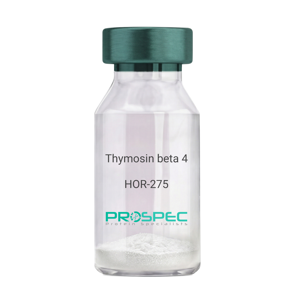
    Thymosin beta 4

    Description:

    Thymosin β4

    Thymosin beta-4. TB500, TB-500

    Product # :

    HOR-275

    Price :

    Quantity :

    Shipping Method :

    Room Temp Icon

    Shipped at Room temp

    Add To Cart

    More Info

    • description
    • formulation
    • purity
    • More Info
    • Citations

    Description

    Thymosin b4 is a 43 amino acid peptide which is regarded as the main intracellular G-actin sequestering peptide. It has a molecular weight of 4963.55 Da, and its molecular formula is: C212H350N56O78S1. Extracellular Thymosin b4 may contribute to physiological processes such as angiogenesis, wound healing, and regulation of inflammation.

    Formulation

    The protein (1mg/ml) was lyophilized with no additives.

    Purity

    Greater than 98.0% as determined by RP-HPLC.

    Citations

    More Info

    • Introduction

      Thymosin is a hormone secreted from the thymus. Its primary function is to stimulate the production of T cells, which are an important part of the immune system. Thymosin also assists in the development of B cells to plasma cells to produce antibodies. The predominant form of thymosin, thymosin b4, is a member of a highly conserved family of actin monomer-sequestering proteins. b-thymosins are the primary regulators of unpolymerized actin, and are essential for maintaining the small cytoplasmic pool of free G-actin monomers required for rapid filament elongation and allowing for the flux of monomers between the thymosin-bound pool and F-actin.

    • Synonyms

      Thymosin beta-4. TB500, TB-500

    • Physical Appearance

      Sterile Filtered White lyophilized (freeze-dried) powder.

    • Stability

      Lyophilized Thymosin b4 although stable at room temperature for 3 weeks, should be stored desiccated below -18°C. Upon reconstitution T beta 4 should be stored at 4°C between 2-7 days and for future use below -18°C.For long term storage it is recommended to add a carrier protein (0.1% HSA or BSA).Please prevent freeze-thaw cycles.

    • Solubility

      It is recommended to reconstitute the lyophilized Thymosin beta-4 in sterile 18MΩ-cm H2O not less than 100 µg/ml, which can then be further diluted to other aqueous solutions.

    • Amino Acid Sequence

      Thymosin b4 has an a.a. sequence of Ac-Ser-Asp-Lys-Pro-Asp-Met-Ala-Glu-Ile-Glu-Lys-Phe-Asp-Lys-Ser-Lys-Leu-Lys-Lys-Thr-Glu-Thr-Gln-Glu-Lys-Asn-Pro-Leu-Pro-Ser-Lys-Glu-Thr-Ile-Glu-Gln-Glu-Lys-Gln-Ala-Gly-Glu-Ser-OH.

    • Background

      What is the molecular weight/Mw of THYMOSIN BETA 4 Protein?
      THYMOSIN BETA 4 Protein has a total Mw of 4.96kDa.

      What is the Purity of THYMOSIN BETA 4 Protein?
      THYMOSIN BETA 4 Protein is >98% pure as determined by SDS-PAGE.

      What is the Biological Activity of THYMOSIN BETA 4 Protein?
      The biological functionality of THYMOSIN BETA 4 Protein will be determined in the future.

      What is the amino acid sequence of THYMOSIN BETA 4 Protein?
      Thymosin b4 has an a.a. sequence of Ac-Ser-Asp-Lys-Pro-Asp-Met-Ala-Glu-Ile-Glu-Lys-Phe-Asp-Lys-Ser-Lys-Leu-Lys-Lys-Thr-Glu-Thr-Gln-Glu-Lys-Asn-Pro-Leu-Pro-Ser-Lys-Glu-Thr-Ile-Glu-Gln-Glu-Lys-Gln-Ala-Gly-Glu-Ser-OH.
      What applications can THYMOSIN BETA 4 Protein be used in?
      THYMOSIN BETA 4 Protein can probably be used in western blot, ELISA and Lateral Flow.

      What is the endotoxin level for THYMOSIN BETA 4 Protein?
      The endotoxin level is minimal, THYMOSIN BETA 4 Protein was purified using conventional chromatography techniques.

      Who supplies THYMOSIN BETA 4 that is in stock and available to order?
      ProSpec has the protein in stock and available for immediate delivery.

      Where can I order bulk mg quantities of THYMOSIN BETA 4?
      ProSpec supplies bulk mg quantities at affordable prices that meets research and academic budgets.

    ProSpec's products are furnished for LABORATORY RESEARCH USE ONLY. The product may not be used as drugs, agricultural or pesticidal products, food additives or household chemicals.

    Thymosin Beta 4
  • View Data Sheet

    Name :

    RPL11 Human

    Description:

    Ribosomal Protein L11 Human Recombinant

    Ribosomal Protein L11, Cell Growth-Inhibiting Protein 34, CLL-Associated Antigen KW-12, 60S Ribosomal Protein L11, DBA7, GIG34, L11.

    Product # :

    PRO-1561

    Price :

    Quantity :

    Shipping Method :

    Ice Icon

    Shipped with Ice Packs

    Add To Cart

    More Info

    • description
    • source
    • formulation
    • purity
    • More Info
    • Citations

    Description

    RPL11 Human Recombinant produced in E.coli is a single, non-glycosylated polypeptide chain containing 201 amino acids (1-178) and having a molecular mass of 22.6kDa.RPL11 is fused to a 23 amino acid His-tag at N-terminus & purified by proprietary chromatographic techniques.

    Source

    Escherichia Coli.

    Formulation

    The RPL11 solution (0.5mg/ml) contains 20mM Tris-HCl buffer (pH 8.0), 0.2M NaCl, 1mM DTT, 2mM EDTA, 250mM Imidazole and 50% glycerol.

    Purity

    Greater than 90% as determined by SDS-PAGE.

    Citations

    More Info

    • Introduction

      Ribosomes are the organelles which catalyze protein synthesis, they’re comprised of a small 40S subunit and a large 60S subunit. Ribosomal Protein L11 (RPL11) is a member of the L5P family of ribosomal proteins. RPL11 is a ribosomal protein which is a component of the 60S subunit. RPL11 is found in the cytoplasm. The RPL11 protein most likely associates with the 5S rRNA.

    • Synonyms

      Ribosomal Protein L11, Cell Growth-Inhibiting Protein 34, CLL-Associated Antigen KW-12, 60S Ribosomal Protein L11, DBA7, GIG34, L11.

    • Physical Appearance

      Sterile Filtered clear solution.

    • Stability

      Store at 4°C if entire vial will be used within 2-4 weeks. Store, frozen at -20°C for longer periods of time. For long term storage it is recommended to add a carrier protein (0.1% HSA or BSA).Avoid multiple freeze-thaw cycles.

    • Amino Acid Sequence

      MGSSHHHHHH SSGLVPRGSH MGSMAQDQGE KENPMRELRI RKLCLNICVG ESGDRLTRAA KVLEQLTGQT PVFSKARYTV RSFGIRRNEK IAVHCTVRGA KAEEILEKGL KVREYELRKN NFSDTGNFGF GIQEHIDLGI KYDPSIGIYG LDFYVVLGRP GFSIADKKRR TGCIGAKHRI SKEEAMRWFQ QKYDGIILPG K

    ProSpec's products are furnished for LABORATORY RESEARCH USE ONLY. The product may not be used as drugs, agricultural or pesticidal products, food additives or household chemicals.

    Rpl11 Human
  • View Data Sheet

    Name :

    IFN g Antibody

    Description:

    Interferon-gamma, Mouse Anti-Human

    Immune Interferon, type II interferon, T cell interferon, MAF, IFNG, IFG, IFI, IFN-gamma.

    Product # :

    ANT-123

    Price :

    Quantity :

    Shipping Method :

    Room Temp Icon

    Shipped at Room temp

    Add To Cart

    More Info

    • formulation
    • More Info
    • Citations

    Formulation

    1mg/ml in PBS (after reconstitution).

    Citations

    More Info

    • Introduction

      IFN-gamma produced by lymphocytes activated by specific antigens or mitogens.
      IFN-gamma, in addition to having antiviral activity, has important immunoregulatory functions, it is a potent activator of macrophages, and has antiproliferative effects on transformed cells and it can potentiate the antiviral and antitumor effects of the type I interferons.

    • Synonyms

      Immune Interferon, type II interferon, T cell interferon, MAF, IFNG, IFG, IFI, IFN-gamma.

    • Solubility

      Reconstitute with sterile H20. Mix gently, wash the sides of the vial and wait 30-60 seconds before use.

    • Immunogen

      r.Human IFN-g.

    • Ig Subclass

      Mouse IgG2a.

    • Clone

      NYRhIFNg.

    • Titer

      This antibody loses its antigen binding capacity when immobilized on plastic. Biological activity is determined by virus neutralization assay and MHC class II induction.

    • Shipping Conditions

      Antibody is shipped lyophilized at ambient temperature.

    • Type

      Mouse Anti Human Monoclonal.

    • Storage Procedures

      In lyophilized form, for long periods, store at 4C in a dry environment. After reconstitution, if not intended for use within a month, aliquot and store at -20C.

    • Purification Method

      Ion exchange.

    ProSpec's products are furnished for LABORATORY RESEARCH USE ONLY. The product may not be used as drugs, agricultural or pesticidal products, food additives or household chemicals.

    Interferon Gamma Human Antibody
  • View Data Sheet

    Name :

    CTSF Human, Sf9

    Description:

    Cathepsin-F Human Recombinant, Sf9

    CTSF, CATSF, CLN13.

    Product # :

    ENZ-1167

    Price :

    Quantity :

    Shipping Method :

    Ice Icon

    Shipped with Ice Packs

    Add To Cart

    More Info

    • description
    • source
    • formulation
    • purity
    • biological activity
    • More Info
    • Citations

    Description

    CTSF produced in Sf9 Baculovirus cells is a single, glycosylated polypeptide chain containing 474 amino acids (20-484.a.a) and having a molecular mass of 52.5kDa.CTSF is expressed with a 6 amino acid His tag at C-Terminus and purified by proprietary chromatographic techniques.

    Source

    Sf9, Baculovirus cells.

    Formulation

    CTSF protein solution (0.25mg/ml) contains Phosphate buffered saline (pH7.4) and 40% glycerol.

    Purity

    Greater than 90.0% as determined by SDS-PAGE.

    Biological Activity

    Specific activity is > 5 pmol/min/ug, and is defined as the amount of enzyme that hydrolyze 1pmole of Z-Phe-ArgAMC to Z-Phe-Arg and AMC per minute at pH 5.0 at 37℃.

    Citations

    More Info

    • Introduction

      Cathepsin F (CTSF) is a member of the peptidase C1 family. Cathepsins are papain family cysteine proteinases which are a main component of the lysosomal proteolytic system. The CTSF gene is ubiquitously expressed, and it maps to chromosome 11q13, close to the gene encoding cathepsin W. CTSF plays a role in normal protein catabolism. CTSF is involved in some degradative processes occurring in tumor progression since it is highly expressed in some cancer cell lines.

    • Synonyms

      CTSF, CATSF, CLN13.

    • Physical Appearance

      Sterile filtered colorless solution.

    • Stability

      Store at 4°C if entire vial will be used within 2-4 weeks.Store, frozen at -20°C for longer periods of time.For long term storage it is recommended to add a carrier protein (0.1% HSA or BSA).Avoid multiple freeze-thaw cycles.

    • Amino Acid Sequence

      ADLAPAQPRA ASFQAWGPPS PELLAPTRFA LEMFNRGRAA GTRAVLGLVR GRVRRAGQGS LYSLEATLEE PPCNDPMVCR LPVSKKTLLC SFQVLDELGR HVLLRKDCGP VDTKVPGAGE PKSAFTQGSA MISSLSQNHP DNRNETFSSV ISLLNEDPLS QDLPVKMASI FKNFVITYNR TYESKEEARW RLSVFVNNMV RAQKIQALDR GTAQYGVTKF SDLTEEEFRT IYLNTLLRKE PGNKMKQAKS VGDLAPPEWD WRSKGAVTKV KDQGMCGSCW AFSVTGNVEG WFLNQGTLL SLSEQELLDC DKMDKACMGG LPSNAYSAIK NLGGLETEDD YSYQGHMQSC NFSAEKAKVY INDSVELSQN EQKLAAWLAK RGPISVAINA FGMQFYRHGI SRPLRPLCSP WLIDHAVLLV GYGNRSDVPF WAIKNSWGTD WGEKGYYYLH RGSGACGVNT MASSAVVDHH HHHH

    ProSpec's products are furnished for LABORATORY RESEARCH USE ONLY. The product may not be used as drugs, agricultural or pesticidal products, food additives or household chemicals.

    Ctsf Protein
  • View Data Sheet

    Name :

    CGREF1 Human

    Description:

    Cell Growth Regulator With EF-Hand Domain 1 Human Recombinant

    Cell Growth Regulator With EF-Hand Domain 1, Cell Growth Regulatory Gene 11 Protein, Hydrophobestin, CGR11, Cell Growth Regulator With EF Hand Domain Protein 1, Cell Growth Regulator With EF Hand Domain 1, Cell growth regulator with EF hand domain protein 1.

    Product # :

    PRO-2154

    Price :

    Quantity :

    Shipping Method :

    Ice Icon

    Shipped with Ice Packs

    Add To Cart

    More Info

    • description
    • source
    • formulation
    • purity
    • More Info
    • Citations

    Description

    CGREF1 Human Recombinant produced in E.Coli is a single, non-glycosylated polypeptide chain containing 305 amino acids (20-301 a.a) and having a molecular mass of 32.3kDa. CGREF1 is fused to a 23 amino acid His-tag at N-terminus & purified by proprietary chromatographic techniques.

    Source

    Escherichia Coli.

    Formulation

    CGREF1 protein solution (0.5mg/ml) containing Phosphate Buffered Saline (pH7.4).

    Purity

    Greater than 85% as determined by SDS-PAGE.

    Citations

    More Info

    • Introduction

      Cell Growth Regulator with EF-Hand Domain 1, also known as CGREF1 is a secreted calcium ion binding protein. CGREF1 includes two EF-hand domains & both EF-hands are essential for function. CGREF1 is most likely digested extracellularly by an unfamiliar serine protease generating extremely hydrophobic bioactive peptides. CGREF1 mediates cell-cell adhesion in a calcium-dependent manner. In addition, CGREF1 is capable to inhibit growth in more than a few cell lines.

    • Synonyms

      Cell Growth Regulator With EF-Hand Domain 1, Cell Growth Regulatory Gene 11 Protein, Hydrophobestin, CGR11, Cell Growth Regulator With EF Hand Domain Protein 1, Cell Growth Regulator With EF Hand Domain 1, Cell growth regulator with EF hand domain protein 1.

    • Physical Appearance

      Sterile filtered colorless solution.

    • Stability

      Store at 4°C if entire vial will be used within 2-4 weeks. Store, frozen at -20°C for longer periods of time. For long term storage it is recommended to add a carrier protein (0.1% HSA or BSA).Avoid multiple freeze-thaw cycles.

    • Amino Acid Sequence

      MGSSHHHHHH SSGLVPRGSH MGSAPKDGVT RPDSEVQHQL LPNPFQPGQE QLGLLQSYLK GLGRTEVQLE HLSREQVLLY LFALHDYDQS GQLDGLELLS MLTAALAPGA ANSPTTNPVI LIVDKVLETQ DLNGDGLMTP AELINFPGVA LRHVEPGEPL APSPQEPQAV GRQSLLAKSP LRQETQEAPG PREEAKGQVE ARRESLDPVQ EPGGQAEADG DVPGPRGEAE GQAEAKGDAP GPRGEAGGQA EAEGDAPGPR GEAGGQAEAR ENGEEAKELP GETLESKNTQ NDFEVHIVQV ENDEI.

    ProSpec's products are furnished for LABORATORY RESEARCH USE ONLY. The product may not be used as drugs, agricultural or pesticidal products, food additives or household chemicals.

    Cgref1 Human
  • View Data Sheet

    Name :

    NECAP2 Human

    Description:

    NECAP Endocytosis Associated 2 Human Recombinant

    NECAP endocytosis-associated protein 2, NECAP-2, FLJ10420, Adaptin ear-binding coat-associated protein 2.

    Product # :

    PRO-180

    Price :

    Quantity :

    Shipping Method :

    Ice Icon

    Shipped with Ice Packs

    Add To Cart

    More Info

    • description
    • source
    • formulation
    • purity
    • More Info
    • Citations

    Description

    NECAP2 produced in E.Coli is a single, non-glycosylated polypeptide chain containing 283 amino acids (1-263a.a.) and having a molecular mass of 30.5 kDa. NECAP2 is fused to a 20 amino acid His-tag at N-terminus & purified by proprietary chromatographic techniques.

    Source

    Escherichia Coli.

    Formulation

    The NECAP2 protein solution (1mg/1ml) is formulated in 20mM Tris-HCl buffer (pH8.0), 100mM NaCl, 2mM DTT and 10% glycerol.

    Purity

    Greater than 85% as determined by SDS-PAGE.

    Citations

    More Info

    • Introduction

      NECAP2 which is involved in endocytosis, is a vital protein paralogues for clathrin-mediated membrane trafficking which are enhanced in CCV layers. NECAP2 protein colocalizes with AP-2 at the plasma membrane by binding AP-2s ?-ear domain, and interacts with AP-1, AP-2 and several GAE domain proteins termed GGA1, GGA2 and GGA3.

    • Synonyms

      NECAP endocytosis-associated protein 2, NECAP-2, FLJ10420, Adaptin ear-binding coat-associated protein 2.

    • Physical Appearance

      Sterile Filtered clear solution.

    • Stability

      Store at 4°C if entire vial will be used within 2-4 weeks. Store, frozen at -20°C for longer periods of time. For long term storage it is recommended to add a carrier protein (0.1% HSA or BSA).Avoid multiple freeze-thaw cycles.

    • Amino Acid Sequence

      MGSSHHHHHH SSGLVPRGSH MEESGYESVL CVKPDVHVYR IPPRATNRGY RAAEWQLDQP SWSGRLRITA KGQMAYIKLE DRTSGELFAQ APVDQFPGTA VESVTDSSRY FVIRIEDGNG RRAFIGIGFG DRGDAFDFNV ALQDHFKWVK QQCEFAKQAQ NPDQGPKLDL GFKEGQTIKL NIANMKKKEG AAGNPRVRPA STGGLSLLPP PPGGKTSTLI PPPGEQLAVG GSLVQPAVAP SSGGAPVPWP QPNPATADIW GDFTKSTGST SSQTQPGTGW VQF

    ProSpec's products are furnished for LABORATORY RESEARCH USE ONLY. The product may not be used as drugs, agricultural or pesticidal products, food additives or household chemicals.

    Necap2 Human
  • View Data Sheet

    Name :

    SDC2 Human

    Description:

    Syndecan-2 Human Recombinant

    Syndecan 2, Heparan Sulfate Proteoglycan 1 Cell Surface-Associated, Heparan Sulfate Proteoglycan Core Protein, Syndecan Proteoglycan 2, Fibroglycan, HSPG1, SYND2, HSPG, Cell Surface-Associated Heparan Sulfate Proteoglycan 1, CD362 Antigen, Syndecan-2, CD362, SDC2.

    Product # :

    PRO-2449

    Price :

    Quantity :

    Shipping Method :

    Ice Icon

    Shipped with Ice Packs

    Add To Cart

    More Info

    • description
    • source
    • formulation
    • purity
    • More Info
    • Citations

    Description

    SDC2 Human Recombinant produced in Sf9 Baculovirus cells is a single, glycosylated polypeptide chain containing 135 amino acids (19-144a.a.) and having a molecular mass of 15kDa (Molecular size on SDS-PAGE will appear at approximately 18-28kDa). SDC2 is expressed with a 6 amino acids His tag at C-Terminus and purified by proprietary chromatographic techniques.

    Source

    Sf9, Baculovirus cells.

    Formulation

    SDC2 protein solution (0.25mg/ml) contains Phosphate Buffered Saline (pH 7.4) and 10% glycerol.

    Purity

    Greater than 90.0% as determined by SDS-PAGE.

    Citations

    More Info

    • Introduction

      Syndecan 2 (SDC2) is a conserved family of heparan- and chondroitin-sulfate carrying transmembrane protein. Syndecan 2 is a cell membrane proteoglycan which modulates the activity and dynamics of some growth factor receptors and integrins. SDC2 is expressed in HSCs (hepatic stellate cells), a key cell type involved in matrix deposition in liver fibrotic disorders. SDC2 is implicated in the formation of specialized membrane domains and serves as a direct link between the extracellular environment and the organization of the cortical cytoplasm.

    • Synonyms

      Syndecan 2, Heparan Sulfate Proteoglycan 1 Cell Surface-Associated, Heparan Sulfate Proteoglycan Core Protein, Syndecan Proteoglycan 2, Fibroglycan, HSPG1, SYND2, HSPG, Cell Surface-Associated Heparan Sulfate Proteoglycan 1, CD362 Antigen, Syndecan-2, CD362, SDC2.

    • Physical Appearance

      Sterile filtered colorless solution.

    • Stability

      Store at 4°C if entire vial will be used within 2-4 weeks. Store, frozen at -20°C for longer periods of time. For long term storage it is recommended to add a carrier protein (0.1% HSA or BSA).Avoid multiple freeze-thaw cycles.

    • Amino Acid Sequence

      ADPESRAELT SDKDMYLDNS SIEEASGVYP IDDDDYASAS GSGADEDVES PELTTSRPLP KILLTSAAPK VETTTLNIQN KIPAQTKSPE ETDKEKVHLS DSERKMDPAE EDTNVYTEKH SDSLFKRTEH HHHHH.

    ProSpec's products are furnished for LABORATORY RESEARCH USE ONLY. The product may not be used as drugs, agricultural or pesticidal products, food additives or household chemicals.

    Sdc2 Human
  • View Data Sheet

    Name :

    Zika NS1, HEK

    Description:

    Zika NS1 Protein Recombinant, HEK

    Product # :

    ZKV-003

    Price :

    Quantity :

    Shipping Method :

    Ice Icon

    Shipped with Ice Packs

    Add To Cart

    More Info

    • description
    • source
    • formulation
    • purity
    • More Info
    • Citations

    Description

    The HEK derived recombinant Zika NS1 protein strain MR-766 (Prototype, Uganda, 1947, GenBank accession number AY632535).The Zika NS1 protein is fused to a 6xHis tag at C-terminus and purified by proprietary chromatographic technique.

    Source

    Human Embryonic Kidney 293 cells.

    Formulation

    Zika NS1 protein contains Dulbecco’s Phosphate buffered saline, pH 7.4.

    Purity

    Zika NS1 protein is >90% pure as determined by SDS-PAGE.

    Citations

    More Info

    • Introduction

      Zika virus (ZIKV) belongs to the family Flaviviridae and the genus Flavivirus, it is transmitted by daytime-active Aedes mosquitoes, such as A. aegypti and A. albopictus. The Zika virus is related to the dengue, yellow fever, Japanese encephalitis, and West Nile viruses. Much like the other flaviviruses, Zika virus is enveloped and icosahedral and has a nonsegmented, single-stranded, positive-sense RNA genome. Zika fever is an infection, which often causes no symptoms or only mild ones, like a mild form of dengue fever, and it is treated by rest. As of February 2016, there has been mounting evidence that Zika fever in pregnant women can cause abnormal brain development in their fetuses by mother-to-child transmission, which may result in miscarriage or microcephaly, however it is not yet known whether Zika virus causes microcephaly. Furthermore, a connection has been established with neurologic conditions in infected adults, including Guillain–Barre syndrome.

    • Physical Appearance

      Sterile Filtered solution.

    • Stability

      Zika NS1 protein although stable at 4°C for 1 week, should be stored below -18°C. Please prevent freeze thaw cycles.

    • Applications

      Each laboratory should determine an optimum working titer for use in its particular application.

    ProSpec's products are furnished for LABORATORY RESEARCH USE ONLY. The product may not be used as drugs, agricultural or pesticidal products, food additives or household chemicals.

    Zika Ns1 Hek
  • View Data Sheet

    Name :

    KRT19 Human

    Description:

    Cytokeratin 19 Human Recombinant

    Keratin type I cytoskeletal 19, Cytokeratin-19, CK-19, Keratin-19, K19, KRT19, CK19, K1CS, MGC15366.

    Product # :

    PRO-350

    Price :

    Quantity :

    Shipping Method :

    Room Temp Icon

    Shipped at Room temp

    Add To Cart

    More Info

    • description
    • source
    • formulation
    • purity
    • More Info
    • Citations

    Description

    Cytokeratin 19 Human Recombinant produced in E.Coli is a single, non-glycosylated polypeptide chain having a molecular mass of 44,098 Dalton. The KRT19 is purified by proprietary chromatographic techniques.

    Source

    Escherichia Coli.

    Formulation

    The protein (1mg/ml) was lyophilized after from a sterile solution containing 30mM Tris-HCl pH-8, 9.5M urea, 2mM DTT, 2mM EDTA and 10mM methylammonium chloride.

    Purity

    Greater than 95.0% as determined by SDS-PAGE.

    Citations

    More Info

    • Introduction

      CTK-19 is a member of the keratin family. The keratins are intermediate filament proteins responsible for the structural integrity of epithelial cells and are subdivided into cytokeratins and hair keratins. The type I cytokeratins consist of acidic proteins which are arranged in pairs of heterotypic keratin chains. Unlike its related family members, this smallest known acidic cytokeratin is not paired with a basic cytokeratin in epithelial cells. It is specifically expressed in the periderm, the transiently superficial layer that envelopes the developing epidermis. The type I cytokeratins are clustered in a region of chromosome 17q12-q21.

    • Synonyms

      Keratin type I cytoskeletal 19, Cytokeratin-19, CK-19, Keratin-19, K19, KRT19, CK19, K1CS, MGC15366.

    • Physical Appearance

      Sterile Filtered White lyophilized (freeze-dried) powder.

    • Stability

      Lyophilized KRT19 although stable at room temperature for 3 weeks, should be stored desiccated below -18°C. Upon reconstitution KRT19 should be stored at 4°C between 2-7 days and for future use below -18°C.For long term storage it is recommended to add a carrier protein (0.1% HSA or BSA).Please prevent freeze-thaw cycles.

    • Solubility

      It is recommended to reconstitute the lyophilized KRT19 in sterile 18MΩ-cm H2O not less than 100µg/ml, which can then be further diluted to other aqueous solutions.

    • Reconstitution to filaments

      Performed by mixing equimolar amounts of cytokeratins of type I and type II at concentrations of approx. 0.5 mg/ml, both dissolved in 9.5 M urea buffer (see above). Protofilaments and filament complexes are obtained by dialyzing the resulting polypeptide solution stepwise to a concentration of 4 M urea and then to low salt condition (50 mM NaCl, 2 mM dithiothreitol, 10 mM Tris-HCl, pH 7.4). For immunization purposes, the solution can be further dialyzed against PBS (phosphate buffered saline, e.g. Dulbecco's PBS).

    ProSpec's products are furnished for LABORATORY RESEARCH USE ONLY. The product may not be used as drugs, agricultural or pesticidal products, food additives or household chemicals.

    Krt19 Human
  • View Data Sheet

    Name :

    G CSF Antibody, FITC

    Description:

    Granulocyte Colony Stimulating Factor, Mouse Anti-Human, FITC

    CSF-3, MGI-1G, GM-CSF beta, Pluripoietin, Filgrastim, Lenograstim, G-CSF, MGC45931, GCSF.

    Product # :

    ANT-241

    Price :

    Quantity :

    Shipping Method :

    Room Temp Icon

    Shipped at Room temp

    Add To Cart

    More Info

    • formulation
    • More Info
    • Citations

    Formulation

    1 mg/ml in PBS (after reconstitution).

    Citations

    More Info

    • Introduction

      GCSF is a cytokine that controls the production, differentiation, and function of granulocytes. The active protein is found extracellularly. Three transcript variants encoding three different isoforms have been found for this gene.
      Granulocyte/macrophage colony-stimulating factors are cytokines that act in hematopoiesis by controlling the production, differentiation, and function of 2 related white cell populations of the blood, the granulocytes and the monocytes-macrophages. This csf induces granulocytes.

    • Synonyms

      CSF-3, MGI-1G, GM-CSF beta, Pluripoietin, Filgrastim, Lenograstim, G-CSF, MGC45931, GCSF.

    • Solubility

      Reconstitute with sterile H20. Mix gently, wash the sides of the vial and wait 30-60 seconds before use.

    • Immunogen

      r.Human G-CSF.

    • Ig Subclass

      Mouse IgG.

    • Clone

      NYRhGCSF.

    • Applications

      Direct ELISA, Western Blot, Intra-cellular staining.

    • Titer

      For intra-cellular staining use 10µl per 1,000,000 cells.

    • Shipping Conditions

      Antibody is shipped lyophilized at ambient temperature.

    • Type

      Mouse Anti Human Monoclonal.

    • Storage Procedures

      In lyophilized form, for long periods, store at 4°C in a dry environment. After reconstitution, if not intended for use within a month, aliquot and store at -20°C.

    • Purification Method

      Protein A.

    ProSpec's products are furnished for LABORATORY RESEARCH USE ONLY. The product may not be used as drugs, agricultural or pesticidal products, food additives or household chemicals.

    G Csf Antibody Fitc
  • View Data Sheet

    Name :

    CD300E Human

    Description:

    CD300E Human Recombinant

    CD300e Molecule, CD300e Antigen, CD300LE, IREM2, CLM2, Immune Receptor Expressed On Myeloid Cells 2, Polymeric Immunoglobulin Receptor 2,CD300 Antigen Like Family Member E, CD300 Antigen-Like Family Member E,Poly-Ig Receptor 2, CMRF35-A5, PIgR2, CMRF35-Like Molecule 2, CMRF35A5, CD300E.

    Product # :

    PRO-2114

    Price :

    Quantity :

    Shipping Method :

    Ice Icon

    Shipped with Ice Packs

    Add To Cart

    More Info

    • description
    • source
    • formulation
    • purity
    • More Info
    • Citations

    Description

    CD300E Human Recombinant produced in E.Coli is a single, non-glycosylated polypeptide chain containing 179 amino acids (18-173 a.a) and having a molecular mass of 19.9kDa.CD300E is fused to a 23 amino acid His-tag at N-terminus & purified by proprietary chromatographic techniques.

    Source

    Escherichia Coli.

    Formulation

    CD300E protein solution (1mg/ml) containing 20mM Tris-HCl buffer (pH8.0).

    Purity

    Greater than 90.0% as determined by SDS-PAGE.

    Citations

    More Info

    • Introduction

      CMRF35-like molecule 2 precursor (CD300E) is a member of the CD300 family. CD300E is a 205 amino acid single-pass type I membrane protein which contains one Ig-like V-type (immunoglobulin-like) domain. While interacting with DAP12, CD300E probably performs as an activating receptor. CD300E is present on the surface of mature hematopoietic cells of the monocyte & myeloid lineages.

    • Synonyms

      CD300e Molecule, CD300e Antigen, CD300LE, IREM2, CLM2, Immune Receptor Expressed On Myeloid Cells 2, Polymeric Immunoglobulin Receptor 2,CD300 Antigen Like Family Member E, CD300 Antigen-Like Family Member E,Poly-Ig Receptor 2, CMRF35-A5, PIgR2, CMRF35-Like Molecule 2, CMRF35A5, CD300E.

    • Physical Appearance

      Sterile Filtered clear solution.

    • Stability

      Store at 4°C if entire vial will be used within 2-4 weeks. Store, frozen at -20°C for longer periods of time. For long term storage it is recommended to add a carrier protein (0.1% HSA or BSA).Avoid multiple freeze-thaw cycles.

    • Amino Acid Sequence

      MGSSHHHHHH SSGLVPRGSH MGSLKGPGSV TGTAGDSLTV WCQYESMYKG YNKYWCRGQY DTSCESIVET KGEEKVERNG RVSIRDHPEA LAFTVTMQNL NEDDAGSYWC KIQTVWVLDS WSRDPSDLVR VYVSPAITTP RRTTHPATPP IFLVVNPGRN LSTGEVLTQN SGFRLSSPH.

    ProSpec's products are furnished for LABORATORY RESEARCH USE ONLY. The product may not be used as drugs, agricultural or pesticidal products, food additives or household chemicals.

    Cd300E Human
  • View Data Sheet

    Name :

    IL10 Rat

    Description:

    Interleukin-10 Rat Recombinant

    B-TCGF, CSIF, TGIF, IL-10, IL10A, MGC126450, MGC126451, Cytokine synthesis inhibitory factor.

    Product # :

    CYT-465

    Price :

    Quantity :

    Shipping Method :

    Room Temp Icon

    Shipped at Room temp

    Add To Cart

    More Info

    • description
    • source
    • formulation
    • purity
    • biological activity
    • More Info
    • Citations

    Description

    IL-10 Recombinant Rat produced in E.coli is a single, glycosylated polypeptide chain containing 160 amino acids and having a molecular mass of 18.6 kDa. The Interleukin-10 Mouse is purified by proprietary chromatographic techniques.

    Source

    Escherichia Coli.

    Formulation

    The protein was lyophilized containing 20mM Tris-HCl pH-8.0 and 100mM NaCl.

    Purity

    Greater than 97.0% as determined by:
    (a) Analysis by RP-HPLC.
    (b) Analysis by SDS-PAGE.

    Biological Activity

    The ED50 as determined by the dose dependent inhibition of MC/9 proliferation is less than 10ng/ml concentration corresponding to a Specific Activity of 100,000IU/mg.

    Citations

    More Info

    • Introduction

      IL10 is a cytokine produced primarily by monocytes and to a lesser extent by lymphocytes. This cytokine has pleiotropic effects in immunoregulation and inflammation. It down-regulates the expression of Th1 cytokines, MHC class II Ags, and costimulatory molecules on macrophages. It also enhances B cell survival, proliferation, and antibody production. This cytokine can block NF-kappa B activity, and is involved in the regulation of the JAK-STAT signaling pathway. Knockout studies in mice suggested the function of this cytokine as an essential immunoregulator in the intestinal tract.

    • Synonyms

      B-TCGF, CSIF, TGIF, IL-10, IL10A, MGC126450, MGC126451, Cytokine synthesis inhibitory factor.

    • Physical Appearance

      Sterile Filtered White lyophilized (freeze-dried) powder.

    • Stability

      Lyophilized IL-10 Rat although stable at room temperature for 3 weeks, should be stored desiccated below -18°C. Upon reconstitution Interleukin10 Rat recombinant should be stored at 4°C between 2-7 days and for future use below -18°C.For long term storage it is recommended to add a carrier protein (0.1% HSA or BSA).Please prevent freeze-thaw cycles.

    • Solubility

      It is recommended to reconstitute the lyophilized IL-10 Rat Recombinant in sterile 18MΩ-cm H2O not less than 100 µg/ml, which can then be further diluted to other aqueous solutions.

    • Amino Acid Sequence

      SKGHSIRGDN NCTHFPVSQT HMLRELRAAF SQVKTFFQKK DQLDNILLTD SLLQDFKGYL GCQALSEMIK FYLVEVMPQA ENHGPEIKEH LNSLGEKLKT LWIQLRRCHR FLPCENKSKA VEQVKNDFNK LQDKGVYKAM NEFDIFINCI EAYVTLKMKN.

    • Background

      What is the molecular weight/Mw of IL10 RAT Protein?
      IL10 RAT Protein has a total Mw of 18.6kDa.

      What is the source or expression system of IL10 RAT Protein?
      Escherichia Coli.

      What is the Purity of IL10 RAT Protein?
      IL10 RAT Protein is >97% pure as determined by SDS-PAGE.

      What is the Biological Activity of IL10 RAT Protein?
      The ED50 as determined by the dose dependent inhibition of MC/9 proliferation is less than 10ng/ml concentration corresponding to a Specific Activity of 100,000IU/mg.

      What is the amino acid sequence of IL10 RAT Protein?
      SKGHSIRGDN NCTHFPVSQT HMLRELRAAF SQVKTFFQKK DQLDNILLTD SLLQDFKGYL GCQALSEMIK FYLVEVMPQA ENHGPEIKEH LNSLGEKLKT LWIQLRRCHR FLPCENKSKA VEQVKNDFNK LQDKGVYKAM NEFDIFINCI EAYVTLKMKN.

      What applications can IL10 RAT Protein be used in?
      IL10 RAT Protein can probably be used in western blot, ELISA and Lateral Flow.

      What is the endotoxin level for IL10 RAT Protein?
      The endotoxin level is minimal, IL10 RAT Protein was purified using conventional chromatography techniques.

      Who supplies recombinant IL10 RAT that is in stock and available to order?
      ProSpec has the protein in stock and available for immediate delivery.

      Where can I order bulk mg quantities of IL10 RAT?
      ProSpec supplies bulk mg quantities at affordable prices that meets research and academic budgets.

    ProSpec's products are furnished for LABORATORY RESEARCH USE ONLY. The product may not be used as drugs, agricultural or pesticidal products, food additives or household chemicals.

    Il 10 Rat
  • View Data Sheet

    Name :

    EREG Human, HEK

    Description:

    Epiregulin Human Recombinant, HEK

    EPR, Epiregulin, Ep, ER, Proepiregulin, EREG.

    Product # :

    CYT-1206

    Price :

    Quantity :

    Shipping Method :

    Ice Icon

    Shipped with Ice Packs

    Add To Cart

    More Info

    • description
    • source
    • formulation
    • purity
    • biological activity
    • More Info
    • Citations

    Description

    EREG Human Recombinant produced in HEK293 cells is a single, glycosylated polypeptide chain (63-108a.a) containing 289 amino acids and having a molecular mass of 32.6 kDa.EREG is fused to a 239 amino acid hIgG-His-Tag at C-terminus & purified by proprietary chromatographic techniques.

    Source

    HEK293 cells.

    Formulation

    EREG protein (0.25mg/ml) contains 10% glycerol and Phosphate-Buffered Saline (pH 7.4).

    Purity

    Greater than 90.0% as determined by SDS-PAGE.

    Biological Activity

    Measured in a cell proliferation assay using Balb/3T3 mouse embryonic fibroblast cells. The ED50 range ≤ 1ug/ml.

    Citations

    More Info

    • Introduction

      "Epiregulin is part of the EGF family. Epiregulin functions as a ligand of EGFR, as well as a ligand of most members of the ERBB (v-erb-b2 oncogene homolog) family of tyrosine-kinase receptors.Epiregulin is expressed mostly in the placenta and peripheral blood leukocytes and in specific carcinomas of the bladder, lung, kidney and colon. Epiregulin stimulates the proliferation of keratinocytes, hepatocytes, fibroblasts and vascular smooth muscle cells. Epiregulin inhibits the growth of several tumor-derived epithelial cell lines. Human Epiregulin is initially synthesized as a glycosylated 19.0 kDa transmembrane precursor protein, which is processed by proteolytic cleavage to produce a 6.0 kDa mature secreted sequence."

    • Synonyms

      EPR, Epiregulin, Ep, ER, Proepiregulin, EREG.

    • Physical Appearance

      Sterile filtered colorless solution.

    • Stability

      Store at 4°C if entire vial will be used within 2-4 weeks.Store, frozen at -20°C for longer periods of time.For long term storage it is recommended to add a carrier protein (0.1% HSA or BSA).Avoid multiple freeze-thaw cycles.

    • Amino Acid Sequence

      DGSMVSITKC SSDMNGYCLH GQCIYLVDMS QNYCRCEVGY TGVRCEHFFL LEPKSCDKTH TCPPCPAPEL LGGPSVFLFP PKPKDTLMIS RTPEVTCVVV DVSHEDPEVK FNWYVDGVEV HNAKTKPREE QYNSTYRVVS VLTVLHQDWL NGKEYKCKVS NKALPAPIEK TISKAKGQPR EPQVYTLPPS RDELTKNQVS LTCLVKGFYP SDIAVEWESN GQPENNYKTT PPVLDSDGSF FLYSKLTVDK SRWQQGNVFS CSVMHEALHN HYTQKSLSLS PGKHHHHHH

    • Background

      What is the molecular weight/Mw of EREG Protein?
      EREG Protein has a total Mw of 32.6kDa.

      What is the source or expression system of EREG Protein?
      HEK293 cells.

      What is the Purity of EREG Protein?
      EREG Protein is >90% pure as determined by SDS-PAGE.

      What is the Biological Activity of EREG Protein?
      Measured in a cell proliferation assay using Balb/3T3 mouse embryonic fibroblast cells. The ED50 range ≤ 1ug/ml.

      What is the amino acid sequence of EREG Protein?
      DGSMVSITKC SSDMNGYCLH GQCIYLVDMS QNYCRCEVGY TGVRCEHFFL LEPKSCDKTH TCPPCPAPEL LGGPSVFLFP PKPKDTLMIS RTPEVTCVVV DVSHEDPEVK FNWYVDGVEV HNAKTKPREE QYNSTYRVVS VLTVLHQDWL NGKEYKCKVS NKALPAPIEK TISKAKGQPR EPQVYTLPPS RDELTKNQVS LTCLVKGFYP SDIAVEWESN GQPENNYKTT PPVLDSDGSF FLYSKLTVDK SRWQQGNVFS CSVMHEALHN HYTQKSLSLS PGKHHHHHH

      What applications can EREG Protein be used in?
      EREG Protein can probably be used in western blot, ELISA and Lateral Flow.

      What is the endotoxin level for EREG Protein?
      The endotoxin level is minimal, EREG Protein was purified using conventional chromatography techniques.

    ProSpec's products are furnished for LABORATORY RESEARCH USE ONLY. The product may not be used as drugs, agricultural or pesticidal products, food additives or household chemicals.

    Ereg Human
  • View Data Sheet

    Name :

    KLK3 Human, HEK

    Description:

    Kallikrein-3 Human Recombinant, HEK

    Prostate-specific antigen, PSA, Gamma-seminoprotein, Seminin, Kallikrein-3, P-30 antigen, Semenogelase, KLK3, APS, hK3, KLK2A1

    Product # :

    ENZ-1149

    Price :

    Quantity :

    Shipping Method :

    Ice Icon

    Shipped with Ice Packs

    Add To Cart

    More Info

    • description
    • source
    • formulation
    • purity
    • biological activity
    • More Info
    • Citations

    Description

    KLK3 Human Recombinant produced in HEK cells is a single, glycosylated, polypeptide chain (18-261 a.a) containing a total of 250 amino acids, having a molecular mass of 27.6kDa.KLK3 is fused to a 6 amino acid His-tag at C-terminus,and is purified by proprietary chromatographic techniques.

    Source

    HEK293 Cells.

    Formulation

    The KLK3 solution (0.25mg/ml) contains 10% Glycerol and Phosphate-Buffered Saline (pH 7.4).

    Purity

    Greater than 95.0% as determined by SDS-PAGE.

    Biological Activity

    Specific activity is  > 250pmol/min/ug. Defined by the amount of enzyme that cleaves 1pmole of Succinyl-ArgPro-Tyr-p-Nitroanilide to Succinyl-Arg-Pro-Tyr and p-Nitroanilide per minute at pH 7.5 at 37˚C.

    Citations

    More Info

    • Introduction

      Kallikrein-3 or KLK3 is a serine proteases subgroup, enzymes can cleave peptide bonds. KLK3 is found in the prostate and may be hold accountable to the regulation of semen liquefaction via seminogelin hydrolysis. The protein is expressed in low concentrations in men with healthy prostates serum. When elevated quantities are found it may show prostate cancer or different prostate disorders such as prostatitis and benign prostatic hyperplasia.

    • Synonyms

      Prostate-specific antigen, PSA, Gamma-seminoprotein, Seminin, Kallikrein-3, P-30 antigen, Semenogelase, KLK3, APS, hK3, KLK2A1

    • Physical Appearance

      Sterile Filtered colorless solution.

    • Stability

      Store at 4°C if entire vial will be used within 2-4 weeks. Store, frozen at -20°C for longer periods of time. For long term storage it is recommended to add a carrier protein (0.1% HSA or BSA).Avoid multiple freeze-thaw cycles.

    • Amino Acid Sequence

      APLILSRIVG GWECEKHSQP WQVLVASRGR AVCGGVLVHP QWVLTAAHCI RNKSVILLGR HSLFHPEDTG QVFQVSHSFP HPLYDMSLLK NRFLRPGDDS SHDLMLLRLS EPAELTDAVK VMDLPTQEPA LGTTCYASGW GSIEPEEFLT PKKLQCVDLH VISNDVCAQV HPQKVTKFML CAGRWTGGKS TCSGDSGGPL VCNGVLQGIT SWGSEPCALP ERPSLYTKVV HYRKWIKDTI VANPHHHHHH

    ProSpec's products are furnished for LABORATORY RESEARCH USE ONLY. The product may not be used as drugs, agricultural or pesticidal products, food additives or household chemicals.

    Kallikrein 3 Human
  • View Data Sheet

    Name :

    EGF (Leu 21) Human

    Description:

    Epidermal Growth Factor (Leu-21) Human Recombinant

    Urogastrone, URG, EGF.

    Product # :

    CYT-466

    Price :

    Quantity :

    Shipping Method :

    Room Temp Icon

    Shipped at Room temp

    Add To Cart

    More Info

    • description
    • source
    • formulation
    • purity
    • biological activity
    • More Info
    • Citations

    Description

    EGF 21-Leu Human Recombinant produced in E.Coli is a single, non-glycosylated, polypeptide chain containing 53 amino acids and having a molecular mass of 6205 Dalton.The EGF 21-Leu is purified by proprietary chromatographic techniques.

    Source

    Escherichia Coli.

    Formulation

    The protein was lyophilized from a concentrated (1mg/ml) solution with no additives.

    Purity

    Greater than 98.0% as determined by(a) Analysis by RP-HPLC.
    (b) Analysis by SDS-PAGE.

    Biological Activity

    The ED50, calculated by the dose-dependant proliferation of MDCK cells is < 10ng/ml concentration corresponding to a Specific Activity of 100,000IU/mg.

    Citations

    More Info

    • Introduction

      Epidermal growth factor has a profound effect on the differentiation of specific cells in vivo and is a potent mitogenic factor for a variety of cultured cells of both ectodermal and mesodermal origin. The EGF precursor is believed to exist as a membrane-bound molecule which is proteolytically cleaved to generate the 53-amino acid peptide hormone that stimulates cells to divide. EGF stimulates the growth of various epidermal and epithelial tissues in vivo and in vitro and of some fibroblasts in cell culture.

    • Synonyms

      Urogastrone, URG, EGF.

    • Physical Appearance

      Sterile Filtered White lyophilized (freeze-dried) powder.

    • Stability

      Lyophilized Epidermal Growth Factor 21 Leu although stable at room temperature for 3 weeks, should be stored desiccated below -18°C. Upon reconstitution EGF 21-Leu should be stored at 4°C between 2-7 days and for future use below -18°C.For long term storage it is recommended to add a carrier protein (0.1% HSA or BSA).Please prevent freeze-thaw cycles.

    • Solubility

      It is recommended to reconstitute the lyophilized Epidermal Growth Factor 21-Leu in sterile 18MΩ-cm H2O not less than 100µg/ml, which can then be further diluted to other aqueous solutions.

    • Amino Acid Sequence

      The sequence of the first five N-terminal amino acids was determined and was found to be Asn-Ser-Asp-Ser-Glu.

    • Background

      Deciphering the Potential of Epidermal Growth Factor (Leu-21) Human Recombinant: Unveiling Novel Insights and Therapeutic Implications

      Abstract:

      This concise research paper delves into the enigmatic landscape of Epidermal Growth Factor (Leu-21) Human Recombinant, illuminating its intricate molecular characteristics, signaling pathways, and promising therapeutic avenues. Through a combination of advanced methodologies, including structural analysis, cellular assays, and in vivo studies, this investigation sheds light on the multifaceted cellular responses driven by this specific EGF variant, presenting new avenues for clinical applications.

      Introduction:

      Central to cellular processes, Epidermal Growth Factor (EGF) stands as a pivotal cytokine. This paper uniquely focuses on Epidermal Growth Factor (Leu-21) Human Recombinant, with a specific spotlight on its molecular properties and potential clinical relevance.

      Molecular Insights and Signaling Dynamics:

      The crux of its functionality lies in the interaction between Epidermal Growth Factor (Leu-21) and its cognate receptor, initiating a cascade of intracellular events. Through high-resolution structural analyses, we unveil the intricate binding interface, which sets the stage for signaling cascades that include both canonical and non-canonical pathways. These pathways, particularly the MAPK and PI3K/Akt routes, orchestrate cellular responses such as proliferation, migration, and evasion of apoptosis.

      Experimental Profiling and Cellular Responses:

      In decoding the cellular ramifications, a repertoire of in vitro assays has been meticulously employed. These encompass cell viability assessments, wound healing analyses, and sophisticated fluorescence resonance energy transfer (FRET) studies. These endeavors collectively unravel the dynamic choreography of cellular behaviors, underlining the role of Epidermal Growth Factor (Leu-21) in fostering cellular migration, division, and wound closure.

      In Vivo Implications and Therapeutic Prospects:

      Translating these in vitro insights to clinical potential, in vivo investigations present a compelling narrative. Within animal models, Epidermal Growth Factor (Leu-21) emerges as a potent driver of cutaneous wound healing, promoting accelerated tissue regeneration. Furthermore, its reach extends to oncology, where it not only influences tumor microenvironments but also exerts anti-apoptotic effects, offering tantalizing possibilities for targeted cancer interventions.

      Future Challenges and Prospects:

      While these discoveries hold immense promise, challenges linger. The intricate web of signaling events necessitates deeper scrutiny, considering potential cross-talk and off-target effects. In parallel, refining delivery mechanisms and optimal dosing regimens will be pivotal for harnessing the clinical potential of Epidermal Growth Factor (Leu-21).

      Conclusion:

      In a symphony of complex molecular insights and tangible therapeutic potential, Epidermal Growth Factor (Leu-21) Human Recombinant emerges as a captivating enigma. Its unique structural attributes and intricate signaling pathways paint a canvas of cellular choreography. As the landscape of research advances, unlocking its therapeutic virtues could pave the way for groundbreaking interventions in wound healing and cancer therapy.

      What is the molecular weight/Mw of EGF Protein?
      EGF Protein has a total Mw of 6.2kDa.

      What is the source or expression system of EGF Protein?
      Escherichia Coli.

      What is the Purity of EGF Protein?
      EGF Protein is >98% pure as determined by SDS-PAGE.

      What is the Biological Activity of EGF Protein?
      The ED50, calculated by the dose-dependant proliferation of MDCK cells is < 10ng/ml concentration corresponding to a Specific Activity of 100,000IU/mg.

      What is the amino acid sequence of EGF Protein?
      EGF Protein is composed from 53 amino acids.

      What applications can EGF Protein be used in?
      EGF Protein can probably be used in western blot, ELISA and Lateral Flow.

      What is the endotoxin level for EGF Protein?
      The endotoxin level is minimal, EGF Protein was purified using conventional chromatography techniques.

    ProSpec's products are furnished for LABORATORY RESEARCH USE ONLY. The product may not be used as drugs, agricultural or pesticidal products, food additives or household chemicals.

    Egf 21 Leu Human
  • View Data Sheet

    Name :

    CD300A Human

    Description:

    CD300A Human Recombinant

    CD300a Molecule, CD300a Antigen, IRp60, CD300 Antigen-Like Family Member A, NK Inhibitory Receptor, Immunoglobulin Superfamily Member 12, CMRF35-Like Molecule 8, CMRF35-H, Inhibitory Receptor Protein 60, CMRF-35-H9, CMRF-35H, CLM-8, IRC1, IRC2, CMRF35H Leukocyte Immunoglobulin-Like Receptor, Leukocyte Membrane Antigen, IgSF12.

    Product # :

    PRO-1618

    Price :

    Quantity :

    Shipping Method :

    Ice Icon

    Shipped with Ice Packs

    Add To Cart

    More Info

    • description
    • source
    • formulation
    • purity
    • More Info
    • Citations

    Description

    CD300A Human Recombinant produced in E.coli is a single, non-glycosylated polypeptide chain containing 134 amino acids (18-128) and having a molecular mass of 14.7kDa.CD300A is fused to a 23 amino acid His-tag at N-terminus & purified by proprietary chromatographic techniques.

    Source

    Escherichia Coli.

    Formulation

    The CD300A solution contains 20mM Tris-HCl buffer (pH 8.0), 0.15M NaCl, 1mM DTT and 10% glycerol.

    Purity

    Greater than 90% as determined by SDS-PAGE.

    Citations

    More Info

    • Introduction

      CD300A belongs to the CD300 family and contains 1 Ig-like V-type (immunoglobulin-like) domain. The protein is expressed not only by natural killer (NK) cells but also by T-cell subsets, B-cells, dendritic cells, mast cells, granulocytes and monocytes. CD300A is inhibitory receptor which may contribute to the down-regulation of cytolytic activity in natural killer (NK) cells, and to the down-regulation of mast cell degranulation.

    • Synonyms

      CD300a Molecule, CD300a Antigen, IRp60, CD300 Antigen-Like Family Member A, NK Inhibitory Receptor, Immunoglobulin Superfamily Member 12, CMRF35-Like Molecule 8, CMRF35-H, Inhibitory Receptor Protein 60, CMRF-35-H9, CMRF-35H, CLM-8, IRC1, IRC2, CMRF35H Leukocyte Immunoglobulin-Like Receptor, Leukocyte Membrane Antigen, IgSF12.

    • Physical Appearance

      Sterile filtered colorless solution.

    • Stability

      Store at 4°C if entire vial will be used within 2-4 weeks. Store, frozen at -20°C for longer periods of time. For long term storage it is recommended to add a carrier protein (0.1% HSA or BSA).Avoid multiple freeze-thaw cycles.

    • Amino Acid Sequence

      MGSSHHHHHH SSGLVPRGSH MGSLSKCRTV AGPVGGSLSV QCPYEKEHRT LNKYWCRPPQ IFLCDKIVET KGSAGKRNGR VSIRDSPANL SFTVTLENLT EEDAGTYWCG VDTPWLRDFH DPVVEVEVSV FPAS

    ProSpec's products are furnished for LABORATORY RESEARCH USE ONLY. The product may not be used as drugs, agricultural or pesticidal products, food additives or household chemicals.

    Cd300A Human
  • 1
  • …
  • 22
  • 23
  • 24
  • 25
  • 26
  • …
  • 37
Your browser does not support the video tag.

Products

  • Cytokines
  • Growth Factors
  • Chemokines
  • CD Antigens
  • Neurotrophins
  • Hormones
  • Enzymes
  • Viral Antigens
  • Recombinant Proteins
  • Natural Proteins
  • Monoclonal Antibodies
  • Polyclonal Antibodies
  • Thermal Packaging

About

  • Company
  • Founder
  • Contribution
  • Contact Us
  • My Account

Ordering

  • Ordering Method
  • Ordering - Credit Card
  • Ordering PO
  • Shipping Fees
  • FAQ's

Legal

  • Terms & Conditions
  • Privacy
  • Site Map
  • info@prospecbio.com
  • Facebook
  • Instagram
  • Linkedin Profile

© 2026 ProSpec-Tany TechnoGene Ltd. All Rights Reserved.

  • Choosing a selection results in a full page refresh.
  • Opens in a new window.