Search results
910 results found for “gdp dissociation inhibitor”
Name
Description
Product #
Price
Quantity
Shipping Method
- View Data Sheet
Name :
Prolactin Ovine AntagonistDescription:
Prolactin Ovine Antagonsit Recombinant
Mammotropin, Luteotropic hormone, Luteotropin, PRL.
Product # :
CYT-311Price :
Quantity :
Shipping Method :
Shipped at Room temp
More Info
- description
- source
- formulation
- purity
- biological activity
- More Info
- Citations
Description
Prolactin Ovine Antagonist Recombinant produced in E.Coli is a single, non-glycosylated polypeptide chain containing 199 amino acids and having a molecular mass of 23kDa. Ovine Prolactin Antagonist is purified by proprietary chromatographic techniques.
Source
Escherichia Coli.
Formulation
Ovine Prolactin was lyophilized from a concentrated (1mg/ml) solution with 0.02%-0.03% NaHCO3.
Purity
Greater than 99.0% as determined by SDS-PAGE.
Biological Activity
Ovine Prolactin Antagonist is devoid of agonistic activity and capable of inhibiting biological activity of Ovine Prolactin or other lactogenic hormones as evidenced by proliferation assay of Nb2 or other cells.Citations
More Info
-
Introduction
Prolactin is a lactogenic hormone secreted by the adenohypophysis .Besides its major action on lactation, in some species prolactin exerts effects on reproduction, maternal behavior, fat metabolism, immunomodulation and osmoregulation.Prolactin has been shown also to have cytokine-like activities and to have important immunoregulatory activities. It contributes to the development of lymphoid tissues and the maintenance of physiological immune function and also modulates a variety of T-cell immune responses. Prolactin has been reported to activate cellular proliferation in nonreproductive tissue, such as liver, spleen, and thymus. It induces significant proliferation in aortic smooth muscle cells and also enhances proliferation of these cells induced by PDGF . Prolactin also appears to be directly mitogenic for pancreatic beta cells. Prolactin is also mitogenic for cultured astrocytes.
-
Synonyms
Mammotropin, Luteotropic hormone, Luteotropin, PRL.
-
Physical Appearance
Sterile Filtered White lyophilized (freeze-dried) powder.
-
Stability
Lyophilized Ovine Prolactin Antagonist although stable at room temperature for 3 weeks, should be stored desiccated below -18°C. Upon reconstitution Ovine Prolactin Antagonist should be stored at 4°C between 2-7 days and for future use below -18°C.For long term storage it is recommended to add a carrier protein (0.1% HSA or BSA).Please prevent freeze-thaw cycles.
-
Solubility
It is recommended to reconstitute the lyophilized Ovine Prolactin Antagonist in sterile 18MΩ-cm H2O not less than 100µg/ml, which can then be further diluted to other aqueous solutions.
-
Amino Acid Sequence
The sequence of the first five N-terminal amino acids was determined and was found to be Ala-Thr-Pro-Val-Cys-Pro.
ProSpec's products are furnished for LABORATORY RESEARCH USE ONLY. The product may not be used as drugs, agricultural or pesticidal products, food additives or household chemicals.
- View Data Sheet
Name :
PARP2 AntibodyDescription:
Poly (ADP-Ribose) Polymerase 2, Mouse Anti Human
ADPRT2, ADPRTL2, ADPRTL3, ARTD2, pADPRT-2, PARP-2, Poly [ADP-ribose] polymerase 2, hPARP-2, ADP-ribosyltransferase diphtheria toxin-like 2, NAD (+) ADP-ribosyltransferase 2, Poly [ADP-ribose] synthase 2.
Product # :
ANT-491Price :
Quantity :
Shipping Method :
Shipped with Ice Packs
More Info
- formulation
- More Info
- Citations
Formulation
1mg/ml containing PBS, pH-7.4, 10% Glycerol and 0.02% Sodium Azide.
Citations
More Info
-
Introduction
Poly (ADP-Ribose) Polymerase 2 (PARP2) contains a catalytic domain which is homologous to that of poly (ADP-ribosyl) transferase but lacks an N-terminal DNA binding domain which activates the C-terminal catalytic domain of poly (ADP-ribosyl) transferase. PARP2 is capable of catalyzing a poly (ADP-ribosyl)ation reaction. The basic residues within the N-terminal area of this protein can bear potential DNA-binding properties, and can be engaged in the nuclear and/or nucleolar targeting of the protein. There has been found two alternatively spliced transcript variants encoding distinct isoforms.
-
Synonyms
ADPRT2, ADPRTL2, ADPRTL3, ARTD2, pADPRT-2, PARP-2, Poly [ADP-ribose] polymerase 2, hPARP-2, ADP-ribosyltransferase diphtheria toxin-like 2, NAD (+) ADP-ribosyltransferase 2, Poly [ADP-ribose] synthase 2.
-
Physical Appearance
Sterile filtered colorless solution.
-
Immunogen
Anti-human PARP2 mAb, clone PAT29G4A, is derived from hybridization of mouse F0 myeloma cells with spleen cells from BALB/c mice immunized with a recombinant human PARP2 protein 233-583 amino acids purified from E. coli.
-
Ig Subclass
Mouse IgG2a heavy chain and Kappa light chain.
-
Clone
PAT29G4A.
-
Applications
The antibody has been tested by ELISA, Western blot analysis to assure specificity and reactivity. Since application varies, however, each investigation should be titrated by the reagent to obtain optimal results. Recommended dilution range for Western blot analysis is 1:500 ~ 1:5000. Recommended starting dilution is 1:500.
-
Type
Mouse Anti Human Monoclonal.
-
Storage Procedures
For periods up to 1 month store at 4°C, for longer periods of time, store at -20°C. Prevent freeze thaw cycles.
-
Purification Method
PARP2 antibody was purified from mouse ascitic fluids by protein-A affinity chromatography.
ProSpec's products are furnished for LABORATORY RESEARCH USE ONLY. The product may not be used as drugs, agricultural or pesticidal products, food additives or household chemicals.
- View Data Sheet
Name :
DTD1 HumanDescription:
D-Tyrosyl-tRNA Deacylase 1 Human Recombinant
bA379J5.3, bA555E18.1, C20orf88, DUEB, HARS2, pqn-68, D-tyrosyl-tRNA(Tyr) deacylase 1, DNA-unwinding element-binding protein B.
Product # :
ENZ-671Price :
Quantity :
Shipping Method :
Shipped with Ice Packs
More Info
- description
- source
- formulation
- purity
- More Info
- Citations
Description
DTD1 Human Recombinant produced in E.Coli is a single, non-glycosylated polypeptide chain containing 232 amino acids (1-209a.a.) and having a molecular mass of 25.9kDa.DTD1 is fused to a 23 amino acid His-tag at N-terminus & purified by proprietary chromatographic techniques.
Source
Escherichia Coli.
Formulation
DTD1 protein solution (0.5mg/ml) contains 20mM Tris-HCl buffer (pH 8.0), 0.1M NaCl, 10% glycerol and 1mM DTT.
Purity
Greater than 85% as determined by SDS-PAGE.
Citations
More Info
-
Introduction
D-Tyrosyl-tRNA Deacylase 1 (DTD1) is a member of the DTD family. DTD1 hydrolyzes D-tyrosyl-tRNA(Tyr) into D-tyrosine and free tRNA(Tyr). DTD1 may be a defense mechanism against a damaging effect of D-tyrosine. DTD1 is an ATPase involved in DNA replication, which may facilitate loading of CDC45 onto pre-replication complexes. The DTD1 protein localizes to the DUE (DNA unwinding elements) of active replication origins. DTD1 is expressed in numerous adult and fetal tissues, with the highest levels in the testis, ovary, spleen and in the adult and fetal brain. DTD1 might be a risk factor for AIA (aspirin-intolerant asthma) by catalyzing the hydrolysis of D-tryptophan and interacting with the tyrosyl-tRNA synthetase (tyrRS) enzyme that promotes a pro-inflammatory phenotype.
-
Synonyms
bA379J5.3, bA555E18.1, C20orf88, DUEB, HARS2, pqn-68, D-tyrosyl-tRNA(Tyr) deacylase 1, DNA-unwinding element-binding protein B.
-
Physical Appearance
Sterile Filtered clear solution.
-
Stability
Store at 4°C if entire vial will be used within 2-4 weeks. Store, frozen at -20°C for longer periods of time.For long term storage it is recommended to add a carrier protein (0.1% HSA or BSA).Please avoid freeze thaw cycles.
-
Amino Acid Sequence
MGSSHHHHHH SSGLVPRGSH MGSMKAVVQR VTRASVTVGG EQISAIGRGI CVLLGISLED TQKELEHMVR KILNLRVFED ESGKHWSKSVMDKQYEILCV SQFTLQCVLK GNKPDFHLAM PTEQAEGFYN SFLEQLRKTY RPELIKDGKF GAYMQVHIQN DGPVTIELES PAPGTATSDP KQLSKLEKQQ QRKEKTRAKG PSESSKERNT PRKEDRSASS GAEGDVSSER EP.
ProSpec's products are furnished for LABORATORY RESEARCH USE ONLY. The product may not be used as drugs, agricultural or pesticidal products, food additives or household chemicals.
- View Data Sheet
Name :
DUSP23 Human, ActiveDescription:
Dual Specificity Phosphatase 23 Human Recombinant, Active
Dual specificity protein phosphatase 23, Low molecular mass dual specificity phosphatase 3, LDP-3, VH1-like phosphatase Z, DUSP23, LDP3, VHZ, VH1-Like Member Z, EC 3.1.3.16, EC 3.1.3.48, DUSP25, MOSP, LDP3, Dual Specificity Phosphatase 23, VH1-Like Phosphatase Z, LDP-3, VHZ, Low-Molecular-Mass Dual-Specificity Phosphatase 3, Low Molecular Mass Dual Specificity Phosphatase 3, Dual Specificity Protein, Phosphatase 23, Testicular Tissue Protein Li 59.
Product # :
ENZ-1043Price :
Quantity :
Shipping Method :
Shipped with Ice Packs
More Info
- description
- source
- formulation
- purity
- biological activity
- More Info
- Citations
Description
DUSP23 Human Recombinant produced in E.coli is a single, non-glycosylated polypeptide chain containing 170 amino acids (1-150 a.a) and having a molecular mass of 18.8kDa.DUSP23 is fused to a 20 amino acid His-tag at N-terminus & purified by proprietary chromatographic techniques.
Source
Escherichia Coli.
Formulation
DUSP23 protein solution (1mg/ml) containing 20mM Tris-HCl buffer (pH 8.0), 2mM DTT, 10% glycerol and 100mM NaCl.
Purity
Greater than 90% as determined by SDS-PAGE.
Biological Activity
Specific activity is > 200 units/mg, and is defined as the amount of enzyme that hydrolyzes 1.0 nmole of p-nitrophenyl phosphate (pNPP) per minute at pH 7.5 at 37°C.
Citations
More Info

-
Introduction
DUSP23 is a member of the protein-tyrosine phosphatase family. DUSP23 is a protein phosphatase which facilitates dephosphorylation of phosphorylated proteins on Tyr and Ser/Thr residues. In vitro, DUSP23 dephosphorylate p44-ERK1 (MAPK3) but not p54 SAPK-beta (MAPK10). In addition, DUSP23 enhances activation of JNK and p38(MAPK14).
-
Synonyms
Dual specificity protein phosphatase 23, Low molecular mass dual specificity phosphatase 3, LDP-3, VH1-like phosphatase Z, DUSP23, LDP3, VHZ, VH1-Like Member Z, EC 3.1.3.16, EC 3.1.3.48, DUSP25, MOSP, LDP3, Dual Specificity Phosphatase 23, VH1-Like Phosphatase Z, LDP-3, VHZ, Low-Molecular-Mass Dual-Specificity Phosphatase 3, Low Molecular Mass Dual Specificity Phosphatase 3, Dual Specificity Protein, Phosphatase 23, Testicular Tissue Protein Li 59.
-
Physical Appearance
Sterile Filtered clear solution.
-
Stability
Store at 4°C if entire vial will be used within 2-4 weeks. Store, frozen at -20°C for longer periods of time. For long term storage it is recommended to add a carrier protein (0.1% HSA or BSA).Avoid multiple freeze-thaw cycles.
-
Amino Acid Sequence
MGSSHHHHHH SSGLVPRGSH MGVQPPNFSW VLPGRLAGLA LPRLPAHYQF LLDLGVRHLV SLTERGPPHS DSCPGLTLHR LRIPDFCPPA PDQIDRFVQI VDEANARGEA VGVHCALGFG RTGTMLACYL VKERGLAAGD AIAEIRRLRP GSIETYEQEK AVFQFYQRTK.
ProSpec's products are furnished for LABORATORY RESEARCH USE ONLY. The product may not be used as drugs, agricultural or pesticidal products, food additives or household chemicals.
- View Data Sheet
Name :
ASPA HumanDescription:
Aspartoacylase Human Recombinant
Aspartoacylase, Aminoacylase-2, ACY-2, ASPA, ACY2, ASP
Product # :
ENZ-1135Price :
Quantity :
Shipping Method :
Shipped with Ice Packs
More Info
- description
- source
- formulation
- purity
- More Info
- Citations
Description
ASPA Human Recombinant produced in E.Coli is a single, non-glycosylated polypeptide chain containing 313 amino acids (1-313) and having a molecular mass of 35.7 kDa.ASPA is purified by proprietary chromatographic techniques.
Source
E.coli.
Formulation
The ASPA solution (1mg/ml) contains 10% Glycerol and Phosphate-Buffered Saline (pH 7.4).
Purity
Greater than 90.0% as determined by SDS-PAGE.
Citations
More Info
-
Introduction
Aspartoacylas or ASPA, is a protein, found in several tissues such as skeletal muscle, cerebral white matter, kidney, liver & lungs. ASPA is a homodimer that catalyses the deacetylation of Nacetylaspartic acid. In order to create L-aspartate & acetate.
-
Synonyms
Aspartoacylase, Aminoacylase-2, ACY-2, ASPA, ACY2, ASP
-
Physical Appearance
Sterile Filtered colorless solution.
-
Stability
Store at 4°C if entire vial will be used within 2-4 weeks. Store, frozen at -20°C for longer periods of time. For long term storage it is recommended to add a carrier protein (0.1% HSA or BSA).Avoid multiple freeze-thaw cycles.
-
Amino Acid Sequence
MTSCHIAEEH IQKVAIFGGT HGNELTGVFL VKHWLENGAE IQRTGLEVKP FITNPRAVKK CTRYIDCDLN RIFDLENLGK KMSEDLPYEV RRAQEINHLF GPKDSEDSYD IIFDLHNTTS NMGCTLILED SRNNFLIQMF HYIKTSLAPL PCYVYLIEHP SLKYATTRSI AKYPVGIEVG PQPQGVLRAD ILDQMRKMIK HALDFIHHFN EGKEFPPCAI EVYKIIEKVD YPRDENGEIA AIIHPNLQDQ DWKPLHPGDP MFLTLDGKTI PLGGDCTVYP VFVNEAAYYE KKEAFAKTTK LTLNAKSIRC CLH
ProSpec's products are furnished for LABORATORY RESEARCH USE ONLY. The product may not be used as drugs, agricultural or pesticidal products, food additives or household chemicals.
- View Data Sheet
Name :
BASP1 HumanDescription:
Brain Abundant Membrane Attached Signal Protein 1 Human Recombinant
CAP-23, CAP23, NAP-22, NAP22, Brain acid soluble protein 1, BASP1, BASP1 Human, 22 kDa neuronal tissue-enriched acidic protein, Neuronal axonal membrane protein NAP-22.
Product # :
PRO-1355Price :
Quantity :
Shipping Method :
Shipped with Ice Packs
More Info
- description
- source
- formulation
- purity
- More Info
- Citations
Description
BASP1 Human Recombinant produced in E.coli is a single, non-glycosylated polypeptide chain containing 250 amino acids (1-227) and having a molecular mass of 25 kDa (Molecular size on SDS-PAGE will appear higher). BASP1 is fused to a 23 amino acid His-tag at N-terminus & purified by proprietary chromatographic techniques.
Source
Escherichia Coli.
Formulation
The BASP1 solution (0.25mg/ml) contains 20mM Tris-HCl buffer (pH 8.0), 0.2M NaCl and 20% glycerol.
Purity
Greater than 95.0% as determined by SDS-PAGE.
Citations
More Info
-
Introduction
Brain Abundant Membrane Attached Signal Protein 1 (BASP1) is a membrane bound protein with numerous transient phosphorylation positions and PEST motifs. Preservation of proteins with PEST sequences amongst diverse species supports their functional significance. PEST sequences take place in proteins with high turnover rates. Immunological attributes of this protein are species specific. BASP1 undergoes N-terminal myristoylation.
-
Synonyms
CAP-23, CAP23, NAP-22, NAP22, Brain acid soluble protein 1, BASP1, BASP1 Human, 22 kDa neuronal tissue-enriched acidic protein, Neuronal axonal membrane protein NAP-22.
-
Physical Appearance
Sterile Filtered colorless solution.
-
Stability
Store at 4°C if entire vial will be used within 2-4 weeks. Store, frozen at -20°C for longer periods of time. For long term storage it is recommended to add a carrier protein (0.1% HSA or BSA).Avoid multiple freeze-thaw cycles.
-
Amino Acid Sequence
MGSSHHHHHH SSGLVPRGSH MGSMGGKLSK KKKGYNVNDE KAKEKDKKAE GAATEEEGTP KESEPQAAAE PAEAKEGKEK PDQDAEGKAE EKEGEKDAAA AKEEAPKAEP EKTEGAAEAK AEPPKAPEQE QAAPGPAAGG EAPKAAEAAA APAESAAPAA GEEPSKEEGE PKKTEAPAAP AAQETKSDGA PASDSKPGSS EAAPSSKETP AATEAPSSTP KAQGPAASAE EPKPVEAPAA NSDQTVTVKE.
ProSpec's products are furnished for LABORATORY RESEARCH USE ONLY. The product may not be used as drugs, agricultural or pesticidal products, food additives or household chemicals.
- View Data Sheet
Name :
HSP90B1 HumanDescription:
Heat Shock Protein 90kDa Beta (GRP94) Member 1 Human Recombinant
ECGP, GP96, TRA1, GRP94, HSP90B1, Endoplasmin, Heat shock protein 90 kDa beta member 1, 94 kDa glucose-regulated protein, gp96 homolog, Tumor rejection antigen 1.
Product # :
HSP-091Price :
Quantity :
Shipping Method :
Shipped with Ice Packs
More Info
- description
- source
- formulation
- purity
- More Info
- Citations
Description
HSP90B1 produced in E.Coli is a single, non-glycosylated polypeptide chain containing 819 amino acids (22-803 a.a.) and having a molecular mass of 94.4 kDa.HSP90B1 is expressed with a 36 amino acid His tag at N-Terminus and purified by proprietary chromatographic techniques.
Source
Escherichia Coli.
Formulation
The GRP94 protein solution (1mg/ml) contains 20mM Tris-HCl, pH-8, 1mM EDTA, 0.1M NaCl, 1mM DTT and 10% Glycerol.
Purity
Greater than 90.0% as determined by SDS-PAGE.
Citations
More Info
-
Introduction
HSP90B1 is an abundant molecular chaperone resident endoplasmic reticulum (ER) lumenal stress protein which is part of the Hsp90 family. HSP90B1 is involved in maintaining protein homeostasis in the secretory pathway as well as functioning in the intracellular trafficking of peptides from the extracellular space to the MHC class I antigen processing pathway of antigen presentation cells. HSP90B1 has key roles in signal transduction, protein folding, protein degradation, and morphologic evolution. HSP90B1 protein associates with numerous cochaperones and involved in folding of newly synthesized proteins or stabilizing and refolding denatured proteins after stress. HSP90B1 is highly expressed in human gastric carcinoma BGC-823 cells during the whole cell cycle.
-
Synonyms
ECGP, GP96, TRA1, GRP94, HSP90B1, Endoplasmin, Heat shock protein 90 kDa beta member 1, 94 kDa glucose-regulated protein, gp96 homolog, Tumor rejection antigen 1.
-
Physical Appearance
Sterile filtered colorless solution.
-
Stability
Store at 4°C if entire vial will be used within 2-4 weeks. Store, frozen at -20°C for longer periods of time. For long term storage it is recommended to add a carrier protein (0.1% HSA or BSA).Avoid multiple freeze-thaw cycles.
-
Amino Acid Sequence
MRGSHHHHHH GMASMTGGQQ MGRDLYDDDD KDRWGSMDDE VDVDGTVEED LGKSREGSRT DDEVVQREEE AIQLDGLNAS QIRELREKSE KFAFQAEVNR MMKLIINSLY KNKEIFLREL ISNASDALDK IRLISLTDEN ALSGNEELTV KIKCDKEKNL LHVTDTGVGM TREELVKNLG TIAKSGTSEF LNKMTEAQED GQSTSELIGQ FGVGFYSAFL VADKVIVTSK HNNDTQHIWE SDSNEFSVIA DPRGNTLGRG TTITLVLKEE ASDYLELDTI KNLVKKYSQF INFPIYVWSS KTETVEEPME EEEAAKEEKE ESDDEAAVEE EEEEKKPKTK KVEKTVWDWE LMNDIKPIWQ RPSKEVEEDE YKAFYKSFSK ESDDPMAYIH FTAEGEVTFK SILFVPTSAP RGLFDEYGSK KSDYIKLYVR RVFITDDFHD MMPKYLNFVK GVVDSDDLPL NVSRETLQQH KLLKVIRKKL VRKTLDMIKK IADDKYNDTF WKEFGTNIKL GVIEDHSNRT RLAKLLRFQS SHHPTDITSL DQYVERMKEK QDKIYFMAGS SRKEAESSPF VERLLKKGYE VIYLTEPVDE YCIQALPEFD GKRFQNVAKE GVKFDESEKT KESREAVEKE FEPLLNWMKD KALKDKIEKA VVSQRLTESP CALVASQYGW SGNMERIMKA QAYQTGKDIS TNYYASQKKT FEINPRHPLI RDMLRRIKED EDDKTVLDLA VVLFETATLR SGYLLPDTKA YGDRIERMLR LSLNIDPDAK VEEEPEEEPE ETAEDTTEDT EQDEDEEMDV GTDEEEETAK ESTAEKDEL.
ProSpec's products are furnished for LABORATORY RESEARCH USE ONLY. The product may not be used as drugs, agricultural or pesticidal products, food additives or household chemicals.
- View Data Sheet
Name :
TIMP1 Human, Sf9Description:
Tissue Inhibitor of Metalloprotease 1 Human Recombinant, Sf9
TIMP Metallopeptidase Inhibitor 1, TIMP, EPA, Tissue Inhibitor Of Metalloproteinases 1, Fibroblast Collagenase Inhibitor, Erythroid-Potentiating Activity, Collagenase Inhibitor, TIMP-1, CLGI, Tissue Inhibitor Of Metalloproteinase 1 (Erythroid Potentiating Activity, Collagenase Inhibitor), Erythroid Potentiating Activity, Metalloproteinase Inhibitor 1, EPO, HCI, Metalloproteinase inhibitor 1.
Product # :
ENZ-876Price :
Quantity :
Shipping Method :
Shipped with Ice Packs
More Info
- description
- source
- formulation
- purity
- More Info
- Citations
Description
TIMP1 produced in Sf9 Baculovirus cells is a single, glycosylated polypeptide chain containing 190 amino acids (24-207a.a.) and having a molecular mass of 21.5kDa. TIMP1 is expressed with a 6 amino acid His tag at C-Terminus and purified by proprietary chromatographic techniques.
Source
Sf9, Baculovirus cells.
Formulation
TIMP1 protein solution (0.2mg/ml) contains phosphate buffered saline (pH7.4).
Purity
Greater than 90.0% as determined by SDS-PAGE.
Citations
More Info
-
Introduction
TIMP1 is a member of the TIMP family. TIMP1 is an inducible glycoprotein produced by various cell types. The TIMP1 glycoprotein is a natural inhibitor of the matrix metalloproteinases, which a group of peptidases involved in degradation of the extracellular matrix. TIMP1 binds in a reversible mode to MMPs, with regions in the N-terminal domain binding to the MMP substrate-binding site. On top of its inhibitory function against most of the known MMPs, TIMP1 is able to promote cell proliferation in a broad range of cell types, and may also have an anti-apoptotic role. Furthermore, TIMP1 has erthyroid-potentiating activity via translocation to the nucleus and also inhibits apoptosis in B-cells. The TIMP1 gene is situated within intron 6 of the synapsin I gene and is transcribed in the opposite direction. TIMP1 activity is dependent on the existence of disulfide bonds. TIMP1 transcription is extremely inducible in reaction to many cytokines and hormones.
Increased TIMP1 levels are connected with squamous cell laryngeal carcinoma. TIMP1 overexpression is linked to gastric cancer. -
Synonyms
TIMP Metallopeptidase Inhibitor 1, TIMP, EPA, Tissue Inhibitor Of Metalloproteinases 1, Fibroblast Collagenase Inhibitor, Erythroid-Potentiating Activity, Collagenase Inhibitor, TIMP-1, CLGI, Tissue Inhibitor Of Metalloproteinase 1 (Erythroid Potentiating Activity, Collagenase Inhibitor), Erythroid Potentiating Activity, Metalloproteinase Inhibitor 1, EPO, HCI, Metalloproteinase inhibitor 1.
-
Physical Appearance
Sterile Filtered clear solution.
-
Stability
Store at 4°C if entire vial will be used within 2-4 weeks. Store, frozen at -20°C for longer periods of time. Please avoid freeze thaw cycles.
ProSpec's products are furnished for LABORATORY RESEARCH USE ONLY. The product may not be used as drugs, agricultural or pesticidal products, food additives or household chemicals.
- View Data Sheet
Name :
KGF MouseDescription:
Keratinocye Growth Factor Mouse Recombinant
HBGF-7, FGF7, FGF-7, KGF, Keratinocyte growth factor, Fibroblast growth factor 7, Heparin-binding growth factor 7.
Product # :
CYT-721Price :
Quantity :
Shipping Method :
Shipped at Room temp
More Info
- description
- source
- formulation
- purity
- biological activity
- More Info
- Citations
Description
Keratinocyte Growth Factor-1 Mouse Recombinant produced in E.Coli is a single, non-glycosylated, polypeptide chain containing 164 amino acids and having a molecular mass of 18.9 kDa. The FGF-7 is purified by proprietary chromatographic techniques.
Source
Escherichia Coli.
Formulation
The protein was lyophilized from a concentrated (1mg/ml) solution containing 20mM Phosphate buffer pH-8 and 0.1M NaCl.
Purity
Greater than 95.0% as determined by: (a) Analysis by HPLC. (b) Analysis by SDS-PAGE.
Biological Activity
The ED50, calculated by the dose-dependant stimulation of KGF-responsive BaF3 indicator cells (measured by 3H-thymidine uptake) is < 10ng/ml corresponding to a specific activity of 100,000 Units/mg.Citations
More Info
-
Introduction
KGF is a member of the fibroblast growth factor (FGF) family. FGF family members possess broad mitogenic and cell survival activities, and are involved in a variety of biological processes, including embryonic development, cell growth, morphogenesis, tissue repair, tumor growth and invasion. FGF7 is a potent epithelial cell-specific growth factor, whose mitogenic activity is predominantly exhibited in keratinocytes but not in fibroblasts and endothelial cells. Studies of mouse and rat homologs of this gene implicated roles in morphogenesis of epithelium, reepithelialization of wounds, hair development and early lung organogenesis.
-
Synonyms
HBGF-7, FGF7, FGF-7, KGF, Keratinocyte growth factor, Fibroblast growth factor 7, Heparin-binding growth factor 7.
-
Physical Appearance
Sterile Filtered White lyophilized (freeze-dried) powder.
-
Stability
Lyophilized KGF although stable at room temperature for 3 weeks, should be stored desiccated below -18°C. Upon reconstitution FGF7 should be stored at 4°C between 2-7 days and for future use below -18°C. For long term storage it is recommended to add a carrier protein (0.1% HSA or BSA). Please prevent freeze-thaw cycles.
-
Solubility
It is recommended to reconstitute the lyophilized KGF in sterile 18MΩ-cm H2O not less than 100µg/ml, which can then be further diluted to other aqueous solutions.
-
Amino Acid Sequence
MCNDMSPEQT ATSVNCSSPE RHTRSYDYME GGDIRVRRLF CRTQWYLRID KRGKVKGTQE MKNSYNIMEI RTVAVGIVAI KGVESEYYLA MNKEGKLYAK KECNEDCNFK ELILENHYNT YASAKWTHSG GEMFVALNQK GIPVKGKKTK KEQKTAHFLP MAIT.
ProSpec's products are furnished for LABORATORY RESEARCH USE ONLY. The product may not be used as drugs, agricultural or pesticidal products, food additives or household chemicals.
- View Data Sheet
Name :
UBE2D2 Human, HisDescription:
Ubiquitin Conjugating Enzyme E2D2 Human Recombinant, His Tag
ubiquitin-conjugating enzyme E2D 2 (homologous to yeast UBC4/5), UBC4, Ubiquitin carrier protein D2, Ubiquitin-conjugating enzyme E2-17 kDa 2, Ubiquitin-protein ligase D2, UBCH5B, EC 6.3.2.19, E2(17)KB2, PUBC1, UBC4/5, UBC5B, UBCH4.
Product # :
ENZ-154Price :
Quantity :
Shipping Method :
Shipped with Ice Packs
More Info
- description
- source
- formulation
- purity
- More Info
- Citations
Description
UBE2D2 Human Recombinant produced in E.coli is a single, non-glycosylated polypeptide chain containing 167 amino acids (1-147) and having a molecular mass of 18.9 kDa.The UBE2D2 is fused to a 20 amino acid His-Tag at N-terminus and purified by proprietary chromatographic techniques.
Source
E.coli.
Formulation
The UBE2D2 solution (0.5mg/ml) contains 20mM Tris-HCl buffer (pH 8.0), 1mM DTT, 0.1M NaCl and 10% glycerol.
Purity
Greater than 95% as determined by SDS-PAGE.
Citations
More Info
-
Introduction
UBE2D2 belongs to the E2 ubiquitin-conjugating enzyme family. UBE2D2 takes part in the ubiquitination of the tumor-suppressor protein p53, which is induced by an E3 ubiquitin-protein ligase. UBE2D2 catalyzes ubiquitination of IkB-alpha in a SCFB-TRCP and phosphorylation dependent method.
-
Synonyms
ubiquitin-conjugating enzyme E2D 2 (homologous to yeast UBC4/5), UBC4, Ubiquitin carrier protein D2, Ubiquitin-conjugating enzyme E2-17 kDa 2, Ubiquitin-protein ligase D2, UBCH5B, EC 6.3.2.19, E2(17)KB2, PUBC1, UBC4/5, UBC5B, UBCH4.
-
Physical Appearance
Sterile Filtered colorless solution.
-
Stability
Store at 4°C if entire vial will be used within 2-4 weeks. Store, frozen at -20°C for longer periods of time. For long term storage it is recommended to add a carrier protein (0.1% HSA or BSA).Avoid multiple freeze-thaw cycles.
-
Amino Acid Sequence
MGSSHHHHHH SSGLVPRGSH MALKRIHKEL NDLARDPPAQ CSAGPVGDDM FHWQATIMGP NDSPYQGGVF FLTIHFPTDY PFKPPKVAFT TRIYHPNINS NGSICLDILR SQWSPALTIS KVLLSICSLL CDPNPDDPLV PEIARIYKTD REKYNRIARE WTQKYAM.
ProSpec's products are furnished for LABORATORY RESEARCH USE ONLY. The product may not be used as drugs, agricultural or pesticidal products, food additives or household chemicals.
- View Data Sheet
Name :
G CSF Human, HEKDescription:
Granulocyte-Colony Stimulating Factor Human Recombinant, HEK
CSF-3, MGI-1G, GM-CSF beta, Pluripoietin, Filgrastim, Lenograstim, G-CSF, MGC45931, GCSF.
Product # :
CYT-088Price :
Quantity :
Shipping Method :
Shipped at Room temp
More Info
- description
- source
- formulation
- purity
- biological activity
- More Info
- Citations
Description
G-CSF Human Recombinant produced in HEK cells is a glycosylated monomer, having a molecular weight range of 21-25kDa due to glycosylation.The G-CSF is purified by proprietary chromatographic techniques.
Source
HEK.
Formulation
The G-CSF was lyophilized from 1mg/ml in 1xPBS.
Purity
Greater than 95% as obsereved by SDS-PAGE.
Biological Activity
The specific activity was determined by the dose-dependent stimulation of the proliferation of murine M-NFS-60 cells (Mouse Myeloid Leukemia indicator cell line), the ED50 is <0.01ng/ml.
Citations
More Info
-
Introduction
GCSF is a cytokine that controls the production, differentiation, and function of granulocytes. The active protein is found extracellularly. Three transcript variants encoding three different isoforms have been found for the GCSF gene. Granulocyte/macrophage colony-stimulating factors are cytokines that act in hematopoiesis by controlling the production, differentiation, and function of 2 related white cell populations of the blood, the granulocytes and the monocytes-macrophages. This csf induces granulocytes.
-
Synonyms
CSF-3, MGI-1G, GM-CSF beta, Pluripoietin, Filgrastim, Lenograstim, G-CSF, MGC45931, GCSF.
-
Physical Appearance
Sterile Filtered White lyophilized (freeze-dried) powder.
-
Stability
Lyophilized G-CSF although stable at room temperature for 3 weeks, should be stored desiccated below -18°C. Upon reconstitution G-CSF should be stored at 4°C between 2-7 days and for future use below -18°C.For long term storage it is recommended to add a carrier protein (0.1% HSA or BSA).Please prevent freeze-thaw cycles.
-
Solubility
It is recommended to reconstitute the lyophilized G-CSF in sterile 1xPBS containing 0.1% endotoxin-free recombinant HSA.
-
Background
What is the molecular weight/Mw of G CSF HUMAN, HEK Protein?
G CSF HUMAN, HEK Protein has a total Mw of 23kDa.
What is the source or expression system of G CSF HUMAN, HEK Protein?
HEK.
What is the Purity of G CSF HUMAN, HEK Protein?
G CSF HUMAN, HEK Protein is >95% pure as determined by SDS-PAGE.
What is the Biological Activity of G CSF HUMAN, HEK Protein?
The specific activity was determined by the dose-dependent stimulation of the proliferation of murine M-NFS-60 cells (Mouse Myeloid Leukemia indicator cell line), the ED50 is <0.01ng/ml.
What applications can G CSF HUMAN, HEK Protein be used in?
G CSF HUMAN, HEK Protein can probably be used in western blot, ELISA and Lateral Flow.
What is the endotoxin level for G CSF HUMAN, HEK Protein?
The endotoxin level is minimal, G CSF HUMAN, HEK Protein was purified using conventional chromatography techniques.
ProSpec's products are furnished for LABORATORY RESEARCH USE ONLY. The product may not be used as drugs, agricultural or pesticidal products, food additives or household chemicals.
- View Data Sheet
Name :
SERPINF2 MouseDescription:
Serpin Peptidase Inhibitor, Clade F Member 2 Mouse Recombinant
Alpha-2-antiplasmin, Alpha-2-AP, Alpha-2-plasmin inhibitor, Alpha-2-PI, Serpin F2.
Product # :
PRO-2240Price :
Quantity :
Shipping Method :
Shipped with Ice Packs
More Info
- description
- source
- formulation
- purity
- More Info
- Citations
Description
SERPINF2 produced in Sf9 Baculovirus cells is a single, glycosylated polypeptide chain containing 470 amino acids (28-491 a.a.) and having a molecular mass of 52.9kDa (Migrates at 70-100kDa on SDS-PAGE under reducing conditions). SERPINF2 is expressed with a 6 amino acid His tag at C-Terminus and purified by proprietary chromatographic techniques.
Source
Sf9, Baculovirus cells.
Formulation
SERPINF2 protein solution (1mg/ml) contains Phosphate Buffered Saline (pH 7.4) and 10% glycerol.
Purity
Greater than 90.0% as determined by SDS-PAGE.
Citations
More Info
-
Introduction
Alpha-2-antiplasmin (SERPINF2) is a serine protease inhibitor. The main targets of SERPINF2 are plasmin and trypsin; however SERPINF2 also inactivates matriptase-3/TMPRSS7 and chymotrypsin.
-
Synonyms
Alpha-2-antiplasmin, Alpha-2-AP, Alpha-2-plasmin inhibitor, Alpha-2-PI, Serpin F2.
-
Physical Appearance
Sterile Filtered colorless solution.
-
Stability
Store at 4°C if entire vial will be used within 2-4 weeks. Store, frozen at -20°C for longer periods of time. For long term storage it is recommended to add a carrier protein (0.1% HSA or BSA). Avoid multiple freeze-thaw cycles.
-
Amino Acid Sequence
VDLPGQQPVS EQAQQKLPLP ALFKLDNQDF GDHATLKRSP GHCKSVPTAE ETRRLAQAMM AFTTDLFSLV AQTSTSSNLV LSPLSVALAL SHLALGAQNQ TLHSLHRVLH MNTGSCLPHL LSHFYQNLGP GTIRLAARIY LQKGFPIKDD FLEQSERLFG AKPVKLTGKQ EEDLANINQW VKEATEGKIE DFLSELPDST VLLLLNAIHF HGFWRTKFDP SLTQKDFFHL DERFTVSVDM MHAVSYPLRW FLLEQPEIQV AHFPFKNNMS FVVVMPTYFE WNVSEVLANL TWDTLYHPSL QERPTKVWLP KLHLQQQLDL VATLSQLGLQ ELFQGPDLRG ISEQNLVVSS VQHQSTMELS EAGVEAAAAT SVAMNRMSLS SFTVNRPFLF FIMEDTIGVP LFVGSVRNPN PSALPQLQEQ RDSPDNRLIG QNDKADFHGG KTFGPDLKLA PRMEEDYPQF SSPKHHHHHH.
ProSpec's products are furnished for LABORATORY RESEARCH USE ONLY. The product may not be used as drugs, agricultural or pesticidal products, food additives or household chemicals.
- View Data Sheet
Name :
CUTA HumanDescription:
CutA Divalent Cation Tolerance Homolog Human Recombinant
ACHAP, C6orf82, MGC111154, cutA divalent cation tolerance homolog, CUTA.
Product # :
PRO-812Price :
Quantity :
Shipping Method :
Shipped with Ice Packs
More Info
- description
- source
- formulation
- purity
- More Info
- Citations
Description
CUTA Human Recombinant produced in E.Coli is a single, non-glycosylated, polypeptide chain containing 156 amino acids (33-179 a.a.) and having a molecular mass of 17.1 kDa. CUTA protein is fused to an 8 amino acid His-Tag at C-terminus and purified by standard chromatography.
Source
Escherichia Coli.
Formulation
CUTA Human solution containing 20mM Tris-HCl pH-8, 1mM DTT, and 10% glycerol.
Purity
Greater than 95% as determined by SDS-PAGE.
Citations
More Info
-
Introduction
CUTA is the 179 amino acid mammalian homolog of the cutA E. coli protein and is ubiquitously expressed, particularly in brain tissue. CUTA participates in cellular tolerance to a broad range of divalent cations other than copper. The CUTA protein is a cytoplasmic protein, encoded by the single-gene operon and has been related to divalent cation tolerance.
-
Synonyms
ACHAP, C6orf82, MGC111154, cutA divalent cation tolerance homolog, CUTA.
-
Physical Appearance
Sterile filtered colorless solution.
-
Stability
Store at 4°C if entire vial will be used within 2-4 weeks. Store, frozen at -20°C for longer periods of time. For long term storage it is recommended to add a carrier protein (0.1% HSA or BSA). Please prevent freeze thaw cycles.
-
Amino Acid Sequence
MRLLLLPRVL LTMASGSPPT QPSPASDSGS GYVPGSVSAA FVTCPNEKVA KEIARAVVEK RLAACVNLIP QITSIYEWKG KIEEDSEVLM MIKTQSSLVP ALTDFVRSVH PYEVAEVIAL PVEQGNFPYL QWVRQVTESV SDSITVLPLE HHHHHH.
ProSpec's products are furnished for LABORATORY RESEARCH USE ONLY. The product may not be used as drugs, agricultural or pesticidal products, food additives or household chemicals.
- View Data Sheet
Name :
HSD17B10 HumanDescription:
Hydroxysteroid (17-beta) Dehydrogenase 10 Human Recombinant
17b-HSD10, ABAD, CAMR, DUPXp11.22, ERAB, HADH2, HCD2, MHBD, MRPP2, MRX17, MRX31, SCHAD, MRXS10, SDR5C1.
Product # :
PRO-823Price :
Quantity :
Shipping Method :
Shipped with Ice Packs
More Info
- description
- source
- formulation
- purity
- More Info
- Citations
Description
HSD17B10 Human Recombinant produced in E.Coli is a single, non-glycosylated, polypeptide chain containing 271 amino acids (12-261 a.a.) and having a molecular mass of 28.1 kDa. HSD17B10 protein is fused to a 21 amino acid His-Tag at N-terminus and purified by standard chromatography.
Source
Escherichia Coli.
Formulation
HSD17B10 protein solution (1mg/ml) containing 20mM Tris-HCl buffer (pH8.0), 0.1M NaCl, 1mM DTT & 10% glycerol.
Purity
Greater than 95% as determined by SDS-PAGE.
Citations
More Info
-
Introduction
HSD17B10 functions in mitochondrial tRNA maturation. HSD17B10 is part of mitochondrial ribonuclease P, an enzyme composed of MRPP1/RG9MTD1, MRPP2/HSD17B10 and MRPP3/KIAA0391, which cleaves tRNA molecules in their 5''-ends. HSD17B10 interacts with intracellular amyloid-beta, and contributes to the neuronal dysfunction associated with Alzheimer disease.
-
Synonyms
17b-HSD10, ABAD, CAMR, DUPXp11.22, ERAB, HADH2, HCD2, MHBD, MRPP2, MRX17, MRX31, SCHAD, MRXS10, SDR5C1.
-
Physical Appearance
Sterile filtered colorless solution.
-
Stability
Store at 4°C if entire vial will be used within 2-4 weeks. Store, frozen at -20°C for longer periods of time. For long term storage it is recommended to add a carrier protein (0.1% HSA or BSA).Avoid multiple freeze-thaw cycles.
-
Amino Acid Sequence
MGSSHHHHHH SSGLVPRGSH MVAVITGGAS GLGLATAERL VGQGASAVLL DLPNSGGEAQ AKKLGNNCVF APADVTSEKD VQTALALAKG KFGRVDVAVN CAGIAVASKT YNLKKGQTHT LEDFQRVLDV NLMGTFNVIR LVAGEMGQNE PDQGGQRGVI INTASVAAFE GQVGQAAYSA SKGGIVGMTL PIARDLAPIG IRVMTIAPGL FGTPLLTSLP EKVCNFLASQ VPFPSRLGDP AEYAHLVQAI IENPFLNGEV IRLDGAIRMQ P.
ProSpec's products are furnished for LABORATORY RESEARCH USE ONLY. The product may not be used as drugs, agricultural or pesticidal products, food additives or household chemicals.
- View Data Sheet
Name :
UBE2L3 Human HisDescription:
Ubiquitin-Conjugating Enzyme E2L 3 Human Recombinant, His Tag
Ubiquitin-conjugating enzyme E2 L3, EC 6.3.2.19, Ubiquitin-protein ligase L3,Ubiquitin carrier protein L3, UbcH7, E2-F1, L-UBC, UbcM4.
Product # :
ENZ-344Price :
Quantity :
Shipping Method :
Shipped at Room temp
More Info
- description
- source
- formulation
- purity
- More Info
- Citations
Description
Ubiquitin-Conjugating Enzyme E2L 3 Human Recombinant produced in E.coli is an 18.9 kDa protein containing 162 amino acids.The UBE2L3 protein contains 6xHis tag and is purified by proprietary chromatographic techniques.
Source
Escherichia Coli.
Formulation
Lyophilized from a 0.2μm filtered concentrated (1 mg/ml) solution in 1X PBS and 1mM DTT, pH 7.5.
Purity
Greater than 95.0% as determined by: (a) Analysis by RP-HPLC.
(b) Analysis by SDS-PAGE.Citations
More Info
-
Introduction
Human Ubquitin Conjugating Enzyme 7 (UbcH7) is a class I enzyme which functions in the stress response and the control of transcription factors. The enzyme is ubiquitously expressed with high levels of expression seen in adult muscle. UbcH7 mediates the selective degradation of short-lived and abnormal proteins and is highly homologous to UbcH5. It has been demonstrated to participate in the ubiquitinylation of p53, c-Fos and NF-?B. UbcH7 is one of two E2s (UbcH5 being the other) with which HECT domain proteins interact with UbcH7 being able to efficiently substitute for UbcH5 in E6-AP-dependent ubiquitinylation.
-
Synonyms
Ubiquitin-conjugating enzyme E2 L3, EC 6.3.2.19, Ubiquitin-protein ligase L3,Ubiquitin carrier protein L3, UbcH7, E2-F1, L-UBC, UbcM4.
-
Physical Appearance
Sterile Filtered white lyophilized powder.
-
Stability
Lyophilized UBE2L3 although stable at room temperature for 3 weeks, should be stored desiccated below -18°C. Upon reconstitution UBE2L3 should be stored at 4°C between 2-7 days and for future use below -18°C.For long term storage it is recommended to add a carrier protein (0.1% HSA or BSA).Please prevent freeze-thaw cycles.
-
Solubility
It is recommended to reconstitute the lyophilized UBE2L3 in sterile water not less than 100µg/ml, which can then be further diluted to other aqueous solutions.
-
Amino Acid Sequence
MHHHHHHAMAASRRLMKELEEIRKCGMKNFRNIQVDEANLLTWQGLIVP
DNPPYDKGAFRIEINFPAEYPFKPPKITFKTKIYHPNIDEKGQVCLPVISAEN
WKPATKTDQVIQSLIALVNDPQPEHPLRADLAEEYSKDRKKFCKNAEEFT
KKYGEKRPVD.
ProSpec's products are furnished for LABORATORY RESEARCH USE ONLY. The product may not be used as drugs, agricultural or pesticidal products, food additives or household chemicals.
- View Data Sheet
Name :
PECI HumanDescription:
Peroxisomal D3,D2-Enoyl-CoA Isomerase Human Recombinant
EC 5.3.3.8, ACBD2, DRS1, HCA88, PECI, Peroxisomal 3,2-trans-enoyl-CoA isomerase, Dodecenoyl-CoA isomerase, Delta(3),delta(2)-enoyl-CoA isomerase, D3,D2-enoyl-CoA isomerase, Diazepam-binding inhibitor-related protein 1, DBI-related protein 1, DRS-1, Hepatocellular carcinoma-associated antigen 88, Renal carcinoma antigen NY-REN-1, KIAA0536, dJ1013A10.
Product # :
ENZ-531Price :
Quantity :
Shipping Method :
Shipped with Ice Packs
More Info
- description
- source
- formulation
- purity
- More Info
- Citations
Description
PECI Recombinant Human produced in E.Coli is a single, non-glycosylated polypeptide chain containing 384 amino acids (1-364 a.a.) and having a molecular mass of 42.3 kDa. The PECI is fused to 20 amino acid His-Tag at N-terminus and purified by proprietary chromatographic techniques.
Source
Escherichia Coli.
Formulation
PECI Human solution (1mg/ml) containing 20mM Tris-HCl, pH-8 and 10% glycerol.
Purity
Greater than 95.0% as determined by SDS-PAGE.
Citations
More Info
-
Introduction
PECI is an enzyme that localized to the peroxisomal matrix and encloses one ACB (acyl-CoA-binding) domain. PECI is expressed abundantly in liver, heart and skeletal muscle. PECI functions to catalyze the isomerization of both 3-cis and 3-trans double bonds into the 2-trans form in an array of enoyl-CoA species. PECI takes part in the beta-oxidation of unsaturated fatty acids.
-
Synonyms
EC 5.3.3.8, ACBD2, DRS1, HCA88, PECI, Peroxisomal 3,2-trans-enoyl-CoA isomerase, Dodecenoyl-CoA isomerase, Delta(3),delta(2)-enoyl-CoA isomerase, D3,D2-enoyl-CoA isomerase, Diazepam-binding inhibitor-related protein 1, DBI-related protein 1, DRS-1, Hepatocellular carcinoma-associated antigen 88, Renal carcinoma antigen NY-REN-1, KIAA0536, dJ1013A10.
-
Physical Appearance
Sterile filtered colorless solution.
-
Stability
Store at 4°C if entire vial will be used within 2-4 weeks. Store, frozen at -20°C for longer periods of time. For long term storage it is recommended to add a carrier protein (0.1% HSA or BSA).Avoid multiple freeze-thaw cycles.
-
Amino Acid Sequence
MGSSHHHHHH SSGLVPRGSH MNRTAMRASQ KDFENSMNQV KLLKKDPGNE VKLKLYALYK QATEGPCNMP KPGVFDLINK AKWDAWNALG SLPKEAARQN YVDLVSSLSP SLESSSQVEP GTDRKSTGFE TLVVTSEDGI TKIMFNRPKK KNAINTEMYH EIMRALKAAS KDDSIITVLT GNGDYYSSGN DLTNFTDIPP GGVEEKAKNN AVLLREFVGC FIDFPKPLIA VVNGPAVGIS VTLLGLFDAV YASDRATFHT PFSHLGQSPE GCSSYTFPKI MSPAKATEML IFGKKLTAGE ACAQGLVTEV FPDSTFQKEV WTRLKAFAKL PPNALRISKE VIRKREREKL HAVNAEECNV LQGRWLSDEC TNAVVNFLSR KSKL.
ProSpec's products are furnished for LABORATORY RESEARCH USE ONLY. The product may not be used as drugs, agricultural or pesticidal products, food additives or household chemicals.
- View Data Sheet
Name :
WFDC2 Human, HisDescription:
WAP Four-Disulfide Core Domain 2 Human Recombinant, His Tag
WAP four-disulfide core domain protein 2, Epididymal secretory protein E4, Major epididymis-specific protein E4, Putative protease inhibitor WAP5, WFDC2, HE4, WAP5, EDDM4, dJ461P17.6.
Product # :
PRO-1609Price :
Quantity :
Shipping Method :
Shipped at Room temp
More Info
- description
- source
- formulation
- purity
- More Info
- Citations
Description
WFDC2 Human Recombinant produced in E.Coli is a single, non-glycosylated, polypeptide chain (31-124) containing 104 amino acids including a 10 a.a N-terminal His tag. The total molecular mass is 11.3kDa (calculated).
Source
Escherichia Coli.
Formulation
WFDC2 filtered (0.4µm) and lyophilized from 0.5mg/ml in PBS buffer, pH 7.5.
Purity
Greater than 90.0% as determined by SDS-PAGE.
Citations
More Info
-
Introduction
WAP four-disulfide core domain protein 2 (WFDC2) is a protease inhibitor, which belongs to the WFDC domain family. WFDC2 is effective with a broad range of proteases, e.g. aspartic, serine or thiol proteases. WFDC2 is expressed in several normal tissues, including the male reproductive system, regions of the respiratory tract and nasopharynx. WFDC2 may be involved in sperm maturation. WFDC2 is also highly expressed in a number of tumors cells lines, such ovarian, colon, breast, lung and renal cells lines.
-
Synonyms
WAP four-disulfide core domain protein 2, Epididymal secretory protein E4, Major epididymis-specific protein E4, Putative protease inhibitor WAP5, WFDC2, HE4, WAP5, EDDM4, dJ461P17.6.
-
Physical Appearance
Filtered White lyophilized (freeze-dried) powder.
-
Stability
Store lyophilized protein at -20°C. Aliquot the product after reconstitution to avoid repeated freezing/thawing cycles. Reconstituted protein can be stored at 4°C for a limited period of time; it does not show any change after two weeks at 4°C.
-
Solubility
It is recommended to add deionized water to prepare a working stock solution of approximately 0.5mg/ml and let the lyophilized pellet dissolve completely. WFDC2 is not sterile! Please filter the product by an appropriate sterile filter before using it in the cell culture.
-
Amino Acid Sequence
MKHHHHHHASEKTGVCPELQ ADQNCTQECV SDSECADNLK CCSAGCATFC SLPNDKEGSC PQVNINFPQL GLCRDQCQVD SQCPGQMKCC RNGCGKVSCV TPNF.
ProSpec's products are furnished for LABORATORY RESEARCH USE ONLY. The product may not be used as drugs, agricultural or pesticidal products, food additives or household chemicals.
- View Data Sheet
Name :
TIMP2 Human HEKDescription:
Tissue Inhibitor of Metalloprotease 2 Human Recombinant, HEK
TIMP metallopeptidase inhibitor 2, CSC-21K, tissue inhibitor of metalloproteinase 2, TIMP-2, metalloproteinase inhibitor 2.
Product # :
ENZ-120Price :
Quantity :
Shipping Method :
Shipped at Room temp
More Info
- description
- source
- formulation
- purity
- biological activity
- More Info
- Citations
Description
TIMP2 Human Recombinant produced in HEK-293 cells is a secreted protein with the sequence of Human TIMP-2 (amino acids Cys27-Pro220) and is fused to a polyhistidine tag at the C-terminus.
Source
HEK293 Cells.
Formulation
The TIMP2 protein was lyophilized after extensive dialysis against PBS.
Purity
Greater than 95.0% as determined by SDS-PAGE.
Biological Activity
The IC50 of 3 nM is measured by its ability to inhibit recombinant human MMP-2 cleavage of the colorimetric peptide substrate, Mca-PLGL-DpaAR-NH2.Citations
More Info
-
Introduction
TIMP2 belongs to the TIMP gene family. The proteins encoded by this gene family are natural inhibitors of the matrix metalloproteinases, a group of peptidases that take part in degradation of the extracellular matrix. Besides having an inhibitory role against metalloproteinases, the encoded protein has a exclusive part among TIMPfamily members in its capability to directly suppress the proliferation of endothelial cells. Consequently, the encoded protein is crucial to the conservation of tissue homeostasis by suppressing the production of quiescent tissues as an answer to angiogenic factors, and by inhibiting protease activity in tissues undergoing renovation of the extracellular matrix.
-
Synonyms
TIMP metallopeptidase inhibitor 2, CSC-21K, tissue inhibitor of metalloproteinase 2, TIMP-2, metalloproteinase inhibitor 2.
-
Physical Appearance
Sterile Filtered White lyophilized (freeze-dried) powder.
-
Stability
Lyophilized TIMP2 although stable at room temperature for 3 weeks, should be stored desiccated below -18°C. Upon reconstitution TIMP2 should be stored at 4°C between 2-7 days and for future use below -18°C.For long term storage it is recommended to add a carrier protein (0.1% HSA or BSA).Please prevent freeze-thaw cycles.
-
Solubility
It is recommended to reconstitute the lyophilized TIMP2 in sterile assay buffer (50mM Tris, 10mM CaCl2, 150mM NaCl, 0.05% Brij-35, PH 7.5) not less than 100µg/ml.
ProSpec's products are furnished for LABORATORY RESEARCH USE ONLY. The product may not be used as drugs, agricultural or pesticidal products, food additives or household chemicals.
- View Data Sheet
Name :
IDE Human, ActiveDescription:
Insulin-Degrading Enzyme Human Recombinant
Insulin-Degrading Enzyme, Abeta-Degrading Protease, Insulysin, EC 3.4.24.56, Insulinase, Insulin Protease, INSULYSIN, EC 3.4.24, IDE, insulin-degrading enzyme isoform 1.
Product # :
ENZ-1192Price :
Quantity :
Shipping Method :
Shipped with Ice Packs
More Info
- description
- source
- formulation
- purity
- biological activity
- More Info
- Citations
Description
IDE Human, Active Human Recombinant produced in E.Coli is a single, non-glycosylated, polypeptide chain (42-1019 a.a) containing a total of 984 amino acids, having a molecular mass of 114 kDa. IDE is fused to a 6 amino acid His-tag at C-terminus and is purified by proprietary chromatographic techniques.
Source
Escherichia Coli.
Formulation
The IDE solution (0.5mg/ml) contains 10% Glycerol, 100mM NaCl, 0.05% Brij35 and 20mM Tris-HCl buffer (pH 7.5).
Purity
Greater than 90.0% as determined by SDS-PAGE.
Biological Activity
Specific activity is greater than 3,000 pmol/min/ug in which 1 unit will convert 1.0 pmole of Mca-RPPGFSAFK(Dnp)-OH to MCA-Pro-Leu-OH per minute at pH 7.5 at 25°C.
Citations
More Info
-
Synonyms
Insulin-Degrading Enzyme, Abeta-Degrading Protease, Insulysin, EC 3.4.24.56, Insulinase, Insulin Protease, INSULYSIN, EC 3.4.24, IDE, insulin-degrading enzyme isoform 1.
-
Physical Appearance
Filtered colorless solution.
-
Stability
Store at 4°C if entire vial will be used within 2-4 weeks. Store, frozen at -20°C for longer periods of time. For long term storage it is recommended to add a carrier protein (0.1% HSA or BSA). Avoid multiple freeze-thaw cycles.
-
Amino Acid Sequence
MNNPAIKRIG NHITKSPEDK REYRGLELAN GIKVLLISDP TTDKSSAALD VHIGSLSDPP NIAGLSHFCE HMLFLGTKKY PKENEYSQFL SEHAGSSNAF TSGEHTNYYF DVSHEHLEGA LDRFAQFFLC PLFDESCKDR EVNAVDSEHE KNVMNDAWRL FQLEKATGNP KHPFSKFGTG NKYTLETRPN QEGIDVRQEL LKFHSAYYSS NLMAVCVLGR ESLDDLTNLV VKLFSEVENK NVPLPEFPEH PFQEEHLKQL YKIVPIKDIR NLYVTFPIPD LQKYYKSNPG HYLGHLIGHE GPGSLLSELK SKGWVNTLVG GQKEGARGFM FFIINVDLTE EGLLHVEDII LHMFQYIQKL RAEGPQEWVF QECKDLNAVA FRFKDKERPR GYTSKIAGIL HYYPLEEVLT AEYLLEEFRP DLIEMVLDKL RPENVRVAIV SKSFEGKTDR TEEWYGTQYK QEAIPDEVIK KWQNADLNGK FKLPTKNEFI PTNFEILPLE KEATPYPALI KDTAMSKLWF KQDDKFFLPK ACLNFEFFSP FAYVDPLHCN MAYLYLELLK DSLNEYAYAA ELAGLSYDLQ NTIYGMYLSV KGYNDKQPIL LKKIIEKMAT FEIDEKRFEI IKEAYMRSLN NFRAEQPHQH AMYYLRLLMT EVAWTKDELK EALDDVTLPR LKAFIPQLLS RLHIEALLHG NITKQAALGI MQMVEDTLIE HAHTKPLLPS
QLVRYREVQL PDRGWFVYQQ RNEVHNNCGI EIYYQTDMQS TSENMFLELF CQIISEPCFN TLRTKEQLGY IVFSGPRRAN GIQGLRFIIQ SEKPPHYLES RVEAFLITME KSIEDMTEEA FQKHIQALAI RRLDKPKKLS AECAKYWGEI ISQQYNFDRD NTEVAYLKTL TKEDIIKFYK EMLAVDAPRR HKVSVHVLAR EMDSCPVVGE FPCQNDINLS QAPALPQPEV IQNMTEFKRG LPLFPLVKPH INFMAAKLHH HHHH. -
Background
Insulin-degrading enzyme (IDE) is a crucial protease that plays a significant role in maintaining glucose homeostasis by degrading insulin and other bioactive peptides. Dysregulation of IDE has been implicated in various metabolic disorders, particularly type 2 diabetes mellitus. IDE is also associated with the clearance of amyloid-beta peptides in the brain, making it relevant to Alzheimer's disease pathology. Studying the recombinant form of IDE is fundamental to understanding its functional mechanisms and exploring potential avenues for therapeutic interventions.
The primary goal of this research is to express and purify recombinant IDE using diverse expression systems. Recombinant DNA techniques will be employed to construct expression vectors containing the IDE gene, followed by expression in bacterial, yeast, or mammalian cell-based systems. The recombinant IDE will be purified using affinity chromatography or other appropriate methods, facilitating subsequent biochemical and biophysical characterization.
The second objective is to investigate the substrate specificity and catalytic activity of the purified IDE. In vitro enzymatic assays will be conducted to analyse the ability of the recombinant IDE to degrade insulin and other potential substrates. The effects of various factors, such as pH, temperature, and potential modulators, on IDE activity will be evaluated. Additionally, the interactions between IDE and its substrates will be explored using binding assays.
The third objective is to elucidate the three-dimensional structure of the IDE recombinant using techniques like X-ray crystallography or nuclear magnetic resonance (NMR) spectroscopy. Structural insights into the active site and binding pockets of IDE will provide valuable information for understanding its substrate recognition and catalytic mechanisms. This knowledge could be instrumental in designing targeted therapeutic compounds.
By characterizing the IDE recombinant, this research aims to contribute to our understanding of its role in insulin metabolism, glucose regulation, and potential therapeutic applications. The findings from this study may have implications for the development of novel treatments for diabetes and other related disorders.
ProSpec's products are furnished for LABORATORY RESEARCH USE ONLY. The product may not be used as drugs, agricultural or pesticidal products, food additives or household chemicals.
- View Data Sheet
Name :
BD 3 HumanDescription:
Beta Defensin-3 Human Recombinant
HBD3, HBP3, DEFB3, HBD-3, HBP-3, DEFB103.
Product # :
CYT-461Price :
Quantity :
Shipping Method :
Shipped at Room temp
More Info
- description
- source
- formulation
- purity
- More Info
- Citations
Description
Beta Defensin-3 Human Recombinant produced in E.Coli is a single, non-glycosylated polypeptide chain containing 45 amino acids and having a molecular mass of 5161.2 Dalton. The BD-3 is purified by proprietary chromatographic techniques.
Source
Escherichia Coli.
Formulation
The HBD-3 was lyophilized without additives.
Purity
Greater than 95.0% as determined by(a) Analysis by RP-HPLC.
(b) Analysis by SDS-PAGE.Citations
More Info
-
Introduction
Defensins form a family of microbicidal and cytotoxic peptides made by neutrophils. Members of the defensin family are highly similar in protein sequence. This gene encodes defensin, beta 103A, which has broad spectrum antimicrobial activity and may play an important role in innate epithelial defense.
-
Synonyms
HBD3, HBP3, DEFB3, HBD-3, HBP-3, DEFB103.
-
Physical Appearance
Sterile Filtered lyophilized (freeze-dried) powder.
-
Stability
Lyophilized Beta Defensin-3 Recombinant although stable at room temperature for 3 weeks, should be stored desiccated below -18°C. Upon reconstitution BD-3 should be stored at 4°C between 2-7 days and for future use below -18°C.Please prevent freeze-thaw cycles.
-
Solubility
It is recommended to reconstitute the lyophilized Beta Defensin-3 in sterile 18MΩ-cm H2O not less than 100µg/ml, which can then be further diluted to other aqueous solutions.
-
Amino Acid Sequence
GIINTLQKYY CRVRGGRCAV LSCLPKEEQI GKCSTRGRKC CRRKK.
-
Background
Beta Defensin-3 Human Recombinant: Advancements in Antimicrobial Peptide Therapy
Abstract:
Beta Defensin-3 (hBD-3) human recombinant is a promising antimicrobial peptide with broad-spectrum activity against bacteria, viruses, and fungi. This research paper provides an overview of hBD-3, including its properties, mode of action, and potential applications. Additionally, novel methodologies for the production and optimization of hBD-3 human recombinant are discussed, highlighting its future implications in the field of infectious disease management.Introduction:
The rise of drug-resistant pathogens necessitates exploring alternative therapeutic approaches, such as antimicrobial peptides. Beta Defensin-3 (hBD-3) human recombinant has emerged as a potent candidate due to its broad-spectrum antimicrobial activity. This paper aims to examine the unique features of hBD-3 and propose innovative methodologies for its production and optimization.Properties and Mode of Action:
hBD-3 possesses a distinct structural composition consisting of 45 amino acids, including an N-terminal loop, three antiparallel β-strands, and a C-terminal α-helix. These structural elements contribute to its ability to disrupt microbial membranes and target selectivity. The mode of action involves electrostatic interactions with negatively charged microbial membranes, leading to membrane disruption and subsequent cell death. Furthermore, hBD-3 exhibits immunomodulatory functions by promoting chemotaxis, enhancing phagocytic activity, and modulating the release of pro-inflammatory cytokines.Production of hBD-3 Human Recombinant:
Various expression systems, such as bacterial, yeast, and mammalian cell-based platforms, have been explored for the efficient production of hBD-3 human recombinant. Each system offers distinct advantages and challenges, requiring careful selection to achieve high yields and desired protein quality. Optimization strategies, including codon optimization, fusion protein tags, and appropriate growth conditions, have been employed to enhance production efficiency. Purification techniques, such as chromatography and ultrafiltration, have been optimized to isolate high-quality hBD-3 recombinant.Applications and Future Perspectives:
hBD-3 human recombinant exhibits significant therapeutic potential against drug-resistant pathogens, making it a promising alternative to conventional antibiotics. It also demonstrates promise in wound healing and tissue regeneration by stimulating angiogenesis, extracellular matrix production, and keratinocyte migration. Moreover, the unique physicochemical properties of hBD-3 open avenues for its utilization in nanomedicine, enabling targeted therapy and improved drug delivery.Conclusion:
hBD-3 human recombinant represents a potent antimicrobial peptide with broad-spectrum activity against diverse pathogens. The optimization of production methodologies and further exploration of its mechanisms of action will contribute to its clinical utility. With its potential applications in infectious disease management, wound healing, and nanomedicine, hBD-3 human recombinant holds promise as a versatile therapeutic agent.What is the molecular weight/Mw of BD3 Protein?
BD3 Protein has a total Mw of 5.1kDa.
What is the source or expression system of BD3 Protein?
Escherichia Coli.
What is the Purity of BD3 Protein?
BD3 Protein is >95% pure as determined by SDS-PAGE.
What is the Biological Activity of BD3 Protein?
The biological functionality of BD3 Protein will be determined in the future.
What is the amino acid sequence of BD3 Protein?
GIINTLQKYY CRVRGGRCAV LSCLPKEEQI GKCSTRGRKC CRRKK.
What applications can BD3 Protein be used in?
BD3 Protein can probably be used in western blot, ELISA and Lateral Flow.
What is the endotoxin level for BD3 Protein?
The endotoxin level is minimal, BD3 Protein was purified using conventional chromatography techniques.
ProSpec's products are furnished for LABORATORY RESEARCH USE ONLY. The product may not be used as drugs, agricultural or pesticidal products, food additives or household chemicals.
- View Data Sheet
Name :
G6PD AntibodyDescription:
Glucose-6-Phosphate Dehydrogenase, Mouse Anti Human
G6PD, G6PD1, Glucose-6-phosphate 1-dehydrogenase.
Product # :
ANT-391Price :
Quantity :
Shipping Method :
Shipped with Ice Packs
More Info
- formulation
- More Info
- Citations
Formulation
1mg/ml containing PBS, pH-7.4, & 0.1% Sodium Azide.
Citations
More Info
-
Introduction
G6PD is the rate-limiting enzyme of the pentose phosphate pathway, a metabolic pathway that supplies reducing energy to cells by maintaining the level of NADPH. G6PD converts glucose-6-phosphate into 6-phosphoglucono-δ-lactone and at the same time produces NADPH. The NADPH maintains the level of glutathione in these cells that helps protect the red blood cells against oxidative damage. G6PD deficiency causes acute hemolytic anemia, neonatal jaundice or acute hemolysis. G6PD is a cytosolic enzyme encoded by an X-linked gene whose main function is to produce NADPH, a crucial electron donor in the defense against oxidizing agents and in reductive biosynthetic reactions. G6PD produces pentose sugars for nucleic acid synthesis and is a main producer of NADPH reducing power.
-
Synonyms
G6PD, G6PD1, Glucose-6-phosphate 1-dehydrogenase.
-
Immunogen
Anti-human G6PD mAb is derived from hybridization of mouse F0 myeloma cells with spleen cells from BALB/c mice immunized with recombinant human G6PD amino acids 35-506 purified from E. coli.
-
Ig Subclass
Mouse IgG2b heavy chain and κ light chain.
-
Clone
PAT2F6AT.
-
Type
Mouse Anti Human Monoclonal.
-
Storage Procedures
For periods up to 1 month store at 4°C, for longer periods of time, store at -20°C. Prevent freeze thaw cycles.
-
Purification Method
G6PD antibody was purified from mouse ascitic fluids by protein-G affinity chromatography.
ProSpec's products are furnished for LABORATORY RESEARCH USE ONLY. The product may not be used as drugs, agricultural or pesticidal products, food additives or household chemicals.
- View Data Sheet
Name :
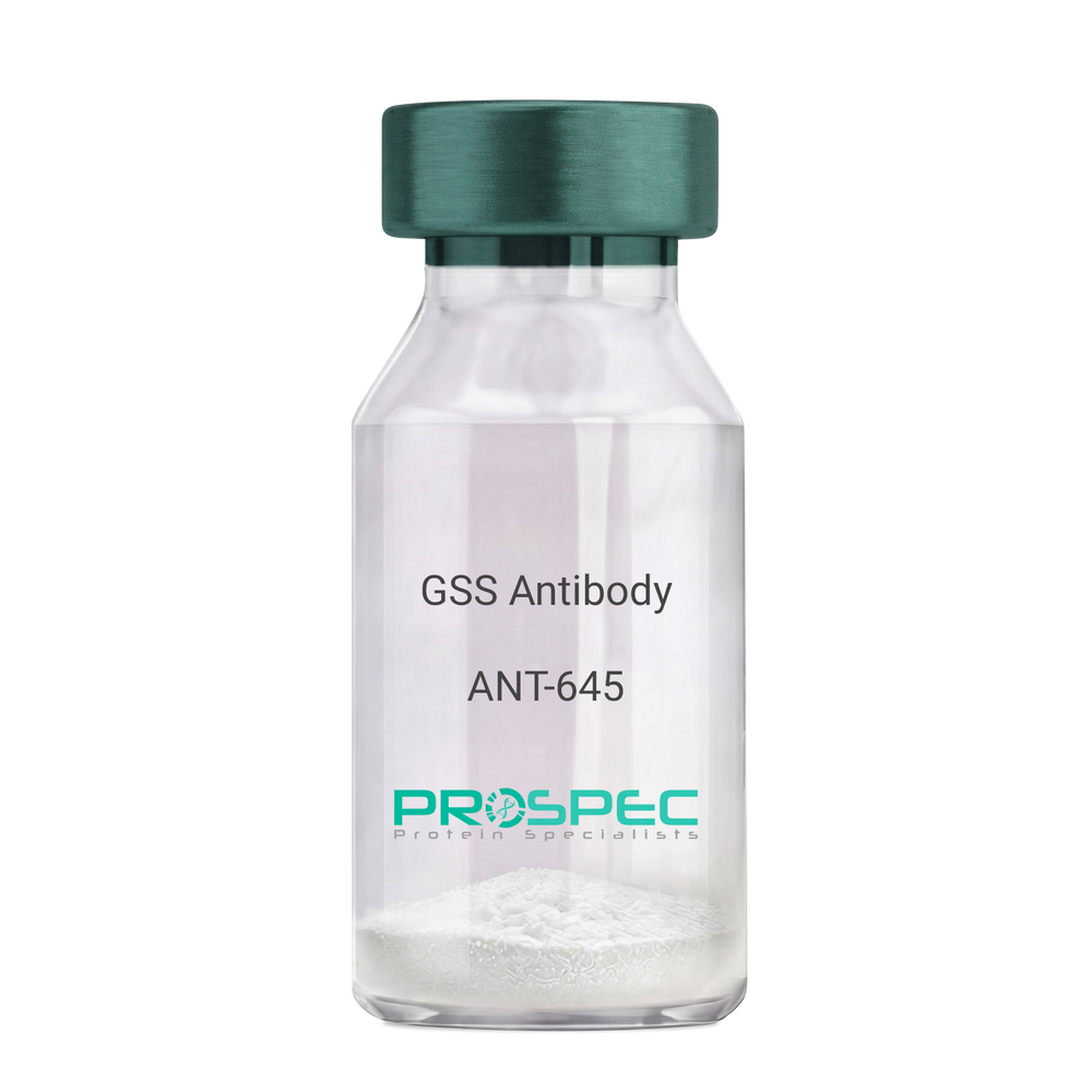
GSS AntibodyDescription:
Glutathione Synthetase, Mouse Anti Human
Glutathione synthetase, GSH synthetase, GSH-S, Glutathione synthase, GSHS, MGC14098, GSS.
Product # :
ANT-645Price :
Quantity :
Shipping Method :
Shipped with Ice Packs
More Info
- formulation
- More Info
- Citations
Formulation
1mg/ml containing PBS, pH-7.4, 10% Glycerol and 0.02% Sodium Azide.
Citations
More Info
-
Introduction
Glutathione synthetase (GSS) is the second enzyme in the glutathione biosynthesis pathway. GSS catalyses the condensation of gamma-glutamylcysteine and glycine to form glutathione. Defects in the GSS are the cause of glutathione synthetase deficiency aka GSS deficiency or 5-oxoprolinuria or pyroglutamic aciduria, which is a severe form characterized by an increased rate of hemolysis and defective function of the central nervous system.
-
Synonyms
Glutathione synthetase, GSH synthetase, GSH-S, Glutathione synthase, GSHS, MGC14098, GSS.
-
Physical Appearance
Sterile filtered colorless solution.
-
Immunogen
Anti-human GSS mAb, is derived from hybridization of mouse F0 myeloma cells with spleen cells from BALB/c mice immunized with recombinant human GSS amino acids 1-474 purified from E. coli.
-
Ig Subclass
Mouse IgG1 heavy chain and k light chain.
-
Clone
PAT12F2AT.
-
Applications
GSS antibody has been tested by ELISA, Western blot analysis and ICC/IF to assure specificity and reactivity. Since application varies, however, each investigation should be titrated by the reagent to obtain optimal results.
-
Type
Mouse Anti Human Monoclonal.
-
Storage Procedures
For periods up to 1 month store at 4°C, for longer periods of time, store at -20°C. Prevent freeze thaw cycles.
-
Purification Method
GSS antibody was purified from mouse ascitic fluids by protein-A affinity chromatography.
ProSpec's products are furnished for LABORATORY RESEARCH USE ONLY. The product may not be used as drugs, agricultural or pesticidal products, food additives or household chemicals.
- View Data Sheet
Name :
GFRA3 Human, Sf9Description:
GDNF Family Receptor Alpha 3 Human Recombinant, Sf9
GDNF Family Receptor Alpha3, GDNFR-alpha-3, GFR-alpha-3, GDNF Receptor Alpha-3, GDNFR3, GDNF Family Receptor Alpha-3, Glial Cell Line-Derived Neurotrophic Factor Receptor Alpha-3, GPI-Linked Receptor, GFRA3.
Product # :
CYT-1013Price :
Quantity :
Shipping Method :
Shipped with Ice Packs
More Info
- description
- source
- formulation
- purity
- More Info
- Citations
Description
GFRA3 Human Recombinant produced in Sf9 Baculovirus cells is a single, glycosylated polypeptide chain containing 585 amino acids (32-374) and having a molecular mass of 65.5kDa (Molecular size on SDS-PAGE will appear at approximately 70-100kDa). GFRA3 is fused to a 239 amino acid hIgG-His-Tag at C-terminus and purified by proprietary chromatographic techniques.
Source
Sf9, Baculovirus cells.
Formulation
GFRA3 protein solution (0.25mg/ml) containing Phosphate Buffered Saline (pH 7.4) and 10% glycerol.
Purity
Greater than 85.0% as determined by SDS-PAGE.
Citations
More Info
-
Introduction
GDNF Family Receptor Alpha 3 (GFRA3) belongs to the GDNF receptor family. GFRA3 creates a signaling receptor complex with RET tyrosine kinase receptor and binds the ligand, artemin (ARTN).
-
Synonyms
GDNF Family Receptor Alpha3, GDNFR-alpha-3, GFR-alpha-3, GDNF Receptor Alpha-3, GDNFR3, GDNF Family Receptor Alpha-3, Glial Cell Line-Derived Neurotrophic Factor Receptor Alpha-3, GPI-Linked Receptor, GFRA3.
-
Physical Appearance
Sterile Filtered clear solution.
-
Stability
Store at 4°C if entire vial will be used within 2-4 weeks. Store, frozen at -20°C for longer periods of time. For long term storage it is recommended to add a carrier protein (0.1% HSA or BSA).Avoid multiple freeze-thaw cycles.
-
Amino Acid Sequence
ADPDPLPTES RLMNSCLQAR RKCQADPTCS AAYHHLDSCT SSISTPLPSE EPSVPADCLE AAQQLRNSSL IGCMCHRRMK NQVACLDIYW TVHRARSLGN YELDVSPYED TVTSKPWKMN LSKLNMLKPD SDLCLKFAML CTLNDKCDRL RKAYGEACSG PHCQRHVCLR QLLTFFEKAA EPHAQGLLLC PCAPNDRGCG ERRRNTIAPN CALPPVAPNC LELRRLCFSD PLCRSRLVDF QTHCHPMDIL GTCATEQSRC LRAYLGLIGT AMTPNFVSNV NTSVALSCTC RGSGNLQEEC EMLEGFFSHN PCLTEAIAAK MRFHSQLFSQ DWPHPTFAVM AHQNENLEPK SCDKTHTCPP CPAPELLGGP SVFLFPPKPK DTLMISRTPE VTCVVVDVSH EDPEVKFNWY VDGVEVHNAK TKPREEQYNS TYRVVSVLTV LHQDWLNGKE YKCKVSNKAL PAPIEKTISK AKGQPREPQV YTLPPSRDEL TKNQVSLTCL VKGFYPSDIA VEWESNGQPE NNYKTTPPVL DSDGSFFLYS KLTVDKSRWQ QGNVFSCSVM HEALHNHYTQ KSLSLSPGKH HHHHH.
-
Background
What is the molecular weight/Mw of GFRA3 HUMAN, SF9 Protein?
GFRA3 HUMAN, SF9 Protein has a total Mw of 65.5kDa.
What is the source or expression system of GFRA3 HUMAN, SF9 Protein?
Sf9, Baculovirus cells.
What is the Purity of GFRA3 HUMAN, SF9 Protein?
GFRA3 HUMAN, SF9 Protein is >85% pure as determined by SDS-PAGE.
What is the Biological Activity of GFRA3 HUMAN, SF9 Protein?
The biological functionality of GFRA3 HUMAN, SF9 Protein will be determined in the future.
What is the amino acid sequence of GFRA3 HUMAN, SF9 Protein?
ADPDPLPTES RLMNSCLQAR RKCQADPTCS AAYHHLDSCT SSISTPLPSE EPSVPADCLE AAQQLRNSSL IGCMCHRRMK NQVACLDIYW TVHRARSLGN YELDVSPYED TVTSKPWKMN LSKLNMLKPD SDLCLKFAML CTLNDKCDRL RKAYGEACSG PHCQRHVCLR QLLTFFEKAA EPHAQGLLLC PCAPNDRGCG ERRRNTIAPN CALPPVAPNC LELRRLCFSD PLCRSRLVDF QTHCHPMDIL GTCATEQSRC LRAYLGLIGT AMTPNFVSNV NTSVALSCTC RGSGNLQEEC EMLEGFFSHN PCLTEAIAAK MRFHSQLFSQ DWPHPTFAVM AHQNENLEPK SCDKTHTCPP CPAPELLGGP SVFLFPPKPK DTLMISRTPE VTCVVVDVSH EDPEVKFNWY VDGVEVHNAK TKPREEQYNS TYRVVSVLTV LHQDWLNGKE YKCKVSNKAL PAPIEKTISK AKGQPREPQV YTLPPSRDEL TKNQVSLTCL VKGFYPSDIA VEWESNGQPE NNYKTTPPVL DSDGSFFLYS KLTVDKSRWQ QGNVFSCSVM HEALHNHYTQ KSLSLSPGKH HHHHH.
What applications can GFRA3 HUMAN, SF9 Protein be used in?
GFRA3 HUMAN, SF9 Protein can probably be used in western blot, ELISA and Lateral Flow.
What is the endotoxin level for GFRA3 HUMAN, SF9 Protein?
The endotoxin level is minimal, GFRA3 HUMAN, SF9 Protein was purified using conventional chromatography techniques.
ProSpec's products are furnished for LABORATORY RESEARCH USE ONLY. The product may not be used as drugs, agricultural or pesticidal products, food additives or household chemicals.
- View Data Sheet
Name :
ACPP Human, Sf9Description:
Acid Phosphatase Prostate, Human Recombinant, sf9
Acid Phosphatase, Prostate, Thiamine Monophosphatase, Ecto-5-Nucleotidase, 5-Nucleotidase, EC 3.1.3.2, TMPase, 5-NT, Prostatic Acid Phosphatase, Prostatic Acid Phosphotase, EC 3.1.3.5, ACP-3 , ACP3, PAP.
Product # :
ENZ-968Price :
Quantity :
Shipping Method :
Shipped with Ice Packs
More Info
- description
- source
- formulation
- purity
- More Info
- Citations
Description
ACPP produced in Sf9 Baculovirus cells is a single, glycosylated polypeptide chain containing 360 amino acids (33-386 a.a.) and having a molecular mass of 41.8kDa (Migrates at 40-57kDa on SDS-PAGE under reducing conditions). ACPP is expressed with a 6 amino acid His tag at C-Terminus and purified by proprietary chromatographic techniques.
Source
Sf9, Baculovirus cells.
Formulation
ACPP protein solution (0.5mg/ml) contains Phosphate Buffered Saline (pH 7.4) and 10% glycerol.
Purity
Greater than 95.0% as determined by SDS-PAGE.
Citations
More Info
-
Introduction
Acid phosphatase, prostate (ACPP) is a non-specific tyrosine phosphatase, which dephosphorylates a varied number of substrates under acidic conditions (pH 4-6) including alkyl, aryl, and acyl orthophosphate monoesters and phosphorylated proteins. ACPP has lipid phosphatase activity and inactivates lysophosphatidic acid in seminal plasma.
-
Synonyms
Acid Phosphatase, Prostate, Thiamine Monophosphatase, Ecto-5-Nucleotidase, 5-Nucleotidase, EC 3.1.3.2, TMPase, 5-NT, Prostatic Acid Phosphatase, Prostatic Acid Phosphotase, EC 3.1.3.5, ACP-3 , ACP3, PAP.
-
Physical Appearance
Sterile Filtered colorless solution.
-
Stability
Store at 4°C if entire vial will be used within 2-4 weeks. Store, frozen at -20°C for longer periods of time. For long term storage it is recommended to add a carrier protein (0.1% HSA or BSA). Avoid multiple freeze-thaw cycles.
-
Amino Acid Sequence
KELKFVTLVF RHGDRSPIDT FPTDPIKESS WPQGFGQLTQ LGMEQHYELG EYIRKRYRKF LNESYKHEQV YIRSTDVDRT LMSAMTNLAA LFPPEGVSIW NPILLWQPIP VHTVPLSEDQ LLYLPFRNCP RFQELESETL KSEEFQKRLH PYKDFIATLG KLSGLHGQDL FGIWSKVYDP LYCESVHNFT LPSWATEDTM TKLRELSELS LLSLYGIHKQ KEKSRLQGGV LVNEILNHMK RATQIPSYKK LIMYSAHDTT VSGLQMALDV YNGLLPPYAS CHLTELYFEK GEYFVEMYYR NETQHEPYPL MLPGCSPSCP LERFAELVGP VIPQDWSTEC MTTNSHQGTE DSTDHHHHHH.
ProSpec's products are furnished for LABORATORY RESEARCH USE ONLY. The product may not be used as drugs, agricultural or pesticidal products, food additives or household chemicals.