Search results
1000 results found for “PTH”
Name
Description
Product #
Price
Quantity
Shipping Method
- View Data Sheet
Name :
YWHAE AntibodyDescription:
Tyr-3/Trp- 5 Monooxygenase Activation Protein Epsilon, Mouse Anti Human
YWHAE, MDS, MDCR, KCIP-1, 14-3-3E, 14-3-3 Epsilon, FLJ45465, Tyr-3/Trp- 5 Monooxygenase Activation Protein Epsilon.
Product # :
ANT-379Price :
Quantity :
Shipping Method :
Shipped with Ice Packs
More Info
- formulation
- More Info
- Citations
Formulation
1mg/ml containing PBS, pH-7.4, 10% Glycerol and 0.02% Sodium Azide.
Citations
More Info
-
Introduction
The 14-3-3 family of proteins plays a key regulatory role in signal transduction, checkpoint control, apoptotic and nutrient-sensing pathways. 14-3-3 proteins are highly conserved and ubiquitously expressed. There are at least seven isoforms, β, γ, ε, σ, ζ, τ and η that have been identified in mammals. The 14-3-3 epsilon, a subtype of the 14-3-3 family of proteins, was thought to be brain and neuron-specific. It has been shown to interact with CDC25 phosphatases, RAF1 and IRS1 proteins, suggesting its role in diverse biochemical activities related to signal transduction, such as cell division and regulation of insulin sensitivity. It has also been implicated in the pathogenesis of small cell lung cancer.
-
Synonyms
YWHAE, MDS, MDCR, KCIP-1, 14-3-3E, 14-3-3 Epsilon, FLJ45465, Tyr-3/Trp- 5 Monooxygenase Activation Protein Epsilon.
-
Immunogen
Anti-human YWHAE mAb is derived from hybridization of mouse FO myeloma cells with spleen cells from BALB/c mice immunized with recombinant human YWHAE amino acids 1-255 purified from E. coli.
-
Ig Subclass
Mouse IgG2b heavy chain and k light chain
-
Clone
PAT4F8AT.
-
Applications
YWHAE antibody has been tested by ELISA, Western blot analysis, Flow cytometry and ICC/IF to assure specificity and reactivity. Since application varies, however, each investigation should be titrated by the reagent to obtain optimal results.
-
Type
Mouse Anti Human Monoclonal.
-
Storage Procedures
For periods up to 1 month store at 4°C, for longer periods of time, store at -20°C. Prevent freeze thaw cycles.
-
Purification Method
YWHAE antibody was purified from mouse ascitic fluids by protein-A affinity chromatography.
ProSpec's products are furnished for LABORATORY RESEARCH USE ONLY. The product may not be used as drugs, agricultural or pesticidal products, food additives or household chemicals.
- View Data Sheet
Name :
PAPP-A NativeDescription:
Pregnancy-Associated Plasma Protein-1 Human
Pappalysin-1, Pregnancy-associated plasma protein A, PAPP-A, IGF-dependent IGFBP-4 protease, IGFBP-4ase, PAPPA, PAPA, DIPLA1, PAPPA1, ASBABP2.
Product # :
ENZ-1204Price :
Quantity :
Shipping Method :
Shipped at Room temp
More Info
- description
- source
- formulation
- purity
- More Info
- Citations
Description
PAPP-A Human native purified from human placenta having a total molecular mass of ~200 kDa. The PAPP-A is purified by proprietary chromatographic techniques.
Source
Human Placenta
Formulation
PAPP-A protein was lyophilized from 10mM Tris-HCl pH-7, 0.15M NaCl, 0.1% NGME & 0.09% NaN3.
Purity
Greater than 95.0% as determined by SDS-PAGE.
Citations
More Info
-
Introduction
PAPPA is a large zinc binding protein, which plays a role as a metalloprotease and specifically cleaves IGFBP-4 and IGFBP-5, resulting in release of bound IGF. PAPP-A regulates IGF bioactivity in various biological systems, including the human ovary and cardiovascular systems. PAPP A levels were higher in patients with unstable angina or acute myocardial infarction. PAPPA is also involved in local proliferative processes such as wound healing and bone remodeling. Moreover, PAPP-A is produced in high concentrations during pregnancy and is released into the maternal circulation. In placenta, PAPP A is expressed in X cells in septa and anchoring villi, and in syncytiotrophoblasts in the chorionic villi.
-
Synonyms
Pappalysin-1, Pregnancy-associated plasma protein A, PAPP-A, IGF-dependent IGFBP-4 protease, IGFBP-4ase, PAPPA, PAPA, DIPLA1, PAPPA1, ASBABP2.
-
Physical Appearance
Brownish lyophilized (freeze-dried) powder.
-
Stability
Lyophilized PAPP-A although stable at room temperature for 3 weeks, should be stored desiccated below -18C. Upon reconstitution PAPP-A should be stored at 4C between 2-7 days and for future use below -18C. For long term storage it is recommended to add a carrier protein (0.1% HSA or BSA). Please prevent freeze-thaw cycles.
-
Solubility
It is recommended to add deionized water to a working concentration of 0.1mg/ml and let the lyophilized pellet dissolve completely.
-
Human Virus Test
Starting material tested and found negative for HIV-I, HIV-II, HCV antibodies and HBsAg antigen.
ProSpec's products are furnished for LABORATORY RESEARCH USE ONLY. The product may not be used as drugs, agricultural or pesticidal products, food additives or household chemicals.
- View Data Sheet
Name :
TesamorelinDescription:
Tesamorelin
Product # :
HOR-062Price :
Quantity :
Shipping Method :
Shipped at Room temp
More Info
- description
- formulation
- purity
- More Info
- Citations
Description
Tesamorelin is a synthetic single, non-glycosylated polypeptide chain containing 44 amino acids, having a molecular mass of 5135.78 Dalton and a Molecular formula of C221H366N72O67S.
Formulation
The protein was lyophilized with no additives.
Purity
Greater than 97.0% as determined by analysis by RP-HPLC.
Citations
More Info
-
Physical Appearance
Sterile Filtered White lyophilized (freeze-dried) powder.
-
Stability
Lyophilized Tesamorelin although stable at room temperature for 3 weeks, should be stored desiccated below -18°C. Upon reconstitution Tesamorelin should be stored at 4°C between 2-7 days and for future use below -18°C.
For long term storage it is recommended to add a carrier protein (0.1% HSA or BSA).
Please prevent freeze-thaw cycles.
-
Solubility
It is recommended to reconstitute the lyophilized Tesamorelin in sterile 18MΩ-cm H2O not less than 100 µg/ml, which can then be further diluted to other aqueous solutions.
-
Amino Acid Sequence
trans-3-hexenoyl-Tyr-Ala-Asp-Ala-IlePhe-Thr-Asn-Ser-Tyr-Arg-Lys-Val-Leu-Gly-Gln-Leu-Ser-Ala-Arg-LysLeu-Leu-Gln-Asp-Ile-Met-Ser-Arg-Gln-Gln-Gly-Glu-Ser-Asn-Gln-GluArg-Gly-Ala-Arg-Ala-Arg-Leu-NH2.
-
Background
Tesamorelin is a growth hormone-releasing hormone (GHRH) analogue. Tesamorelin stimulates the pituitary gland to produce endogenous growth hormone, which targets visceral adipose tissue. Tesamorelin has evolved from a HIV treatment into an effective metabolic and regenerative therapy.
ProSpec's products are furnished for LABORATORY RESEARCH USE ONLY. The product may not be used as drugs, agricultural or pesticidal products, food additives or household chemicals.
- View Data Sheet
Name :
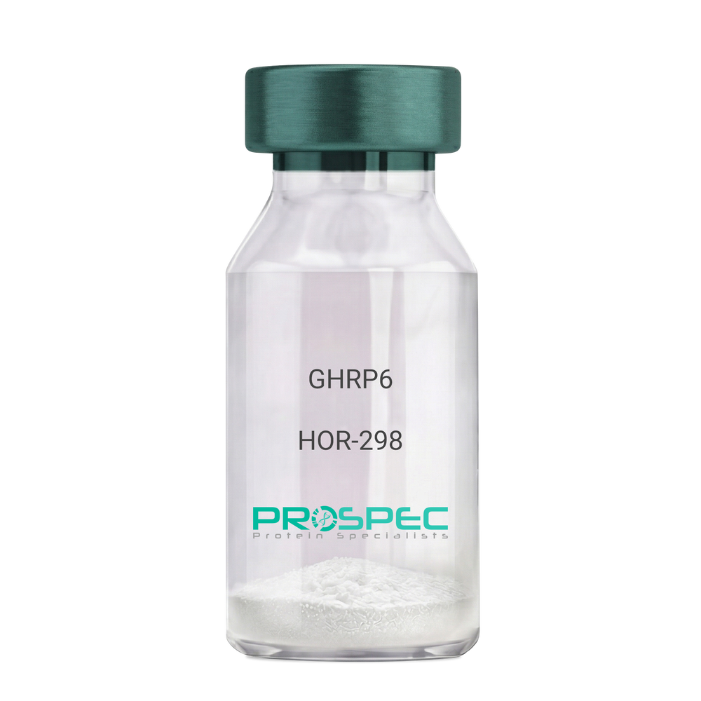
GHRP6Description:
Growth Hormone Releasing Peptide-6
GHRP-6, GHRP6
Product # :
HOR-298Price :
Quantity :
Shipping Method :
Shipped at Room temp
More Info
- description
- formulation
- purity
- More Info
- Citations
Description
Growth Hormone Releasing Peptide-6 Synthetic is a single, non-glycosylated polypeptide chain containing 6 amino acids, having a molecular mass of 873 Dalton and a Molecular formula of C46H56N12O6.
Formulation
The GHRP-6 peptide was lyophilized with no additives.
Purity
Greater than 98.0% as determined by analysis by RP-HPLC.
Citations
More Info
-
Introduction
GH-releasing peptides (GHRPs) are synthetic peptides that like GHRH act directly on pituitary somatotrophs to stimulate GH release. Growth hormone (GH) release is stimulated by a variety of synthetic secretagogues, of which growth hormone-releasing hexapeptide (GHRP-6) has been most thoroughly studied; it is thought to have actions at both pituitary and hypothalamic site.
-
Synonyms
GHRP-6, GHRP6
-
Physical Appearance
Sterile Filtered White lyophilized (freeze-dried) powder.
-
Stability
Lyophilized Growth Hormone Releasing Peptide-6 although stable at room temperature for 3 weeks, should be stored desiccated below -18°C. Upon reconstitution GHRP-6 should be stored at 4°C between 2-7 days and for future use below -18°C. For long term storage it is recommended to add a carrier protein (0.1% HSA or BSA).Please prevent freeze-thaw cycles.
-
Solubility
It is recommended to reconstitute the lyophilized GHRP-6 in sterile 18MΩ-cm H2O not less than 100 µg/ml, which can then be further diluted to other aqueous solutions.
-
Amino Acid Sequence
His-D-Trp-Ala-Trp-D-Phe-Lys-NH2.
-
Background
What is the molecular weight/Mw of GHRP6 Protein?
GHRP6 Protein has a total Mw of 0.873kDa.
What is the Purity of GHRP6 Protein?
GHRP6 Protein is >98% pure as determined by SDS-PAGE.
What is the Biological Activity of GHRP6 Protein?
The biological functionality of GHRP6 Protein will be determined in the future.
What is the amino acid sequence of GHRP6 Protein?
His-D-Trp-Ala-Trp-D-Phe-Lys-NH2.
What applications can GHRP6 Protein be used in?
GHRP6 Protein can probably be used in western blot, ELISA and Lateral Flow.
What is the endotoxin level for GHRP6 Protein?
The endotoxin level is minimal, GHRP6 Protein was purified using conventional chromatography techniques.
Who supplies GHRP6 that is in stock and available to order?
ProSpec has the protein in stock and available for immediate delivery.
Where can I order bulk mg quantities of GHRP6?
ProSpec supplies bulk mg quantities at affordable prices that meets research and academic budgets.
ProSpec's products are furnished for LABORATORY RESEARCH USE ONLY. The product may not be used as drugs, agricultural or pesticidal products, food additives or household chemicals.
- View Data Sheet
Name :
GHK-CuDescription:
GHK-Cu
Copper Tripeptide-1
Product # :
HOR-063Price :
Quantity :
Shipping Method :
Shipped at Room temp
More Info
- description
- formulation
- purity
- More Info
- Citations
Description
GHK-Cu is a synthetic single, non-glycosylated polypeptide chain containing 3 amino acids, having a molecular mass of 401.91 Dalton and a Molecular formula of C14H22N6O4Cu.
Formulation
The protein was lyophilized with no additives.
Purity
Greater than 97.0% as determined by analysis by RP-HPLC.
Citations
More Info
-
Physical Appearance
Sterile Filtered White lyophilized (freeze-dried) powder.
-
Stability
Lyophilized GHK-Cu although stable at room temperature for 3 weeks, should be stored desiccated below -18°C. Upon reconstitution GHK-Cu should be stored at 4°C between 2-7 days and for future use below -18°C.
For long term storage it is recommended to add a carrier protein (0.1% HSA or BSA).
Please prevent freeze-thaw cycles.
-
Solubility
It is recommended to reconstitute the lyophilized GHK-Cu in sterile 18MΩ-cm H2O not less than 100 µg/ml, which can then be further diluted to other aqueous solutions.
-
Amino Acid Sequence
Gly-His-Lys.
-
Background
GHK-Cu is located in human plasma and has an ability to modify gene expression to a healthier state, influencing more than 4,000 human genes. GHK-Cu can change pathological gene expression to a healthy mode, mainly in chronic conditions as metastatic cancer, COPD and Ulcerative Colitis. GHK-Cu was discovered to be effective in systemic repair and neuroprotection.
What is the molecular weight/Mw of GHK-CU Protein?
GHK-CU Protein has a total Mw of 0.40kDa.
What is the Purity of GHK-CU Protein?
GHK-CU Protein is >97% pure as determined by SDS-PAGE.
What is the Biological Activity of GHK-CU Protein?
The biological functionality of GHK-CU Protein will be determined in the future.
What is the amino acid sequence of GHK-CU Protein?
Gly-His-Lys.
What applications can GHK-CU Protein be used in?
GHK-CU Protein can probably be used in western blot, ELISA and Lateral Flow.
What is the endotoxin level for GHK-CU Protein?
The endotoxin level is minimal, GHK-CU Protein was purified using conventional chromatography techniques.
Who supplies GHK-CU that is in stock and available to order?
ProSpec has the protein in stock and available for immediate delivery.
Where can I order bulk mg quantities of GHK-CU?
ProSpec supplies bulk mg quantities at affordable prices that meets research and academic budgets.
ProSpec's products are furnished for LABORATORY RESEARCH USE ONLY. The product may not be used as drugs, agricultural or pesticidal products, food additives or household chemicals.
- View Data Sheet
Name :
PSIP1 HumanDescription:
PC4 And SFRS1 Interacting Protein 1 Human Recombinant
PC4 and SFRS1 Interacting Protein 1, Lens Epithelium-Derived Growth Factor, Transcriptional Coactivator P75/P52, Dense Fine Speckles 70 KDa Protein, CLL-Associated Antigen KW-7, DFS 70, PSIP2, DFS70, LEDGF, PC4 And SFRS1 Interacting Protein 2, Transcriptional Coactivator P52/P75, PAIP, P52, P75, PSIP1.
Product # :
PRO-2267Price :
Quantity :
Shipping Method :
Shipped with Ice Packs
More Info
- description
- source
- formulation
- purity
- More Info
- Citations
Description
Recombinant Human PSIP1 produced in SF9 is a glycosylated, polypeptide chain having a calculated molecular mass of 71 kDa. PSIP1 is expressed with a 10xHis tag at N-terminus and purified by proprietary chromatographic techniques.
Source
Sf9 insect cells.
Formulation
PSIP1 is supplied in 20mM HEPES buffer pH-8.0, 200mM NaCl and 20% glycerol.
Purity
Greater than 84.0% as determined by SDS-PAGE.
Citations
More Info
-
Introduction
PC4 and SFRS1 Interacting Protein 1 (PSIP1) is a transcriptional coactivator involved in neuroepithelial stem cell differentiation and neurogenesis. PSIP1 is also involved particularly in lens epithelial cell gene regulation and stress responses. PSIP1 has a significant role in lens epithelial to fiber cell terminal differentiation. In addition, PSIP1 plays a protective role during stress-induced apoptosis. Furthermore, PSIP1 is a cellular cofactor for lentiviral integration.
-
Synonyms
PC4 and SFRS1 Interacting Protein 1, Lens Epithelium-Derived Growth Factor, Transcriptional Coactivator P75/P52, Dense Fine Speckles 70 KDa Protein, CLL-Associated Antigen KW-7, DFS 70, PSIP2, DFS70, LEDGF, PC4 And SFRS1 Interacting Protein 2, Transcriptional Coactivator P52/P75, PAIP, P52, P75, PSIP1.
-
Physical Appearance
Sterile Filtered clear solution.
-
Stability
Store at 4°C if entire vial will be used within 2-4 weeks. Store, frozen at -20°C for longer periods of time. Avoid multiple freeze-thaw cycles.
-
Immunological Functions
1. Binds IgG type human auto-antibodies.2. Immunodot test with positive/negative samples.
ProSpec's products are furnished for LABORATORY RESEARCH USE ONLY. The product may not be used as drugs, agricultural or pesticidal products, food additives or household chemicals.
- View Data Sheet
Name :
GPD2 HumanDescription:
Glycerol-3-Phosphate Dehydrogenase 2 Human Recombinant
Glycerol-3-phosphate dehydrogenase mitochondrial, glycerol-3-phosphate dehydrogenase 2 (mitochondrial), GPDH-M, GPD-M, mtGPD, GPD2, GDH2, GPDM, mGPDH.
Product # :
ENZ-437Price :
Quantity :
Shipping Method :
Shipped with Ice Packs
More Info
- description
- source
- formulation
- purity
- More Info
- Citations
Description
GPD2 Human Recombinant produced in E.Coli is single, a non-glycosylated, Polypeptide chain containing 558 amino acids fragment (43-600) corresponding to the GlpA domain fragment of the mature protein, having a total molecular mass of 66.26kDa and fused with a 4.5kDa amino-terminal hexahistidine tag. The GPD2 is purified by proprietary chromatographic techniques.
Source
Escherichia Coli.
Formulation
GPD2 protein solution is supplied in 20mM Tris-HCl pH 8, 1mM EDTA and 50% glycerol.
Purity
Greater than 95.0% as determined by SDS-PAGE.
Citations
More Info
-
Introduction
GPD2 (Mitochondrial glycerol-3-phosphate dehydrogenase) is a Ca2+-sensitive, FAD-binding protein, which is located on the outer surface of the inner mitochondrial membrane. Two isoforms have been identified for mGPD: Isoform 1 is comprised of 727 a.a. residues, while isoform 2 lacks 126 a.a. residues of the N-terminus. GPD2 catalyses the oxidation of glycerol-3-phosphate to DHAP (dihydroxyacetone phosphate) with associated reduction of the enzyme-bound FAD. GPD2 is a testis-specific promoter of mitochondrial GPDH. GPD2 along with a cytosolic NAD-linked GPD forms the glycerol phosphate shuttle that uses the interconversion of G-3-P and DHAP to transfer reducing equivalents into mitochondria, which results in the reoxidation of NADH produced during glycolysis.
GPD2 deficiency contributes to the impairment of glucose-stimulated INS discharge in a number of animal models of non-INS dependent diabetes mellitus. GPD2 up-regulation as a result of a highly glycolytic environment contributes to the general increase in ROS generation and may lead to the progression of prostate cancer. -
Synonyms
Glycerol-3-phosphate dehydrogenase mitochondrial, glycerol-3-phosphate dehydrogenase 2 (mitochondrial), GPDH-M, GPD-M, mtGPD, GPD2, GDH2, GPDM, mGPDH.
-
Physical Appearance
Sterile Filtered clear solution.
-
Stability
Store at 4°C if entire vial will be used within 2-4 weeks. Store, frozen at -20°C for longer periods of time. Please avoid freeze thaw cycles.
ProSpec's products are furnished for LABORATORY RESEARCH USE ONLY. The product may not be used as drugs, agricultural or pesticidal products, food additives or household chemicals.
- View Data Sheet
Name :
UGDH HumanDescription:
UDP-Glucose Dehydrogenase Human Recombinant
GDH, UDP-GlcDH, UDPGDH, UGD, EC=1.1.1.22, UDP-Glc dehydrogenase, UDP-glucose 6-dehydrogenase, UGDH.
Product # :
ENZ-535Price :
Quantity :
Shipping Method :
Shipped with Ice Packs
More Info
- description
- source
- formulation
- purity
- biological activity
- More Info
- Citations
Description
UGDH Recombinant Human produced in E.Coli is a single, non-glycosylated polypeptide chain containing 533 amino acids (1-494 a.a.) and having a molecular mass of 59.5 kDa. The UGDH is fused to a 39 amino acid His-Tag at N-terminus and purified by proprietary chromatographic techniques.
Source
Escherichia Coli.
Formulation
UGDH Human solution (0.5mg/ml) containing 20mM Tris-HCl pH-8, 1mM DTT, 1mM EDTA, 0.1M NaCl & 20% glycerol.
Purity
Greater than 95.0% as determined by SDS-PAGE.
Biological Activity
Specific activity is > 2500pmol/min/ug, and is defined as the amount of enzyme that convert 1.0pmole of UDPglucose to UDP-glucuronate per minute at pH 8.7 at 37C.
Citations
More Info
-
Introduction
UGDH is part of the UDP-glucose/GDP-mannose dehydrogenase family and is a widely expressed enzyme localized in the liver. UGDH transfers UDP-glucose to UDP-glucuronate and thus takes part in the biosynthesis of glycosaminoglycans such as hyaluronan, chondroitin sulfate, and heparan sulfate. These glycosylated products are ordinary molecules of the extracellular matrix and participate in signal transduction, cell migration, and cancer growth and metastasis.
-
Synonyms
GDH, UDP-GlcDH, UDPGDH, UGD, EC=1.1.1.22, UDP-Glc dehydrogenase, UDP-glucose 6-dehydrogenase, UGDH.
-
Physical Appearance
Sterile filtered colorless solution.
-
Stability
Store at 4°C if entire vial will be used within 2-4 weeks. Store, frozen at -20°C for longer periods of time. For long term storage it is recommended to add a carrier protein (0.1% HSA or BSA).Avoid multiple freeze-thaw cycles.
-
Amino Acid Sequence
MRGSHHHHHH GMASMTGGQQ MGRDLYDDDD KDRWGSELEM FEIKKICCIG AGYVGGPTCS VIAHMCPEIR VTVVDVNESR INAWNSPTLP IYEPGLKEVV ESCRGKNLFF STNIDDAIKE ADLVFISVNT PTKTYGMGKG RAADLKYIEA CARRIVQNSN GYKIVTEKST VPVRAAESIR RIFDANTKPN LNLQVLSNPE FLAEGTAIKD LKNPDRVLIG GDETPEGQRA VQALCAVYEH WVPREKILTT NTWSSELSKL AANAFLAQRI SSINSISALC EATGADVEEV ATAIGMDQRI GNKFLKASVG FGGSCFQKDV LNLVYLCEAL NLPEVARYWQ QVIDMNDYQR RRFASRIIDS LFNTVTDKKI AILGFAFKKD TGDTRESSSI YISKYLMDEG AHLHIYDPKV PREQIVVDLS HPGVSEDDQV SRLVTISKDP YEACDGAHAV VICTEWDMFK ELDYERIHKK MLKPAFIFDG RRVLDGLHNE LQTIGFQIET IGKKVSSKRI PYAPSGEIPK FSLQDPPNKK PKV.
ProSpec's products are furnished for LABORATORY RESEARCH USE ONLY. The product may not be used as drugs, agricultural or pesticidal products, food additives or household chemicals.
- View Data Sheet
Name :
Hepsin HumanDescription:
Hepsin Human Recombinant
HPN, TMPRSS1, Serine protease hepsin, Transmembrane protease serine 1.
Product # :
PRO-2846Price :
Quantity :
Shipping Method :
Shipped at Room temp
More Info
- description
- source
- formulation
- purity
- biological activity
- More Info
- Citations
Description
Hepsin Human Recombinant produced in Cho cells is a covalently-linked heterodimer having a total molecular mass of 43.0kDa. Hepsin is fused to a 6 amino acid His-tag at C-terminus & purified by proprietary chromatographic techniques.
Source
CHO cells.
Formulation
The Hepsin protein was Lyophilized from a 0.2µm filtered concentrated solution in 20mM Tris and 150mM NaCl, pH 8.0.
Purity
Greater than 95.0% as determined by SDS-PAGE.
Biological Activity
The specific activity is >20,000 pmol/min/μg and was measured by its ability to cleave tert-butoxycarbonyl-Gln-Arg-Arg-7-amino-4-methylcoumarin (Boc-QRR-AMC).Citations
More Info
-
Synonyms
HPN, TMPRSS1, Serine protease hepsin, Transmembrane protease serine 1.
-
Physical Appearance
Filtered White lyophilized (freeze-dried) powder.
-
Stability
Lyophilized Hepsin Active although stable at room temperature for 3 weeks, should be stored desiccated below -18°C. Upon reconstitution Hepsin Active should be stored at 4°C between 2-7 days and for future use below -18°C.
Please prevent freeze-thaw cycles. -
Solubility
It is recommended to reconstitute the lyophilized Hepsin in sterile 18MΩ-cm H2O not less than 100µg/ml, which can then be further diluted to other aqueous solutions.
-
Amino Acid Sequence
Light Chain (Non-catalytic Chain)
RSDQEPLYPV QVSSADARLM VFDKTEGTWR LLCSSRSNAR VAGLSCEEMG FLRALTHSEL DVRTAGANGT SGFFCVDEGR LPHTQRLLE VISVCDCPRGR FLAAICQDCG RRKLPVDR.
Heavy Chain (Catalytic Chain)
IVGGRDTSLG RWPWQVSLRY DGAHLCGGSL LSGDWVLTAA HCFPERNRVL SRWRVFAGAV AQASPHGLQL GVQAVVYHGG YLPFRDPNSE ENSNDIALVH LSSPLPLTEY IQPVCLPAAG QALVDGKICT VTGWGNTQYY GQQAGVLQEA RVPIISNDVC NGADFYGNQI KPKMFCAGYP EGGIDACQGD SGGPFVCEDS ISRTPRWRLC GIVSWGTGCA LAQKPGVYTK VSDFREWIFQ AIKTHSEASG MVTQLHHHHH H.
-
Background
Hepsin is a type II transmembrane serine protease expressed primarily on epithelial cells, it takes part in extracellular proteolysis by activating precursor proteins such as pro-HGF and contributes to tissue remodeling, cell signaling, and normal epithelial function. Recombinant Hepsin is used to study prostate cancer, extracellular matrix remodelling, protease signalling pathways, tumor invasion, metastasis, HGF/MET signaling, and for screening inhibitors that target serine proteases
What is the molecular weight / Mw of Hepsin Protein?
Hepsin Protein has a total Mw of 43kDa.
What is the source or expression system of Hepsin Protein?
CHO Cells
What is the Purity of Hepsin Protein?
Hepsin Protein is >95% pure as determined by SDS-PAGE.
What is the Biological Activity of Hepsin Protein?
The enzymatic activity was measured by its ability to cleave tert-butoxycarbonyl-Gln-Arg-Arg-7-amino-4-methylcoumarin (Boc-QRR-AMC). The specific activity is >20,000 pmol/min/μg.
What is the amino acid sequence of Hepsin Protein?
Light Chain (Non-catalytic Chain)
RSDQEPLYPV QVSSADARLM VFDKTEGTWR LLCSSRSNAR VAGLSCEEMG FLRALTHSEL DVRTAGANGT SGFFCVDEGR LPHTQRLLE VISVCDCPRGR FLAAICQDCG RRKLPVDR
Heavy Chain (Catalytic Chain)
IVGGRDTSLG RWPWQVSLRY DGAHLCGGSL LSGDWVLTAA HCFPERNRVL SRWRVFAGAV AQASPHGLQL GVQAVVYHGG YLPFRDPNSE ENSNDIALVH LSSPLPLTEY IQPVCLPAAG QALVDGKICT VTGWGNTQYY GQQAGVLQEA RVPIISNDVC NGADFYGNQI KPKMFCAGYP EGGIDACQGD SGGPFVCEDS ISRTPRWRLC GIVSWGTGCA LAQKPGVYTK VSDFREWIFQ AIKTHSEASG MVTQLHHHHH H
What applications can Hepsin Protein be used in?
Hepsin Protein can probably be used in western blot, ELISA and Lateral Flow.
What is the endotoxin level for Hepsin Protein?
The endotoxin level is minimal, Hepsin Protein was purified using conventional chromatography techniques.
ProSpec's products are furnished for LABORATORY RESEARCH USE ONLY. The product may not be used as drugs, agricultural or pesticidal products, food additives or household chemicals.
- View Data Sheet
Name :
PDGF BB HumanDescription:
Platelet-Derived Growth Factor BB Human Recombinant
Glioma-derived growth factor, GDGF, Osteosarcoma-derived Growth Factor, ODGF, SIS, SSV, PDGF2, c-sis, FLJ12858, PDGF-BB, PDGF B-chain, Platelet-derived growth factor beta polypeptide.
Product # :
CYT-501Price :
Quantity :
Shipping Method :
Shipped at Room temp
More Info
- description
- source
- formulation
- purity
- biological activity
- More Info
- Citations
Description
Platelet-Derived Growth Factor BB Human Recombinant is a homodimeric, non-glycosylated, polypeptide chain containing 2x10⁹ amino acids (218 amino acids in total) and having a molecular mass of 24.3 kDa. PDGF-BB is purified by proprietary chromatographic techniques.
Source
Escherichia Coli.
Formulation
Lyophilized from a 0.2µm filtered concentrated solution in PBS, pH7.4.
Purity
Greater than 95.0% as determined by:
(a) Analysis by RP-HPLC.
(b) Analysis by SDS-PAGE.Biological Activity
Determined by the dose-dependent stimulation of the proliferation of Balb/c 3T3 cells.
The expected ED50 for this effect is 1.0-3.0 ng/ml.Citations
More Info
-
Introduction
PDGF-BB is a member of the platelet-derived growth factor family. The four members of this family are mitogenic factors for cells of mesenchymal origin and are characterized by a motif of eight cysteines. This gene product can exist either as a homodimer (PDGF-BB) or as a heterodimer with the platelet-derived growth factor alpha polypeptide (PDGF-AB), where the dimers are connected by disulfide bonds. Mutations in this gene are associated with meningioma. Reciprocal translocations between chromosomes 22 and 7, at sites where this gene and that for COL1A1 are located, are associated with a particular type of skin tumor called dermatofibrosarcoma protuberans resulting from unregulated expression of growth factor. Two splice variants have been identified for this gene.
-
Synonyms
Glioma-derived growth factor, GDGF, Osteosarcoma-derived Growth Factor, ODGF, SIS, SSV, PDGF2, c-sis, FLJ12858, PDGF-BB, PDGF B-chain, Platelet-derived growth factor beta polypeptide.
-
Physical Appearance
Sterile Filtered White lyophilized (freeze-dried) powder.
-
Stability
Lyophilized Platelet-derived Growth Factor BB although stable at room temperature for 3 weeks, should be stored desiccated below -18°C. Upon reconstitution PDGF BB should be stored at 4°C between 2-7 days and for future use below -18°C.Please prevent freeze-thaw cycles.
-
Solubility
It is recommended to reconstitute the lyophilized Platelet-derived Growth Factor-BB in sterile 18MΩ-cm H2O not less than 100µg/ml, which can then be further diluted to other aqueous solutions.
-
Amino Acid Sequence
SLGSLTIAEP AMIAECKTRT EVFEISRRLI DRTNANFLVW PPCVEVQRCS GCCNNRNVQC RPTQVQLRPV QVRKIEIVRK KPIFKKATVT LEDHLACKCE TVAAARPVT.
-
Background
PDGF BB HUMAN: Overview of Its Production, Properties, and Clinical Significance
PDGF BB HUMAN, standing for Platelet-Derived Growth Factor BB, is a powerful protein in the field of medical research, particularly in cell growth and healing. This growth factor plays a crucial role in the development and repair of tissues by stimulating cells primarily of mesenchymal origin.
Characteristics and Production
PDGF is produced as a recombinant protein in E. coli. It presents as a homodimer consisting of two identical polypeptide chains, each containing 109 amino acids, culminating in a total molecular mass of 24.3 kDa. The production process ensures a high-purity product, which is essential for reliable scientific results.
Physical Properties and Formulation
This growth factor appears as a white, sterile, lyophilized powder. It is formulated in a buffered solution (PBS, pH 7.4) and then filtered to ensure sterility and purity, essential for laboratory use. The formulation process is designed to maintain the stability and activity of the protein under various research conditions.
Solubility and Storage Instructions
PDGF BB is recommended to be reconstituted in sterile water to achieve a concentration of no less than 100µg/ml. This solution can then be diluted further to meet experimental needs.
Once reconstituted, the protein should be stored at 4°C for short-term use (2-7 days) and below -18°C for long-term storage. Avoiding freeze-thaw cycles is crucial to preserve its biological activities.
Stability and Purity
The lyophilized form of PDGF BB remains stable at room temperature for up to three weeks but requires desiccation for longer storage.
Moreover, the purity of this growth factor exceeds 95%, as confirmed by rigorous testing, including RP-HPLC and SDS-PAGE, ensuring that researchers receive a highly effective product.
Research Applications
PDGF BB HUMAN is widely used in laboratory research to explore various biological processes, including wound healing, angiogenesis, and the development of certain types of cancers. It is also instrumental in studying the cellular mechanisms underlying tissue repair and regeneration.
Biological Activity
The effectiveness of PDGF BB is measured by its ability to stimulate the proliferation of Balb/c 3T3 cells, with an effective dose (ED50) ranging from 1.0 to 3.0 ng/ml. This high level of activity underscores its utility in promoting cell growth, making it an invaluable tool in tissue engineering and regenerative medicine.
Usage Guidelines
It is important to note that PDGF BB HUMAN is intended solely for laboratory research and is not suitable for drug, food, or cosmetic applications. Researchers must handle this growth factor under controlled conditions to ensure safety and efficacy.
Essentially, PDGF BB is a pivotal component in the toolkit of biomedical researchers, offering profound insights into cellular processes and potential therapeutic approaches. Also, its well-defined properties and controlled production make it a staple in studies focused on cell growth and tissue repair.
ProSpec's products are furnished for LABORATORY RESEARCH USE ONLY. The product may not be used as drugs, agricultural or pesticidal products, food additives or household chemicals.
- View Data Sheet
Name :
FGF17 Human, HisDescription:
Fibroblast Growth Factor 17 Human Recombinant, His Tag
Fibroblast growth factor 17, FGF-17, FGF17, FGF-13, HH20.
Product # :
CYT-755Price :
Quantity :
Shipping Method :
Shipped with Ice Packs
More Info
- description
- source
- formulation
- purity
- More Info
- sds-page
- Citations
Description
FGF17 Human Recombinant produced in E.Coli is a single, non-glycosylated polypeptide chain containing 219 amino acids (23-216 a.a) and having a molecular mass of 25.2kDa.FGF17 is fused to a 25 amino acid His-tag at N-terminus & purified by proprietary chromatographic techniques.
Source
E.coli.
Formulation
FGF17 protein solution (1mg/ml) contains 20mM Tris-HCl buffer (pH 8.0), 0.4M urea and 10% glycerol.
Purity
Greater than 90% as determined by SDS-PAGE.
sds-page
Citations
More Info
-
Introduction
Fibroblast Growth Factor 17 (FGF17) belongs to the fibroblast growth factor (FGF) family. FGF family members have broad mitogenic and cell survival activities, and are involved in various biological processes including embryonic development cell growth, morphogenesis, tissue repair, tumor growth and invasion. The FGF17 gene is highly expressed in the cerebellum and cortex. The mouse homolog of the FGF17 gene is localized to specific sites in the midline structures of the forebrain, the midbrain-hindbrain junction, developing skeleton and developing arteries, suggesting a role in central nervous system, bone and vascular development.
-
Synonyms
Fibroblast growth factor 17, FGF-17, FGF17, FGF-13, HH20.
-
Physical Appearance
Sterile Filtered colorless solution.
-
Stability
Store at 4°C if entire vial will be used within 2-4 weeks. Store, frozen at -20°C for longer periods of time. For long term storage it is recommended to add a carrier protein (0.1% HSA or BSA).Avoid multiple freeze-thaw cycles.
-
Amino Acid Sequence
MGSSHHHHHH SSGLVPRGSH MGSHMTQGEN HPSPNFNQYV RDQGAMTDQL SRRQIREYQL YSRTSGKHVQ VTGRRISATA EDGNKFAKLI VETDTFGSRV RIKGAESEKY ICMNKRGKLI GKPSGKSKDC VFTEIVLENN YTAFQNARHE GWFMAFTRQG RPRQASRSRQ NQREAHFIKR LYQGQLPFPN HAEKQKQFEF VGSAPTRRTK RTRRPQPLT.
-
Background
What is the molecular weight/Mw of FGF17 HIS Protein?
FGF17 HIS Protein has a total Mw of 25.2kDa.
What is the source or expression system of FGF17 HIS Protein?
Escherichia Coli.
What is the Purity of FGF17 HIS Protein?
FGF17 HIS Protein is >90% pure as determined by SDS-PAGE.
What is the Biological Activity of FGF17 HIS Protein?
The biological functionality of FGF17 HIS Protein will be determined in the future.
What is the amino acid sequence of FGF17 HIS Protein?
MGSSHHHHHH SSGLVPRGSH MGSHMTQGEN HPSPNFNQYV RDQGAMTDQL SRRQIREYQL YSRTSGKHVQ VTGRRISATA EDGNKFAKLI VETDTFGSRV RIKGAESEKY ICMNKRGKLI GKPSGKSKDC VFTEIVLENN YTAFQNARHE GWFMAFTRQG RPRQASRSRQ NQREAHFIKR LYQGQLPFPN HAEKQKQFEF VGSAPTRRTK RTRRPQPLT.
What applications can FGF17 HIS Protein be used in?
FGF17 HIS Protein can probably be used in western blot, ELISA and Lateral Flow.
What is the endotoxin level for FGF17 HIS Protein?
The endotoxin level is minimal, FGF17 HIS Protein was purified using conventional chromatography techniques.
ProSpec's products are furnished for LABORATORY RESEARCH USE ONLY. The product may not be used as drugs, agricultural or pesticidal products, food additives or household chemicals.
- View Data Sheet
Name :
TNFRSF14 Human, HisDescription:
HVEM Human Recombinant, His Tag
Tumor Necrosis Factor Receptor Superfamily Member 14, HVEM, TR2, Herpes Virus Entry Mediator A, Tumor Necrosis Factor Receptor-Like 2, Herpesvirus Entry Mediator, HVEA, ATAR, CD270, LIGHTR, CD40-Like Protein, Tumor Necrosis Factor Receptor-Like Gene2.
Product # :
CYT-800Price :
Quantity :
Shipping Method :
Shipped with Ice Packs
More Info
- description
- source
- formulation
- purity
- More Info
- Citations
Description
TNFRSF14 Human Recombinant produced in E. Coli is a single, glycosylated polypeptide chain containing 187 amino acids (39-202) and having a molecular mass of 19.7kDa.TNFRSF14 is fused to a 23 amino acid His-tag at N-terminus & purified by proprietary chromatographic techniques.
Source
E.coli.
Formulation
The TNFRSF14 solution (0.5mg/ml) containing 20mM Tris-HCl (pH 8.0), 0.1M NaCl and 10% glycerol.
Purity
Greater than 85% as determined by SDS-PAGE.
Citations
More Info
-
Introduction
TNFRSF14, a member of the TNF receptor superfamily, is a type I transmembrane protein. TNFRSF14 is expressed in peripheral blood T cells, B cells, monocytes and in various tissues enriched in lymphoid cells. TNFRSF14 operates as a co-stimulatory factor for the activation of lymphoid cells and as a deterrent to infection by herpesvirus. Additionally, TNFRSF14 encourages the proliferation of T cells, and triggers apoptosis of various tumor cells.
-
Synonyms
Tumor Necrosis Factor Receptor Superfamily Member 14, HVEM, TR2, Herpes Virus Entry Mediator A, Tumor Necrosis Factor Receptor-Like 2, Herpesvirus Entry Mediator, HVEA, ATAR, CD270, LIGHTR, CD40-Like Protein, Tumor Necrosis Factor Receptor-Like Gene2.
-
Physical Appearance
Sterile filtered colorless solution.
-
Stability
Store at 4°C if entire vial will be used within 2-4 weeks. Store, frozen at -20°C for longer periods of time. For long term storage it is recommended to add a carrier protein (0.1% HSA or BSA).Avoid multiple freeze-thaw cycles.
-
Amino Acid Sequence
MGSSHHHHHH SSGLVPRGSH MGSLPSCKED EYPVGSECCP KCSPGYRVKE ACGELTGTVC EPCPPGTYIA HLNGLSKCLQ CQMCDPAMGL RASRNCSRTE NAVCGCSPGH FCIVQDGDHC AACRAYATSS PGQRVQKGGT ESQDTLCQNC PPGTFSPNGT LEECQHQTKC SWLVTKAGAG TSSSHWV.
ProSpec's products are furnished for LABORATORY RESEARCH USE ONLY. The product may not be used as drugs, agricultural or pesticidal products, food additives or household chemicals.
- View Data Sheet
Name :
Leptin qA Human, PEGDescription:
Leptin Quadruple Antagonist Pegylated Human Recombinant
Product # :
CYT-1251Price :
Quantity :
Shipping Method :
Shipped at Room temp
More Info
- description
- source
- formulation
- purity
- biological activity
- More Info
- Citations
Description
Leptin Pegylated Quadruple Antagonist Human Recombinant is a single non-glycosilated polypeptide chain containing 146 amino and an additional Ala at N-terminus acids. The Human Leptin antagonist is bound to 20 kDa mono-PEG at N-terminus, resulting in 35.6 kDa. The Human Leptin Pegylated Quadruple Antagonist was mutated, resulting in D23L/L39A/D40A/F41A that was purified by proprietary chromatographic techniques.
Source
Escherichia coli.
Formulation
The Human Leptin Pegylated Quadruple Antagonist was lyophilized from a concentrated (0.65mg/ml) solution with 0.003mM NaHCO3.
Purity
Greater than 98.0% as determined by:
(a) Gel filtration analysis.
(b) Analysis by SDS-PAGE.
Biological Activity
Human Leptin Pegylated Quadruple Antagonist inhibits leptin-induced proliferation of BAF/3 cells stably transfected with the long form of human leptin receptor. Its in vitro activity is 6-8 fold lower than the non-pegylated human leptin antagonist but in vivo it has profound weight gain effect (as compared to the non-pegylated human leptin antagonist), resulting mainly from increased food intake. The in vivo activity of human pegylated super leptin antagonist was compared to that of human pegylated leptin antagonist is 9-27 fold higher.
Citations
More Info
-
Physical Appearance
White lyophilized (freeze-dried) powder.
-
Stability
Lyophilized Human Leptin Pegylated Quadruple Antagonist although stable at room temperature for several weeks, should be stored desiccated below -18°C. Upon reconstitution at > 0.1 and up to 2mM of Human pegylated leptin antagonist and filter sterilization Human pegylated leptin antagonist can be stored at 4°C or even room temperature for several weeks making it suitable for long term infusion studies using osmotic pumps. At lower concentration addition of a carrier protein (0.1% HSA or BSA) is suggested. Please prevent freeze-thaw cycles.
-
Solubility
It is recommended to reconstitute the lyophilized Human Leptin Pegylated Quadruple Antagonist in sterile water or sterile 0.4% NaHCO3 adjusted to pH 8-9, not less than 100µg/ml, which can then be further diluted with other aqueous solutions.
-
Background
Leptin is a~16 kDa protein which is encoded by the obese gene. Leptin is a hormone which participates in regulating body weight, reproductive function and metabolism. leptin is expressed predominantly by adipocytes, which supports the idea that body weight is sensed as the total mass of fat in the body. Smaller amounts of leptin are also secreted by cellsin the epithelium of the stomach and in the placenta. Leptin receptors are highly expressed in areas of the hypothalamus which regulates body weight, as well as in T lymphocytes and vascular endothelial cells.
-
Protein content
Protein quantization was carried out by UV spectroscopy at 280 nm using the absorbency value of 0.88 as the extinction coefficient for a 0.1% (1mg/ml) solution at pH 8.0. This value is calculated by the PC GENE computer analysis program of protein sequences (IntelliGenetics).
ProSpec's products are furnished for LABORATORY RESEARCH USE ONLY. The product may not be used as drugs, agricultural or pesticidal products, food additives or household chemicals.
- View Data Sheet
Name :
GPT Human, ActiveDescription:
Glutamic-Pyruvate Transaminase Human Recombinant, Active
Alanine aminotransferase 1, ALT1, EC 2.6.1.2, Glutamate pyruvate transaminase 1, GPT 1, Glutamic--alanine transaminase 1, Glutamic--pyruvic transaminase 1, GPT, AAT1, GPT1.
Product # :
ENZ-280Price :
Quantity :
Shipping Method :
Shipped with Ice Packs
More Info
- description
- source
- formulation
- purity
- biological activity
- More Info
- Citations
Description
Alanine Aminotransferase Human Recombinant produced in E.Coli is a homodimer, non-glycosylated, polypeptide chain containing 495a.a and having a molecular mass of 54,479 Dalton. The amino acid sequence is the same as that of native form of human liver ALT.The ALT is purified by proprietary chromatographic techniques.
Source
Escherichia Coli.
Formulation
The protein was dialyzed against 40mM sodium acetate buffer (pH 5.5), 1mM DTT,1mM EDTA, 5mM 2-oxoglutarate and 0.1mM pyridoxal-5'-phosphate.
Purity
Greater than 95.0% as determined by Analysis by SDS-PAGE.
Biological Activity
The specific activity was found to be 839 U/mg.
Citations
More Info
-
Introduction
Alanine transaminase or ALT is a transaminaseenzyme.
ALT is found in serumand in various bodily tissues, but is most commonly associated with the liver; It catalyzes the transfer of an aminogroup from alanineto a-ketoglutarate, the products of this reversible transaminationreaction being pyruvateand glutamatealanine+ a-ketoglutarate= pyruvate+ glutamate
It is commonly measured clinically as a part of a diagnostic liver function test, to determine liver health. It is also called serum glutamate pyruvate transaminase (SGPT) or alanine aminotransferase (ALAT). Diagnostically, it is almost always measured in units/litre (U/L). -
Synonyms
Alanine aminotransferase 1, ALT1, EC 2.6.1.2, Glutamate pyruvate transaminase 1, GPT 1, Glutamic--alanine transaminase 1, Glutamic--pyruvic transaminase 1, GPT, AAT1, GPT1.
-
Physical Appearance
Sterile liquid formulation.
-
Stability
AAT1 although stable at 10°C for 5 days, should be stored below -18°C.Please prevent freeze-thaw cycles.
ProSpec's products are furnished for LABORATORY RESEARCH USE ONLY. The product may not be used as drugs, agricultural or pesticidal products, food additives or household chemicals.
- View Data Sheet
Description:
Vasoactive Intestinal Peptide
Product # :
HOR-043Price :
Quantity :
Shipping Method :
Shipped at Room temp
More Info
- description
- formulation
- purity
- More Info
- Citations
Description
Vasoactive Intestinal Peptide Synthetic is a single, non-glycosylated polypeptide chain containing 28 amino acids, having a molecular mass of 3325 Dalton and a Molecular formula of C147H238N44O42S .
Formulation
The protein was lyophilized with no additives.
Purity
Greater than 97.0% as determined by analysis by RP-HPLC.
Citations
More Info
-
Physical Appearance
Sterile Filtered White lyophilized (freeze-dried) powder.
-
Stability
Lyophilized Vasoactive Intestinal Peptide although stable at room temperature for 3 weeks, should be stored desiccated below -18°C. Upon reconstitution Vasoactive Intestinal Peptide should be stored at 4°C between 2-7 days and for future use below -18°C. For long term storage it is recommended to add a carrier protein (0.1% HSA or BSA). Please prevent freeze-thaw cycles.
-
Solubility
It is recommended to reconstitute the lyophilized Vasoactive Intestinal Peptide in sterile 18MΩ-cm H2O not less than 100 µg/ml, which can then be further diluted to other aqueous solutions.
-
Amino Acid Sequence
H-His-Ser-Asp-Ala-Val-Phe-Thr-Asp-Asn-Tyr-Thr-Arg-Leu-Arg-Lys-Gln-Met-Ala-Val-Lys-Lys-Tyr-Leu-Asn-Ser-Ile-Leu-Asn-NH2.
-
Background
Vasoactive Intestinal Peptide (VIP), a prominent member of the glucagon-secretin peptide superfamily, plays a multifaceted role as a neuropeptide and neurotransmitter. This research paper aims to provide an extensive analysis of VIP, exploring its biochemical properties, diverse physiological functions, and potential therapeutic applications in various disease conditions.
Vasoactive Intestinal Peptide (VIP), originally identified for its potent vasodilatory properties in the gastrointestinal system, has since been recognized for its wide-ranging physiological effects throughout the body. As a neuropeptide and neurotransmitter.
VIP, a 28-amino acid peptide, exhibits a diverse range of biological activities due to its interactions with multiple G protein-coupled receptors (GPCRs) from the VIP/PACAP receptor family (Harmar et al., 2012). These receptors are expressed in various tissues and cells, enabling VIP to exert its pleiotropic effects.
VIP is involved in the regulation of numerous physiological processes. It functions as a potent vasodilator, modulates smooth muscle activity in the gastrointestinal tract, and participates in neurotransmission in the central and peripheral nervous systems (Lelievre et al., 2008). Additionally, VIP has immunomodulatory properties, influencing immune cell functions and inflammatory responses (Delgado et al., 2004).
VIP serves as a neurotransmitter in the central nervous system, where it contributes to various neuronal functions, including learning, memory, and sensory perception (Deng et al., 2015). Moreover, VIP has demonstrated neuroprotective effects in neurodegenerative disorders, making it a potential therapeutic target for neuroprotection and neuroregeneration (Delgado et al., 2002).
VIP's diverse physiological functions offer potential therapeutic applications in various disease contexts. Research has explored its potential in the treatment of inflammatory diseases, neurodegenerative disorders, and gastrointestinal disorders (Delgado et al., 2013). Furthermore, VIP-based therapies are being investigated for their neuroprotective potential in brain injuries and stroke.
As the research on VIP continues to progress, further investigation is warranted to uncover the precise mechanisms of action and potential clinical applications in medicine. VIP stands as a remarkable example of a neuropeptide with extensive physiological roles and therapeutic potential in various disease conditions.
What is the molecular weight/Mw of VASOACTIVE INTESTINAL PEPTIDE Protein?
VASOACTIVE INTESTINAL PEPTIDE Protein has a total Mw of 3.32kDa.
What is the Purity of VASOACTIVE INTESTINAL PEPTIDE Protein?
VASOACTIVE INTESTINAL PEPTIDE Protein is >97% pure as determined by SDS-PAGE.
What is the Biological Activity of VASOACTIVE INTESTINAL PEPTIDE Protein?
The biological functionality of VASOACTIVE INTESTINAL PEPTIDE Protein will be determined in the future.
What is the amino acid sequence of VASOACTIVE INTESTINAL PEPTIDE Protein?
H-His-Ser-Asp-Ala-Val-Phe-Thr-Asp-Asn-Tyr-Thr-Arg-Leu-Arg-Lys-Gln-Met-Ala-Val-Lys-Lys-Tyr-Leu-Asn-Ser-Ile-Leu-Asn-NH2.
What applications can VASOACTIVE INTESTINAL PEPTIDE Protein be used in?
VASOACTIVE INTESTINAL PEPTIDE Protein can probably be used in western blot, ELISA and Lateral Flow.
What is the endotoxin level for VASOACTIVE INTESTINAL PEPTIDE Protein?
The endotoxin level is minimal, VASOACTIVE INTESTINAL PEPTIDE Protein was purified using conventional chromatography techniques.
ProSpec's products are furnished for LABORATORY RESEARCH USE ONLY. The product may not be used as drugs, agricultural or pesticidal products, food additives or household chemicals.
- View Data Sheet
Name :
PGRN HumanDescription:
Progranulin Human Recombinant
GRN, PGRN, granulin, Acrogranin, propithelin, PC cell derived growth Factor, GEP, GP88, PEPI, PCDGF.
Product # :
CYT-524Price :
Quantity :
Shipping Method :
Shipped at Room temp
More Info
- description
- source
- formulation
- purity
- biological activity
- More Info
- Citations
Description
Progranulin Human Recombinant produced in HEK is a single, glycosylated, polypeptide chain containing 1-593 amino acids and having a molecular mass of 74kDa. The Progranulin is purified by standard chromatographic techniques.
Source
HEK 293 cells.
Formulation
The protein contains 1xPBS.
Purity
Greater than 98.0% as determined by SDS-PAGE.
Biological Activity
Activates phospho-ERK1/2 in neuronal mouse P19 cells and regulates food intake and body weight.
Citations
More Info
-
Introduction
A 88-kDa progranulin, also called proepithelin and PC cell-derived growth factor, is a single precursor protein of granulins which are a family of secreted, glycosylated peptides that are cleaved from a single precursor protein with 7.5 repeats of a highly conserved 12-cysteine granulin/epithelin motif. Granulins are a variety of active, 6 kDa peptides and named granulin A (epithelin 1), granulin B (epithelin 2), granulin C, etc. Both the peptides and intact progranulin protein regulate cell growth. However, different members of the granulin protein family may act as inhibitors, stimulators, or have dual actions on cell growth. Granulin family members are important in normal development, wound healing, and tumorigenesis.
-
Synonyms
GRN, PGRN, granulin, Acrogranin, propithelin, PC cell derived growth Factor, GEP, GP88, PEPI, PCDGF.
-
Physical Appearance
Sterile Filtered White lyophilized (freeze-dried) powder.
-
Stability
Lyophilized Progranulin although stable at room temperature for 3 weeks, should be stored desiccated below -18C. Upon reconstitution PGRN should be stored at 4C between 2-7 days and for future use below -18C. For long term storage it is recommended to add a carrier protein (0.1% HSA or BSA). Please prevent freeze-thaw cycles.
-
Solubility
It is recommended to reconstitute the lyophilized Progranulin in sterile 18MΩ-cm H2O not less than 100µg/ml, which can then be further diluted to other aqueous solutions.
ProSpec's products are furnished for LABORATORY RESEARCH USE ONLY. The product may not be used as drugs, agricultural or pesticidal products, food additives or household chemicals.
- View Data Sheet
Name :
PAP HumanDescription:
Prostate Acid Phosphatase Human
Product # :
ENZ-1171Price :
Quantity :
Shipping Method :
Shipped at Room temp
More Info
- description
- source
- formulation
- purity
- More Info
- Citations
Description
Human Prostate Acid Phosphatase produced in Pooled human seminal fluid having a molecular mass of approximately 100kD.
Source
Pooled human seminal fluid.
Formulation
PAP Human is lyophilized (0.2 µm filtered) from 0.02M NH4HCO3.
Purity
Greater than 96.0% as determined by SDS-PAGE.
Citations
More Info
-
Introduction
Prostatic acid phosphatase, also known as PAP, is an enzyme produced by the prostate. PAP may be found in increased amounts in men with prostate cancer.
PAP’s physiological function may be associated with the liquefaction process of semen.
The highest levels of PAP are found in metastasized prostate cancer. Diseases of the bone, such as Paget's disease or hyperparathyroidism, diseases of blood cells (sickle-cell disease) or multiple myeloma or lysosomal storage diseases (Gaucher's disease), will show moderately higher levels.
Certain medications can cause temporary changes in PAP levels. Manipulation of the prostate gland through rectal exam, biopsy or massage may increase the level. -
Physical Appearance
Sterile Filtered White lyophilized (freeze-dried) powder.
-
Stability
PAP Human although stable at room temperature for 3 weeks, should be stored between 2-8°C.
-
Solubility
It is recommended to reconstitute the lyophilized PAP Human in phosphate buffer containing 0.15M NaCl.
-
Human Virus Test
Starting material tested and found negative for HIV I & II antibodies, Hepatitis B surface antigen, Hepatitis C antibodies and Syphilis.
ProSpec's products are furnished for LABORATORY RESEARCH USE ONLY. The product may not be used as drugs, agricultural or pesticidal products, food additives or household chemicals.
- View Data Sheet
Name :
PKAkt1/PKBa HumanDescription:
Protein Kinase Akt1/PKB alpha, Inactive enzyme Human Recombinant
RAC-alpha serine/threonine-protein kinase, EC 2.7.11.1, RAC-PK-alpha, Protein kinase B, PKB, C-AKT, AKT1, AKT, RAC, PRKBA, MGC99656, RAC-ALPHA.
Product # :
PKA-207Price :
Quantity :
Shipping Method :
Shipped with Ice Packs
More Info
- description
- source
- formulation
- purity
- biological activity
- More Info
- Citations
Description
PKAkt1 is a glycosilated polypeptide having a molecular mass of 59.1 kDa, fused with a polyhistidine tag at N-terminus (to facilitate removal of Akt1 kinase from the reaction mixture).Inactive enzyme, suitable for negative control experiments or for phosphorylation as a substrate.Recombinant Protein Kinase B is purified by proprietary chromatographic techniques.
Source
Sf9 insect cells.
Formulation
PKAkt1 in 50mM Tris-HCl, 100mM NaCl, 1mM DTT, 25mM beta glycerophosphate, 50% glycerol, pH 8.5.
Purity
Greater than 90% as determined by SDS-PAGE.
Biological Activity
No protease activity (Twinning test).
The Specific activity is 235 U/mg.Citations
More Info
-
Introduction
Akt1, also known as "Akt" or protein kinaseB (PKB) is an important molecule in mammaliancellular signaling.
In humans, there are three genes in the "Akt family": Akt1, Akt2, and Akt3. These enzymesare members of the serine/threonine-specific protein kinasefamily (EC2.7.11.1).
Akt1 is involved in cellular survival pathways, by inhibiting apoptoticprocesses. Akt1 is also able to induce protein synthesispathways, and is therefore a key signaling protein in the cellular pathways that lead to skeletal muscle hypertrophy, and general tissue growth. Since it can block apoptosis, and thereby promote cell survival, Akt1 has been implicated as a major factor in many types of cancer. Akt (now also called Akt1) was originally identified as the oncogenein the transforming retrovirus, AKT8. -
Synonyms
RAC-alpha serine/threonine-protein kinase, EC 2.7.11.1, RAC-PK-alpha, Protein kinase B, PKB, C-AKT, AKT1, AKT, RAC, PRKBA, MGC99656, RAC-ALPHA.
-
Physical Appearance
Sterile Filtered clear solution.
-
Stability
Store at 4°C if entire vial will be used within 2-4 weeks. Store, frozen at -20°C for longer periods of time. Avoid multiple freeze-thaw cycles.
-
Unit Definition
1 Unit is defined as 1 picomole phosphate transferred to the synthetic peptide (RPRAATF) per min at 30°C.
ProSpec's products are furnished for LABORATORY RESEARCH USE ONLY. The product may not be used as drugs, agricultural or pesticidal products, food additives or household chemicals.
- View Data Sheet
Name :
C-PeptideDescription:
C-Peptide
Product # :
HOR-046Price :
Quantity :
Shipping Method :
Shipped at Room temp
More Info
- description
- formulation
- purity
- More Info
- Citations
Description
C-Peptide Synthetic is a single, non-glycosylated polypeptide chain containing 31 amino acids, having a molecular mass of 3020 Dalton and a Molecular formula of C129H211N35O48S .
Formulation
The protein was lyophilized with no additives.
Purity
Greater than 97.0% as determined by analysis by RP-HPLC.
Citations
More Info
-
Physical Appearance
Sterile Filtered White lyophilized (freeze-dried) powder.
-
Stability
Lyophilized C-Peptide although stable at room temperature for 3 weeks, should be stored desiccated below -18°C. Upon reconstitution C-Peptide should be stored at 4°C between 2-7 days and for future use below -18°C. For long term storage it is recommended to add a carrier protein (0.1% HSA or BSA). Please prevent freeze-thaw cycles.
-
Solubility
It is recommended to reconstitute the lyophilized C-Peptide in sterile 18MΩ-cm H2O not less than 100 µg/ml, which can then be further diluted to other aqueous solutions.
-
Amino Acid Sequence
H-Glu-Ala-Glu-Asp-Leu-Gln-Val-Gly-Gln-Val-Glu-Leu-Gly-Gly-Gly-Pro-Gly-Ala-Gly-Ser-Leu-Gln-Pro-Leu-Ala-Leu-Glu-Gly-Ser-Leu-Gln-OH.
-
Background
C-peptide, a small peptide derived from proinsulin, has garnered increasing attention in recent years for its multifaceted physiological functions and clinical significance, particularly in the context of diabetes mellitus. While initially considered a mere byproduct of insulin synthesis, research has revealed that C-peptide exerts unique biological effects, extending beyond glycemic control. This research endeavors to comprehensively investigate the roles of C-peptide, shedding light on its diverse functions and potential applications in diabetes management and other areas of healthcare.
The primary objective of this study is to unravel the mechanisms underlying the physiological actions of C-peptide. In vitro experiments using cellular and tissue models will be conducted to explore how C-peptide interacts with cellular receptors and signaling pathways. This includes investigations into its influence on insulin secretion, glucose uptake, and endothelial function. Understanding these mechanisms is essential for delineating the potential therapeutic applications of C-peptide.
The second objective is to assess the clinical relevance of C-peptide in diabetes management. Clinical trials involving individuals with type 1 and type 2 diabetes will be conducted to evaluate the effects of exogenous C-peptide administration on glycemic control, insulin sensitivity, and microvascular complications. These studies may provide valuable insights into the use of C-peptide as an adjunct therapy in diabetes treatment.
The third objective is to explore the broader implications of C-peptide in healthcare. Research will investigate the potential role of C-peptide in conditions beyond diabetes, such as its effects on cardiovascular health, neuroprotection, and wound healing. Understanding the multifunctional properties of C-peptide may open up new avenues for therapeutic interventions in various medical specialties.
By delving into the diverse functions of C-peptide, this research aims to expand our knowledge of its physiological roles and clinical applications. The findings may lead to innovative approaches for diabetes management and offer insights into the broader healthcare implications of C-peptide.
ProSpec's products are furnished for LABORATORY RESEARCH USE ONLY. The product may not be used as drugs, agricultural or pesticidal products, food additives or household chemicals.
- View Data Sheet
Name :
G CSF Human, PEGDescription:
Granulocyte-Colony Stimulating Factor Pegylated Human Recombinant
CSF-3, MGI-1G, GM-CSF beta, Pluripoietin, Filgrastim, Lenograstim, G-CSF, MGC45931, GCSF.
Product # :
CYT-018Price :
Quantity :
Shipping Method :
Shipped with Ice Packs
More Info
- description
- source
- formulation
- purity
- biological activity
- More Info
- Citations
Description
Granulocyte Colony Stimulating Factor Human Recombinant produced in E.coli is a single, non-glycosylated, polypeptide chain containing 175 amino acids and having a molecular mass of 18.8kDa. The Pegylated G-CSF is produced by attaching a 20kDa methoxypolyethylene glycol propionaldehyde (mPEG-ALD) to the N-terminal amino acid of G-CSF giving a total molecular mass of 38.8kDa. G-CSF is purified by proprietary chromatographic techniques.
Source
Escherichia Coli.
Formulation
G-CSF is supplied in solution (0.69mg/ml) containing 10mM Acetate Buffer (pH 4.0), and 0.004% Polysorbate 80.
Purity
Greater than 95.0% as determined by SEC-HPLC.
Biological Activity
The ED50, calculated by the dose-dependent proliferation of murine NFS-60 indicator cells is less than 0.1 ng/ml, corresponding to a Specific Activity of 10,000,000IU/mg.
Citations
More Info
-
Introduction
GCSF is a cytokine that controls the production, differentiation, and function of granulocytes. The active protein is found extracellularly. Three transcript variants encoding three different isoforms have been found for this gene. Granulocyte/macrophage colony-stimulating factors are cytokines that act in hematopoiesis by controlling the production, differentiation, and function of 2 related white cell populations of the blood, the granulocytes and the monocytes-macrophages. This csf induces granulocytes.
-
Synonyms
CSF-3, MGI-1G, GM-CSF beta, Pluripoietin, Filgrastim, Lenograstim, G-CSF, MGC45931, GCSF.
-
Physical Appearance
Colorless, clear and transparent solution.
-
Stability
G-CSF PEG should be stored refrigerated at 2° to 8°C. Vials should be kept in theirpackaging to protect from light until the time of use. Shaking and freezing should be avoided.
-
Background
What is the molecular weight/Mw of G CSF HUMAN, PEG Protein?
G CSF HUMAN, PEG Protein has a total Mw of 18.8kDa.
What is the source or expression system of G CSF HUMAN, PEG Protein?
Escherichia Coli.
What is the Purity of G CSF HUMAN, PEG Protein?
G CSF HUMAN, PEG Protein is >95% pure as determined by SDS-PAGE.
What is the Biological Activity of G CSF HUMAN, PEG Protein?
The ED50, calculated by the dose-dependent proliferation of murine NFS-60 indicator cells is less than 0.1 ng/ml, corresponding to a Specific Activity of 10,000,000IU/mg.
What is the amino acid sequence of G CSF HUMAN, PEG Protein?
G CSF HUMAN, PEG Protein is composed from 175 amino acids.
What applications can G CSF HUMAN, PEG Protein be used in?
G CSF HUMAN, PEG Protein can probably be used in western blot, ELISA and Lateral Flow.
What is the endotoxin level for G CSF HUMAN, PEG Protein?
The endotoxin level is minimal, G CSF HUMAN, PEG Protein was purified using conventional chromatography techniques.
ProSpec's products are furnished for LABORATORY RESEARCH USE ONLY. The product may not be used as drugs, agricultural or pesticidal products, food additives or household chemicals.
- View Data Sheet
Name :
CTF1 HumanDescription:
Cardiotrophin-1 Human Recombinant
CTF1, CT1, CT-1, Cardiophin 1, Cardiotrophin-1.
Product # :
CYT-944Price :
Quantity :
Shipping Method :
Shipped at Room temp
More Info
- description
- source
- formulation
- purity
- biological activity
- More Info
- Citations
Description
Cardiotrophin-1 Human Recombinant produced in E.coli is a single, non-glycosylated, polypeptide chain containing 201 amino acids and having a molecular mass of 21.2kDa.The CTF1 is purified by proprietary chromatographic techniques.
Source
Escherichia Coli.
Formulation
CTF-1 protein was lyophilized from a 0.2µm filtered concentrated solution in 30% Acetonitrile and 0.1% TFA.
Purity
Greater than 95.0% as determined by:
(a) Analysis by RP-HPLC.
(b) Analysis by SDS-PAGE.Biological Activity
The ED50 as determined by a cell proliferation assay using human TF-1 cells is less than 1.0 ng/ml, corresponding to a specific activity of > 1.0×10⁶ IU/mg.Citations
More Info
-
Introduction
Cardiotrophin 1 (CT-1) is a 201 amino acid member of the interleukin-6 superfamily. It was identified by its ability to induce hypertrophic response in cardiac myocytes. CT-1 mRNA levels were found both in cardiac myocytes and in cardiac nonmyocytes. CT 1 was also detected in abundance in normal adult human lung and was expressed in both fetal and adult airway smooth muscle cells. CT 1 activates gp130 dependent signaling and stimulates the Janus kinase/signal transducers and activators of transcription (JAK/STAT) pathway to transduce hypertrophic and cytoprotective signals in cardiac myocytes.
CT 1 has also a neurotrophic function. CTF1 deficiency causes increased motoneuron cell death in spinal cord and brainstem nuclei of mice during a period between embryonic day 14 and the first postnatal week. Moreover, CT-1 is a hepatocyte survival factor that efficiently reduces hepatocellular damage in animal models of acute liver injury. Cardiotrophin 1 expression is augmented after hypoxic stimulation and it can protect cardiac cells when added either prior to simulated ischaemia or at the time of reoxygenation following simulated ischaemia. Cardiotrophin 1 can induce expression of the protective heat shock proteins (hsps) in cardiac cells.
Cardiotrophin-1 increased ventricular expression of ANP, brain natriuretic peptide (BNP) and angiotensinogen mRNA.
Cardiophin 1 levels were significantly elevated in patients with heart failure, patients with dilatative cardiomyopathy, moderate/severe mitral regurgitation, stable and unstable angina and after acute myocardial infarction. -
Synonyms
CTF1, CT1, CT-1, Cardiophin 1, Cardiotrophin-1.
-
Physical Appearance
Sterile Filtered White lyophilized (freeze-dried) powder.
-
Stability
Lyophilized CTF1 although stable at room temperature for 3 weeks, should be stored desiccated below -18°C. Upon reconstitution CTF-1 should be stored at 4°C between 2-7 days and for future use below -18°C. For long term storage it is recommended to add a carrier protein (0.1% HSA or BSA). Please prevent freeze-thaw cycles.
-
Solubility
It is recommended to reconstitute the lyophilized CTF1 in sterile 4mM HCl not less than 100µg/ml, which can then be further diluted to other aqueous solutions.
-
Amino Acid Sequence
MSRREGSLED PQTDSSVSLL PHLEAKIRQT HSLAHLLTKY AEQLLQEYVQ LQGDPFGLPS FSPPRLPVAG LSAPAPSHAG LPVHERLRLD AAALAALPPL LDAVCRRQAE LNPRAPRLLR RLEDAARQAR ALGAAVEALL AALGAANRGP RAEPPAATAS AASATGVFPA KVLGLRVCGL YREWLSRTEG DLGQLLPGGS A.
-
Background
Title: Cardiotrophin-1 Human Recombinant: A Potential Therapeutic Target for Cardiovascular Diseases
Abstract:
Cardiotrophin-1 (CT-1) is a cytokine that plays a crucial role in cardiac development and homeostasis. This research paper provides a comprehensive analysis of human recombinant CT-1, focusing on its production, characterization, and potential therapeutic implications in cardiovascular diseases. The paper discusses the significance of CT-1 in cardiac cell survival, hypertrophy, and regeneration. Furthermore, it elucidates the ongoing research and clinical trials exploring the therapeutic potential of recombinant CT-1 in cardiovascular disorders. The information presented in this paper aims to enhance the understanding of human recombinant CT-1 and its utility as a research tool and a potential therapeutic agent for cardiovascular diseases.Introduction:
Cardiotrophin-1 (CT-1) is a member of the interleukin-6 cytokine family, primarily produced by cardiac cells. It exerts its effects by binding to the CT-1 receptor complex, leading to the activation of various signaling pathways. Human recombinant CT-1, produced through genetic engineering techniques, provides researchers with a valuable tool to explore its biological functions and therapeutic potential.Production and Characterization:
Recombinant CT-1 is typically produced using expression systems such as mammalian cells or bacteria. The protein is then purified and characterized to ensure its structural integrity and functional activity. Quality control measures are implemented to confirm the specificity and bioactivity of the recombinant CT-1.Role in Cardiovascular Physiology:
CT-1 plays a critical role in cardiac cell survival, hypertrophy, and regeneration. It promotes cardiomyocyte growth and survival, contributing to the adaptation of the heart to stress and injury. CT-1 also exhibits angiogenic properties, stimulating the formation of new blood vessels in the heart. These functions make recombinant CT-1 an important tool for studying cardiac physiology and exploring potential therapeutic interventions.Therapeutic Implications:
The dysregulation of CT-1 signaling has been implicated in various cardiovascular diseases, including heart failure, myocardial infarction, and cardiac hypertrophy. Recombinant CT-1 holds promise as a potential therapeutic agent for these conditions. Clinical trials are underway to evaluate the safety and efficacy of CT-1-based therapies, including recombinant CT-1 administration and gene therapy approaches.Conclusion:
Human recombinant CT-1 is a valuable research tool and a potential therapeutic target for cardiovascular diseases. Its production, characterization, and applications in cardiac cell signaling contribute to our understanding of cardiovascular physiology and the development of novel treatments. Continued research and clinical trials exploring the therapeutic potential of recombinant CT-1 hold promise for improving outcomes in patients with cardiovascular disorders.What is the molecular weight/Mw of CTF1 Protein?
CTF1 Protein has a total Mw of 21.2kDa.
What is the source or expression system of CTF1 Protein?
Escherichia Coli.
What is the Purity of CTF1 Protein?
CTF1 Protein is >95% pure as determined by SDS-PAGE.
What is the Biological Activity of CTF1 Protein?
The ED50 as determined by a cell proliferation assay using human TF-1 cells is less than 1.0 ng/ml, corresponding to a specific activity of > 1.0×10⁶ IU/mg.
What is the amino acid sequence of CTF1 Protein?
MSRREGSLED PQTDSSVSLL PHLEAKIRQT HSLAHLLTKY AEQLLQEYVQ LQGDPFGLPS FSPPRLPVAG LSAPAPSHAG LPVHERLRLD AAALAALPPL LDAVCRRQAE LNPRAPRLLR RLEDAARQAR ALGAAVEALL AALGAANRGP RAEPPAATAS AASATGVFPA KVLGLRVCGL YREWLSRTEG DLGQLLPGGS A.
What applications can CTF1 Protein be used in?
CTF1 Protein can probably be used in western blot, ELISA and Lateral Flow.
What is the endotoxin level for CTF1 Protein?
The endotoxin level is minimal, CTF1 Protein was purified using conventional chromatography techniques.
ProSpec's products are furnished for LABORATORY RESEARCH USE ONLY. The product may not be used as drugs, agricultural or pesticidal products, food additives or household chemicals.
- View Data Sheet
Name :
AITRL Human, T7Description:
AITRL Human Recombinant, T7 Tag
Osteostat, TNFSF18, Activation-induced TNFR member Ligand, GITRL,TL6, AITRL, Glucocorticoid-induced TNF-related ligand, hGITRL, Tumor necrosis factor ligand superfamily member 18, MGC138237.
Product # :
CYT-807Price :
Quantity :
Shipping Method :
Shipped with Ice Packs
More Info
- description
- source
- formulation
- purity
- More Info
- Citations
Description
AITRL Human Recombinant produced in E.Coli is a single, non-glycosylated polypeptide chain containing 134 amino acids (81-199 a.a) and having a molecular mass of 15kDa.AITRL is fused to a 15 amino acid T7-tag at N-terminus & purified by proprietary chromatographic techniques.
Source
Escherichia Coli.
Formulation
AITRL protein solution (0.25mg/ml) contains 20mM Tris-HCl buffer (pH 8.0), 10% glycerol and 0.4M Urea.
Purity
Greater than 90% as determined by SDS-PAGE.
Citations
More Info
-
Introduction
Osteostat is the cytokine that binds to TNFRSF18/AITR/GITR and is important for interactions between activated T-lymphocytes and endothelial cells and may modulate T-lymphocyte survival in peripheral tissues. Osteostat is expressed at high levels in the small intestine, ovary, testis, kidney and endothelial cells after stimulation by lipopolysaccharides.
Osteostat protein is detectable in human microvascular EC and is highly up-regulated by IFN-alpha and IFN-beta. Osteostat inhibit differentiation of osteoclasts from monocytic precursor cells. Osteostat suppresses the early stage of osteoclastogenesis via inhibition of macrophage colony-stimulating factorinduced receptor activator of NF-kappaB (RANK) expression in the osteoclast precursor cells. Osteostat does not inhibit lipopolysaccharide-induced RANK expression in monocytes and dendritic cells, or activation-induced RANK expression in T cells. Osteostat is a novel regulator of osteoclast generation and substantiate the major role played by the endothelium in bone physiology. -
Synonyms
Osteostat, TNFSF18, Activation-induced TNFR member Ligand, GITRL,TL6, AITRL, Glucocorticoid-induced TNF-related ligand, hGITRL, Tumor necrosis factor ligand superfamily member 18, MGC138237.
-
Physical Appearance
Sterile Filtered colorless solution.
-
Stability
Store at 4°C if entire vial will be used within 2-4 weeks. Store, frozen at -20°C for longer periods of time. For long term storage it is recommended to add a carrier protein (0.1% HSA or BSA).Avoid multiple freeze-thaw cycles.
-
Amino Acid Sequence
MASMTGGQQM GRGSHMAKFG PLPSKWQMAS SEPPCVNKVS DWKLEILQNG LYLIYGQVAP NANYNDVAPF EVRLYKNKDM IQTLTNKSKI QNVGGTYELH VGDTIDLIFN SEHQVLKNNT YWGIILLANP QFIS.
-
Background
What is the molecular weight/Mw of AITRL Protein?
AITRL Protein has a total Mw of 15kDa.
What is the source or expression system of AITRL Protein?
Escherichia Coli.
What is the Purity of AITRL Protein?
AITRL Protein is >90% pure as determined by SDS-PAGE.
What is the Biological Activity of AITRL Protein?
The biological functionality of AITRL Protein will be determined in the future.
What is the amino acid sequence of AITRL Protein?
MASMTGGQQM GRGSHMAKFG PLPSKWQMAS SEPPCVNKVS DWKLEILQNG LYLIYGQVAP NANYNDVAPF EVRLYKNKDM IQTLTNKSKI QNVGGTYELH VGDTIDLIFN SEHQVLKNNT YWGIILLANP QFIS.
What applications can AITRL Protein be used in?
AITRL Protein can probably be used in western blot, ELISA and Lateral Flow.
What is the endotoxin level for AITRL Protein?
The endotoxin level is minimal, AITRL Protein was purified using conventional chromatography techniques.
ProSpec's products are furnished for LABORATORY RESEARCH USE ONLY. The product may not be used as drugs, agricultural or pesticidal products, food additives or household chemicals.
- View Data Sheet
Name :
MT3 HumanDescription:
Metallothionein 3 Human Recombinant
GIF, GIFB, GRIF, ZnMT3, Metallothionein-3, MT-3, Growth inhibitory factor, Metallothionein-III, MT-III, MT3.
Product # :
PRO-1856Price :
Quantity :
Shipping Method :
Shipped with Ice Packs
More Info
- description
- source
- formulation
- purity
- More Info
- Citations
Description
MT3 Human Recombinant produced in E. coli is a single polypeptide chain containing 91 amino acids (1-68) and having a molecular mass of 9.3kDa (molecular size on SDS-PAGE will appear higher).MT3 is fused to a 23 amino acid His-tag at N-terminus & purified by proprietary chromatographic techniques.
Source
Escherichia Coli.
Formulation
The MT3 solution (0.5mg/ml) contains 20mM Tris-HCl buffer (pH 8.0), 0.15M NaCl and 10% glycerol.
Purity
Greater than 85% as determined by SDS-PAGE.
Citations
More Info
-
Introduction
Metallothionein 3 (MT3) contains 3 zinc and 3 copper atoms per polypeptide chain and a minor amount of cadmium. MT3 inhibits survival and neurite formation of cortical neurons in vitro. MT3 also binds heavy metals.
-
Synonyms
GIF, GIFB, GRIF, ZnMT3, Metallothionein-3, MT-3, Growth inhibitory factor, Metallothionein-III, MT-III, MT3.
-
Physical Appearance
Sterile Filtered clear solution.
-
Stability
Store at 4°C if entire vial will be used within 2-4 weeks. Store frozen at -20°C for longer periods of time. For long term storage it is recommended to add a carrier protein (0.1% HSA or BSA).Avoid multiple freeze-thaw cycles.
-
Amino Acid Sequence
MGSSHHHHHH SSGLVPRGSH MGSMDPETCP CPSGGSCTCA DSCKCEGCKC TSCKKSCCSC CPAECEKCAK DCVCKGGEAA EAEAEKCSCC Q.
ProSpec's products are furnished for LABORATORY RESEARCH USE ONLY. The product may not be used as drugs, agricultural or pesticidal products, food additives or household chemicals.
- View Data Sheet
Name :
SHH (C24II) HumanDescription:
Sonic HedgeHog (C24II) Human Recombinant
Sonic Hedgehog, MCOPCB5, SMMCI, HLP3, HPE3, Sonic Hedgehog (Drosophila) Homolog, Sonic Hedgehog Homolog (Drosophila), Sonic Hedgehog Homolog, Sonic Hedgehog Protein, TPTPS, HHG-1, HHG1, TPT, SHH (C24II).
Product # :
CYT-888Price :
Quantity :
Shipping Method :
Shipped with Ice Packs
More Info
- description
- source
- formulation
- purity
- More Info
- Citations
Description
SHH Human Recombinant produced in E.Coli is a single, non-glycosylated, polypeptide chain containing 176 amino acids (24-197 a.a.) and having a molecular mass of 19.8kDa (The Cys at position 24 has been replaced with 2 Ile).The SHH is purified by proprietary chromatographic techniques.
Source
Escherichia Coli.
Formulation
The SHH protein solution (1mg/ml) containing Phosphate buffered saline (pH7.4).
Purity
Greater than 95% as determined by SDS-PAGE.
Citations
More Info
-
Introduction
Sonic Hedgehog is part of a small group of secreted proteins which are vital for development in both vertebrates and invertebrates. 3 mammalian hedgehog genes (sonic, desert, Indian) share about 60% homology. The Human Sonic Hedgehog is 99% homologous to the mouse gene. Sonic HedgeHog is a protein that is vital in guding the early embryo. It has been associated as the major inductive signal in patterning of the ventral neural tube, the anterior-posterior limb axis, and the ventral somites. Sonic HedgeHog binds to the patched receptor, which functions in association with smoothened, to activate the transcription of target genes. In the absence of sonic HedgeHog, patched receptor represses the constitutive signaling activity of smoothened. Sonic HedgeHog also regulates another factor, the gli oncogene. Sonic HedgeHog intercellular signal is essential for a various patterning events during development: signal produced by the notochord that induces ventral cell fate in the neural tube and somites, and the polarizing signal for patterning of the anterior-posterior axis of the developing limb bud. Sonic HedgeHog exhibits both floor plate- and motor neuron-inducing activity. Mutations in a long-range Sonic HedgeHog enhancer located in an intron of the limb region 1 gene result in preaxial polydactyly.
-
Synonyms
Sonic Hedgehog, MCOPCB5, SMMCI, HLP3, HPE3, Sonic Hedgehog (Drosophila) Homolog, Sonic Hedgehog Homolog (Drosophila), Sonic Hedgehog Homolog, Sonic Hedgehog Protein, TPTPS, HHG-1, HHG1, TPT, SHH (C24II).
-
Physical Appearance
Sterile filtered colorless solution.
-
Stability
Store at 4°C if entire vial will be used within 2-4 weeks. Store, frozen at -20°C for longer periods of time. For long term storage it is recommended to add a carrier protein (0.1% HSA or BSA).Avoid multiple freeze-thaw cycles.
-
Amino Acid Sequence
MIIGPGRGFG KRRHPKKLTP LAYKQFIPNV AEKTLGASGR YEGKISRNSE RFKELTPNYN PDIIFKDEEN TGADRLMTQR CKDKLNALAI SVMNQWPGVK LRVTEGWDED GHHSEESLHY EGRAVDITTS DRDRSKYGML ARLAVEAGFD WVYYESKAHI HCSVKAENSV AAKSGG
ProSpec's products are furnished for LABORATORY RESEARCH USE ONLY. The product may not be used as drugs, agricultural or pesticidal products, food additives or household chemicals.