Search results
1000 results found for “Neuregulin”
Name
Description
Product #
Price
Quantity
Shipping Method
- View Data Sheet
Name :
SERPINA8 HumanDescription:
Serpin Peptidase Inhibitor, Clade A Member 8 Human Recombinant
Angiotensinogen, Serpin A8, AGT, SERPINA8, ANHU.
Product # :
PRO-1572Price :
Quantity :
Shipping Method :
Shipped at Room temp
More Info
- description
- source
- formulation
- purity
- More Info
Description
SERPINA8 Human Recombinant produced in HEK cells is a single, glycosylated, polypeptide chain (a.a 34-485) containing a total of 462 amino acids, having a molecular mass of 51.0kDa (calculated) and fused to a 2 a.a C-terminal linker and an 8 a.a Flag tag at C-Terminus.The Human SERPINA8 is purified by proprietary chromatographic techniques.
Source
HEK 293.
Formulation
Filtered (0.4µm) and lyophilized from 0.5mg/ml in 20mM Tris buffer and 50mM NaCl, pH 7.5.
Purity
Greater than 95.0% as determined by SDS-PAGE.
More Info
-
Introduction
Serpin Peptidase Inhibitor, Clade A Member 8 (SERPINA8), which is a pre-angiotensinogen or angiotensinogen precursor, is expressed in the liver and cleaved by the enzyme renin in response to lowered blood pressure. The ensuing product, angiotensin I, is at that point cleaved by angiotensin converting enzyme (ACE) to produce the physiologically active enzyme angiotensin II. SERPINA8 protein is involved in maintaining blood pressure and in the pathogenesis of essential hypertension and preeclampsia. SERPINA8 gene mutations are linked with susceptibility to essential hypertension, and may cause renal tubular dysgenesis, which is a severe disorder of renal tubular development. Defects in the SERPINA8 gene are also linked with non-familial structural atrial fibrillation, and inflammatory bowel disease.
-
Synonyms
Angiotensinogen, Serpin A8, AGT, SERPINA8, ANHU.
-
Physical Appearance
Filtered White lyophilized (freeze-dried) powder.
-
Stability
Store lyophilized protein at -20°C. Aliquot the product after reconstitution to avoid repeated freezing/thawing cycles. Reconstituted protein can be stored at 4°C for a limited period of time; it does not show any change after two weeks at 4°C.
-
Solubility
It is recommended to add deionized water to a working concentration of 0.5mg/ml and let the lyophilized pellet dissolve completely. SERPINA8 is not sterile! Please filter the product by an appropriate sterile filter before using it in the cell culture.
-
Amino Acid Sequence
DRVYIHPFHL VIHNESTCEQ LAKANAGKPK DPTFIPAPIQ AKTSPVDEKA LQDQLVLVAA KLDTEDKLRA AMVGMLANFL GFRIYGMHSE LWGVVHGATV LSPTAVFGTL ASLYLGALDH TADRLQAILG VPWKDKNCTS RLDAHKVLSA LQAVQGLLVA QGRADSQAQL LLSTVVGVFT APGLHLKQPF VQGLALYTPV VLPRSLDFTE LDVAAEKIDR FMQAVTGWKT GCSLTGASVD STLAFNTYVH FQGKMKGFSL LAEPQEFWVD NSTSVSVPML SGMGTFQHWS DIQDNFSVTQ VSFTESACLL LIQPHYASDL DKVEGLTFQQ NSLNWMKKLS PRTIHLTMPQ LVLQGSYDLQ DLLAQAELPA ILHTELNLQK LSNDRIRVGE VLNSIFFELE ADEREPTEST QQLNKPEVLE VTLNRPFLFA VYDQSATALH FLGRVANPLS TART DYKDDD DK.
ProSpec's products are furnished for LABORATORY RESEARCH USE ONLY. The product may not be used as drugs, agricultural or pesticidal products, food additives or household chemicals.
- View Data Sheet
Name :
PFN2 HumanDescription:
Profilin-2 Human Recombinant
Profilin-II, PFN2, Profilin-2, PFL, D3S1319E.
Product # :
PRO-809Price :
Quantity :
Shipping Method :
Shipped with Ice Packs
More Info
- description
- source
- formulation
- purity
- More Info
Description
PFN2 Human Recombinant produced in E.Coli is a single, non-glycosylated, polypeptide chain containing 160 amino acids (1-140 a.a.) and having a molecular mass of 17.2 kDa. PFN2 protein is fused to a 20 amino acid His-Tag at N-terminus and purified by standard chromatography.
Source
Escherichia Coli.
Formulation
PFN2 protein solution (1mg/ml) containing 20mM Tris-HCl pH-8, 1mM DTT and 10% glycerol.
Purity
Greater than 95% as determined by SDS-PAGE.
More Info
-
Introduction
PFN2 is a ubiquitous actin monomer-binding protein which is part of the profilin family. PFN2 regulates actin polymerization in response to extra cellular signals. PFN2 binds to actin and affects the structure of the cytoskeleton. At high concentrations, profilin prevents the polymerization of actin, while it increases it at low concentrations. PFN2 binds to PIP2, it inhibits the formation of IP3 and DG.
-
Synonyms
Profilin-II, PFN2, Profilin-2, PFL, D3S1319E.
-
Physical Appearance
Sterile filtered colorless solution.
-
Stability
Store at 4°C if entire vial will be used within 2-4 weeks. Store, frozen at -20°C for longer periods of time. For long term storage it is recommended to add a carrier protein (0.1% HSA or BSA).Avoid multiple freeze-thaw cycles.
-
Amino Acid Sequence
MGSSHHHHHH SSGLVPRGSH MAGWQSYVDN LMCDGCCQEA AIVGYCDAKY VWAATAGGVF QSITPIEIDM IVGKDREGFF TNGLALGAKK CSVIRDSLYV DGDCTMDIRT KSQGGEPTYN VAVGRAGRVL VFVMGKEGVH GGGLNKKAYS MAKYLRDSGF.
ProSpec's products are furnished for LABORATORY RESEARCH USE ONLY. The product may not be used as drugs, agricultural or pesticidal products, food additives or household chemicals.
- View Data Sheet
Name :
FLRT3 HumanDescription:
Fibronectin Leucine Rich Transmembrane Protein 3 Human Recombinant
Fibronectin Leucine Rich Transmembrane Protein 3, Fibronectin-Like Domain, Containing Leucine-Rich Transmembrane Protein 3, HH21, Leucine-Rich Repeat, Transmembrane Protein FLRT3, KIAA1469.
Product # :
PRO-2181Price :
Quantity :
Shipping Method :
Shipped with Ice Packs
More Info
- description
- source
- formulation
- purity
- More Info
Description
FLRT3 produced in Sf9 insect cells is a single, glycosylated polypeptide chain containing 508 amino acids (29-528a.a.) and having a molecular mass of 57.6kDa. (Molecular size on SDS-PAGE will appear at approximately 70-100kDa).FLRT3 is expressed with an 8 amino acid His tag at C-Terminus and purified by proprietary chromatographic techniques.
Source
Sf9, Insect cells.
Formulation
FLRT3 protein solution (1mg/ml) contains Phosphate Buffered Saline (pH 7.4) and 10% glycerol.
Purity
Greater than 90.0% as determined by SDS-PAGE.
More Info
-
Introduction
Fibronectin Leucine Rich Transmembrane Protein 3, also known as FLRT3 is a member of the fibronectin leucine rich transmembrane protein (FLRT) family. FLRT3 contains one fibronectin type-III domain as well as 10 LRR (leucine-rich) repeats and is expressed in the kidney, brain, pancreas, skeletal muscle, lung, liver, placenta, and heart. In addition, the members of the FLRT family play a role in cell adhesion as well as receptor signaling. FLRT3 has been implicated in neurite outgrowth after nerve damage, as a positive regulator of FGF signalling and in homotypic cell adhesion. Furthermore, FLRT3 has an essential function in regulating cellular adhesion among the epithelial apical ridge and the underlying mesenchyme and also in the establishment of the dorso-ventral position of the ridge.
-
Synonyms
Fibronectin Leucine Rich Transmembrane Protein 3, Fibronectin-Like Domain, Containing Leucine-Rich Transmembrane Protein 3, HH21, Leucine-Rich Repeat, Transmembrane Protein FLRT3, KIAA1469.
-
Physical Appearance
Sterile filtered colorless solution.
-
Stability
Store at 4°C if entire vial will be used within 2-4 weeks.Store, frozen at -20°C for longer periods of time. For long term storage it is recommended to add a carrier protein (0.1% HSA or BSA).Avoid multiple freeze-thaw cycles.
-
Amino Acid Sequence
KSCPSVCRCD AGFIYCNDRF LTSIPTGIPE DATTLYLQNN QINNAGIPSD LKNLLKVERI YLYHNSLDEF PTNLPKYVKE LHLQENNIRT ITYDSLSKIP YLEELHLDDN SVSAVSIEEG AFRDSNYLRL LFLSRNHLST IPWGLPRTIE ELRLDDNRIS TISSPSLQGL TSLKRLVLDG NLLNNHGLGD KVFFNLVNLT ELSLVRNSLT AAPVNLPGTN LRKLYLQDNH INRVPPNAFS YLRQLYRLDM SNNNLSNLPQ GIFDDLDNIT QLILRNNPWY CGCKMKWVRD WLQSLPVKVN VRGLMCQAPE KVRGMAIKDL NAELFDCKDS GIVSTIQITT AIPNTVYPAQ GQWPAPVTKQ PDIKNPKLTK DHQTTGSPSR KTITITVKSV TSDTIHISWK LALPMTALRL SWLKLGHSPA FGSITETIVT GERSEYLVTA LEPDSPYKVC MVPMETSNLY LFDETPVCIE TETAPLRMYN PTTTLNREQE KEPYKNPNLP LEHHHHHH.
ProSpec's products are furnished for LABORATORY RESEARCH USE ONLY. The product may not be used as drugs, agricultural or pesticidal products, food additives or household chemicals.
- View Data Sheet
Name :
CGREF1 HumanDescription:
Cell Growth Regulator With EF-Hand Domain 1 Human Recombinant
Cell Growth Regulator With EF-Hand Domain 1, Cell Growth Regulatory Gene 11 Protein, Hydrophobestin, CGR11, Cell Growth Regulator With EF Hand Domain Protein 1, Cell Growth Regulator With EF Hand Domain 1, Cell growth regulator with EF hand domain protein 1.
Product # :
PRO-2154Price :
Quantity :
Shipping Method :
Shipped with Ice Packs
More Info
- description
- source
- formulation
- purity
- More Info
Description
CGREF1 Human Recombinant produced in E.Coli is a single, non-glycosylated polypeptide chain containing 305 amino acids (20-301 a.a) and having a molecular mass of 32.3kDa. CGREF1 is fused to a 23 amino acid His-tag at N-terminus & purified by proprietary chromatographic techniques.
Source
Escherichia Coli.
Formulation
CGREF1 protein solution (0.5mg/ml) containing Phosphate Buffered Saline (pH7.4).
Purity
Greater than 85% as determined by SDS-PAGE.
More Info
-
Introduction
Cell Growth Regulator with EF-Hand Domain 1, also known as CGREF1 is a secreted calcium ion binding protein. CGREF1 includes two EF-hand domains & both EF-hands are essential for function. CGREF1 is most likely digested extracellularly by an unfamiliar serine protease generating extremely hydrophobic bioactive peptides. CGREF1 mediates cell-cell adhesion in a calcium-dependent manner. In addition, CGREF1 is capable to inhibit growth in more than a few cell lines.
-
Synonyms
Cell Growth Regulator With EF-Hand Domain 1, Cell Growth Regulatory Gene 11 Protein, Hydrophobestin, CGR11, Cell Growth Regulator With EF Hand Domain Protein 1, Cell Growth Regulator With EF Hand Domain 1, Cell growth regulator with EF hand domain protein 1.
-
Physical Appearance
Sterile filtered colorless solution.
-
Stability
Store at 4°C if entire vial will be used within 2-4 weeks. Store, frozen at -20°C for longer periods of time. For long term storage it is recommended to add a carrier protein (0.1% HSA or BSA).Avoid multiple freeze-thaw cycles.
-
Amino Acid Sequence
MGSSHHHHHH SSGLVPRGSH MGSAPKDGVT RPDSEVQHQL LPNPFQPGQE QLGLLQSYLK GLGRTEVQLE HLSREQVLLY LFALHDYDQS GQLDGLELLS MLTAALAPGA ANSPTTNPVI LIVDKVLETQ DLNGDGLMTP AELINFPGVA LRHVEPGEPL APSPQEPQAV GRQSLLAKSP LRQETQEAPG PREEAKGQVE ARRESLDPVQ EPGGQAEADG DVPGPRGEAE GQAEAKGDAP GPRGEAGGQA EAEGDAPGPR GEAGGQAEAR ENGEEAKELP GETLESKNTQ NDFEVHIVQV ENDEI.
ProSpec's products are furnished for LABORATORY RESEARCH USE ONLY. The product may not be used as drugs, agricultural or pesticidal products, food additives or household chemicals.
- View Data Sheet
Name :
CXCL4 Variant 1 HumanDescription:
Platelet Factor-4 Variant 1 Human Recombinant
CXCL4, PF-4, PF4, Iroplact, Oncostatin-A, SCYB4, MGC138298.
Product # :
CHM-243Price :
Quantity :
Shipping Method :
Shipped at Room temp
More Info
- description
- source
- formulation
- purity
- More Info
Description
CXCL4 Variant-1 Human Recombinant produced in E.Coli is a single, non-glycosylated polypeptide chain containing 77 amino acids and having a molecular mass of 8.7 kDa. The CXCL4 Variant-1 is fused to 6xHis tag at N-Terminus and purified by standard chromatography techniques.
Source
Escherichia Coli.
Formulation
The protein was lyophilized without additives.
Purity
Greater than 95.0% as determined by SDS-PAGE.
More Info
-
Introduction
Platelet factor-4 is a 70-amino acid protein that is released from the alpha-granules of activated platelets . Its major physiologic role appears to be neutralization of molecules on the endothelial surface of blood vessels, thereby inhibiting local antithrombin III activity and promoting coagulation. As a strong chemoattractant for neutrophils and fibroblasts, PF4 probably has a role in inflammation and wound repair. Oncostatin-A is a member of the CXC chemocinfamily. Human PF4 is used for the proof of induced thrombocytopenia. Furthermore it is used as an inhibitor in the angiogenesis during tumor therapy.
-
Synonyms
CXCL4, PF-4, PF4, Iroplact, Oncostatin-A, SCYB4, MGC138298.
-
Physical Appearance
Sterile Filtered white lyophilized powder.
-
Stability
Human CXCL4 although stable at 25°C 1 week, should be stored desiccated below -18°C. Please prevent freeze-thaw cycles.
-
Solubility
It is recommended to reconstitute the lyophilized CXCL4 in sterile 18MΩ-cm H2O not less than 100µg/ml, which can then be further diluted to other aqueous solutions.
-
Amino Acid Sequence
MHHHHHHEAE EDGDLQCLCV KTTSQVRPRH ITSLEVIKAG PHCPTAQLIA TLKNGRKICL DLQALLYKKI IKEHLES.
ProSpec's products are furnished for LABORATORY RESEARCH USE ONLY. The product may not be used as drugs, agricultural or pesticidal products, food additives or household chemicals.
- View Data Sheet
Name :
Follistatin HumanDescription:
Follistatin Human Recombinant
FST, FS
Product # :
CYT-232Price :
Quantity :
Shipping Method :
Shipped at Room temp
More Info
- description
- source
- formulation
- purity
- biological activity
- More Info
Description
Follistatin Human Recombinant produced in E.Coli is a single, non-glycosylated polypeptide chain containing 288 amino acids and having a total molecular mass of 31.5kDa.The FST is purified by proprietary chromatographic techniques.
Source
Escherichia Coli.
Formulation
Lyophilized from a concentrated (1mg/ml) solution containing no additives.
Purity
Greater than 95.0% as determined by:
(a) Analysis by RP-HPLC.
(b) Analysis by SDS-PAGE.Biological Activity
The activity is determined by the ability to neutralize ACTV inhibitory effect of mouse MPC-11 cells. The expected ED50 is 100-400ng/ml, corresponding to a Specific Activity of 2,500-10,000units/mg in the presence of 7.5ng/ml ACTV A.
More Info
-
Introduction
Follistatin is a single-chain gonadal protein that specifically inhibits follicle-stimulating hormone release. The single FST gene encodes two isoforms, FST317 and FST344 containing 317 and 344 amino acids respectively, resulting from alternative splicing of the precursor mRNA. In a study in which 37 candidate genes were tested for linkage and association with polycystic ovary syndrome (PCOS) or hyperandrogenemia in 150 families, evidence was found for linkage between PCOS and follistatin. Follistatin functions as an ACTV antagonist. specific inhibitor of the biosynthesis and secretion of pituitary follicle stimulating hormone (fsh).
-
Synonyms
FST, FS
-
Physical Appearance
Sterile Filtered White lyophilized (freeze-dried) powder.
-
Stability
Lyophilized Follistatin although stable at room temperature for 3 weeks, should be stored desiccated below -18°C. Upon reconstitution FST should be stored at 4°C between 2-7 days and for future use below -18°C.For long term storage it is recommended to add a carrier protein (0.1% HSA or BSA).Please prevent freeze-thaw cycles.
-
Solubility
It is recommended to reconstitute the lyophilized Follistatin in sterile 18MΩ-cm H2O not less than 100µg/ml, which can then be further diluted to other aqueous solutions.
-
Amino Acid Sequence
The sequence of the first five N-terminal amino acids was determined and was found to be Gly-Asn-Cys-Trp-Leu.
-
Background
What is the molecular weight/Mw of FOLLISTATIN HUMAN Protein?
FOLLISTATIN HUMAN Protein has a total Mw of 31.5kDa.
What is the source or expression system of FOLLISTATIN HUMAN Protein?
Escherichia Coli.
What is the Purity of FOLLISTATIN HUMAN Protein?
FOLLISTATIN HUMAN Protein is >95% pure as determined by SDS-PAGE.
What is the Biological Activity of FOLLISTATIN HUMAN Protein?
The activity is determined by the ability to neutralize ACTV inhibitory effect of mouse MPC-11 cells. The expected ED50 is 100-400ng/ml, corresponding to a Specific Activity of 2,500-10,000units/mg in the presence of 7.5ng/ml ACTV A.
What is the amino acid sequence of FOLLISTATIN HUMAN Protein?
The sequence of the first five N-terminal amino acids was determined and was found to be Gly-Asn-Cys-Trp-Leu.
What applications can FOLLISTATIN HUMAN Protein be used in?
FOLLISTATIN HUMAN Protein can probably be used in western blot, ELISA and Lateral Flow.
What is the endotoxin level for FOLLISTATIN HUMAN Protein?
The endotoxin level is minimal, FOLLISTATIN HUMAN Protein was purified using conventional chromatography techniques.
ProSpec's products are furnished for LABORATORY RESEARCH USE ONLY. The product may not be used as drugs, agricultural or pesticidal products, food additives or household chemicals.
- View Data Sheet
Name :
FGB HumanDescription:
Fibrinogen Beta Chain Human Recombinant
Fibrinogen beta chain, Fibrinopeptide B, Fibrinogen, B Beta Polypeptide, Epididymis Secretory Sperm Binding Protein Li 78p, HEL-S-78p.
Product # :
PRO-2210Price :
Quantity :
Shipping Method :
Shipped with Ice Packs
More Info
- description
- source
- formulation
- purity
- More Info
Description
FGB Human Recombinant produced in E.Coli is a single, non-glycosylated polypeptide chain containing 351 amino acids (164-491a.a) and having a molecular mass of 40kDa. FGB is fused to a 23 amino acid His-tag at N-terminus & purified by proprietary chromatographic techniques.
Source
Escherichia Coli.
Formulation
FGB protein solution (1mg/ml) containing 20mM Tris-HCl buffer (pH8.0) and 10% glycerol.
Purity
Greater than 85.0% as determined by SDS-PAGE.
More Info
-
Introduction
Fibrinogen Beta Chain 9 (FGB) is the beta component of fibrinogen, a blood-borne glycoprotein composed of 3 pairs of nonidentical polypeptide chains. When vascular injury accrues, fibrinogen is cleaved by thrombin to form fibrin which is found in abundance in blood clots.
-
Synonyms
Fibrinogen beta chain, Fibrinopeptide B, Fibrinogen, B Beta Polypeptide, Epididymis Secretory Sperm Binding Protein Li 78p, HEL-S-78p.
-
Physical Appearance
Sterile Filtered colorless solution.
-
Stability
Store at 4°C if entire vial will be used within 2-4 weeks. Store, frozen at -20°C for longer periods of time. For long term storage it is recommended to add a carrier protein (0.1% HSA or BSA). Avoid multiple freeze-thaw cycles.
-
Amino Acid Sequence
MGSSHHHHHH SSGLVPRGSH MGSDNENVVN EYSSELEKHQ LYIDETVNSN IPTNLRVLRS ILENLRSKIQ KLESDVSAQM EYCRTPCTVS CNIPVVSGKE CEEIIRKGGE TSEMYLIQPD SSVKPYRVYC DMNTENGGWT VIQNRQDGSV DFGRKWDPYK QGFGNVATNT DGKNYCGLPG EYWLGNDKIS QLTRMGPTEL LIEMEDWKGD KVKAHYGGFT VQNEANKYQI SVNKYRGTAG NALMDGASQL MGENRTMTIH NGMFFSTYDR DNDGWLTSDP RKQCSKEDGG GWWYNRCHAA NPNGRYYWGG QYTWDMAKHG TDDGVVWMNW KGSWYSMRKM SMKIRPFFPQ Q.
ProSpec's products are furnished for LABORATORY RESEARCH USE ONLY. The product may not be used as drugs, agricultural or pesticidal products, food additives or household chemicals.
- View Data Sheet
Name :
CALM2 HumanDescription:
Calmodulin-2 Human Recombinant
PHKD, CAMII, PHKD2, Calmodulin-2, CALM2, CALM1 protein, Phosphorylase kinase delta.
Product # :
PRO-618Price :
Quantity :
Shipping Method :
Shipped with Ice Packs
More Info
- description
- source
- formulation
- purity
- More Info
Description
Recombinant CALM2 produced in E.Coli is a single, non-glycosylated polypeptide chain containing 149 amino acids and having a molecular mass of 16 kDa. CALM2 is purified by conventional chromatography techniques.
Source
Escherichia Coli.
Formulation
The CALM2 solution (1mg/ml) contains 20mM Tris-HCl, pH-7.5.
Purity
Greater than 90.0% as determined by:
(a) Analysis by RP-HPLC.
(b) Analysis by SDS-PAGE.More Info
-
Introduction
Calmodulin-2 acts as an intracellular calcium sensor protein. When the intracellular Ca2+ concentration increases, calmodulin can bind up to four Ca2+, changing its conformation and regulating cellular functions such as activation or inhibition of a large number of enzymes, ion channels, and receptors. P53 protein stimulates CALM2 gene expression in 041 cells. CALM-2 is involved in the processes of Ca(2+)-induced neuronal cell death and the blockage of calmodulin attenuates brain injury after cerebral ischemia. Calmodulin-2 mediates the control of a large number of enzymes and other proteins by ca(2+). among the enzymes to be stimulated by the calmodulin-ca(2+) complex are a number of protein kinases and phosphatases.
-
Synonyms
PHKD, CAMII, PHKD2, Calmodulin-2, CALM2, CALM1 protein, Phosphorylase kinase delta.
-
Physical Appearance
Sterile filtered colorless solution.
-
Stability
Store at 4°C if entire vial will be used within 2-4 weeks. Store, frozen at -20°C for longer periods of time. For long term storage it is recommended to add a carrier protein (0.1% HSA or BSA). Avoid multiple freeze-thaw cycles.
-
Amino Acid Sequence
MADQLTEEQI AEFKEAFSLF DKDGDGTITT KELGTVMRSL GQNPTEAELQ DMINEVDADG NGTIDFPEFL TMMARKMKDT DSEEEIREAF RVFDKDGNGY ISAAELRHVM TNLGEKLTDE EVDEMIREAD IDGDGQVNYE EFVQMMTAK.
ProSpec's products are furnished for LABORATORY RESEARCH USE ONLY. The product may not be used as drugs, agricultural or pesticidal products, food additives or household chemicals.
- View Data Sheet
Name :
KLRK1 HumanDescription:
Killer Cell lectin-Like Receptor Subfamily K, Member 1 Human Recombinant
CD314, D12S2489E, KLR, NKG2-D, NKG2D, Killer cell lectin-like receptor subfamily K member 1, NK cell receptor D, NKG2-D-activating NK receptor, CD314.
Product # :
PRO-1290Price :
Quantity :
Shipping Method :
Shipped with Ice Packs
More Info
- description
- source
- formulation
- purity
- More Info
Description
KLRK1 Human Recombinant produced in E.Coli is a single, non-glycosylated polypeptide chain containing 168 amino acids (73-216 a.a.) and having a molecular mass of 19.2kDa. KLRK1 is fused to a 24 amino acid His-tag at N-terminus & purified by proprietary chromatographic techniques.
Source
Escherichia Coli.
Formulation
KLRK1 protein solution (0.5mg/ml) contains 20mM Tris-HCl buffer (pH8.0), 10% glycerol and 0.4M Urea.
Purity
Greater than 90.0% as determined by SDS-PAGE.
More Info
-
Introduction
Killer Cell lectin-Like Receptor Subfamily K, Member 1 (KLRK1) is an activating receptor which has recently generated considerable interest. The most fascinating of these, are a couple of closely related proteins known as MICA and MICB. These are cell-surface molecules distantly related to MHC class I proteins, and their genes have elements of heat shock promoters. Thus, MICA and MICB are expressed in the course of cell stress and are up-regulated in tumor cells and during viral infections. This receptor-ligand combination has a crucial role in the immune response to various pathologies.
-
Synonyms
CD314, D12S2489E, KLR, NKG2-D, NKG2D, Killer cell lectin-like receptor subfamily K member 1, NK cell receptor D, NKG2-D-activating NK receptor, CD314.
-
Physical Appearance
Sterile Filtered clear solution.
-
Stability
Store at 4°C if entire vial will be used within 2-4 weeks. Store, frozen at -20°C for longer periods of time. For long term storage it is recommended to add a carrier protein (0.1% HSA or BSA).Avoid multiple freeze-thaw cycles.
-
Amino Acid Sequence
MGSSHHHHHH SSGLVPRGSH MGSMIWSAVF LNSLFNQEVQ IPLTESYCGP CPKNWICYKN NCYQFFDESK NWYESQASCM SQNASLLKVY SKEDQDLLKL VKSYHWMGLV HIPTNGSWQW EDGSILSPNL LTIIEMQKGD CALYASSFKG YIENCSTPNT YICMQRTV.
ProSpec's products are furnished for LABORATORY RESEARCH USE ONLY. The product may not be used as drugs, agricultural or pesticidal products, food additives or household chemicals.
- View Data Sheet
Name :
CHRNA6 HumanDescription:
Cholinergic Receptor Nicotinic, Alpha 6 Human Recombinant
CHNRA6, Neuronal acetylcholine receptor subunit alpha-6, CHRNA6.
Product # :
PRO-1878Price :
Quantity :
Shipping Method :
Shipped with Ice Packs
More Info
- description
- source
- formulation
- purity
- More Info
Description
CHRNA6 Human Recombinant produced in E.Coli is a single, non-glycosylated polypeptide chain containing 250 amino acids (26-239 a.a) and having a molecular mass of 29.3kDa. CHRNA6 is fused to a 36 amino acid His-tag at N-terminus & purified by proprietary chromatographic techniques.
Source
Escherichia Coli.
Formulation
CHRNA6 protein solution (1mg/ml) containing 20mM Tris-HCl buffer (pH 8.0), 0.4M Urea and 10% glycerol.
Purity
Greater than 85.0% as determined by SDS-PAGE.
More Info
-
Introduction
Cholinergic Receptor Nicotinic, Alpha 6 (CHRNA6) is a part of the nicotinic acetylcholine receptors (nAChRs) family. nAChRs are heteropentameric ligand-gated ion channels generating from the assembly of alpha and beta protein subunits. After binding acetylcholine, the AChRs respond by a major change in conformation which affects all subunits and leads to opening of an ion-conducting channel across the plasma membrane. CHRNA6 receptors have a rather selective localization to the nigrostriatal pathway.
-
Synonyms
CHNRA6, Neuronal acetylcholine receptor subunit alpha-6, CHRNA6.
-
Physical Appearance
Sterile Filtered colorless solution.
-
Stability
Store at 4°C if entire vial will be used within 2-4 weeks. Store, frozen at -20°C for longer periods of time. For long term storage it is recommended to add a carrier protein (0.1% HSA or BSA).Avoid multiple freeze-thaw cycles.
-
Amino Acid Sequence
MRGSHHHHHH GMASMTGGQQ MGRDLYDDDD KDRWGSKGCV GCATEERLFH KLFSHYNQFI RPVENVSDPV TVHFEVAITQ LANVDEVNQI METNLWLRHI WNDYKLRWDP MEYDGIETLR VPADKIWKPD IVLYNNAVGD FQVEGKTKAL LKYNGMITWT PPAIFKSSCP MDITFFPFDH QNCSLKFGSW TYDKAEIDLL IIGSKVDMND FWENSEWEII DASGYKHDIK YNCCEEIYTD ITYSFYIRRL.
ProSpec's products are furnished for LABORATORY RESEARCH USE ONLY. The product may not be used as drugs, agricultural or pesticidal products, food additives or household chemicals.
- View Data Sheet
Name :
CXCL1 RatDescription:
GRO-Alpha Rat Recombinant (CXCL1)
Growth-regulated protein alpha, CXCL1, Melanoma growth stimulatory activity, MGSA, Neutrophil-activating protein 3, NAP-3, GRO-alpha(1-73), chemokine (C-X-C motif) ligand 1, GRO1, GROa, SCYB1, MGSA-a, MGSA alpha.
Product # :
CHM-375Price :
Quantity :
Shipping Method :
Shipped at Room temp
More Info
- description
- source
- formulation
- purity
- biological activity
- More Info
Description
CXCL1 Rat Recombinant produced in E.Coli is a single,non-glycosylated, polypeptide chain containing 73 amino acids and having a molecular mass of 7.8 kDa. The Rat CXCL1 is purified by proprietary chromatographic techniques.
Source
Escherichia Coli.
Formulation
Lyophilized from a 0.2μm filtered concentrated (1mg/ml) solution in 20mM PB, pH 7.4, and 150mM NaCl.
Purity
Greater than 97.0% as determined by:
(a) Analysis by RP-HPLC.
(b) Analysis by SDS-PAGE.Biological Activity
Determined by its ability to chemoattract rat neutrophils using a concentration range of 10-100ng/ml corresponding to a Specific Activity of 10,000-100,000IU/mg.More Info
-
Introduction
Chemokine (C-X-C motif) ligand 1 (CXCL1) is a small cytokine belonging to the CXC chemokine family that was previously called GRO1 oncogene, Neutrophil-activating protein 3 (NAP-3) and melanoma growth stimulating activity, alpha (MSGA-α). It is secreted by human melanoma cells, has mitogenic properties and is implicated in melanoma pathogenesis. CXCL1 is expressed by macrophages, neutrophils and epithelial cells, and has neutrophil chemoattractant activity. CXCL1 plays a role in spinal cord development by inhibiting the migration of oligodendrocyte precursors and is involved in the processes of angiogenesis, inflammation, wound healing, and tumorigenesis. This chemokine elicits its effects by signaling through the chemokine receptor CXCR2. The gene for CXCL1 is located on human chromosome 4 amongst genes for other CXC chemokines.
-
Synonyms
Growth-regulated protein alpha, CXCL1, Melanoma growth stimulatory activity, MGSA, Neutrophil-activating protein 3, NAP-3, GRO-alpha(1-73), chemokine (C-X-C motif) ligand 1, GRO1, GROa, SCYB1, MGSA-a, MGSA alpha.
-
Physical Appearance
Sterile Filtered White lyophilized (freeze-dried) powder.
-
Stability
Lyophilized CXCL1 although stable at room temperature for 3 weeks, should be stored desiccated below -18°C. Upon reconstitution CXCL1 should be stored at 4°C between 2-7 days and for future use below -18°C.For long term storage it is recommended to add a carrier protein (0.1% HSA or BSA).Please prevent freeze-thaw cycles.
-
Solubility
It is recommended to reconstitute the lyophilized CXCL1 in sterile 18MΩ-cm H2O not less than 100µg/ml, which can then be further diluted to other aqueous solutions.
-
Amino Acid Sequence
APVANELRCQ CLQTVAGIHF KNIQSLKVMP PGPHCTQTEV IATLKNGREA CLDPEAPMVQ KIVQKMLKGV PK.
-
Background
What is the molecular weight/Mw of CXCL1 RAT Protein?
CXCL1 RAT Protein has a total Mw of 7.8kDa.
What is the source or expression system of CXCL1 RAT Protein?
Escherichia Coli.
What is the Purity of CXCL1 RAT Protein?
CXCL1 RAT Protein is >97% pure as determined by SDS-PAGE.
What is the Biological Activity of CXCL1 RAT Protein?
Determined by its ability to chemoattract rat neutrophils using a concentration range of 10-100ng/ml corresponding to a Specific Activity of 10,000-100,000IU/mg.
What is the amino acid sequence of CXCL1 RAT Protein?
APVANELRCQ CLQTVAGIHF KNIQSLKVMP PGPHCTQTEV IATLKNGREA CLDPEAPMVQ KIVQKMLKGV PK.
What applications can CXCL1 RAT Protein be used in?
CXCL1 RAT Protein can probably be used in western blot, ELISA and Lateral Flow.
What is the endotoxin level for CXCL1 RAT Protein?
The endotoxin level is minimal, CXCL1 RAT Protein was purified using conventional chromatography techniques.
ProSpec's products are furnished for LABORATORY RESEARCH USE ONLY. The product may not be used as drugs, agricultural or pesticidal products, food additives or household chemicals.
- View Data Sheet
Name :
CNPY3 HumanDescription:
Canopy 3 Homolog Human Recombinant
CAG4A, ERDA5, PRAT4A, TNRC5, Protein canopy homolog 3, CTG repeat protein 4a, Expanded repeat-domain protein CAG/CTG 5, Protein associated with TLR4, Trinucleotide repeat-containing gene 5 protein,CNPY3.
Product # :
PRO-1386Price :
Quantity :
Shipping Method :
Shipped with Ice Packs
More Info
- description
- source
- formulation
- purity
- More Info
Description
CNPY3 Human Recombinant produced in E.Coli is a single, non-glycosylated polypeptide chain containing 271 amino acids (31-278 a.a.) and having a molecular mass of 29.9kDa. CNPY3 is fused to a 23 amino acid His-tag at N-terminus & purified by proprietary chromatographic techniques.
Source
E.coli.
Formulation
CNPY3 protein solution (0.5mg/ml) contains 20mM Tris-HCl buffer (pH 8.0), 0.15M NaCl, 10% glycerol and 1mM DTT.
Purity
Greater than 85% as determined by SDS-PAGE.
More Info
-
Introduction
Canopy 3 homolog (CNPY3) is a 271 a.aToll-like receptor specific co-chaperone for HSP90B1. CNPY3 is necessitated for proper TLR folding, excluding that of TLR3, and so controls TLR exit from the endoplasmic reticulum. Therefore, CNPY3i s required for both innate and adaptive immune responses.
-
Synonyms
CAG4A, ERDA5, PRAT4A, TNRC5, Protein canopy homolog 3, CTG repeat protein 4a, Expanded repeat-domain protein CAG/CTG 5, Protein associated with TLR4, Trinucleotide repeat-containing gene 5 protein,CNPY3.
-
Physical Appearance
Sterile Filtered colorless solution.
-
Stability
Store at 4°C if entire vial will be used within 2-4 weeks. Store, frozen at -20°C for longer periods of time. For long term storage it is recommended to add a carrier protein (0.1% HSA or BSA).Avoid multiple freeze-thaw cycles.
-
Amino Acid Sequence
MGSSHHHHHH SSGLVPRGSH MGSGPSQAGA EENDWVRLPS KCEVCKYVAV ELKSAFEETG KTKEVIGTGY GILDQKASGV KYTKSDLRLI EVTETICKRL LDYSLHKERT GSNRFAKGMS ETFETLHNLV HKGVKVVMDI PYELWNETSA EVADLKKQCD VLVEEFEEVI EDWYRNHQEE DLTEFLCANH VLKGKDTSCL AEQWSGKKGD TAALGGKKSK KKSSRAKAAG GRSSSSKQRK ELGGLEGDPS PEEDEGIQKA SPLTHSPPDE L.
ProSpec's products are furnished for LABORATORY RESEARCH USE ONLY. The product may not be used as drugs, agricultural or pesticidal products, food additives or household chemicals.
- View Data Sheet
Name :
RLN3 HumanDescription:
Relaxin-3 Human Recombinant
Relaxin 3, Prorelaxin H3, RXN3, Insl7, ZINS4, H3, Relaxin 3 (H3), Relaxin-3, RLN3.
Product # :
PRO-2176Price :
Quantity :
Shipping Method :
Shipped at Room temp
More Info
- description
- source
- formulation
- purity
- biological activity
- More Info
Description
Relaxin-3 Human Recombinant produced in E.Coli is a disulfide-linked heterodimeric, non-glycosylated, polypeptide chain containing 24 amino acids for A chain and 27 amino acids for B chain and having a molecular mass of 2.5kDa for A chain and 3kDa for B chain.The Relaxin-3 is purified by proprietary chromatographic techniques.
Source
Escherichia Coli.
Formulation
Lyophilized from a 0.2µm filtered solution in Acetonitrile and TFA.
Purity
Greater than 95.0% as determined by SDS-PAGE.
Biological Activity
The ED50, as measured by cAMP accumulation in human THP-1 cells, is less than 17.5ng/ml.More Info
-
Introduction
Relaxin-3 (RLN3) belongs to the relaxin family. Relaxins are endocrine and autocrine/paracrine hormones. Relaxin, which is produced by the ovary, targets the mammalian reproductive system to ripen the cervix, elongate the pubic symphysis and inhibit uterine contraction. Unlike human Relaxins 1 and 2, Relaxin 3 does not seem to have a role in reproduction; however it is involved in stress response in the brain stem. RLN3 is the only known ligand for the G-protein-coupled receptor GPCR135, titled RXFP3. In addition, RLN3 binds the LGR7 (RXFP1) receptor, however with lower affinity than Relaxin-2. Even though binding of RLN3 to LGR7 increases intracellular cAMP, binding to GPCR135 suppresses cAMP accumulation, indicating coupling to Gi, Go, or Gz by this receptor.
-
Synonyms
Relaxin 3, Prorelaxin H3, RXN3, Insl7, ZINS4, H3, Relaxin 3 (H3), Relaxin-3, RLN3.
-
Physical Appearance
Sterile Filtered White lyophilized (freeze-dried) powder.
-
Stability
Lyophilized RLN3 although stable at room temperature for 3 weeks, should be stored desiccated below -18°C. Upon reconstitution Relaxin-3 should be stored at 4°C between 2-7 days and for future use below -18°C.For long term storage it is recommended to add a carrier protein (0.1% HSA or BSA).Please prevent freeze-thaw cycles.
-
Solubility
It is recommended to reconstitute the lyophilized RLN3 in sterile 18M-cm H2O not less than 100µg/ml, which can then be further diluted to other aqueous solutions.
-
Amino Acid Sequence
A chain: DVLAGLSSSC CKWGCSKSEI SSLC.
B chain: RAAPYGVRLCG REFIRAVIFT CGGSRW.
ProSpec's products are furnished for LABORATORY RESEARCH USE ONLY. The product may not be used as drugs, agricultural or pesticidal products, food additives or household chemicals.
- View Data Sheet
Name :
IFIH1 HumanDescription:
Interferon Induced With Helicase C Domain 1 Human Recombinant
Interferon-induced helicase C domain-containing protein 1, Clinically amyopathic dermatomyositis autoantigen 140 kDa, CADM-140 autoantigen, Helicase with 2 CARD domains, Helicard, Interferon-induced with helicase C domain protein 1, Melanoma differentiation-associated protein 5, MDA-5, Murabutide down-regulated protein, RIG-I-like receptor 2, RLR-2, RNA helicase-DEAD box protein 116, IFIH1, MDA5, RH116, Hlcd, IDDM19.
Product # :
PRO-1505Price :
Quantity :
Shipping Method :
Shipped with Ice Packs
More Info
- description
- source
- formulation
- purity
- More Info
Description
IFIH1 Human Recombinant produced in SF9 is a glycosylated, polypeptide chain having a calculated molecular mass of 152,000 Dalton. IFIH1 is expressed with a -10xHis tag at N-terminus and purified by proprietary chromatographic techniques.
Source
Sf9 Insect Cells.
Formulation
IFIH1 is supplied in 20mM HEPES buffer pH-7.9, 550mM NaCl and 6M Urea.
Purity
Greater than 93.0% as determined by SDS-PAGE.
More Info
-
Introduction
IFIH1 is a DEAD box protein which is upregulated in response to treatment with beta-interferon and a protein kinase C-activating compound, mezerein. Irreversible reprogramming of melanomas can be attained by therapy with both these agents; treatment with either agent alone only achieves reversible differentiation. DEAD box proteins are implicated in several cellular processes involving alteration of RNA secondary structure such as translation initiation, nuclear and mitochondrial splicing, and ribosome and spliceosome assembly.
-
Synonyms
Interferon-induced helicase C domain-containing protein 1, Clinically amyopathic dermatomyositis autoantigen 140 kDa, CADM-140 autoantigen, Helicase with 2 CARD domains, Helicard, Interferon-induced with helicase C domain protein 1, Melanoma differentiation-associated protein 5, MDA-5, Murabutide down-regulated protein, RIG-I-like receptor 2, RLR-2, RNA helicase-DEAD box protein 116, IFIH1, MDA5, RH116, Hlcd, IDDM19.
-
Physical Appearance
Sterile Filtered clear solution.
-
Stability
Store at 4°C if entire vial will be used within 2-4 weeks. Store, frozen at -20°C for longer periods of time. Avoid multiple freeze-thaw cycles.
ProSpec's products are furnished for LABORATORY RESEARCH USE ONLY. The product may not be used as drugs, agricultural or pesticidal products, food additives or household chemicals.
- View Data Sheet
Name :
BAFFR Human, HEKDescription:
BAFF (BLyS) Receptor Human Recombinant, HEK
TNFRSF13C, CD268, BAFF-R, MGC138235, B cell-activating factor receptor.
Product # :
CYT-1224Price :
Quantity :
Shipping Method :
Shipped with Ice Packs
More Info
- description
- source
- formulation
- purity
- biological activity
- More Info
Description
BAFFR Human Recombinant is a single, glycosylated, polypeptide chain (1-78 a.a) containing a total of 314 amino acids and having a molecular mass of 34.4 kDa. BAFFR is fused to 233 a.a hIgG-Tag at C-terminus and is purified by proprietary chromatographic techniques.
Source
HEK293 Cells.
Formulation
The BAFFR solution (1mg/ml) contains 10% Glycerol and Phosphate-Buffered Saline (pH 7.4).
Purity
Greater than 95.0% as determined by SDS-PAGE.
Biological Activity
The ED50 is ≤0.7 ug/ml, measured by its ability in a functional ELISA with BAFF Human.
More Info
-
Synonyms
TNFRSF13C, CD268, BAFF-R, MGC138235, B cell-activating factor receptor.
-
Physical Appearance
Sterile Filtered colorless solution.
-
Stability
Store at 4°C if entire vial will be used within 2-4 weeks. Store, frozen at -20°C for longer periods of time. For long term storage it is recommended to add a carrier protein (0.1% HSA or BSA).Avoid multiple freeze-thaw cycles.
-
Amino Acid Sequence
DGSMRRGPRS LRGRDAPAPT PCVPAECFDL LVRHCVACGL LRTPRPKPAG ASSPAPRTAL QPQESVGAGA GEAALPLPGL LLEPKSCDKT HTCPPCPAPE LLGGPSVFLF PPKPKDTLMI SRTPEVTCVV VDVSHEDPEV KFNWYVDGVE VHNAKTKPRE EQYNSTYRVV SVLTVLHQDW LNGKEYKCKV SNKALPAPIE KTISKAKGQP REPQVYTLPP SRDELTKNQV SLTCLVKGFY PSDIAVEWES NGQPENNYKT TPPVLDSDGS FFLYSKLTVD KSRWQQGNVF SCSVMHEALH NHYTQKSLSL SPGK.
-
Background
B-cell Activating Factor (BAFF) and its corresponding receptor, BAFF-R, are integral components of the immune system, orchestrating crucial processes in B-cell survival, maturation, and differentiation. As we delve into the intricate world of immunology, the study of BAFF and its receptor has unveiled essential pathways that govern the immune responses in health and disease. This research investigates the multifaceted role of BAFF Receptor Protein, shedding light on its structural complexities, signaling mechanisms, and its pivotal contributions to immune regulation. By exploring the interactions between BAFF and its receptor, scientists aim to decipher the delicate balance that underlies immune homeostasis and explore potential therapeutic avenues.
Structural Architecture of BAFF Receptor Protein:
BAFF Receptor, a transmembrane protein predominantly expressed on B cells, belongs to the tumor necrosis factor receptor (TNFR) superfamily. Its intricate structure involves various domains, each playing a unique role in ligand binding, receptor activation, and downstream signaling. Understanding the structural intricacies of BAFF Receptor is paramount to unraveling the molecular events that govern B-cell fate decisions and immune responses.
Physiological Significance in B-Cell Biology:
BAFF Receptor, upon binding with its ligand BAFF, initiates a cascade of events critical for B-cell survival and function. This interaction promotes B-cell maturation, prevents premature apoptosis, and influences the formation of immune synapses. Additionally, BAFF Receptor signaling is tightly regulated to prevent excessive B-cell activation, ensuring immune tolerance and preventing autoimmune responses. Disruptions in these pathways can lead to autoimmune disorders, underscoring the crucial role of BAFF Receptor in maintaining immune equilibrium.
Regulation of Immune Responses:
BAFF Receptor signaling not only affects B-cell development but also has broader implications for immune responses. By modulating antibody production, B-cell activation, and immune memory, BAFF Receptor plays a vital role in shaping adaptive immunity. Its dysregulation has been implicated in various autoimmune conditions, making it an attractive target for therapeutic interventions aimed at restoring immune balance.
BAFF Receptor as a Therapeutic Target:
The intricate involvement of BAFF Receptor in autoimmune diseases, such as rheumatoid arthritis and systemic lupus erythematosus, has positioned it as a promising therapeutic target. Researchers are exploring monoclonal antibodies and other targeted therapies that aim to modulate BAFF Receptor signaling, providing a new frontier in autoimmune disease management. Additionally, understanding the BAFF-BAFF Receptor axis offers potential insights into the development of vaccines and immunotherapies, fostering innovative approaches in the fight against infectious diseases and malignancies.
BAFF Receptor Protein, as a key player in immune regulation, embodies the complexities of immunology. Its interactions with BAFF orchestrate fundamental processes in B-cell biology and adaptive immunity. As scientists unravel the intricate signaling pathways and structural nuances of BAFF Receptor, they pave the way for novel therapeutic strategies and innovative treatments for autoimmune disorders and beyond. This research not only deepens our understanding of immune regulation but also holds the promise of transformative advancements in immunotherapy, ultimately shaping the future of immune-related healthcare.
What is the molecular weight/Mw of BAFF-R Protein?
BAFF-R Protein has a total Mw of 34.4kDa.
What is the source or expression system of BAFF-R Protein?
HEK293 Cells.
What is the Purity of BAFF-R Protein?
BAFF-R Protein is >95% pure as determined by SDS-PAGE.
What is the Biological Activity of BAFF-R Protein?
The ED50 is ≤0.7 ug/ml, measured by its ability in a functional ELISA with BAFF Human.
What is the amino acid sequence of BAFF-R Protein?
DGSMRRGPRS LRGRDAPAPT PCVPAECFDL LVRHCVACGL LRTPRPKPAG ASSPAPRTAL QPQESVGAGA GEAALPLPGL LLEPKSCDKT HTCPPCPAPE LLGGPSVFLF PPKPKDTLMI SRTPEVTCVV VDVSHEDPEV KFNWYVDGVE VHNAKTKPRE EQYNSTYRVV SVLTVLHQDW LNGKEYKCKV SNKALPAPIE KTISKAKGQP REPQVYTLPP SRDELTKNQV SLTCLVKGFY PSDIAVEWES NGQPENNYKT TPPVLDSDGS FFLYSKLTVD KSRWQQGNVF SCSVMHEALH NHYTQKSLSL SPGK.
What applications can BAFF-R Protein be used in?
BAFF-R Protein can probably be used in western blot, ELISA and Lateral Flow.
What is the endotoxin level for BAFF-R Protein?
The endotoxin level is minimal, BAFF-R Protein was purified using conventional chromatography techniques.
ProSpec's products are furnished for LABORATORY RESEARCH USE ONLY. The product may not be used as drugs, agricultural or pesticidal products, food additives or household chemicals.
- View Data Sheet
Name :
KRT18 BovineDescription:
Cytokeratin-18 Bovine
Keratin type I cytoskeletal 18, Cytokeratin-18, CK-18, Keratin-18, K18, KRT18,CYK18,Cell proliferation-inducing gene 46 protein.
Product # :
PRO-2785Price :
Quantity :
Shipping Method :
Shipped at Room temp
More Info
- description
- source
- formulation
- purity
- More Info
Description
KRT18 Bovine having a calculated molecular mass of 45 kDa, pI-5.4.
Source
Bovine liver.
Formulation
KRT18 was lyophilized from a 1mg/ml solution containing 30mM Tris/HCI pH 8, 9M urea, 2mM EDTA, 2mM DTT and 10mM methylammonium chloride.
Purity
Greater than 95.0% as determined by SDS-PAGE.
More Info
-
Synonyms
Keratin type I cytoskeletal 18, Cytokeratin-18, CK-18, Keratin-18, K18, KRT18,CYK18,Cell proliferation-inducing gene 46 protein.
-
Physical Appearance
Sterile Filtered White lyophilized (freeze-dried) powder.
-
Stability
Store the lyophilized KRT18 between 2-8°C, do not freeze. Upon reconstitution KRT18 should be stored at -20°C.Please prevent freeze-thaw cycles.
-
Solubility
It is recommended to reconstitute the lyophilized KRT18 in sterile 18MΩ-cm H2O not less than 100µg/ml, which can then be further diluted to other aqueous solutions.
-
Background
Keratin-18 (K18) is an intermediate filament protein that plays a vital role in maintaining the structural integrity of epithelial cells. Extensive research has been conducted on K18 in human and murine models, shedding light on its functions and implications for various epithelial tissues.
However, the study of K18 in bovine tissues is an emerging area with potential for advancing our understanding of epithelial cell biology and its applications in veterinary medicine and biotechnology. Bovine tissues, such as the liver and gastrointestinal tract, are of particular interest due to their relevance in cattle production and food safety.
This research aims to provide a comprehensive exploration of K18 in bovine tissues, elucidating its functions, structural significance, and potential applications.
The primary objective of this research is to elucidate the role of K18 in bovine tissues, particularly in maintaining the structural integrity of epithelial cells.In vitro and ex vivo experiments, utilizing bovine epithelial cell cultures and tissue specimens, will be conducted to investigate how K18 contributes to cellular morphology, cytoskeletal organization, and tissue resilience. Understanding these mechanisms is fundamental for deciphering the complexities of epithelial cell biology in bovine species.
The second objective is to assess the relevance of bovine K18 in veterinary medicine and cattle production. Studies involving bovine models will be conducted to evaluate the impact of K18 mutations or variations on tissue health, disease susceptibility, and meat quality. These investigations may provide valuable insights into potential applications in cattle breeding and food safety.
The third objective is to explore the potential biotechnological applications of bovine K18. Research will investigate the use of K18-expressing bovine cells as models for studying epithelial-related diseases and for developing tissue engineering approaches for veterinary medicine and biotechnology.
By delving into the functions and roles of K18 in bovine tissues, this research aims to expand our knowledge of epithelial cell biology, its implications for veterinary medicine, and its potential applications in biotechnology and cattle production.
ProSpec's products are furnished for LABORATORY RESEARCH USE ONLY. The product may not be used as drugs, agricultural or pesticidal products, food additives or household chemicals.
- View Data Sheet
Name :
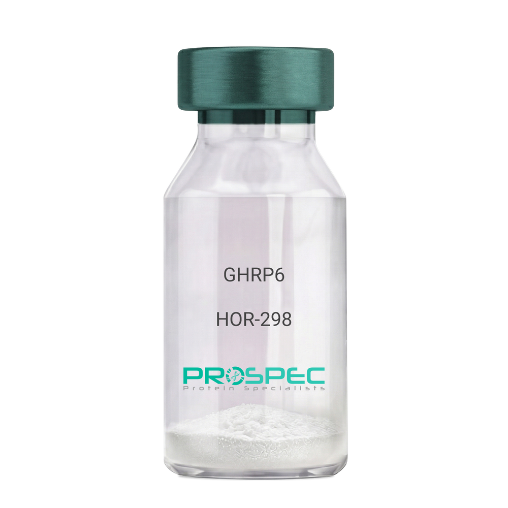
GHRP6Description:
Growth Hormone Releasing Peptide-6
GHRP-6, GHRP6
Product # :
HOR-298Price :
Quantity :
Shipping Method :
Shipped at Room temp
More Info
- description
- formulation
- purity
- More Info
Description
Growth Hormone Releasing Peptide-6 Synthetic is a single, non-glycosylated polypeptide chain containing 6 amino acids, having a molecular mass of 873 Dalton and a Molecular formula of C46H56N12O6.
Formulation
The GHRP-6 peptide was lyophilized with no additives.
Purity
Greater than 98.0% as determined by analysis by RP-HPLC.
More Info
-
Introduction
GH-releasing peptides (GHRPs) are synthetic peptides that like GHRH act directly on pituitary somatotrophs to stimulate GH release. Growth hormone (GH) release is stimulated by a variety of synthetic secretagogues, of which growth hormone-releasing hexapeptide (GHRP-6) has been most thoroughly studied; it is thought to have actions at both pituitary and hypothalamic site.
-
Synonyms
GHRP-6, GHRP6
-
Physical Appearance
Sterile Filtered White lyophilized (freeze-dried) powder.
-
Stability
Lyophilized Growth Hormone Releasing Peptide-6 although stable at room temperature for 3 weeks, should be stored desiccated below -18°C. Upon reconstitution GHRP-6 should be stored at 4°C between 2-7 days and for future use below -18°C. For long term storage it is recommended to add a carrier protein (0.1% HSA or BSA).Please prevent freeze-thaw cycles.
-
Solubility
It is recommended to reconstitute the lyophilized GHRP-6 in sterile 18MΩ-cm H2O not less than 100 µg/ml, which can then be further diluted to other aqueous solutions.
-
Amino Acid Sequence
His-D-Trp-Ala-Trp-D-Phe-Lys-NH2.
ProSpec's products are furnished for LABORATORY RESEARCH USE ONLY. The product may not be used as drugs, agricultural or pesticidal products, food additives or household chemicals.
- View Data Sheet
Name :
KIR2DL4 HumanDescription:
Killer Cell Immunoglobulin-Like Receptor, 2 Domains Long Cytoplasmic Tail 4 Human Recombinant
Killer Cell Immunoglobulin Like Receptor, Two Ig Domains And Long Cytoplasmic Tail 4,Killer Cell Immunoglobulin-Like Receptor, Two Domains, Long Cytoplasmic Tail 4,Killer Cell Inhibitory Receptor 103AS, MHC Class I NK Cell Receptor KIR103AS,CD158 Antigen-Like Family Member D, KIR-103AS, KIR103AS, CD158D, G9P, Killer Cell Immunoglobulin-Like Receptor 2DL4, CD158d Antigen, KIR-2DL4, KIR103, KIR2DL4.
Product # :
PRO-2460Price :
Quantity :
Shipping Method :
Shipped with Ice Packs
More Info
- description
- source
- formulation
- purity
- More Info
Description
KIR2DL4 Human Recombinant produced in Sf9 Insect cells is a single, glycosylated polypeptide chain containing 458 amino acids (24-242 a.a.) and having a molecular mass of 51kDa (Molecular size on SDS-PAGE will appear at approximately 50-70kDa). KIR2DL4 is expressed with a 239 amino acids hIgG-His tag at C-Terminus and purified by proprietary chromatographic techniques.
Source
Sf9, Baculovirus cells.
Formulation
KIR2DL4 protein solution (0.25mg/ml) contains Phosphate buffered saline (pH7.4) and 10% glycerol.
Purity
Greater than 85.0% as determined by SDS-PAGE.
More Info
-
Introduction
Killer cell immunoglobulin-like receptor 2DL4, also known as KIR2DL4 is part of the killer cell Ig-like receptor (KIR) family. KIR proteins with the long cytoplasmic domain transduce inhibitory signals through an immune tyrosine-based inhibitory motif (ITIM), whereas KIR proteins which contain the short cytoplasmic domain, lack the ITIM motif and as a substitute associate with the TYRO protein tyrosine kinase binding protein to transduce activating signals. KIR2DL4 stimulates NK cells to produce IFN-gamma and stimulation with IL-2 upregulates cell surface expression on CD56dimcells and leads to the inhibition of the cytolytic NK cell function.
-
Synonyms
Killer Cell Immunoglobulin Like Receptor, Two Ig Domains And Long Cytoplasmic Tail 4,Killer Cell Immunoglobulin-Like Receptor, Two Domains, Long Cytoplasmic Tail 4,Killer Cell Inhibitory Receptor 103AS, MHC Class I NK Cell Receptor KIR103AS,CD158 Antigen-Like Family Member D, KIR-103AS, KIR103AS, CD158D, G9P, Killer Cell Immunoglobulin-Like Receptor 2DL4, CD158d Antigen, KIR-2DL4, KIR103, KIR2DL4.
-
Physical Appearance
Sterile Filtered colorless solution.
-
Stability
Store at 4°C if entire vial will be used within 2-4 weeks. Store, frozen at -20°C for longer periods of time. For long term storage it is recommended to add a carrier protein (0.1% HSA or BSA).Avoid multiple freeze-thaw cycles.
-
Amino Acid Sequence
HVGGQDKPFC SAWPSAVVPQ GGHVTLRCHY RRGFNIFTLY KKDGVPVPEL YNRIFWNSFL ISPVTPAHAG TYRCRGFHPH SPTEWSAPSN PLVIMVTGLY EKPSLTARPG PTVRAGENVT LSCSSQSSFD IYHLSREGEA HELRLPAVPS INGTFQADFP LGPATHGETY RCFGSFHGSP
YEWSDPSDPL PVSVTGNPSS SWPSPTEPSF KTGIARHLHL EPKSCDKTHT CPPCPAPELL GGPSVFLFPP KPKDTLMISR TPEVTCVVVD VSHEDPEVKF NWYVDGVEVH NAKTKPREEQ YNSTYRVVSV LTVLHQDWLN GKEYKCKVSN KALPAPIEKT ISKAKGQPRE PQVYTLPPSR
DELTKNQVSL TCLVKGFYPS DIAVEWESNG QPENNYKTTP PVLDSDGSFF LYSKLTVDKS RWQQGNVFSC SVMHEALHNH YTQKSLSLSP GKHHHHHH
ProSpec's products are furnished for LABORATORY RESEARCH USE ONLY. The product may not be used as drugs, agricultural or pesticidal products, food additives or household chemicals.
- View Data Sheet
Name :
Leptin Human, PEGDescription:
Leptin Human Recombinant, PEG
OB Protein, Obesity Protein, OBS, Obesity factor.
Product # :
CYT-1108Price :
Quantity :
Shipping Method :
Shipped at Room temp
More Info
- description
- source
- formulation
- purity
- biological activity
- More Info
Description
Pegylated Leptin Human Recombinant produced in E.Coli is a single non-glycosilated polypeptide chain containing 146 amino acids, an additional Ala at N-terminus and one molecule of PEG 20 kDa at its N-terminus acids and having a molecular weight of 35.6kDa. However due to enlarged hydrodymanic volume it runs on the SDS-PAGE as 48 kDa protein and in gel-filtration on Superdex 200 as over 100 kDa protein. Pegylated Leptin Human Recombinant was purified by proprietary chromatographic techniques.
Source
Escherichia Coli.
Formulation
Lyophilized from a concentrated (1mg/ml) solution with 0.0045mM NaHCO3.
Purity
Greater than 98.0% as determined by:
(a) Gel filtration analysis.
(b) Analysis by SDS-PAGE.Biological Activity
Biological Activity is < than 0.1% as determined by inducing proliferation of BAF/3 cells stably transfected with the long form of human leptin receptor. It’s in vitro activity is 5-7 fold lower than the non-pegylated recombinant human leptin but in vivo it has profound weight reducing effect, resulting mainly from reduced food intake.
More Info
-
Introduction
Leptin takes an important part in the regulation of energy balance and body weight control.After entering the circulation, Leptin binds LEPRwhich results in the activation of several major signalling pathways. In the hypothalamus Leptin acts as an appetite-regulating factor that induces a decrease in food intake and an increase in energy consumption and also regulates bone mass and secretion of hypothalamo-pituitary-adrenal hormones. In the periphery, increases basal metabolism, regulates pancreatic beta-cell function and insulin secretion and affects innate and adaptive immunity.
-
Synonyms
OB Protein, Obesity Protein, OBS, Obesity factor.
-
Physical Appearance
Sterile Filtered White lyophilized (freeze-dried) powder.
-
Stability
Lyophilized Pegylated leptin although stable at room temperature for 3 weeks, should be stored desiccated below -18°C. Upon reconstitution leptin N82K should be stored at 4°C between 2-7 days and for future use below -18°C. For long term storage it is recommended to add a carrier protein (0.1% HSA or BSA). Please prevent freeze-thaw cycles.
-
Solubility
It is recommended to reconstitute the lyophilized Pegylated leptin in sterile water or 0.4% NaHCO3 adjusted to pH 8-9, not less than 100µg/ml, which can then be further diluted to other aqueous solutions.
-
Protein content
Protein quantitation was carried out by UV spectroscopy at 280 nm using the absorbency value of 0.87 as the extinction coefficient for a 0.1% (1mg/ml) solution at pH 8.0. This value is calculated by the PC GENE computer analysis program of protein sequences (IntelliGenetics).
ProSpec's products are furnished for LABORATORY RESEARCH USE ONLY. The product may not be used as drugs, agricultural or pesticidal products, food additives or household chemicals.
- View Data Sheet
Name :
NME1 AntibodyDescription:
Non-Metastatic Cells 1, Mouse Anti Human
Nucleoside diphosphate kinase A, NDP kinase A, NDK A, Tumor metastatic process-associated protein, Metastasis inhibition factor nm23, nm23-H1, Granzyme A-activated DNase, GAAD, NME1, NDPKA, NM23, NB, AWD, NBS, NDPK-A.
Product # :
ANT-691Price :
Quantity :
Shipping Method :
Shipped with Ice Packs
More Info
- formulation
- More Info
Formulation
1mg/ml containing PBS, pH-7.4, 10% Glycerol and 0.02% Sodium Azide.
More Info
-
Introduction
NDK (Nucleoside diphosphate kinase) exists as a hexamer composed of 'A' (encoded by NME1) and 'B' (encoded by NME2) isoforms. NME1 is involved in cell proliferation, differentiation and development, signal transduction, G protein-coupled receptor endocytosis, and gene expression. It also has tumor metastasis-suppressive capacity. NME1 has a key role in the synthesis of nucleoside triphosphates other than ATP. NME1 is essential for neural development including neural patterning and cell future determination.
The NME1 gene is expressed in various tumor types where its levels have been alternatively linked to reduced or increased metastatic potential.
Decrease in NME1 expression is notably connected to aggressive behavior in melanoma, breast, colon, and gastric carcinomas. In contrast, elevated levels of NME1 gene expression are noted in the advanced stage of thyroid carcinomas.
Somatic mutations of the NME1 gene are found in neuroblastoma. Increased NME1 in neuroblastoma is linked to features of the disease that are associated with aggressive tumors. -
Synonyms
Nucleoside diphosphate kinase A, NDP kinase A, NDK A, Tumor metastatic process-associated protein, Metastasis inhibition factor nm23, nm23-H1, Granzyme A-activated DNase, GAAD, NME1, NDPKA, NM23, NB, AWD, NBS, NDPK-A.
-
Physical Appearance
Sterile filtered colorless solution.
-
Immunogen
Anti-human NME1 mAb, is derived from hybridization of mouse F0 myeloma cells with spleen cells from BALB/c mice immunized with recombinant human NME1 amino acids 1-152 purified from E. coli.
-
Ig Subclass
Mouse IgG2b heavy chain and k light chain.
-
Clone
PAT5F4AT.
-
Applications
NME1 antibody has been tested by ELISA, Western blot analysis, Flow cytometry and ICC/IF to assure specificity and reactivity. Since application varies, however, each investigation should be titrated by the reagent to obtain optimal results.
-
Type
Mouse Anti Human Monoclonal.
-
Storage Procedures
For periods up to 1 month store at 4°C, for longer periods of time, store at -20°C. Prevent freeze thaw cycles.
-
Purification Method
NME1 antibody was purified from mouse ascitic fluids by protein-A affinity chromatography.
ProSpec's products are furnished for LABORATORY RESEARCH USE ONLY. The product may not be used as drugs, agricultural or pesticidal products, food additives or household chemicals.
- View Data Sheet
Name :
PDCD12 AntibodyDescription:
Programmed Cell Death 12, Mouse Anti Human
Cell death regulator Aven, AVEN, PDCD12, Programmed Cell Death 12.
Product # :
ANT-457Price :
Quantity :
Shipping Method :
Shipped with Ice Packs
More Info
- formulation
- More Info
Formulation
1mg/ml containing PBS, pH-7.4, & 0.1% Sodium Azide.
More Info
-
Introduction
PDCD12 (AVEN) represents a new class of cell death regulator. PDCD12 is a conserved intracellular membrane protein which blocks apoptosis impeded by Apaf-1 mediated caspase activation. BAD and Bcl-2 also interact with PDCD12 to prevent apoptosis. PDCD12 is expressed in a wide variety of normal tissues including the heart, skeletal muscle, kidney, liver, testis, and also in diseases such as cancer.
-
Synonyms
Cell death regulator Aven, AVEN, PDCD12, Programmed Cell Death 12.
-
Immunogen
Anti-human PDCD12 mAb is derived from hybridization of mouse F0 myeloma cells with spleen cells from BALB/c mice immunized with recombinant human PDCD12 amino acids 254-362 purified from E. coli.
-
Ig Subclass
Mouse IgG2a heavy chain and κ light chain.
-
Clone
P3G4AT.
-
Applications
PDCD12 antibody has been tested by ELISA, Western blot and immunohistochemistry analysis to assure specificity and reactivity. Since application varies, however, each investigation should be titrated by the reagent to obtain optimal results. Recommended dilution range for Western blot is 1:1,000~1:2,000 and immunohistochemistry analysis is 1:50~100. Recommended starting dilution for Western blot is 1:1,000 and Immunohistochemistry is 1:50.
-
Type
Mouse Anti Human Monoclonal.
-
Storage Procedures
For periods up to 1 month store at 4°C, for longer periods of time, store at -20°C. Prevent freeze thaw cycles.
-
Purification Method
PDCD12 antibody was purified from mouse ascitic fluids by protein-G affinity chromatography.
ProSpec's products are furnished for LABORATORY RESEARCH USE ONLY. The product may not be used as drugs, agricultural or pesticidal products, food additives or household chemicals.
- View Data Sheet
Name :
KLRD1 AntibodyDescription:
Mouse Anti Human Killer Cell Lectin Like Receptor D1
Killer Cell Lectin Like Receptor D1, Killer Cell Lectin-Like Receptor Subfamily D, Member 1, NK Cell Receptor, CD94 Antigen, CD94, KP43, Killer Cell Lectin-Like Receptor Subfamily D Member 1, Natural Killer Cells Antigen CD94, KLRD1.
Product # :
ANT-717Price :
Quantity :
Shipping Method :
Shipped with Ice Packs
More Info
- formulation
- More Info
Formulation
1mg/ml containing PBS, pH-7.4, 10% Glycerol and 0.02% Sodium Azide.
More Info
-
Introduction
KLRD1, also known as Killer Cell Lectin Like Receptor D1, is expressed on the surface of natural killer cells in the innate immune system. KLRD1 functions as a receptor for the recognition of MHC class I HLA-E molecules by NK cells and some cytotoxic T-cells. KLRD1 can create disulfide-bonded heterodimer with NKG2 family members. CD94 & NKG2 complex interacts with (HLA)-E, Human Leukocyte Antigen on target cells on the surface of natural killer cells.
-
Synonyms
Killer Cell Lectin Like Receptor D1, Killer Cell Lectin-Like Receptor Subfamily D, Member 1, NK Cell Receptor, CD94 Antigen, CD94, KP43, Killer Cell Lectin-Like Receptor Subfamily D Member 1, Natural Killer Cells Antigen CD94, KLRD1.
-
Physical Appearance
Sterile filtered colorless solution.
-
Immunogen
Anti-human KLRD1 mAb, is derived from hybridization of mouse F0 myeloma cells with spleen cells from BALB/c mice immunized with a recombinant human KLRD1 protein 32-279 amino acids purified from E. coli.
-
Ig Subclass
Mouse IgG1 heavy chain and k light chain.
-
Clone
PAT13E3AT.
-
Applications
KLRD1 antibody has been tested by ELISA and Western blot analysis to assure specificity and reactivity. Since application varies, however, each investigation should be titrated by the reagent to obtain optimal results.
-
Type
Mouse Anti Human Monoclonal.
-
Storage Procedures
For periods up to 1 month store at 4°C, for longer periods of time, store at -20°C. Prevent freeze thaw cycles.
-
Purification Method
KLRD1 antibody was purified from mouse ascitic fluids by protein-A affinity chromatography.
ProSpec's products are furnished for LABORATORY RESEARCH USE ONLY. The product may not be used as drugs, agricultural or pesticidal products, food additives or household chemicals.
- View Data Sheet
Name :
Tamm HorsfallDescription:
Recombinant Human Tamm Horsfall Glycoprotein
Tamm-Horsfall urinary glycoprotein, THP, FJHN, HNFJ, THGP, MCKD2, ADMCKD2, UMOD, Uromodulin.
Product # :
ENZ-1206Price :
Quantity :
Shipping Method :
Shipped at Room temp
More Info
- description
- source
- formulation
- purity
- More Info
- SDS-PAGE
Description
Uromodulin Human Recombinant protein produced from HEK Cells, is a polypeptide chain containing 595 amino acids ( 25-613 a.a. ) and having a total Mw of 65 kDa.
Source
HEK293
Formulation
The UMOD protein was lyophilized from 0.4μm filtered solution containing 50mM NaCl, 0.02M TRIS, pH 7.5.
Purity
Greater than 90.0% as determined by SDS-PAGE analysis.
SDS-PAGE
More Info
-
Introduction
Uromodulin (Tamm–Horsfall protein) is produced mainly by cells in the kidney’s thick ascending limb and is the most abundant protein in normal urine.
Uromodulin takes part in salt and water regulation, helps prevent urinary tract infections and kidney stones, and influences inflammation and immune activity in the kidney.
Reduced Uromodulin levels is associated with chronic kidney disease.
UMOD mutations causes unproper protein folding and thus transported incorrectly, resulting in its accumulation inside kidney tubular cells that damages the tubules and cause kidney disease (ADTKD-UMOD), associated with high uric acid, gout, and progressive kidney failure.
-
Synonyms
Tamm-Horsfall urinary glycoprotein, THP, FJHN, HNFJ, THGP, MCKD2, ADMCKD2, UMOD, Uromodulin.
-
Physical Appearance
Filtered White lyophilized (freeze-dried) powder.
-
Stability
Lyophilized UMOD although stable at room temperature for 3 weeks, should be stored at -18C. Upon reconstitution UMOD should be stored at 4C between 2-7 days and for future use below -18C.
For long term storage it is recommended to add a carrier protein (0.1% HSA or BSA).
Please prevent freeze-thaw cycles.
-
Solubility
Add deionized water to prepare a working stock solution of approximately 0.5mg/ml and let the lyophilized pellet dissolve completely. Product is not sterile! Please filter the product by an appropriate sterile filter before using it in the cell culture.
-
Amino Acid Sequence
DTSEARWCSE CHSNATCTED EAVTTCTCQE GFTGDGLTCV DLDECAIPGA HNCSANSSCV NTPGSFSCVC PEGFRLSPGL GCTDVDECAE PGLSHCHALA TCVNVVGSYL CVCPAGYRGD GWHCECSPGS CGPGLDCVPE GDALVCADPC QAHRTLDEYW RSTEYGEGYA CDTDLRGWYR FVGQGGARMA ETCVPVLRCN TAAPMWLNGT HPSSDEGIVS RKACAHWSGH CCLWDASVQV KACAGGYYVY NLTAPPECHL AYCTDPSSVE GTCEECSIDE DCKSNNGRWH CQCKQDFNIT DISLLEHRLE CGANDMKVSL GKCQLKSLGF DKVFMYLSDS RCSGFNDRDN RDWVSVVTPA RDGPCGTVLT RNETHATYSN TLYLADEIII RDLNIKINFA CSYPLDMKVS LKTALQPMVS ALNIRVGGTG MFTVRMALFQ TPSYTQPYQG SSVTLSTEAF LYVGTMLDGG DLSRFALLMT NCYATPSSNA TDPLKYFIIQ DRCPHTRDST IQVVENGESS QGRFSVQMFR FAGNYDLVYL HCEVYLCDTM NEKCKPTCSG TRFRSGSVID QSRVLNLGPI TRKGVQATVH HHHHH
-
Background
Uromodulin is the most abundant protein in normal urine. Its secretion in urine follows proteolytic cleavage of the ectodomain of its glycosyl phosphatidylinosital-anchored counterpart that is situated on the luminal cell surface of the loop of Henle. Uromodulin plays a role as a constitutive inhibitor of calcium crystallization in renal fluids. Secretion of uromodulin in urine provides protection against urinary tract infections caused by uropathogenic bacteria. Defects in Uromodulin expression are associated with the autosomal dominant renal disorders medullary cystic kidney disease-2 (MCKD2) and familial juvenile hyperuricemic nephropathy (FJHN). These disorders are characterized by juvenile onset of hyperuricemia, gout, and progressive renal failure. While several transcript variants may exist for this gene, the full-length natures of only two have been described to date. UMOD is involved in regulating the circulating activity of cytokines as it binds to il-1, il-2 and tnf with high affinity.
What is the molecular weight/Mw of UMOD Protein?
UMOD Protein has a total Mw of 65kDa.
What is the source or expression system of UMOD Protein?
HEK293.
What is the Purity of UMOD Protein?
UMOD Protein is >90% pure as determined by SDS-PAGE.
What is the Biological Activity of UMOD Protein?
The biological functionality of UMOD Protein will be determined in the future.
What is the amino acid sequence of UMOD Protein?
UMOD Protein is composed from 595 amino acids.
What applications can UMOD Protein be used in?
UMOD Protein can probably be used in western blot, ELISA and Lateral Flow.
What is the endotoxin level for UMOD Protein?
The endotoxin level is minimal, UMOD Protein was purified using conventional chromatography techniques.
ProSpec's products are furnished for LABORATORY RESEARCH USE ONLY. The product may not be used as drugs, agricultural or pesticidal products, food additives or household chemicals.
- View Data Sheet
Name :
TNFRSF10A Human FcDescription:
TRAIL Receptor-1 Human Recombinant Fc
Tumor Necrosis Factor Receptor Superfamily Member 10a, TRAILR1, APO2, DR4, TNF-Related Apoptosis-Inducing Ligand Receptor 1, Death Receptor 4, TRAIL Receptor 1, TRAIL-R1, Tumor Necrosis Factor Receptor Superfamily Member 10a Variant 2, Tumor Necrosis Factor Receptor Superfamily Member 10A, Cytotoxic TRAIL Receptor, CD261 Antigen, TRAILR-1, CD261, TNFRSF10A.
Product # :
CYT-877Price :
Quantity :
Shipping Method :
Shipped at Room temp
More Info
- description
- source
- formulation
- purity
- biological activity
- More Info
Description
TRAIL R1 Fc Human Recombinant (24-240) produced in E.Coli is a non-glycosylated, disulfide-linked homodimer containing two polypeptide chains of 217 amino acids each and having a molecular mass of 41kDa [including tag] each chain. TRAILR1 is fused to a 236 amino acid Fc tag at C-terminal and purified by proprietary chromatographic techniques.
Source
Escherichia Coli.
Formulation
Lyophilized from a 0.2µm filtered solution in PBS.
Purity
Greater than 95.0% as determined by SDS-PAGE.
Biological Activity
The ED50, as measured by its ability to inhibit Trail-mediated cytotoxicity in mouse L-929 cells, is less than 1.25ng/ml.More Info
-
Introduction
Human TNFRSF10A is a type 1 transmembrane protein in the TNF R superfamily. TNFRSF10A is not expressed in rodents. The trimeric ligand Trail binds TNFRSF10A and induces apoptosis, and recombinant, soluble forms of the receptor inhibit Trail-induced apoptosis. TNFRSF10A is expressed generally in damaged, infected, and malignant cells. TNFRSF10A functions in immune surveillance, inducing apoptosis in cancer cells but not normal cells.
-
Synonyms
Tumor Necrosis Factor Receptor Superfamily Member 10a, TRAILR1, APO2, DR4, TNF-Related Apoptosis-Inducing Ligand Receptor 1, Death Receptor 4, TRAIL Receptor 1, TRAIL-R1, Tumor Necrosis Factor Receptor Superfamily Member 10a Variant 2, Tumor Necrosis Factor Receptor Superfamily Member 10A, Cytotoxic TRAIL Receptor, CD261 Antigen, TRAILR-1, CD261, TNFRSF10A.
-
Physical Appearance
Sterile Filtered White lyophilized (freeze-dried) powder.
-
Stability
Lyophilized TNFRSF10A although stable at room temperature for 3 weeks, should be stored desiccated below -18°C. Upon reconstitution TRAIL-R1 should be stored at 4°C between 2-7 days and for future use below -18°C.For long term storage it is recommended to add a carrier protein (0.1% HSA or BSA).Please prevent freeze-thaw cycles.
-
Solubility
It is recommended to reconstitute the lyophilized TNFRSF10A in sterile 18M-cm H2O not less than 100µg/ml, which can then be further diluted to other aqueous solutions.
-
Amino Acid Sequence
ASGTEAAAAT PSKVWGSSAG RIEPRGGGRG ALPTSMGQHG PSARARAGRA PGPRPAREAS PRLRVHKTFK FVVVGVLLQV VPSSAATIKL HDQSIGTQQW EHSPLGELCP PGSHRSERPG ACNRCTEGVG YTNASNNLFA CLPCTACKSD EEERSPCTTT RNTACQCKPG TFRNDNSAEM CRKCSTGCPR GMVKVKDCTP WSDIECVHKE SGNGHNIEGR MDPKSCDKTH TCPPCPAPEL LGGPSVFLFP PKPKDTLMIS RTPEVTCVVV DVSHEDPEVK FNWYVDGVEV HNAKTKPREE QYNSTYRVVS VLTVLHQDWL NGKEYKCKVS NKALPAPIEK TISKAKGQPR EPQVYTLPPS RDELTKNQVS LTCLVKGFYP SDIAVEWESN GQPENNYKTT PPVLDSDGSF FLYSKLTVDK SRWQQGNVFS CSVMHEALHN HYTQKSLSLS PGK.
ProSpec's products are furnished for LABORATORY RESEARCH USE ONLY. The product may not be used as drugs, agricultural or pesticidal products, food additives or household chemicals.