Search results
1000 results found for “pleiotrophin”
Name
Description
Product #
Price
Quantity
Shipping Method
- View Data Sheet
Name :
LHRH ProteinDescription:
Luteinizing Hormone Releasing Hormone Human Recombinant
LHRH, GRH, GNRH, LNRH.
Product # :
HOR-268Price :
Quantity :
Shipping Method :
Shipped with Ice Packs
More Info
- description
- source
- formulation
- purity
- More Info
Description
LHRH Human Recombinant produced in E.Coli is a single, non-glycosylated polypeptide chain containing 92 amino acids (24-92 a.a.) and having a molecular mass of 10.3kDa.LHRH is fused to a 23 amino acid His-tag at N-terminus & purified by proprietary chromatographic techniques.
Source
Escherichia Coli.
Formulation
LHRH protein solution (0.5mg/ml) containing 20mM Tris-HCl buffer (pH 8.0), 0.15M NaCl, 20% glycerol and 1mM DTT.
Purity
Greater than 90.0% as determined by SDS-PAGE.
More Info
-
Introduction
GNRH1 also known as Luteinising-hormone releasing hormone (LHRH), is a peptide hormone responsible for the release of FSH and LH from the anterior pituitary. GNRH1 is synthesized and released by the hypothalamus.
At the pituitary, GNRH1 stimulates the synthesis and secretion of the follicle-stimulating hormone (FSH) and luteinizing hormone (LH). These processes are controlled by the size and frequency of GNRH1 pulses, as well as by feedback from androgens and estrogens. Low requency GNRH1 pulses lead to FSH release, whereas high frequency GNRH1 pulses stimulate LH release.
There are differences in GNRH1 secretion between males and females. In males, GNRH1 is secreted in pulses at a constant frequency, but in females the frequency of the pulses varies during the menstrual cycle and there is a large surge of GNRH1 just before ovulation.
GNRH1 secretion is pulsatile in all vertebrates, and is necessary for correct reproductive function. Thus, a single hormone, GNRH1, controls a complex process of follicular growth, ovulation, and corpus luteum maintenance in the female, and spermatogenesis in the male. -
Synonyms
LHRH, GRH, GNRH, LNRH.
-
Physical Appearance
Sterile filtered colorless solution.
-
Stability
Store at 4°C if entire vial will be used within 2-4 weeks. Store, frozen at -20°C for longer periods of time. For long term storage it is recommended to add a carrier protein (0.1% HSA or BSA).Avoid multiple freeze-thaw cycles.
-
Amino Acid Sequence
MGSSHHHHHH SSGLVPRGSH MGSQHWSYGL RPGGKRDAEN LIDSFQEIVK EVGQLAETQR FECTTHQPRS PLRDLKGALE SLIEEETGQK KI.
ProSpec's products are furnished for LABORATORY RESEARCH USE ONLY. The product may not be used as drugs, agricultural or pesticidal products, food additives or household chemicals.
- View Data Sheet
Name :
COPS8 HumanDescription:
COP9 Constitutive Photomorphogenic 8 Human Recombinant
COP9 signalosome complex subunit 8, SGN8, Signalosome subunit 8, COP9 homolog, hCOP9, JAB1-containing signalosome subunit 8, COPS8, CSN8, COP9.
Product # :
PRO-983Price :
Quantity :
Shipping Method :
Shipped with Ice Packs
More Info
- description
- source
- formulation
- purity
- More Info
Description
COPS8 Human Recombinant produced in E.coli is a single, non-glycosylated polypeptide chain containing 229 amino acids (1-209) and having a molecular mass of 25.3kDa.COPS8 is fused to a 20 amino acid His-tag at N-terminus & purified by proprietary chromatographic techniques.
Source
Escherichia Coli.
Formulation
The COPS8 solution (1mg/ml) contains 20mM Tris-HCl buffer (pH 8.0), 0.4M Urea and 10% glycerol.
Purity
Greater than 90.0% as determined by SDS-PAGE.
More Info
-
Introduction
COP9 signalosome complex subunit 8 isoform 1 (COPS8) is one of the 8 subunits of COP9 signalosome, which is a much conserved protein complex that functions as an imperative regulator in multiple signaling pathways. The structure and function of COP9 signalosome is analogous to that of the 19S regulatory particle of 26S proteasome. COP9 signalosome interacts with SCF-type E3 ubiquitin ligases and acts as a positive regulator of E3 ubiquitin ligases.
-
Synonyms
COP9 signalosome complex subunit 8, SGN8, Signalosome subunit 8, COP9 homolog, hCOP9, JAB1-containing signalosome subunit 8, COPS8, CSN8, COP9.
-
Physical Appearance
Sterile Filtered colorless solution.
-
Stability
Store at 4°C if entire vial will be used within 2-4 weeks. Store, frozen at -20°C for longer periods of time. For long term storage it is recommended to add a carrier protein (0.1% HSA or BSA).Avoid multiple freeze-thaw cycles.
-
Amino Acid Sequence
MGSSHHHHHH SSGLVPRGSH MPVAVMAESA FSFKKLLDQC ENQELEAPGG IATPPVYGQL LALYLLHNDM NNARYLWKRI PPAIKSANSE LGGIWSVGQR IWQRDFPGIY TTINAHQWSE TVQPIMEALR DATRRRAFAL VSQAYTSIIA DDFAAFVGLP VEEAVKGILE QGWQADSTTR
MVLPRKPVAG ALDVSFNKFI PLSEPAPVPP IPNEQQLARL TDYVAFLEN.
ProSpec's products are furnished for LABORATORY RESEARCH USE ONLY. The product may not be used as drugs, agricultural or pesticidal products, food additives or household chemicals.
- View Data Sheet
Name :
FGF12 Human, HisDescription:
Recombinant Human Fibroblast Growth Factor 12, His Tag
FGF-12, FGF12, FGF12B, FHF1, Fibroblast growth factor 12, Fibroblast growth factor homologous factor 1, FHF-1, Myocyte-activating factor.
Product # :
CYT-620Price :
Quantity :
Shipping Method :
Shipped with Ice Packs
More Info
- description
- source
- formulation
- purity
- More Info
Description
The FGF-12 Human recombinant protein is a single, non-glycosylated polypeptide chain produced in E. coli, having a molecular weight of 22.6kDa and containing 201 amino acids (1-181). The FGF12 is fused to a 20 amino acid His tag at the N-terminus.
Source
Escherichia Coli.
Formulation
The FGF-12 solution (1mg/ml) contains 20mM Tris pH-7.5, 1mM DTT, 2mM EDTA and 10% glycerol.
Purity
Greater than 90% as determined by SDS-PAGE.
More Info
-
Introduction
FGF12 is part of the Fibroblast Growth Factor (FGF) family which has a vast mitogenic and cell survival functions, and play a role in a range of biological activities, among them are embryonic development, cell growth, morphogenesis, tissue repair, tumor growth, and invasion. FGF-12 doesn’t obtain the N-terminal signal sequence present in the majority of the FGF family members, but it contains clusters of basic residues that act as a nuclear localization signal. When transfected into mammalian cells, FGF12 accumulated in the nucleus, but was not secreted. FGF12 is involved in nervous system development and function. FGF12 binds to IB2 (islet brain-2), a cellular kinase scaffold, and voltage gated sodium channels and is also involved in intracellular signaling and ion exchange.
-
Synonyms
FGF-12, FGF12, FGF12B, FHF1, Fibroblast growth factor 12, Fibroblast growth factor homologous factor 1, FHF-1, Myocyte-activating factor.
-
Physical Appearance
Sterile liquid colorless solution.
-
Stability
Store FGF12 at -20°C. Can be stored at 4°C for a limited period of time of 7 days.
-
Amino Acid Sequence
MSSHHHHHH SSGLVPRGSH MESKEPQLKG IVTRLFSQQG YFLQMHPDGT IDGTKDENSD YTLFNLIPVG LRVVAIQGVK ASLYVAMNGE YLYSSDVFT PECKFKESVF ENYYVIYSST LYRQQESGRA WFLGLNKEGQ IMKGNRVKKT KPSSHFVPKP IEVCMYREQS LHEIGEKQGR RKSSGTPTM NGGKVVNQDS T.
-
Background
What is the molecular weight/Mw of FGF12 Protein?
FGF12 Protein has a total Mw of 22.6kDa.
What is the source or expression system of FGF12 Protein?
Escherichia Coli.
What is the Purity of FGF12 Protein?
FGF12 Protein is >90% pure as determined by SDS-PAGE.
What is the Biological Activity of FGF12 Protein?
The biological functionality of FGF12 Protein will be determined in the future.
What is the amino acid sequence of FGF12 Protein?
MSSHHHHHH SSGLVPRGSH MESKEPQLKG IVTRLFSQQG YFLQMHPDGT IDGTKDENSD YTLFNLIPVG LRVVAIQGVK ASLYVAMNGE YLYSSDVFT PECKFKESVF ENYYVIYSST LYRQQESGRA WFLGLNKEGQ IMKGNRVKKT KPSSHFVPKP IEVCMYREQS LHEIGEKQGR RKSSGTPTM NGGKVVNQDS T.
What applications can FGF12 Protein be used in?
FGF12 Protein can probably be used in western blot, ELISA and Lateral Flow.
What is the endotoxin level for FGF12 Protein?
The endotoxin level is minimal, FGF12 Protein was purified using conventional chromatography techniques.
ProSpec's products are furnished for LABORATORY RESEARCH USE ONLY. The product may not be used as drugs, agricultural or pesticidal products, food additives or household chemicals.
- View Data Sheet
Name :
ElamipretideDescription:
Elamipretide
SS-31, MTP-131, Bendavia.
Product # :
HOR-040Price :
Quantity :
Shipping Method :
Shipped at Room temp
More Info
- description
- source
- formulation
- purity
- More Info
- HPLC, MS
Description
Elamipretide Synthetic is a single, non-glycosylated polypeptide chain containing 4 amino acids, having a molecular mass of 639.79 Dalton and a Molecular formula of C32H49N9O5
.Source
Synthetic Peptide
Formulation
The protein was lyophilized with no additives.
Purity
Greater than 97.0% as determined by analysis by RP-HPLC.
HPLC, MS
More Info
-
Synonyms
SS-31, MTP-131, Bendavia.
-
Physical Appearance
Sterile Filtered White lyophilized (freeze-dried) powder.
-
Stability
Lyophilized Elamipretide although stable at room temperature for 3 weeks, should be stored desiccated below -18°C. Upon reconstitution Elamipretide should be stored at 4°C between 2-7 days and for future use below -18°C.For long term storage it is recommended to add a carrier protein (0.1% HSA or BSA). Please prevent freeze-thaw cycles.
-
Solubility
It is recommended to reconstitute the lyophilized Elamipretide in sterile 18MΩ-cm H2O not less than 100 µg/ml, which can then be further diluted to other aqueous solutions.
-
Amino Acid Sequence
H-D-Arg-(2',6'-dimethyl-Tyr)-Lys-Phe-NH2
-
Background
What is the molecular weight/Mw of Elamipretide Protein?
Elamipretide Protein has a total Mw of 639Da.
What is the source or expression system of Elamipretide Protein?
Synthetic peptide
What is the Purity of Elamipretide Protein?
Elamipretide Protein is >97% pure as determined by SDS-PAGE.
What is the Biological Activity of Elamipretide Protein?
The biological functionality of Elamipretide Protein will be determined in the future.
What is the amino acid sequence of Elamipretide Protein?
H-D-Arg-(2',6'-dimethyl-Tyr)-Lys-Phe-NH2
What applications can Elamipretide Protein be used in?
Elamipretide Protein can probably be used in western blot, ELISA and Lateral Flow.
What is the endotoxin level for Elamipretide Protein?
The endotoxin level is minimal, Elamipretide Protein was purified using conventional chromatography techniques.Elamipretide (also known as SS-31) is a novel mitochondrial-targeted peptide with immense promise as a therapeutic agent in mitochondrial dysfunction-related disorders. This research paper aims to provide a comprehensive analysis of Elamipretide, delving into its biochemical properties, mechanisms of action, and potential applications in various disease conditions.
Elamipretide, a mitochondria-targeting tetrapeptide, has garnered attention for its unique ability to protect mitochondria from oxidative stress and attenuate mitochondrial dysfunction (Siegel et al., 2013). This paper endeavors to explore Elamipretide's biochemical basis and its potential as a therapeutic agent in various diseases linked to mitochondrial impairment.
Elamipretide selectively accumulates within the inner mitochondrial membrane, where it exerts its cytoprotective effects. By reducing reactive oxygen species (ROS) production and enhancing electron transport chain efficiency, Elamipretide plays a crucial role in mitochondrial homeostasis (Kloner et al., 2015).
The mitochondrial protective actions of Elamipretide arise from its interaction with cardiolipin, a phospholipid predominantly localized in the inner mitochondrial membrane. By binding to cardiolipin, Elamipretide stabilizes mitochondrial cristae, improves membrane integrity, and enhances oxidative phosphorylation (Minkler et al., 2015).
Elamipretide's potential applications extend to a myriad of disease conditions characterized by mitochondrial dysfunction. In preclinical studies, Elamipretide has shown promise in mitigating tissue damage following ischemia-reperfusion injury, preserving cardiac function after myocardial infarction, and ameliorating neurodegenerative processes (Birk et al., 2017; Cho et al., 2015).
As the research on Elamipretide progresses, further investigation is warranted to better understand its pharmacokinetics, long-term safety, and potential off-target effects. Clinical trials exploring its therapeutic efficacy in human diseases offer exciting prospects for the future.
Elamipretide, a mitochondria-targeting peptide, emerges as a promising candidate in combating mitochondrial dysfunction-related disorders. Its unique ability to stabilize mitochondrial membranes and enhance cellular bioenergetics positions Elamipretide as a novel therapeutic option for a diverse range of diseases.
ProSpec's products are furnished for LABORATORY RESEARCH USE ONLY. The product may not be used as drugs, agricultural or pesticidal products, food additives or household chemicals.
- View Data Sheet
Name :
MCSFR HumanDescription:
Colony Stimulating Factor 1 Receptor Human Recombinant
Macrophage colony-stimulating factor 1 receptor, CSF-1 receptor (EC:2.7.10.1), CSF-1-R, CSF-1R, M-CSF-R, Proto-oncogene c-Fms, CD115, CSF1R, FMS.
Product # :
CYT-1068Price :
Quantity :
Shipping Method :
Shipped with Ice Packs
More Info
- description
- source
- formulation
- purity
- biological activity
- More Info
Description
MCSFR produced in Sf9 Baculovirus cells is a single, glycosylated polypeptide chain containing 737 amino acids (20-517a.a.) and having a molecular mass of 82.1kDa. (Molecular size on SDS-PAGE will appear at approximately 70-100kDa).MCSFR is expressed with a 239 amino acid hIgG-His tag at C-Terminus and purified by proprietary chromatographic techniques.
Source
Sf9, Baculovirus cells.
Formulation
MCSFR protein solution (0.25mg/ml) contains Phosphate Buffered Saline (pH 7.4) and 10% glycerol.
Purity
Greater than 90.0% as determined by SDS-PAGE.
Biological Activity
Measured by its ability to inhibit M-CSF dependent proliferation of M-NFS-60 mouse myelogenous leukemia lymphoblast cells. The ED50 for this effect is less or equal to 100ng/ml in the presence of 10 ng/ml M-CSF.
More Info
-
Introduction
MCSFR which is also familiar as CSF1R, is part of the type3 subfamily of receptor tyrosine kinases. MCSFR is expressed mainly on cells of the monocyte and macrophage lineage, stem cells, and in the growing placenta. Most of the biological effects of this cytokine are mediated by MCSFR. MCSFR contains an extracellular ligand-binding domain, a single membrane-spanning segment, and an intracellular tyrosine kinase domain. Originally CSF1 and this receptor were implicated as vital for normal trophoblastic implantation as well as monocyte development. The role of CSF1/CSF1R in normal mammary gland development is actually very interesting since this connection has also been discovered in the biology of breast cancer with the results of abnormal expression of CSF1 and its receptor. Likewise, in Alzheimer's disease and after brain injuries an increased level of CSF1R was found in the microglia, which causes the microglia to become more active.
-
Synonyms
Macrophage colony-stimulating factor 1 receptor, CSF-1 receptor (EC:2.7.10.1), CSF-1-R, CSF-1R, M-CSF-R, Proto-oncogene c-Fms, CD115, CSF1R, FMS.
-
Physical Appearance
Sterile filtered colorless solution.
-
Stability
Store at 4°C if entire vial will be used within 2-4 weeks. Store, frozen at -20°C for longer periods of time. For long term storage it is recommended to add a carrier protein (0.1% HSA or BSA).Avoid multiple freeze-thaw cycles.
-
Amino Acid Sequence
IPVIEPSVPE LVVKPGATVT LRCVGNGSVE WDGPPSPHWT LYSDGSSSIL STNNATFQNT GTYRCTEPGD PLGGSAAIHL YVKDPARPWN VLAQEVVVFE DQDALLPCLL TDPVLEAGVS LVRVRGRPLM RHTNYSFSPW HGFTIHRAKF IQSQDYQCSA LMGGRKVMSI SIRLKVQKVI PGPPALTLVP AELVRIRGEA AQIVCSASSV DVNFDVFLQH NNTKLAIPQQ SDFHNNRYQK VLTLNLDQVD FQHAGNYSCV ASNVQGKHST SMFFRVVESA YLNLSSEQNL IQEVTVGEGL NLKVMVEAYP GLQGFNWTYL GPFSDHQPEP KLANATTKDT YRHTFTLSLP RLKPSEAGRY SFLARNPGGW RALTFELTLR YPPEVSVIWT FINGSGTLLC AASGYPQPNV TWLQCSGHTD RCDEAQVLQV WDDPYPEVLS QEPFHKVTVQ SLLTVETLEH NQTYECRAHN SVGSGSWAFI PISAGAHTHP PDEFLFTPLE PKSCDKTHTC PPCPAPELLG GPSVFLFPPK PKDTLMISRT PEVTCVVVDV SHEDPEVKFN WYVDGVEVHN AKTKPREEQY NSTYRVVSVL TVLHQDWLNG KEYKCKVSNK ALPAPIEKTI SKAKGQPREP QVYTLPPSRD ELTKNQVSLT CLVKGFYPSD IAVEWESNGQ PENNYKTTPP VLDSDGSFFL YSKLTVDKSR WQQGNVFSCS VMHEALHNHY TQKSLSLSPG KHHHHHH.
ProSpec's products are furnished for LABORATORY RESEARCH USE ONLY. The product may not be used as drugs, agricultural or pesticidal products, food additives or household chemicals.
- View Data Sheet
Name :
Leptin PAT7E10AT AntibodyDescription:
Leptin Clone PAT7E10AT, Mouse Anti Human
OB Protein, Obesity Protein, OBS, Obesity factor.
Product # :
ANT-690Price :
Quantity :
Shipping Method :
Shipped with Ice Packs
More Info
- formulation
- More Info
Formulation
1mg/ml containing PBS, pH-7.4, 10% Glycerol and 0.02% Sodium Azide.
More Info
-
Introduction
A 16-kDa peptide hormone secreted from white adipocytes and implicated in the regulation of food intake and energy balance. Leptin provides the key afferent signal from fat cells in the feedback system that controls body fat stores.
-
Synonyms
OB Protein, Obesity Protein, OBS, Obesity factor.
-
Physical Appearance
Sterile filtered colorless solution.
-
Immunogen
Anti-human Leptin mAb, is derived from hybridization of mouse F0 myeloma cells with spleen cells from BALB/c mice immunized with recombinant human Leptin amino acids 22-167 purified from E. coli.
-
Ig Subclass
Mouse IgG2b heavy chain and k light chain.
-
Clone
PAT7E10AT.
-
Applications
Leptin antibody has been tested by ELISA, Western blot analysis and ICC/IF to assure specificity and reactivity. Since application varies, however, each investigation should be titrated by the reagent to obtain optimal results.
-
Type
Mouse Anti Human Monoclonal.
-
Storage Procedures
For periods up to 1 month store at 4°C, for longer periods of time, store at -20°C. Prevent freeze thaw cycles.
-
Purification Method
Leptin antibody was purified from mouse ascitic fluids by protein-A affinity chromatography.
ProSpec's products are furnished for LABORATORY RESEARCH USE ONLY. The product may not be used as drugs, agricultural or pesticidal products, food additives or household chemicals.
- View Data Sheet
Name :
PSMD10 AntibodyDescription:
Gankyrin, Mouse Anti Human
26S proteasome non-ATPase regulatory subunit 10, 26S proteasome regulatory subunit p28, Gankyrin, PSMD10, p28, dJ889N15.2.
Product # :
ANT-609Price :
Quantity :
Shipping Method :
Shipped with Ice Packs
More Info
- formulation
- More Info
Formulation
1mg/ml containing PBS, pH-7.4, 10% Glycerol and 0.02% Sodium Azide.
More Info
-
Introduction
Gankyrin (proteasome 26S subunit) is a multicatalytic proteinase oncoprotein commonly overexpressed in most hepatocellular carcinomas. Proteasomes are found throughout eukaryotic cells at a high concentrations and cleave peptides in an ATP/ubiquitin-dependent process in a non-lysosomal pathway. Gankyrin interacts with S6 ATPase of the 19S regulatory particle of the 26S proteasome. Gankyrin is involved in theregulation of the phosphorylation of the retinoblastoma protein by CDK4, and to enhance the ubiquitinylation of p53 by MDM2. Gankyrin consists of 7 ankyrin repeats and is structurally similar to I kappa Bs. Gankyrin acts as a regulatory subunit of the 26s proteasome which is involved in the atp-dependent degradation of ubiquitinated proteins. Gankyrin is involved in progression of esophageal squamous cell carcinoma. gankyrin plays an oncogenic role especially in early stages of human epatocarcinogenesis. Gankyrin binds to NF-kappaB and suppresses its activity at the transcription level by modulating acetylation through SIRT1. Structural comparison between Gankyrin & p16(INK4A) identified numerous residues of gankyrin that are potentially important for CDK4 binding.
-
Synonyms
26S proteasome non-ATPase regulatory subunit 10, 26S proteasome regulatory subunit p28, Gankyrin, PSMD10, p28, dJ889N15.2.
-
Physical Appearance
Sterile filtered colorless solution.
-
Immunogen
Anti-human PSMD10 mAb, is derived from hybridization of mouse F0 myeloma cells with spleen cells from BALB/c mice immunized with a recombinant human PSMD10 protein 1-226 amino acids purified from E.coli.
-
Ig Subclass
Mouse IgG1 heavy chain and k light chain.
-
Clone
PAT1F4AT.
-
Applications
PSMD10 antibody has been tested by ELISA, Western blot analysis, Flow cytometry and ICC/IF to assure specificity and reactivity. Since application varies, however, each investigation should be titrated by the reagent to obtain optimal results.
-
Type
Mouse Anti Human Monoclonal.
-
Storage Procedures
For periods up to 1 month store at 4°C, for longer periods of time, store at -20°C. Prevent freeze thaw cycles.
-
Purification Method
PSMD10 antibody was purified from mouse ascitic fluids by protein-A affinity chromatography.
ProSpec's products are furnished for LABORATORY RESEARCH USE ONLY. The product may not be used as drugs, agricultural or pesticidal products, food additives or household chemicals.
- View Data Sheet
Name :
iL22RA (Y51A) Mouse, PEGDescription:
Interleukin-22 Receptor Antagonist (Y51A), PEG Mouse Recombinant
Product # :
CYT-1248Price :
Quantity :
Shipping Method :
Shipped at Room temp
More Info
- description
- source
- formulation
- purity
- biological activity
- More Info
Description
Interleukin 22 Y51A mutan Mouse Recombinant is a single non-glycosilated polypeptide chain containing 147 amino acids and additional Ala at N-terminus. The iL22RA is bound to 20 kDa PEG molecule at N-terminus, resulting in 36.0 kDa. The Mouse iL22RA (Y51A) Pegylated runs as a 50 kDa due to enlarged hydrodymanic volume. iL22RA Mutant Mouse Recombinant was purified by proprietary chromatographic techniques.
Source
Escherichia coli.
Formulation
The Mouse iL22RA (Y51A) was lyophilized from a concentrated (1mg/ml) solution with 0.003mM NaHCO3.
Purity
Greater than 98.0% as determined by:
(a) Gel filtration analysis.
(b) Analysis by SDS-PAGE.
Biological Activity
Pegylated Mouse iL22RA inhibits mouse IL-22 induced STAT3 phosphorylation in HepG cells. Its inhibitory acrtivity in vitro is ~ 10-20% compared to the non-pegylated mIL22 (Y51A) mutant.
More Info
-
Physical Appearance
White lyophilized (freeze-dried) powder.
-
Stability
Lyophilized iL22RA (Y51A) Mutant Mouse Recombinant although stable at room temperature for several weeks, should be stored desiccated below -18°C. Upon reconstitution at > 0.1 mg/ml and up to 2mM and filter sterilization iL22RA mutant can be stored at 4°C or even room temperature for several weeks making it suitable for long term infusion studies using osmotic pumps. At lower concentration addition of a carrier protein (0.1% HSA or BSA) is suggested. Please prevent freeze-thaw cycles.
-
Solubility
It is recommended to reconstitute the lyophilized iL22RA (Y51A) Mutant Mouse Recombinant in sterile water or sterile 0.4% NaHCO3 adjusted to pH 8-9, not less than 100µg/ml, which can then be further diluted with other aqueous solutions.
-
Background
IL-22 is a part of the IL-10 family of regulatory cytokines and produced by several populations of immune cells at a site of inflammation. Members of this family share partial homology in their amino acid sequences, but varies in their biological functions. IL-22 takes effect on non-hematopoietic cells. IL-22 takes part in wound healing and in protection against microbs. Produced by T lymphocytes, IL-22 inhibits IL-4 production by Th2 cells, and induces acute phase reactants in the pancreas and liver.
-
Protein content
Protein quantization was carried out by UV spectroscopy at 280 nm using the absorbency value of 0.18 as the extinction coefficient for a 0.1% (1mg/ml) solution at pH 8.0. This value is calculated by the DNAman computer analysis program of protein sequences.
ProSpec's products are furnished for LABORATORY RESEARCH USE ONLY. The product may not be used as drugs, agricultural or pesticidal products, food additives or household chemicals.
- View Data Sheet
Name :
FGF23 Human, HisDescription:
Fibroblast Growth Factor-23 Human Recombinant, His Tag
Tumor-derived hypophosphatemia-inducing factor, HYPF, ADHR, HPDR2, PHPTC, FGF23, FGF-23, Fibroblast Growth Factor-23.
Product # :
CYT-374Price :
Quantity :
Shipping Method :
Shipped at Room temp
More Info
- description
- source
- formulation
- purity
- biological activity
- More Info
Description
Fibroblast Growth Factor-23 Human Recombinant produced in E.Coli is a single, non-glycosylated polypeptide chain expressed with a -6xHis tag containing a total of 257 amino acids (251 a.a. FGF23+ 6 a.a. His tag) and having a molecular mass of 28629.5 Dalton. The FGF-23 is and purified by chromatographic techniques.
Source
Escherichia Coli.
Formulation
The protein (0.5mg/ml) was lyophilized from 25mM Tris pH7.5 and 0.6M NaCl solution.
Purity
Greater than 90.0% as determined by:
(a) Analysis by RP-HPLC.
(b) Analysis by SDS-PAGE.Biological Activity
Treatment with hrFGF23 has been shown to induce FGFR mediated Erk phosphorylation, reduce plasma PTH levels in rats and to reduce blood phosphate levels.More Info
-
Introduction
FGF-23 is a member of the fibroblast growth factor (FGF) family. FGF family members possess broad mitogenic and cell survival activities and are involved in a variety of biological processes including embryonic development, cell growth, morphogenesis, tissue repair, tumor growth and invasion. FGF-23 inhibits renal tubular phosphate transport. This gene was identified by its mutations associated with autosomal dominant hypophosphatemic rickets (ADHR), an inherited phosphate wasting disorder. Abnormally high level expression of FGF23 was found in oncogenic hypophosphatemic osteomalacia (OHO), a phenotypically similar disease caused by abnormal phosphate metabolism. Mutations FGF23 have also been shown to cause familial tumoral calcinosis with hyperphosphatemia.
-
Synonyms
Tumor-derived hypophosphatemia-inducing factor, HYPF, ADHR, HPDR2, PHPTC, FGF23, FGF-23, Fibroblast Growth Factor-23.
-
Physical Appearance
Sterile Filtered white lyophilized powder.
-
Stability
Lyophilized Fibroblast Growth Factor 23 although stable at room temperature for 3 weeks, should be stored desiccated below -18°C. Upon reconstitution FGF-23 should be stored at 4°C between 2-7 days and for future use below -18°C.For long term storage it is recommended to add a carrier protein (0.1% HSA or BSA).Please prevent freeze-thaw cycles.
-
Solubility
It is recommended to reconstitute the lyophilized Fibroblast Growth Factor-23 in sterile 18MΩ-cm H2O not less than 100µg/ml, which can then be further diluted to other aqueous solutions.
-
Amino Acid Sequence
MLGARLRLWVCALCSVCSMSVLRAYPNASPLLGSSWGGLIHLYTATARN
SYHLQIHKNGHVDGAPHQTIYSALMIRSEDAGFVVITGVMSRRYLCMDFR
GNIFGSHYFDPENCRFQHQTLENGYDVYHSPQYHFLVSLGRAKRAFLPG
MNPPPYSQFLSRRNEIPLIHFNTPIPRRHTRSAEDDSERDPLNVLKPRAR
MTPAPASCSQELPSAEDNSPMASDPLGVVRGGRVNTHAGGTGPEGCRP
FAKFIHHHHHH. -
Background
What is the molecular weight/Mw of FGF23 HUMAN, HIS Protein?
FGF23 HUMAN, HIS Protein has a total Mw of 28.6kDa.
What is the source or expression system of FGF23 HUMAN, HIS Protein?
Escherichia Coli.
What is the Purity of FGF23 HUMAN, HIS Protein?
FGF23 HUMAN, HIS Protein is >90% pure as determined by SDS-PAGE.
What is the Biological Activity of FGF23 HUMAN, HIS Protein?
Treatment with hrFGF23 has been shown to induce FGFR mediated Erk phosphorylation, reduce plasma PTH levels in rats and to reduce blood phosphate levels.
What is the amino acid sequence of FGF23 HUMAN, HIS Protein?
MLGARLRLWVCALCSVCSMSVLRAYPNASPLLGSSWGGLIHLYTATARN
SYHLQIHKNGHVDGAPHQTIYSALMIRSEDAGFVVITGVMSRRYLCMDFR
GNIFGSHYFDPENCRFQHQTLENGYDVYHSPQYHFLVSLGRAKRAFLPG
MNPPPYSQFLSRRNEIPLIHFNTPIPRRHTRSAEDDSERDPLNVLKPRAR
MTPAPASCSQELPSAEDNSPMASDPLGVVRGGRVNTHAGGTGPEGCRP
FAKFIHHHHHH.
What applications can FGF23 HUMAN, HIS Protein be used in?
FGF23 HUMAN, HIS Protein can probably be used in western blot, ELISA and Lateral Flow.
What is the endotoxin level for FGF23 HUMAN, HIS Protein?
The endotoxin level is minimal, FGF23 HUMAN, HIS Protein was purified using conventional chromatography techniques.
ProSpec's products are furnished for LABORATORY RESEARCH USE ONLY. The product may not be used as drugs, agricultural or pesticidal products, food additives or household chemicals.
- View Data Sheet
Name :
KLK13 Human, sf9Description:
Kallikrein-13 Human Recombinant, sf9
Kallikrein-Related Peptidase 13, KLKL4, Kallikrein-Like Protein 4, Kallikrein 13, KLK-L4, Kallikrein-Like Gene 4, Kallikrein-13, EC 3.4.21.-, EC 3.4.21, Kallikrein-13.
Product # :
ENZ-911Price :
Quantity :
Shipping Method :
Shipped with Ice Packs
More Info
- description
- source
- formulation
- purity
- biological activity
- More Info
Description
KLK13 produced in Sf9 Baculovirus cells is a single, glycosylated polypeptide chain containing 267 amino acids (17-277a.a.) and having a molecular mass of 29.7kDa. (Molecular size on SDS-PAGE will appear at approximately 28-40kDa). KLK13 is expressed with a 6 amino acid His tag at C-Terminus and purified by proprietary chromatographic techniques.
Source
Sf9, Baculovirus cells.
Formulation
KLK13 protein solution (0.5mg/ml) contains Phosphate Buffered Saline (pH 7.4) and 10% glycerol.
Purity
Greater than 90% as determined by SDS-PAGE.
Biological Activity
Specific activity is > 8,000 pmol/min/ug. One unit will hydrolyze 1.0 pmole of BAEE to Na-Benzoyl-L-arginine per minute at pH8.0 at 25C.More Info
-
Introduction
Kallikreins are a subgroup of serine proteases having various physiological functions. Many kallikreins are implicated in carcinogenesis and some have potential of becoming novel cancer and other disease biomarkers. Kallikrein-13 (KLK13) is one of the fifteen kallikrein subfamily members located in a cluster on chromosome 19. KLK13 gene expression is regulated by steroid hormones and may be useful as a marker for breast cancer.
-
Synonyms
Kallikrein-Related Peptidase 13, KLKL4, Kallikrein-Like Protein 4, Kallikrein 13, KLK-L4, Kallikrein-Like Gene 4, Kallikrein-13, EC 3.4.21.-, EC 3.4.21, Kallikrein-13.
-
Physical Appearance
Sterile Filtered colorless solution.
-
Stability
Store at 4°C if entire vial will be used within 2-4 weeks. Store, frozen at -20°C for longer periods of time. For long term storage it is recommended to add a carrier protein (0.1% HSA or BSA).Avoid multiple freeze-thaw cycles.
-
Amino Acid Sequence
GGVSQESSKV LNTNGTSGFL PGGYTCFPHS QPWQAALLVQ GRLLCGGVLV HPKWVLTAAH CLKEGLKVYL GKHALGRVEA GEQVREVVHS IPHPEYRRSP THLNHDHDIM LLELQSPVQL TGYIQTLPLS HNNRLTPGTT CRVSGWGTTT SPQVNYPKTL QCANIQLRSD EECRQVYPGK ITDNMLCAGT KEGGKDSCEG DSGGPLVCNR TLYGIVSWGD FPCGQPDRPG VYTRVSRYVL WIRETIRKYE TQQQKWLKGP QHHHHHH.
ProSpec's products are furnished for LABORATORY RESEARCH USE ONLY. The product may not be used as drugs, agricultural or pesticidal products, food additives or household chemicals.
- View Data Sheet
Name :
VEGF AntibodyDescription:
Vascular Endothelial Cell Growth Factor, Mouse Anti-Human
Vascular endothelial growth factor A, VEGF-A, Vascular permeability factor, VPF, VEGF, MGC70609.
Product # :
ANT-125Price :
Quantity :
Shipping Method :
Shipped at Room temp
More Info
- More Info
More Info
-
Introduction
Vascular endothelial growth factoris an important signaling proteininvolved in both vasculogenesisand angiogenesis. As its name implies, VEGF activity has been mostly studied on cells of the vascular endothelium, although it does have effects on a number of other cell types (e.g. stimulation monocyte/macrophagemigration, neurons, cancer cells, kidney epithelial cells ).VEGF mediates increased vascular permeability, induces angiogenesis, vasculogenesis and endothelial cell growth, promotes cell migration, and inhibits apoptosis. In vitro, VEGF has been shown to stimulate endothelial cell mitogenesisand cell migration. VEGF is also a vasodilator and increases microvascular permeability and was originally referred to as vascular permeability factor.
Elevated levels of this protein is linked to POEMS syndrome, also known as Crow-Fukase syndrome. Mutations in this gene have been associated with proliferative and nonproliferative diabetic retinopathy. -
Synonyms
Vascular endothelial growth factor A, VEGF-A, Vascular permeability factor, VPF, VEGF, MGC70609.
-
Solubility
Reconstitute with sterile H20. Mix gently, wash the sides of the vial and wait 30-60 seconds before use.
-
Immunogen
r.HumanVEGF.
-
Ig Subclass
Mouse IgM.
-
Clone
NYRhVEGF.
-
Applications
Direct ELISA, Western Blot, Immuneprecipitation.
-
Titer
By direct ELISA, 1:10,000 dilution will yield 0.15 O.D using alkaline phosphatase conjugated rabbit anti-mouse Ig (Jackson Laboratories).
-
Shipping Conditions
Antibody is shipped lyophilized at ambient temperature.
-
Type
Mouse Anti Human Monoclonal.
-
Storage Procedures
In lyophilized form, for long periods, store at 4oC in a dry environment. After reconstitution, if not intended for use within a month, aliquot and store at -20oC.
-
Purification Method
Boric acid precipitation Protein concentration1mg/ml in PBS (after reconstitution).
ProSpec's products are furnished for LABORATORY RESEARCH USE ONLY. The product may not be used as drugs, agricultural or pesticidal products, food additives or household chemicals.
- View Data Sheet
Name :
EPO MouseDescription:
Erythropoietin Mouse Recombinant
Erythropoietin, erythropoietin isoform 1 precursor, Epo.
Product # :
CYT-1171Price :
Quantity :
Shipping Method :
Shipped with Ice Packs
More Info
- description
- source
- formulation
- purity
- biological activity
- More Info
- sds-page
Description
EPO Mouse Recombinant produced in Baculovirus is a single glycosylated polypeptide chain containing 176 amino acids (27-192 aa) and having a molecular mass of 19.8kDa.EPO is fused to a 9 amino acid His tag at C-terminus and purified by proprietary chromatographic techniques.
Source
Sf9, Baculovirus cells.
Formulation
EPO Mouse protein (0.25mg/ml) contains 10% glycerol and Phosphate-Buffered Saline (pH 7.4).
Purity
Greater than 95.0% as determined by SDS-PAGE.
Biological Activity
Measured in a cell proliferation assay using TF-1 human erythroleukemic cells. The ED50 range ≤ 2ng/ml.
sds-page
More Info
-
Introduction
Erythropoietin or EPO is a hormone (glycoprotein), part of the type I cytokine group of proteins. EPO is found mainly in the kidney tissue, produced from fibroblast-like cortical interstitial cells near the proximal tubules. EPO is also present in the blood, where it acts as red cell production regulator, by the promotion of differentiation of erythroid and thereby starts hemoglobin synthesis. Furthermore, EPO has neuroprotective activity towards brain injuries & anti-apoptotic activity in different tissues.
-
Synonyms
Erythropoietin, erythropoietin isoform 1 precursor, Epo.
-
Physical Appearance
Sterile Filtered colorless solution.
-
Stability
Store at 4°C if entire vial will be used within 2-4 weeks. Store, frozen at -20°C for longer periods of time. For long term storage it is recommended to add a carrier protein (0.1% HSA or BSA).Avoid multiple freeze-thaw cycles.
-
Amino Acid Sequence
ADPMAPPRLI CDSRVLERYI LEAKEAENVT MGCAEGPRLS ENITVPDTKV NFYAWKRMEV EEQAIEVWQG LSLLSEAILQ AQALLANSSQ PPETLQLHID KAISGLRSLT SLLRVLGAQK ELMSPPDTTP PAPLRTLTVD TFCKLFRVYA NFLRGKLKLY TGEVCRRGDR HHHHHH.
-
Background
What is the molecular weight/Mw of EPO Protein?
EPO Protein has a total Mw of 19.8kDa.
What is the source or expression system of EPO Protein?
Sf9, Baculovirus cells.
What is the Purity of EPO Protein?
EPO Protein is >95% pure as determined by SDS-PAGE.
What is the Biological Activity of EPO Protein?
Measured in a cell proliferation assay using TF-1 human erythroleukemic cells. The ED50 range ≤ 2ng/ml.
What is the amino acid sequence of EPO Protein?
ADPMAPPRLI CDSRVLERYI LEAKEAENVT MGCAEGPRLS ENITVPDTKV NFYAWKRMEV EEQAIEVWQG LSLLSEAILQ AQALLANSSQ PPETLQLHID KAISGLRSLT SLLRVLGAQK ELMSPPDTTP PAPLRTLTVD TFCKLFRVYA NFLRGKLKLY TGEVCRRGDR HHHHHH.
What applications can EPO Protein be used in?
EPO Protein can probably be used in western blot, ELISA and Lateral Flow.
What is the endotoxin level for EPO Protein?
The endotoxin level is minimal, EPO Protein was purified using conventional chromatography techniques.
ProSpec's products are furnished for LABORATORY RESEARCH USE ONLY. The product may not be used as drugs, agricultural or pesticidal products, food additives or household chemicals.
- View Data Sheet
Name :
IL 11 Human, PichiaDescription:
Interleukin-11 Human Recombinant, Pichia
Interleukin-11, IL-11, Adipogenesis inhibitory factor, AGIF, Oprelvekin, IL11.
Product # :
CYT-013Price :
Quantity :
Shipping Method :
Shipped at Room temp
More Info
- description
- source
- formulation
- purity
- biological activity
- More Info
Description
IL11 Human Recombinant produced in Pichia Pastoris is a single, non-glycosylated, Polypeptide chain containing 177 amino acids (it differs from the 178 amino acid length of the native IL11 only in lack of the N-terminal praline residue) and having a molecular mass of 19kDa.The IL11 is purified by proprietary chromatographic techniques.
Source
Pichia Pastoris.
Formulation
IL11 was Lyophilized from a 0.2 µm filtered concentrated solution of 20mM PB, pH7.2 and 2% Glycine buffer.
Purity
Greater than 95% as determined by SDS-PAGE.
Biological Activity
The ED50 as determined by the dose-dependent stimulation of the proliferation of murine 7TD1 was found to be less then 0.2ng-0.8ng/ml, corresponding to a Specific Activity of greater than 1,000,000 IU/ mg.
More Info
-
Introduction
IL11 is a member of the gp130 family of cytokines. These cytokines drive the assembly of multisubunit receptor complexes, all of which contain at least one molecule of the transmembrane signaling receptor IL6ST (gp130). IL-11 is shown to stimulate the T-cell-dependent development of immunoglobulin-producing B cells. It is also found to support the proliferation of hematopoietic stem cells and megakaryocyte progenitor cells.
-
Synonyms
Interleukin-11, IL-11, Adipogenesis inhibitory factor, AGIF, Oprelvekin, IL11.
-
Physical Appearance
Sterile Filtered White lyophilized (freeze-dried) powder.
-
Stability
Lyophilized IL11 although stable at room temperature for 3 weeks, should be stored desiccated below -18°C. Upon reconstitution IL11 should be stored at 4°C between 2-7 days and for future use below -18°C.Please prevent freeze-thaw cycles.
-
Solubility
It is recommended to reconstitute the lyophilized Interleukin -11 in sterile 18MΩ-cm H2O not less than 100µg/ml, which can then be further diluted to other aqueous solutions.
-
Amino Acid Sequence
The sequence of the first five N-terminal amino acids was determined and was found to be Gly-Pro-Pro-Pro-Gly.
ProSpec's products are furnished for LABORATORY RESEARCH USE ONLY. The product may not be used as drugs, agricultural or pesticidal products, food additives or household chemicals.
- View Data Sheet
Name :
LIFR HumanDescription:
Leukemia Inhibitory Factor Receptor Alpha Human Recombinant
Leukemia Inhibitory Factor Receptor Alpha, CD118 Antigen, LIF Receptor, LIF-R, Leukemia Inhibitory Factor Receptor, CD118, SJS2, STWS, SWS, Leukemia inhibitory factor receptor, LIF receptor, LIF-R.
Product # :
CYT-949Price :
Quantity :
Shipping Method :
Shipped with Ice Packs
More Info
- description
- source
- formulation
- purity
- More Info
Description
LIFR produced in Sf9 Baculovirus cells is a single, glycosylated polypeptide chain containing 798 amino acids (45-833a.a.) and having a molecular mass of 90.5kDa (Molecular size on SDS-PAGE will appear at approximately 100-150kDa). LIFR is expressed with a 6 amino acid His tag at C-Terminus and purified by proprietary chromatographic techniques.
Source
Sf9, Baculovirus cells.
Formulation
LIFR protein solution (0.5mg/ml) contains Phosphate Buffered Saline (pH 7.4) and 10% glycerol.
Purity
Greater than 90.0% as determined by SDS-PAGE.
More Info
-
Introduction
Leukemia inhibitory factor receptor (LIFR) is the receptor for leukemia inhibitory factor, a pleiotropic cytokine affecting the differentiation, survival, and proliferation of various cells in the adult and the embryo. LIFR plays an imperative role in a number of aspects of early pregnancy such as blastocyst implantation in the uterus.
-
Synonyms
Leukemia Inhibitory Factor Receptor Alpha, CD118 Antigen, LIF Receptor, LIF-R, Leukemia Inhibitory Factor Receptor, CD118, SJS2, STWS, SWS, Leukemia inhibitory factor receptor, LIF receptor, LIF-R.
-
Physical Appearance
Sterile filtered colorless solution.
-
Stability
Store at 4°C if entire vial will be used within 2-4 weeks. Store, frozen at -20°C for longer periods of time. For long term storage it is recommended to add a carrier protein (0.1% HSA or BSA). Avoid multiple freeze-thaw cycles.
-
Amino Acid Sequence
ADPQKKGAPH DLKCVTNNLQ VWNCSWKAPS GTGRGTDYEV CIENRSRSCY QLEKTSIKIP ALSHGDYEIT INSLHDFGSS TSKFTLNEQN VSLIPDTPEI LNLSADFSTS TLYLKWNDRG SVFPHRSNVI WEIKVLRKES MELVKLVTHN TTLNGKDTLH HWSWASDMPL ECAIHFVEIR CYIDNLHFSG LEEWSDWSPV KNISWIPDSQ TKVFPQDKVI LVGSDITFCC VSQEKVLSAL IGHTNCPLIH LDGENVAIKI RNISVSASSG TNVVFTTEDN IFGTVIFAGY PPDTPQQLNC ETHDLKEIIC SWNPGRVTAL VGPRATSYTL VESFSGKYVR LKRAEAPTNE SYQLLFQMLP NQEIYNFTLN AHNPLGRSQS TILVNITEKV YPHTPTSFKV KDINSTAVKL SWHLPGNFAK INFLCEIEIK KSNSVQEQRN VTIKGVENSS YLVALDKLNP YTLYTFRIRC STETFWKWSK WSNKKQHLTT EASPSKGPDT WREWSSDGKN LIIYWKPLPI NEANGKILSY NVSCSSDEET QSLSEIPDPQ HKAEIRLDKN DYIISVVAKN SVGSSPPSKI ASMEIPNDDL KIEQVVGMGK GILLTWHYDP NMTCDYVIKW CNSSRSEPCL MDWRKVPSNS TETVIESDEF RPGIRYNFFL YGCRNQGYQL LRSMIGYIEE LAPIVAPNFT VEDTSADSIL VKWEDIPVEE LRGFLRGYLF YFGKGERDTS KMRVLESGRS DIKVKNITDI SQKTLRIADL QGKTSYHLVL RAYTDGGVGP EKSMYVVTKE NSHHHHHH.
ProSpec's products are furnished for LABORATORY RESEARCH USE ONLY. The product may not be used as drugs, agricultural or pesticidal products, food additives or household chemicals.
- View Data Sheet
Name :
EFNA1 Human, Sf9Description:
Ephrin A1 Human Recombinant, Sf9
Ephrin A1, Immediate Early Response Protein B61, TNF Alpha-Induced Protein 4, Ephrin-A1, TNFAIP4, LERK-1, EPLG1, LERK1, Tumor Necrosis Factor, Alpha-Induced Protein 4, Eph-Related Receptor Tyrosine Kinase Ligand 1, EPH-Related Receptor Tyrosine Kinase Ligand 1, Tumor Necrosis Factor Alpha-Induced Protein 4, Ligand Of Eph-Related Kinase 1, ECKLG, EFL1, B61, EPH-related receptor tyrosine kinase ligand 1, LERK-1.
Product # :
PRO-2538Price :
Quantity :
Shipping Method :
Shipped with Ice Packs
More Info
- description
- source
- formulation
- purity
- More Info
Description
EFNA1 produced in Sf9 Baculovirus cells is a single, glycosylated polypeptide chain containing 406 amino acids (19-182a.a.) and having a molecular mass of 46.6kDa. (Molecular size on SDS-PAGE will appear at approximately 40-57kDa).EFNA1 is expressed with a 242 amino acid hIgG-His-tag at C-Terminus and purified by proprietary chromatographic techniques.
Source
Sf9, Baculovirus cells.
Formulation
EFNA1 protein solution (0.5mg/ml) contains 10% glycerol & Phosphate Buffered Saline (pH 7.4).
Purity
Greater than 90% as determined by SDS-PAGE.
More Info
-
Introduction
EFNA1 belongs to the ephrin (EPH) family. The EPH subfamily is the biggest group of receptor protein kinases and they take part in vital nervous system function and development.
-
Synonyms
Ephrin A1, Immediate Early Response Protein B61, TNF Alpha-Induced Protein 4, Ephrin-A1, TNFAIP4, LERK-1, EPLG1, LERK1, Tumor Necrosis Factor, Alpha-Induced Protein 4, Eph-Related Receptor Tyrosine Kinase Ligand 1, EPH-Related Receptor Tyrosine Kinase Ligand 1, Tumor Necrosis Factor Alpha-Induced Protein 4, Ligand Of Eph-Related Kinase 1, ECKLG, EFL1, B61, EPH-related receptor tyrosine kinase ligand 1, LERK-1.
-
Physical Appearance
Sterile Filtered colorless solution.
-
Stability
Store at 4°C if entire vial will be used within 2-4 weeks.Store, frozen at -20°C for longer periods of time. For long term storage it is recommended to add a carrier protein (0.1% HSA or BSA).Avoid multiple freeze-thaw cycles.
-
Amino Acid Sequence
ADPDRHTVFW NSSNPKFRNE DYTIHVQLND YVDIICPHYE DHSVADAAME QYILYLVEHE EYQLCQPQSK DQVRWQCNRP SAKHGPEKLS EKFQRFTPFT LGKEFKEGHS YYYISKPIHQ HEDRCLRLKV TVSGKITHSP QAHVNPQEKR LAADDPEVRV LHSIGHSLEP KSCDKTHTCP PCPAPELLGG PSVFLFPPKP KDTLMISRTP EVTCVVVDVS HEDPEVKFNW YVDGVEVHNA KTKPREEQYN STYRVVSVLT VLHQDWLNGK EYKCKVSNKA LPAPIEKTIS KAKGQPREPQ VYTLPPSRDE LTKNQVSLTC LVKGFYPSDI AVEWESNGQP ENNYKTTPPV LDSDGSFFLY SKLTVDKSRW QQGNVFSCSV MHEALHNHYT QKSLSLSPGK HHHHHH
ProSpec's products are furnished for LABORATORY RESEARCH USE ONLY. The product may not be used as drugs, agricultural or pesticidal products, food additives or household chemicals.
- View Data Sheet
Name :
PDCD6 HumanDescription:
Programmed Cell Death 6 Human Recombinant
Programmed cell death 6, ALG-2, PEF1B, Apoptosis-linked gene 2 protein, Probable calcium-binding protein ALG-2, FLJ46208, MGC111017, MGC119050, MGC9123.
Product # :
PRO-184Price :
Quantity :
Shipping Method :
Shipped with Ice Packs
More Info
- description
- source
- formulation
- purity
- More Info
Description
PDCD6 produced in E.Coli is a single, non-glycosylated polypeptide chain containing 211 amino acids (1-191a.a.) and having a molecular mass of 24.0 kDa. PDCD6 is fused to a 20 amino acid His-tag at N-terminus & purified by proprietary chromatographic techniques.
Source
Escherichia Coli.
Formulation
The PDCD6 protein solution (0.5mg/1ml) is formulated in 10mM sodium citrate (pH 3.5) and 40% glycerol.
Purity
Greater than 95% as determined by SDS-PAGE.
More Info
-
Introduction
PDCD6 is a calcium-binding protein and is a member of the penta-EF-hand protein family. Calcium binding is vital for homodimerization and for conformational changes essential for binding to other protein partners. PDCD6 takes part in T cell receptor-, Fas-, and glucocorticoid-induced programmed cell death. However, tests showed that in mice deficient for PDCD6, apoptosis was not blocked which suggests that PDCD6 is functionally redundant.
-
Synonyms
Programmed cell death 6, ALG-2, PEF1B, Apoptosis-linked gene 2 protein, Probable calcium-binding protein ALG-2, FLJ46208, MGC111017, MGC119050, MGC9123.
-
Physical Appearance
Sterile Filtered clear solution.
-
Stability
Store at 4°C if entire vial will be used within 2-4 weeks. Store, frozen at -20°C for longer periods of time. For long term storage it is recommended to add a carrier protein (0.1% HSA or BSA).Avoid multiple freeze-thaw cycles.
-
Amino Acid Sequence
MGSSHHHHHH SSGLVPRGSH MAAYSYRPGP GAGPGPAAGA ALPDQSFLWN VFQRVDKDRS GVISDTELQQ ALSNGTWTPF NPVTVRSIIS MFDRENKAGV NFSEFTGVWK YITDWQNVFR TYDRDNSGMI DKNELKQALS GFGYRLSDQF HDILIRKFDR QGRGQIAFDD FIQGCIVLQR LTDIFRRYDT DQDGWIQVSY EQYLSMVFSI V
ProSpec's products are furnished for LABORATORY RESEARCH USE ONLY. The product may not be used as drugs, agricultural or pesticidal products, food additives or household chemicals.
- View Data Sheet
Name :
POU5F1 Human, PolyRDescription:
POU Class 5 Homeobox 1 Human Recombinant, Polyarginine-Tag
POU domain class 5 transcription factor 1, Octamer-binding protein 3, Oct-3, Octamer-binding protein 4, Oct-4, Octamer-binding transcription factor 3, OTF-3, POU5F1, OCT3, OCT4, OTF3, OTF4, MGC22487.
Product # :
PRO-091Price :
Quantity :
Shipping Method :
Shipped with Ice Packs
More Info
- description
- source
- formulation
- purity
- More Info
Description
POU5F1 Human Recombinant produced in E.Coli is a single, non-glycosylated, Polypeptide chain containing 360 amino acids and having a molecular mass of 38.6kDa.The POU5F1 is fused to a C-terminal poly-arginine tag and purified by proprietary chromatographic techniques.
Source
Escherichia Coli.
Formulation
POU5F1 0.2µm filtered solution in PBS, pH7.4 and 5% glycerol.
Purity
Greater than 95.0% as determined by:
(a) Analysis by SEC-HPLC.
(b) Analysis by SDS-PAGE.More Info
-
Introduction
POU5F1 is a homeodomain transcription factor of the POU family, expressed in embryonic stem (ES) cells and embryonic carcinoma (EC) cells.POU5F1 is significantly involved in the signaling pathway for maintaining self-renewal and pluripotency of ES cells. POU5F1 has 2 distinct DNA binding domains which independently bind half-sites of the canonical octamer motif. This flexibility allows POU5F1 to bind with distinct DNA motifs by forming heterodimers with other transcription factors or by forming homodimers in several conformations. Human POU5F1 contains a 75aa POU specific (POUS) domain and a 60aa POU-Homeo-(POUH) domain connected by a linker region. The Human POU5F1 specifically interacts with Octamer motif ATGCAAAT. In addition, 2 proline-rich domains in the N-terminal and C-terminal regions are vital for POU5F1 transactivation. POU5F1 regulates a number of target genes and has been shown to work jointly with other transcription factors including Sox2 as well as Nanog to sustain stem cell potency and self-renewal.
-
Synonyms
POU domain class 5 transcription factor 1, Octamer-binding protein 3, Oct-3, Octamer-binding protein 4, Oct-4, Octamer-binding transcription factor 3, OTF-3, POU5F1, OCT3, OCT4, OTF3, OTF4, MGC22487.
-
Physical Appearance
Sterile filtered colorless solution.
-
Stability
Store at 4°C if entire vial will be used within 2-4 weeks. Store, frozen at -20°C for longer periods of time.Avoid multiple freeze-thaw cycles.
-
Amino Acid Sequence
The sequence of the first five N-terminal amino acids was determined and was found to be Met-Ala-Gly-His-Leu.
ProSpec's products are furnished for LABORATORY RESEARCH USE ONLY. The product may not be used as drugs, agricultural or pesticidal products, food additives or household chemicals.
- View Data Sheet
Name :
GHBP HumanDescription:
GHBP Human Recombinant
Product # :
CYT-238Price :
Quantity :
Shipping Method :
Shipped at Room temp
More Info
- description
- source
- formulation
- purity
- biological activity
- More Info
Description
GHBP Human Recombinant produced in E.Coli is a single, non-glycosylated, polypeptide chain containing 248 amino acids and having a molecular mass of 28107.01 Dalton. GHR is purified by proprietary chromatographic techniques.
Source
Escherichia Coli.
Formulation
GHBP was lyophilized from a concentrated (1mg/ml) solution with 0.0045mM NaHCO3.
Purity
Greater than 98.0% as determined by:
(a) Analysis by SEC-HPLC.
(b) Analysis by SDS-PAGE.Biological Activity
GHBP is fully biologically active as evidenced by its ability of forming 2:1 complex with G.H.
More Info
-
Introduction
GHBP is a transmembrane receptor for GH. Binding of GH to the receptor leads to receptor dimerization and the activation of an intra- and intercellular signal transduction pathway leading to growth. A common alternate allele of this gene, called GHRd3, lacks exon three and has been well-characterized. Mutations in this gene have been associated with Laron syndrome, also known as the GH insensitivity syndrome (GHIS), a disorder characterized by short stature. Other splice variants, including one encoding a soluble form of the protein (GHRtr), have been observed but have not been thoroughly characterized.
-
Physical Appearance
Sterile Filtered White lyophilized (freeze-dried) powder.
-
Stability
Lyophilized GHBP although stable at room temperature for 3 weeks, should be stored desiccated below -18°C. Upon reconstitution GHBP should be stored at 4°C between 2-7 days and for future use below -18°C.For long term storage it is recommended to add a carrier protein (0.1% HSA or BSA).Please prevent freeze-thaw cycles.
-
Solubility
It is recommended to reconstitute the lyophilized GHBP in sterile 18MΩ-cm H2O not less than 100µg/ml, which can then be further diluted to other aqueous solutions.
-
Amino Acid Sequence
AFSGSEATAAILSRAPWSLQSVNPGLKTNS SKEPKFTKCRSPERETFSCHWTDEVHHGTK
NLGPIQLFYTRRNTQEWTQEWKECPDYVSA GENSCYFNSSFTSIWIPYCIKLTSNGGTVD
EKCFSVDEIVQPDPPIALNWTLLNVSLTGI HADIQVRWEAPRNADIQKGWMVLEYELQYK
EVNETKWKMMDPILTTSVPVYSLKVDKEYE VRVRSKQRNSGNYGEFSEVLYVTLPQMSQF
TCEEDFYF. -
Protein content
Protein quantitation was carried out by two independent methods:1. UV spectroscopy at 280 nm using the absorbency value of 2.6 as the extinction coefficient for a 0.1% (1mg/ml) solution. This value is calculated by the PC GENE computer analysis program of protein sequences (IntelliGenetics). 2. Analysis by RP-HPLC, using a calibrated solution of GHBP as a Reference Standard.
ProSpec's products are furnished for LABORATORY RESEARCH USE ONLY. The product may not be used as drugs, agricultural or pesticidal products, food additives or household chemicals.
- View Data Sheet
Name :
VEGFR2 Fc HumanDescription:
Vascular Endothelial Growth Factor Receptor-2 Fc Chimera Human Recombinant
KDR D1-7, sKDR D1-7, Kinase insert domain receptor, Protein-tyrosine kinase receptor Flk-1, CD309, type III receptor tyrosine kinase, FLK1, VEGFR-2.
Product # :
PKA-243Price :
Quantity :
Shipping Method :
Shipped at Room temp
More Info
- description
- source
- formulation
- purity
- biological activity
- More Info
Description
Soluble VEGFR2 Fc Human Recombinant fused with the Fc part of human IgG1 produced in baculovirus is a disulfide-linked homodimeric, glycosylated, polypeptide containing 968 amino acids and having a molecular mass of 145 kDa. The soluble receptor protein contains only the first 7 extracellular domains, which contain all the information necessary for ligand binding.The sKDR Fc Chimera is purified by proprietary chromatographic techniques.
Source
Insect Cells.
Formulation
KDR fusion protein was lyophilized from a concentrated (1mg/ml) sterile solution containing 1xPBS pH-7.2.
Purity
Greater than 90.0% as determined by SDS-PAGE.
Biological Activity
The activity of sVEGFR2/Fc was determined by its ability to inhibit the VEGF-dependent proliferation of human umbilical vein endothelial cells.
More Info
-
Introduction
Endothelial cells express three different vascular endothelial growth factor (VEGF) receptors, belonging to the family of receptor tyrosine kinases (RTKs). They are named VEGFR-1 (Flt-1), VEGFR-2 (KDR/Flk-1), VEGFR-3 (Flt-4). Their expression is almost exclusively restricted to endothelial cells, but VEGFR-1 can also be found on monocytes. All VEGF-receptors have seven immunoglobulin-like extracellular domains, a single transmembrane region and an intracellular split tyrosine kinase domain. VEGFR-2 has a lower affinity for VEGF than the Flt-1 receptor, but a higher signaling activity. Mitogenic activity in endothelial cells is mainly mediated by VEGFR-2 leading to their proliferation.
Differential splicing of the flt-1 gene leads to the formation of a secreted, soluble variant of VEGFR-1 (sVEGFR-1). No naturally occuring, secreted forms of VEGFR-2 have so far been reported. The binding of VEGF165 to VEGFR-2 is dependent on heparin. -
Synonyms
KDR D1-7, sKDR D1-7, Kinase insert domain receptor, Protein-tyrosine kinase receptor Flk-1, CD309, type III receptor tyrosine kinase, FLK1, VEGFR-2.
-
Physical Appearance
Sterile Filtered White lyophilized (freeze-dried) powder.
-
Stability
Lyophilized VEGFR-2 Fc/Chimera protein although stable at room temperature for 3 weeks, should be stored desiccated below -18°C. Upon reconstitution FLK1 should be stored at 4°C between 2-7 days and for future use below -18°C.For long term storage it is recommended to add a carrier protein (0.1% HSA or BSA).Please prevent freeze-thaw cycles.
-
Solubility
It is recommended to reconstitute the lyophilized VEGFR2 in sterile water not less than 50 µg/ml, which can then be further diluted to other aqueous solutions.
-
Amino Acid Sequence
ASVGLPSVSL DLPRLSIQKD ILTIKANTTL QITCRGQRDL DWLWPNNQSG SEQRVEVTEC SDGLFCKTLT IPKVIGNDTG AYKCFYRETD LASVIYVYVQ DYRSPFIASV SDQHGVVYIT ENKNKTVVIP CLGSISNLNV SLCARYPEKR FVPDGNRISW DSKKGFTIPS YMISYAGMVF CEAKINDESY QSIMYIVVVV GYRIYDVVLS PSHGIELSVG EKLVLNCTAR TELNVGIDFN WEYPSSKHQH KKLVNRDLKT QSGSEMKKFL STLTIDGVTR SDQGLYTCAA SSGLMTKKNS TFVRVHEKPF VAFGSGMESL VEATVGERVR IPAKYLGYPP PEIKWYKNGI PLESNHTIKA GHVLTIMEVS ERDTGNYTVI LTNPISKEKQ SHVVSLVVYV PPQIGEKSLI SPVDSYQYGT TQTLTCTVYA IPPPHHIHWY WQLEEECANE PSQAVSVTNP YPCEEWRSVE DFQGGNKIEV NKNQFALIEG KNKTVSTLVI QAANVSALYK CEAVNKVGRG ERVISFHVTR GPEITLQPDM QPTEQESVSL WCTADRSTFE NLTWYKLGPQ PLPIHVGELP TPVCKNLDTL WKLNATMFSN STNDILIMEL KNASLQDQGD YVCLAQDRKT KKRHCVVRQL TVLERVAPTI TGNLENQTTS IGESIEVSCT ASGNPPPQIM WFKDNETLVE DSGIVLKDGN RNLTIRRVRK EDEGLYTCQA CSVLGCAKVE AFFIIEGANA SDKTHTCPPC PAPELLGGPS VFLFPPKPKD TLMISRTPEV TCVVVDVSHE DPEVKFNWYV DGVEVHNAKT KPREEQYNST YRVVSVLTVL HQDWLNGKEY KCKVSNKALP APIEKTISKA KGQPREPQVY TLPPSREEMT KNQVSLTCLV KGFYPSDIAV EWESNGQPEN NYKTTPPMLD SDGSFFLYSK LTVDKSRWQQ GNVFSCSVMH EALHNHYTQK SLSLSPGK.
ProSpec's products are furnished for LABORATORY RESEARCH USE ONLY. The product may not be used as drugs, agricultural or pesticidal products, food additives or household chemicals.
- View Data Sheet
Name :
TPO Mouse, HEKDescription:
Thrombopoietin Mouse Recombinant, HEK
Thpo, C-mpl ligand,ML, Megakaryocyte colony-stimulating factor, Megakaryocyte growth and development factor, MGDF, Myeloproliferative leukemia virus oncogene ligand, Mgdf, Ml, Mpllg, Tpo, thrombopoietin isoform 1.
Product # :
CYT-1157Price :
Quantity :
Shipping Method :
Shipped with Ice Packs
More Info
- description
- source
- formulation
- purity
- biological activity
- More Info
Description
TPO Mouse Recombinant produced in HEK293 cells is a single, non-glycosylated polypeptide chain containing 341 amino acids ( 22-356 a.a) and having a molecular mass of 36.4kDa. TPO is fused to a 6 amino acid His-tag at C-terminus & purified by proprietary chromatographic techniques.
Source
HEK293.
Formulation
TPO protein (0.5mg/ml) contains Phosphate-Buffered Saline (pH 7.4) and 10% glycerol.
Purity
Measured in a cell proliferation assay using MO7e human megakaryocytic leukemic cells. The ED50 range ≤4ng/ml.
Biological Activity
Measured by its ability to agglutinate human red blood cells. The ED50 for this effect is ≥ 25ug/ml.
More Info
-
Introduction
Thrombopoietin or TPO or MGDF, is a protein (glycoprotein hormone) that can be found in the liver and kidney tissues. TPO regulates the creation of platelets. The protein enhances the production & differentiation of cells such as megakaryocytes that are part of the bone marrow cells that secretes a wide number of platelets. The cellular development process that ends up in the production of platelet calls (Megakaryocytopoiesis). Humoral growth factor is needed for the megakaryocyte proliferation & maturation of megakaryocyte, not apart from thrombopoiesis.
-
Synonyms
Thpo, C-mpl ligand,ML, Megakaryocyte colony-stimulating factor, Megakaryocyte growth and development factor, MGDF, Myeloproliferative leukemia virus oncogene ligand, Mgdf, Ml, Mpllg, Tpo, thrombopoietin isoform 1.
-
Physical Appearance
Sterile Filtered colorless solution.
-
Stability
Store at 4°C if entire vial will be used within 2-4 weeks. Store, frozen at -20°C for longer periods of time. For long term storage it is recommended to add a carrier protein (0.1% HSA or BSA).Avoid multiple freeze-thaw cycles.
-
Amino Acid Sequence
SPVAPACDPR LLNKLLRDSH LLHSRLSQCP DVDPLSIPVL LPAVDFSLGE WKTQTEQSKA QDILGAVSLL LEGVMAARGQ LEPSCLSSLL GQLSGQVRLL LGALQGLLGT QLPLQGRTTA HKDPNALFLS LQQLLRGKVR FLLLVEGPTL CVRRTLPTTA VPSSTSQLLT LNKFPNRTSG LLETNFSVTA RTAGPGLLSR LQGFRVKITP GQLNQTSRSP VQISGYLNRT HGPVNGTHGL FAGTSLQTLE ASDISPGAFN KGSLAFNLQG GLPPSPSLAP DGHTPFPPSP ALPTTHGSPP QLHPLFPDPS TTMPNSTAPH PVTMYPHPRN LSQETHHHHH H.
ProSpec's products are furnished for LABORATORY RESEARCH USE ONLY. The product may not be used as drugs, agricultural or pesticidal products, food additives or household chemicals.
- View Data Sheet
Name :
IGF1 Human Des1-3Description:
Insulin-Like Growth Factor 1 Des (1-3) Human Recombinant
Somatomedin C, IGF-I, IGFI, IGF1, IGF-IA, Mechano growth factor, MGF, Des(1-3), Des1-3, Des 1-3, Des (1-3), IGF-1 (4-70).
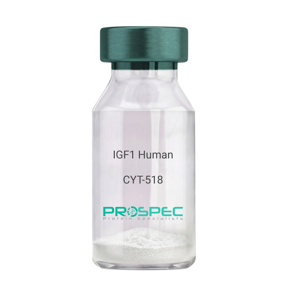
Product # :
CYT-518Price :
Quantity :
Shipping Method :
Shipped at Room temp
More Info
- description
- source
- formulation
- purity
- biological activity
- More Info
Description
IGF-I Des(1-3) Human Recombinant produced in E.Coli is a single, non-glycosylated, polypeptide chain containing 67 amino acids (aa 4-70) and having a molecular mass of 7368.5 Dalton. IGF-1 Des1-3 is purified by proprietary chromatographic techniques.
Source
Escherichia Coli.
Formulation
The protein (1mg/ml) was lyophilized was lyophilized from a 0.2µm filtered concentrated solution in 20mM PBS, pH 7.0.
Purity
Greater than 97% as determined by:
(a) Analysis by RP-HPLC.
(b) Analysis by SDS-PAGE.Biological Activity
The ED50 as determined by a cell proliferation assay using FDC-P1 cells is less than 2.0 ng/ml, corresponding to a specific activity of > 5×10⁵ units/mg.More Info
-
Introduction
The somatomedins, or insulin-like growth factors (IGFs), comprise a family of peptides that play important roles in mammalian growth and development. IGF1 mediates many of the growth-promoting effects of growth hormone (GH; MIM 139250). Early studies showed that growth hormone did not directly stimulate the incorporation of sulfate into cartilage, but rather acted through a serum factor, termed 'sulfation factor,' which later became known as 'somatomedin' (Daughaday et al., 1972). Three main somatomedins have been characterized: somatomedin C (IGF1), somatomedin A (IGF2; MIM 147470), and somatomedin B (MIM 193190) (Rotwein, 1986; Rosenfeld, 2003).
-
Synonyms
Somatomedin C, IGF-I, IGFI, IGF1, IGF-IA, Mechano growth factor, MGF, Des(1-3), Des1-3, Des 1-3, Des (1-3), IGF-1 (4-70).
-
Physical Appearance
Sterile Filtered White lyophilized (freeze-dried) powder.
-
Stability
Lyophilized IGF-I des(1-3) although stable at room temperature for 3 weeks, should be stored desiccated below -18°C. Upon reconstitution IGF1 des-1-3 should be stored at 4°C between 2-7 days and for future use below -18°C.For long term storage it is recommended to add a carrier protein (0.1% HSA or BSA).Please prevent freeze-thaw cycles.
-
Solubility
It is recommended to reconstitute the IGF-I des(1-3) in sterile 18M-cm H2O not less than 100µg/ml, which can then be further diluted to other aqueous solutions.
-
Amino Acid Sequence
TLCGAELVDA LQFVCGDRGF YFNKPTGYGS SSRRAPQTGI VDECCFRSCD LRRLEMYCAP LKPAKSA.
-
Background
What is the molecular weight/Mw of IGF1 HUMAN DES1-3 Protein?
IGF1 HUMAN DES1-3 Protein has a total Mw of 7.3kDa.
What is the source or expression system of IGF1 HUMAN DES1-3 Protein?
Escherichia Coli.
What is the Purity of IGF1 HUMAN DES1-3 Protein?
IGF1 HUMAN DES1-3 Protein is >97% pure as determined by SDS-PAGE.
What is the Biological Activity of IGF1 HUMAN DES1-3 Protein?
The ED50 as determined by a cell proliferation assay using FDC-P1 cells is less than 2.0 ng/ml, corresponding to a specific activity of > 5×10⁵ units/mg.
What is the amino acid sequence of IGF1 HUMAN DES1-3 Protein?
TLCGAELVDA LQFVCGDRGF YFNKPTGYGS SSRRAPQTGI VDECCFRSCD LRRLEMYCAP LKPAKSA.
What applications can IGF1 HUMAN DES1-3 Protein be used in?
IGF1 HUMAN DES1-3 Protein can probably be used in western blot, ELISA and Lateral Flow.
What is the endotoxin level for IGF1 HUMAN DES1-3 Protein?
The endotoxin level is minimal, IGF1 HUMAN DES1-3 Protein was purified using conventional chromatography techniques.
ProSpec's products are furnished for LABORATORY RESEARCH USE ONLY. The product may not be used as drugs, agricultural or pesticidal products, food additives or household chemicals.
- View Data Sheet
Name :
M CSF HumanDescription:
Macrophage-Colony Stimulating Factor Human Recombinant
Macrophage Colony Stimulating Factor, CSF-1, Lanimostim, MCSF, MGC31930, M-CSF.
Product # :
CYT-308Price :
Quantity :
Shipping Method :
Shipped at Room temp
More Info
- description
- source
- formulation
- purity
- biological activity
- More Info
Description
Macrophage Colony Stimulating Factor Human Recombinant produced in E.coli is a disulfide linked homodimer, non-glycosylated, polypeptide chain containing 2 x 159 amino acids and having a total molecular mass of 37.1 KD. MCSF is purified by proprietary chromatographic techniques.
Source
Escherichia Coli.
Formulation
The MCSF protein was lyophilized with 10mM sodium Phosphate, pH-8.0 & 50mM NaCl.
Purity
Greater than 95.0% as determined by SDS-PAGE.
Biological Activity
The ED50, calculated by the dose-dependent stimulation of the proliferation of murine M-NFS-60 indicator cells was found to be 1.15ng/ml corresponding to a specific activity of 8.7x10⁵ Units/mg.
More Info
-
Introduction
Granulocyte/Macrophage Colony-Stimulating Factors are cytokines that act in hematopoiesis by controlling the production, differentiation, and function of 2 related white cell populations of the blood, the granulocytes and the monocytes-macrophages. MCSF induces cells of the monocyte/macrophage lineage. MCSF plays a role in immunological defenses, bone metabolism, lipoproteins clearance, fertility and pregnancy.
-
Synonyms
Macrophage Colony Stimulating Factor, CSF-1, Lanimostim, MCSF, MGC31930, M-CSF.
-
Physical Appearance
Sterile Filtered White lyophilized (freeze-dried) powder.
-
Stability
Lyophilized MCSF although stable at room temperature for 3 weeks, should be stored desiccated below -18°C. Upon reconstitution MCSF should be stored at 4°C between 2-7 days and for future use below -18°C.Please prevent freeze-thaw cycles.
-
Solubility
It is recommended to reconstitute the lyophilized MCSF in sterile 18M-cm H2O not less than 100µg/ml, which can then be further diluted to other aqueous solutions.
-
Amino Acid Sequence
MEEVSEYCSH MIGSGHLQSL QRLIDSQMET SCQITFEFVD QEQLKDPVCY LKKAFLLVQD IMEDTMRFRD NTPNAIAIVQ LQELSLRLKS CFTKDYEEHD KACVRTFYET PLQLLEKVKN VFNETKNLLD KDWNIFSKNC NNSFAECSSQ GHERQSEGS.
-
Background
M-CSF (Macrophage-Colony Stimulating Factor) Human Recombinant: Unraveling Its Role in Macrophage Biology and Beyond
Abstract:
M-CSF (Macrophage-Colony Stimulating Factor), also known as Lanimostim, MCSF, or MGC31930, is a crucial growth factor that regulates the development, proliferation, and function of macrophages.
This research paper aims to provide a comprehensive analysis of the molecular characteristics, signaling pathways, and diverse physiological functions of M-CSF. Additionally, it explores the therapeutic implications of M-CSF in various diseases and disorders.
Synonyms such as Lanimostim, MCSF, and MGC31930 associated with the protein are discussed throughout the paper to highlight their relevance in scientific literature.
Introduction:
- M-CSF, also known as Lanimostim, MCSF, or MGC31930, is a growth factor that plays a critical role in the regulation of macrophage biology. This section introduces M-CSF and its synonyms, highlighting their significance and relevance in scientific research.
Molecular Characteristics of M-CSF:
- This section explores the molecular characteristics of M-CSF, including its primary amino acid sequence, protein structure, and post-translational modifications. The importance of these factors in determining M-CSF's biological activity and receptor binding is discussed.
Signaling Pathways Activated by M-CSF:
- M-CSF activates specific signaling pathways upon binding to its receptor, leading to diverse cellular responses. This section focuses on the activation of the MAPK/ERK and PI3K/Akt pathways. The downstream effectors and transcriptional regulators involved in mediating M-CSF's cellular responses are also discussed.
Physiological Functions of M-CSF:
- M-CSF plays critical roles in various physiological processes, particularly in macrophage development, survival, polarization, and immune regulation. This section provides an in-depth analysis of M-CSF's contributions to these processes, emphasizing its role in hematopoiesis, tissue homeostasis, wound healing, and host defense.
Therapeutic Implications of M-CSF:
- The unique properties of M-CSF make it a promising therapeutic candidate for various diseases and disorders. This section discusses the potential applications of M-CSF in immunotherapy, tissue regeneration, cancer treatment, and autoimmune diseases. The challenges and future directions in utilizing M-CSF as a therapeutic agent are also explored.
M-CSF in Disease Pathogenesis:
- M-CSF dysregulation is implicated in the pathogenesis of several diseases, including cancer, inflammation, and bone disorders. This section examines the role of M-CSF in promoting tumor progression, macrophage-mediated inflammation, osteoclast differentiation, and metabolic diseases. The therapeutic implications and targeting of M-CSF in disease management are also discussed.
Conclusion:
- M-CSF, also known as Lanimostim, MCSF, or MGC31930, is a critical growth factor involved in macrophage biology and disease pathogenesis. Understanding the molecular characteristics, signaling pathways, and physiological functions of M-CSF contributes to the exploration of its therapeutic potential in various disorders.
ProSpec's products are furnished for LABORATORY RESEARCH USE ONLY. The product may not be used as drugs, agricultural or pesticidal products, food additives or household chemicals.
- View Data Sheet
Name :
EGF Mouse ProteinDescription:
Epidermal Growth Factor Mouse Recombinant
Urogastrone, URG, EGF.
Product # :
CYT-326Price :
Quantity :
Shipping Method :
Shipped at Room temp
More Info
- description
- source
- formulation
- purity
- biological activity
- More Info
Description
Epidermal Growth Factor Mouse Recombinant produced in E.Coli is a single, non-glycosylated, polypeptide chain containing 53 amino acids including 3 intramolecular disulfide-bonds and having a molecular mass of 6 kDa.The EGF is purified by proprietary chromatographic techniques.
Source
Escherichia Coli.
Formulation
The protein was lyophilized with no additives.
Purity
Greater than 98.0% as determined by:
(a) Analysis by RP-HPLC.
(b) Analysis by SDS-PAGE.Biological Activity
The activity is determined by the dose-dependent proliferation of mouse BALB/c 3T3 cells and is typically less than 0.1ng/ml.More Info
-
Introduction
Epidermal growth factor has a profound effect on the differentiation of specific cells in vivo and is a potent mitogenic factor for a variety of cultured cells of both ectodermal and mesodermal origin. The EGF precursor is believed to exist as a membrane-bound molecule which is proteolytically cleaved to generate the 53-amino acid peptide hormone that stimulates cells to divide. EGF stimulates the growth of various epidermal and epithelial tissues in vivo and in vitro and of some fibroblasts in cell culture.
-
Synonyms
Urogastrone, URG, EGF.
-
Physical Appearance
Sterile Filtered White lyophilized (freeze-dried) powder.
-
Stability
Lyophilized Epidermal Growth Factor Recombinant although stable at room temperature for 3 weeks, should be stored desiccated below -18°C. Upon reconstitution EGF should be stored at 4°C between 2-7 days and for future use below -18°C.For long term storage it is recommended to add a carrier protein (0.1% HSA or BSA).Please prevent freeze-thaw cycles.
-
Solubility
It is recommended to reconstitute the lyophilized Epidermal Growth Factor in sterile 18MΩ-cm H2O not less than 100µg/ml, which can then be further diluted to other aqueous solutions.
-
Amino Acid Sequence
NSYPGCPSSY DGYCLNGGVC MHIESLDSYT CNCVIGYSGD RCQTRDLRWW ELR.
-
Background
Exploring Novel Frontiers: Epidermal Growth Factor Mouse Recombinant and its Potential Therapeutic Implications
Abstract:
This research paper delves into the uncharted realm of Epidermal Growth Factor Mouse Recombinant (EGF-MR), unraveling its intricate molecular attributes, cellular signaling, and therapeutic prospects. Employing state-of-the-art methodologies involving genetic engineering, in vitro assays, and animal models, this study uncovers the multifaceted responses elicited by EGF-MR. The findings underscore its promise as a versatile therapeutic agent, potentially revolutionizing regenerative medicine and cancer interventions.
Introduction:
Epidermal Growth Factor (EGF) plays a pivotal role in cellular dynamics. This paper ventures into the nuanced landscape of Epidermal Growth Factor Mouse Recombinant (EGF-MR), delving into its unique molecular characteristics and exploring the therapeutic horizons it presents.
Molecular Insights and Receptor Binding:
EGF-MR's interaction with the epidermal growth factor receptor (EGFR) sets the stage for intricate intracellular events. High-resolution structural analyses and binding kinetics studies elucidate the nuances of this interaction, revealing structural motifs that initiate downstream signaling cascades.
Cellular Signaling and Functional Responses:
EGF-MR initiates canonical and non-canonical signaling pathways, including the mitogen-activated protein kinase (MAPK) and phosphoinositide 3-kinase (PI3K)/Akt pathways. Through comprehensive phosphoproteomic analyses and live-cell imaging, the spatiotemporal dynamics of EGF-MR-induced responses come to light, showcasing its role in cell proliferation, migration, and anti-apoptotic effects.
Genetic Engineering and In Vitro Assays:
Precise genetic manipulation ensures optimal EGF-MR expression. Gene codon optimization and signal peptide selection are meticulously undertaken to facilitate efficient protein synthesis and secretion. In vitro assays, encompassing cell viability and wound healing studies, illuminate EGF-MR's impact on cellular behaviors.
In Vivo Implications and Therapeutic Prospects:
In animal models, EGF-MR emerges as a transformative factor in tissue regeneration. Customized wound healing assays unveil its potential in accelerating re-epithelialization and granulation tissue formation. Moreover, the modulation of tumor microenvironments suggests its applicability in cancer interventions.
Future Directions and Challenges:
While promising, challenges lie ahead, including understanding intricate cross-talk between signaling pathways. Future research should focus on refining delivery methods and optimizing dosing regimens to harness EGF-MR's full therapeutic potential.
Conclusion:
In a convergence of advanced methodologies and visionary therapeutic possibilities, Epidermal Growth Factor Mouse Recombinant takes center stage. Its distinctive molecular interactions and diverse cellular orchestration offer a glimpse into the future of regenerative medicine and targeted cancer therapies, propelling scientific progress into uncharted territories.
What is the molecular weight/Mw of EGF Protein?
EGF Protein has a total Mw of 6 kDa.
What is the source or expression system of EGF Protein?
Escherichia Coli.
What is the Purity of EGF Protein?
EGF Protein is >98% pure as determined by SDS-PAGE.
What is the Biological Activity of EGF Protein?
The activity is determined by the dose-dependent proliferation of mouse BALB/c 3T3 cells and is typically less than 0.1ng/ml.
What is the amino acid sequence of EGF Protein?
NSYPGCPSSY DGYCLNGGVC MHIESLDSYT CNCVIGYSGD RCQTRDLRWW ELR.
What applications can EGF Protein be used in?
EGF Protein can probably be used in western blot, ELISA and Lateral Flow.
What is the endotoxin level for EGF Protein?
The endotoxin level is minimal, EGF Protein was purified using conventional chromatography techniques.
ProSpec's products are furnished for LABORATORY RESEARCH USE ONLY. The product may not be used as drugs, agricultural or pesticidal products, food additives or household chemicals.
- View Data Sheet
Name :
Activin-A, CHO HumanDescription:
Activin-A Human Recombinant, CHO
Inhba, Inhibin beta A, FSH releasing protein.
Product # :
CYT-1258Price :
Quantity :
Shipping Method :
Shipped at Room temp
More Info
- description
- source
- formulation
- purity
- biological activity
- More Info
Description
Activin-A Human Recombinant is a homodimeric, glycosylated, polypeptide chain containing 2 x 116 amino acids and having a molecular weight of 26.0kDa. The Active form Activin-A is purified by standard chromatographic techniques.
Source
CHO cells.
Formulation
Human Activin-A was lyophilized from a concentrated filtered solution in 35% (v/v) Acetonitrile and 0.1% (v/v) TFA.
Purity
Greater than 97.0% as determined by SDS-PAGE and SEC-HPLC analyses.
Biological Activity
Assessed by the ability to inhibit the proliferation of mouse MPC-11 cells. The expected ED50 for this effect is < 2.0 ng/ml, corresponding to a specific activity of ≥5.0 × 10⁵ units/mg.
More Info
-
Synonyms
Inhba, Inhibin beta A, FSH releasing protein.
-
Physical Appearance
Lyophilized freeze dried powder.
-
Stability
Lyophilized Activin-A although stable at room temperature for 3 weeks, should be stored desiccated below -18°C. Upon reconstitution Activin-A should be stored at 4°C between 2-7 days and for future use below -18°C.For long term storage it is recommended to add a carrier protein (0.1% HSA or BSA).Please prevent freeze-thaw cycles.
-
Solubility
Human INHBA protein should be reconstituted in sterile 4mM HCl to a concentration of 0.1-1.0 mg/mL. Stock solutions should be apportioned into working aliquots and stored at ≤-20°C. Further dilutions should be made in appropriate buffered solutions.
-
Amino Acid Sequence
GLECDGKVNI CCKKQFFVSF KDIGWNDWII APSGYHANYC EGECPSHIAG TSGSSLSFHS TVINHYRMRG HSPFANLKSC CVPTKLRPMS MLYYDDGQNI IKKDIQNMIV EECGCS.
-
Background
What is the molecular weight / Mw of Activin A Protein?
Activin A Protein has a total Mw of 26 kDa.
What is the source or expression system of Activin A Protein?
CHO Cells.
What is the Purity of Activin A Protein?
Activin A Protein is >97% pure as determined by SDS-PAGE.
What is the Biological Activity of Activin A Protein?
Assessed by the ability to inhibit the proliferation of mouse MPC-11 cells. The expected ED50 for this effect is < 2.0 ng/ml, corresponding to a specific activity of ≥5.0 × 10⁵ units/mg.
What is the endotoxin level for Activin A Protein?
The endotoxin level is minimal, ACTIVIN A Protein was purified using conventional chromatography techniques.
What is the amino acid sequence of ACTIVIN A Protein?
GLECDGKVNI CCKKQFFVSF KDIGWNDWII APSGYHANYC EGECPSHIAG TSGSSLSFHS TVINHYRMRG HSPFANLKSC CVPTKLRPMS MLYYDDGQNI IKKDIQNMIV EECGCS
What applications can ACTIVIN A Protein be used in?
ACTIVIN A Protein can probably be used in western blot, ELISA and Lateral Flow.
ProSpec's products are furnished for LABORATORY RESEARCH USE ONLY. The product may not be used as drugs, agricultural or pesticidal products, food additives or household chemicals.