Search results
1000 results found for “myostatin”
Name
Description
Product #
Price
Quantity
Shipping Method
- View Data Sheet
Name :
TIPIN HumanDescription:
TIMELESS Interacting Protein Human Recombinant
TIMELESS Interacting Protein, CSM3 Homolog.
Product # :
PRO-1710Price :
Quantity :
Shipping Method :
Shipped with Ice Packs
More Info
- description
- source
- formulation
- purity
- More Info
Description
TIPIN Human Recombinant produced in E.coli is a single, non-glycosylated polypeptide chain containing 324 amino acids (1-301) and having a molecular mass of 36.9kDa.TIPIN is fused to a 23 amino acid His-tag at N-terminus & purified by proprietary chromatographic techniques.
Source
Escherichia Coli.
Formulation
The TIPIN solution (0.5mg/ml) contains 20mM Tris-HCl buffer (pH8.0), 0.15M NaCl, 1mM DTT and 10% glycerol.
Purity
Greater than 85.0% as determined by SDS-PAGE.
More Info
-
Introduction
TIPIN is a member of the CSM3 family. TIPIN protein is essential for normal advancement of S-phase and vital for cell existence after DNA damage or replication stress. TIPIN is specifically necessary for the ATR - CHEK1 pathway in the replication checkpoint induced by ultraviolet light.
-
Synonyms
TIMELESS Interacting Protein, CSM3 Homolog.
-
Physical Appearance
Sterile Filtered clear solution.
-
Stability
Store at 4°C if entire vial will be used within 2-4 weeks. Store, frozen at -20°C for longer periods of time. For long term storage it is recommended to add a carrier protein (0.1% HSA or BSA).Avoid multiple freeze-thaw cycles.
-
Amino Acid Sequence
MGSSHHHHHH SSGLVPRGSH MGSMLEPQEN GVIDLPDYEH VEDETFPPFP PPASPERQDG EGTEPDEESG NGAPVPVPPK RTVKRNIPKL DAQRLISERG LPALRHVFDK AKFKGKGHEA EDLKMLIRHM EHWAHRLFPK LQFEDFIDRV EYLGSKKEVQ TCLKRIRLDL PILHEDFVSN NDEVAENNEH DVTSTELDPF LTNLSESEMF ASELSRSLTE EQQQRIERNK QLALERRQAK LLSNSQTLGN DMLMNTPRAH TVEEVNTDED QKEESNGLNE DILDNPCNDA IANTLNEEET LLDQSFKNVQ QQLDATSRNI TEAR
ProSpec's products are furnished for LABORATORY RESEARCH USE ONLY. The product may not be used as drugs, agricultural or pesticidal products, food additives or household chemicals.
- View Data Sheet
Name :
SBDS HumanDescription:
Shwachman-Bodian-Diamond Syndrome Human Recombinant
SDS, SWDS, Shwachman-Bodian-Diamond syndrome, Ribosome Maturation protein SBDS.
Product # :
PRO-272Price :
Quantity :
Shipping Method :
Shipped with Ice Packs
More Info
- description
- source
- formulation
- purity
- More Info
Description
SBDS produced in E.Coli is a single, non-glycosylated polypeptide chain containing 270 amino acids (1-250a.a.) and having a molecular mass of 30.9kDa.SBDS is fused to a 20 amino acid His-tag at N-terminus & purified by proprietary chromatographic techniques.
Source
Escherichia Coli.
Formulation
The SBDS protein solution (0.5mg/1ml) is formulated in 20mM Tris-HCl buffer (pH8.0) 2mM DTT, 50mM NaCl, 0.1mM EDTA, and 20% glycerol.
Purity
Greater than 95% as determined by SDS-PAGE.
More Info
-
Introduction
SBDS is part of an extremely preserved protein family which exists from archaea to vertebrates and plants. SBDS protein functions in RNA metabolism and has a role in the biogenesis of the 60S ribosomal subunit and translational activation of ribosomes. Shwachman-Diamond syndrome is a rare autosomal recessive disorder produced by mutations in the SBDS gene.
-
Synonyms
SDS, SWDS, Shwachman-Bodian-Diamond syndrome, Ribosome Maturation protein SBDS.
-
Physical Appearance
Sterile Filtered clear solution.
-
Stability
Store at 4°C if entire vial will be used within 2-4 weeks. Store, frozen at -20°C for longer periods of time.Please avoid freeze thaw cycles.
-
Amino Acid Sequence
MGSSHHHHHH SSGLVPRGSH MSIFTPTNQI RLTNVAVVRM KRAGKRFEIA CYKNKVVGWR SGVEKDLDEV LQTHSVFVNV SKGQVAKKED LISAFGTDDQ TEICKQILTK GEVQVSDKER HTQLEQMFRD IATIVADKCV NPETKRPYTV ILIERAMKDI HYSVKTNKST KQQALEVIKQ LKEKMKIERA HMRLRFILPV NEGKKLKEKL KPLIKVIESE DYGQQLEIVC LIDPGCFREI DELIKKETKG KGSLEVLNLK DVEEGDEKFE
ProSpec's products are furnished for LABORATORY RESEARCH USE ONLY. The product may not be used as drugs, agricultural or pesticidal products, food additives or household chemicals.
- View Data Sheet
Name :
Eotaxin MouseDescription:
Eotaxin Mouse Recombinant (CCL11)
Small inducible cytokine A11, CCL11, Eosinophil chemotactic protein, chemokine (C-C motif) ligand 11, SCYA11.
Product # :
CHM-308Price :
Quantity :
Shipping Method :
Shipped at Room temp
More Info
- description
- source
- formulation
- purity
- biological activity
- More Info
Description
Eotaxin Mouse Recombinant produced in E.Coli is a single, non-glycosylated polypeptide chain containing 74 amino acids and having a molecular mass of 8403.2 Dalton. The CCL11 is purified by proprietary chromatographic techniques.
Source
Escherichia Coli.
Formulation
Lyophilized from a concentrated (1mg/ml) solution in water containing no additives.
Purity
Greater than 95.0% as determined by:
(a) Analysis by RP-HPLC.
(b) Analysis by SDS-PAGE.Biological Activity
The Biological activity was determined by measuring the dose dependent phosphorylation of ERK1 and ERK2 in CCR3 transfected 293 cells. Significant ERK phosphorylation is observed with >100 ng/ml (corresponding to a Specific Activity of 10,000IU/mg) of recombinant mouse eotaxin.More Info
-
Introduction
Chemokine (C-C motif) ligand 11 (CCL11) is a small cytokine belonging to the CC chemokine family that is also known as eotaxin. CCL11 selectively recruits eosinophils by inducing their chemotaxis, and therefore, is implicated in allergic responses. The effects of CCL11 are mediated by its binding to a G-protein-linked receptor known as a chemokine receptor. Chemokine receptors for which CCL11 is a ligand include CCR2, CCR3 and CCR5. The gene for human CCL11 (scya11) is encoded on three exons and is located on chromosome 17.
-
Synonyms
Small inducible cytokine A11, CCL11, Eosinophil chemotactic protein, chemokine (C-C motif) ligand 11, SCYA11.
-
Physical Appearance
Sterile Filtered White lyophilized (freeze-dried) powder.
-
Stability
Lyophilized Eotaxin although stable at room temperature for 3 weeks, should be stored desiccated below -18°C. Upon reconstitution CCL11 should be stored at 4°C between 2-7 days and for future use below -18°C. For long term storage it is recommended to add a carrier protein (0.1% HSA or BSA).Please prevent freeze-thaw cycles.
-
Solubility
It is recommended to reconstitute the lyophilized Eotaxin in sterile 18MΩ-cm H2O not less than 100µg/ml, which can then be further diluted to other aqueous solutions.
-
Amino Acid Sequence
The sequence of the first five N-terminal amino acids was determined and was found to be His-Pro-Gly-Ser-Ile.
-
Background
What is the molecular weight/Mw of EOTAXIN MOUSE Protein?
EOTAXIN MOUSE Protein has a total Mw of 8.4kDa.
What is the source or expression system of EOTAXIN MOUSE Protein?
Escherichia Coli.
What is the Purity of EOTAXIN MOUSE Protein?
EOTAXIN MOUSE Protein is >95% pure as determined by SDS-PAGE.
What is the Biological Activity of EOTAXIN MOUSE Protein?
The Biological activity was determined by measuring the dose dependent phosphorylation of ERK1 and ERK2 in CCR3 transfected 293 cells. Significant ERK phosphorylation is observed with >100 ng/ml (corresponding to a Specific Activity of 10,000IU/mg) of recombinant mouse eotaxin.
What is the amino acid sequence of EOTAXIN MOUSE Protein?
The sequence of the first five N-terminal amino acids was determined and was found to be His-Pro-Gly-Ser-Ile.
What applications can EOTAXIN MOUSE Protein be used in?
EOTAXIN MOUSE Protein can probably be used in western blot, ELISA and Lateral Flow.
What is the endotoxin level for EOTAXIN MOUSE Protein?
The endotoxin level is minimal, EOTAXIN MOUSE Protein was purified using conventional chromatography techniques.
ProSpec's products are furnished for LABORATORY RESEARCH USE ONLY. The product may not be used as drugs, agricultural or pesticidal products, food additives or household chemicals.
- View Data Sheet
Name :
MoeXDescription:
Mycobacterium Tuberculosis MoeX Recombinant
Product # :
PRO-1919Price :
Quantity :
Shipping Method :
Shipped with Ice Packs
More Info
- description
- source
- formulation
- purity
- More Info
Description
Recombinant Mycobacterium Tuberculosis MoeX produced in E.Coli is a single, non-glycosylated, polypeptide chain having a molecular mass of 38 kDa. The MoeX is fused to a His tag at C-terminus and purified by proprietary chromatographic techniques.
Source
Escherichia Coli.
Formulation
MoeX protein solution 0.75mg/ml contains 1xPBS, 25mM arginine and 0.02% NaN3.
Purity
Protein is >95% pure as determined by 12% PAGE (coomassie staining).
More Info
-
Physical Appearance
Sterile Filtered clear solution.
-
Stability
MoeX Recombinant although stable at 4°C for 1 week, should be stored below -18°C. Please prevent freeze thaw cycles.
ProSpec's products are furnished for LABORATORY RESEARCH USE ONLY. The product may not be used as drugs, agricultural or pesticidal products, food additives or household chemicals.
- View Data Sheet
Name :
S100b Mouse, HisDescription:
S100 Calcium Binding Protein B Mouse Recombinant, His Tag
Protein S100-B, S-100 protein beta chain, S-100 protein subunit beta, S100 calcium-binding protein B, Bpb, AI850290, MGC74317, S100b.
Product # :
PRO-853Price :
Quantity :
Shipping Method :
Shipped with Ice Packs
More Info
- description
- source
- formulation
- purity
- More Info
Description
S100B Mouse Recombinant produced in E.Coli is a single, non-glycosylated polypeptide chain containing 92 amino acids (1-112 a.a.) and having a molecular mass of 12.8 kDa. The Mouse S100B is fused to a 20 amino acid His-Tag at N-terminus and purified by proprietary chromatographic techniques.
Source
Escherichia Coli.
Formulation
S100B Mouse solution containing 20mM Tris-HCl pH-8, 1mM DTT & 10% glycerol.
Purity
Greater than 85.0% as determined by SDS-PAGE.
More Info
-
Introduction
S100B faintly binds calcium but binds zinc extremely strongly-distinct binding sites with different affinities exist for both ions on each monomer. Physiological concentrations of potassium ion antagonize the binding of both divalent cations, especially affecting high-affinity calcium-binding sites. S100B binds to and initiates the activation of STK38 by releasing autoinhibitory intramolecular interactions within the kinase. Interaction with AGER after myocardial infarction participates in myocyte apoptosis by activating ERK1/2 and p53/TP53 signaling.
-
Synonyms
Protein S100-B, S-100 protein beta chain, S-100 protein subunit beta, S100 calcium-binding protein B, Bpb, AI850290, MGC74317, S100b.
-
Physical Appearance
Sterile filtered colorless solution.
-
Stability
Store at 4°C if entire vial will be used within 2-4 weeks. Store, frozen at -20°C for longer periods of time. For long term storage it is recommended to add a carrier protein (0.1% HSA or BSA).Avoid multiple freeze-thaw cycles.
-
Amino Acid Sequence
MGSSHHHHHH SSGLVPRGSH MSELEKAMVA LIDVFHQYSG REGDKHKLKK SELKELINNE LSHFLEEIKE QEVVDKVMET LDEDGDGECD FQEFMAFVAM VTTACHEFFE HE.
ProSpec's products are furnished for LABORATORY RESEARCH USE ONLY. The product may not be used as drugs, agricultural or pesticidal products, food additives or household chemicals.
- View Data Sheet
Name :
MAK16 HumanDescription:
MAK16 Human Recombinant
Protein MAK16 homolog, NNP78, Protein RBM13, RBM13, MAK16L, RBM13.
Product # :
PRO-2086Price :
Quantity :
Shipping Method :
Shipped with Ice Packs
More Info
- description
- source
- formulation
- purity
- More Info
Description
MAK16 Human Recombinant produced in E.Coli is a single, non-glycosylated polypeptide chain containing 323 amino acids (1-300 a.a) and having a molecular mass of 37.8kDa (Molecular size on SDS-PAGE will appear higher).MAK16 is fused to a 23 amino acid His-tag at N-terminus & purified by proprietary chromatographic techniques.
Source
Escherichia Coli.
Formulation
MAK16 protein solution (0.25mg/ml) contains Phosphate buffered saline (pH7.4), 30% glycerol and 1mM DTT.
Purity
Greater than 90.0% as determined by SDS-PAGE.
More Info
-
Introduction
MAK16 is a member of the MAK16 family.
-
Synonyms
Protein MAK16 homolog, NNP78, Protein RBM13, RBM13, MAK16L, RBM13.
-
Physical Appearance
Sterile Filtered clear solution.
-
Stability
Store at 4°C if entire vial will be used within 2-4 weeks. Store, frozen at -20°C for longer periods of time. For long term storage it is recommended to add a carrier protein (0.1% HSA or BSA).Avoid multiple freeze-thaw cycles.
-
Amino Acid Sequence
MGSSHHHHHH SSGLVPRGSH MGSMQSDDVI WDTLGNKQFC SFKIRTKTQS FCRNEYSLTG LCNRSSCPLA NSQYATIKEE KGQCYLYMKV IERAAFPRRL WERVRLSKNY EKALEQIDEN LIYWPRFIRH KCKQRFTKIT QYLIRIRKLT LKRQRKLVPL SKKVERREKR REEKALIAAQ LDNAIEKELL ERLKQDTYGD IYNFPIHAFD KALEQQEAES DSSDTEEKDD DDDDEEDVGK REFVEDGEVD ESDISDFEDM DKLDASSDED QDGKSSSEEE EEKALSAKHK GKMPLRGPLQ RKRAYVEIEY EQETEPVAKA KTT.
ProSpec's products are furnished for LABORATORY RESEARCH USE ONLY. The product may not be used as drugs, agricultural or pesticidal products, food additives or household chemicals.
- View Data Sheet
Name :
JOSD1 HumanDescription:
Josephin Domain Containing 1 Human Recombinant
Josephin-1, Josephin domain-containing 1, JOSD1, JSPH1, KIAA0063, dJ508I15.2.
Product # :
PRO-1251Price :
Quantity :
Shipping Method :
Shipped with Ice Packs
More Info
- description
- source
- formulation
- purity
- More Info
Description
JOSD1 Human Recombinant produced in E.Coli is a single, non-glycosylated polypeptide chain containing 225 amino acids (1-202 a.a.) and having a molecular mass of 25.6kDa.JOSD1 is fused to a 23 amino acid His-tag at N-terminus & purified by proprietary chromatographic techniques.
Source
Escherichia Coli.
Formulation
JOSD1 protein solution (0.5mg/ml) containing 20mM Tris-HCl buffer (pH 8.0), 0.2M NaCl, 40% glycerol, 5mM DTT and 2mM EDTA.
Purity
Greater than 90.0% as determined by SDS-PAGE.
More Info
-
Introduction
Josephin-1 (JOSD1) has minimal protease activity towards poly-ubiquitin chains (in vitro). JOSD1 functions as a deubiquitinating enzyme. Ubiquitin-binding sites in the Josephin domain are a factor in ubiquitin chain binding and cleavage.
-
Synonyms
Josephin-1, Josephin domain-containing 1, JOSD1, JSPH1, KIAA0063, dJ508I15.2.
-
Physical Appearance
Sterile Filtered colorless solution.
-
Stability
Store at 4°C if entire vial will be used within 2-4 weeks. Store, frozen at -20°C for longer periods of time. For long term storage it is recommended to add a carrier protein (0.1% HSA or BSA).Avoid multiple freeze-thaw cycles.
-
Amino Acid Sequence
MGSSHHHHHH SSGLVPRGSH MGSMSCVPWK GDKAKSESLE LPQAAPPQIY HEKQRRELCA LHALNNVFQD SNAFTRDTLQ EIFQRLSPNT MVTPHKKSML GNGNYDVNVI MAALQTKGYE AVWWDKRRDV GVIALTNVMG FIMNLPSSLC WGPLKLPLKR QHWICVREVG GAYYNLDSKL KMPEWIGGES ELRKFLKHHL RGKNCELLLV VPEEVEAHQS WRTDV.
ProSpec's products are furnished for LABORATORY RESEARCH USE ONLY. The product may not be used as drugs, agricultural or pesticidal products, food additives or household chemicals.
- View Data Sheet
Name :
TerlipressinDescription:
Terlipressin
Product # :
HOR-287Price :
Quantity :
Shipping Method :
Shipped at Room temp
More Info
- description
- formulation
- purity
- More Info
Description
Terlipressin contains 12 amino acids Gly-Gly-Gly-c[Cys-Tyr-Phe-Gln-Asn-Cys]-Pro-Lys-Gly-NH2 and having a molecular weight of 1227.37 Dalton.
Formulation
The protein (1 mg/ml) was lyophilized with no additives.
Purity
Greater than 98.0% as determined by:
(a) Analysis by RP-HPLC.
(b) Analysis by SDS-PAGE.More Info
-
Introduction
Terlipressin is similar to a naturally occurring hormone present in the body, known as antidiuretic hormone (ADH) or vasopressin. ADH has two main effects in the body. Firstly, it causes narrowing of blood vessels (vasoconstriction), thereby limiting blood flow to a particular area of the body. It also acts on receptors in the kidney to retain water in the body, which helps to prevent excessive loss of water in the urine. Terlipressin is commonly used to stop bleeding of varices in the food pipe (oesophagus). Varices are fragile distended veins that can occur in various parts of the body such as the oesophagus. This is caused by an increase in blood pressure in certain diseases such as severe liver disease. These fragile varices can rupture and lead to life threatening bleeding. Terlipressin is therefore given to narrow blood vessels, and so restricting blood flow to the varices and stopping the bleeding.
-
Physical Appearance
Sterile Filtered White lyophilized (freeze-dried) powder.
-
Stability
Lyophilized Terlipressin although stable at room temperature for 3 weeks, should be stored desiccated below -18°C. Upon reconstitution Terlipressin should be stored at 4°C between 2-7 days and for future use below -18°C.For long term storage it is recommended to add a carrier protein (0.1% HSA or BSA).Please prevent freeze-thaw cycles.
-
Solubility
It is recommended to reconstitute the lyophilized Terlipressin18MΩ-cm H2O not less than 100 µg/ml, which can then be further diluted to other aqueous solutions.
ProSpec's products are furnished for LABORATORY RESEARCH USE ONLY. The product may not be used as drugs, agricultural or pesticidal products, food additives or household chemicals.
- View Data Sheet
Name :
MT3 HumanDescription:
Metallothionein 3 Human Recombinant
GIF, GIFB, GRIF, ZnMT3, Metallothionein-3, MT-3, Growth inhibitory factor, Metallothionein-III, MT-III, MT3.
Product # :
PRO-1856Price :
Quantity :
Shipping Method :
Shipped with Ice Packs
More Info
- description
- source
- formulation
- purity
- More Info
Description
MT3 Human Recombinant produced in E. coli is a single polypeptide chain containing 91 amino acids (1-68) and having a molecular mass of 9.3kDa (molecular size on SDS-PAGE will appear higher).MT3 is fused to a 23 amino acid His-tag at N-terminus & purified by proprietary chromatographic techniques.
Source
Escherichia Coli.
Formulation
The MT3 solution (0.5mg/ml) contains 20mM Tris-HCl buffer (pH 8.0), 0.15M NaCl and 10% glycerol.
Purity
Greater than 85% as determined by SDS-PAGE.
More Info
-
Introduction
Metallothionein 3 (MT3) contains 3 zinc and 3 copper atoms per polypeptide chain and a minor amount of cadmium. MT3 inhibits survival and neurite formation of cortical neurons in vitro. MT3 also binds heavy metals.
-
Synonyms
GIF, GIFB, GRIF, ZnMT3, Metallothionein-3, MT-3, Growth inhibitory factor, Metallothionein-III, MT-III, MT3.
-
Physical Appearance
Sterile Filtered clear solution.
-
Stability
Store at 4°C if entire vial will be used within 2-4 weeks. Store frozen at -20°C for longer periods of time. For long term storage it is recommended to add a carrier protein (0.1% HSA or BSA).Avoid multiple freeze-thaw cycles.
-
Amino Acid Sequence
MGSSHHHHHH SSGLVPRGSH MGSMDPETCP CPSGGSCTCA DSCKCEGCKC TSCKKSCCSC CPAECEKCAK DCVCKGGEAA EAEAEKCSCC Q.
ProSpec's products are furnished for LABORATORY RESEARCH USE ONLY. The product may not be used as drugs, agricultural or pesticidal products, food additives or household chemicals.
- View Data Sheet
Name :
GMNN HumanDescription:
Geminin Human Recombinant
GMNN, Geminin, DNA Replication Inhibitor, Gem, RP3-369A17.3.
Product # :
PRO-579Price :
Quantity :
Shipping Method :
Shipped with Ice Packs
More Info
- description
- source
- formulation
- purity
- More Info
Description
Geminin Human Recombinant fused to N-terminal His-Tag produced in E.Coli is a single, non-glycosylated polypeptide chain containing 245 amino acids and having a molecular mass of 27.7 kDa.
Source
Escherichia Coli.
Formulation
The protein solution contains 20mM Tris pH 8, 100mM NaCl and 10% glycerol.
Purity
Greater than 95.0% as determined by:
(a) Analysis by RP-HPLC.
(b) Analysis by SDS-PAGE.More Info
-
Introduction
Geminin is a 25 kDa nuclear protein, which inhibits DNA replication and is degraded during the mitotic phase of the cell cycle. Geminin controls replication by binding to the licensing factor Cdt1, and is involved in neural differentiation. In addition, Geminin directly interacts with Six3 and Hox homeodomain proteins during embryogenesis and inhibits their functions. Geminin can also promote DNA replication. Geminin has 2 roles in 2 different stages of the cell cycle: Geminin is a negative regulator of DNA replication during the “S phase” of the cell cycle. Inhibition of Geminin during the “S phase” (by RNAi) results in an additional round of replication of portions of the genome. During the “M phase” of the cell cycle (mitosis) Geminin stabilizes the replication factor Cdt1 promoting DNA replication during the next cell cycle. Moreover, inhibition of Geminin during mitosis (by RNAi) causes destabilization of Cdt1 protein and impairment of DNA replication during the next cell cycle. Geminin thus guarantees that only one round of replication occurs during each cell cycle. It was discovered that Geminin is overexpressed in a number of malignancies and cancer cell lines. This maintains the concept that Geminin has also a positive role in DNA replication and cell cycle progression. Geminin accumulates through S, G2 and M phases of the cell cycle but is absent during the G1 phase. During the metaphase/anaphase transition of mitosis Geminin levels decrease.
-
Synonyms
GMNN, Geminin, DNA Replication Inhibitor, Gem, RP3-369A17.3.
-
Physical Appearance
Sterile Filtered colorless solution.
-
Stability
Store at 4°C if entire vial will be used within 2-4 weeks. Store, frozen at -20°C for longer periods of time. For long term storage it is recommended to add a carrier protein (0.1% HSA or BSA).Avoid multiple freeze-thaw cycles.
-
Amino Acid Sequence
MRGSHHHHHH GMASMTGGQQ MGRDLYDDDD KDRWGSMNPS MKQKQEEIKE NIKNSSVPRR TLKMIQPSAS GSLVGRENEL SAGLSKRKHR NDHLTSTTSS PGVIVPESSE NKNLGGVTQE SFDLMIKENP SSQYWKEVAE KRRKALYEAL KENEKLHKEI EQKDNEIARL KKENKELAEV AEHVQYMAEL IERLNGEPLD NFESLDNQEF DSEEETVEDS LVEDSEIGTC AEGTVSSSTD
AKPCI.
ProSpec's products are furnished for LABORATORY RESEARCH USE ONLY. The product may not be used as drugs, agricultural or pesticidal products, food additives or household chemicals.
- View Data Sheet
Name :
SCF RatDescription:
Stem Cell Factor Rat Recombinant
Kit ligand Precursor, C-kit ligand, SCF, Mast cell growth factor, MGF, SF, KL-1, Kitl, DKFZp686F2250, Hematopoietic growth factor KL.
Product # :
CYT-323Price :
Quantity :
Shipping Method :
Shipped at Room temp
More Info
- description
- source
- formulation
- purity
- biological activity
- More Info
Description
Stem cell factor Rat Recombinant produced in E.Coli is a single, non-glycosylated polypeptide chain containing 164 amino acids (26-189) and having a molecular mass of 18.4 kDa.The Rat SCF is purified by proprietary chromatographic techniques.
Source
Escherichia Coli.
Formulation
Lyophilized from a concentrated (1mg/ml) solution in water containing 0.02% NaHCO3.
Purity
Greater than 98.0% as determined by:
(a) Analysis by SEC-HPLC.
(b) Analysis by SDS-PAGE.Biological Activity
The ED50 is determined by the dose-dependant stimulation of the proliferation of human TF-1 cells which is < 10 ng/ml, corresponding to a specific activity of 100,000units/mg.More Info
-
Introduction
Stem cell factor / KIT ligand (SCF) is a cytokine which binds CD117 (c-Kit). SCF is also known as "steel factor" or "c-kit ligand". SCF exists in two forms, cell surface bound SCF and soluble (or free) SCF. Soluble SCF is produced by the cleavage of surface bound SCF by metalloproteases.
SCF is a growth factor important for the survival, proliferation, and differentiation of hematopoietic stem cells and other hematopoietic progenitor cells. One of its roles is to change the BFU-E (burst-forming unit-erythroid) cells, which are the earliest erythrocyte precursors in the erythrocytic series, into the CFU-E (colony-forming unit-erythroid). -
Synonyms
Kit ligand Precursor, C-kit ligand, SCF, Mast cell growth factor, MGF, SF, KL-1, Kitl, DKFZp686F2250, Hematopoietic growth factor KL.
-
Physical Appearance
Sterile Filtered White lyophilized (freeze-dried) powder.
-
Stability
Lyophilized rat SCF although stable at room temperature for 3 weeks, should be stored desiccated below -18°C. Upon reconstitution SCF should be stored at 4°C between 2-7 days and for future use below -18°C.For long term storage it is recommended to add a carrier protein (0.1% HSA or BSA).Please prevent freeze-thaw cycles.
-
Solubility
It is recommended to reconstitute the lyophilized SCF in sterile 18MΩ-cm H2O not less than 100µg/ml, which can then be further diluted to other aqueous solutions.
-
Amino Acid Sequence
MQEICRNPVT DNVKDITKLV ANLPNDYMIT LNYVAGMDVL PSHCWLRDMV THLSVSLTTL LDKFSNISEG LSNYSIIDKL GKIVDDLVAC MEENAPKNVK ESLKKPETRN FTPEEFFSIF NRSIDAFKDF MVASDTSDCV LSSTLGPEKD SRVSVTKPFM LPPVA.
ProSpec's products are furnished for LABORATORY RESEARCH USE ONLY. The product may not be used as drugs, agricultural or pesticidal products, food additives or household chemicals.
- View Data Sheet
Name :
IL6ST HumanDescription:
Interleukin-6 Signal Transducer Human Recombinant
Interleukin 6 signal transducer, oncostatin M receptor, IL6ST, CD130, CDw130, GP130, GP130-RAPS, IL6R-beta
Product # :
CYT-1156Price :
Quantity :
Shipping Method :
Shipped with Ice Packs
More Info
- description
- source
- formulation
- purity
- More Info
Description
IL6ST Human produced in Sf9 Baculovirus cells is a single, glycosylated polypeptide chain containing 605 amino acids (23-619 a.a) and having a molecular mass of 68.9kDa.IL6ST is fused to an 8 amino acid His tag at C-terminus and purified by proprietary chromatographic techniques.
Source
Sf9, Baculovirus cells.
Formulation
The IL6ST solution (0.25mg/ml) contains 10% Glycerol and Phosphate-Buffered Saline (pH 7.4).
Purity
Greater than 95% as determined by SDS-PAGE.
More Info
-
Introduction
Interleukin-6 Signal Transducer or IL6ST is a receptor, part of the family of class 1 cytokine receptor. IL6ST binds to IL-6 through membrane-anchored or soluble IL-6R starts a connection between another complex of IL6ST and IL-6 , thus forming a homo-dimer and a signal transduction occurs.
-
Synonyms
Interleukin 6 signal transducer, oncostatin M receptor, IL6ST, CD130, CDw130, GP130, GP130-RAPS, IL6R-beta
-
Physical Appearance
Sterile Filtered colorless solution.
-
Stability
Store at 4°C if entire vial will be used within 2-4 weeks. Store, frozen at -20°C for longer periods of time. For long term storage it is recommended to add a carrier protein (0.1% HSA or BSA).Avoid multiple freeze-thaw cycles.
-
Amino Acid Sequence
ELLDPCGYIS PESPVVQLHS NFTAVCVLKE KCMDYFHVNA NYIVWKTNHF TIPKEQYTII NRTASSVTFT DIASLNIQLT CNILTFGQLE QNVYGITIIS GLPPEKPKNL SCIVNEGKKM RCEWDRGRET HLETNFTLKS EWATHKFADC KAKRDTPTSC TVDYSTVYFV NIEVWVEAEN ALGKVTSDHI NFDPVYKVKP NPPHNLSVIN SEELSSILKL TWTNPSIKSV IILKYNIQYR TKDASTWSQI PPEDTASTRS SFTVQDLKPF TEYVFRIRCM KEDGKGYWSD WSEEASGITY EDRPSKAPSF WYKIDPSHTQ GYRTVQLVWK TLPPFEANGK ILDYEVTLTR WKSHLQNYTV NATKLTVNLT NDRYVATLTV RNLVGKSDAA VLTIPACDFQ ATHPVMDLKA FPKDNMLWVE WTTPRESVKK YILEWCVLSD KAPCITDWQQ EDGTVHRTYL RGNLAESKCY LITVTPVYAD GPGSPESIKA YLKQAPPSKG PTVRTKKVGK NEAVLEWDQL PVDVQNGFIR NYTIFYRTII GNETAVNVDS SHTEYTLSSL TSDTLYMVRM AAYTDEGGKD GPEFTFTTPK FAQGEIELEH HHHHH
-
Background
Significance of Human Recombinant Interleukin-6 Signal Transducer: Insights and Implications
Abstract:
The Interleukin-6 (IL-6) signal transducer holds a pivotal role in the complex IL-6 signaling pathway, orchestrating crucial cellular responses. This paper delves into the significance of Human Recombinant IL-6 Signal Transducer in unraveling IL-6-mediated cellular communication and highlights its potential applications in research and therapeutic development. The review sheds light on the methodology of producing this transducer and its relevance in advancing our understanding of cytokine signaling.
Introduction:
IL-6, a pleiotropic cytokine, is known to exert its diverse effects through a complex signaling cascade, wherein the signal transducer plays a crucial role. The availability of Human Recombinant IL-6 Signal Transducer enables the investigation of its role in health and disease. This transducer is vital for transmitting IL-6 signals, influencing processes like immune response, inflammation, and cell differentiation.
IL-6 Signal Transduction Pathway:
The IL-6 signal transduction pathway involves binding of IL-6 to its receptor, leading to the recruitment of the signal transducer and subsequent activation of downstream signaling molecules such as JAK/STAT pathway. This orchestrated response modulates gene expression, thereby impacting cellular behavior.
Methods of Production:
Human Recombinant IL-6 Signal Transducer is produced by expressing the corresponding gene in a suitable expression system, often utilizing bacterial or mammalian cells. Proper post-translational modifications are necessary to ensure its biological activity and appropriate functioning within the signaling cascade.
Applications in Research:
Human Recombinant IL-6 Signal Transducer serves as a fundamental tool in elucidating IL-6-mediated signaling mechanisms. Its availability facilitates the exploration of how aberrant signaling contributes to diseases such as autoimmune disorders, inflammatory conditions, and certain cancers. The transducer's interaction with other signaling pathways is also of interest for comprehensive pathway analysis.
Therapeutic Implications:
Understanding the IL-6 signal transduction pathway has led to the development of targeted therapies for IL-6-related diseases. Modulation of this pathway presents potential opportunities for novel therapeutic interventions, thereby offering a new dimension in precision medicine.
Challenges and Future Directions:
While the availability of Human Recombinant IL-6 Signal Transducer has significantly advanced our knowledge, challenges remain in deciphering the intricate nuances of IL-6 signaling. Developing strategies to selectively intervene in this pathway without disturbing physiological homeostasis presents an ongoing challenge.
Conclusion:
The Human Recombinant IL-6 Signal Transducer serves as a cornerstone in unraveling the complexities of IL-6 signaling, shedding light on its roles in health and disease. Its applications span from fundamental research to therapeutic development, showcasing its potential to shape the future of precision medicine.
ProSpec's products are furnished for LABORATORY RESEARCH USE ONLY. The product may not be used as drugs, agricultural or pesticidal products, food additives or household chemicals.
- View Data Sheet
Name :
C5a MouseDescription:
Complement Component C5a Mouse Recombinant
Complement C5, Hemolytic complement, C5, Hc, He, C5a.
Product # :
PRO-085Price :
Quantity :
Shipping Method :
Shipped at Room temp
More Info
- description
- source
- formulation
- purity
- biological activity
- More Info
Description
C5a Mouse Recombinant produced in E.Coli is a single, non-glycosylated, Polypeptide chain containing 77 amino acids and having a molecular mass of 9kDa.The Mouse C5a is purified by proprietary chromatographic techniques.
Source
Escherichia Coli.
Formulation
Mouse C5a was lyophilized from a concentrated (1mg/ml) solution in 20mM PB, pH 7.5 and 350mM NaCl.
Purity
Greater than 95.0% as determined by:
(a) Analysis by RP-HPLC.
(b) Analysis by SDS-PAGE.Biological Activity
The ED50 of Recombinant Mouse C5a as determined by its ability to induce N-acetyl-b-D-glucosaminidase release from differentiated U937 human histiocytic lymphoma cells was 5-20ng/ml.More Info
-
Introduction
Mouse Complement 5a (C5a) is a glycoprotein which is a member of a family of structurally and functionally related proteins known as anaphylatoxins. C5a is a 77 a.a. peptide created by the C5a convertase proteolytic cleavage of C5 ? chain in the classical and alternative complement pathway (C4b2a3b, C3bBb3b). The mouse C5a has four ? helices and three intrachain disulfide bonds which form a triple loop structure. C5a functions through G-protein coupled receptor (GPCR) (C5aR/CD88). C5a is a effective chemoattractant and anaphylatoxin which functions on all classes of leukocytes and on many other cell types including endothelial, smooth muscle, kidney, liver, and neural cells. Mouse C5a also mediates IL-8 release from bronchial epithelial cells. Furthermore, it triggers an oxidative surge in macrophages and neutrophils, causing the release of histamine in basophils and mast cells. The C5a anaphylatoxin activity on hepatocytes results indirectly from interaction with nonparenchymal cell via prostanoid secretion.
-
Synonyms
Complement C5, Hemolytic complement, C5, Hc, He, C5a.
-
Physical Appearance
Sterile Filtered White lyophilized (freeze-dried) powder.
-
Stability
Lyophilized Mouse C5a although stable at room temperature for 3 weeks, should be stored desiccated below -18°C. Upon reconstitution Mouse C5a should be stored at 4°C between 2-7 days and for future use below -18°C.Please prevent freeze-thaw cycles.
-
Solubility
It is recommended to reconstitute the lyophilized Mouse C5a in sterile 18MΩ-cm H2O not less than 100µg/ml, which can then be further diluted to other aqueous solutions.
-
Amino Acid Sequence
The sequence of the first five N-terminal amino acids was determined and was found to be Asn-Leu-His-Leu-Leu.
ProSpec's products are furnished for LABORATORY RESEARCH USE ONLY. The product may not be used as drugs, agricultural or pesticidal products, food additives or household chemicals.
- View Data Sheet
Name :
FTH1 HumanDescription:
Ferritin Human Recombinant, Heavy Chain
Ferritin heavy chain, Cell proliferation-inducing gene 15 protein, FTH1, FHC, FTH, PLIF, FTHL6, PIG15, MGC104426.
Product # :
PRO-658Price :
Quantity :
Shipping Method :
Shipped with Ice Packs
More Info
- description
- source
- formulation
- purity
- More Info
Description
FTH1 Human Recombinant produced in E.Coli is a single, non-glycosylated polypeptide chain containing 183 amino acids and having a molecular mass of 21 kDa.
Source
Escherichia Coli.
Formulation
The FTH1 protein solution contains 20mM Tris-HCl pH-7.5, 1mM DTT and 10% glycerol.
Purity
Greater than 95.0% as determined by SDS-PAGE.
More Info
-
Introduction
Ferritin is a fairly large, iron-storage heteropolymeric protein composed of 2 subunit types, light Ferritin & heavy Ferritin polypeptides, which is expressed in most kinds of cells and co-assemble in different proportion in a tissue-specific manner. Ferritin is composed of 24 self-assembled polypeptide subunits of the heavy and light ferritin chains and is characterized by the capacity to remove Fe (II) from solution in the presence of oxygen.
Ferritin light polypeptide protein is the main intracellular iron storage protein in prokaryotes and eukaryotes. Variation in ferritin subunit composition influence the rates of iron uptake and release in various tissues. A key function of ferritin is the storage of iron in a soluble and nontoxic state. Defects in this light chain ferritin gene are associated with several neurodegenerative diseases and hyper ferrit anemia-cataract syndrome.
Ferritin stores iron in a soluble, nontoxic, readily accessible form. Ferritin is needed for iron homeostasis. Iron is taken up in the ferrous form and deposited as ferric hydroxides after it has been oxidized. -
Synonyms
Ferritin heavy chain, Cell proliferation-inducing gene 15 protein, FTH1, FHC, FTH, PLIF, FTHL6, PIG15, MGC104426.
-
Physical Appearance
Sterile Filtered colorless solution.
-
Stability
Store at 4°C if entire vial will be used within 2-4 weeks. Store, frozen at -20°C for longer periods of time. For long term storage it is recommended to add a carrier protein (0.1% HSA or BSA).Avoid multiple freeze-thaw cycles.
-
Amino Acid Sequence
MTTASTSQVR QNYHQDSEAA INRQINLELY ASYVYLSMSY YFDRDDVALK NFAKYFLHQS HEEREHAEKL MKLQNQRGGR IFLQDIKKPD CDDWESGLNA MECALHLEKN VNQSLLELHK LATDKNDPHL CDFIETHYLN EQVKAIKELG DHVTNLRKMG APESGLAEYL FDKHTLGDSD NES.
ProSpec's products are furnished for LABORATORY RESEARCH USE ONLY. The product may not be used as drugs, agricultural or pesticidal products, food additives or household chemicals.
- View Data Sheet
Name :
EGF Mouse ProteinDescription:
Epidermal Growth Factor Mouse Recombinant
Urogastrone, URG, EGF.
Product # :
CYT-326Price :
Quantity :
Shipping Method :
Shipped at Room temp
More Info
- description
- source
- formulation
- purity
- biological activity
- More Info
Description
Epidermal Growth Factor Mouse Recombinant produced in E.Coli is a single, non-glycosylated, polypeptide chain containing 53 amino acids including 3 intramolecular disulfide-bonds and having a molecular mass of 6 kDa.The EGF is purified by proprietary chromatographic techniques.
Source
Escherichia Coli.
Formulation
The protein was lyophilized with no additives.
Purity
Greater than 98.0% as determined by:
(a) Analysis by RP-HPLC.
(b) Analysis by SDS-PAGE.Biological Activity
The activity is determined by the dose-dependent proliferation of mouse BALB/c 3T3 cells and is typically less than 0.1ng/ml.More Info
-
Introduction
Epidermal growth factor has a profound effect on the differentiation of specific cells in vivo and is a potent mitogenic factor for a variety of cultured cells of both ectodermal and mesodermal origin. The EGF precursor is believed to exist as a membrane-bound molecule which is proteolytically cleaved to generate the 53-amino acid peptide hormone that stimulates cells to divide. EGF stimulates the growth of various epidermal and epithelial tissues in vivo and in vitro and of some fibroblasts in cell culture.
-
Synonyms
Urogastrone, URG, EGF.
-
Physical Appearance
Sterile Filtered White lyophilized (freeze-dried) powder.
-
Stability
Lyophilized Epidermal Growth Factor Recombinant although stable at room temperature for 3 weeks, should be stored desiccated below -18°C. Upon reconstitution EGF should be stored at 4°C between 2-7 days and for future use below -18°C.For long term storage it is recommended to add a carrier protein (0.1% HSA or BSA).Please prevent freeze-thaw cycles.
-
Solubility
It is recommended to reconstitute the lyophilized Epidermal Growth Factor in sterile 18MΩ-cm H2O not less than 100µg/ml, which can then be further diluted to other aqueous solutions.
-
Amino Acid Sequence
NSYPGCPSSY DGYCLNGGVC MHIESLDSYT CNCVIGYSGD RCQTRDLRWW ELR.
-
Background
Exploring Novel Frontiers: Epidermal Growth Factor Mouse Recombinant and its Potential Therapeutic Implications
Abstract:
This research paper delves into the uncharted realm of Epidermal Growth Factor Mouse Recombinant (EGF-MR), unraveling its intricate molecular attributes, cellular signaling, and therapeutic prospects. Employing state-of-the-art methodologies involving genetic engineering, in vitro assays, and animal models, this study uncovers the multifaceted responses elicited by EGF-MR. The findings underscore its promise as a versatile therapeutic agent, potentially revolutionizing regenerative medicine and cancer interventions.
Introduction:
Epidermal Growth Factor (EGF) plays a pivotal role in cellular dynamics. This paper ventures into the nuanced landscape of Epidermal Growth Factor Mouse Recombinant (EGF-MR), delving into its unique molecular characteristics and exploring the therapeutic horizons it presents.
Molecular Insights and Receptor Binding:
EGF-MR's interaction with the epidermal growth factor receptor (EGFR) sets the stage for intricate intracellular events. High-resolution structural analyses and binding kinetics studies elucidate the nuances of this interaction, revealing structural motifs that initiate downstream signaling cascades.
Cellular Signaling and Functional Responses:
EGF-MR initiates canonical and non-canonical signaling pathways, including the mitogen-activated protein kinase (MAPK) and phosphoinositide 3-kinase (PI3K)/Akt pathways. Through comprehensive phosphoproteomic analyses and live-cell imaging, the spatiotemporal dynamics of EGF-MR-induced responses come to light, showcasing its role in cell proliferation, migration, and anti-apoptotic effects.
Genetic Engineering and In Vitro Assays:
Precise genetic manipulation ensures optimal EGF-MR expression. Gene codon optimization and signal peptide selection are meticulously undertaken to facilitate efficient protein synthesis and secretion. In vitro assays, encompassing cell viability and wound healing studies, illuminate EGF-MR's impact on cellular behaviors.
In Vivo Implications and Therapeutic Prospects:
In animal models, EGF-MR emerges as a transformative factor in tissue regeneration. Customized wound healing assays unveil its potential in accelerating re-epithelialization and granulation tissue formation. Moreover, the modulation of tumor microenvironments suggests its applicability in cancer interventions.
Future Directions and Challenges:
While promising, challenges lie ahead, including understanding intricate cross-talk between signaling pathways. Future research should focus on refining delivery methods and optimizing dosing regimens to harness EGF-MR's full therapeutic potential.
Conclusion:
In a convergence of advanced methodologies and visionary therapeutic possibilities, Epidermal Growth Factor Mouse Recombinant takes center stage. Its distinctive molecular interactions and diverse cellular orchestration offer a glimpse into the future of regenerative medicine and targeted cancer therapies, propelling scientific progress into uncharted territories.
What is the molecular weight/Mw of EGF Protein?
EGF Protein has a total Mw of 6 kDa.
What is the source or expression system of EGF Protein?
Escherichia Coli.
What is the Purity of EGF Protein?
EGF Protein is >98% pure as determined by SDS-PAGE.
What is the Biological Activity of EGF Protein?
The activity is determined by the dose-dependent proliferation of mouse BALB/c 3T3 cells and is typically less than 0.1ng/ml.
What is the amino acid sequence of EGF Protein?
NSYPGCPSSY DGYCLNGGVC MHIESLDSYT CNCVIGYSGD RCQTRDLRWW ELR.
What applications can EGF Protein be used in?
EGF Protein can probably be used in western blot, ELISA and Lateral Flow.
What is the endotoxin level for EGF Protein?
The endotoxin level is minimal, EGF Protein was purified using conventional chromatography techniques.
ProSpec's products are furnished for LABORATORY RESEARCH USE ONLY. The product may not be used as drugs, agricultural or pesticidal products, food additives or household chemicals.
- View Data Sheet
Name :
SNAI1 HumanDescription:
Snail Family Zinc Finger 1 Human Recombinant
Snail Family Zinc Finger 1, Protein Sna, Protein Snail Homolog 1, SNAH, Snail 1 (Drosophila Homolog), Zinc Finger Protein, Snail Homolog 1 (Drosophila), SLUGH2, SNA, SNAIL, SNAIL1, dJ710H13.1, Snail 1 Homolog, Snail 1 Zinc Finger Protein, Snail 1, Zinc Finger Protein, Snail Homolog 1, Zinc Finger Protein SNAI1.
Product # :
PRO-1759Price :
Quantity :
Shipping Method :
Shipped with Ice Packs
More Info
- description
- source
- formulation
- purity
- More Info
Description
SNAI1 Human Recombinant produced in E.Coli is a single, non-glycosylated polypeptide chain containing 287 amino acids (1-264 a.a) and having a molecular mass of 31.5kDa.SNAI1 is fused to a 23 amino acid His-tag at N-terminus & purified by proprietary chromatographic techniques.
Source
Escherichia Coli.
Formulation
SNAI1 protein solution (1mg/ml) containing 20mM Tris-HCl buffer (pH 8.0), 0.4M urea and 10% glycerol.
Purity
Greater than 85% as determined by SDS-PAGE.
More Info
-
Introduction
Snail homolog 1 (SNAI1) is involved in induction of the epithelial to mesenchymal transition (EMT), formation and maintenance of embryonic mesoderm, growth arrest, survival and cell migration. SNAI1 binds to 3 E-boxes of the E-cadherin gene promoter and represses its transcription. The nuclear protein encoded by SNAI1 is structurally identical to the Drosophila snail protein, and is considered as well to be vital for mesoderm formation in the developing embryo. At least two variants of a similar processed pseudogene have been found on chromosome 2. Among the diseases associated with SNAI1 are waardenburg syndrome type iid, and inappropriate adh syndrome.
-
Synonyms
SnailFamily ZincFinger 1, Protein Sna, Protein Snail Homolog 1, SNAH, Snail 1 (Drosophila Homolog), Zinc Finger Protein, Snail Homolog 1 (Drosophila), SLUGH2, SNA, SNAIL, SNAIL1, dJ710H13.1, Snail 1 Homolog, Snail 1 Zinc Finger Protein, Snail 1, Zinc Finger Protein, Snail Homolog 1, Zinc Finger Protein SNAI1.
-
Physical Appearance
Sterile Filtered clear solution.
-
Stability
Store at 4°C if entire vial will be used within 2-4 weeks. Store, frozen at -20°C for longer periods of time.For long term storage it is recommended to add a carrier protein (0.1% HSA or BSA).Please avoid freeze thaw cycles.
-
Amino Acid Sequence
MGSSHHHHHH SSGLVPRGSH MGSMPRSFLV RKPSDPNRKP NYSELQDSNP EFTFQQPYDQ AHLLAAIPPP EILNPTASLP MLIWDSVLAP QAQPIAWASL RLQESPRVAE LTSLSDEDSG KGSQPPSPPS PAPSSFSSTS VSSLEAEAYA AFPGLGQVPK QLAQLSEAKD LQARKAFNCK YCNKEYLSLG ALKMHIRSHT LPCVCGTCGK AFSRPWLLQG HVRTHTGEKP FSCPHCSRAF ADRSNLRAHL QTHSDVKKYQ CQACARTFSR MSLLHKHQES GCSGCPR
ProSpec's products are furnished for LABORATORY RESEARCH USE ONLY. The product may not be used as drugs, agricultural or pesticidal products, food additives or household chemicals.
- View Data Sheet
Name :
ATG4B HumanDescription:
ATG4 Autophagy Related 4 Homolog B Human Recombinant
Cysteine protease ATG4B, AUT-like 1 cysteine endopeptidasem, Autophagin-1, Autophagy-related cysteine endopeptidase 1, Autophagy-related protein 4 homolog B, hAPG4B, ATG4B, APG4B, AUTL1, KIAA0943.
Product # :
PRO-1048Price :
Quantity :
Shipping Method :
Shipped with Ice Packs
More Info
- description
- source
- formulation
- purity
- More Info
Description
ATG4B Human Recombinant produced in E.Coli is a single, non-glycosylated polypeptide chain containing 401 amino acids (1-393 a.a.) and having a molecular mass of 45.4kDa.ATG4B is fused to an 8 amino acid His-tag at C-terminus & purified by proprietary chromatographic techniques.
Source
Escherichia Coli.
Formulation
ATG4B protein solution (0.5mg/ml) containing 20mM Tris-HCl buffer (pH8.0), 20% glycerol, 1mM DTT and 0.1mM PMSF.
Purity
Greater than 90.0% as determined by SDS-PAGE.
More Info
-
Introduction
Cysteine protease ATG4B (ATG4B) belongs to the autophagin protein family. Autophagy is the manner by which endogenous proteins and damaged organelles are destroyed intracellularly. Autophagy is vital for cell homeostasis and cell remodeling during differentiation, metamorphosis, non-apoptotic cell death, and aging. ATG4B is a cysteine protease necessary for autophagy, which cleaves the C-terminal part of either MAP1LC3, GABARAPL2 or GABARAP, allowing the liberation of form I. A subpopulation of form I is then transformed to a smaller form (form II). Form II, with an exposed C-terminal glycine, is deemed to be the phosphatidylethanolamine (PE)-conjugated form, and is capable of binding to autophagosomes. Reduced levels of autophagy are seen in some malignant tumors; therefore autophagy may have a role in controlling the unregulated cell growth linked to cancer.
-
Synonyms
Cysteine protease ATG4B, AUT-like 1 cysteine endopeptidasem, Autophagin-1, Autophagy-related cysteine endopeptidase 1, Autophagy-related protein 4 homolog B, hAPG4B, ATG4B, APG4B, AUTL1, KIAA0943.
-
Physical Appearance
Sterile Filtered clear solution.
-
Stability
Store at 4°C if entire vial will be used within 2-4 weeks. Store, frozen at -20°C for longer periods of time. For long term storage it is recommended to add a carrier protein (0.1% HSA or BSA).Avoid multiple freeze-thaw cycles.
-
Amino Acid Sequence
MDAATLTYDT LRFAEFEDFP ETSEPVWILG RKYSIFTEKD EILSDVASRL WFTYRKNFPA IGGTGPTSDT GWGCMLRCGQ MIFAQALVCR HLGRDWRWTQ RKRQPDSYFS VLNAFIDRKD SYYSIHQIAQ MGVGEGKSIG QWYGPNTVAQ VLKKLAVFDT WSSLAVHIAM DNTVVMEEIR RLCRTSVPCA GATAFPADSD RHCNGFPAGA EVTNRPSPWR PLVLLIPLRL GLTDINEAYV ETLKHCFMMP QSLGVIGGKP NSAHYFIGYV GEELIYLDPH TTQPAVEPTD GCFIPDESFH CQHPPCRMSI AELDPSIAVG FFCKTEDDFN DWCQQVKKLS LLGGALPMFE LVEQQPSHLA CPDVLNLSLD SSDVERLERF FDSEDEDFEI LSLLEHHHHH H.
ProSpec's products are furnished for LABORATORY RESEARCH USE ONLY. The product may not be used as drugs, agricultural or pesticidal products, food additives or household chemicals.
- View Data Sheet
Name :
TSG101 HumanDescription:
Tumor Susceptibility Gene 101 Human Recombinant
TSG10, VPS23, TSG101, ESCRT-I complex subunit TSG101, Tumor susceptibility gene 101 protein.
Product # :
PRO-805Price :
Quantity :
Shipping Method :
Shipped with Ice Packs
More Info
- description
- source
- formulation
- purity
- More Info
Description
TSG101 Human Recombinant produced in E.Coli is a single, non-glycosylated, polypeptide chain containing 181 amino acids (1-145 a.a.) and having a molecular mass of 20.7 kDa. TSG101 protein is fused to a 36 amino acid His-Tag at N-terminus and purified by standard chromatography.
Source
Escherichia Coli.
Formulation
TSG101 protein solution (0.5mg/ml) containing 20mM Tris-HCl pH-8, 1mM DTT and 10% glycerol.
Purity
Greater than 90% as determined by SDS-PAGE.
More Info
-
Introduction
TSG101 is a member of apparently inactive homologs of ubiquitin-conjugating enzymes. TSG101 contains a coiled-coil domain that interacts with stathmin, a cytosolic phosphoprotein implicated in tumorigenesis. TSG101 is involved in cell growth and differentiation and acts as a negative growth regulator. TSG101 in vitro steady-state expression is important for maintenance of genomic stability and cell cycle regulation. TSG101 mutations and alternative splicing occur in high rate in breast cancer and implicate that defects occur during breast cancer tumorigenesis and/or progression. TSG101 is a factor of the ESCRT-I complex, a monitor of vesicular trafficking process. TSG101 binds to ubiquitinated cargo proteins and is needed for the sorting of endocytic ubiquitinated cargos into multivesicular bodies. TSG101 is needed for completion of cytokinesis and is involved in cell growth and differentiation.
-
Synonyms
TSG10, VPS23, TSG101, ESCRT-I complex subunit TSG101, Tumor susceptibility gene 101 protein.
-
Physical Appearance
Sterile filtered colorless solution.
-
Stability
Store at 4°C if entire vial will be used within 2-4 weeks. Store, frozen at -20°C for longer periods of time. For long term storage it is recommended to add a carrier protein (0.1% HSA or BSA).Avoid multiple freeze-thaw cycles.
-
Amino Acid Sequence
MRGSHHHHHH GMASMTGGQQ MGRDLYDDDD KDRWGSMAVS ESQLKKMVSK YKYRDLTVRE TVNVITLYKD LKPVLDSYVF NDGSSRELMN LTGTIPVPYR GNTYNIPICL WLLDTYPYNP PICFVKPTSS MTIKTGKHVD ANGKIYLPYL HEWKHPQSDL LGLIQVMIVV FGDEPPVFSR P.
ProSpec's products are furnished for LABORATORY RESEARCH USE ONLY. The product may not be used as drugs, agricultural or pesticidal products, food additives or household chemicals.
- View Data Sheet
Name :
CXCL9 BovineDescription:
MIG (CXCL9) Bovine Recombinant
Small inducible cytokine B9, CXCL9, Gamma INF-induced monokine, MIG, chemokine (C-X-C motif) ligand 9, CMK, Humig, SCYB9, crg-10, monokine induced by gamma-INF.
Product # :
CHM-041Price :
Quantity :
Shipping Method :
Shipped at Room temp
More Info
- description
- source
- formulation
- purity
- biological activity
- More Info
Description
MIG (CXCL9) BovineRecombinant produced in E.Coli is a non-glycosylated polypeptide chain containing 104 amino acids and having a molecular mass of approximately 18.0kDa.MIG is purified by proprietary chromatographic techniques.
Source
Escherichia Coli.
Formulation
Lyophilized from a 0.2μm filtered concentrated solution in 20 mM PB and 500mM NaCl, pH 7.0.
Purity
Greater than 96.0% as determined by:
(a) Analysis by RP-HPLC.
(b) Analysis by SDS-PAGE.Biological Activity
The biological activity determined by a chemotaxis bioassay using human lymphocytes is 0.1-1.0 ng/ml.
More Info
-
Introduction
Chemokine (C-X-C motif) ligand 9 (CXCL9) is a small cytokine belongs to the CXC chemokine family that is also known as Monokine induced by gamma INF (MIG). CXCL9 is closely related to two other CXC chemokines called CXCL10 and CXCL11, whose genes are located near the gene for CXCL9 on human chromosome 4. CXCL9, CXCL10 and CXCL11 all elicit their chemotactic functions by interacting with the chemokine receptor CXCR3.
-
Synonyms
Small inducible cytokine B9, CXCL9, Gamma INF-induced monokine, MIG, chemokine (C-X-C motif) ligand 9, CMK, Humig, SCYB9, crg-10, monokine induced by gamma-INF.
-
Physical Appearance
Sterile Filtered White lyophilized (freeze-dried) powder.
-
Stability
Lyophilized MIG although stable at room temperature for 3 weeks, should be stored desiccated below -18°C. Upon reconstitution MIG (CXCL9) should be stored at 4°C between 2-7 days and for future use below -18°C.Please prevent freeze-thaw cycles.
-
Solubility
It is recommended to reconstitute the lyophilized MIG (CXCL9) in sterile 18MΩ-cm H2O not less than 100µg/ml, which can then be further diluted to other aqueous solutions.
-
Amino Acid Sequence
VPAIRNGRCS CINTSQGMIH PKSLKDLKQF APSPSCEKTE IIATMKNGNE ACLNPDLPEV KELIKEWEKQ VNQKKKQRKG KKYKKTKKVP KVKRSQRPSQ KKTT.
-
Background
What is the molecular weight/Mw of CXCL9 BOVINE Protein?
CXCL9 BOVINE Protein has a total Mw of 18kDa.
What is the source or expression system of CXCL9 BOVINE Protein?
Escherichia Coli.
What is the Purity of CXCL9 BOVINE Protein?
CXCL9 BOVINE Protein is > 96% pure as determined by SDS-PAGE.
What is the Biological Activity of CXCL9 BOVINE Protein?
The biological activity determined by a chemotaxis bioassay using human lymphocytes is 0.1-1.0 ng/ml.
What is the amino acid sequence of CXCL9 BOVINE Protein?
VPAIRNGRCS CINTSQGMIH PKSLKDLKQF APSPSCEKTE IIATMKNGNE ACLNPDLPEV KELIKEWEKQ VNQKKKQRKG KKYKKTKKVP KVKRSQRPSQ KKTT.
What applications can CXCL9 BOVINE Protein be used in?
CXCL9 BOVINE Protein can probably be used in western blot, ELISA and Lateral Flow.
What is the endotoxin level for CXCL9 BOVINE Protein?
The endotoxin level is minimal, CXCL9 BOVINE Protein was purified using conventional chromatography techniques.
ProSpec's products are furnished for LABORATORY RESEARCH USE ONLY. The product may not be used as drugs, agricultural or pesticidal products, food additives or household chemicals.
- View Data Sheet
Name :
GOT1 MouseDescription:
Glutamic-Oxaloacetic Transaminase 1 Mouse Recombinant
Aspartate aminotransferase, cytoplasmic, cAspAT, Cysteine aminotransferase, cytoplasmic, Cysteine transaminase, cytoplasmic, cCAT, Glutamate oxaloacetate transaminase 1, Transaminase A.
Product # :
ENZ-872Price :
Quantity :
Shipping Method :
Shipped with Ice Packs
More Info
- description
- source
- formulation
- purity
- More Info
Description
GOT1 Mouse Recombinant produced in E.Coli is a single, non-glycosylated polypeptide chain containing 436 amino acids (1-413a.a) and having a molecular mass of 48.6kDa.GOT1 is fused to a 23 amino acid His-tag at N-terminus & purified by proprietary chromatographic techniques.
Source
Escherichia Coli.
Formulation
GOT1 protein solution (0.5mg/ml) containing Phosphate buffered saline (pH7.4), 10% glycerol and 1mM DTT.
Purity
Greater than 95.0% as determined by SDS-PAGE.
More Info
-
Introduction
GOT1 is a pyridoxal phosphate-dependent enzyme which exists in cytoplasmic and mitochondrial forms, GOT1 and GOT2, which participate in amino acid metabolism and the urea and tricarboxylic acid cycles. Both enzymes are homodimeric and show close homology.
-
Synonyms
Aspartate aminotransferase, cytoplasmic, cAspAT, Cysteine aminotransferase, cytoplasmic, Cysteine transaminase, cytoplasmic, cCAT, Glutamate oxaloacetate transaminase 1, Transaminase A.
-
Physical Appearance
Sterile filtered colorless solution.
-
Stability
Store at 4°C if entire vial will be used within 2-4 weeks.Store, frozen at -20°C for longer periods of time.For long term storage it is recommended to add a carrier protein (0.1% HSA or BSA).Avoid multiple freeze-thaw cycles.
-
Amino Acid Sequence
MGSSHHHHHH SSGLVPRGSH MGSMAPPSVF AQVPQAPPVL VFKLTADFRD DPDPRKVNLG VGAYRTDESQ PWVLPVVRKV EQKIANDNSL NHEYLPILGL AEFRSCASRL VLGDNSPAIR ENRVGGVQSL GGTGALRIGA DFLGRWYNGT DNKNTPIYVS SPTWENHNAV FSAAGFKDIR PYCYWDAEKR GLDLQGFLND LENAPEFSIF VLHACAHNPT GTDPTPEQWK QIAAVMQRRF LFPFFDSAYQ GFASGDLEKD AWAIRYFVSE GFELFCAQSF SKNFGLYNER VGNLTVVGKE SDSVLRVLSQ MEKIVRITWS NPPAQGARIV AATLSDPELF KEWKGNVKTM ADRILTMRSE LRARLEALKT PGTWSHITEQ IGMFSFTGLN PKQVEYLVNE KHIYLLPSGR INMCGLTTKN LDYVATSIHE AVTKIQ.
ProSpec's products are furnished for LABORATORY RESEARCH USE ONLY. The product may not be used as drugs, agricultural or pesticidal products, food additives or household chemicals.
- View Data Sheet
Name :
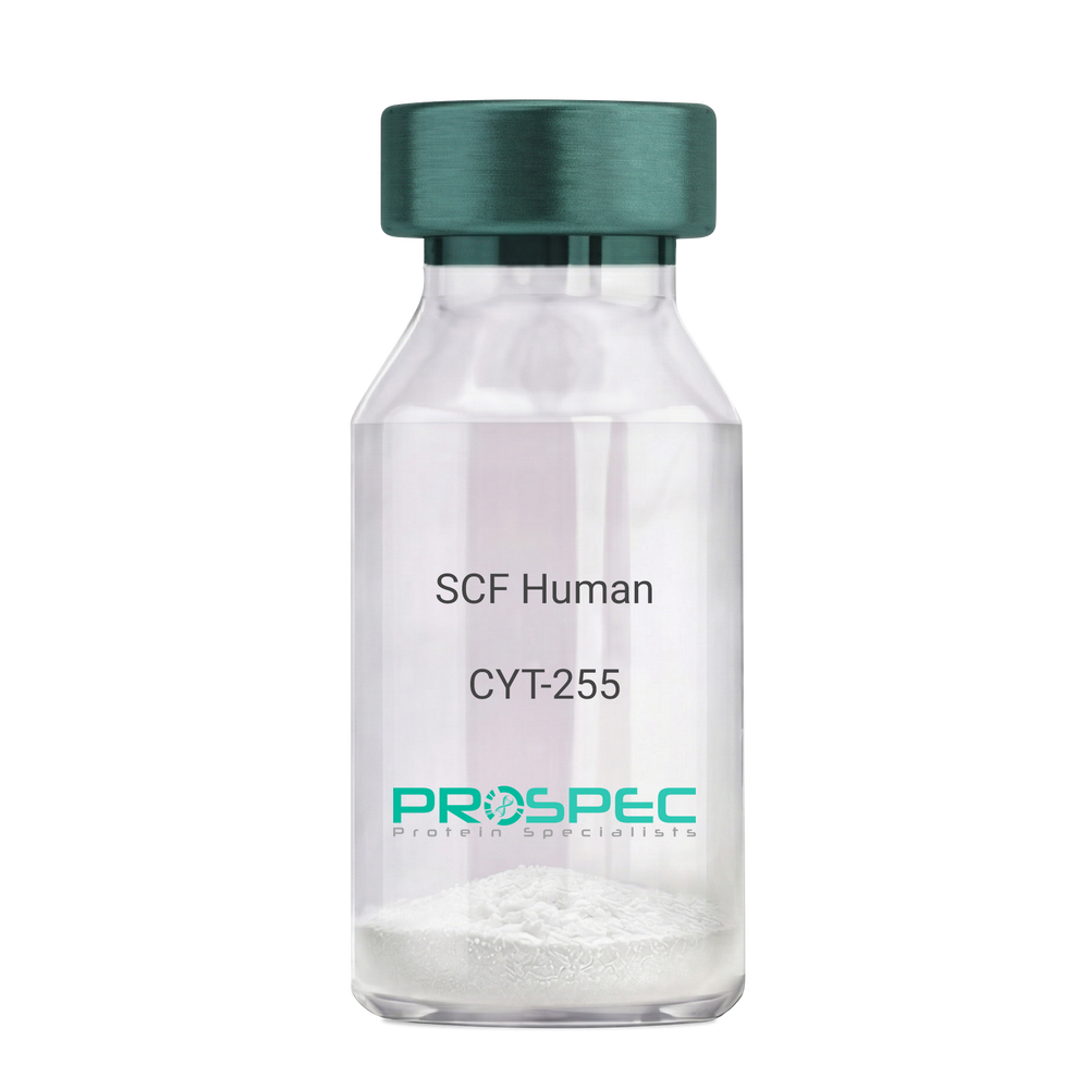
SCF HumanDescription:
Stem Cell Factor Human Recombinant
Kit ligand Precursor, C-kit ligand, SCF, Mast cell growth factor, MGF, SF, KL-1, Kitl, DKFZp686F2250.
Product # :
CYT-255Price :
Quantity :
Shipping Method :
Shipped at Room temp
More Info
- description
- source
- formulation
- purity
- biological activity
- More Info
Description
Stem Cell Factor Human Recombinant produced in E.Coli is a single, non-glycosylated polypeptide chain containing 165 amino acids and having a molecular mass of 18409 Dalton. The SCF is purified by proprietary chromatographic techniques.
Source
Escherichia Coli.
Formulation
Lyophilized from a concentrated (1mg/ml) solution in water containing 0.02% NaHCO3.
Purity
Greater than 98.0% as determined by:
(a) Analysis by SEC-HPLC.
(b) Analysis by SDS-PAGE.Biological Activity
The ED50 as determined by the dose-dependant stimulation of Human TF-1 cells is < 2 ng/ml, corresponding to a Specific Activity of 500,000 IU/mg.More Info
-
Introduction
Stem cell factor / KIT ligand (SCF) is a cytokine which binds CD117(c-Kit). SCF is also known as "steel factor" or "c-kit ligand". SCF exists in two forms, cell surface bound SCF and soluble (or free) SCF. Soluble SCF is produced by the cleavage of surface bound SCF by metalloproteases. SCF is a growth factor important for the survival, proliferation, and differentiation of hematopoietic stem cells and other hematopoietic progenitor cells. One of its roles is to change the BFU-E (burst-forming unit-erythroid) cells, which are the earliest erythrocyte precursors in the erythrocytic series, into the CFU-E (colony-forming unit-erythroid).
-
Synonyms
Kit ligand Precursor, C-kit ligand, SCF, Mast cell growth factor, MGF, SF, KL-1, Kitl, DKFZp686F2250.
-
Physical Appearance
Sterile Filtered White lyophilized (freeze-dried) powder.
-
Stability
Lyophilized KIT ligand although stable at room temperature for 3 weeks, should be stored desiccated below -18°C. Upon reconstitution SCF should be stored at 4°C between 2-7 days and for future use below -18°C.For long term storage it is recommended to add a carrier protein (0.1% HSA or BSA).Please prevent freeze-thaw cycles.
-
Solubility
It is recommended to reconstitute the lyophilized Stem Cell Factor in sterile 18MΩ-cm H2O not less than 100µg/ml, which can then be further diluted to other aqueous solutions.
-
Amino Acid Sequence
The sequence of the first five N-terminal amino acids was determined and was found to be Met-Glu-Gly-Ile-Cys.
-
Protein content
Protein quantitation was carried out by two independent methods1. UV spectroscopy at 280 nm using the absorbency value of 0.52 as the extinction coefficient for a 0.1% (1mg/ml) solution. This value is calculated by the PC GENE computer analysis program of protein sequences (IntelliGenetics). 2. Analysis by RP-HPLC, using a standard solution of Stem Cell Factor as a Reference Standard.
ProSpec's products are furnished for LABORATORY RESEARCH USE ONLY. The product may not be used as drugs, agricultural or pesticidal products, food additives or household chemicals.
- View Data Sheet
Name :
GoserelinDescription:
Goserelin
Product # :
HOR-256Price :
Quantity :
Shipping Method :
Shipped at Room temp
More Info
- description
- formulation
- purity
- More Info
Description
Goserelin contains 10 amino acids Glu1-His2-Trp3-Ser4-Tyr5-D-Ser(tBu)6-Leu7-Arg8-Pro9-AzGly10-NH2 and having a molecular weight of 1269.43 Dalton.
Formulation
The Goserelin peptide was lyophilized with no additives.
Purity
Greater than 98.0% as determined by:
(a) Analysis by RP-HPLC.
(b) Analysis by SDS-PAGE.More Info
-
Introduction
Goserelin is a hormone similar to the one normally released from the hypothalamus gland in the brain (GnRH super-agonist). It is used to treat for prostate and breast cancer.
Goserelin decreases the amount of estrogen and testosterone by this treating endometriosis and cancer of the breast, and can help thin the uterus lining before surgery. Goserelin prevents the growth of tissue associated with endometriosis.
Reducing the amount of testosterone is one way of treating prostate cancer. -
Physical Appearance
Sterile Filtered White lyophilized (freeze-dried) powder.
-
Stability
Lyophilized Goserelin although stable at room temperature for 3 weeks, should be stored desiccated below -18°C. Upon reconstitution Goserelin should be stored at 4°C between 2-7 days and for future use below -18°C. For long term storage it is recommended to add a carrier protein (0.1% HSA or BSA).Please prevent freeze-thaw cycles.
-
Solubility
It is recommended to reconstitute the lyophilized Goserelin in sterile 18MΩ-cm H2O not less than 100 µg/ml, which can then be further diluted to other aqueous solutions.
ProSpec's products are furnished for LABORATORY RESEARCH USE ONLY. The product may not be used as drugs, agricultural or pesticidal products, food additives or household chemicals.
- View Data Sheet
Name :
TRIM21 Human BiotinDescription:
Tripartite Motif Containing 21 (RO52) Human Recombinant, Biotinylated
52 kDa Ro protein, Sjoegren syndrome type A antigen, SS-A, Ro(SS-A), 52 kDa ribonucleoprotein autoantigen Ro/SS-A, Tripartite motif-containing protein 21, RING finger protein 81, TRIM21, RNF81, RO52, SSA1, SSA, RO-52.
Product # :
PRO-2559Price :
Quantity :
Shipping Method :
Shipped with Ice Packs
More Info
- description
- source
- formulation
- purity
- More Info
Description
TRIM21 Human Recombinant, Biotin produced in SF9 is a glycosylated, polypeptide chain having a calculated molecular mass of 52kDa. TRIM21 is expressed with a -6x His tag at N-terminus and purified by proprietary chromatographic techniques.
Source
Sf9 insect cells.
Formulation
TRIM21 solution is supplied in 20mM HEPES pH-7.6, 0.01mM EDTA and 0.02% SDS.
Purity
Greater than 80% as determined by SDS-PAGE.
More Info
-
Introduction
TRIM21 is a member of the tripartite motif (TRIM) family. The TRIM motif includes three zinc-binding domains, a RING, a B-box type 1 and a B-box type 2, and a coiled-coil region. The 52 kDa Ro protein is part of the RoSSA ribonucleoprotein, which includes a single polypeptide and one of four small RNA molecules. The RoSSA particle localizes to both the cytoplasm and the nucleus. Ro/SSA interacts with autoantigens in patients with Sjogren syndrome and systemic lupus erythematosus. Ribonucleoprotein particle is composed of a single polypeptide and one of four small RNA molecules. The RoSSA is present in all mammalian cells studied but has no known function. At least 2 isoforms are present in nucleated and red blood cells, and tissue specific differences in Ro/SSA proteins were identified.
-
Synonyms
52 kDa Ro protein, Sjoegren syndrome type A antigen, SS-A, Ro(SS-A), 52 kDa ribonucleoprotein autoantigen Ro/SS-A, Tripartite motif-containing protein 21, RING finger protein 81, TRIM21, RNF81, RO52, SSA1, SSA, RO-52.
-
Physical Appearance
Sterile Filtered clear solution.
-
Stability
Store at 4°C if entire vial will be used within 2-4 weeks. Store, frozen at -20°C for longer periods of time. Avoid multiple freeze-thaw cycles.
-
Immunological Functions
1. Binds IgG type human auto antibodies.2. Functional Streptavidin based ELISA test (analysis of positive/negative samples.)
ProSpec's products are furnished for LABORATORY RESEARCH USE ONLY. The product may not be used as drugs, agricultural or pesticidal products, food additives or household chemicals.
- View Data Sheet
Name :
MIA HumanDescription:
Melanoma Inhibitory Activity Human Recombinant
Melanoma-derived growth regulatory protein precursor, Cartilage-derived retinoic acid-sensitive protein, CD-RAP, MIA.
Product # :
CYT-310Price :
Quantity :
Shipping Method :
Shipped at Room temp
More Info
- description
- source
- formulation
- purity
- biological activity
- More Info
Description
Melanoma Inhibitory Activity Human Recombinant produced in E.Coli is a single, non-glycosylated, polypeptide chain consisting of 108 amino having a total molecular mass of 12237 Dalton.The MIA is purified by proprietary chromatographic techniques.
Source
Escherichia Coli.
Formulation
The protein was lyophilized from a concentrated (1mg/ml) solution containing 20mM Potassium-phosphate pH=7 and 150mM potassium chloride.
Purity
Greater than 95.0% as determined by:
(a) Analysis by RP-HPLC.
(b) Analysis by SDS-PAGE.Biological Activity
The biological activity is calculated by the inhibiting effect on the invasion of Mel In Tumor cells and found active in Mel In assay.More Info
-
Introduction
The Melanoma Inhibitory protein (MIA) was identified as an inhibitor of in vitro growth of malignant melanoma cells. The protein contains a SH3 domain.
MIA acts as a potent tumor cell growth inhibitor for malignant melanoma cells and some other neuroectodermal tumors, including gliomas, in an autocrine fashion. In a study of human melanoma cell lines with different metastatic capacity MIA mRNA expression appeared to be inversely correlated with pigmentation. MIA has been shown to represent a very sensitive and specific serum marker for systemic malignant melanoma that might be useful for staging of primary melanomas, detection of progression from localized to metastatic disease during follow-up, and monitoring therapy of advanced melanomas. -
Synonyms
Melanoma-derived growth regulatory protein precursor, Cartilage-derived retinoic acid-sensitive protein, CD-RAP, MIA.
-
Physical Appearance
Sterile Filtered White lyophilized (freeze-dried) powder.
-
Stability
Lyophilized MIA although stable at room temperature for 3 weeks, should be stored desiccated below -18°C. Upon reconstitution MIA should be stored at 4°C between 2-7 days and for future use below -18°C.Please prevent freeze-thaw cycles.
-
Solubility
It is recommended to reconstitute the lyophilized Melanoma Inhibitory Activity in sterile 18MΩ-cm H2O not less than 100µg/ml, which can then be further diluted to other aqueous solutions.
-
Amino Acid Sequence
Agrees with the sequence of native MIA human with an addition N-terminal Methionine residue.
MGPMPKLADRKLCADQECSSHPISMAVALQDYMAPDCRFLTIHRGQVV
YVFSLKGRGRFLWGGSVQGDYYGDLAARLGYFPSSIVREDQTLKVDVKT
DKWDFYCQ. -
Protein content
UV spectroscopy at 280 nm using the absorption coefficient of 19300 M-1cm-1.
ProSpec's products are furnished for LABORATORY RESEARCH USE ONLY. The product may not be used as drugs, agricultural or pesticidal products, food additives or household chemicals.