Search results
648 results found for “killer cell lectin-like receptor”
Name
Description
Product #
Price
Quantity
Shipping Method
- View Data Sheet
Name :
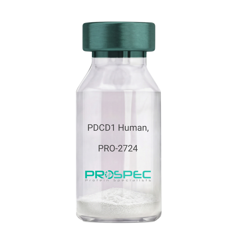
PDCD1 Human, ActiveDescription:
Programmed Cell Death 1 Human Recombinant, Active
Programmed cell death protein 1, CD279, hPD-1, SLEB2, hPD-l, hSLE1, PD-1, PD1, PDCD1.
Product # :
PRO-2724Price :
Quantity :
Shipping Method :
Shipped with Ice Packs
More Info
- description
- source
- formulation
- purity
- biological activity
- More Info
- Citations
Description
PDCD1 Human Recombinant produced in HEK293 Cells is a single, glycosylated polypeptide chain containing 383 amino acids (21-170 a.a) and having a molecular mass of 42.9kDa.PDCD1 is fused to a 233 amino acid hIgG-His-Tag at C-terminus & purified by proprietary chromatographic techniques.
Source
HEK293 Cells.
Formulation
The PDCD1 solution (0.5mg/1ml) contains phosphate buffered saline (pH7.4) and 10% glycerol.
Purity
Greater than 95.0% as determined by SDS-PAGE.
Biological Activity
Measured by its binding ability in a functional ELISA with Human PD-L1/B7-H1
Citations
More Info
-
Introduction
PDCD1 is a cell surface membrane protein of the immunoglobulin superfamily. PDCD1 is expressed in pro-B-cells and is thought to have a role in their differentiation. In mice, expression of PDCD1 gene is induced in the thymus when anti-CD3 antibodies are injected and large numbers of thymocytes undergo apoptosis. Mice deficient for the PDCD1 gene bred on a BALB/c background developed dilated cardiomyopathy and died from congestive heart failure. These studies suggest that the PDCD1 gene product may also be important in T cell function and contribute to the prevention of autoimmune diseases.
-
Synonyms
Programmed cell death protein 1, CD279, hPD-1, SLEB2, hPD-l, hSLE1, PD-1, PD1, PDCD1.
-
Physical Appearance
Sterile filtered colorless solution.
-
Stability
Store at 4°C if entire vial will be used within 2-4 weeks.Store, frozen at -20°C for longer periods of time.For long term storage it is recommended to add a carrier protein (0.1% HSA or BSA).Avoid multiple freeze-thaw cycles.
-
Amino Acid Sequence
PGWFLDSPDR PWNPPTFSPA LLVVTEGDNA TFTCSFSNTS ESFVLNWYRM SPSNQTDKLA AFPEDRSQPG QDCRFRVTQL PNGRDFHMSV VRARRNDSGT YLCGAISLAP KAQIKESLRA ELRVTERRAE VPTAHPSPSP RPAGQFQTLV LEPKSCDKTH TCPPCPAPEL LGGPSVFLFP PKPKDTLMIS RTPEVTCVVV DVSHEDPEVK FNWYVDGVEV HNAKTKPREE QYNSTYRVVS VLTVLHQDWL NGKEYKCKVS NKALPAPIEK TISKAKGQPR EPQVYTLPPS RDELTKNQVS LTCLVKGFYP SDIAVEWESN GQPENNYKTT PPVLDSDGSF FLYSKLTVDK SRWQQGNVFS CSVMHEALHN HYTQKSLSLS PGK
ProSpec's products are furnished for LABORATORY RESEARCH USE ONLY. The product may not be used as drugs, agricultural or pesticidal products, food additives or household chemicals.
- View Data Sheet
Name :
ATP1B2 HumanDescription:
ATPaseTransporting Beta 2 Human Recombinant
ATPase Na+/K+ Transporting Beta 2 Polypeptide, Sodium-Potassium ATPase Subunit Beta 2 (Non-Catalytic), Sodium/Potassium-Dependent ATPase Beta-2 Subunit, Sodium/Potassium-Transporting ATPase Beta-2 Chain, Adhesion Molecule On Glia, Na K-ATPase Beta-2 Polypeptide, Sodium Pump Subunit Beta-2, AMOG.
Product # :
PRO-1647Price :
Quantity :
Shipping Method :
Shipped with Ice Packs
More Info
- description
- source
- formulation
- purity
- More Info
- Citations
Description
ATP1B2 Human Recombinant produced in E.coli is a single, non-glycosylated polypeptide chain containing 246 amino acids (68-290) and having a molecular mass of 27.8kDa. ATP1B2 is fused to a 23 amino acid His-tag at N-terminus & purified by proprietary chromatographic techniques.
Source
Escherichia Coli.
Formulation
The ATP1B2 solution contains 20mM Tris-HCl buffer (pH 8.0), 0.4M Urea and 10% glycerol.
Purity
Greater than 90% as determined by SDS-PAGE.
Citations
More Info
-
Introduction
ATPaseTransporting Beta 2 (ATP1B2) is the non-catalytic component of the active enzyme, which catalyzes the hydrolysis of ATP coupled with the exchange of Na+ and K+ ions across the plasma membrane. The precise function of the beta-2 subunit is not known. The ATP1B2 protein is composed of 3 subunits: alpha (catalytic), beta and gamma.
-
Synonyms
ATPase Na+/K+ Transporting Beta 2 Polypeptide, Sodium-Potassium ATPase Subunit Beta 2 (Non-Catalytic), Sodium/Potassium-Dependent ATPase Beta-2 Subunit, Sodium/Potassium-Transporting ATPase Beta-2 Chain, Adhesion Molecule On Glia, Na K-ATPase Beta-2 Polypeptide, Sodium Pump Subunit Beta-2, AMOG.
-
Physical Appearance
Sterile filtered colorless solution.
-
Stability
Store at 4°C if entire vial will be used within 2-4 weeks. Store, frozen at -20°C for longer periods of time. For long term storage it is recommended to add a carrier protein (0.1% HSA or BSA).Avoid multiple freeze-thaw cycles.
-
Amino Acid Sequence
MGSSHHHHHH SSGLVPRGSH MGSDHTPKYQ DRLATPGLMI RPKTENLDVI VNVSDTESWD QHVQKLNKFL EPYNDSIQAQ KNDVCRPGRY YEQPDNGVLN YPKRACQFNR TQLGNCSGIG DSTHYGYSTG QPCVFIKMNR VINFYAGANQ SMNVTCAGKR DEDAENLGNF VMFPANGNID LMYFPYYGKK FHVNYTQPLV AVKFLNVTPN VEVNVECRIN AANIATDDER DKFAGRVAFK LRINKT
ProSpec's products are furnished for LABORATORY RESEARCH USE ONLY. The product may not be used as drugs, agricultural or pesticidal products, food additives or household chemicals.
- View Data Sheet
Name :
BCL2A1 HumanDescription:
BCL2-Related Protein A1 Human Recombinant
BCL2-related protein A1, Hemopoietic-specific early response protein, hematopoietic BCL2-related protein A1, Bcl-2-like protein 5, Protein BFL-1, BCL2L5, Protein GRS, HBPA1, ACC-1, ACC-2.
Product # :
PRO-1102Price :
Quantity :
Shipping Method :
Shipped with Ice Packs
More Info
- description
- source
- formulation
- purity
- More Info
- Citations
Description
BCL2A1 Human Recombinant produced in E. coli is a single polypeptide chain containing 176 amino acids (1-152) and having a molecular mass of 20.2kDa.BCL2A1 is fused to a 24 amino acid His-tag at N-terminus & purified by conventional chromatography.
Source
E.coli.
Formulation
The BCL2A1 solution (0.5mg/1ml) contains 20mM Tris-HCl buffer (pH 8.0) and 10% glycerol.
Purity
Greater than 95% as determined by SDS-PAGE.
Citations
More Info
-
Introduction
BCL2A1 delays apoptosis induced by IL-3 deficiency. BCL2A1 takes part in the response of hemopoietic cells to external signals and in preserving endothelial survival during infection.
-
Synonyms
BCL2-related protein A1, Hemopoietic-specific early response protein, hematopoietic BCL2-related protein A1, Bcl-2-like protein 5, Protein BFL-1, BCL2L5, Protein GRS, HBPA1, ACC-1, ACC-2.
-
Physical Appearance
Sterile Filtered colorless solution.
-
Stability
Store at 4°C if entire vial will be used within 2-4 weeks. Store, frozen at -20°C for longer periods of time. For long term storage it is recommended to add a carrier protein (0.1% HSA or BSA).Avoid multiple freeze-thaw cycles.
-
Amino Acid Sequence
MGSSHHHHHH SSGLVPRGSH MGSHMTDCEF GYIYRLAQDY LQYVLQIPQP GSGPSKTSRV LQKVAFSVQK EVEKNLKSCL DNVNVVSVDT ARTLFNQVME KEFEDDIINW GRIVTIFAFE GILIKKLLRQ QIAPDVDTYK EISYFVAEFI MNNTGEWIRQ NGGWENGFVK KFEPKS.
ProSpec's products are furnished for LABORATORY RESEARCH USE ONLY. The product may not be used as drugs, agricultural or pesticidal products, food additives or household chemicals.
- View Data Sheet
Name :
EPHA3 MouseDescription:
EPH Receptor A3 Mouse Recombinant
Cek4, EK4, End3, ETK1, Hek, Hek4, Mek4, Tyro4, ephrin type-A receptor 3, ephrin type-A receptor 3 isoform1, receptor protein-tyrosine kinase, EPH-like kinase 4, tyrosine-protein kinase TYRO4, tyrosine-protein kinase receptor ETK1, Eph receptor A3, AW492086.
Product # :
PRO-2817Price :
Quantity :
Shipping Method :
Shipped with Ice Packs
More Info
- description
- source
- formulation
- purity
- biological activity
- More Info
- Citations
Description
EPHA3 Mouse Recombinant produced in HEK293 cells is a single, polypeptide chain containing 527 amino acids (21-541 a.a.) and having a molecular mass of 59.5kDa. EPHA3 is expressed with a 6 amino acid His tag at C-Terminus and purified by proprietary chromatographic techniques.
Source
HEK293 Cells.
Formulation
EPHA3 protein solution (1mg/ml) containing Phosphate Buffered Saline (pH 7.4) and 10% glycerol.
Purity
Greater than 95% as determined by SDS-PAGE.
Biological Activity
Measured by its binding ability in a functional ELISA with cat#pro-2816.
Citations
More Info
-
Synonyms
Cek4, EK4, End3, ETK1, Hek, Hek4, Mek4, Tyro4, ephrin type-A receptor 3, ephrin type-A receptor 3 isoform1, receptor protein-tyrosine kinase, EPH-like kinase 4, tyrosine-protein kinase TYRO4, tyrosine-protein kinase receptor ETK1, Eph receptor A3, AW492086.
-
Physical Appearance
Sterile Filtered colorless solution.
-
Stability
Store at 4°C if entire vial will be used within 2-4 weeks. Store, frozen at -20°C for longer periods of time. For long term storage it is recommended to add a carrier protein (0.1% HSA or BSA). Avoid multiple freeze-thaw cycles.
-
Amino Acid Sequence
ELSPQPSNEV NLLDSKTIQG ELGWISYPSH GWEEISGVDE HYTPIRTYQV CNVMDHSQNN WLRTNWVPRN SAQKIYVELK FTLRDCNSIP LVLGTCKETF NLYYMESDDD HGVKFREHQF TKIDTIAADE SFTQMDLGDR ILKLNTEIRE VGPVNKKGFY LAFQDVGACV ALVSVRVYFK KCPFTVKNLA MFPDTVPMDS QSLVEVRGSC VNNSKEEDPP RMYCSTEGEW LVPIGKCTCN AGYEERGFIC QACRPGFYKA SDGAAKCAKC PPHSSTQEDG SMNCRCENNY FRAEKDPPSM ACTRPPSAPR NVISNINETS VILDWSWPLD TGGRKDITFN IICKKCGWNV RQCEPCSPNV RFLPRQLGLT NTTVTVTDLL AHTNYTFEID AVNGVSELSS PPRQYAAVSI TTNQAAPSPV MTIKKDRTSR NSISLSWQEP EHPNGIILDY EVKYYEKQEQ ETSYTILRAR GTNVTISSLK PDTTYVFQIR ARTAAGYGTN SRKFEFETSP DSFSISGENS HHHHHHH.
-
Background
Research on EphA3 in mice has highlighted its critical involvement in the development of the nervous system, cardiovascular system, and skeletal system. EphA3 signaling influences axon pathfinding, synaptic plasticity, and the formation of tissue boundaries, ensuring proper tissue and organ development. Dysregulation of EphA3 has been implicated in various pathologies, including cancer, where it contributes to tumor growth, angiogenesis, and metastasis.
ProSpec's products are furnished for LABORATORY RESEARCH USE ONLY. The product may not be used as drugs, agricultural or pesticidal products, food additives or household chemicals.
- View Data Sheet
Name :
TNFSF8 Human, Sf9Description:
CD30 Ligand Human Recombinant, Sf9
Tumor Necrosis Factor Superfamily Member 8, Tumor Necrosis Factor (Ligand) Superfamily, Member 8, CD153 Antigen, CD30 Ligand, CD30LG, CD30-L, CD30L, Tumor Necrosis Factor (Ligand) Superfamily Member 8, Tumor Necrosis Factor Ligand 3A, CD30 Antigen Ligand, TNLG3A, CD153, Tumor necrosis factor ligand superfamily member 8, TNFSF8, CD30 ligand.
Product # :
CYT-954Price :
Quantity :
Shipping Method :
Shipped with Ice Packs
More Info
- description
- source
- formulation
- purity
- More Info
- Citations
Description
TNFSF8 produced in Sf9 Baculovirus cells is a single, glycosylated polypeptide chain containing 181 amino acids (63-234a.a.) and having a molecular mass of 20.7kDa (Molecular size on SDS-PAGE will appear at approximately 18-40kDa). TNFSF8 is expressed with a 6 amino acid His tag at C-Terminus and purified by proprietary chromatographic techniques.
Source
Sf9, Baculovirus cells.
Formulation
TNFSF8 protein solution (0.5mg/ml) contains Phosphate Buffered Saline (pH 7.4) and 10% glycerol.
Purity
Greater than 85% as determined by SDS-PAGE.
Citations
More Info
-
Introduction
CD30 Ligand (TNFSF8) is a cytokine which is a member of the tumor necrosis factor (TNF) ligand family. The TNFSF8 cytokine is a ligand for TNFRSF8/CD30, which is a cell surface antigen and a marker for Hodgkin lymphoma and related hematologic malignancies. The employment of the TNFSF8 cytokine expressed on B cell surface has an inhibitory role in modulating Ig class switch. TNFSF8 enhances cell proliferation of some lymphoma cell lines, while inducing cell death and reducing cell proliferation of other lymphoma cell lines. The pleiotropic biological activities of the TNFSF8 cytokine on different CD30+ lymphoma cell lines has a pathophysiologic role in Hodgkin's and some non-Hodgkin's lymphomas.
-
Synonyms
Tumor Necrosis Factor Superfamily Member 8, Tumor Necrosis Factor (Ligand) Superfamily, Member 8, CD153 Antigen, CD30 Ligand, CD30LG, CD30-L, CD30L, Tumor Necrosis Factor (Ligand) Superfamily Member 8, Tumor Necrosis Factor Ligand 3A, CD30 Antigen Ligand, TNLG3A, CD153, Tumor necrosis factor ligand superfamily member 8, TNFSF8, CD30 ligand.
-
Physical Appearance
Sterile filtered colorless solution.
-
Stability
Store at 4°C if entire vial will be used within 2-4 weeks. Store, frozen at -20°C for longer periods of time. For long term storage it is recommended to add a carrier protein (0.1% HSA or BSA).Avoid multiple freeze-thaw cycles.
-
Amino Acid Sequence
ADPQRTDSIP NSPDNVPLKG GNCSEDLLCI LKRAPFKKSW AYLQVAKHLN KTKLSWNKDG ILHGVRYQDG NLVIQFPGLY FIICQLQFLV QCPNNSVDLK LELLINKHIK KQALVTVCES GMQTKHVYQN LSQFLLDYLQ VNTTISVNVD TFQYIDTSTF PLENVLSIFL YSNSDHHHHH H.
ProSpec's products are furnished for LABORATORY RESEARCH USE ONLY. The product may not be used as drugs, agricultural or pesticidal products, food additives or household chemicals.
- View Data Sheet
Name :
FLT1 Human, HEK ActiveDescription:
Vascular Endothelial Growth Factor receptor-1 Human Recombinant, HEK Active
FLT-1, FLT1, Tyrosine-protein kinase receptor FLT, Flt-1, Tyrosine-protein kinase FRT, Fms-like tyrosine kinase 1, VEGFR-1.
Product # :
PKA-134Price :
Quantity :
Shipping Method :
Shipped with Ice Packs
More Info
- description
- source
- formulation
- purity
- biological activity
- More Info
- Citations
Description
FLT1 Human Recombinant is a single, glycosylated polypeptide chain containing 535 amino acids (27-328a.a) and having a molecular mass of 60.3kDa (calculated). FLT1 is fused to a 233 amino acid hIgG-Tag at C-terminus and is purified by proprietary chromatographic techniques.
Source
HEK293
Formulation
FLT1 protein solution (0.5mg/ml) contains 10% Glycerol and Phosphate-Buffered Saline (pH 7.4).
Purity
Greater than 90.0% as determined by SDS-PAGE.
Biological Activity
The ED50 range ≤ 60ng/ml and is measured by its ability to inhibit proliferation using HUVEC human umbilical vein endothelial cells in the
presence of Human VEGF165.Citations
More Info
-
Synonyms
FLT-1, FLT1, Tyrosine-protein kinase receptor FLT, Flt-1, Tyrosine-protein kinase FRT, Fms-like tyrosine kinase 1, VEGFR-1.
-
Physical Appearance
Filtered clear solution.
-
Stability
Store at 4°C if entire vial will be used within 2-4 weeks.Store, frozen at -20°C for longer periods of time.Avoid multiple freeze-thaw cycles.
-
Amino Acid Sequence
SKLKDPELSL KGTQHIMQAG QTLHLQCRGE AAHKWSLPEM VSKESERLSI TKSACGRNGK QFCSTLTLNT AQANHTGFYS CKYLAVPTSK KKETESAIYI FISDTGRPFV EMYSEIPEII HMTEGRELVI PCRVTSPNIT VTLKKFPLDT LIPDGKRIIW DSRKGFIISN ATYKEIGLLT CEATVNGHLY KTNYLTHRQT NTIIDVQIST PRPVKLLRGH TLVLNCTATT PLNTRVQMTW SYPDEKNKRA SVRRRIDQSN SHANIFYSVL TIDKMQNKDK GLYTCRVRSG PSFKSVNTSV HILEPKSCDK THTCPPCPAP ELLGGPSVFL FPPKPKDTLM ISRTPEVTCV VVDVSHEDPE VKFNWYVDGV EVHNAKTKPR EEQYNSTYRV VSVLTVLHQD WLNGKEYKCK VSNKALPAPI EKTISKAKGQ PREPQVYTLP PSRDELTKNQ VSLTCLVKGF YPSDIAVEWE SNGQPENNYK TTPPVLDSDG SFFLYSKLTV DKSRWQQGNV FSCSVMHEAL HNHYTQKSLS LSPGK.
-
Background
VEGFR-1 (Vascular Endothelial Growth Factor Receptor-1), also known as Flt-1 (Fms-like tyrosine kinase 1), is a critical receptor involved in angiogenesis and vascular development. This research paper delves into the structure, function, and therapeutic implications of VEGFR-1, shedding light on its multifaceted role in various physiological and pathological processes.
VEGFR-1 is a transmembrane receptor tyrosine kinase belonging to the VEGF receptor family. It is primarily expressed on endothelial cells and plays a pivotal role in mediating the cellular responses to VEGF ligands. Upon ligand binding, VEGFR-1 initiates intracellular signaling cascades that regulate endothelial cell proliferation, migration, and survival, ultimately contributing to the formation of new blood vessels.
The structure of VEGFR-1 comprises distinct domains, including an extracellular ligand-binding domain, a transmembrane domain, and an intracellular tyrosine kinase domain. The extracellular domain facilitates the interaction between VEGF ligands and the receptor, while the intracellular domain transduces downstream signals by phosphorylating specific tyrosine residues.
VEGFR-1 exhibits not only ligand-dependent but also ligand-independent functions. In addition to its role as a VEGF receptor, it can act as a decoy receptor, sequestering VEGF and modulating the bioavailability of VEGF ligands. This unique property allows VEGFR-1 to regulate VEGF signaling and influence angiogenic processes.
The signaling pathways activated by VEGFR-1 are diverse and intricate, involving multiple downstream effectors, such as PI3K/AKT, MAPK/ERK, and STAT proteins. These pathways regulate endothelial cell behaviors, including proliferation, migration, and differentiation, which are crucial for angiogenesis. Perturbations in VEGFR-1 signaling have been implicated in various pathological conditions, including cancer, retinopathy, and inflammatory disorders.
The therapeutic targeting of VEGFR-1 has gained considerable attention for its potential in managing angiogenesis-related diseases. Inhibitors specifically designed to block VEGFR-1 have been developed to suppress aberrant angiogenesis and impede tumor growth. Moreover, VEGFR-1-based therapies have been explored for ocular diseases like wet age-related macular degeneration (AMD) and diabetic retinopathy, aiming to alleviate pathological neovascularization.
The availability of VEGFR-1 human recombinant proteins has facilitated in-depth research and the development of potential therapeutic interventions. Recombinant VEGFR-1 proteins serve as valuable tools for investigating VEGF-VEGFR-1 interactions, screening drug candidates, and elucidating the underlying molecular mechanisms of VEGFR-1-mediated signaling pathways.
ProSpec's products are furnished for LABORATORY RESEARCH USE ONLY. The product may not be used as drugs, agricultural or pesticidal products, food additives or household chemicals.
- View Data Sheet
Name :
ZG16 HumanDescription:
Zymogen Granule Protein 16 Homolog Human Recombinant
Zymogen granule membrane protein 16, Zymogen granule protein 16, hZG16, Secretory lectin ZG16, ZG16, JCLN, JCLN1, ZG16A, FLJ43571, FLJ92276, MGC34820, MGC183567.
Product # :
PRO-478Price :
Quantity :
Shipping Method :
Shipped with Ice Packs
More Info
- description
- source
- formulation
- purity
- More Info
- Citations
Description
ZG16 Human Recombinant produced in E. coli is a single polypeptide chain containing 174 amino acids (17-167) and having a molecular mass of 19kDa. ZG16 is fused to a 23 amino acid His-tag at N-terminus & purified by proprietary chromatographic techniques.
Source
Escherichia Coli.
Formulation
The ZG16 solution (0.25mg/ml) contains 20mM Tris-HCl buffer (pH 8.0), 0.15M NaCl, 20% glycerol and 1mM DTT.
Purity
Greater than 95% as determined by SDS-PAGE.
Citations
More Info
-
Introduction
Zymogen Granule Protein 16 Homolog (ZG16) is a member of the jacalin lectin family. ZG16 may have a role in protein trafficking and may act as a linker molecule between the submembranous matrix on the luminal side of zymogen granule membrane (ZGM) and aggregated secretory proteins during granule formation in the TGN. ZG16 is highly expressed in the liver, but can also be detected at lower levels in colon, ileum and jejunum.
-
Synonyms
Zymogen granule membrane protein 16, Zymogen granule protein 16, hZG16, Secretory lectin ZG16, ZG16, JCLN, JCLN1, ZG16A, FLJ43571, FLJ92276, MGC34820, MGC183567.
-
Physical Appearance
Sterile filtered colorless solution.
-
Stability
Store at 4°C if entire vial will be used within 2-4 weeks. Store, frozen at -20°C for longer periods of time. For long term storage it is recommended to add a carrier protein (0.1% HSA or BSA). Avoid multiple freeze-thaw cycles.
-
Amino Acid Sequence
MGSSHHHHHH SSGLVPRGSH MGSNAIQARS SSYSGEYGGG GGKRFSHSGN QLDGPITALR VRVNTYYIVG LQVRYGKVWS DYVGGRNGDL EEIFLHPGES VIQVSGKYKW YLKKLVFVTD KGRYLSFGKD SGTSFNAVPL HPNTVLRFIS GRSGSLIDAI GLHWDVYPTS CSRC.
ProSpec's products are furnished for LABORATORY RESEARCH USE ONLY. The product may not be used as drugs, agricultural or pesticidal products, food additives or household chemicals.
- View Data Sheet
Name :
CD3 Antibody, BiotinDescription:
CD3, Mouse Anti-Human, Biotin
Product # :
ANT-138Price :
Quantity :
Shipping Method :
Shipped at Room temp
More Info
- formulation
- More Info
- Citations
Formulation
1mg/ml in PBS (after reconstitution).
Citations
More Info
-
Introduction
CD3 has four peptide chains (gamma, delta, epsilon and zeta) that form CD3. Defects in CD3 are associated with T cell immunodeficiency CD3 complex mediates signal transduction.
-
Solubility
Reconstitute with of H20. Mix gently, wash the sides of the vial and wait 30-60 seconds before use.
-
Immunogen
Purified human PBL T cells.
-
Ig Subclass
Mouse IgG2a.
-
Clone
hCD3.
-
Applications
Blocking, staining and activating antibody. For staining, use 10µl/1,000,000 cells. Titer for blocking and activating T cells should be determined by the investigator.
-
Available Conjugates
This antibody is also available conjugated to FITC.
-
Type
Mouse Anti Human Monoclonal.
-
Storage Procedures
Lyophilized: store at 4oC. After reconstitution, if not intended for use within a month, aliquot and store at -20oC.
-
Purification Method
Ion exchange column.
ProSpec's products are furnished for LABORATORY RESEARCH USE ONLY. The product may not be used as drugs, agricultural or pesticidal products, food additives or household chemicals.
- View Data Sheet
Name :
TACI Human, HisDescription:
Tumor Necrosis Factor Receptor 13B Human Recombinant, His Tag
CD267, CVID, CVID2, TACI, TNFRSF14B, Tumor necrosis factor receptor superfamily, member 13B, Tumor necrosis factor receptor superfamily, member 13B, isoform CRA_a, TNFRSF13B.
Product # :
CYT-032Price :
Quantity :
Shipping Method :
Shipped with Ice Packs
More Info
- description
- source
- formulation
- purity
- More Info
- Citations
Description
TACI Human Recombinant fused with a 23 amino acid His tag at N-terminus produced in E.Coli is a single, non-glycosylated, polypeptide chain containing 188 amino acids (1- 165a.a.) and having a molecular mass of 20.9kDa. The TACI is purified by proprietary chromatographic techniques.
Source
E.coli.
Formulation
The TACI solution (1mg/1ml) contains 20mM Tris-HCl buffer (pH8.0), 10% glycerol and 0.4M Urea.
Purity
Greater than 90% as determined by SDS-PAGE.
Citations
More Info
-
Introduction
TNFRSF13B (TACI) is a transmembrane receptor protein found predominantly on the surface of B cells (a significant part of the immune system). TACI was at first discovered owing to its ability to interact with calcium-modulator and cyclophilin ligand (CAML). Later on, it was found that TACI plays a key role in humoral immunity by interacting with two members of the TNF family. Also, TACI controls T cell-independent B cell antibody responses, isotype switching, and B cell homeostasis.
-
Synonyms
CD267, CVID, CVID2, TACI, TNFRSF14B, Tumor necrosis factor receptor superfamily, member 13B, Tumor necrosis factor receptor superfamily, member 13B, isoform CRA_a, TNFRSF13B.
-
Physical Appearance
Sterile Filtered colorless solution.
-
Stability
Store at 4°C if entire vial will be used within 2-4 weeks. Store, frozen at -20°C for longer periods of time. For long term storage it is recommended to add a carrier protein (0.1% HSA or BSA).Avoid multiple freeze-thaw cycles.
-
Amino Acid Sequence
MGSSHHHHHH SSGLVPRGSH MGSMSGLGRS RRGGRSRVDQ EERFPQGLWT GVAMRSCPEE QYWDPLLGTC MSCKTICNHQ SQRTCAAFCR SLSCRKEQGK FYDHLLRDCI SCASICGQHP KQCAYFCENK LRSPVNLPPE LRRQRSGEVE NNSDNSGRYQ GLEHRGSEAS PALPGLKLSA DQVALVYS.
ProSpec's products are furnished for LABORATORY RESEARCH USE ONLY. The product may not be used as drugs, agricultural or pesticidal products, food additives or household chemicals.
- View Data Sheet
Name :
IL7R HumanDescription:
Interleukin-7 Receptor Human Recombinant
IL7R, Interleukin 7 Receptor, IL-7 Receptor Subunit Alpha, IL-7R Subunit Alpha,CD127 Antigen, IL-7R-Alpha, CDW127, Interleukin-7 Receptor Subunit Alpha, Interleukin 7 Receptor Isoform H5-6, Interleukin 7 Receptor Alpha Chain, IL-7RA,CD127, IL7RA, ILRA.
Product # :
CYT-1046Price :
Quantity :
Shipping Method :
Shipped with Ice Packs
More Info
- description
- source
- formulation
- purity
- More Info
- SDS-PAGE
- Citations
Description
IL7R produced in Sf9 Baculovirus cells is a single, glycosylated polypeptide chain containing 461 amino acids (21-239a.a.) and having a molecular mass of 52.5kDa. (Molecular size on SDS-PAGE will appear at approximately 50-70kDa). IL7R is expressed with an 242 amino acid hIgG-His tag at C-Terminus and purified by proprietary chromatographic techniques.
Source
Sf9, Baculovirus cells.
Formulation
IL7R protein solution (0.5mg/ml) contains Phosphate Buffered Saline (pH 7.4) and 10% glycerol.
Purity
Greater than 90.0% as determined by SDS-PAGE.
SDS-PAGE
Citations
More Info
-
Introduction
Interleukin-7 receptor (IL7R) ,is a hematopoietin receptor superfamily member. IL7R takes a vital part in lymphocyte differentiation, proliferation, multiple sclerosis, as well as survival. IL7R protein signaling is vital for T-cell development and regulation of native and memory T-cell homeostasis. Likewise, IL7R is critically needed for the proper function & development of lymphoid cells.
-
Synonyms
IL7R, Interleukin 7 Receptor, IL-7 Receptor Subunit Alpha, IL-7R Subunit Alpha,CD127 Antigen, IL-7R-Alpha, CDW127, Interleukin-7 Receptor Subunit Alpha, Interleukin 7 Receptor Isoform H5-6, Interleukin 7 Receptor Alpha Chain, IL-7RA,CD127, IL7RA, ILRA.
-
Physical Appearance
Sterile Filtered colorless solution.
-
Stability
Store at 4°C if entire vial will be used within 2-4 weeks. Store, frozen at -20°C for longer periods of time. For long term storage it is recommended to add a carrier protein (0.1% HSA or BSA). Avoid multiple freeze-thaw cycles.
-
Amino Acid Sequence
ADLESGYAQN GDLEDAELDD YSFSCYSQLE VNGSQHSLTC AFEDPDVNIT NLEFEICGAL VEVKCLNFRK LQEIYFIETK KFLLIGKSNI CVKVGEKSLT CKKIDLTTIV KPEAPFDLSV VYREGANDFV VTFNTSHLQK KYVKVLMHDV AYRQEKDENK WTHVNLSSTK LTLLQRKLQP AAMYEIKVRS IPDHYFKGFW SEWSPSYYFR TPEINNSSGE MDLEPKSCDK THTCPPCPAP ELLGGPSVFL FPPKPKDTLM ISRTPEVTCV VVDVSHEDPE VKFNWYVDGV EVHNAKTKPR EEQYNSTYRV VSVLTVLHQD WLNGKEYKCK VSNKALPAPI EKTISKAKGQ PREPQVYTLP PSRDELTKNQ VSLTCLVKGF YPSDIAVEWE SNGQPENNYK TTPPVLDSDG SFFLYSKLTV DKSRWQQGNV FSCSVMHEAL HNHYTQKSLS LSPGKHHHHH H
-
Background
What is the molecular weight/Mw of IL7R HUMAN Protein?
IL7R HUMAN Protein has a total Mw of 52.5kDa.
What is the source or expression system of IL7R HUMAN Protein?
Sf9, Baculovirus cells.
What is the Purity of IL7R HUMAN Protein?
IL7R HUMAN Protein is >90% pure as determined by SDS-PAGE.
What is the Biological Activity of IL7R HUMAN Protein?
The biological functionality of IL7R HUMAN Protein will be determined in the future.
What is the amino acid sequence of IL7R HUMAN Protein?
ADLESGYAQN GDLEDAELDD YSFSCYSQLE VNGSQHSLTC AFEDPDVNIT NLEFEICGAL VEVKCLNFRK LQEIYFIETK KFLLIGKSNI CVKVGEKSLT CKKIDLTTIV KPEAPFDLSV VYREGANDFV VTFNTSHLQK KYVKVLMHDV AYRQEKDENK WTHVNLSSTK LTLLQRKLQP AAMYEIKVRS IPDHYFKGFW SEWSPSYYFR TPEINNSSGE MDLEPKSCDK THTCPPCPAP ELLGGPSVFL FPPKPKDTLM ISRTPEVTCV VVDVSHEDPE VKFNWYVDGV EVHNAKTKPR EEQYNSTYRV VSVLTVLHQD WLNGKEYKCK VSNKALPAPI EKTISKAKGQ PREPQVYTLP PSRDELTKNQ VSLTCLVKGF YPSDIAVEWE SNGQPENNYK TTPPVLDSDG SFFLYSKLTV DKSRWQQGNV FSCSVMHEAL HNHYTQKSLS LSPGKHHHHH H.
What applications can IL7R HUMAN Protein be used in?
IL7R HUMAN Protein can probably be used in western blot, ELISA and Lateral Flow.
What is the endotoxin level for IL7R HUMAN Protein?
The endotoxin level is minimal, IL7R HUMAN Protein was purified using conventional chromatography techniques.
Who supplies recombinant IL7R HUMAN that is in stock and available to order?
ProSpec has the protein in stock and available for immediate delivery.
Where can I order bulk mg quantities of IL7R HUMAN?
ProSpec supplies bulk mg quantities at affordable prices that meets research and academic budgets.
ProSpec's products are furnished for LABORATORY RESEARCH USE ONLY. The product may not be used as drugs, agricultural or pesticidal products, food additives or household chemicals.
- View Data Sheet
Name :
ANXA1 AntibodyDescription:
Annexin A1, Mouse Anti Human
ANX1, LPC1, ANXA1, Lipocortin I, Calpactin II, Chrombindin-9, p35, Annexin-1, Phospholipase A2 inhibitory protein, Annexin I, Annexin A1.
Product # :
ANT-562Price :
Quantity :
Shipping Method :
Shipped with Ice Packs
More Info
- formulation
- More Info
- Citations
Formulation
1mg/ml containing PBS, pH-7.4, 10% Glycerol and 0.02% Sodium Azide.
Citations
More Info
-
Introduction
ANXA1 is part of the family of Ca(2+)-dependent phospholipid binding proteins which have a Mw between 35kDa-40kDa and are situated on the cytosolic face of the plasma membrane. ANXA1 protein has a Mw of 40kDa, with phospholipase A2 inhibitory activity to bind from two to four calcium ions with high affinity. Since phospholipase A2 is necessary for the biosynthesis of the potent mediators of inflammation, prostaglandins and leukotrienes, ANXA1 might have potential anti-inflammatory activity. ANXA1 promotes membrane fusion and iplays a role in exocytosis. The recognition of ANXA1 protein by immunocytochemical leads a simple, highly sensitive and specific assay for diagnosis of hairy cell leukemia.
-
Synonyms
ANX1, LPC1, ANXA1, Lipocortin I, Calpactin II, Chrombindin-9, p35, Annexin-1, Phospholipase A2 inhibitory protein, Annexin I, Annexin A1.
-
Physical Appearance
Sterile filtered colorless solution.
-
Immunogen
Anti-human ANXA1 mAb, clone PAT2G5AT, is derived from hybridization of mouse F0 myeloma cells with spleen cells from BALB/c mice immunized with a recombinant human ANXA1 protein 1-346 amino acids purified from E. coli.
-
Ig Subclass
Mouse IgG1 heavy chain and k light chain.
-
Clone
PAT2G5AT
-
Applications
The antibody has been tested by ELISA, Western blot analysis to assure specificity and reactivity. Since application varies, however, each investigation should be titrated by the reagent to obtain optimal results.
-
Type
Mouse Anti Human Monoclonal.
-
Storage Procedures
For periods up to 1 month store at 4°C, for longer periods of time, store at -20°C. Prevent freeze thaw cycles.
-
Purification Method
ANXA1 antibody was purified from mouse ascitic fluids by protein-A affinity chromatography.
ProSpec's products are furnished for LABORATORY RESEARCH USE ONLY. The product may not be used as drugs, agricultural or pesticidal products, food additives or household chemicals.
- View Data Sheet
Name :
DDR2 HumanDescription:
Discoidin Domain Receptor Tyrosine Kinase 2 Human Recombinant
Discoidin domain-containing receptor 2, DDR2, MIG20a, NTRKR3, TKT, TYR010, Discoidin Domain Receptor Tyrosine Kinase 2, CD167 antigen-like family member B, Discoidin domain-containing receptor tyrosine kinase 2, Neurotrophic tyrosine kinase, receptor-related 3, Receptor protein-tyrosine kinase TKT, Tyrosine-protein kinase TYRO10, CD_antigen: CD167b.
Product # :
PKA-329Price :
Quantity :
Shipping Method :
Shipped with Ice Packs
More Info
- description
- source
- formulation
- purity
- More Info
- Citations
Description
DDR2 Human Recombinant produced in Sf9 Baculovirus cells is a single, glycosylated polypeptide chain containing 386 amino acids (22-399a.a) and having a molecular mass of 43.7kDa. (Molecular size on SDS-PAGE will appear at approximately 40-57kDa).DDR2 is fused to 8 amino acid His-tag at C-terminus & purified by proprietary chromatographic techniques.
Source
Sf9, Baculovirus cells.
Formulation
DDR2 solution (0.5mg/ml) contains Phosphate Buffered Saline (pH 7.4) and 10% glycerol.
Purity
Greater than 90% as determined by SDS-PAGE.
Citations
More Info
-
Introduction
Discoidin Domain Receptor Tyrosine Kinase 2 (DDR2) which is mostly expressed in mesenchymal cells is a part of the discoidin-like domain containing subfamily of receptor tyrosine kinases. DDR2 whose ligand is fibrillar collagen rather than a growth factor-like peptide is unique among RTKs. DDR2 regulates cell differentiation, remodeling of the extracellular matrix, cell migration and cell proliferation. DDR2 is also essential for normal bone development.
-
Synonyms
Discoidin domain-containing receptor 2, DDR2, MIG20a, NTRKR3, TKT, TYR010, Discoidin Domain Receptor Tyrosine Kinase 2, CD167 antigen-like family member B, Discoidin domain-containing receptor tyrosine kinase 2, Neurotrophic tyrosine kinase, receptor-related 3, Receptor protein-tyrosine kinase TKT, Tyrosine-protein kinase TYRO10, CD_antigen: CD167b.
-
Physical Appearance
Sterile Filtered colorless solution.
-
Stability
Store at 4°C if entire vial will be used within 2-4 weeks. Store, frozen at -20°C for longer periods of time. For long term storage it is recommended to add a carrier protein (0.1% HSA or BSA).Avoid multiple freeze-thaw cycles.
-
Amino Acid Sequence
KAQVNPAICR YPLGMSGGQI PDEDITASSQ WSESTAAKYG RLDSEEGDGA WCPEIPVEPD DLKEFLQIDL HTLHFITLVG TQGRHAGGHG IEFAPMYKIN YSRDGTRWIS WRNRHGKQVL DGNSNPYDIF LKDLEPPIVA RFVRFIPVTD HSMNVCMRVE LYGCVWLDGL VSYNAPAGQQ FVLPGGSIIY LNDSVYDGAV GYSMTEGLGQ LTDGVSGLDD FTQTHEYHVW PGYDYVGWRN ESATNGYIEI MFEFDRIRNF TTMKVHCNNM FAKGVKIFKE VQCYFRSEAS EWEPNAISFP LVLDDVNPSA RFVTVPLHHR MASAIKCQYH FADTWMMFSE ITFQSDAAMY NNSEALPTSP MAPTTYDPML KVDDSNTRLE HHHHHH.
ProSpec's products are furnished for LABORATORY RESEARCH USE ONLY. The product may not be used as drugs, agricultural or pesticidal products, food additives or household chemicals.
- View Data Sheet
Name :
KLK7 Human, sf9Description:
Kallikrein-7 Human Recombinant, sf9
Kallikrein Related Peptidase 7, Kallikrein 7 (Chymotryptic, Stratum Corneum), Stratum Corneum Chymotryptic Enzyme, Serine Protease 6, PRSS6, SCCE, HK7, Kallikrein-Related Peptidase 7, Signal Protein, EC 3.4.21.117, EC 3.4.21, HSCCE.
Product # :
ENZ-962Price :
Quantity :
Shipping Method :
Shipped with Ice Packs
More Info
- description
- source
- formulation
- purity
- More Info
- Citations
Description
KLK7 produced in Sf9 Baculovirus cells is a single, glycosylated polypeptide chain containing 190 amino acids (1-181 a.a.) and having a molecular mass of 20.9kDa (Migrates at 28-40kDa on SDS-PAGE under reducing conditions). KLK7 is expressed with a 6 amino acid His tag at C-Terminus and purified by proprietary chromatographic techniques.
Source
Sf9, Baculovirus cells.
Formulation
KLK7 protein solution (0.5mg/ml) contains Phosphate Buffered Saline (pH 7.4) and 10% glycerol.
Purity
Greater than 95.0% as determined by SDS-PAGE.
Citations
More Info
-
Introduction
KLK7 catalyzes the degradation of intercellular cohesive structures in the cornified layer of the skin in the continuous shedding of cells from the skin surface. Specific for amino acid residues with aromatic side chains in the P1 position. KLK7 cleaves insulin B chain at ''6-Leu- -Cys-7'', ''16-Tyr- -Leu-17'', ''25-Phe- -Tyr-26'' and ''26-Tyr--Thr-27''. KLK7 is involved in the activation of precursors to inflammatory cytokines.
-
Synonyms
Kallikrein Related Peptidase 7, Kallikrein 7 (Chymotryptic, Stratum Corneum), Stratum Corneum Chymotryptic Enzyme, Serine Protease 6, PRSS6, SCCE, HK7, Kallikrein-Related Peptidase 7, Signal Protein, EC 3.4.21.117, EC 3.4.21, HSCCE.
-
Physical Appearance
Sterile Filtered colorless solution.
-
Stability
Store at 4°C if entire vial will be used within 2-4 weeks. Store, frozen at -20°C for longer periods of time. For long term storage it is recommended to add a carrier protein (0.1% HSA or BSA). Avoid multiple freeze-thaw cycles.
-
Amino Acid Sequence
ADPMNEYTVH LGSDTLGDRR AQRIKASKSF RHPGYSTQTH VNDLMLVKLN SQARLSSMVK KVRLPSRCEP PGTTCTVSGW GTTTSPDVTF PSDLMCVDVK LISPQDCTKV YKDLLENSML CAGIPDSKKN ACNGDSGGPL VCRGTLQGLV SWGTFPCGQP NDPGVYTQVC KFTKWINDTM KKHRHHHHHH.
ProSpec's products are furnished for LABORATORY RESEARCH USE ONLY. The product may not be used as drugs, agricultural or pesticidal products, food additives or household chemicals.
- View Data Sheet
Name :
ICAM1 Human, Sf9Description:
Intercellular Adhesion Molecule-1 Human Recombinant, SF9
Intercellular Adhesion Molecule 1, Major Group Rhinovirus Receptor, ICAM-1, Intercellular Adhesion Molecule 1 (CD54), Human Rhinovirus Receptor, Cell Surface Glycoprotein P3.58, Human Rhinovirus Receptor, CD54 Antigen, P3.58, CD54, BB2, Intercellular adhesion molecule 1.
Product # :
PRO-2200Price :
Quantity :
Shipping Method :
Shipped with Ice Packs
More Info
- description
- source
- formulation
- purity
- biological activity
- More Info
- Citations
Description
ICAM1 produced in Sf9 Baculovirus cells is a single, glycosylated polypeptide chain containing 692 amino acids (28-480a.a.) and having a molecular mass of 76.5kDa. (Molecular size on SDS-PAGE will appear at approximately 70-100kDa). ICAM1 is expressed with a 239 amino acid His tag at C-Terminus and purified by proprietary chromatographic techniques.
Source
Sf9, Baculovirus cells.
Formulation
ICAM1 protein solution (0.25mg/ml) contains phosphate buffered saline (pH7.4) and 10% glycerol.
Purity
Greater than 90.0% as determined by SDS-PAGE.
Biological Activity
The ED50 range ≤ 2 ug/ml is measured by the ability of the immobilized protein to support the adhesion of HL-60 human promyelocytic cells. When cells are added to Human ICAM-1/CD54 coated plates.
Citations
More Info
-
Introduction
ICAM-1 also called CD54 is a single chain membrane glycoprotein expressed on the surface of a variety of non-haematopoietic and haematopoietic cell types and has roles in signal transduction, cell signaling and lymphocyte adhesion. ICAM1 binds to integrins such as CD11a / CD18, or CD11b / CD18. ICAM1 is also used by Rhinovirus as a receptor. ICAM-1 is an intercellular adhesion molecule constantly present in low concentrations in the membranes of leukocytes and endothelial cells. When stimulated by cytokine the concentrations significantly increase. ICAM-1 can be stimulated by interleukin-1 (IL-1) and tumor necrosis factor alpha (TNFA) and is expressed by the vascular endothelium, macrophages and lymphocytes. ICAM-1 is a ligand for LFA-1 which is a receptor found on leukocytes. Upon activation, leukocytes bind to endothelial cells via ICAM-1/LFA-1 and then transmigrate into tissues. ICAM-1 is implicated in subarachnoid hemorrhage (SAH). Levels of ICAM-1 are shown to be notably elevated in patients with SAH. Soluble ICAM-1 is detectable in the plasma and is elevated in patients with various inflammatory conditions.
-
Synonyms
Intercellular Adhesion Molecule 1, Major Group Rhinovirus Receptor, ICAM-1, Intercellular Adhesion Molecule 1 (CD54), Human Rhinovirus Receptor, Cell Surface Glycoprotein P3.58, Human Rhinovirus Receptor, CD54 Antigen, P3.58, CD54, BB2, Intercellular adhesion molecule 1.
-
Physical Appearance
Sterile Filtered colorless solution.
-
Stability
Store at 4°C if entire vial will be used within 2-4 weeks. Store, frozen at -20°C for longer periods of time. For long term storage it is recommended to add a carrier protein (0.1% HSA or BSA).Avoid multiple freeze-thaw cycles.
-
Amino Acid Sequence
QTSVSPSKVI LPRGGSVLVT CSTSCDQPKL LGIETPLPKK ELLLPGNNRK VYELSNVQED SQPMCYSNCP DGQSTAKTFL TVYWTPERVE LAPLPSWQPV GKNLTLRCQV EGGAPRANLT VVLLRGEKEL KREPAVGEPA EVTTTVLVRR DHHGANFSCR TELDLRPQGL ELFENTSAPY QLQTFVLPAT PPQLVSPRVL EVDTQGTVVC SLDGLFPVSE AQVHLALGDQ RLNPTVTYGN DSFSAKASVS VTAEDEGTQR LTCAVILGNQ SQETLQTVTI YSFPAPNVIL TKPEVSEGTE VTVKCEAHPR AKVTLNGVPA QPLGPRAQLL LKATPEDNGR SFSCSATLEV AGQLIHKNQT RELRVLYGPR LDERDCPGNW TWPENSQQTP MCQAWGNPLP ELKCLKDGTF PLPIGESVTV TRDLEGTYLC RARSTQGEVT REVTVNVLSP RYEVEPKSCD KTHTCPPCPA PELLGGPSVF LFPPKPKDTL MISRTPEVTC VVVDVSHEDP EVKFNWYVDG VEVHNAKTKP REEQYNSTYR VVSVLTVLHQ DWLNGKEYKC KVSNKALPAP IEKTISKAKG QPREPQVYTL PPSRDELTKN QVSLTCLVKG FYPSDIAVEW ESNGQPENNY KTTPPVLDSD GSFFLYSKLT VDKSRWQQGN VFSCSVMHEA LHNHYRQKSL SLSPGKHHHH HH.
-
Background
Intercellular adhesion molecule-1 (ICAM-1) is a cell surface glycoprotein that plays a pivotal role in immune responses and inflammatory processes. This research aims to investigate the function and significance of ICAM-1 protein in various physiological and pathological conditions. Understanding the molecular mechanisms and regulatory roles of ICAM-1 can provide valuable insights into its potential as a therapeutic target for immune-related disorders.
Structure and Expression of ICAM-1 Protein:
ICAM-1 belongs to the immunoglobulin superfamily and is composed of five immunoglobulin-like domains. It is primarily expressed on the surfaces of endothelial cells, leukocytes, and other immune cells. ICAM-1 expression can be induced by pro-inflammatory cytokines, such as tumor necrosis factor-alpha (TNF-α) and interleukin-1 (IL-1), during immune responses and inflammatory conditions.
ICAM-1 and Leukocyte Adhesion:
One of the critical functions of ICAM-1 is its involvement in leukocyte adhesion and migration. ICAM-1 interacts with its primary receptor, lymphocyte function-associated antigen-1 (LFA-1), expressed on leukocytes. This interaction facilitates the firm adhesion of leukocytes to endothelial cells, leading to their transmigration into inflamed tissues. The ICAM-1/LFA-1 axis plays a crucial role in immune surveillance, inflammation, and host defense against pathogens.
Implications of ICAM-1 in Inflammatory Disorders:
ICAM-1 has been implicated in various inflammatory diseases, including rheumatoid arthritis, inflammatory bowel disease, and multiple sclerosis. Enhanced expression of ICAM-1 on endothelial cells promotes leukocyte recruitment and contributes to the perpetuation of chronic inflammation. Therefore, targeting ICAM-1-mediated leukocyte adhesion has emerged as a potential therapeutic strategy to alleviate inflammation and attenuate disease progression.
ICAM-1 in Viral Infections:
ICAM-1 also plays a role in viral infections, as several viruses exploit ICAM-1 to facilitate their entry into host cells. For instance, rhinoviruses, which cause the common cold, utilize ICAM-1 as a receptor for attachment and entry into respiratory epithelial cells. The interaction between ICAM-1 and viral proteins promotes viral internalization and subsequent infection. Understanding the mechanisms of ICAM-1-mediated viral entry can aid in the development of antiviral strategies.
Therapeutic Targeting of ICAM-1:
Given its crucial involvement in immune responses and disease pathogenesis, ICAM-1 has emerged as a potential therapeutic target. Strategies aimed at blocking ICAM-1/LFA-1 interactions have shown promise in preclinical and clinical studies. Monoclonal antibodies targeting ICAM-1 or LFA-1 have been developed to prevent leukocyte adhesion and reduce inflammation. Additionally, small molecule inhibitors and gene therapies targeting ICAM-1 expression are being explored as potential therapeutic interventions.
Challenges and Future Directions:
Although therapeutic targeting of ICAM-1 shows promise, several challenges need to be addressed. Specific targeting of ICAM-1 without affecting its physiological functions and potential off-target effects are important considerations. Additionally, the complex and dynamic nature of ICAM-1 expression and regulation require further investigation to optimize therapeutic strategies.
Conclusion:
The investigation of ICAM-1 protein provides insights into its pivotal role in immune responses, leukocyte adhesion, and inflammatory processes. Understanding the molecular mechanisms and functional implications of ICAM-1 opens avenues for the development of targeted therapies for inflammatory disorders and viral infections. Further research on ICAM-1 protein
ProSpec's products are furnished for LABORATORY RESEARCH USE ONLY. The product may not be used as drugs, agricultural or pesticidal products, food additives or household chemicals.
- View Data Sheet
Name :
EBAG9 HumanDescription:
Estrogen Receptor Binding Site Associated Antigen 9 Human Recombinant
EB9, PDAF, RCAS1, EBAG9, Receptor-binding cancer antigen expressed on SiSo cells, Cancer-associated surface antigen RCAS1, Estrogen receptor-binding fragment-associated gene 9 protein.
Product # :
PRO-343Price :
Quantity :
Shipping Method :
Shipped with Ice Packs
More Info
- description
- source
- formulation
- purity
- More Info
- Citations
Description
EBAG9 Human Recombinant produced in E.Coli is a single, non-glycosylated polypeptide chain containing 207 amino acids (28-213) and having a molecular mass of 23.4 kDa. The EBAG9 protein is fused to a 20 amino acid His-Tag at N-terminus and purified by standard chromatography techniques.
Source
Escherichia Coli.
Formulation
The EBAG9 protein solution contains 20mM Tris-HCl pH-8, 1mM DTT, 2mM EDTA and 10% glycerol.
Purity
Greater than 90% as determined by SDS-PAGE.
Citations
More Info
-
Introduction
EBAG9 is a type III transmembrane protein that is primarily located in the Golgi. EBAG9 is a tumor-associated antigen that is expressed at high frequency in a variety of cancers, such as advanced breast and prostate cancers. while the EBAG9 acts to inhibit the growth of receptor-binding cells and induced apoptosis of immune cells, cancer cells may evade immune surveillance. Thus immunodetection of EBAG9 expression can be a negative prognostic indicator. EBAG9 promotes progression of bladder cancer. In rheumatoid arthritis, the deficiency of EBAG9 induces CTL infiltration through failure to evade immune attack, therefore leading to apoptosis of the synovial lining cells. EBAG9 is involved in controlling exocytosis processes. EBAG9 expression plays a role in the progression of oral squamous cell carcinoma. EBAG9 plays a specific role in premature stages of breast carcinogenesis.
-
Synonyms
EB9, PDAF, RCAS1, EBAG9, Receptor-binding cancer antigen expressed on SiSo cells, Cancer-associated surface antigen RCAS1, Estrogen receptor-binding fragment-associated gene 9 protein.
-
Physical Appearance
Sterile Filtered colorless solution.
-
Stability
Store at 4°C if entire vial will be used within 2-4 weeks. Store, frozen at -20°C for longer periods of time. For long term storage it is recommended to add a carrier protein (0.1% HSA or BSA).Avoid multiple freeze-thaw cycles.
-
Amino Acid Sequence
MGSSHHHHHH SSGLVPRGSH MRSGRGRKLS GDQITLPTTV DYSSVPKQTD VEEWTSWDED APTSVKIEGG NGNVATQQNS LEQLEPDYFK DMTPTIRKTQ KIVIKKREPL NFGIPDGSTG FSSRLAATQD LPFIHQSSEL GDLDTWQENT NAWEEEEDAA WQAEEVLRQQ KLADREKRAA EQQRKKMEKE AQRLMKKEQN KIGVKLS.
ProSpec's products are furnished for LABORATORY RESEARCH USE ONLY. The product may not be used as drugs, agricultural or pesticidal products, food additives or household chemicals.
- View Data Sheet
Name :
IL18R1 HumanDescription:
Interleukin-18 Receptor-1 Recombinant Human
Interleukin 18 Receptor 1, CD218 Antigen-Like Family Member A, IL1 Receptor-Related Protein, IL-18R-1, CDw218a, IL-18R1, IL-1Rrp, IL1R-Rp, IL1RRP, Interleukin-18 Receptor 1, Cytokine Receptor, CD218a Antigen, IL18Ralpha2, CD218a, IL18RA.
Product # :
CYT-1078Price :
Quantity :
Shipping Method :
Shipped with Ice Packs
More Info
- description
- source
- formulation
- purity
- More Info
- Citations
Description
IL18R1 produced in Sf9 Baculovirus cells is a single, glycosylated polypeptide chain containing a total of 550 amino acids (19-329 a.a.) and having a molecular mass of 62.7kDa (Molecular size on SDS-PAGE will appear at approximately 70-100kDa). IL18R1 is expressed with a 239 amino acid hIgG-His tag at C-Terminus and purified by proprietary chromatographic techniques.
Source
Sf9, Baculovirus cells.
Formulation
IL18R1 protein solution (0.5mg/ml) contains 10% glycerol & Phosphate buffered saline (pH7.4).
Purity
Greater than 90.0% as determined by SDS-PAGE.
Citations
More Info
-
Introduction
IL18R1 (interleukin 18 receptor 1) is an interleukin receptor which is part of the immunoglobulin superfamily. IL18R1 comprises 3 Ig-like C2-type domains, on TIR domain IL18R1 shares many immuno-regulatory functions with IL12. IL12 and IFN-alpha are found to induce the expression of IL18 receptor in NK & T cells. Moreover, IL18R1 explicitly binds interleukin 18, and is vital for IL18 mediated signal transduction. IL18R1 does not bind IL1A or IL1B beta, however it does bind to the agonist which causes NF-kappa-B to become active.
-
Synonyms
Interleukin 18 Receptor 1, CD218 Antigen-Like Family Member A, IL1 Receptor-Related Protein, IL-18R-1, CDw218a, IL-18R1, IL-1Rrp, IL1R-Rp, IL1RRP, Interleukin-18 Receptor 1, Cytokine Receptor, CD218a Antigen, IL18Ralpha2, CD218a, IL18RA.
-
Physical Appearance
Sterile filtered colorless solution.
-
Stability
Store at 4°C if entire vial will be used within 2-4 weeks. Store, frozen at -20°C for longer periods of time. For long term storage it is recommended to add a carrier protein (0.1% HSA or BSA).Avoid multiple freeze-thaw cycles.
-
Amino Acid Sequence
AESCTSRPHI TVVEGEPFYL KHCSCSLAHE IETTTKSWYK SSGSQEHVEL NPRSSSRIAL HDCVLEFWPV ELNDTGSYFF QMKNYTQKWK LNVIRRNKHS CFTERQVTSK IVEVKKFFQI TCENSYYQTL VNSTSLYKNC KKLLLENNKN PTIKKNAEFE DQGYYSCVHF LHHNGKLFNI TKTFNITIVE DRSNIVPVLL GPKLNHVAVE LGKNVRLNCS ALLNEEDVIY WMFGEENGSD PNIHEEKEMR IMTPEGKWHA SKVLRIENIG ESNLNVLYNC TVASTGGTDT KSFILVRKAD MADIPGHVFT RVEPKSCDKT HTCPPCPAPE LLGGPSVFLF PPKPKDTLMI SRTPEVTCVV VDVSHEDPEV KFNWYVDGVE VHNAKTKPRE EQYNSTYRVV SVLTVLHQDW LNGKEYKCKV SNKALPAPIE KTISKAKGQP REPQVYTLPP SRDELTKNQV SLTCLVKGFY PSDIAVEWES NGQPENNYKT TPPVLDSDGS FFLYSKLTVD KSRWQQGNVF SCSVMHEALH NHYTQKSLSL
SPGKHHHHHH
ProSpec's products are furnished for LABORATORY RESEARCH USE ONLY. The product may not be used as drugs, agricultural or pesticidal products, food additives or household chemicals.
- View Data Sheet
Name :
CLMP Human, SF9Description:
CXADR-Like Membrane Protein, Sf9 Human Recombinant
CXADR like membrane protein, ACAM, ASAM, CSBM, CSBS, Adipocyte adhesion molecule, Coxsackie- and adenovirus receptor-like membrane protein, CAR-like membrane protein.
Product # :
PRO-2519Price :
Quantity :
Shipping Method :
Shipped with Ice Packs
More Info
- description
- source
- formulation
- purity
- More Info
- Citations
Description
CLMP produced in Sf9 Baculovirus cells is a single, glycosylated polypeptide chain containing 226 amino acids (19-235a.a.) and having a molecular mass of 25.6kDa. (Molecular size on SDS-PAGE will appear at approximately 28-40kDa).CLMP is expressed with a 9 amino acid His tag at C-Terminus and purified by proprietary chromatographic techniques.
Source
Sf9, Baculovirus cells.
Formulation
CLMP protein solution (0.5mg/ml) contains phosphate buffered saline (pH7.4), 1mM DTT and 20%glycerol
Purity
Greater than 95.0% as determined by SDS-PAGE.
Citations
More Info
-
Introduction
CXADR Like Membrane Protein or CLMP, part of the CTX family within the immunoglobulin superfamily, is a type 1 transmembrane peptide. CLMP is expressed in the placenta and small intensine, skeletal muscle, colon, lungs and heart. CLMP is located in junction complexes across endothelial and epithelial cells and can take part in cell adhesion. The protein has influence on development of obesity via adipocyte differentiation.
-
Synonyms
CXADR like membrane protein, ACAM, ASAM, CSBM, CSBS, Adipocyte adhesion molecule, Coxsackie- and adenovirus receptor-like membrane protein, CAR-like membrane protein.
-
Physical Appearance
Sterile Filtered clear solution.
-
Stability
Store at 4°C if entire vial will be used within 2-4 weeks. Store, frozen at -20°C for longer periods of time. For long term storage it is recommended to add a carrier protein (0.1% HSA or BSA).Avoid multiple freeze-thaw cycles.
-
Amino Acid Sequence
ADPTHTEIKR VAEEKVTLPC HHQLGLPEKD TLDIEWLLTD NEGNQKVVIT YSSRHVYNNL TEEQKGRVAF ASNFLAGDAS LQIEPLKPSD EGRYTCKVKN SGRYVWSHVI LKVLVRPSKP KCELEGELTE GSDLTLQCES SSGTEPIVYY WQRIREKEGE DERLPPKSRI DYNHPGRVLL
QNLTMSYSGL YQCTAGNEAG KESCVVRVTV QYVQSIGMVA HHHHHH.
ProSpec's products are furnished for LABORATORY RESEARCH USE ONLY. The product may not be used as drugs, agricultural or pesticidal products, food additives or household chemicals.
- View Data Sheet
Name :
CD2 HumanDescription:
CD2 Human Recombinant
T-cell surface antigen CD2, T-cell surface antigen T11/Leu-5, LFA-2, LFA-3 receptor, Erythrocyte receptor, Rosette receptor, CD2 antigen, CD2, T11, SRBC.
Product # :
PRO-1337Price :
Quantity :
Shipping Method :
Shipped with Ice Packs
More Info
- description
- source
- formulation
- purity
- More Info
- Citations
Description
CD2 Human Recombinant produced in E.Coli is a single, non-glycosylated polypeptide chain containing 209 amino acids (25-209 a.a) and having a molecular mass of 23.8kDa.CD2 is fused to a 24 amino acid His-tag at N-terminus & purified by proprietary chromatographic techniques.
Source
Escherichia Coli.
Formulation
CD2 protein solution (1mg/ml) containing 20mM Tris-HCl buffer (pH8.0), 10% glycerol and 0.4M Urea.
Purity
Greater than 85.0% as determined by SDS-PAGE.
Citations
More Info
-
Introduction
CD2 which is also known as E-rosette receptor, T11 and lymphocytefunction antigen-2 (LFA-2) is expressed on T cells, thymocytes, and subset of natural killer cells. Human CD2 functions as the receptor for sheep erythrocytes, human CD58 (LFA-3), and CD15s (Sialyl Lewis X). p56lck, p59fyn, CD3eta and CD3epsilon are tyrosine-phosphorylated after CD2 stimulation. CD2 plays a role in T cell activation, T- or NK-cell-mediated cytolysis, apoptosis in activated peripheral T cells, and regulation of T cell anergy.
-
Synonyms
T-cell surface antigen CD2, T-cell surface antigen T11/Leu-5, LFA-2, LFA-3 receptor, Erythrocyte receptor, Rosette receptor, CD2 antigen, CD2, T11, SRBC.
-
Physical Appearance
Sterile Filtered clear solution.
-
Stability
Store at 4°C if entire vial will be used within 2-4 weeks. Store, frozen at -20°C for longer periods of time. For long term storage it is recommended to add a carrier protein (0.1% HSA or BSA).Avoid multiple freeze-thaw cycles.
-
Amino Acid Sequence
MGSSHHHHHH SSGLVPRGSH MGSMKEITNA LETWGALGQD INLDIPSFQM SDDIDDIKWE KTSDKKKIAQ FRKEKETFKE KDTYKLFKNG TLKIKHLKTD DQDIYKVSIY DTKGKNVLEK IFDLKIQERV SKPKISWTCI NTTLTCEVMN GTDPELNLYQ DGKHLKLSQR VITHKWTTSL
SAKFKCTAGN KVSKESSVEP VSCPEKGLD.
ProSpec's products are furnished for LABORATORY RESEARCH USE ONLY. The product may not be used as drugs, agricultural or pesticidal products, food additives or household chemicals.
- View Data Sheet
Name :
CD21 Antibody, FITCDescription:
CD21, Mouse Anti-Human, FITC
Complement receptor type 2, Cr2, Complement C3d receptor, Epstein-Barr virus receptor, EBV receptor, CD21 antigen, CR2, C3DR, CD21, SLEB9.
Product # :
ANT-259Price :
Quantity :
Shipping Method :
Shipped at Room temp
More Info
- formulation
- More Info
- Citations
Formulation
1mg/ml in PBS (after reconstitution).
Citations
More Info
-
Introduction
CD21 is expressed strongly on mature B cells, follicular dentritic cells and weakly on immature thymocytes and T lymphocytes. In B-cell ontogeny, CD21 appears after the pre-B-stage, is maintained during peripheral B-cell development and is lost upon terminal differentiation into plasma cells. CD21 expression is also gradually lost after stimulation of B cells in vitro. CD21 functions as receptor for C3d, C3dg and iC3b Complement components, for EBV and for IFNalpha. CD21 binds to CD23 and associates with CD19, CD81 and Leu13 to form a large signal-transduction complex involved in B cell activation.
-
Synonyms
Complement receptor type 2, Cr2, Complement C3d receptor, Epstein-Barr virus receptor, EBV receptor, CD21 antigen, CR2, C3DR, CD21, SLEB9.
-
Solubility
Reconstitute with of H20. Mix gently, wash the sides of the vial and wait 30-60 seconds before use.
-
Immunogen
Purified human B-Cells.
-
Ig Subclass
Mouse IgG2a.
-
Clone
hCD21.
-
Applications
Staining antibody. For staining, use 10µl/1,000,000 cells.
-
Available Conjugates
This antibody is also available unconjugated and Biotin. For staining with biotin or FITC-conjugated antibody use 5-10µl/106 cells.
-
Type
Mouse Anti Human Monoclonal.
-
Storage Procedures
Lyophilized: store at 4°C. After reconstitution, if not intended for use within a month, aliquot and store at -20°C.
-
Purification Method
Protein-A.
ProSpec's products are furnished for LABORATORY RESEARCH USE ONLY. The product may not be used as drugs, agricultural or pesticidal products, food additives or household chemicals.
- View Data Sheet
Name :
BRCC3 AntibodyDescription:
Lys-63-specific deubiquitinase BRCC36, Mouse Anti Human
Lys-63-specific deubiquitinase BRCC36, BRCA1-A complex subunit BRCC36, BRISC complex subunit BRCC36, BRCA1/BRCA2-containing complex subunit 3, BRCA1/BRCA2-containing complex subunit 36, BRCC3, BRCC36, C6.1A, CXorf53, C6.1A, RP11-143H17.2.
Product # :
ANT-448Price :
Quantity :
Shipping Method :
Shipped with Ice Packs
More Info
- formulation
- More Info
- Citations
Formulation
1mg/ml containing PBS, pH-7.4, & 0.1% Sodium Azide.
Citations
More Info
-
Introduction
BRCC3 is a subunit of the BRCA1-BRCA2-containing complex (BRCC), which is an E3 ubiquitin ligase. BRCC3 is also thought to be involved in the cellular response to ionizing radiation and progression through the G2/M checkpoint.
-
Synonyms
Lys-63-specific deubiquitinase BRCC36, BRCA1-A complex subunit BRCC36, BRISC complex subunit BRCC36, BRCA1/BRCA2-containing complex subunit 3, BRCA1/BRCA2-containing complex subunit 36, BRCC3, BRCC36, C6.1A, CXorf53, C6.1A, RP11-143H17.2.
-
Immunogen
Anti-human BRCC3 mAb, is derived from hybridization of mouse F0 myeloma cells with spleen cells from BALB/c mice immunized with recombinant human BRCC3 amino acids 1-316 purified from E. coli.
-
Ig Subclass
Mouse IgG1 heavy chain and κ light chain.
-
Clone
PAT3B1AT.
-
Applications
BRCC3 antibody has been tested by ELISA and Western blot analysis to assure specificity and reactivity. Since application varies, however, each investigation should be titrated by the reagent to obtain optimal results. Recommended dilution range for Western blot analysis is 1:1000 ~ 2000. Recommended starting dilution is 1:1000.
-
Type
Mouse Anti Human Monoclonal.
-
Storage Procedures
For periods up to 1 month store at 4°C, for longer periods of time, store at -20°C. Prevent freeze thaw cycles.
-
Purification Method
BRCC3 antibody was purified from mouse ascitic fluids by protein-G affinity chromatography.
ProSpec's products are furnished for LABORATORY RESEARCH USE ONLY. The product may not be used as drugs, agricultural or pesticidal products, food additives or household chemicals.
- View Data Sheet
Name :
LCAT Human, HEKDescription:
Lecithin-Cholesterol Acyltransferase Human Recombinant, HEK
Phosphatidylcholine-sterol acyltransferase, Lecithin-cholesterol acyltransferase, Phospholipid-cholesterol acyltransferase, LCAT.
Product # :
ENZ-254Price :
Quantity :
Shipping Method :
Shipped at Room temp
More Info
- description
- source
- formulation
- purity
- More Info
- Citations
Description
LCAT Human Recombinant produced in HEK is a single, glycosylated, polypeptide chain containing 429 amino acids (25-440) which includes a 13 amino acid Flag Tag fused at N-terminus and having a total molecular mass of 48.5 kDa. LCAT Human Recombinant is purified by proprietary chromatographic techniques.
Source
Human Embryonic Kidney 293 cells
Formulation
The LCAT protein was lyophilized from 0.4um filtered solution at a concentration of 0.5mg/ml containing 20mM Tris buffer, and 50mM NaCl, pH 7.5.
Purity
Greater than 95.0% as determined by SDS-PAGE.
Citations
More Info
-
Introduction
LCAT is an extracellular cholesterol esterifying enzyme, lecithin-cholesterol acyltransferase. The esterification of cholesterol is required for cholesterol transport. LCAT is a essential enzyme in the extracellular metabolism of plasma lipoproteins.
-
Synonyms
Phosphatidylcholine-sterol acyltransferase, Lecithin-cholesterol acyltransferase, Phospholipid-cholesterol acyltransferase, LCAT.
-
Physical Appearance
Filtered White lyophilized (freeze-dried) powder.
-
Stability
Lyophilized LCAT although stable at room temperature for 3 weeks, should be stored desiccated below -18°C. Upon reconstitution LCAT should be stored at 4°C between 2-7 days and for future use below -18°C.For long term storage it is recommended to add a carrier protein (0.1% HSA or BSA).Please prevent freeze-thaw cycles.
-
Solubility
Add deionized water to prepare a working stock solution of approximately 0.5 mg/mL and let the lyophilized pellet dissolve completely. LCAT HEK is not sterile! Please filter the product by an appropriate sterile filter before using it in the cell culture.
-
Amino Acid Sequence
HVDYKDDDDK PAGFWLLNVL FPPHTTPKAE LSNHTRPVIL VPGCLGNQLE AKLDKPDVVN WMCYRKTEDF FTIWLDLNMF LPLGVDCWID NTRVVYNRSS GLVSNAPGVQ IRVPGFGKTY SVEYLDSSKL AGYLHTLVQN LVNNGYVRDE TVRAAPYDWR LEPGQQEEYY RKLAGLVEEM HAAYGKPVFL IGHSLGCLHL LYFLLRQPQA WKDRFIDGFI SLGAPWGGSI KPMLVLASGD NQGIPIMSSI KLKEEQRITT TSPWMFPSRM AWPEDHVFIS TPSFNYTGRD FQRFFADLHF EEGWYMWLQS RDLLAGLPAP GVEVYCLYGV GLPTPRTYIY DHGFPYTDPV GVLYEDGDDT VATRSTELCG LWQGRQPQPV HLLPLHGIQH LNMVFSNLTL EHINAILLGA YRQGPPASPT ASPEPPPPE
ProSpec's products are furnished for LABORATORY RESEARCH USE ONLY. The product may not be used as drugs, agricultural or pesticidal products, food additives or household chemicals.
- View Data Sheet
Name :
LIFR HumanDescription:
Leukemia Inhibitory Factor Receptor Alpha Human Recombinant
Leukemia Inhibitory Factor Receptor Alpha, CD118 Antigen, LIF Receptor, LIF-R, Leukemia Inhibitory Factor Receptor, CD118, SJS2, STWS, SWS, Leukemia inhibitory factor receptor, LIF receptor, LIF-R.
Product # :
CYT-949Price :
Quantity :
Shipping Method :
Shipped with Ice Packs
More Info
- description
- source
- formulation
- purity
- More Info
- Citations
Description
LIFR produced in Sf9 Baculovirus cells is a single, glycosylated polypeptide chain containing 798 amino acids (45-833a.a.) and having a molecular mass of 90.5kDa (Molecular size on SDS-PAGE will appear at approximately 100-150kDa). LIFR is expressed with a 6 amino acid His tag at C-Terminus and purified by proprietary chromatographic techniques.
Source
Sf9, Baculovirus cells.
Formulation
LIFR protein solution (0.5mg/ml) contains Phosphate Buffered Saline (pH 7.4) and 10% glycerol.
Purity
Greater than 90.0% as determined by SDS-PAGE.
Citations
More Info
-
Introduction
Leukemia inhibitory factor receptor (LIFR) is the receptor for leukemia inhibitory factor, a pleiotropic cytokine affecting the differentiation, survival, and proliferation of various cells in the adult and the embryo. LIFR plays an imperative role in a number of aspects of early pregnancy such as blastocyst implantation in the uterus.
-
Synonyms
Leukemia Inhibitory Factor Receptor Alpha, CD118 Antigen, LIF Receptor, LIF-R, Leukemia Inhibitory Factor Receptor, CD118, SJS2, STWS, SWS, Leukemia inhibitory factor receptor, LIF receptor, LIF-R.
-
Physical Appearance
Sterile filtered colorless solution.
-
Stability
Store at 4°C if entire vial will be used within 2-4 weeks. Store, frozen at -20°C for longer periods of time. For long term storage it is recommended to add a carrier protein (0.1% HSA or BSA). Avoid multiple freeze-thaw cycles.
-
Amino Acid Sequence
ADPQKKGAPH DLKCVTNNLQ VWNCSWKAPS GTGRGTDYEV CIENRSRSCY QLEKTSIKIP ALSHGDYEIT INSLHDFGSS TSKFTLNEQN VSLIPDTPEI LNLSADFSTS TLYLKWNDRG SVFPHRSNVI WEIKVLRKES MELVKLVTHN TTLNGKDTLH HWSWASDMPL ECAIHFVEIR CYIDNLHFSG LEEWSDWSPV KNISWIPDSQ TKVFPQDKVI LVGSDITFCC VSQEKVLSAL IGHTNCPLIH LDGENVAIKI RNISVSASSG TNVVFTTEDN IFGTVIFAGY PPDTPQQLNC ETHDLKEIIC SWNPGRVTAL VGPRATSYTL VESFSGKYVR LKRAEAPTNE SYQLLFQMLP NQEIYNFTLN AHNPLGRSQS TILVNITEKV YPHTPTSFKV KDINSTAVKL SWHLPGNFAK INFLCEIEIK KSNSVQEQRN VTIKGVENSS YLVALDKLNP YTLYTFRIRC STETFWKWSK WSNKKQHLTT EASPSKGPDT WREWSSDGKN LIIYWKPLPI NEANGKILSY NVSCSSDEET QSLSEIPDPQ HKAEIRLDKN DYIISVVAKN SVGSSPPSKI ASMEIPNDDL KIEQVVGMGK GILLTWHYDP NMTCDYVIKW CNSSRSEPCL MDWRKVPSNS TETVIESDEF RPGIRYNFFL YGCRNQGYQL LRSMIGYIEE LAPIVAPNFT VEDTSADSIL VKWEDIPVEE LRGFLRGYLF YFGKGERDTS KMRVLESGRS DIKVKNITDI SQKTLRIADL QGKTSYHLVL RAYTDGGVGP EKSMYVVTKE NSHHHHHH.
ProSpec's products are furnished for LABORATORY RESEARCH USE ONLY. The product may not be used as drugs, agricultural or pesticidal products, food additives or household chemicals.
- View Data Sheet
Name :
CCL21 Mouse, Sf9Description:
Exodus-2 (CCL21) Mouse Recombinant, Sf9
Ccl21a, C-C motif chemokine 21a, 6Ckine, Beta-chemokine exodus-2, Small-inducible cytokine A21a,Thymus-derived chemotactic agent 4, TCA4, Ccl21a, Scya21, Scya21a, 6CKBAC2, 6Ckine, ALP,AW987545, CKb9, plt, Scya21b, SLC.
Product # :
CHM-043Price :
Quantity :
Shipping Method :
Shipped with Ice Packs
More Info
- description
- source
- formulation
- purity
- More Info
- Citations
Description
Exodus-2 Mouse Recombinant produced in Sf9 Baculovirus cells is a single, glycosylated polypeptide chain containing 119 amino acids (24-133aa) and having a molecular mass of 13.1kDa.Exodus-2 is fused to a 9 amino acid His tag at C-terminus and purified by proprietary chromatographic techniques.
Source
Sf9, Baculovirus cells.
Formulation
The Exodus-2 solution (0.5mg/ml) contains Phosphate-Buffered Saline (pH 7.4) and 10% glycerol.
Purity
Greater than 90.0% as determined by SDS-PAGE.
Citations
More Info
-
Introduction
Exodus-2 protein or CCL21 is a cytokine, part of the CC chemokines subgroup. Because of its 6 conserved residues of the cysteine amino acid (instead of the 4 residues we usually see in chemokines), the protein is also named 6Ckine and SLC (secondary lymphoid-tissue chemokine). The gene that codes for this protein is located on chromosome 9 in humans. The Exodus-2 is binding to the CCR7 receptor, which is a chemokine receptor located to the cell’s surface.
-
Synonyms
Ccl21a, C-C motif chemokine 21a, 6Ckine, Beta-chemokine exodus-2, Small-inducible cytokine A21a,Thymus-derived chemotactic agent 4, TCA4, Ccl21a, Scya21, Scya21a, 6CKBAC2, 6Ckine, ALP,AW987545, CKb9, plt, Scya21b, SLC.
-
Physical Appearance
Sterile Filtered colorless solution.
-
Stability
Store at 4°C if entire vial will be used within 2-4 weeks. Store, frozen at -20°C for longer periods of time. For long term storage it is recommended to add a carrier protein (0.1% HSA or BSA).Avoid multiple freeze-thaw cycles.
-
Amino Acid Sequence
ADPSDGGGQD CCLKYSQKKI PYSIVRGYRK QEPSLGCPIP AILFSPRKHS KPELCANPEE GWVQNLMRRL DQPPAPGKQS PGCRKNRGTS KSGKKGKGSK GCKRTEQTQP SRGHHHHHH
-
Background
What is the molecular weight/Mw of CCL21 MOUSE, SF9 Protein?
CCL21 MOUSE, SF9 Protein has a total Mw of 13.1kDa.
What is the source or expression system of CCL21 MOUSE, SF9 Protein?
Sf9, Baculovirus cells.
What is the Purity of CCL21 MOUSE, SF9 Protein?
CCL21 MOUSE, SF9 Protein is >90% pure as determined by SDS-PAGE.
What is the Biological Activity of CCL21 MOUSE, SF9 Protein?
The biological functionality of CCL21 MOUSE, SF9 Protein will be determined in the future.
What is the amino acid sequence of CCL21 MOUSE, SF9 Protein?
ADPSDGGGQD CCLKYSQKKI PYSIVRGYRK QEPSLGCPIP AILFSPRKHS KPELCANPEE GWVQNLMRRL DQPPAPGKQS PGCRKNRGTS KSGKKGKGSK GCKRTEQTQP SRGHHHHHH
What applications can CCL21 MOUSE, SF9 Protein be used in?
CCL21 MOUSE, SF9 Protein can probably be used in western blot, ELISA and Lateral Flow.
What is the endotoxin level for CCL21 MOUSE, SF9 Protein?
The endotoxin level is minimal, CCL21 MOUSE, SF9 Protein was purified using conventional chromatography techniques.
Who supplies recombinant CCL21 MOUSE, SF9 that is in stock and available to order?
ProSpec has the protein in stock and available for immediate delivery.
Where can I order bulk mg quantities of CCL21 MOUSE, SF9?
ProSpec supplies bulk mg quantities at affordable prices that meets research and academic budgets.
ProSpec's products are furnished for LABORATORY RESEARCH USE ONLY. The product may not be used as drugs, agricultural or pesticidal products, food additives or household chemicals.
- View Data Sheet
Name :
BAG2 HumanDescription:
BCL2-Associated Athanogene 2 Human Recombinant
BAG family molecular chaperone regulator 2, BAG-2, Bcl-2-associated athanogene 2, BAG2, KIAA0576, MGC149462, dJ417I1.2.
Product # :
PRO-168Price :
Quantity :
Shipping Method :
Shipped with Ice Packs
More Info
- description
- source
- formulation
- purity
- More Info
- Citations
Description
BAG2 Human Recombinant fused with a 20 amino acid His tag at N-terminus produced in E.Coli is a single, non-glycosylated, polypeptide chain containing 231 amino acids (1-211 a.a.) and having a molecular mass of 25.9kDa. The BAG2 is purified by proprietary chromatographic techniques.
Source
Escherichia Coli.
Formulation
The BAG2 solution (1 mg/ml) contains 20mM Tris-HCl buffer (pH 8.0), 1mM DTT, 20% glycerol and 0.1M NaCl.
Purity
Greater than 95.0% as determined by SDS-PAGE.
Citations
More Info
-
Introduction
BCL2-associated athanogene 2 (BAG2) belongs to the Bag family of proteins. BAG2 is a chief component of the HSC 70/CHIP chaperone-dependent ubiquitin ligase complex and acts to upset CHIP-mediated ubiquitylation. BAG proteins compete with Hip for attaching to the Hsc70/Hsp70 ATPase domain and promote substrate discharge. All the BAG proteins have a roughly 45 amino acid BAG domain by the C-terminus but vary clearly in their N-terminal regions. The BAG domains of BAG1, BAG2, and BAG3 act together expressly with the Hsc70 ATPase domain in vitro and in mammalian cells. All three proteins attach with high affinity to the ATPase domain of Hsc70 and obstruct its chaperone activity in a Hip-repressible manner.
-
Synonyms
BAG family molecular chaperone regulator 2, BAG-2, Bcl-2-associated athanogene 2, BAG2, KIAA0576, MGC149462, dJ417I1.2.
-
Physical Appearance
Sterile Filtered colorless solution.
-
Stability
Store at 4°C if entire vial will be used within 2-4 weeks. Store, frozen at -20°C for longer periods of time. For long term storage it is recommended to add a carrier protein (0.1% HSA or BSA).Avoid multiple freeze-thaw cycles.
-
Amino Acid Sequence
MGSSHHHHHH SSGLVPRGSH MAQAKINAKA NEGRFCRSSS MADRSSRLLE SLDQLELRVE ALREAATAVE QEKEILLEMI HSIQNSQDMR QISDGEREEL NLTANRLMGR TLTVEVSVET IRNPQQQESL KHATRIIDEV VNKFLDDLGN AKSHLMSLYS ACSSEVPHGP VDQKFQSIVI GCALEDQKKI KRRLETLLRN IENSDKAIKL LEHSKGAGSK TLQQNAESRF N.
ProSpec's products are furnished for LABORATORY RESEARCH USE ONLY. The product may not be used as drugs, agricultural or pesticidal products, food additives or household chemicals.