Skip to content

High-quality research proteins.

  • 1-800-805-6531

  • Mon - Sat 8.00-18.00, Sun - Closed

  • info@prospecbio.com

  • PO Box 6591 East Brunswick NJ 08816

  • Products
    • Cytokines
    • Growth factors
    • Chemokines
    • Cd antigens
    • Neurotrophins
    • Hormones
    • Enzymes
    • Viral antigens
    • Recombinant proteins
    • Natural proteins
    • Monoclonal antibodies
    • Polyclonal antibodies
    • Thermal Packaging
  • About
    • Company
    • Founder
    • Contribution
  • Customers
    • Eastern Asia
    • Eastern Europe
    • North America
    • South America
    • South East Asia
    • Southern Asia
    • Western Europe
  • Ordering
    • Ordering Method
    • Ordering - Credit Card
    • Ordering - PO
    • Shipping Fees
    • FAQs
  • Services
    • DNA Cloning
    • Protein Expression
    • Monoclonal Antibody
    • Fermentation
    • Technology License
  • Contact us
Log in

Country/region

  • USD $ | Albania
  • USD $ | Andorra
  • USD $ | Argentina
  • USD $ | Armenia
  • USD $ | Australia
  • USD $ | Austria
  • USD $ | Azerbaijan
  • USD $ | Belarus
  • USD $ | Belgium
  • USD $ | Bolivia
  • USD $ | Brazil
  • USD $ | Bulgaria
  • USD $ | Cambodia
  • USD $ | Canada
  • USD $ | Chile
  • USD $ | China
  • USD $ | Colombia
  • USD $ | Costa Rica
  • USD $ | Croatia
  • USD $ | Cyprus
  • USD $ | Czechia
  • USD $ | Denmark
  • USD $ | Estonia
  • USD $ | Finland
  • USD $ | France
  • USD $ | Georgia
  • USD $ | Germany
  • USD $ | Greece
  • USD $ | Hong Kong SAR
  • USD $ | Hungary
  • USD $ | Iceland
  • USD $ | India
  • USD $ | Ireland
  • USD $ | Israel
  • USD $ | Italy
  • USD $ | Japan
  • USD $ | Kazakhstan
  • USD $ | Latvia
  • USD $ | Liechtenstein
  • USD $ | Lithuania
  • USD $ | Madagascar
  • USD $ | Malta
  • USD $ | Mexico
  • USD $ | Moldova
  • USD $ | Monaco
  • USD $ | Netherlands
  • USD $ | New Zealand
  • USD $ | North Macedonia
  • USD $ | Norway
  • USD $ | Panama
  • USD $ | Paraguay
  • USD $ | Peru
  • USD $ | Philippines
  • USD $ | Poland
  • USD $ | Portugal
  • USD $ | Romania
  • USD $ | Singapore
  • USD $ | Slovakia
  • USD $ | Slovenia
  • USD $ | South Africa
  • USD $ | South Korea
  • USD $ | Spain
  • USD $ | Sri Lanka
  • USD $ | Sweden
  • USD $ | Switzerland
  • USD $ | Taiwan
  • USD $ | Thailand
  • USD $ | Türkiye
  • USD $ | Ukraine
  • USD $ | United Kingdom
  • USD $ | United States
  • USD $ | Uruguay
  • USD $ | Vatican City
  • USD $ | Venezuela
  • USD $ | Vietnam
  • Facebook
  • Instagram
logoProSpec - Recombinant Protein Manufacturer for academic and R&D industry
Become a distributor
Account Log in
Wishlist
Cart Cart(0)
  • Products
    +
    • Cytokines
    • Growth factors
    • Chemokines
    • Cd antigens
    • Neurotrophins
    • Hormones
    • Enzymes
    • Viral antigens
    • Recombinant proteins
    • Natural proteins
    • Monoclonal antibodies
    • Polyclonal antibodies
    • Thermal Packaging
  • About
    +
    • Company
    • Founder
    • Contribution
  • Customers
    +
    • Eastern Asia
    • Eastern Europe
    • North America
    • South America
    • South East Asia
    • Southern Asia
    • Western Europe
  • Ordering
    +
    • Ordering Method
    • Ordering - Credit Card
    • Ordering - PO
    • Shipping Fees
    • FAQs
  • Services
    +
    • DNA Cloning
    • Protein Expression
    • Monoclonal Antibody
    • Fermentation
    • Technology License
  • Contact us

Item added to your cart

View cart
View All
  • Cytokines
  • Growth factors
  • Chemokines
  • Cd antigens
  • Neurotrophins
  • Hormones
  • Enzymes
  • Viral antigens
  • Recombinant proteins
  • Natural proteins
  • Monoclonal antibodies
  • Polyclonal antibodies
  • Thermal Packaging
  • Tumor Necrosis Factor

    Tumor Necrosis Factor

    View All
  • 4-1BB

    4-1BB

    View All
  • Adiponectin

    Adiponectin

    View All
  • AIF1

    AIF1

    View All
  • Other Cytokines

    Other Cytokines

    View All
  • AITRL

    AITRL

    View All
  • Angiopoietin

    Angiopoietin

    View All
  • Apolipoprotein

    Apolipoprotein

    View All
  • B-Cell Activating Factor

    B-Cell Activating Factor

    View All
  • Beta Defensin

    Beta Defensin

    View All
  • Bone Morphogenetic Protein

    Bone Morphogenetic Protein

    View All
  • B type Natriuretic Peptide

    B type Natriuretic Peptide

    View All
  • BST

    BST

    View All
  • Betacellulin

    Betacellulin

    View All
  • Cardiotrophin

    Cardiotrophin

    View All
  • Activin

    Activin

    View All
  • Fibroblast Growth Factor

    Fibroblast Growth Factor

    View All
  • Other Growth Factors

    Other Growth Factors

    View All
  • CSF

    CSF

    View All
  • CTGF

    CTGF

    View All
  • IGFBP

    IGFBP

    View All
  • VEGF Protein

    VEGF Protein

    View All
  • EGF

    EGF

    View All
  • Epigen

    Epigen

    View All
  • Erythropoietin

    Erythropoietin

    View All
  • Insulin-Like Growth Factor

    Insulin-Like Growth Factor

    View All
  • Growth Hormone

    Growth Hormone

    View All
  • HDGF

    HDGF

    View All
  • Hepatocyte Growth Factor

    Hepatocyte Growth Factor

    View All
  • Insulin

    Insulin

    View All
  • BCA-1/ BLC (CXCL13)

    BCA-1/ BLC (CXCL13)

    View All
  • BRAK (CXCL14)

    BRAK (CXCL14)

    View All
  • C-10 (CCL6)

    C-10 (CCL6)

    View All
  • Other Chemokines

    Other Chemokines

    View All
  • MEC (CCL28)

    MEC (CCL28)

    View All
  • LD78-beta (CCL3L1)

    LD78-beta (CCL3L1)

    View All
  • CTACK (CCL27)

    CTACK (CCL27)

    View All
  • CXCL16

    CXCL16

    View All
  • CXCL17

    CXCL17

    View All
  • Platelet Factor-4 (CXCL4)

    Platelet Factor-4 (CXCL4)

    View All
  • ENA-78 (CXCL5)

    ENA-78 (CXCL5)

    View All
  • Eotaxin (CCL11,24,26)

    Eotaxin (CCL11,24,26)

    View All
  • Exodus-2 (CCL21)

    Exodus-2 (CCL21)

    View All
  • Fractalkine (CX3CL1)

    Fractalkine (CX3CL1)

    View All
  • CXCL6

    CXCL6

    View All
  • Other CD Antigens

    Other CD Antigens

    View All
  • CD14

    CD14

    View All
  • CD164

    CD164

    View All
  • CD1

    CD1

    View All
  • CD2

    CD2

    View All
  • CD200

    CD200

    View All
  • CD207

    CD207

    View All
  • CD226

    CD226

    View All
  • CD23

    CD23

    View All
  • CD244

    CD244

    View All
  • CD247

    CD247

    View All
  • CD27

    CD27

    View All
  • CD274

    CD274

    View All
  • CD300

    CD300

    View All
  • CD33

    CD33

    View All
  • Other Neurotrophins

    Other Neurotrophins

    View All
  • Beta-NGF

    Beta-NGF

    View All
  • BDNF

    BDNF

    View All
  • CDNF

    CDNF

    View All
  • CNTF

    CNTF

    View All
  • GDNF

    GDNF

    View All
  • Glia Maturation Factor

    Glia Maturation Factor

    View All
  • MANF

    MANF

    View All
  • Midkine

    Midkine

    View All
  • Neuroglobin

    Neuroglobin

    View All
  • Neurotrophic factors

    Neurotrophic factors

    View All
  • Neuregulin

    Neuregulin

    View All
  • Neuropilin

    Neuropilin

    View All
  • Pigment Epithelium-Derived Factor

    Pigment Epithelium-Derived Factor

    View All
  • Pleiotrophin

    Pleiotrophin

    View All
  • Peptide Hormones

    Peptide Hormones

    View All
  • Other Hormones

    Other Hormones

    View All
  • HCG

    HCG

    View All
  • Endothelin

    Endothelin

    View All
  • Exendin

    Exendin

    View All
  • FSH

    FSH

    View All
  • Glucagon

    Glucagon

    View All
  • GHRP

    GHRP

    View All
  • GLP

    GLP

    View All
  • Thyrostimulin

    Thyrostimulin

    View All
  • Inhibin A

    Inhibin A

    View All
  • LHRH

    LHRH

    View All
  • Melanotan

    Melanotan

    View All
  • Procalcitonin

    Procalcitonin

    View All
  • PTH

    PTH

    View All
  • Peroxiredoxin

    Peroxiredoxin

    View All
  • Peptidase

    Peptidase

    View All
  • Synthetase

    Synthetase

    View All
  • Transferase

    Transferase

    View All
  • Hydrolase

    Hydrolase

    View All
  • Dehydrogenase

    Dehydrogenase

    View All
  • ACE2

    ACE2

    View All
  • Esterase

    Esterase

    View All
  • Protein Kinases

    Protein Kinases

    View All
  • Other Enzymes

    Other Enzymes

    View All
  • Phosphatase

    Phosphatase

    View All
  • Deaminase

    Deaminase

    View All
  • Oxygenase

    Oxygenase

    View All
  • Adenylate Kinase

    Adenylate Kinase

    View All
  • Lyase

    Lyase

    View All
  • Chagas

    Chagas

    View All
  • SARS

    SARS

    View All
  • Influenza

    Influenza

    View All
  • ASFV

    ASFV

    View All
  • Astrovirus

    Astrovirus

    View All
  • Borrelia

    Borrelia

    View All
  • Helicobacter Pylori

    Helicobacter Pylori

    View All
  • Chikungunya

    Chikungunya

    View All
  • Chlamydia

    Chlamydia

    View All
  • Cytomegalo

    Cytomegalo

    View All
  • Dengue

    Dengue

    View All
  • EBV

    EBV

    View All
  • Ebola

    Ebola

    View All
  • Feline Leukemia Virus

    Feline Leukemia Virus

    View All
  • Hepatitis A

    Hepatitis A

    View All
  • Other

    Other

    View All
  • Actin

    Actin

    View All
  • ADAM

    ADAM

    View All
  • Complement Component

    Complement Component

    View All
  • Ag85

    Ag85

    View All
  • Eukaryotic Translation Initiation Factor

    Eukaryotic Translation Initiation Factor

    View All
  • Anterior Gradient Protein

    Anterior Gradient Protein

    View All
  • Heat Shock Protein

    Heat Shock Protein

    View All
  • Alpha-2-HS-Glycoprotein

    Alpha-2-HS-Glycoprotein

    View All
  • Hemoglobin

    Hemoglobin

    View All
  • Allergy

    Allergy

    View All
  • Anaplasma

    Anaplasma

    View All
  • Angiogenin

    Angiogenin

    View All
  • Ankyrin Repeat Domain

    Ankyrin Repeat Domain

    View All
  • Annexin

    Annexin

    View All
  • Other Natural Proteins

    Other Natural Proteins

    View All
  • Aprotinin

    Aprotinin

    View All
  • Transferrin

    Transferrin

    View All
  • Avidin

    Avidin

    View All
  • Anti Coagulation Factors

    Anti Coagulation Factors

    View All
  • Natural Albumin

    Natural Albumin

    View All
  • Natural Coagulation Factors

    Natural Coagulation Factors

    View All
  • Fibronectin

    Fibronectin

    View All
  • Thrombin

    Thrombin

    View All
  • Anti Human Enzyme

    Anti Human Enzyme

    View All
  • Other Monoclonal Antibodies

    Other Monoclonal Antibodies

    View All
  • Anti Human Cytokine

    Anti Human Cytokine

    View All
  • Anti Human Heat Shock Protein

    Anti Human Heat Shock Protein

    View All
  • Anti Mouse Lymphocyte

    Anti Mouse Lymphocyte

    View All
  • Anti Human Chemokine

    Anti Human Chemokine

    View All
  • Anti Human Lymphocyte

    Anti Human Lymphocyte

    View All
  • Anti Viral Monoclonal

    Anti Viral Monoclonal

    View All
  • Anti Mouse Cytokine

    Anti Mouse Cytokine

    View All
  • Anti-GST Monoclonal

    Anti-GST Monoclonal

    View All
  • Other Polyclonal Antibodies

    Other Polyclonal Antibodies

    View All
  • Anti Viral

    Anti Viral

    View All
  • Anti-GST Monoclonal

    Anti-GST Monoclonal

    View All
Back

Search results

1000 results found for “aprotinin”

Name

Description

Product #

Price

Quantity

Shipping Method

  • View Data Sheet

    Name :

    C6 Human

    Description:

    Complement C6 Human

    Complement component C6, C6.

    Product # :

    PRO-2693

    Price :

    Quantity :

    Shipping Method :

    Ice Icon

    Shipped with Ice Packs

    Add To Cart

    More Info

    • description
    • source
    • formulation
    • purity
    • More Info

    Description

    Human Complement C6 produced in Human plasma having a molecular mass of 105 kDa.

    Source

    Human Plasma.

    Formulation

    C6 protein solution contains phosphate buffer saline, pH 7.2.

    Purity

    Greater than 90.0% as determined by SDS-PAGE.

    More Info

    • Introduction

      C6 is necessary for membrane attack complex formation and is activated by binding to recently-formed C5b at the cell membrane.Each pathway of complement activation generates proteolytic enzyme complexes which are bound to the target surface. These enzymes cleave a peptide bond in the larger alpha chain of C5 releasing C5a and activating C5b. Although C5b is unstable it remains bound to the activating complex for a few minutes during which it binds a single C6 from the surrounding fluid or it decays and is no longer capable of forming MAC. The C5b,6 complex may also remain connected to the C3/C5 convertase where the binding of a single C7 exposes a membrane-binding region and C5b,6,7 can enter into the bilipid layer of the target cell.

    • Synonyms

      Complement component C6, C6.

    • Physical Appearance

      Sterile filtered solution.

    • Stability

      C6 Human is stable at 4°C if entire vial will be used within 2-4 weeks.Store, frozen below -20°C for longer periods of time.For long term storage it is recommended to add a carrier protein (0.1% HSA or BSA).Avoid multiple freeze-thaw cycles.

    • Human Virus Test

      Plasma from each donor has been tested and found negative for antibody to HIV-1, HIV-2, HCV and HBSAG.

    ProSpec's products are furnished for LABORATORY RESEARCH USE ONLY. The product may not be used as drugs, agricultural or pesticidal products, food additives or household chemicals.

    C6 Human
  • View Data Sheet

    Name :

    C4BP Human

    Description:

    Complement Component 4 Binding Protein Human

    C4bp, C4BP, C4b-binding protein alpha chain, Proline-rich protein, PRP, C4BPA

    Product # :

    PRO-2734

    Price :

    Quantity :

    Shipping Method :

    Ice Icon

    Shipped with Ice Packs

    Add To Cart

    More Info

    • description
    • source
    • formulation
    • purity
    • More Info

    Description

    C4BP Human produced in Human Plasma having a molecular mass of 540 kDa.

    Source

    Human Plasma.

    Formulation

    C4BP solution (1mg/ml) contains PBS, pH 7.2.

    Purity

    Greater than 90% as determined by SDS-PAGE.

    More Info

    • Introduction

      C4BP has a flower shaped structure with each alpha chain radiating out from a central core. The arms are linked by disulfide bonds in the centre and the ends of each arm bind C4b. C4BP acts as a cofactor allowing factor I to split and permanently inactivate C4b. C4b binding protein modulates complement activation of the classical and lectin pathways. C4BP binds to C4b and accelerates the dissociation of C2a from the C3/C5 convertase C4b,C2a. This inactivates the central enzyme of both pathways. Studies have shown that on surfaces densely coated with C4b, C4BP can bind up to four C4b molecules simultaneously.

    • Synonyms

      C4bp, C4BP, C4b-binding protein alpha chain, Proline-rich protein, PRP, C4BPA

    • Physical Appearance

      Sterile filtered solution.

    • Stability

      C4BP Human is stable at 4°C if entire vial will be used within 2-4 weeks.Store, frozen below -20°C for longer periods of time.For long term storage it is recommended to add a carrier protein (0.1% HSA or BSA).Avoid multiple freeze-thaw cycles.

    • Human Virus Test

      Plasma from each donor has been tested and found negative for antibody to HIV-1, HIV-2, HCV, HTLV-I &II, STS and HBSAG.

    ProSpec's products are furnished for LABORATORY RESEARCH USE ONLY. The product may not be used as drugs, agricultural or pesticidal products, food additives or household chemicals.

    C4Bp Human
  • View Data Sheet

    Name :

    OmpA

    Description:

    Outer Membrane Protein-A Bacterial Recombinant

    Outer Membrane Protein-A, OmpA.

    Product # :

    PRO-571

    Price :

    Quantity :

    Shipping Method :

    Room Temp Icon

    Shipped at Room temp

    Add To Cart

    More Info

    • description
    • source
    • formulation
    • purity
    • biological activity
    • More Info

    Description

    The recombinant form was found to be undistinguishable from the wild type when examined by SDS-PAGE and gel filtration chromatography yielding a 50.5 kDa monomeric protein. The immunological similarity of the protein samples was demonstrated by employing polyclonal and monoclonal antibodies in ELISA and Western Blot techniques. All forms of A-protein were found to activate the secretion of tumour necrosis factor alpha from murine macrophage. For ref see Maurice et al. (1999) Protein Expression and Purification 16, 396-404.The OmpA is purified by proprietary chromatographic techniques.

    Source

    Escherichia Coli.

    Formulation

    The OmpA protein was lyophilized from a concentrated (1mg/ml) solution with 0.02% NaHCO3.

    Purity

    Greater than 98.0% as determined by:
    (a) Analysis by Gel filtration.
    (b) Analysis by SDS-PAGE.

    Biological Activity

    The interaction of bacterial and recombinant A-layer protein with murine macrophages was directed at determining the effect of A-protein on intracellular events that occur in primed macrophages. This was accomplished by measuring the cytotoxic product produced by peritoneal macrophages when exposed to A-protein coated latex beads. Thioglycolate elicited macrophages exhibited a low level of activation (18% cytotoxicity) that was significantly increased (48% cytotoxicity) in the presence of latex beads. Coating of the latex beads with each of the three A-protein products resulted in an increase of cytoxicity (mean +/- SEM) from 48% to 91%.

    More Info

    • Introduction

      The OmpA protein is one of the main outer-membrane proteins of a large array of Gram-negative bacteria such as A.salmonicida, Shigella dysenteriae and E.coli.OmpA’s major physiological functions include maintenance of the structural integrity and morphology of the cells and porin activity, as well as a role in conjugation and bacteriophage binding.Achromogenic atypical Aeromonas salmonicida is the causative agent of goldfish ulcer disease.Virulence of this bacterium is associated with the production of a paracrystalline outer membrane A-layer protein.The species specific structural gene for the monomeric form of A-protein was cloned into a pET-3d plasmid in order to express and produce a recombinant form of the protein in E.coli BL21(DE3). The induced protein was isolated from inclusion bodies by a simple solubilization-renaturation procedure and purified by ion exchange chromatography on Q-Sepharose to over 95% pure monomeric protein.Recombinant A-protein was compared by biochemical, immunological and molecular methods with the A-protein isolated from atypical A.salmonicida bacterial cells by the glycine and the membrane extraction methods.

    • Synonyms

      Outer Membrane Protein-A, OmpA.

    • Physical Appearance

      Sterile Filtered White lyophilized (freeze-dried) powder.

    • Stability

      Lyophilized Bacterial Outer Membrane Protein-A although stable at room temperature for 3 weeks, should be stored desiccated below -18°C. Upon C between 2-7 days and for future use reconstituted OmpA should be stored at 4 below -18°C. For long term storage it is recommended to add a carrier protein (0.1% HSA or BSA). Please avoid freeze-thaw cycles.

    • Solubility

      It is recommended to reconstitute the lyophilized OmpA in sterile 0.4% NaHCO3, not less than 100µg/ml, which can then be further diluted to other aqueous solutions.

    • Amino Acid Sequence

      mdvvispndn tfvttslasv tkqpvldfst aqqnltlnfs evgdlknngf ivleiqgegq fndaeirqwl sngfwrrpft gllvnpndhg nfansgevnd vrkffkiisd gtqltivhti dsngkrlrla lasdveetin fadaevelkl nlanqafklt sgsqgtvalt agalwnasyt adpvatkplf klgklfqlsl tnagkatalv segflklnig danisatdfa itnvttnqti qrdkvnltlt gdvsafkkda ngnlvnkaga sigwkaaadg qsatavlgag nmaggvqnal aafgtlyvaa dntvpvpavn fnvkaeiqgd sqatynyfkd eladlfiltr dgmkfdtitt gttsanlihi rdvsnilpte ggkifvtite yadhaangrg egtvlvtrka lsvtlpsgga vtlkpadvaa dvgasitagr qarlvfevet nqgevavkks naegvdiqng trgtaplvdf tl.

    ProSpec's products are furnished for LABORATORY RESEARCH USE ONLY. The product may not be used as drugs, agricultural or pesticidal products, food additives or household chemicals.

    Outer Membrane Protein A
  • View Data Sheet

    Name :

    TGFB1 (113 a.a.) Human

    Description:

    Transforming Growth Factor-Beta 1 (113 a.a.) Human Recombinant

    Transforming growth factor beta-1, TGF-beta-1, CED, DPD1, TGFB, TGF-b 1.

    Product # :

    CYT-679

    Price :

    Quantity :

    Shipping Method :

    Ice Icon

    Shipped with Ice Packs

    Add To Cart

    More Info

    • description
    • source
    • formulation
    • purity
    • More Info

    Description

    TGF-b 1 Human Recombinant produced in E.Coli is a single, non-glycosylated, polypeptide chain containing 113 amino acids (279-390 a.a.) and having a total molecular mass of 12.9 kDa. TGF-b 1 (113 a.a.) is purified by proprietary chromatographic techniques.

    Source

    Escherichia Coli.

    Formulation

    The TGF-b 1 solution contains 10mM Sodium Citrate (pH3.5) and 10% glycerol.

    Purity

    Greater than 95.0% as determined by SDS-PAGE.

    More Info

    • Introduction

      Transforming growth factor betas (TGFBetas) mediate many cell-cell interactions that occur during embryonic development. Three TGFBetas have been identified in mammals. TGFBeta1, TGFBeta2 and TGFBeta3 are each synthesized as precursor proteins that are very similar in that each is cleaved to yield a 112 amino acid polypeptide that remains associated with the latent portion of the molecule.

    • Synonyms

      Transforming growth factor beta-1, TGF-beta-1, CED, DPD1, TGFB, TGF-b 1.

    • Physical Appearance

      Sterile Filtered colorless solution.

    • Stability

      Store at 4°C if entire vial will be used within 2-4 weeks. Store, frozen at -20°C for longer periods of time. For long term storage it is recommended to add a carrier protein (0.1% HSA or BSA).Avoid multiple freeze-thaw cycles.

    • Amino Acid Sequence

      MALDTNYCFS STEKNCCVRQ LYIDFRKDLG WKWIHEPKGY HANFCLGPCP YIWSLDTQYS KVLALYNQHN PGASAAPCCV PQALEPLPIVYYVGRKPKVE QLSNMIVRSC KCS.

    • Background

      Title: Transforming Growth Factor-Beta 1 (113 a.a.) Human Recombinant: A Key Regulator of Cellular Processes with Therapeutic Potential

      Abstract:


      Transforming Growth Factor-Beta 1 (TGF-β1) is a multifunctional cytokine that plays a crucial role in various cellular processes, including cell growth, differentiation, and immune modulation. The development of TGF-β1 human recombinant proteins has provided valuable tools for studying its biological functions and therapeutic applications. This research paper explores the production process, characteristics, and potential therapeutic uses of TGF-β1 human recombinant, highlighting its importance and clinical significance.

      Introduction:


      TGF-β1 is a pivotal cytokine involved in numerous physiological and pathological processes, such as embryonic development, tissue repair, and immune regulation. Harnessing the therapeutic potential of TGF-β1 has been facilitated by the development of TGF-β1 human recombinant proteins using recombinant DNA technology. These recombinant proteins have become valuable tools for investigating the biological functions of TGF-β1 and exploring its therapeutic applications.

      Production Process and Characteristics:


      TGF-β1 human recombinant proteins are produced using recombinant DNA technology, allowing for the expression of the TGF-β1 gene in different host systems. The resulting recombinant proteins possess similar structural and functional characteristics to native TGF-β1. They exhibit the ability to bind to the TGF-β receptor, initiate intracellular signaling pathways, and modulate various cellular responses.

      Therapeutic Applications:


      TGF-β1 human recombinant proteins have shown promise in a wide range of therapeutic applications. They have been investigated for their potential in tissue regeneration and wound healing, as TGF-β1 plays a crucial role in promoting cell proliferation and extracellular matrix production. Additionally, TGF-β1 has been studied in the context of fibrotic diseases, such as pulmonary fibrosis and liver fibrosis, where it is implicated in the fibrotic cascade. Furthermore, TGF-β1 has been explored as a potential target for antitumor therapies due to its involvement in tumor progression and immune evasion.

      Advantages and Challenges:


      The use of TGF-β1 human recombinant proteins offers several advantages, including the ability to study and manipulate its biological functions in a controlled manner. Recombinant proteins also provide a consistent and reproducible source of TGF-β1, overcoming the challenges associated with sourcing native TGF-β1 from biological samples. However, challenges remain in optimizing production processes, ensuring correct protein folding, and maintaining protein stability.

      Conclusion:


      TGF-β1 human recombinant proteins have emerged as valuable tools for studying the biological functions of TGF-β1 and exploring its therapeutic applications. The production of TGF-β1 recombinant proteins using recombinant DNA technology allows for the investigation of its diverse roles in cellular processes. The therapeutic potential of TGF-β1 human recombinant proteins extends to tissue regeneration, fibrotic diseases, and cancer research. Continued research and development efforts are essential to further optimize production processes, address challenges, and fully exploit the clinical benefits of TGF-β1 human recombinant proteins.

    ProSpec's products are furnished for LABORATORY RESEARCH USE ONLY. The product may not be used as drugs, agricultural or pesticidal products, food additives or household chemicals.

    Tgf Beta 1 Human 113 Aa
  • View Data Sheet

    Name :

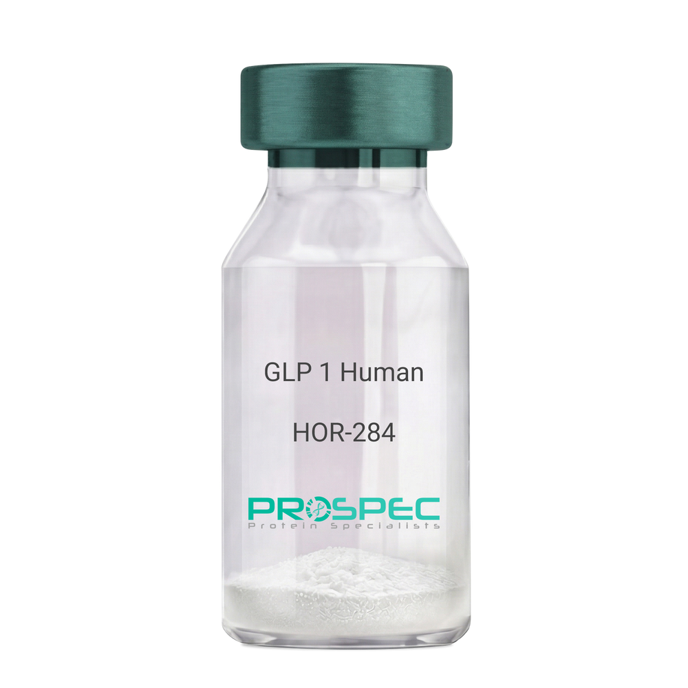
    GLP 1 Human

    Description:

    Human Glucagon Like Peptide-1

    GLP1, Glucagon Like Peptide-1, Incretin hormone.

    Product # :

    HOR-284

    Price :

    Quantity :

    Shipping Method :

    Room Temp Icon

    Shipped at Room temp

    Add To Cart

    More Info

    • description
    • formulation
    • purity
    • More Info

    Description

    Glucagon Like Peptide-1 is a single, non-glycosylated, polypeptide chain containing 30 amino acids and having a molecular mass of 3297.7 Dalton.The GLP-1 is purified by proprietary chromatographic techniques.

    Formulation

    The GLP-1 peptide was lyophilized with no additives.

    Purity

    Greater than 96.0% as determined by RP-HPLC.

    More Info

    • Introduction

      Glucagon-like peptide-1 (GLP-1) is derived from the transcription product of the proglucagon gene. The major source of GLP-1 in the body is the intestinal L cell that secretes GLP-1 as a guthormone. The biologically active forms of GLP-1 are: GLP-1-(7-37) and GLP-1-(7-36)NH2.
      GLP-1 secretion by L cells is dependent on the presence of nutrients in the lumen of the small intestine. The secretagogues (agents that causes or stimulates secretion) of this hormone include major nutrients like carbohydrate, proteinand lipid. Once in the circulation, GLP-1 has a half life of less than 2 minutes, due to rapid degradation by the enzyme dipeptidyl peptidase-4.
      GLP-1 possesses several physiological properties that make it a subject of intensive investigation as a potential treatment of diabetes mellitus. The known physiological functions of GLP-1 include: Increases insulin secretion from the pancreas in a glucose-dependent manner, decreases glucagon secretion from the pancreas, increases beta cells mass and insulin gene expression, inhibits acid secretion and gastric emptying in the stomach, decreases food intake by increasing satiety.

    • Synonyms

      GLP1, Glucagon Like Peptide-1, Incretin hormone.

    • Physical Appearance

      Sterile Filtered White lyophilized (freeze-dried) powder.

    • Stability

      Lyophilized Glucagon Like Peptide-1 although stable at room temperature for 3 weeks, should be stored desiccated below -18°C. Upon reconstitution GLP-1 should be stored at 4°C between 2-7 days and for future use below -18°C. For long term storage it is recommended to add a carrier protein (0.1% HSA or BSA).Please prevent freeze-thaw cycles.

    • Solubility

      It is recommended to reconstitute the lyophilized Glucagon Like Peptide-1 in sterile H2O at 0.5mg/ml, which can then be further diluted to other aqueous solutions.

    • Amino Acid Sequence

      H-His-Ala-Glu-Gly-Thr-Phe-Thr-Ser-Asp-Val-Ser-Ser-Tyr-Leu-Glu-Gly-Gln-Ala-Ala-Lys-Glu-Phe-Ile-Ala-Trp-Leu-Val-Lys-Gly-Arg-NH2.

    ProSpec's products are furnished for LABORATORY RESEARCH USE ONLY. The product may not be used as drugs, agricultural or pesticidal products, food additives or household chemicals.

    Glp 1 Human
  • View Data Sheet

    Name :

    Activin-A Human Active

    Description:

    Activin-A Human Recombinant, Active

    Inhba, Inhibin beta A, FSH releasing protein.

    Product # :

    CYT-145

    Price :

    Quantity :

    Shipping Method :

    Room Temp Icon

    Shipped at Room temp

    Add To Cart

    More Info

    • description
    • source
    • formulation
    • purity
    • biological activity
    • More Info

    Description

    Active form Activin-A Human Recombinant produced in e.coli is a homodimeric, non-glycosylated, polypeptide chain containing 2 x 117 amino acids and having a molecular weight of 26.2kDa.The Active form Activin-A is purified by standard chromatographic techniques.

    Source

    E.Coli.

    Formulation

    Human Activin-A was lyophilized from a concentrated 1mg/ml protein solution containing 0.1% TFA.

    Purity

    Greater than 95% as obsereved by SDS-PAGE.

    Biological Activity

    Biological activity is assessed by the ability to induce cytotoxicity of MPC-11 cells and was found to be 8.95ng/ml corresponding to a specific activity of 1.1 x 10⁵ units/mg.

    More Info

    • Introduction

      Activins are homodimers or heterodimers of the different β subunit isoforms, part of the TGFβ family. Mature Activin A has two 116 amino acids residues βA subunits (βA-βA). Activin displays an extensive variety of biological activities, including mesoderm induction, neural cell differentiation, bone remodelling, haematopoiesis, and reproductive physiology. Activins takes part in the production and regulation of hormones such as FSH, LH, GnRH and ACTH. Cells that are identified to express Activin A include fibroblasts, endothelial cells, hepatocytes, vascular smooth muscle cells, macrophages, keratinocytes, osteoclasts, bone marrow monocytes, prostatic epithelium, neurons, chondrocytes, osteoblasts, Leydig cells, Sertoli cells, and ovarian granulosa cells.

    • Synonyms

      Inhba, Inhibin beta A, FSH releasing protein.

    • Physical Appearance

      Lyophilized freeze dried powder.

    • Stability

      Lyophilized Activin-A although stable at room temperature for 3 weeks, should be stored desiccated below -18°C. Upon reconstitution Activin-A should be stored at 4°C between 2-7 days and for future use below -18°C.For long term storage it is recommended to add a carrier protein (0.1% HSA or BSA).Please prevent freeze-thaw cycles.

    • Solubility

      Human INHBA protein should be reconstituted in distilled pyrogen free water to a concentration of 100ug /ml which can then be further diluted to other aqueous solutions.

    • Amino Acid Sequence

      MGLECDGKVN ICCKKQFFVS FKDIGWNDWI IAPSGYHANY CEGECPSHIA GTSGSSLSFH STVINHYRMR GHSPFANLKS CCVPTKLRPM SMLYYDDGQN IIKKDIQNMI VEECGCS.

    • Background

      Title: Research on Activin A Human Recombinant: Molecular Characteristics, Signaling Pathways, Physiological Functions, and Therapeutic Potential

      Introduction:

      Activin A, a member of the transforming growth factor-beta (TGF-β) superfamily, is a multifunctional cytokine that plays a significant role in various biological processes in the human body. Its involvement in diverse physiological and pathological functions has garnered considerable attention in scientific research. This paper aims to provide an overview of Activin A, encompassing its molecular characteristics, signaling pathways, physiological functions, and therapeutic potential.

      Activin A is encoded by the INHBA gene and is produced as a precursor protein that undergoes post-translational modifications to generate the mature form. The mature Activin A protein consists of two β-subunits held together by disulfide bonds. These structural features contribute to its functional properties and interactions with specific receptors.

      Upon binding to its cell surface receptors, Activin A triggers intracellular signaling cascades, leading to various cellular responses. Canonical SMAD-dependent pathway as well as non-SMAD pathways, such as MAPK/ERK, PI3K/Akt, and JNK signaling, are activated by Activin A. The intricate network of signaling pathways enables Activin A to regulate diverse biological processes, including cell proliferation, differentiation, apoptosis, and tissue homeostasis.

      Activin A exerts its physiological functions in a tissue-specific manner. It plays a critical role in embryonic development, particularly in organogenesis and patterning. Additionally, Activin A is involved in reproductive biology, where it participates in folliculogenesis, spermatogenesis, and hormonal regulation. It also contributes to neural development, immune system modulation, and skeletal homeostasis.

      The multifunctional properties of Activin A have positioned it as a potential therapeutic target for various diseases. Its involvement in cancer, neurodegenerative disorders, fibrosis, and reproductive disorders has prompted extensive research to explore its therapeutic potential. Understanding the molecular mechanisms underlying Activin A's actions provides valuable insights for developing innovative therapeutic strategies.

      In conclusion, Activin A is a versatile cytokine with diverse roles in human biology. This research aims to deepen our understanding of its molecular characteristics, signaling pathways, physiological functions, and therapeutic potential. By elucidating the complexities of Activin A, we strive to pave the way for novel therapeutic interventions in various human diseases.

    ProSpec's products are furnished for LABORATORY RESEARCH USE ONLY. The product may not be used as drugs, agricultural or pesticidal products, food additives or household chemicals.

    Inhba Human
  • View Data Sheet

    Name :

    PRL R Rabbit

    Description:

    Prolactin Rabbit Soluble Receptor Recombinant

    PRL-R.

    Product # :

    CYT-268

    Price :

    Quantity :

    Shipping Method :

    Room Temp Icon

    Shipped at Room temp

    Add To Cart

    More Info

    • description
    • source
    • formulation
    • purity
    • biological activity
    • More Info

    Description

    Prolactin Receptor Rabbit Extra Celleular Domain Recombinant ?produced in E.Coli is a non-glycosylated, Polypeptide chain containing 207 amino acids and having a molecular mass of 23972 Dalton. The Prolactin Receptor is purified by proprietary chromatographic techniques.

    Source

    Escherichia Coli.

    Formulation

    The protein was lyophilized from a concentrated (1mg/ml) solution with 0.0045mM NaHCO3.

    Purity

    Greater than 97.0% as determined by:
    (a) Analysis by SEC-HPLC.
    (b) Analysis by SDS-PAGE.

    Biological Activity

    Activity is determined by the dose-dependant inhibition of Prolactin-stimuled proliferation of Nb2 cells and by high affinity binding of oPLR and other lactogenic hormones. Refs:
    1) Bignon et al. (1994) JBC 269; 3318-24
    2) Gertler et al. (1996) JBC 271; 24482-91.

    More Info

    • Introduction

      Prolactin is a pituitary hormone involved in the stimulation of milk production, salt and water regulation, growth, development and reproduction. The initial step in its action is the binding to a specific membrane receptor (prolactin receptor) which belongs to the superfamily of class 1 cytokine receptors. The function of the prolactin receptor is mediated, at least in part, by two families of signaling molecules: Janus kinases and signal transducers and activators of transcription. Prolactin (PRL) is a hormone involved in a variety of important functions including ion transport and osmoregulation, stimulation of milk, protein synthesis as well as the regulation of numerous reproductive functions. PRL exerts its influence on different cell types through a signal transduction pathway which begins with the binding of the hormone to a transmembrane PRL receptor. Immunoreactive PRL receptor, a member of the cytokine receptor family, varies in size (short and long forms) with tissue source and species, from ~40 kDa to 100 kDa. The PRL receptor consists of at least three separate domains: an extracellular region with 5 cysteines which contains the prolactin binding site, a single transmembrane domain and a cytoplasmic region, the length of which appears to influence ligand binding and regulate cellular function.

    • Synonyms

      PRL-R.

    • Physical Appearance

      Sterile Filtered White lyophilized (freeze-dried) powder.

    • Stability

      Lyophilized PRL-R although stable at room temperature for 3 weeks, should be stored desiccated below -18°C. Upon reconstitution Prolactin Receptor should be stored at 4°C between 2-7 days and for future use below -18°C.For long term storage it is recommended to add a carrier protein (0.1% HSA or BSA).Please prevent freeze-thaw cycles.

    • Solubility

      It is recommended to reconstitute the lyophilized PRL-R in sterile 18MΩ-cm H2O not less than 100µg/ml, which can then be further diluted to other aqueous solutions.

    • Amino Acid Sequence

      The sequence of the first five N-terminal amino acids was determined and was found to be Gly-Lys-Pro-Phe-Ile.

    • Protein content

      UV spectroscopy at 280 nm using the absorbency value of 2.63 as the extinction coefficient for a 0.1% (1mg/ml) solution. This value is calculated by the PC GENEcomputer analysis program of protein sequences (IntelliGenetics).

    ProSpec's products are furnished for LABORATORY RESEARCH USE ONLY. The product may not be used as drugs, agricultural or pesticidal products, food additives or household chemicals.

    Prlr Rabbit
  • View Data Sheet

    Name :

    A2M Human

    Description:

    Macroglobulin Alpha-2 Human

    Alpha-2-macroglobulin, Alpha-2-M, A2M, CPAMD5, FWP007, S863-7, alpha 2M, DKFZp779B086.

    Product # :

    PRO-551

    Price :

    Quantity :

    Shipping Method :

    Room Temp Icon

    Shipped at Room temp

    Add To Cart

    More Info

    • description
    • source
    • formulation
    • purity
    • More Info

    Description

    Human Alpha-2 Macroglobulin is a tetrameric glycoprotein, produced in Human plasma and having a molecular mass of 725 kDa.

    Source

    Human Plasma.

    Formulation

    Lyophilized from a concentrated solution containing 5mM potassium phosphate buffer, pH 6.5 and 1:1 ratio (w/w) of Glycine.

    Purity

    Greater than 95.0%.

    More Info

    • Introduction

      Alpha-2 Macroglobulin is a serine proteases inhibitor, which inhibits coagulation by inactivating thrombin and Kallikrein, it inhibits fibrinolysis by inactivating plasmin and involved in transport. Alpha-2-Macroglobulin is a large plasma protein, which is produced by the liver, it’s composed of 4 identical subunits bound together by -S-S- bonds. A2M is able to inactivate many kinds of proteinases (including serine-, cysteine-, aspartic- and metalloproteinases). A2M has a 35 amino acid "bait" region in its structure. Proteinases bind and cleave the “bait” region become bound to A2M. Macrophage receptors recognize the proteinase-A2M complex and clear it from the system. A2M binds to and removes MMP-2 and MMP-9 (active forms of the gelatinase) from the circulation using scavenger receptors on the phagocytes. The levels of Alpha-2-macroglobulin are increased in nephrotic syndrome which is a condition where the kidneys start to leak out some of the smaller blood proteins. Due to its large size, A2-macroglobulin is retained in the bloodstream. Increased production of all proteins causes A2-macroglobulin concentration to increase. Chronic renal failure might lead to amyloid by alpha-2-macroglobulin. A2M is raised in cirrhosis, pregnancy and diabetes.

    • Synonyms

      Alpha-2-macroglobulin, Alpha-2-M, A2M, CPAMD5, FWP007, S863-7, alpha 2M, DKFZp779B086.

    • Physical Appearance

      Sterile Filtered White lyophilized (freeze-dried) powder.

    • Stability

      Lyophilized A2M protein although stable at room temperature for 3 weeks, should be stored desiccated below -18°C. Upon reconstitution and filter sterilization A2M can be stored at 4°C for up to 4 weeks. For long term storage and more diluted solutions it is recommended to add a carrier protein (0.1% HSA or BSA). Please prevent freeze-thaw cycles.

    • Solubility

      It is recommended to reconstitute the lyophilized A2M in sterile 18MΩ-cm H2O.

    • Human Virus Test

      Serum from each donor has been tested and found negative for antibody to HIV-1, HIV-2, HCV and HBV.

    ProSpec's products are furnished for LABORATORY RESEARCH USE ONLY. The product may not be used as drugs, agricultural or pesticidal products, food additives or household chemicals.

    Macroglobulin Alpha 2 Human
  • View Data Sheet

    Name :

    SPA Long

    Description:

    Staphylococcal Protein-A Long Form Recombinant

    Immunoglobulin G-binding protein A, IgG-binding protein A, Staphylococcal protein A, SPA.

    Product # :

    PRO-1926

    Price :

    Quantity :

    Shipping Method :

    Room Temp Icon

    Shipped at Room temp

    Add To Cart

    More Info

    • description
    • source
    • formulation
    • purity
    • More Info

    Description

    SPA Recombinant Long Form produced in E.Coli is a single non-glycosylated polypeptide chain. SPA Long Form is comprised of 5 IgG-binding domains E-D-A-B-C aligned in series containing 422 amino acids and having a molecular mass of 46.6kDa.

    Source

    Escherichia Coli.

    Formulation

    SPA protein was lyophilized with no additives.

    Purity

    Greater than 97.0% as determined by:
    (a) Analysis by HPLC.
    (b) Analysis by SDS-PAGE.

    More Info

    • Introduction

      Protein A is a cell wall component produced by several strains of Staphylococcus aureus. The recombinant Protein A is genetically engineered protein and holds 5 IgG-binding regions of protein A. Recombinant Protein A functions basically the same as native Protein A and is perfect for purification of polyclonal or monoclonal IgG antibodies. Protein A binds to human IgG1, IgG2 and IgG4, mouse IgG2a, IgG2b and IgG3 and rat IgG2c. Protein A also binds to total IgG from rabbit, pig, dog, cat, and guinea pig.

    • Synonyms

      Immunoglobulin G-binding protein A, IgG-binding protein A, Staphylococcal protein A, SPA.

    • Physical Appearance

      Sterile Filtered White lyophilized (freeze-dried) powder.

    • Stability

      Lyophilized SPA although stable at room temperature for 3 weeks, should be stored desiccated below -18°C. Upon reconstitution SPA should be stored at 4°C between 2-7 days and for future use below -18°C. For long term storage it is recommended to add a carrier protein (0.1% HSA or BSA). Please prevent freeze-thaw cycles.

    • Solubility

      It is recommended to reconstitute the lyophilized SPA in sterile 18MΩ-cm H2O not less than 0.1mg/ml, which can then be further diluted to other aqueous solutions.

    • Amino Acid Sequence

      AQHDEAQQNA FYQVLNMPNL NADQRNGFIQ SLKDDPSQSA NVLGEAQKLN DSQAPKADAQ QNNFNKDQQS AFYEILNMPN LNEAQRNGFI QSLKDDPSQS TNVLGEAKKL NESQAPKADN NFNKEQQNAF YEILNMPNLN EEQRNGFIQS LKDDPSQSAN LLSEAKKLNE SQAPKADNKF NKEQQNAFYE ILHLPNLNEE QRNGFIQSLK DDPSQSANLL AEAKKLNDAQ APKADNKFNK EQQNAFYEIL HLPNLTEEQR NGFIQSLKDD PSVSKEILAE AKKLNDAQAP KEEDNKKPGK EDGNKPGKED GNKPGKEDNK KPGKEDGNKP GKEDNNKPGK EDGNKPGKED NNKPGKEDGN KPGKEDGNKP GKEDGNGVHV VKPGDTVNDI AKANGTTADK IAADNKLADK NMIKPGQELV VD.

    ProSpec's products are furnished for LABORATORY RESEARCH USE ONLY. The product may not be used as drugs, agricultural or pesticidal products, food additives or household chemicals.

    Spa Long
  • View Data Sheet

    Name :

    S.Typhi OMP

    Description:

    Salmonella Typhi Outer Membrane Protein Recombinant

    Product # :

    STY-002

    Price :

    Quantity :

    Shipping Method :

    Ice Icon

    Shipped with Ice Packs

    Add To Cart

    More Info

    • description
    • source
    • formulation
    • purity
    • More Info

    Description

    Recombinant Salmonella Typhi Outer Membrane Protein produced in E.coli contains 315 amino acids, and fused to a 6 His Tag at C-terminus, migrating as a 33kDa band on SDS-PAGE.S. typhi outer membrane protein is a central pathogen in S. typhi infection, and is directly exposed to the outside to interact with the human immune system.

    Source

    Escherichia Coli.

    Formulation

    Sterile Filtered solution containing 10mM Tris-HCl, 1mM EDTA and 50mM arginine.

    Purity

    Protein is >95% pure as determined by 12% PAGE (coomassie staining).

    More Info

    • Introduction

      Salmonella Typhi is a pathogen causing typhoid fever, affecting over 17 million people with approximately 600,000 deaths annually worldwide. If untreated, typhoid fever cases result in mortality rates ranging from 12-30%.

    • Physical Appearance

      Sterile Filtered solution.

    • Stability

      S.Typhi OMP although stable at 4°C for 1 week, should be stored below -18°C. Please prevent freeze thaw cycles.

    ProSpec's products are furnished for LABORATORY RESEARCH USE ONLY. The product may not be used as drugs, agricultural or pesticidal products, food additives or household chemicals.

    Styphi Omp
  • View Data Sheet

    Name :

    Ebola Sudan Protein

    Description:

    Ebola Sudan Recombinant Protein

    Product # :

    EVD-079

    Price :

    Quantity :

    Shipping Method :

    Ice Icon

    Shipped with Ice Packs

    Add To Cart

    More Info

    • description
    • source
    • formulation
    • purity
    • More Info

    Description

    Nucleoprotein (NP) of Ebola virus has strong antigenicity in immune reactions. C-terminal of EBO-S nucleoprotein was expressed and purified from E. coli, it migrated at 15kDa on SDS-PAGE and pI is 4.52. Ebola Sudan was purified by a proprietary chromatographic technique.

    Source

    Escherichia Coli.

    Formulation

    Ebola Sudan protein solution is supplied in Phosphate buffer and 0.02% sodium azide.

    Purity

    >95% pure as determined by 12% SDS-PAGE (Coomassie blue stain).

    More Info

    • Introduction

      Ebolavirus (EVD) belongs to the Filoviridae family of proteins which is comprised of a single-strand, non-infectious RNA genome. EVD genome is about 19,000 base pairs long and covers 7 genes in the order 3'-UTR-NP-VP35-VP40-GP-VP30-VP24-L-5'-UTR. There are 4 different ebolaviruses such as: Zaire (EBO-Z), Sudan (EBO-S), Cote d’Ivoire (EBO-CI) and Reston (EBO-R) that differ in amino acid sequence and location of where the gene overlaps. Similar to filoviruses and ebolavirions, EVD is a filamentous element that could appear in 3 forms: shepherd's crook, U shape or a 6 shape. Ebolaviruses may be coiled, toroid, or branched. Most ebolavirions are 80nm wideand 14,000nm long.

    • Stability

      Store at 4°C if entire vial will be used within 2-4 weeks. Store, frozen at -20°C for longer periods of time. For long term storage it is recommended to add a carrier protein (0.1% HSA or BSA).Avoid multiple freeze-thaw cycles.

    • Applications

      Immunoassay.

    ProSpec's products are furnished for LABORATORY RESEARCH USE ONLY. The product may not be used as drugs, agricultural or pesticidal products, food additives or household chemicals.

    Ebola Sudan
  • View Data Sheet

    Name :

    Leptin-B Tilapia

    Description:

    Leptin-B Tilapia Recombinant

    Product # :

    CYT-1110

    Price :

    Quantity :

    Shipping Method :

    Room Temp Icon

    Shipped at Room temp

    Add To Cart

    More Info

    • description
    • source
    • formulation
    • purity
    • biological activity
    • More Info

    Description

    Leptin-B Tilapia Recombinant produced in E.Coli is a single, non-glycosylated polypeptide chain containing 152 amino acids and having a molecular mass of 15,243 Dalton. The Leptin-B Tilapia is purified by proprietary chromatographic techniques.

    Source

    Escherichia Coli.

    Formulation

    Lyophilized from a concentrated (1mg/ml) solution containing NaHCO3 at 1:2 salt: protein ratio.

    Purity

    Greater than 95.0% as determined by:
    (a) Gel filtration analysis.
    (b) Analysis by SDS-PAGE.

    Biological Activity

    Tilapia leptins were found to be biologically active in promoting proliferation of BAF/3 cells stably transfected with the long form of human leptin receptor, but their activity was lower than that of mammalian leptin. Furthermore, the Tilapia leptins were biologically active in promoting STAT‐LUC activation in COS7 cells transfected with Tilapia leptin receptor but not in cells transfected with human leptin receptor. Tilapia Leptin A was more active than Tilapia Leptin B.

    More Info

    • Introduction

      Leptin is a protein hormone. It is mainly produced in adipose cells that regulate energy homeostasis by restraining hunger. Leptin ties to nuclear receptors in the hypothalamus (arcuate nucleus). Similar to insulin resistance in type II diabetes, in obesity there is a decrease in the sensitivity towards leptin, ending in a failure to identify satiety, even in high levels of energy stores or leptin itself. Full-length cDNA encoding 2 leptin sequences (tLepA and tLepB) and 1 leptin receptor sequence (tLepR) exists in tilapia (Oreochromis niloticus). The full-length cDNA of tLepR is 3423 bp, encoding a protein of 1140 amino acid which contained all functionally important domains conserved among vertebrate leptin receptors. The cDNAs of tLepA and tLepB are 486 bp and 459 bp in length, encoding proteins of 161 aa and 152 aa, respectively. The three-dimensional structures of tLepA and tLepB demonstrates strong conservation of tertiary structure with that of human leptin comprised of 4 helixes.

    • Physical Appearance

      Sterile Filtered White lyophilized (freeze-dried) powder.

    • Stability

      Lyophilized Leptin-B Tilapia although stable at room temperature for 3 weeks, should be stored desiccated below -18°C. Upon reconstitution Leptin-B Tilapia should be stored at 4°C between 2-7 days and for future use below -18°C. For long term storage it is recommended to add a carrier protein (0.1% HSA or BSA). Please prevent freeze-thaw cycles.

    • Solubility

      It is recommended to reconstitute the lyophilized Leptin-B Tilapia in sterile water or 0.4% NaHCO3 adjusted to pH 8-9, not less than 100µg/ml, which can then be further diluted to other aqueous solutions.

    • Amino Acid Sequence

      The first six N-terminal amino acids of recombinant Tilapia leptin B are Ala-Leu-Leu-Thr-Lys-Gly.

    • Protein content

      Protein quantitation was carried out by UV spectroscopy at 280 nm using the absorbency value of 0.19 for 1 mg/ml Leptin-B Tilapia as the extinction coefficient for a 0.1% (1mg/ml) solution at pH 8.0. This value is calculated by the DNAman computer analysis program of protein sequences.

    ProSpec's products are furnished for LABORATORY RESEARCH USE ONLY. The product may not be used as drugs, agricultural or pesticidal products, food additives or household chemicals.

    Leptin B
  • View Data Sheet

    Name :

    AMH (452-560) Human

    Description:

    Anti-Mullerian Hormone (452-560) Human Recombinant

    Anti-Muellerian hormone, AMH, Muellerian-inhibiting substance, MIS, MIF. 

    Product # :

    PRO-2843

    Price :

    Quantity :

    Shipping Method :

    Room Temp Icon

    Shipped at Room temp

    Add To Cart

    More Info

    • description
    • source
    • formulation
    • purity
    • biological activity
    • More Info

    Description

    Anti-Mullerian Hormone Human Recombinant produced in CHO is a Homodimer, glycosylated, polypeptide chain containing 109 amino acids (Ser452-Arg560) and having a total molecular mass of 23.4Da.

    Anti-Mullerian Hormone is purified by proprietary chromatographic techniques.

    Source

    CHO Cells.

    Formulation

    AMH Lyophilized from a 0.2µm filtered solution in 35% (v/v) Acetonitrile and 0.1% (v/v) TFA.

    Purity

    Greater than 97% as determined by SDS-PAGE and SEC-HPLC analyses.

    Biological Activity

    Immobilized Recombinant Human AMH at 3 µg/mL (100 µL/well) will bind Recombinant Human MIS RII Fc Chimera with a linear range of 2.0-100 ng/mL. The activity was measured by its binding ability in a functional ELISA.

    More Info

    • Introduction

      Anti-Mullerian Hormone also known as AMH is a member of the TGF-beta family. AMH is a glycoprotein which is produced by the Sertoli cells of the testis, causes regression of the Muellerian duct. AMH inhibits the growth of tumors derived from tissues of Muellerian duct origin. Moreover, AMH participates in Leydig cell differentiation and function in addition to follicular development in adult females.

    • Synonyms

      Anti-Muellerian hormone, AMH, Muellerian-inhibiting substance, MIS, MIF.

    • Physical Appearance

      Filtered White lyophilized (freeze-dried) powder.

    • Stability

      Store lyophilized protein at -20°C. Aliquot the product after reconstitution to avoid repeated freezing/thawing cycles. Reconstituted protein can be stored at 4°C for a limited period of time; it does not show any change after two weeks at 4°C.

    • Solubility

      It is recommended to reconstitute in sterile 4mM HCl to a concentration of 100µg/ml, which can then be further diluted to other aqueous solutions.

    • Amino Acid Sequence

      SAGATAADGP CALRELSVDL RAERSVLIPE TYQANNCQGV CGWPQSDRNP RYGNHVVLLL KMQVRGAALA RPPCCVPTAY AGKLLISLSE ERISAHHVPN MVATECGCR.

    ProSpec's products are furnished for LABORATORY RESEARCH USE ONLY. The product may not be used as drugs, agricultural or pesticidal products, food additives or household chemicals.

    Anti Mullerian Hormone Cho
  • View Data Sheet

    Name :

    CFD Human

    Description:

    Complement Factor D Human

    Complement factor D, Adipsin, C3 convertase activator, Properdin factor D, CFD, DF, PFD.

    Product # :

    PRO-2699

    Price :

    Quantity :

    Shipping Method :

    Ice Icon

    Shipped with Ice Packs

    Add To Cart

    More Info

    • description
    • source
    • formulation
    • purity
    • More Info

    Description

    Human Complement Factor D produced in Human plasma is glycosylated polypeptide chain having a total molecular mass of 24kDa.

    Source

    Human Plasma.

    Formulation

    CFD protein solution contains 10mM Sodium phosphate and 145mM NaCl, pH 7.3.

    Purity

    Greater than 95.0% as determined by SDS-PAGE.

    More Info

    • Introduction

      CFD is an important component of the alternative pathway of complement activation. CFD cleaves and activates factor B when it binds C3b or a C3b-like protein such as C3 or CVF. CFD is a serine protease that exists as a mature protease, but it exhibits a highly restricted specificity and it appears to be substrate activated. CFD cleaves factor B bound to C3b leading to the release of the Ba fragment and leaving the Bb fragment bound to C3b. The C3b,Bb complex is called a C3 or C5 convertase because it converts these proteins to their active forms by cleaving off the small peptides C3a and C5a, respectively.

    • Synonyms

      Complement factor D, Adipsin, C3 convertase activator, Properdin factor D, CFD, DF, PFD.

    • Physical Appearance

      Sterile filtered solution.

    • Stability

      CFD Human is stable at 4°C if entire vial will be used within 2-4 weeks.Store, frozen below -20°C for longer periods of time.For long term storage it is recommended to add a carrier protein (0.1% HSA or BSA).Avoid multiple freeze-thaw cycles.

    • Human Virus Test

      Plasma from each donor has been tested and found negative for antibody to HIV-1, HIV-2, HCV and HBSAG.

    ProSpec's products are furnished for LABORATORY RESEARCH USE ONLY. The product may not be used as drugs, agricultural or pesticidal products, food additives or household chemicals.

    Cfd Human
  • View Data Sheet

    Name :

    CST11 Human

    Description:

    Cystatin 11 Human Recombinant

    CST8L, CTES2, dJ322G13.6, SC13.

    Product # :

    PRO-1475

    Price :

    Quantity :

    Shipping Method :

    Ice Icon

    Shipped with Ice Packs

    Add To Cart

    More Info

    • description
    • source
    • formulation
    • purity
    • More Info

    Description

    CST11 Human Recombinant produced in E.Coli is a single, non-glycosylated polypeptide chain containing 101 amino acids (26-103 a.a.) and having a molecular mass of 11.8kDa.CST11 is fused to a 23 amino acid His-tag at N-terminus & purified by proprietary chromatographic techniques.

    Source

    E.coli.

    Formulation

    CST11 protein solution (1mg/ml) contains 20mM Tris-HCl buffer, (pH 8.0), 0.4M urea and 10% glycerol.

    Purity

    Greater than 90% as determined by SDS-PAGE.

    More Info

    • Introduction

      Cystatin 11 (CST11) is a member of the Cystatin family. The Cystatin superfamily is comprised of proteins with multiple cystatin-like sequences. Several family members are active cysteine protease inhibitors, other family members have lost or perhaps never developed this inhibitory activity. The Cystatin superfamily consists of three inhibitory families, including the type 1 cystatins (stefins), type 2 cystatins and the kininogens. The type 2 cystatin proteins are a class of cysteine proteinase inhibitors found in a variety of human fluids and secretions. The majority of the type 2 cystatin genes and pseudogenes are contained in the cystatin locus on chromosome 20. CST11 is located in the cystatin locus and encodes an epididymal-specific protein whose specific function has not been verified yet.

    • Synonyms

      CST8L, CTES2, dJ322G13.6, SC13.

    • Physical Appearance

      Sterile Filtered colorless solution.

    • Stability

      Store at 4°C if entire vial will be used within 2-4 weeks. Store, frozen at -20°C for longer periods of time. For long term storage it is recommended to add a carrier protein (0.1% HSA or BSA).Avoid multiple freeze-thaw cycles.

    • Amino Acid Sequence

      MGSSHHHHHH SSGLVPRGSH MGSARKKTFL SVHEVMAVEN YAKDSLQWIT DQYNKESDDK YHFRIFRVLK VQRQQVNCFF SVFAVPWFEQ YKILNKSCSS D.

    ProSpec's products are furnished for LABORATORY RESEARCH USE ONLY. The product may not be used as drugs, agricultural or pesticidal products, food additives or household chemicals.

    Cst11 Human
  • View Data Sheet

    Name :

    SAP18 Human

    Description:

    Sin3A-Associated Protein 18kDa Human Recombinant

    Histone deacetylase complex subunit SAP18, Sin3-associated polypeptide 18 kDa, Sin3-associated polypeptide p18, Cell growth-inhibiting protein 38, 2HOR0202, SAP18, GIG38.

    Product # :

    PRO-707

    Price :

    Quantity :

    Shipping Method :

    Ice Icon

    Shipped with Ice Packs

    Add To Cart

    More Info

    • description
    • source
    • formulation
    • purity
    • More Info

    Description

    SAP18 Human Recombinant fused with 20 amino acid His tag at N-terminus produced in E.Coli is a single, non-glycosylated, polypeptide chain containing 173 amino acids (20- 172 a.a.) and having a molecular mass of 19.7 kDa.The SAP18 is purified by proprietary chromatographic techniques.

    Source

    Escherichia Coli.

    Formulation

    The SAP18 solution contains 20mM Tris-HCl buffer (pH8.0), 0.1M NaCl, 30% glycerol, 1mM DTT.

    Purity

    Greater than 90.0% as determined by SDS-PAGE.

    More Info

    • Introduction

      SAP18 is a component of the histone deacetylase complex which has a significant role in the regulation of eukaryotic gene expression. SAP18 directly interacts with SIN3 and enhances SIN3-mediated transcriptional repression once bound to the promoter. SAP18 plays an important part in gene-specific recruitment of the HDAC complex by a number of transcription factors including Gli, GAGA, and Bicoid. Histone acetylation is significant in the regulation of eukaryotic gene expression. Histone acetylation and deacetylation is catalyzed by multisubunit complexes. SAP18 is a component of the histone deacetylase complex, which includes SIN3, SAP30, HDAC1, HDAC2, RbAp46, RbAp48, and other polypeptides. SAP18 is a component of a splicing-dependent multiprotein EJC (exon junction complex) placed at splice junction on mRNAs.

    • Synonyms

      Histone deacetylase complex subunit SAP18, Sin3-associated polypeptide 18 kDa, Sin3-associated polypeptide p18, Cell growth-inhibiting protein 38, 2HOR0202, SAP18, GIG38.

    • Physical Appearance

      Sterile Filtered colorless solution.

    • Stability

      Store at 4°C if entire vial will be used within 2-4 weeks. Store, frozen at -20°C for longer periods of time. For long term storage it is recommended to add a carrier protein (0.1% HSA or BSA).Avoid multiple freeze-thaw cycles.

    • Amino Acid Sequence

      MGSSHHHHHH SSGLVPRGSH MAVESRVTQE EIKKEPEKPI DREKTCPLLL RVFTTNNGRH HRMDEFSRGN VPSSELQIYT WMDATLKELT SLVKEVYPEA RKKGTHFNFA IVFTDVKRPG YRVKEIGSTM SGRKGTDDSM TLQSQKFQIG DYLDIAITPP NRAPPPSGRM RPY.

    ProSpec's products are furnished for LABORATORY RESEARCH USE ONLY. The product may not be used as drugs, agricultural or pesticidal products, food additives or household chemicals.

    Sap18 Human
  • View Data Sheet

    Name :

    AHCY Human

    Description:

    Adenosylhomocysteinase Human Recombinant

    EC 3.3.1.1, SAHH, AdoHcyase, S-adenosyl-L-homocysteine hydrolase, Adenosylhomocysteinase.

    Product # :

    ENZ-532

    Price :

    Quantity :

    Shipping Method :

    Ice Icon

    Shipped with Ice Packs

    Add To Cart

    More Info

    • description
    • source
    • formulation
    • purity
    • More Info

    Description

    AHCY Recombinant Human produced in E.Coli is a single, non-glycosylated polypeptide chain containing 452 amino acids (1-432 a.a.) and having a molecular mass of 49.8 kDa. The AHCY is fused to a 20 amino acids His-Tag at N-terminus and purified by proprietary chromatographic techniques.

    Source

    Escherichia Coli.

    Formulation

    AHCY Human solution containing 20mM Tris pH-8, & 10% glycerol.

    Purity

    Greater than 95.0% as determined by SDS-PAGE.

    More Info

    • Introduction

      AHCY is an enzyme that catalyzes the reversible hydrolysis of S-adenosylhomocysteine (AdoHcy) to adenosine (Ado) and L-homocysteine (Hcy). AHCY controls the intracellular S-adenosylhomocysteine (SAH) concentration that is crucial for transmethylation reactions. AHCY deficiency causes hypermethioninemia.

    • Synonyms

      EC 3.3.1.1, SAHH, AdoHcyase, S-adenosyl-L-homocysteine hydrolase, Adenosylhomocysteinase.

    • Physical Appearance

      Sterile filtered colorless solution.

    • Stability

      Store at 4°C if entire vial will be used within 2-4 weeks. Store, frozen at -20°C for longer periods of time. For long term storage it is recommended to add a carrier protein (0.1% HSA or BSA).Avoid multiple freeze-thaw cycles.

    • Amino Acid Sequence

      MGSSHHHHHH SSGLVPRGSH MSDKLPYKVA DIGLAAWGRK ALDIAENEMP GLMRMRERYS ASKPLKGARI AGCLHMTVET AVLIETLVTL GAEVQWSSCN IFSTQDHAAA AIAKAGIPVY AWKGETDEEY LWCIEQTLYF KDGPLNMILD DGGDLTNLIH TKYPQLLPGI RGISEETTTG VHNLYKMMAN GILKVPAINV NDSVTKSKFD NLYGCRESLI DGIKRATDVM IAGKVAVVAG YGDVGKGCAQ ALRGFGARVI ITEIDPINAL QAAMEGYEVT TMDEACQEGN IFVTTTGCID IILGRHFEQM KDDAIVCNIG HFDVEIDVKW LNENAVEKVN IKPQVDRYRL KNGRRIILLA EGRLVNLGCA MGHPSFVMSN SFTNQVMAQI ELWTHPDKYP VGVHFLPKKL DEAVAEAHLG KLNVKLTKLT EKQAQYLGMS CDGPFKPDHY RY.

    ProSpec's products are furnished for LABORATORY RESEARCH USE ONLY. The product may not be used as drugs, agricultural or pesticidal products, food additives or household chemicals.

    Ahcy Human
  • View Data Sheet

    Name :

    MORF4L2 Human

    Description:

    Mortality Factor 4 Like 2 Human Recombinant

    Mortality factor 4-like protein 2, MORF-related gene X protein, Protein MSL3-2, Transcription factor-like protein MRGX, MORF4L2, KIAA0026, MRGX, MORFL2.

    Product # :

    PRO-475

    Price :

    Quantity :

    Shipping Method :

    Ice Icon

    Shipped with Ice Packs

    Add To Cart

    More Info

    • description
    • source
    • formulation
    • purity
    • More Info

    Description

    MORF4L2 produced in E.Coli is a single, non-glycosylated polypeptide chain containing 308 amino acids (1-288 a.a.) and having a molecular mass of 34.4kDa (Molecular weight on SDS-PAGE will appear higher).MORF4L2 is fused to a 20 amino acid His-tag at N-terminus & purified by proprietary chromatographic techniques.

    Source

    Escherichia Coli.

    Formulation

    The MORF4L2 solution (0.5 mg/ml) contains 20mM Tris-HCl buffer (pH8.0), 20% glycerol and 1mM DTT.

    Purity

    Greater than 85.0% as determined by SDS-PAGE.

    More Info

    • Introduction

      MORF4L2 belongs to the mortality factor (MORF) family of transcriptional regulator which is involved in cell growth, regulation and senescence. MORF4L2 localizes to the nucleus, and it has a protein kinase C phosphorylation site as well as a tyrosine phosphorylation site. MORF4L2 interacts with the Rb tumor suppressor through its helix-loop-helix and leucine zipper regions. Furthermore, MORF4L2 has histone deacetylase activity and can either repress or promote the activity of the B-Myb promoter depending on the tissue.

    • Synonyms

      Mortality factor 4-like protein 2, MORF-related gene X protein, Protein MSL3-2, Transcription factor-like protein MRGX, MORF4L2, KIAA0026, MRGX, MORFL2.

    • Physical Appearance

      Sterile Filtered colorless solution.

    • Stability

      Store at 4°C if entire vial will be used within 2-4 weeks. Store, frozen at -20°C for longer periods of time. For long term storage it is recommended to add a carrier protein (0.1% HSA or BSA).Avoid multiple freeze-thaw cycles.

    • Amino Acid Sequence

      MGSSHHHHHH SSGLVPRGSH MSSRKQGSQP RGQQSAEEEN FKKPTRSNMQ RSKMRGASSG KKTAGPQQKN LEPALPGRWG GRSAENPPSG SVRKTRKNKQ KTPGNGDGGS TSEAPQPPRK KRARADPTVE SEEAFKNRME VKVKIPEELK PWLVEDWDLV TRQKQLFQLP AKKNVDAILE EYANCKKSQG NVDNKEYAVN EVVAGIKEYF NVMLGTQLLY KFERPQYAEI LLAHPDAPMS QVYGAPHLLR LFVRIGAMLA YTPLDEKSLA LLLGYLHDFL KYLAKNSASL FTASDYKVAS AEYHRKAL.

    ProSpec's products are furnished for LABORATORY RESEARCH USE ONLY. The product may not be used as drugs, agricultural or pesticidal products, food additives or household chemicals.

    Morf4L2 Human
  • View Data Sheet

    Name :

    SCG3 Human

    Description:

    Secretogranin III Human Recombinant

    Secretogranin III, secretogranin-3, SGIII.

    Product # :

    PRO-1556

    Price :

    Quantity :

    Shipping Method :

    Ice Icon

    Shipped with Ice Packs

    Add To Cart

    More Info

    • description
    • source
    • formulation
    • purity
    • More Info

    Description

    SCG3 Human Recombinant produced in E.coli is a single, non-glycosylated polypeptide chain containing 472 amino acids (20-468) and having a molecular mass of 53.0kDa.SCG3 is fused to a 23 amino acid His-tag at N-terminus & purified by proprietary chromatographic techniques.

    Source

    Escherichia Coli.

    Formulation

    The SCG3 solution (0.5mg/ml) contains 20mM Tris-HCl buffer (pH 8.0), 0.15M NaCl, 1mM DTT and 20% glycerol.

    Purity

    Greater than 85% as determined by SDS-PAGE.

    More Info

    • Introduction

      SCG3 belongs to the chromogranin/secretogranin family of neuroendocrine secretory proteins. Though, the function of SCG3 is unknown, Granins operate as precursors for biologically active peptides. Several granins are known to serve as helper proteins in sorting and proteolytic processing of prohormones.

    • Synonyms

      Secretogranin III, secretogranin-3, SGIII.

    • Physical Appearance

      Sterile Filtered clear solution.

    • Stability

      Store at 4°C if entire vial will be used within 2-4 weeks. Store, frozen at -20°C for longer periods of time. For long term storage it is recommended to add a carrier protein (0.1% HSA or BSA).Avoid multiple freeze-thaw cycles.

    • Amino Acid Sequence

      MGSSHHHHHH SSGLVPRGSH MGSFPKPGGS QDKSLHNREL SAERPLNEQI AEAEEDKIKK TYPPENKPGQ SNYSFVDNLN LLKAITEKEK IEKERQSIRS SPLDNKLNVE DVDSTKNRKL IDDYDSTKSG LDHKFQDDPD GLHQLDGTPL TAEDIVHKIA ARIYEENDRA VFDKIVSKLL NLGLITESQA HTLEDEVAEV LQKLISKEAN NYEEDPNKPT SWTENQAGKI PEKVTPMAAI QDGLAKGEND ETVSNTLTLT NGLERRTKTY SEDNFEELQY FPNFYALLKS IDSEKEAKEK ETLITIMKTL IDFVKMMVKY GTISPEEGVS YLENLDEMIA LQTKNKLEKN ATDNISKLFP APSEKSHEET DSTKEEAAKM EKEYGSLKDS TKDDNSNPGG KTDEPKGKTE AYLEAIRKNI EWLKKHDKKG NKEDYDLSKM RDFINKQADA YVEKGILDKE EAEAIKRIYS SL.

    ProSpec's products are furnished for LABORATORY RESEARCH USE ONLY. The product may not be used as drugs, agricultural or pesticidal products, food additives or household chemicals.

    Scg3 Human
  • View Data Sheet

    Name :

    CYTIP Human

    Description:

    Cytohesin 1 Interacting Protein Human Recombinant

    Cytohesin 1 Interacting Protein, Cytohesin Binder And Regulator, PSCDBP, Pleckstrin Homology, Sec7 And Coiled-Coil Domains, Binding Protein,Pleckstrin Homology Sec7 And Coiled-Coil Domains-Binding Protein, Cytohesin-Associated Scaffolding Protein, Cytohesin Binding Protein HE, Cytohesin-Binding Protein HE, Cbp HE, CASP, CYBR, HE Pleckstrin Homology, Sec7 And Coiled/Coil Domains, Binding Protein, Cytohesin-1 Interacting Protein, Cytohesin-Interacting Protein, CYTHIP, B3-1, CYTIP.

    Product # :

    PRO-2172

    Price :

    Quantity :

    Shipping Method :

    Ice Icon

    Shipped with Ice Packs

    Add To Cart

    More Info

    • description
    • source
    • formulation
    • purity
    • More Info

    Description

    CYTIP Human Recombinant produced in E.Coli is a single, non-glycosylated polypeptide chain containing 384 amino acids (1-359 a.a) and having a molecular mass of 42.6kDa.CYTIP is fused to a 25 amino acid His-tag at N-terminus & purified by proprietary chromatographic techniques.

    Source

    E.coli.

    Formulation

    CYTIP protein solution (1mg/ml) containing 20mM Tris-HCl (pH 8.0) and 10% glycerol.

    Purity

    Greater than 85% as determined by SDS-PAGE.

    More Info

    • Introduction

      Cytohesin-interacting protein, also known as CYTIP contains 2 leucine zipper domains as well as a putative C-terminal nuclear targeting signal. However, CYTIP does not contain any hydrophobic regions. In addition, CYTIP is expressed weakly in resting NK and T cells. It also modulates the activation of ARF genes by CYTH1.

    • Synonyms

      Cytohesin 1 Interacting Protein, Cytohesin Binder And Regulator, PSCDBP, Pleckstrin Homology, Sec7 And Coiled-Coil Domains, Binding Protein,Pleckstrin Homology Sec7 And Coiled-Coil Domains-Binding Protein, Cytohesin-Associated Scaffolding Protein, Cytohesin Binding Protein HE, Cytohesin-Binding Protein HE, Cbp HE, CASP, CYBR, HE Pleckstrin Homology, Sec7 And Coiled/Coil Domains, Binding Protein, Cytohesin-1 Interacting Protein, Cytohesin-Interacting Protein, CYTHIP, B3-1, CYTIP.

    • Physical Appearance

      Sterile Filtered colorless solution.

    • Stability

      Store at 4°C if entire vial will be used within 2-4 weeks. Store, frozen at -20°C for longer periods of time. For long term storage it is recommended to add a carrier protein (0.1% HSA or BSA).Avoid multiple freeze-thaw cycles.

    • Amino Acid Sequence

      MGSSHHHHHH SSGLVPPGSH MGSEFMSLQR LLQHSSNGNL ADFCAGPAYS SYSTLTGSLT MDDNRRIQML ADTVATLPRG RKQLALTRSS SLSDFSWSQR KLVTVEKQDN ETFGFEIQSY RPQNQNACSS EMFTLICKIQ EDSPAHCAGL QAGDVLANIN GVSTEGFTYK QVVDLIRSSG NLLTIETLNG TMILKRTELE AKLQVLKQTL KQKWVEYRSL QLQEHRLLHG DAANCPSLEN MDLDELSLFG PLPGPGPALV DRNRLSSESS CKSWLSSMTM DSEDGYQTCV SEDSSRGAFS RQTSTDDECF IPKEGDDFLR RSSSRRNRSI SNTSSGSMSP LWEGNLSSMF GTLPRKSRKG SVRKQLLKFI PGLHRAVEEE ESRF

    ProSpec's products are furnished for LABORATORY RESEARCH USE ONLY. The product may not be used as drugs, agricultural or pesticidal products, food additives or household chemicals.

    Cytip Human
  • View Data Sheet

    Name :

    AKR1C1 Human, His

    Description:

    Aldo-Keto Reductase Family 1 Member C1 Human Recombinant, His Tag

    DDH1, DDH, HAKRC, 20-alpha-HSD, DD1/DD2, HBAB, C9, DD1, H-37, MBAB, MGC8954, 2-ALPHA-HSD, AKR1C1, Aldo-keto reductase family 1 member C1, 20-alpha-hydroxysteroid dehydrogenase, Trans-1,2-dihydrobenzene-1,2-diol dehydrogenase, Indanol dehydrogenase, Dihydrodiol dehydrogenase 1/2, Chlordecone reductase homolog HAKRC, High-affinity hepatic bile acid-binding protein.

    Product # :

    ENZ-496

    Price :

    Quantity :

    Shipping Method :

    Ice Icon

    Shipped with Ice Packs

    Add To Cart

    More Info

    • description
    • source
    • formulation
    • purity
    • biological activity
    • More Info

    Description

    AKR1C1 Human Recombinant fused to a 20 amino acid His Tag at N-terminus produced in E.Coli is a single, non-glycosylated, polypeptide chain containing 343 amino acids (1-323 a.a.) and having a molecular mass of 38.9 kDa. The AKR1C1 is fused to a 20 a.a. His Tag at n-terminal and purified by proprietary chromatographic techniques.

    Source

    Escherichia Coli.

    Formulation

    The AKR1C1 protein solution (0.5mg/ml) contains 20mM Tris-HCl pH-8, 1mM DTT and 20% glycerol.

    Purity

    Greater than 90% as determined by SDS-PAGE.

    Biological Activity

    Specific activity is > 500 pmol/min/ug, and is defined as the amount of enzyme that catalyze the oxidation of 1.0 pmole 1-Acenaphthenol in the presence of NADP per minute at pH 8.8 at 25°C.

    More Info

    • Introduction

      AKR1C1 transfers progesterone to its inactive state or in other words catalyzes the reaction of 20-alpha-hydroxy progesterone (20-alpha-OHP). In the liver and intestine. AKR1C1 transfers bile and monitors the intrahepatic bile acid concentration though it has a low bile-binding ability. AKR1C1 participates in myelin formation. AKR1C1 is part of the aldo/keto reductase superfamily, which has over 40 known enzymes which catalyze the conversion of aldehydes and ketones to their corresponding alcohols by utilizing NADH and/or NADPH as cofactors thus display overlapping but distinct substrate specificity.

    • Synonyms

      DDH1, DDH, HAKRC, 20-alpha-HSD, DD1/DD2, HBAB, C9, DD1, H-37, MBAB, MGC8954, 2-ALPHA-HSD, AKR1C1, Aldo-keto reductase family 1 member C1, 20-alpha-hydroxysteroid dehydrogenase, Trans-1,2-dihydrobenzene-1,2-diol dehydrogenase, Indanol dehydrogenase, Dihydrodiol dehydrogenase 1/2, Chlordecone reductase homolog HAKRC, High-affinity hepatic bile acid-binding protein.

    • Physical Appearance

      Sterile Filtered clear colorless solution.

    • Stability

      Store at 4°C if entire vial will be used within 2-4 weeks. Store, frozen at -20°C for longer periods of time. For long term storage it is recommended to add a carrier protein (0.1% HSA or BSA).Avoid multiple freeze-thaw cycles.

    • Amino Acid Sequence

      MGSSHHHHHH SSGLVPRGSH MDSKYQCVKL NDGHFMPVLG FGTYAPAEVP KSKALEATKL AIEAGFRHID SAHLYNNEEQ VGLAIRSKIA DGSVKREDIF YTSKLWCNSH RPELVRPALE RSLKNLQLDY VDLYLIHFPV SVKPGEEVIP KDENGKILFD TVDLCATWEA VEKCKDAGLA KSIGVSNFNR RQLEMILNKP GLKYKPVCNQ VECHPYFNQR KLLDFCKSKD IVLVAYSALG SHREEPWVDP NSPVLLEDPV LCALAKKHKR TPALIALRYQ LQRGVVVLAK SYNEQRIRQN VQVFEFQLTS EEMKAIDGLN RNVRYLTLDI FAGPPNYPFS DEY.

    ProSpec's products are furnished for LABORATORY RESEARCH USE ONLY. The product may not be used as drugs, agricultural or pesticidal products, food additives or household chemicals.

    Akr1C1 Human
  • View Data Sheet

    Name :

    SELE Human, HEK

    Description:

    E-Selectin Human Recombinant, HEK

    E-selectin, Endothelial leukocyte adhesion molecule 1, ELAM-1, Leukocyte-endothelial cell adhesion molecule 2, LECAM2, CD62E antigen, SELE, ELAM1, ELAM, ESEL, CD62E.

    Product # :

    PRO-1645

    Price :

    Quantity :

    Shipping Method :

    Room Temp Icon

    Shipped at Room temp

    Add To Cart

    More Info

    • description
    • source
    • formulation
    • purity
    • More Info

    Description

    SELE Human Recombinant produced by mammalian expression system in human cells is a single polypeptide chain containing 543 amino acids (22-556). SELE is fused to an 8 amino acid His-tag at C-terminus is purified by proprietary chromatographic techniques.

    Source

    HEK293 cells.

    Formulation

    SELE was lyophilized from a 0.2 µM filtered solution of PBS and 4% Mannitol, pH 7.5.

    Purity

    Greater than 95% as determined by SDS-PAGE.

    More Info

    • Introduction

      E-selectin which is also called Endothelial leukocyte adhesion molecule 1, ELAM1, ELAM belongs to a family of divalent cation-dependent carbohydrate-binding glycoproteins or adhesion molecules. Eselectin is expressed on the surface of endothelial cells and mediates the interaction of leukocytes and platelets with endothelial cells during an inflammatory response. E-selectin is present in single copy in the human genome and contains 14 exons spanning about 13 kb of DNA.

    • Synonyms

      E-selectin, Endothelial leukocyte adhesion molecule 1, ELAM-1, Leukocyte-endothelial cell adhesion molecule 2, LECAM2, CD62E antigen, SELE, ELAM1, ELAM, ESEL, CD62E.

    • Physical Appearance

      Sterile Filtered White lyophilized (freeze-dried) powder.

    • Stability

      Lyophilized SELE although stable at room temperature for 3 weeks, should be stored desiccated below -18°C. Upon reconstitution SELE should be stored at 4°C between 2-7 days and for future use below -18°C. Please prevent freeze-thaw cycles.

    • Solubility

      It is recommended to reconstitute the lyophilized SELE in 1xPBS to a concentration no less than 100 µg/ml, which can then be further diluted to other aqueous solutions.

    • Amino Acid Sequence

      WSYNTSTEAMTYDEASAYCQQRYTHLVAIQNKEEIEYLNSILSYSPSYYWIGIRKVNNVW
      VWVGTQKPLTEEAKNWAPGEPNNRQKDEDCVEIYIKREKDVGMWNDERCSKKKLALCYTA
      ACTNTSCSGHGECVETINNYTCKCDPGFSGLKCEQIVNCTALESPEHGSLVCSHPLGNFSY
      NSSCSISCDRGYLPSSMETMQCMSSGEWSAPIPACNVVECDAVTNPANGFVECFQNPGSFPW
      NTTCTFDCEEGFELMGAQSLQCTSSGNWDNEKPTCKAVTCRAVRQPQNGSVRCSHSPAGEFT
      FKSSCNFTCEEGFMLQGPAQVECTTQGQWTQQIPVCEAFQCTALSNPERGYMNCLPSASGSFR
      YGSSCEFSCEQGFVLKGSKRLQCGPTGEWDNEKPTCEAVRCDAVHQPPKGLVRCAHSPIGEFTY
      KSSCAFSCEEGFELHGSTQLECTSQGQWTEEVPSCQVVKCSSLAVPGKINMSCSGEPVFGTVCKF
      ACPEGWTLNGSAARTCGATGHWSGLLPTCEAPTESNIPVDHHHHHH.

    ProSpec's products are furnished for LABORATORY RESEARCH USE ONLY. The product may not be used as drugs, agricultural or pesticidal products, food additives or household chemicals.

    Sele Human Hek
  • View Data Sheet

    Name :

    SELE Human, Sf9

    Description:

    E-Selectin Human Recombinant, Sf9

    E-selectin, Endothelial leukocyte adhesion molecule 1, ELAM-1, Leukocyte-endothelial cell adhesion molecule 2, LECAM2, CD62E antigen, SELE, ELAM1, ELAM, ESEL, CD62E

    Product # :

    PRO-2712

    Price :

    Quantity :

    Shipping Method :

    Ice Icon

    Shipped with Ice Packs

    Add To Cart

    More Info

    • description
    • source
    • formulation
    • purity
    • biological activity
    • More Info

    Description

    SELE Human Recombinant produced in Sf9 Baculovirus cells is a single, glycosylated polypeptide chain containing 541 amino acids (22-556 a.a) and having a molecular mass of 59.4kDa.SELE is fused to a 6 amino acid His-tag at C-terminus & purified by proprietary chromatographic techniques.

    Source

    Sf9, Baculovirus cells.

    Formulation

    The SELE solution (0.5mg/1ml) contains Phosphate-Buffered Saline (pH 7.4) and 10% glycerol.

    Purity

    Greater than 95.0% as determined by SDS-PAGE.

    Biological Activity

    Measured by the ability of the immobilized protein to support the adhesion of U937 human histiocytic lymphoma cells, which are added to human E-Seletin/CD62E coated plates 2ug/ml.  This effect is > 40%.

    More Info

    • Introduction

      E-selectin (SELE) is a part of a family of divalent cation-dependent carbohydrate-binding glycoproteins or adhesion molecules. E-selectin is expressed on the surface of endothelial cells and mediates the interaction of leukocytes and platelets with endothelial cells during an inflammatory response. E-selectin is present in single copy in the human genome and contains 14 exons spanning about 13 kb of DNA.

    • Synonyms

      E-selectin, Endothelial leukocyte adhesion molecule 1, ELAM-1, Leukocyte-endothelial cell adhesion molecule 2, LECAM2, CD62E antigen, SELE, ELAM1, ELAM, ESEL, CD62E

    • Physical Appearance

      Sterile filtered colorless solution.

    • Stability

      Store at 4°C if entire vial will be used within 2-4 weeks.Store, frozen at -20°C for longer periods of time.For long term storage it is recommended to add a carrier protein (0.1% HSA or BSA).Avoid multiple freeze-thaw cycles.

    • Amino Acid Sequence

      WSYNTSTEAM TYDEASAYCQ QRYTHLVAIQ NKEEIEYLNS ILSYSPSYYW IGIRKVNNVW VWVGTQKPLT EEAKNWAPGE PNNRQKDEDC VEIYIKREKD VGMWNDERCS KKKLALCYTA ACTNTSCSGH GECVETINNY TCKCDPGFSG LKCEQIVNCT ALESPEHGSL VCSHPLGNFS YNSSCSISCD RGYLPSSMET MQCMSSGEWS APIPACNVVE CDAVTNPANG FVECFQNPGS FPWNTTCTFD CEEGFELMGA QSLQCTSSGN WDNEKPTCKA VTCRAVRQPQ NGSVRCSHSP AGEFTFKSSC NFTCEEGFML QGPAQVECTT QGQWTQQIPV CEAFQCTALS NPERGYMNCL PSASGSFRYG SSCEFSCEQG FVLKGSKRLQ CGPTGEWDNE KPTCEAVRCD AVHQPPKGLV RCAHSPIGEF TYKSSCAFSC EEGFELHGST QLECTSQGQW TEEVPSCQVV KCSSLAVPGK INMSCSGEPV FGTVCKFACP EGWTLNGSAA RTCGATGHWS GLLPTCEAPT ESNIPHHHHH H

    ProSpec's products are furnished for LABORATORY RESEARCH USE ONLY. The product may not be used as drugs, agricultural or pesticidal products, food additives or household chemicals.

    E Selectin Protein
  • View Data Sheet

    Name :

    ANGPT1 Human

    Description:

    Angiopoietin-1 Human Recombinant

    Angiopoietin 1, KIAA0003, ANG-1, AGP1, AGPT.

    Product # :

    CYT-074

    Price :

    Quantity :

    Shipping Method :

    Ice Icon

    Shipped with Ice Packs

    Add To Cart

    More Info

    • description
    • source
    • formulation
    • purity
    • More Info
    • sds-page

    Description

    ANGPT1 Human Recombinant produced in E.coli is a single, non-glycosylated polypeptide chain containing 480 amino acids (20-498) and having a molecular mass of 55.6 kDa.The ANGPT1 is purified by proprietary chromatographic techniques.

    Source

    E.coli.

    Formulation

    The ANGPT1 solution (1mg/ml) contains 20mM Tris-HCl buffer (pH 8.0), 1M Urea and 5% glycerol.

    Purity

    Greater than 85% as determined by SDS-PAGE.

    sds-page

    ANGPT1-sds-page - Product image 1

    More Info

    • Introduction

      ANGPT1 is an angiogenic factor which intervenes in blood vessel maturation and takes part in endothelial development. ANGPT1 is a secreted ligand for Tie-2, a cell surface receptor tyrosine kinase expressed in endothelial and hemopoietic cells. The glycosylated ANGPT1 protein has a coiled-coil region in the amino terminus and a fibrinogen-like domain at the carboxy terminus.

    • Synonyms

      Angiopoietin 1, KIAA0003, ANG-1, AGP1, AGPT.

    • Physical Appearance

      Sterile Filtered colorless solution.

    • Stability

      Store at 4°C if entire vial will be used within 2-4 weeks. Store, frozen at -20°C for longer periods of time. For long term storage it is recommended to add a carrier protein (0.1% HSA or BSA).Avoid multiple freeze-thaw cycles.

    • Amino Acid Sequence

      MSNQRRSPEN SGRRYNRIQH GQCAYTFILP EHDGNCREST TDQYNTNALQ RDAPHVEPDF SSQKLQHLEH VMENYTQWLQ KLENYIVENM KSEMAQIQQN AVQNHTATML EIGTSLLSQT AEQTRKLTDV ETQVLNQTSR LEIQLLENSL STYKLEKQLL QQTNEILKIH EKNSLLEHKI LEMEGKHKEE LDTLKEEKEN LQGLVTRQTY IIQELEKQLN RATTNNSVLQ KQQLELMDTV HNLVNLCTKE GVLLKGGKRE EEKPFRDCAD VYQAGFNKSG IYTIYINNMP EPKKVFCNMD VNGGGWTVIQ HREDGSLDFQ RGWKEYKMGF GNPSGEYWLG NEFIFAITSQ RQYMLRIELM DWEGNRAYSQ YDRFHIGNEK QNYRLYLKGH TGTAGKQSSL ILHGADFSTK DADNDNCMCK CALMLTGGWW FDACGPSNLN GMFYTAGQNH GKLNGIKWHY FKGPSYSLRS TTMMIRPLDF

    • Background

      This factor was found in the conditioned medium of the human neuroepithelioma cell line SHEP1 and the mouse myoblast cell line C2C12ras. The cDNA encoding a protein of 498 amino acids was isolated by using a secretion-trap expression cloning procedure exploiting the presence of a signal sequence present in growth factors that are secreted by producer cells (Davis et al, 1996). Murine and human factors show 97 % identity at the amino acid level. For a related factor see: CDT6. The human gene has been mapped to chromosome 8q22.3-q23 (Cheung et al, 1998).

      The expression of angiopoietin-1 mRNA is downregulated by PDGF, EGF, IL1-beta, and TGF-beta (Enholm et al, 1997).

      Angiopoietin-1 is a ligand for the receptor-like tyrosine kinase designated TIE-2 (Davis et al, 1996). Binding of Angiopoietin-1 to its receptor induces tyrosine phosphorylation of the cytoplasmic receptor domain. A naturally occuring antagonist of Angiopoietin-1 binding to TIE-2 is Angiopoietin-2. Angiopoietin-1 also binds to the TIE-1 receptor and this interaction appears to be critical for the development of the right-hand side venous system. It is dispensable for the left-hand side venous system, suggesting that right-hand and left-hand side vascular networks are established early before asymmetrical features of the network become morphologically discernible Loughna and Sato, 2001).

      Angiopoietin-1 does not directly promote the growth of cultured endothelial cells. Angiopoietin-1 is chemotactic for endothelial cells (Witzenbichler et al, 1998). Excess soluble TIE-2 receptors abolish the chemotactic response of endothelial cells toward angiopoietin-1. Angiopoietin-1 has been shown to counteract cell death by apoptosis in cultured endothelial cells (Holash et al, 1999). Angiopoietin-1 also acts as an apoptosis survival factor for endothelial cells and this effect is augmented by the presence of VEGF (Kwak et al, 1999).

      Angiopoietin-2 dose-dependently blocks directed migration toward Angiopoietin-1. Carlson et al (2001) have shown that Ang-1 binds rather selectively to vitronectin and that Ang-1 can directly support adhesion of human umbilical vein endothelial cells and fibroblasts in a process mediated by integrins.

      The physiologic roles of Angiopoietin-1 and its receptor are limited to angiogenic processes that occur subsequent to the earlier vasculogenic and angiogenic actions of the VEGF family and their receptors. However, it is unlike most of the known angiogenesis factors such as VEGF and other classical endothelial cell growth factors in that addition of the factor to cultures of endothelial cells does not directly promote cell growth even though the TIE-2 receptor becomes activated. Angiopoietin-1 also appears to be incapable of inducing the formation of tubules by endothelial cells.

      Angiopoietins can potentiate the effects of other angiogenic cytokines. An investigation of the impact of angiopoietins on neovascularization in vivo in the cornea micropocket assay of neovascularization demonstrates that neither Angiopoietin-1 nor Angiopoietin-2 alone promote neovascularization. Holash et al (1999) have shown that a subset of tumors initially grows by coopting existing host vessels. Regression of these vessels via a process that involves disruption of interactions between endothelial cells and smooth muscle cells as well as cell death by apoptosis of endothelial cells first causes loss of tumour cells before angiogenesis begins at the tumor margin and the tumor is rescued under the influence of VEGF, Angiopoietin-1, and probably other angiogenic stimuli.

      The embryonic expression pattern of Angiopoietin-1 suggests that it plays a particularly important role in the developing heart. Angiopoietin-1 is expressed highly in the myocardial wall surrounding the endocardium expressing TIE-2. Expression of Angiopoietin-1 becomes much more widespread later in development.

      ANGPT1 Protein has a total Mw of 55.6kDa.

      What is the source or expression system of ANGPT1 Protein?
      Escherichia Coli.

      What is the Purity of ANGPT1 Protein?
      ANGPT1 Protein is >85% pure as determined by SDS-PAGE.

      What is the Biological Activity of ANGPT1 Protein?
      The biological functionality of ANGPT1 Protein will be determined in the future.

      What is the amino acid sequence of ANGPT1 Protein?
      MSNQRRSPEN SGRRYNRIQH GQCAYTFILP EHDGNCREST TDQYNTNALQ RDAPHVEPDF SSQKLQHLEH VMENYTQWLQ KLENYIVENM KSEMAQIQQN AVQNHTATML EIGTSLLSQT AEQTRKLTDV ETQVLNQTSR LEIQLLENSL STYKLEKQLL QQTNEILKIH EKNSLLEHKI LEMEGKHKEE LDTLKEEKEN LQGLVTRQTY IIQELEKQLN RATTNNSVLQ KQQLELMDTV HNLVNLCTKE GVLLKGGKRE EEKPFRDCAD VYQAGFNKSG IYTIYINNMP EPKKVFCNMD VNGGGWTVIQ HREDGSLDFQ RGWKEYKMGF GNPSGEYWLG NEFIFAITSQ RQYMLRIELM DWEGNRAYSQ YDRFHIGNEK QNYRLYLKGH TGTAGKQSSL ILHGADFSTK DADNDNCMCK CALMLTGGWW FDACGPSNLN GMFYTAGQNH GKLNGIKWHY FKGPSYSLRS TTMMIRPLDF

      What applications can ANGPT1 Protein be used in?
      ANGPT1 Protein can probably be used in western blot, ELISA and Lateral Flow.

      What is the endotoxin level for ANGPT1 Protein?
      The endotoxin level is minimal, ANGPT1 Protein was purified using conventional chromatography techniques.

    ProSpec's products are furnished for LABORATORY RESEARCH USE ONLY. The product may not be used as drugs, agricultural or pesticidal products, food additives or household chemicals.

    Angpt1 Human
  • 1
  • …
  • 21
  • 22
  • 23
  • 24
  • 25
  • …
  • 42
Your browser does not support the video tag.

Products

  • Cytokines
  • Growth Factors
  • Chemokines
  • CD Antigens
  • Neurotrophins
  • Hormones
  • Enzymes
  • Viral Antigens
  • Recombinant Proteins
  • Natural Proteins
  • Monoclonal Antibodies
  • Polyclonal Antibodies
  • Thermal Packaging

About

  • Company
  • Founder
  • Contribution
  • Contact Us
  • My Account

Ordering

  • Ordering Method
  • Ordering - Credit Card
  • Ordering PO
  • Shipping Fees
  • FAQ's

Legal

  • Terms & Conditions
  • Privacy
  • Site Map
  • info@prospecbio.com
  • Facebook
  • Instagram
  • Linkedin Profile

© 2026 ProSpec-Tany TechnoGene Ltd. All Rights Reserved.

  • Choosing a selection results in a full page refresh.
  • Opens in a new window.