Search results
1000 results found for “Insulin-Like Growth Factor”
Name
Description
Product #
Price
Quantity
Shipping Method
- View Data Sheet
Name :
IFNG Human, His ActiveDescription:
Interferon-gamma Human Recombinant, His Tag Active
Immune Interferon, type II interferon, T cell interferon, MAF, IFNG, IFG, IFI, IFN-gamma.
Product # :
CYT-1053Price :
Quantity :
Shipping Method :
Shipped with Ice Packs
More Info
- description
- source
- formulation
- purity
- biological activity
- More Info
- SDS-PAGE
Description
IFNG Human Recombinant produced in E.Coli is a single, non-glycosylated polypeptide chain containing 159 amino acids (24-161 a.a) and having a molecular mass of 18.4kDa. IFNG is fused to a 21 amino acid His-tag at N-terminus & purified by proprietary chromatographic techniques.
Source
Escherichia Coli.
Formulation
IFNG protein solution (1mg/ml) containing Phosphate buffered saline (pH 7.4) and 20% glycerol.
Purity
Greater than 90.0% as determined by SDS-PAGE.
Biological Activity
Measured in a cytotoxicity assay using WiDr cells. The ED50 for this effect is ≤ 5 ng/ml.
SDS-PAGE
More Info
-
Introduction
IFN-gamma produced by lymphocytes activated by specific antigens or mitogens.
IFN-gamma, in addition to having antiviral activity, has important immunoregulatory functions, it is a potent activator of macrophages, and has antiproliferative effects on transformed cells and it can potentiate the antiviral and antitumor effects of the type I interferons. -
Synonyms
Immune Interferon, type II interferon, T cell interferon, MAF, IFNG, IFG, IFI, IFN-gamma.
-
Physical Appearance
Sterile Filtered colorless liquid.
-
Stability
Store at 4°C if entire vial will be used within 2-4 weeks. Store, frozen at -20°C for longer periods of time. For long term storage it is recommended to add a carrier protein (0.1% HSA or BSA).Avoid multiple freeze-thaw cycles.
-
Amino Acid Sequence
MGSSHHHHHH SSGLVPRGSH MQDPYVKEAE NLKKYFNAGH SDVADNGTLF LGILKNWKEE SDRKIMQSQI VSFYFKLFKN FKDDQSIQKS VETIKEDMNV KFFNSNKKKR DDFEKLTNYS VTDLNVQRKA IHELIQVMAE LSPAAKTGKR KRSQMLFRG.
-
Background
What is the molecular weight/Mw of IFNG HUMAN, HIS ACTIVE Protein?
IFNG HUMAN, HIS ACTIVE Protein has a total Mw of 18.4kDa.
What is the source or expression system of IFNG HUMAN, HIS ACTIVE Protein?
Escherichia Coli.
What is the Purity of IFNG HUMAN, HIS ACTIVE Protein?
IFNG HUMAN, HIS ACTIVE Protein is >90% pure as determined by SDS-PAGE.
What is the Biological Activity of IFNG HUMAN, HIS ACTIVE Protein?
Measured in a cytotoxicity assay using WiDr cells. The ED50 for this effect is ≤ 5 ng/ml.
What is the amino acid sequence of IFNG HUMAN, HIS ACTIVE Protein?
MGSSHHHHHH SSGLVPRGSH MQDPYVKEAE NLKKYFNAGH SDVADNGTLF LGILKNWKEE SDRKIMQSQI VSFYFKLFKN FKDDQSIQKS VETIKEDMNV KFFNSNKKKR DDFEKLTNYS VTDLNVQRKA IHELIQVMAE LSPAAKTGKR KRSQMLFRG.
What applications can IFNG HUMAN, HIS ACTIVE Protein be used in?
IFNG HUMAN, HIS ACTIVE Protein can probably be used in western blot, ELISA and Lateral Flow.
What is the endotoxin level for IFNG HUMAN, HIS ACTIVE Protein?
The endotoxin level is minimal, IFNG HUMAN, HIS ACTIVE Protein was purified using conventional chromatography techniques.
ProSpec's products are furnished for LABORATORY RESEARCH USE ONLY. The product may not be used as drugs, agricultural or pesticidal products, food additives or household chemicals.
- View Data Sheet
Name :
C19ORF80 MouseDescription:
Chromosome 19 Open Reading Frame 80 Mouse Recombinant
Betatrophin, Angiopoietin-like protein 8, Lipasin, Refeeding-induced fat and liver protein, Gm6484, Angptl8, Rifl, EG624219.
Product # :
PRO-1576Price :
Quantity :
Shipping Method :
Shipped at Room temp
More Info
- description
- source
- formulation
- purity
- More Info
Description
C19ORF80 Mouse Recombinant produced in E.Coli is a single, non-glycosylated, polypeptide chain (a.a 16-198) containing 193 amino acids including a 10 a.a N-terminal His tag. The total molecular mass is 21.8kDa (calculated).
Source
Escherichia Coli.
Formulation
C19ORF80 filtered (0.4 µm) and lyophilized from 0.5mg/ml in 30mM acetate buffer, pH-4.
Purity
Greater than 95.0% as determined by SDS-PAGE.
More Info
-
Introduction
Chromosome 19 Open Reading Frame 80 (C19ORF80) is a significant new regulator of lipid metabolism which regulates serum triglyceride levels, possibly by promoting ANGPTL3 cleavage. C19ORF80 belongs to the ANGPTL protein family. C19ORF80 is a hormone which specifically promotes pancreatic beta cell proliferation and beta cell mass expansion, thus improving glucose tolerance. C19ORF80 is mainly expressed in the liver; it is also expressed in adipose tissues. C19ORF80 is expressed in response to food intake and stimulated by insulin.
-
Synonyms
Betatrophin, Angiopoietin-like protein 8, Lipasin, Refeeding-induced fat and liver protein, Gm6484, Angptl8, Rifl, EG624219.
-
Physical Appearance
Filtered White lyophilized (freeze-dried) powder.
-
Stability
Store lyophilized protein at -20°C. Aliquot the product after reconstitution to avoid repeated freezing/thawing cycles. Reconstituted protein can be stored at 4°C for a limited period of time; it does not show any change after two weeks at 4°C.
-
Solubility
It is recommended to add 0.1M Acetate buffer pH-4 to prepare a working stock solution of approximately 0.5mg/ml and let the lyophilized pellet dissolve completely. For conversion into higher pH value, we recommend intensive dilution by relevant buffer to a concentration of 10µg/ml. In higher concentrations the solubility of this antigen is limited. C19ORF80 is not sterile! Please filter the product by an appropriate sterile filter before using it in the cell culture.
-
Amino Acid Sequence
MKHHHHHHASVRPAPVAPLG GPEPAQYEEL TLLFHGALQL GQALNGVYRA TEARLTEAGH SLGLYDRALE FLGTEVRQGQ DATQELRTSL SEIQVEEDAL HLRAEATARS LGEVARAQQA LRDTVRRLQV QLRGAWLGQA HQEFETLKAR ADKQSHLLWA LTGHVQRQQR EMAEQQQWLR QIQQRLHTAA LPA.
ProSpec's products are furnished for LABORATORY RESEARCH USE ONLY. The product may not be used as drugs, agricultural or pesticidal products, food additives or household chemicals.
- View Data Sheet
Name :
LGALS3 HumanDescription:
Galectin-3 Human Recombinant
Galectin-3, GAL3, MAC2, CBP35, GALB, GALIG, LGALS2, LGALS3, Galactose-specific lectin 3, Mac-2 antigen, IgE-binding protein, 35 kDa lectin, Carbohydrate-binding protein 35, CBP 35, Laminin-binding protein, Lectin L-29, L-31, Galactoside-binding protein, GALBP.
Product # :
CYT-606Price :
Quantity :
Shipping Method :
Shipped at Room temp
More Info
- description
- source
- formulation
- purity
- More Info
Description
LGALS3 Human Recombinant produced in E.Coli is a single, non-glycosylated, polypeptide chain containing 250 amino acids and having a molecular mass of 26.2kDa. The LGALS3 is purified by proprietary chromatographic techniques.
Source
Escherichia Coli.
Formulation
The Galectin-3 protein was lyophilized from a sterile 0.2 micron filtered aqueous solution containing 10 mM sodium phosphate and 50mM sodium chloride, pH 7.5.
Purity
Greater than 95.0% as determined by analysis by SDS-PAGE.
More Info
-
Introduction
Galectin-3 mediates with the alpha-3, beta-1 integrin the stimulation by cspg4 of endothelial cells migration. Galectin-3 plays an necessary part during the acquisition of vasculogenic mimicry and angiogenic properties associated with melanoma progression. LGALS3 overexpression is highly expressed in early stages of papillary carcinoma, and its expression intensity declines during tumor progression. Serum levels of LGALS3 are high in patients with thyroid malignancy but there is considerable overlap in serum LGALS3 concentrations between those with benign and malignant nodular thyroid disease. LGLAS3 takes part as an immune regulator to inhibit T-cell immune responses and promote tumor growth, as a result providing a new mechanism for tumor immune tolerance.
-
Synonyms
Galectin-3, GAL3, MAC2, CBP35, GALB, GALIG, LGALS2, LGALS3, Galactose-specific lectin 3, Mac-2 antigen, IgE-binding protein, 35 kDa lectin, Carbohydrate-binding protein 35, CBP 35, Laminin-binding protein, Lectin L-29, L-31, Galactoside-binding protein, GALBP.
-
Physical Appearance
Sterile Filtered White lyophilized (freeze-dried) powder.
-
Stability
Lyophilized Galectin-3 Recombinant although stable at room temperature for 3 weeks, should be stored desiccated below -18°C. Upon reconstitution Galectin-3 should be stored at 4°C between 2-7 days and for future use below -18°C.For long term storage it is recommended to add a carrier protein (0.1% HSA or BSA).Please prevent freeze-thaw cycles.
-
Solubility
It is recommended to reconstitute the lyophilized LGALS3 in sterile 18MΩ-cm H2O not less than 100µg/ml, which can then be further diluted to other aqueous solutions.
-
Amino Acid Sequence
MADNFSLHDAL SGSGNPNPQG WPGAWGNQPA GAGGYPGASY PGAYPGQAPP GAYPGQAPPG AYPGAPGAYP GAPAPGVYPG PPSGPGAYPS SGQPSATGAY PATGPYGAPA GPLIVPYNLP LPGGVVPRML ITILGTVKPN ANRIALDFQR GNDVAFHFNP RFNENNRRVI VCNTKLDNNW GREERQSVFP FESGKPFKIQ VLVEPDHFKV AVNDAHLLQY NHRVKKLNEI SKLGISGDID LTSASYTMI.
-
Background
What is the molecular weight/Mw of LGALS3 HUMAN Protein?
LGALS3 HUMAN Protein has a total Mw of 26.2kDa.
What is the source or expression system of LGALS3 HUMAN Protein?
Escherichia Coli.
What is the Purity of LGALS3 HUMAN Protein?
LGALS3 HUMAN Protein is >95% pure as determined by SDS-PAGE.
What is the Biological Activity of LGALS3 HUMAN Protein?
The biological functionality of LGALS3 HUMAN Protein will be determined in the future.
What is the amino acid sequence of LGALS3 HUMAN Protein?
MADNFSLHDAL SGSGNPNPQG WPGAWGNQPA GAGGYPGASY PGAYPGQAPP GAYPGQAPPG AYPGAPGAYP GAPAPGVYPG PPSGPGAYPS SGQPSATGAY PATGPYGAPA GPLIVPYNLP LPGGVVPRML ITILGTVKPN ANRIALDFQR GNDVAFHFNP RFNENNRRVI VCNTKLDNNW GREERQSVFP FESGKPFKIQ VLVEPDHFKV AVNDAHLLQY NHRVKKLNEI SKLGISGDID LTSASYTMI.
What applications can LGALS3 HUMAN Protein be used in?
LGALS3 HUMAN Protein can probably be used in western blot, ELISA and Lateral Flow.
What is the endotoxin level for LGALS3 HUMAN Protein?
The endotoxin level is minimal, LGALS3 HUMAN Protein was purified using conventional chromatography techniques.
ProSpec's products are furnished for LABORATORY RESEARCH USE ONLY. The product may not be used as drugs, agricultural or pesticidal products, food additives or household chemicals.
- View Data Sheet
Name :
SDF 1b HumanDescription:
Stromal Cell Derived Factor-1 Beta Human Recombinant (CXCL12)
SDF-1, CXCL12, Pre-B cell growth-stimulating factor, PBSF, hIRH, chemokine (C-X-C motif) ligand 12, SDF1, SDF1B, TPAR1, SCYB12, SDF-1b, TLSF-b.
Product # :
CHM-325Price :
Quantity :
Shipping Method :
Shipped at Room temp
More Info
- description
- source
- formulation
- purity
- biological activity
- More Info
Description
Stromal Cell-Derived Factor-1 beta Human Recombinant produced in E.Coli is a non-glycosylated, Polypeptide chain containing 72 amino acids and having a molecular mass of 8508 Dalton. The SDF-1b is purified by proprietary chromatographic techniques.
Source
Escherichia Coli.
Formulation
The CXCL12 protein was lyophilized from a concentrated (1mg/ml) sterile solution containing no additives.
Purity
Greater than 98.0% as determined by:
(a) Analysis by RP-HPLC.
(b) Analysis by SDS-PAGE.Biological Activity
The specific activity as determined by its ability to chemoattract human peripheral T cells activated with PHA and IL-2 using a concentation of 20-80ng/ml corresponding to a Specific Activity of 12,500-50,000IU/mg.More Info
-
Introduction
SDF-1 (stromal cell-derived factor-1) is small cytokine belonging to the chemokine family that is officially designated Chemokine (C-X-C motif) ligand 12 (CXCL12). It is produced in two forms, SDF-1?/CXCL12a and SDF-1?/CXCL12b, by alternate splicing of the same gene. Chemokines are characterized by the presence of four conserved cysteines, which form two disulfide bonds. The CXCL12 proteins belong to the group of CXC chemokines, whose initial pair of cysteines are separated by one intervening amino acid. CXCL12 is strongly chemotactic for lymphocytes and has been implicated as an important cell co-ordinator during development. During embryogenesis it directs the migration of hematopoietic cells from foetal liver to bone marrow. Mice which were knocked-out for CXCL12 gene were lethal before the birth or within just 1 hour of life. As another role, CXCL12a alters also the electrophysiology of neurons. CXCL12 was shown to be expressend in many tissues in mice (including brain, thymus, heart, lung, liver, kidney, spleen and bone marrow).
The receptor for this chemokine is CXCR4, which was previously called fusin. This CXCL12-CXCR4 interaction used to be considered exclusive (unlike for other chemokines and their receptors), but recently it was suggested that CXCL12 is also bound by CXCR7 receptor.
The gene for CXCL12 is located on human chromosome 10. In human and mouse both CXCL12 and CXCR4 show high identity of sequence: 99% and 90%, respectively. -
Synonyms
SDF-1, CXCL12, Pre-B cell growth-stimulating factor, PBSF, hIRH, chemokine (C-X-C motif) ligand 12, SDF1, SDF1B, TPAR1, SCYB12, SDF-1b, TLSF-b.
-
Physical Appearance
Sterile Filtered White lyophilized (freeze-dried) powder.
-
Stability
Lyophilized SDF-1b although stable at room temperature for 3 weeks, should be stored desiccated below -18°C. Upon reconstitution CXCL12 should be stored at 4°C between 2-7 days and for future use below -18°C. For long term storage it is recommended to add a carrier protein (0.1% HSA or BSA).Please prevent freeze-thaw cycles.
-
Solubility
It is recommended to reconstitute the lyophilized Stromal Cell-Derived Factor-1b in sterile 18MΩ-cm H2O not less than 100µg/ml, which can then be further diluted to other aqueous solutions.
-
Amino Acid Sequence
The sequence of the first five N-terminal amino acids was determined and was found to be Lys-Pro-Val-Ser-Leu.
-
Protein content
Protein quantitation was carried out by two independent methods: 1. UV spectroscopy at 280 nm using the absorbency value of 1.06 as the extinction coefficient for a 0.1% (1mg/ml) solution. This value is calculated by the PC GENE computer analysis program of protein sequences (IntelliGenetics). 2. Analysis by RP-HPLC, using a standard solution of SDF-1b as a Reference Standard.
ProSpec's products are furnished for LABORATORY RESEARCH USE ONLY. The product may not be used as drugs, agricultural or pesticidal products, food additives or household chemicals.
- View Data Sheet
Name :
LGALS14 HumanDescription:
Galectin-14 Human Recombinant
Placental protein 13-like, Charcot-Leyden crystal protein 2, CLC2, Galectin-14, Gal-14, LGALS14, PPL13, MGC22235.
Product # :
CYT-003Price :
Quantity :
Shipping Method :
Shipped with Ice Packs
More Info
- description
- source
- formulation
- purity
- More Info
Description
LGALS14 Human Recombinant fused with a 23 amino acid His tag at N-terminus produced in E.Coli is a single, non-glycosylated, polypeptide chain containing 162 amino acids (1-139 a.a.) and having a molecular mass of 18.5kDa. The LGALS14 is purified by proprietary chromatographic techniques.
Source
Escherichia Coli.
Formulation
The LGALS14 solution (0.5mg/ml) contains 20mM Tris-HCl buffer (pH8.0), 20% glycerol, 0.1M NaCl and 1mM DTT.
Purity
Greater than 90.0% as determined by SDS-PAGE.
More Info
-
Introduction
Galectin14, aka LGALS14, is a member of the galectin family of carbohydrate binding proteins. Galectin family members contain one or two carbohydrate recognition domains, which can bind beta-galactoside. The LGALS14 gene is predominantly expressed in the placenta. LGALS14 is expressed intracellularly in the placenta and eosinophils and is released by eosinophils following allergen stimulation. LGALS14 may be involved in the development of allergic inflammation.
-
Synonyms
Placental protein 13-like, Charcot-Leyden crystal protein 2, CLC2, Galectin-14, Gal-14, LGALS14, PPL13, MGC22235.
-
Physical Appearance
Sterile Filtered colorless solution.
-
Stability
Store at 4°C if entire vial will be used within 2-4 weeks. Store, frozen at -20°C for longer periods of time. For long term storage it is recommended to add a carrier protein (0.1% HSA or BSA).Avoid multiple freeze-thaw cycles.
-
Amino Acid Sequence
MGSSHHHHHH SSGLVPRGSH MGSMSSLPVP YTLPVSLPVG SCVIITGTPI LTFVKDPQLE VNFYTGMDED SDIAFQFRLH FGHPAIMNSR VFGIWRYEEK CYYLPFEDGK PFELCIYVRH KEYKVMVNGQ RIYNFAHRFP PASVKMLQVL RDISLTRVLI SD.
-
Background
What is the molecular weight/Mw of LGALS14 HUMAN Protein?
LGALS14 HUMAN Protein has a total Mw of 18.5kDa.
What is the source or expression system of LGALS14 HUMAN Protein?
Escherichia Coli.
What is the Purity of LGALS14 HUMAN Protein?
LGALS14 HUMAN Protein is >90% pure as determined by SDS-PAGE.
What is the Biological Activity of LGALS14 HUMAN Protein?
The biological functionality of LGALS14 HUMAN Protein will be determined in the future.
What is the amino acid sequence of LGALS14 HUMAN Protein?
MGSSHHHHHH SSGLVPRGSH MGSMSSLPVP YTLPVSLPVG SCVIITGTPI LTFVKDPQLE VNFYTGMDED SDIAFQFRLH FGHPAIMNSR VFGIWRYEEK CYYLPFEDGK PFELCIYVRH KEYKVMVNGQ RIYNFAHRFP PASVKMLQVL RDISLTRVLI SD.
What applications can LGALS14 HUMAN Protein be used in?
LGALS14 HUMAN Protein can probably be used in western blot, ELISA and Lateral Flow.
What is the endotoxin level for LGALS14 HUMAN Protein?
The endotoxin level is minimal, LGALS14 HUMAN Protein was purified using conventional chromatography techniques.
ProSpec's products are furnished for LABORATORY RESEARCH USE ONLY. The product may not be used as drugs, agricultural or pesticidal products, food additives or household chemicals.
- View Data Sheet
Name :
ITGB1BP3 HumanDescription:
Integrin Beta 1 Binding Protein 3 Human Recombinant
Nicotinamide riboside kinase 2, Ribosylnicotinamide kinase 2, Ribosylnicotinic acid kinase 2, ITGB1BP3, MIBP, NRK2, NmR-K 2.
Product # :
PRO-1177Price :
Quantity :
Shipping Method :
Shipped with Ice Packs
More Info
- description
- source
- formulation
- purity
- More Info
Description
ITGB1BP3 Human Recombinant produced in E. coli is a single polypeptide chain containing 253 amino acids (1-230) and having a molecular mass of 28.4 kDa.ITGB1BP3 is fused to a 23 amino acid His-tag at N-terminus & purified by proprietary chromatographic techniques.
Source
E.coli.
Formulation
The ITGB1BP3 solution (0.25mg/1ml) contains 20mM Tris-HCl buffer (pH 8.0), 100mM NaCl, 1mM DTT and 40% glycerol.
Purity
Greater than 90% as determined by SDS-PAGE.
More Info
-
Introduction
Nicotinamide riboside kinase 2 (ITGB1BP3) is a member of the uridine kinase family and NRK subfamily. ITGB1BP3 catalyzes the phosphorylation of nicotinic acid riboside and nicotinamide riboside to create nicotinic acid mononucleotide and nicotinamide mononucleotide. ITGB1BP3 reduces laminin matrix deposition and cell adhesion to laminin, but not to fibronectin. ITGB1BP3 is involved in the regulation of PXN at the protein level and of PXN tyrosine phosphorylation. ITGB1BP3 also has a role in the regulation of terminal myogenesis.
-
Synonyms
Nicotinamide riboside kinase 2, Ribosylnicotinamide kinase 2, Ribosylnicotinic acid kinase 2, ITGB1BP3, MIBP, NRK2, NmR-K 2.
-
Physical Appearance
Sterile Filtered colorless solution.
-
Stability
Store at 4°C if entire vial will be used within 2-4 weeks. Store, frozen at -20°C for longer periods of time. For long term storage it is recommended to add a carrier protein (0.1% HSA or BSA).Avoid multiple freeze-thaw cycles.
-
Amino Acid Sequence
MGSSHHHHHH SSGLVPRGSH MGSMKLIVGI GGMTNGGKTT LTNSLLRALP NCCVIHQDDF FKPQDQIAVG EDGFKQWDVL ESLDMEAMLD TVQAWLSSPQ KFARAHGVSV QPEASDTHIL LLEGFLLYSY KPLVDLYSRR YFLTVPYEEC KWRRSTRNYT VPDPPGLFDG HVWPMYQKYR QEMEANGVEV VYLDGMKSRE ELFREVLEDI QNSLLNRSQE SAPSPARPAR TQGPGRGCGH RTARPAASQQ DSM
ProSpec's products are furnished for LABORATORY RESEARCH USE ONLY. The product may not be used as drugs, agricultural or pesticidal products, food additives or household chemicals.
- View Data Sheet
Name :
GDNF HumanDescription:
Glial-Derived Neurotrophic Factor Human Recombinant
ATF1, ATF2, HFB1-GDNF, GDNF.
Product # :
CYT-305Price :
Quantity :
Shipping Method :
Shipped at Room temp
More Info
- description
- source
- formulation
- purity
- biological activity
- More Info
- sds-page
Description
Glial derived Neurotrophic Factor Human Recombinant produced in E.Coli is a non-glycosylated disulfide-linked homodimer containing 2 x 135 amino acids and having a total molecular mass of approximately 30kDa. GDNF is purified by proprietary chromatographic techniques.
Source
Escherichia Coli.
Formulation
GDNF was lyophilized from a 0.2µm filtered concentrated solution in 1×PBS, pH 7.4 and 5% Trehalose.
Purity
Greater than 95.0% as determined by analysis by SDS-PAGE.
Biological Activity
The ED50 was determined by the proliferation of rat C6 cells is < 0.1 ng/ml, corresponding to a specific activity of > 1.0x10⁷ units/mg.
sds-page
More Info
-
Introduction
GDNF promotes the survival and differentiation of minergic neurons in culture, and is able to prevent apoptosis of motor neurons induced by axotomy. The encoded protein is processed to a mature secreted form that exists as a homodimer. The mature form of the protein is a ligand for the product of the RET (rearranged during transfection) protooncogene. In addition to the transcript encoding GDNF, two additional alternative transcripts encoding distinct proteins, referred to as astrocyte-derived trophic factors, have also been described. Mutations in this gene may be associated with Hirschsprung disease.
GDNF enhances survival and morphological differentiation of minergic neurons and increases their high-affinity uptake. -
Synonyms
ATF1, ATF2, HFB1-GDNF, GDNF.
-
Physical Appearance
Sterile Filtered White lyophilized (freeze-dried) powder.
-
Stability
Lyophilized Glial-derived Neurotrophic Factor although stable at room temperature for 3 weeks, should be stored desiccated below -18°C. Upon reconstitution GDNF should be stored at 4°C between 2-7 days and for future use below -18°C.For long term storage it is recommended to add a carrier protein (0.1% HSA or BSA).Please prevent freeze-thaw cycles.
-
Solubility
It is recommended to reconstitute the lyophilized Glial Derived Neurotrophic Factor in sterile 18MΩ-cm H2O not less than 100µg/ml, which can then be further diluted to other aqueous solutions.
-
Amino Acid Sequence
SPDKQMAVLP RRERNRQAAA ANPENSRGKG RRGQRGKNRG CVLTAIHLNV TDLGLGYETK EELIFRYCSG SCDAAETTYD KILKNLSRNR RLVSDKVGQA CCRPIAFDDD LSFLDDNLVY HILRKHSAKR CGCI.
-
Background
What is the molecular weight/Mw of GDNF HUMAN Protein?
GDNF HUMAN Protein has a total Mw of 30kDa.
What is the source or expression system of GDNF HUMAN Protein?
Escherichia Coli.
What is the Purity of GDNF HUMAN Protein?
GDNF HUMAN Protein is >95% pure as determined by SDS-PAGE.
What is the Biological Activity of GDNF HUMAN Protein?
The ED50 was determined by the proliferation of rat C6 cells is < 0.1 ng/ml, corresponding to a specific activity of > 1.0x10⁷ units/mg.
What is the amino acid sequence of GDNF HUMAN Protein?
SPDKQMAVLP RRERNRQAAA ANPENSRGKG RRGQRGKNRG CVLTAIHLNV TDLGLGYETK EELIFRYCSG SCDAAETTYD KILKNLSRNR RLVSDKVGQA CCRPIAFDDD LSFLDDNLVY HILRKHSAKR CGCI.
What applications can GDNF HUMAN Protein be used in?
GDNF HUMAN Protein can probably be used in western blot, ELISA and Lateral Flow.
What is the endotoxin level for GDNF HUMAN Protein?
The endotoxin level is minimal, GDNF HUMAN Protein was purified using conventional chromatography techniques.
ProSpec's products are furnished for LABORATORY RESEARCH USE ONLY. The product may not be used as drugs, agricultural or pesticidal products, food additives or household chemicals.
- View Data Sheet
Name :
G CSF AntibodyDescription:
Granulocyte Colony Stimulating Factor, Mouse Anti-Human
CSF-3, MGI-1G, GM-CSF beta, Pluripoietin, Filgrastim, Lenograstim, G-CSF, MGC45931, GCSF.
Product # :
ANT-184Price :
Quantity :
Shipping Method :
Shipped at Room temp
More Info
- formulation
- More Info
Formulation
1mg/ml in PBS (after reconstitution).
More Info
-
Introduction
GCSF is a cytokine that controls the production, differentiation, and function of granulocytes. The active protein is found extracellularly. Three transcript variants encoding three different isoforms have been found for this gene.
Granulocyte/macrophage colony-stimulating factors are cytokines that act in hematopoiesis by controlling the production, differentiation, and function of 2 related white cell populations of the blood, the granulocytes and the monocytes-macrophages. This csf induces granulocytes. -
Synonyms
CSF-3, MGI-1G, GM-CSF beta, Pluripoietin, Filgrastim, Lenograstim, G-CSF, MGC45931, GCSF.
-
Solubility
Reconstitute with sterile H20. Mix gently, wash the sides of the vial and wait 30-60 seconds before use.
-
Immunogen
r.Human G-CSF.
-
Ig Subclass
Mouse IgG.
-
Clone
NYRhGCSF.
-
Applications
Direct ELISA, Western Blot, Immuneprecipitation.
-
Titer
By direct ELISA, 1:10,000 dilution will yield 0.4 O.D using alkaline phosphatase conjugated rabbit anti-mouse Ig (Jackson Laboratories).
-
Shipping Conditions
Antibody is shipped lyophilized at ambient temperature.
-
Type
Mouse Anti Human Monoclonal.
-
Storage Procedures
In lyophilized form, for long periods, store at 4oC in a dry environment. After reconstitution, if not intended for use within a month, aliquot and store at -20oC.
-
Purification Method
Protein A.
ProSpec's products are furnished for LABORATORY RESEARCH USE ONLY. The product may not be used as drugs, agricultural or pesticidal products, food additives or household chemicals.
- View Data Sheet
Name :
ENHO HumanDescription:
Energy Homeostasis Associated Human Recombinant
Adropin, Energy homeostasis-associated protein, ENHO, C9orf165, UNQ470.
Product # :
PRO-1569Price :
Quantity :
Shipping Method :
Shipped at Room temp
More Info
- description
- source
- formulation
- purity
- More Info
Description
ENHO Human Recombinant produced in E.Coli is a single, non-glycosylated, polypeptide chain (a.a 34-76) containing 121 amino acids including extra 78 N-terminal amino acids. The total molecular mass is 13.05kDa (calculated).
Source
Escherichia Coli.
Formulation
ENHO filtered (0.4 µm) and lyophilized from 0.5mg/ml in phosphate buffered saline.
Purity
Greater than 90.0% as determined by SDS-PAGE.
More Info
-
Introduction
Energy Homeostasis Associated (ENHO) participates in glucose homeostasis maintenance and lipid metabolism. ENHO is expressed in the liver and the brain. The role of ENHO in obesity or diabetes is studied.
-
Synonyms
Adropin, Energy homeostasis-associated protein, ENHO, C9orf165, UNQ470.
-
Physical Appearance
Filtered White lyophilized (freeze-dried) powder.
-
Stability
Store lyophilized protein at -20°C. Aliquot the product after reconstitution to avoid repeated freezing/thawing cycles. Reconstituted protein can be stored at 4°C for a limited period of time; it does not show any change after two weeks at 4°C.
-
Solubility
It is recommended to add deionized water to prepare a working stock solution of approximately 0.5mg/ml and let the lyophilized pellet dissolve completely. ENHO is not sterile! Please filter the product by an appropriate sterile filter before using it in the cell culture.
-
Amino Acid Sequence
MGGKSNGEKK YIVGFKQGFK SCAKKEDVIS EKGGKLQKCF KYVDAASATL NEKAVEELKK DPSVAYVEED KLFKALTSCHSRSADVDSLS ESSPNSSPGP CPEKAPPPQK PSHEGSYLLQ P.
ProSpec's products are furnished for LABORATORY RESEARCH USE ONLY. The product may not be used as drugs, agricultural or pesticidal products, food additives or household chemicals.
- View Data Sheet
Name :
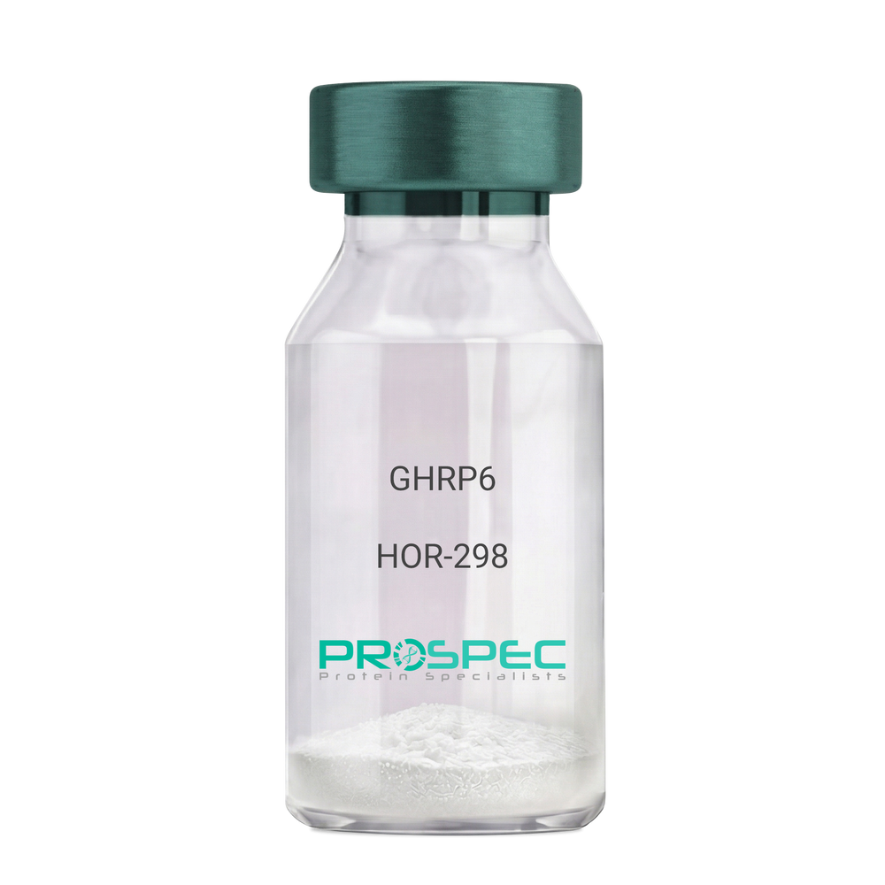
GHRP6Description:
Growth Hormone Releasing Peptide-6
GHRP-6, GHRP6
Product # :
HOR-298Price :
Quantity :
Shipping Method :
Shipped at Room temp
More Info
- description
- formulation
- purity
- More Info
Description
Growth Hormone Releasing Peptide-6 Synthetic is a single, non-glycosylated polypeptide chain containing 6 amino acids, having a molecular mass of 873 Dalton and a Molecular formula of C46H56N12O6.
Formulation
The GHRP-6 peptide was lyophilized with no additives.
Purity
Greater than 98.0% as determined by analysis by RP-HPLC.
More Info
-
Introduction
GH-releasing peptides (GHRPs) are synthetic peptides that like GHRH act directly on pituitary somatotrophs to stimulate GH release. Growth hormone (GH) release is stimulated by a variety of synthetic secretagogues, of which growth hormone-releasing hexapeptide (GHRP-6) has been most thoroughly studied; it is thought to have actions at both pituitary and hypothalamic site.
-
Synonyms
GHRP-6, GHRP6
-
Physical Appearance
Sterile Filtered White lyophilized (freeze-dried) powder.
-
Stability
Lyophilized Growth Hormone Releasing Peptide-6 although stable at room temperature for 3 weeks, should be stored desiccated below -18°C. Upon reconstitution GHRP-6 should be stored at 4°C between 2-7 days and for future use below -18°C. For long term storage it is recommended to add a carrier protein (0.1% HSA or BSA).Please prevent freeze-thaw cycles.
-
Solubility
It is recommended to reconstitute the lyophilized GHRP-6 in sterile 18MΩ-cm H2O not less than 100 µg/ml, which can then be further diluted to other aqueous solutions.
-
Amino Acid Sequence
His-D-Trp-Ala-Trp-D-Phe-Lys-NH2.
ProSpec's products are furnished for LABORATORY RESEARCH USE ONLY. The product may not be used as drugs, agricultural or pesticidal products, food additives or household chemicals.
- View Data Sheet
Name :
ZNF689 HumanDescription:
Zinc Finger Protein 689 Human Recombinant
Zinc Finger Protein 689, Transcription-Involved Protein Upregulated in HCC 1, TIPUH1.
Product # :
PRO-1737Price :
Quantity :
Shipping Method :
Shipped with Ice Packs
More Info
- description
- source
- formulation
- purity
- More Info
Description
ZNF689 Human Recombinant produced in E. coli is a single, non-glycosylated polypeptide chain containing 523 amino acids (1-500a.a) and having a molecular mass of 59.3kDa.ZNF689 is fused to a 23 amino acid His-tag at N-terminus & purified by proprietary chromatographic techniques.
Source
Escherichia Coli.
Formulation
ZNF689 protein solution (1.0mg/ml) containing 20mM Tris-HCl buffer (pH 8.0), 10% glycerol and 0.4M Urea.
Purity
Greater than 85.0% as determined by SDS-PAGE.
More Info
-
Introduction
Zinc Finger Protein 689 (ZNF689) is a member of the krueppel C2H2-type zinc-finger protein family. The ZNF689 protein contains 12 C2H2-type zinc fingers and 1 KRAB domain. ZNF689 may be involved in transcriptional regulation.
-
Synonyms
Zinc Finger Protein 689, Transcription-Involved Protein Upregulated in HCC 1, TIPUH1.
-
Physical Appearance
Sterile filtered colorless solution.
-
Stability
Store at 4°C if entire vial will be used within 2-4 weeks.Store, frozen at -20°C for longer periods of time. For long term storage it is recommended to add a carrier protein (0.1% HSA or BSA).Avoid multiple freeze-thaw cycles.
-
Amino Acid Sequence
MGSSHHHHHH SSGLVPRGSH MGSMAPPSAP LPAQGPGKAR PSRKRGRRPR ALKFVDVAVY FSPEEWGCLR PAQRALYRDV MRETYGHLGA LGCAGPKPAL ISWLERNTDD WEPAALDPQE YPRGLTVQRK SRTRKKNGEK EVFPPKEAPR KGKRGRRPSK PRLIPRQTSG GPICPDCGCT FPDHQALESH KCAQNLKKPY PCPDCGRRFS YPSLLVSHRR AHSGECPYVC DQCGKRFSQR KNLSQHQVIH TGEKPYHCPD CGRCFRRSRS LANHRTTHTG EKPHQCPSCG RRFAYPSLLA IHQRTHTGEK PYTCLECNRR FRQRTALVIH QRIHTGEKPY PCPDCERRFS SSSRLVSHRR VHSGERPYAC EHCEARFSQR STLLQHQLLH TGEKPYPCPD CGRAFRRSGS LAIHRSTHTE EKLHACDDCG RRFAYPSLLA SHRRVHSGER PYACDLCSKR FAQWSHLAQH QLLHTGEKPF PCLECGRCFR QRWSLAVHKC SPKAPNCSPR SAIGGSSQRG NAH.
ProSpec's products are furnished for LABORATORY RESEARCH USE ONLY. The product may not be used as drugs, agricultural or pesticidal products, food additives or household chemicals.
- View Data Sheet
Name :
IL17F HumanDescription:
Interleukin 17F Human Recombinant
Cytokine ML-1, IL-17F, Interleukin-17F precursor, IL17F, ML1, ML-1.
Product # :
CYT-587Price :
Quantity :
Shipping Method :
Shipped at Room temp
More Info
- description
- source
- formulation
- purity
- More Info
Description
Interleukin-17F Human Recombinant produced in E.Coli is a homodimeric, cysteine linked, non-glycosylated polypeptide chain containing 2 x 134 amino acids and having a total molecular mass of 30.1 kDa.The IL-17F Human is purified by proprietary chromatographic techniques.
Source
Escherichia Coli.
Formulation
Lyophilized from a concentrated (1mg/ml) solution containing 10mM sodium citrate buffer pH-3.
Purity
Greater than 97.0% as determined by(a) Analysis by RP-HPLC.
(b) Analysis by SDS-PAGE.More Info
-
Introduction
IL-17F having an accession number of Q96PD4 is a cytokine that shares sequence similarity with IL17. IL-17F is expressed by activated T cells, and has been shown to stimulate the production of several other cytokines, including IL6, IL8, and CSF2/GM_CSF. IL-17F inhibits the angiogenesis of endothelial cells and induce endothelial cells to produce IL2, TGFB1/TGFB, and monocyte chemoattractant protein-1. IL-17F induces stromal cells to produce proinflammatory and hematopoietic cytokines. Intestinal IL17F gene expression is increased in active CD.
IL-17A & IL-17F alleles influence the susceptibility to and pathophysiological features of ulcerative colitis independently. IL-17F and MIF gene polymorphisms are significantly associated with the development of functional dyspepsia.
The initiation of IL-17F/IL-17R signaling pathway requires the receptor ubiquitination by TRAF6. IL-17F induces expression of IFN-gamma-inducible protein 10 (IP-10) by activating Raf1-mitogen-activated protein kinase 1/2-extracellular-regulated kinase 1/2-p90 ribosomal S6 kinase-cyclic AMP response element-binding protein signaling pathway. -
Synonyms
Cytokine ML-1, IL-17F, Interleukin-17F precursor, IL17F, ML1, ML-1.
-
Physical Appearance
Sterile Filtered White lyophilized (freeze-dried) powder.
-
Stability
Lyophilized Interleukin Human IL17F although stable at room temperature for 3 weeks, should be stored desiccated below -18°C. Upon reconstitution Human IL-17F should be stored at 4°C between 2-7 days and for future use below -18°C.For long term storage it is recommended to add a carrier protein (0.1% HSA or BSA).Please prevent freeze-thaw cycles.
-
Solubility
It is recommended to reconstitute the lyophilized Interleukin Human IL17F in sterile water not more than 1mg/ml, which can then be further diluted to other aqueous solutions.
-
Amino Acid Sequence
MRKIPKVGHT FFQKPESCPP VPGGSMKLDI GIINENQRVS MSRNIESRST SPWNYTVTWD PNRYPSEVVQ AQCRNLGCIN AQGKEDISMN SVPIQQETLV VRRKHQGCSV SFQLEKVLVT VGCTCVTPVI HHVQ.
ProSpec's products are furnished for LABORATORY RESEARCH USE ONLY. The product may not be used as drugs, agricultural or pesticidal products, food additives or household chemicals.
- View Data Sheet
Name :
Epoetin Human, Sf9Description:
Erythropoietin-alpha Human Recombinant, Sf9
Erythropoietin, Epoetin, MVCD2, EP, Erythropoietin-Alpha, EPO-a, EPO-alpha.
Product # :
CYT-934Price :
Quantity :
Shipping Method :
Shipped with Ice Packs
More Info
- description
- source
- formulation
- purity
- biological activity
- More Info
- sds-page
Description
Erythropoietin-alpha Human Recombinant produced in Sf9 Insect cells is a single, glycosylated polypeptide chain containing 174 amino acids (28-193a.a.) and having a molecular mass of 19.5kDa (Molecular size on SDS-PAGE will appear at approximately 28-40kDa).EPO-a is expressed with an 8 amino acid His tag at C-Terminus and purified by proprietary chromatographic techniques.
Source
Sf9, Insect cells.
Formulation
EPO a protein solution (0.5mg/ml) contains phosphate buffered saline (pH7.4) and 10% glycerol.
Purity
Greater than 95.0% as determined by SDS-PAGE.
Biological Activity
Measured in a cell proliferation assay using TF-1 human erythroleukemic cells. The ED50 for this effect is ≤ 0.5 ng/ml.sds-page
More Info
-
Introduction
This gene is a member of the EPO/TPO family and encodes a secreted, glycosylated cytokine composed of four alpha helical bundles. The protein is found in the plasma and regulates red cell production by promoting erythroid differentiation and initiating hemoglobin synthesis. This protein also has neuroprotective activity against a variety of potential brain injuries and antiapoptotic functions in several tissue types.
-
Synonyms
Erythropoietin, Epoetin, MVCD2, EP, Erythropoietin-Alpha, EPO-a, EPO-alpha.
-
Physical Appearance
Sterile filtered colorless solution.
-
Stability
Store at 4°C if entire vial will be used within 2-4 weeks.Store, frozen at -20°C for longer periods of time. For long term storage it is recommended to add a carrier protein (0.1% HSA or BSA).Avoid multiple freeze-thaw cycles.
-
Amino Acid Sequence
APPRLICDSR VLERYLLEAK EAENITTGCA EHCSLNENIT VPDTKVNFYA WKRMEVGQQA VEVWQGLALL SEAVLRGQAL LVNSSQPWEP LQLHVDKAVS GLRSLTTLLR ALRAQKEAIS PPDAASAAPL RTITADTFRK LFRVYSNFLR GKLKLYTGEA CRTGDRLEHH HHHH.
-
Background
What is the molecular weight/Mw of EPOETIN Protein?
EPOETIN Protein has a total Mw of 19.5kDa.
What is the source or expression system of EPOETIN Protein?
Sf9, Insect cells.
What is the Purity of EPOETIN Protein?
EPOETIN Protein is >95% pure as determined by SDS-PAGE.
What is the Biological Activity of EPOETIN Protein?
Measured in a cell proliferation assay using TF-1 human erythroleukemic cells. The ED50 for this effect is ≤ 0.5 ng/ml.
What is the amino acid sequence of EPOETIN Protein?
APPRLICDSR VLERYLLEAK EAENITTGCA EHCSLNENIT VPDTKVNFYA WKRMEVGQQA VEVWQGLALL SEAVLRGQAL LVNSSQPWEP LQLHVDKAVS GLRSLTTLLR ALRAQKEAIS PPDAASAAPL RTITADTFRK LFRVYSNFLR GKLKLYTGEA CRTGDRLEHH HHHH.
What applications can EPOETIN Protein be used in?
EPOETIN Protein can probably be used in western blot, ELISA and Lateral Flow.
What is the endotoxin level for EPOETIN Protein?
The endotoxin level is minimal, EPOETIN Protein was purified using conventional chromatography techniques.
ProSpec's products are furnished for LABORATORY RESEARCH USE ONLY. The product may not be used as drugs, agricultural or pesticidal products, food additives or household chemicals.
- View Data Sheet
Name :
OSM Human, HisDescription:
Oncostatin-M Human Recombinant, His Tag
OSM, MGC20461.
Product # :
CYT-060Price :
Quantity :
Shipping Method :
Shipped with Ice Packs
More Info
- description
- source
- formulation
- purity
- More Info
Description
OSM Human Recombinant produced in E.coli is a single, non-glycosylated polypeptide chain containing 230 amino acids (26-234) and having a molecular mass of 25.9 kDa.The OSM is fused to a 21 amino acid His-Tag at N-terminus and purified by proprietary chromatographic techniques.
Source
Escherichia Coli.
Formulation
OSM protein (0.5mg/ml) is supplied in 20mM Tris-HCl, pH-8, 1mM DTT and 20% Glycerol.
Purity
Greater than 95.0% as determined by SDS-PAGE.
More Info
-
Introduction
POU2AF1 is a lymphocyte specific transcription coactivator protein. POU2AF1 cooperates only with the Oct1/2 proteins using sub domains in the POU domain of the Oct1/2 proteins, increasing their transcriptional efficiency. Even though POU2AF1 have no basic ability to bind DNA, it links firmly with the octomer motif in the presence of Oct1 and Oct2. POU2AF1 is expressed at maximum levels in spleen and peripheral blood leukocytes.
-
Synonyms
OSM, MGC20461.
-
Physical Appearance
Sterile Filtered colorless solution.
-
Stability
Store at 4°C if entire vial will be used within 2-4 weeks. Store, frozen at -20°C for longer periods of time. For long term storage it is recommended to add a carrier protein (0.1% HSA or BSA).Avoid multiple freeze-thaw cycles.
-
Amino Acid Sequence
MGSSHHHHHH SSGLVPRGSH MAAIGSCSKE YRVLLGQLQK QTDLMQDTSR LLDPYIRIQG LDVPKLREHC RERPGAFPSE ETLRGLGRRG FLQTLNATLG CVLHRLADLE QRLPKAQDLE RSGLNIEDLE KLQMARPNIL GLRNNIYCMA QLLDNSDTAE PTKAGRGASQ PPTPTPASDA FQRKLEGCRF LHGYHRFMHS VGRVFSKWGE SPNRSRRHSP HQALRKGVRR.
ProSpec's products are furnished for LABORATORY RESEARCH USE ONLY. The product may not be used as drugs, agricultural or pesticidal products, food additives or household chemicals.
- View Data Sheet
Name :
Leptin Receptor ChickenDescription:
Leptin Receptor Chicken Recombinant
OB Protein, Obesity Protein, OBS, Obesity factor, Leptin Receptor.
Product # :
CYT-509Price :
Quantity :
Shipping Method :
Shipped with Ice Packs
More Info
- description
- source
- formulation
- purity
- More Info
Description
Leptin Binding Domain Chicken Recombinant also called Leptin Receptor produced in E.Coli is a single, non-glycosylated, polypeptide chain containing 208 amino acids and having a molecular mass of 24.5 kDa. Chicken Leptin Receptor consists of the cytokine binding domain of leptin receptor amino acids 420-626 of chicken leptin receptor.The Leptin Binding Domain is purified by proprietary chromatographic techniques.
Source
Escherichia Coli.
Formulation
The protein was filter sterilized and stored at 4°C (0.2 to 0.5 mg/ml) solution of Tris-HCl buffer, pH 9.0 with 150mM NaCl.
Purity
Greater than 99.0% as determined by:
(a) Analysis by SEC-HPLC.
(b) Analysis by SDS-PAGE.More Info
-
Introduction
Leptin Receptor is a part of the gp130 family of cytokine receptors that stimulate gene transcription by activating cytosolic STAT proteins. Leptin Receptor plays a role in the regulation of fat metabolism and in novel hematopoietic pathway that is obligatory for normal lymphopoiesis. Leptin Receptorparticipates in the regulation of counter-regulatory response to hypoglycemia by inhibiting neurons of the parabrachial nucleus.Leptin Receptoraffectsspecifically on T lymphocyte responses.
-
Synonyms
OB Protein, Obesity Protein, OBS, Obesity factor, Leptin Receptor.
-
Physical Appearance
Sterile Filtered colorless solution at a concentration of 0.4 mg/ml.
-
Stability
Sterile solutions at 0.5mg/ml or less are stable at 4°C for several months.
-
Amino Acid Sequence
The sequence of the first six N-terminal amino acids was determined and was found to be Ala-Ile-Asp-Val-Asn-Ile Biological ActivityBiological Activity is evidenced by high affinity binding of mammalian leptins at 1:1 molar ratio.
-
Protein content
Protein quantitation was carried out by two independent methods1. UV spectroscopy at 280 nm using the absorbency value of 2.45 as the extinction coefficient for a 0.1% (1mg/ml) solution. This value is calculated by the PC GENE computer analysis program of protein sequences (IntelliGenetics). 2. Analysis by RP-HPLC, using a calibrated solution of Leptin Binding Domain as a Reference Standard.
ProSpec's products are furnished for LABORATORY RESEARCH USE ONLY. The product may not be used as drugs, agricultural or pesticidal products, food additives or household chemicals.
- View Data Sheet
Name :
b NGF MouseDescription:
Beta-Nerve Growth Factor Mouse
Beta Polypeptide, NGF, NGFB, HSAN5, Beta-NGF, MGC161426, MGC161428.
Product # :
CYT-440Price :
Quantity :
Shipping Method :
Shipped at Room temp
More Info
- description
- source
- formulation
- purity
- biological activity
- More Info
Description
NGF beta Mouse produced in Submaxillary Gland of Grown Mouse is a homodimer, non-glycosylated, polypeptide chain containing 2 identical 120 amino acids and having a molecular mass of 13,471 Dalton each.The NGF beta Mouse is purified by advanced biology purification technology.
Source
Submaxillary Gland of Grown Mouse.
Formulation
The NGF beta Mouse was lyophilized from solution containing 5% mannitol and 1% HSA.
Purity
Greater than 98% as determined by(a) Analysis by RP-HPLC.
(b) Analysis by SDS-PAGE .Biological Activity
The method used to test the bioassay is the NGF-dependent survival of dorsal root ganglia neurons of chick embryo, corresponding to a Specific Activity of 500,000IU/mg.More Info
-
Introduction
NGF-beta has nerve growth stimulating activity and the complex is involved in the regulation of growth and the differentiation of sympathetic and certain sensory neurons. Mutations in this gene have been associated with hereditary sensory and autonomic neuropathy, type 5 (HSAN5), and dysregulation of this gene's expression is associated with allergic rhinitis.
-
Synonyms
Beta Polypeptide, NGF, NGFB, HSAN5, Beta-NGF, MGC161426, MGC161428.
-
Physical Appearance
Sterile Filtered White lyophilized (freeze-dried) powder.
-
Stability
Lyophilized Mouse Beta-NGF although stable at room temperature for 3 weeks, should be stored desiccated below -18°C. Upon reconstitution Murine NGF-Beta should be stored at 4°C between 2-7 days and for future use below -18°C.Please prevent freeze-thaw cycles.
-
Solubility
It is recommended to reconstitute the lyophilized Murine NGF-beta in sterile 18MΩ-cm H2O not less than 100µg/ml, which can then be further diluted to other aqueous solutions.
-
Amino Acid Sequence
SSTHPVFHMGEF SVCDSVSVWV GDKTTATDIK GKEVTVLAEV NINNSVFRQY FFETKCRASN PVESGCRGID SKHWNSYCTT THTFVKALTT DEKQAAWRFI RIDTACVCVL SRKATRRG.
-
Background
What is the molecular weight/Mw of B NGF Protein?
B NGF Protein has a total Mw of 13kDa.
What is the source or expression system of B NGF Protein?
Submaxillary Gland of Grown Mouse.
What is the Purity of B NGF Protein?
B NGF Protein is >98% pure as determined by SDS-PAGE.
What is the Biological Activity of B NGF Protein?
The method used to test the bioassay is the NGF-dependent survival of dorsal root ganglia neurons of chick embryo, corresponding to a Specific Activity of 500,000IU/mg.
What is the amino acid sequence of B NGF Protein?
SSTHPVFHMGEF SVCDSVSVWV GDKTTATDIK GKEVTVLAEV NINNSVFRQY FFETKCRASN PVESGCRGID SKHWNSYCTT THTFVKALTT DEKQAAWRFI RIDTACVCVL SRKATRRG.
What applications can B NGF Protein be used in?
B NGF Protein can probably be used in western blot, ELISA and Lateral Flow.
What is the endotoxin level for XXX Protein?
The endotoxin level is minimal, XXX Protein was purified using conventional chromatography techniques.
ProSpec's products are furnished for LABORATORY RESEARCH USE ONLY. The product may not be used as drugs, agricultural or pesticidal products, food additives or household chemicals.
- View Data Sheet
Name :
SDF 1b Human, HisDescription:
Stromal Cell-Derived Factor-1 beta Human Recombinant (CXCL12), His Tag
SDF-1, CXCL12, Pre-B cell growth-stimulating factor, PBSF, hIRH, chemokine (C-X-C motif) ligand 12, SDF1, SDF1B, TPAR1, SCYB12, SDF-1b, TLSF-b, 12-O-tetradecanoylphorbol 13-acetate repressed protein 1, Thymic lymphoma cell-stimulating factor, TLSF.
Product # :
CHM-249Price :
Quantity :
Shipping Method :
Shipped with Ice Packs
More Info
- description
- source
- formulation
- purity
- More Info
Description
SDF-1 beta Human Recombinant produced in E.Coli is a non-glycosylated, Polypeptide chain containing 93 amino acids (22-93 a.a.) and having a molecular mass of 10.8 KDa. The SDF-1b is fused to 20 amino acid His-Tag at N-terminus and purified by proprietary chromatographic techniques.
Source
Escherichia Coli.
Formulation
The SDF-1 beta His Tag protein contains 20mM Tris-HCl buffer pH-8 and 10% glycerol.
Purity
Greater than 90% as determined by Analysis by SDS-PAGE.
More Info
-
Introduction
SDF-1 (stromal cell-derived factor-1) is small cytokine belonging to the chemokine family that is officially designated Chemokine (C-X-C motif) ligand 12 (CXCL12). It is produced in two forms, SDF-1?/CXCL12a and SDF-1?/CXCL12b, by alternate splicing of the same gene. Chemokines are characterized by the presence of four conserved cysteines, which form two disulfide bonds. The CXCL12 proteins belong to the group of CXC chemokines, whose initial pair of cysteines are separated by one intervening amino acid. CXCL12 is strongly chemotactic for lymphocytes and has been implicated as an important cell co-ordinator during development. During embryogenesis it directs the migration of hematopoietic cells from foetal liver to bone marrow. Mice which were knocked-out for CXCL12 gene were lethal before the birth or within just 1 hour of life. As another role, CXCL12a alters also the electrophysiology of neurons. CXCL12 was shown to be expressend in many tissues in mice (including brain, thymus, heart, lung, liver, kidney, spleen and bone marrow).
The receptor for this chemokine is CXCR4, which was previously called fusin. This CXCL12-CXCR4 interaction used to be considered exclusive (unlike for other chemokines and their receptors), but recently it was suggested that CXCL12 is also bound by CXCR7 receptor. The gene for CXCL12 is located on human chromosome 10. In human and mouse both CXCL12 and CXCR4 show high identity of sequence: 99% and 90%, respectively. -
Synonyms
SDF-1, CXCL12, Pre-B cell growth-stimulating factor, PBSF, hIRH, chemokine (C-X-C motif) ligand 12, SDF1, SDF1B, TPAR1, SCYB12, SDF-1b, TLSF-b, 12-O-tetradecanoylphorbol 13-acetate repressed protein 1, Thymic lymphoma cell-stimulating factor, TLSF.
-
Physical Appearance
Sterile filtered colorless solution.
-
Stability
Store at 4°C if entire vial will be used within 2-4 weeks. Store, frozen at -20°C for longer periods of time. For long term storage it is recommended to add a carrier protein (0.1% HSA or BSA). Avoid multiple freeze-thaw cycles.
-
Amino Acid Sequence
MGSSHHHHHH SSGLVPRGSH MKPVSLSYRC PCRFFESHVA RANVKHLKIL NTPNCALQIV ARLKNNNRQV CIDPKLKWIQ EYLEKALNKR FKM.
ProSpec's products are furnished for LABORATORY RESEARCH USE ONLY. The product may not be used as drugs, agricultural or pesticidal products, food additives or household chemicals.
- View Data Sheet
Name :
GH1 AntibodyDescription:
Growth Hormone-1, Mouse Anti Human
GH1, GH, GHN, GH-N, hGH-N, Pituitary growth hormone, Growth hormone 1, Somatotropin.
Product # :
ANT-230Price :
Quantity :
Shipping Method :
Shipped with Ice Packs
More Info
- formulation
- More Info
Formulation
1mg/ml containing PBS, pH-7.4, & 0.1% sodium azide.
More Info
-
Introduction
GH is a member of the somatotropin/prolactin family of hormones which play an important role in growth control. The gene, along with four other related genes, is located at the growth hormone locus on chromosome 17 where they are interspersed in the same transcriptional orientation; an arrangement which is thought to have evolved by a series of gene duplications. The five genes share a remarkably high degree of sequence identity. Alternative splicing generates additional isoforms of each of the five growth hormones, leading to further diversity and potential for specialization. This particular family member is expressed in the pituitary but not in placental tissue as is the case for the other four genes in the growth hormone locus. Mutations in or deletions of the gene lead to growth hormone deficiency and short stature.
-
Synonyms
GH1, GH, GHN, GH-N, hGH-N, Pituitary growth hormone, Growth hormone 1, Somatotropin.
-
Physical Appearance
Sterile Filtered solution.
-
Immunogen
GH Antibody is derived from hybridization of mouse SP2/O myeloma cells with spleen cells from BALB/c mice immunized with recombinant Growth hormone.
-
Ig Subclass
Mouse IgG2b heavy chain and k light chain.
-
Clone
PG3H5AT.
-
Applications
The GH antibody has been tested by ELISA and by Western blot analysis to assure specificity and reactivity. Since application vary, however, each investigation should titrate the reagent to obtain optimal results. Recommended dilution range for Western blot analysis: 1:500 ~ 1:1000. Recommended starting dilution: 1:500.
-
Type
Mouse Anti Human Monoclonal.
-
Storage Procedures
For periods up to 1 month store at 4 C, for longer periods of time, store at -20 C. Prevent freeze thaw cycles.
-
Purification Method
GH antibody was purified from mouse ascitic fluids by protein-G affinity chromatography.
ProSpec's products are furnished for LABORATORY RESEARCH USE ONLY. The product may not be used as drugs, agricultural or pesticidal products, food additives or household chemicals.
- View Data Sheet
Name :
EIF1B HumanDescription:
Eukaryotic Translation Initiation Factor 1B Human Recombinant
Eukaryotic translation initiation factor 1b, eIF1b, Protein translation factor SUI1 homolog GC20, EIF1B, GC20.
Product # :
PRO-175Price :
Quantity :
Shipping Method :
Shipped with Ice Packs
More Info
- description
- source
- formulation
- purity
- More Info
Description
EIF1B Human Recombinant fused with a 20 amino acid His tag at N-terminus produced in E.Coli is a single, non-glycosylated, polypeptide chain containing 133 amino acids (1-113 a.a.) and having a molecular mass of 15kDa. The EIF1B is purified by proprietary chromatographic techniques.
Source
Escherichia Coli.
Formulation
The EIF1B solution (1 mg/ml) contains 20mM Tris-HCl buffer(pH 8.0), 10% glycerol, 2mM DTT and 0.1M NaCl.
Purity
Greater than 95.0% as determined by SDS-PAGE.
More Info
-
Introduction
EIF1B is critical for the scanning process in vitro. EIF1B is an element of a complex involved in recognition of the initiator codon during the scanning process. Translation is also initiated by the function of EIF1B in regulating the activity of ribosomal subunits 43S, 48S and 40S. EIF1B enables 43S ribosomal complexes to distinguish between cognate and near-cognate initiation codons, perceiving the nucleotide content of initiation codons.
-
Synonyms
Eukaryotic translation initiation factor 1b, eIF1b, Protein translation factor SUI1 homolog GC20, EIF1B, GC20.
-
Physical Appearance
Sterile Filtered colorless solution.
-
Stability
Store at 4°C if entire vial will be used within 2-4 weeks. Store, frozen at -20°C for longer periods of time. For long term storage it is recommended to add a carrier protein (0.1% HSA or BSA).Avoid multiple freeze-thaw cycles.
-
Amino Acid Sequence
MGSSHHHHHH SSGLVPRGSH MSTIQNLQSF DPFADATKGD DLLPAGTEDY IHIRIQQRNG RKTLTTVQGI ADDYDKKKLV KAFKKKFACN GTVIEHPEYG EVIQLQGDQR KNICQFLLEV GIVKEEQLKV HGF.
ProSpec's products are furnished for LABORATORY RESEARCH USE ONLY. The product may not be used as drugs, agricultural or pesticidal products, food additives or household chemicals.
- View Data Sheet
Name :
Leptin HumanDescription:
Human Leptin
OB Protein, Obesity Protein, OBS, Obesity factor, Leptin.
Product # :
CYT-683Price :
Quantity :
Shipping Method :
Shipped at Room temp
More Info
- description
- formulation
- purity
- More Info
Description
Leptin Human produced syntheticaly contains 35 amino acids (22-56 a.a.) having a molecular mass of 3950.6 Dalton.
Formulation
The protein was lyophilized from a concentrated (1mg/ml) solution with no additives.
Purity
Greater than 95.0% as determined by RP-HPLC.
More Info
-
Introduction
A 16kDa peptide hormone secreted from white adipocytes and implicated in the regulation of food intake and energy balance. Leptin provides the key afferent signal from fat cells in the feedback system that controls body fat stores.
-
Synonyms
OB Protein, Obesity Protein, OBS, Obesity factor, Leptin.
-
Physical Appearance
Sterile Filtered White lyophilized (freeze-dried) powder.
-
Stability
Lyophilized Leptin although stable at room temperature, should be stored desiccated below -20°C. Reconstituted Leptin is best stored refrigerated at 4°C.
-
Solubility
The lyophilized Leptin is very soluble in water and most aqueous buffers below and above the isoelectric point.
-
Amino Acid Sequence
Val-Pro-Ile-Gln-Lys-Val-Gln-Asp-Asp-Thr-Lys-Thr-Leu-Ile-Lys-Thr-Ile-Val-Thr-Arg-Ile-Asn-Asp-Ile-Ser-His-Thr-Gln-Ser-Val-Ser-Ser-Lys-Gln-Lys.
ProSpec's products are furnished for LABORATORY RESEARCH USE ONLY. The product may not be used as drugs, agricultural or pesticidal products, food additives or household chemicals.
- View Data Sheet
Name :
Prolactin MouseDescription:
Prolactin Mouse Recombinant
Mammotropin, Luterotropic hormone, Lutetropin, PRL.
Product # :
CYT-321Price :
Quantity :
Shipping Method :
Shipped at Room temp
More Info
- description
- source
- formulation
- purity
- biological activity
- More Info
Description
Prolactin Mouse Recombinant produced in E.Coli is a single, non-glycosylated polypeptide chain containing 198 amino acids and having a molecular mass of 22.5 kDa. The Prolactin is purified by proprietary chromatographic techniques.
Source
Escherichia Coli.
Formulation
The protein was lyophilized from a concentrated (1mg/ml) solution with 10mM sodium Phosphate buffer pH=8 and 50mM Nacl.
Purity
Greater than 95.0% as determined by:
(a) Analysis by RP-HPLC.
(b) Analysis by SDS-PAGE.Biological Activity
The sequence of the first five N-terminal amino acids was determined and was found to be Ala-Leu-Pro-Ile-Cys-Ser.
More Info
-
Introduction
Prolactin is a neuroendocrine hormone synthesized primarily by the pituitary gland but also a variety of other cell types including the placenta, brain and uterus. Its primary function is to promote and maintain lactation but has also been shown to have a role in breast cancer development, regulation of reproductive function and immunoregulation.
-
Synonyms
Mammotropin, Luterotropic hormone, Lutetropin, PRL.
-
Physical Appearance
Sterile Filtered White lyophilized (freeze-dried) powder.
-
Stability
Lyophilized Prolactin although stable at room temperature for 3 weeks, should be stored desiccated below -18°C. Upon reconstitution Prolactin should be stored at 4°C between 2-7 days and for future use below -18°C.For long term storage it is recommended to add a carrier protein (0.1% HSA or BSA).Please prevent freeze-thaw cycles.
-
Solubility
It is recommended to reconstitute the lyophilized Prolactin in sterile 18MΩ-cm H2O not less than 100µg/ml, which can then be further diluted to other aqueous solutions.
-
Amino Acid Sequence
The sequence of the first five N-terminal amino acids was determined and was found to be Met-Leu-Pro-Ile-Cys.
ProSpec's products are furnished for LABORATORY RESEARCH USE ONLY. The product may not be used as drugs, agricultural or pesticidal products, food additives or household chemicals.
- View Data Sheet
Name :
LGALS1 HumanDescription:
Galectin-1 Human Recombinant
Galectin-1, GAL1, GAL-1, Lectin galactoside-binding soluble 1, Beta-galactoside- binding lectin L-14-I, Lactose-binding lectin 1, S-Lac lectin 1, Galaptin, 14 kDa lectin, HPL, HBL, Putative MAPK-activating protein PM12, GBP, DKFZp686E23103.
Product # :
CYT-544Price :
Quantity :
Shipping Method :
Shipped at Room temp
More Info
- description
- source
- formulation
- purity
- biological activity
- More Info
Description
LGALS1 Human Recombinant produced in E.Coli is a single, non-glycosylated, polypeptide chain containing 135 amino acids and having a molecular mass of 14.7kDa.The LGALS1 is purified by proprietary chromatographic techniques.
Source
Escherichia Coli.
Formulation
The Galectin-1 protein was lyophilized from a concentrated (1mg/ml) containing 10mM sodium phosphate, pH-7.5.
Purity
Greater than 95.0% as determined by SDS-PAGE.
Biological Activity
The activity of Human Galectin-1 which is determined by the ability to induce chemotaxis of human THP-1 cells is detectable starting at 100ng/ml, corresponding to a specific activity of 1.0x10⁴ units/mg.More Info
-
Introduction
The galectins are a family of beta-galactoside-binding proteins implicated in modulating cell-cell and cell-matrix interactions. Galectin-1 is an autocrine negative growth factor that regulates cell proliferation. Galectin-1 regulates cell apoptosis and cell differentiation. Galectin-1 binds CD45, CD3 and CD4 & inhibits CD45 protein phosphatase activity and therefore the dephosphorylation of lyn kinase. Galectin-1 and its ligands are one of the master regulators of immune responses as T-cell homeostasis and survival, T-cell immune disorders, inflammation and allergies as well as host–pathogen interactions. Galectin-1 expression or overexpression in tumors and/or the tissue surrounding them must be considered as a sign of the malignant tumor progression that is often related to the long-range dissemination of tumoral cells (metastasis), to their dissemination into the surrounding normal tissue, and to tumor immune-escape. Galectin-1 in its oxidized form plays a number of important roles in the regeneration of the central nervous system after injury. The targeted overexpression (or delivery) of Galectin-1 should be considered as a method of choice for the treatment of some kinds of inflammation-related diseases, neurodegenerative pathologies and muscular dystrophies. In contrast, the targeted inhibition of Galectin-1 expression is what should be developed for therapeutic applications against cancer progression. Galectin-1 is thus a promising molecular target for the development of new and original therapeutic tools. There is 88% homology between the human and mouse galectin-1.
-
Synonyms
Galectin-1, GAL1, GAL-1, Lectin galactoside-binding soluble 1, Beta-galactoside- binding lectin L-14-I, Lactose-binding lectin 1, S-Lac lectin 1, Galaptin, 14 kDa lectin, HPL, HBL, Putative MAPK-activating protein PM12, GBP, DKFZp686E23103.
-
Physical Appearance
Sterile Filtered White lyophilized (freeze-dried) powder.
-
Stability
Lyophilized Galectin-1 Recombinant although stable at room temperature for 3 weeks, should be stored desiccated below -18°C. Upon reconstitution Galectin-1 should be stored at 4°C between 2-7 days and for future use below -18°C.For long term storage it is recommended to add a carrier protein (0.1% HSA or BSA).Please prevent freeze-thaw cycles.
-
Solubility
It is recommended to reconstitute the lyophilized LGALS1 in sterile 18MΩ-cm H2O not less than 100µg/ml, which can then be further diluted to other aqueous solutions.
-
Amino Acid Sequence
MACGLVASNL NLKPGECLRV RGEVAPDAKS FVLNLGKDSN NLCLHFNPRF NAHGDANTIV CNSKDGGAWG TEQREAVFPF QPGSVAEVCI TFDQANLTVK LPDGYEFKFP NRLNLEAINY MAADGDFKIK CVAFD.
-
Background
What is the molecular weight/Mw of LGALS1 HUMAN Protein?
LGALS1 HUMAN Protein has a total Mw of 14.7kDa.
What is the source or expression system of LGALS1 HUMAN Protein?
Escherichia Coli.
What is the Purity of LGALS1 HUMAN Protein?
LGALS1 HUMAN Protein is >95% pure as determined by SDS-PAGE.
What is the Biological Activity of LGALS1 HUMAN Protein?
The activity of Human Galectin-1 which is determined by the ability to induce chemotaxis of human THP-1 cells is detectable starting at 100ng/ml, corresponding to a specific activity of 1.0x10⁴ units/mg.
What is the amino acid sequence of LGALS1 HUMAN Protein?
MACGLVASNL NLKPGECLRV RGEVAPDAKS FVLNLGKDSN NLCLHFNPRF NAHGDANTIV CNSKDGGAWG TEQREAVFPF QPGSVAEVCI TFDQANLTVK LPDGYEFKFP NRLNLEAINY MAADGDFKIK CVAFD.
What applications can LGALS1 HUMAN Protein be used in?
LGALS1 HUMAN Protein can probably be used in western blot, ELISA and Lateral Flow.
What is the endotoxin level for LGALS1 HUMAN Protein?
The endotoxin level is minimal, LGALS1 HUMAN Protein was purified using conventional chromatography techniques.
-
Protein content
UV spectroscopy at 280 nm using the absorbency value of 0.59 as the extinction coefficient for a 0.1% (1mg/ml) solution. This value is calculated by the PC GENE computer analysis program of protein sequences (IntelliGenetics).
ProSpec's products are furnished for LABORATORY RESEARCH USE ONLY. The product may not be used as drugs, agricultural or pesticidal products, food additives or household chemicals.
- View Data Sheet
Name :
BMP3 HumanDescription:
Bone Morphogenetic protein-3 Human Recombinant
Bone Morphogenetic Protein 3, Osteogenin, Bone Morphogenetic Protein 3 (Osteogenic), Bone Morphogenetic Protein 3A, BMP-3A, BMP-3, Bone Morphogenetic Protein-3, BMP3A, BMP3.
Product # :
CYT-937Price :
Quantity :
Shipping Method :
Shipped at Room temp
More Info
- description
- source
- formulation
- purity
- biological activity
- More Info
Description
BMP3 Human Recombinant produced in E.coli is a non-glycosylated disulfide linked homodimer containing 2 chains of 110 amino acids and having a molecular mass of 24.8kDa.The BMP-3 is purified by proprietary chromatographic techniques.
Source
Escherichia Coli.
Formulation
BMP-3 protein was lyophilized from a 0.2µm filtered concentrated solution in 30% Acetonitrile and 0.1% TFA.
Purity
Greater than 95.0% as determined by: (a) Analysis by HPLC. (b) Analysis by SDS-PAGE.
Biological Activity
The ED50 as determined by its ability to inhibit BMP-2-induced activity in murine MC3T3- E1 cells.More Info
-
Introduction
Bone Morphogenetic Protein 3 (BMP3) is one of the BMPs, some of which are members of the TGF-beta superfamily (BMP2-7). There are more than 13 BMPs, which are involved in inducing cartilage and bone formation, embryogenesis and morphogenesis of various tissues and organs. In addition, BMPs regulate the growth, differentiation, chemotaxis, and apoptosis of various cell types. Akin to most other TGF-beta family proteins, BMPs are extremely conserved across animal species. At the amino acid sequence level, mature human and rat BMP3 are 98% identical.
-
Synonyms
Bone Morphogenetic Protein 3, Osteogenin, Bone Morphogenetic Protein 3 (Osteogenic), Bone Morphogenetic Protein 3A, BMP-3A, BMP-3, Bone Morphogenetic Protein-3, BMP3A, BMP3.
-
Physical Appearance
Sterile Filtered White lyophilized (freeze-dried) powder.
-
Stability
Lyophilized BMP3 although stable at room temperature for 3 weeks, should be stored desiccated below -18°C. Upon reconstitution BMP-3 should be stored at 4°C between 2-7 days and for future use below -18°C. For long term storage it is recommended to add a carrier protein (0.1% HSA or BSA). Please prevent freeze-thaw cycles.
-
Solubility
It is recommended to reconstitute the lyophilized BMP3 in sterile 4mM HCl not less than 100µg/ml, which can then be further diluted to other aqueous solutions.
-
Amino Acid Sequence
QWIEPRNCAR RYLKVDFADI GWSEWIISPK SFDAYYCSGA CQFPMPKSLK PSNHATIQSI VRAVGVVPGI PEPCCVPEKM SSLSILFFDE NKNVVLKVYP NMTVESCACR.
-
Background
Bone Morphogenetic Protein-3 Human Recombinant: Unveiling the Potential of a Key Regulator in Tissue Regeneration
Abstract:
Bone Morphogenetic Protein-3 (BMP-3) human recombinant is a critical member of the bone morphogenetic protein family, known for its role in tissue development, repair, and regeneration. This research paper provides a comprehensive analysis of BMP-3, including its characteristics, signaling pathways, and potential therapeutic applications. Additionally, innovative methodologies for the production and optimization of BMP-3 human recombinant are proposed, shedding light on its future implications in the field of regenerative medicine.
Introduction:
Tissue regeneration is a complex biological process requiring precise molecular cues. BMP-3, a crucial member of the BMP family, plays a significant role in tissue development and regeneration. This paper explores the unique features of BMP-3 and presents novel approaches for its production and optimization, aiming to unlock its therapeutic potential in various regenerative contexts.
Characteristics and Signaling Pathways:
BMP-3 is a secreted protein that binds to cell surface receptors, initiating intracellular signaling cascades. It influences cell differentiation, proliferation, and extracellular matrix synthesis through both Smad-dependent and Smad-independent signaling pathways. BMP-3 signaling regulates critical processes involved in tissue regeneration, including chondrogenesis and osteogenesis.
Production of BMP-3 Human Recombinant:
Efficient production methodologies are essential for harnessing the therapeutic potential of BMP-3 human recombinant. Recombinant protein expression systems, such as Escherichia coli or mammalian cells, have been utilized to produce functional BMP-3. Optimization strategies, including codon optimization, signal peptide engineering, and protein folding optimization, have been employed to enhance the yield and activity of BMP-3 recombinant protein.
Potential Therapeutic Applications:
BMP-3 human recombinant holds significant promise in the field of regenerative medicine. It plays a crucial role in bone and cartilage regeneration, making it a potential candidate for the treatment of skeletal disorders and tissue injuries. Additionally, BMP-3 signaling influences tissue remodeling and wound healing, suggesting its broader therapeutic applications in other regenerative processes.
Conclusion:
BMP-3 human recombinant represents a key regulator in tissue regeneration, with immense potential in regenerative medicine. Optimizing production methodologies and further unraveling its signaling mechanisms will enhance its therapeutic applications. With its implications in bone and cartilage regeneration and its role in tissue remodeling, BMP-3 human recombinant emerges as a promising tool for promoting tissue repair and regeneration.
What is the molecular weight/Mw of BMP3 Protein?
BMP3 Protein has a total Mw of 24.8kDa.
What is the source or expression system of BMP3 Protein?
Escherichia Coli.
What is the Purity of BMP3 Protein?
BMP3 Protein is >95% pure as determined by SDS-PAGE.
What is the Biological Activity of BMP3 Protein?
The ED50 as determined by its ability to inhibit BMP-2-induced activity in murine MC3T3- E1 cells.
What is the amino acid sequence of BMP3 Protein?
QWIEPRNCAR RYLKVDFADI GWSEWIISPK SFDAYYCSGA CQFPMPKSLK PSNHATIQSI VRAVGVVPGI PEPCCVPEKM SSLSILFFDE NKNVVLKVYP NMTVESCACR.
What applications can BMP3 Protein be used in?
BMP3 Protein can probably be used in western blot, ELISA and Lateral Flow.
What is the endotoxin level for BMP3 Protein?
The endotoxin level is minimal, BMP3 Protein was purified using conventional chromatography techniques.
ProSpec's products are furnished for LABORATORY RESEARCH USE ONLY. The product may not be used as drugs, agricultural or pesticidal products, food additives or household chemicals.
- View Data Sheet
Name :
Leptin tA Mouse, PEG (D23L)Description:
Leptin Triple Antagonist (D23L) Pegylated Mouse Recombinant
OB Protein, Obesity Protein, OBS, Obesity factor.
Product # :
CYT-1242Price :
Quantity :
Shipping Method :
Shipped at Room temp
More Info
- description
- source
- formulation
- purity
- biological activity
- More Info
Description
Leptin Antagonist Triple Mutant D23L Mouse Recombinant is a single non-glycosilated polypeptide chain containing 146 amino and additional Ala at N-terminus. The Leptin antagonist was mutated, resulting in L39A/D40A/F41A mutant. The Mouse Leptin antagonist is bound to 20 kDa mono-PEG at N-terminus, resulting in 35.6 kDa. The Mouse Leptin triple anatagonist runs as a 48 kDa. Leptin Antagonist Triple Mutant Mouse Recombinant was purified by proprietary chromatographic techniques.
Source
Escherichia coli.
Formulation
The Mouse Leptin triple anatagonist was lyophilized from a concentrated (0.65mg/ml) solution with 0.003mM NaHCO3.
Purity
Greater than 99.0% as determined by:
(a) Gel filtration analysis.
(b) Analysis by SDS-PAGE.
Biological Activity
Leptin Antagonist Triple Mutant D23L Mouse Recombinant is capable of stimulating proliferation of BAF/3 cells stably transfected with the long form of human leptin receptor. Its in vitro activity is only slightly lower than the non-pegylated recombinant mouse leptin but in vivo it has profound weight reducing effect (as compared to the non-pegylated recombinant mouse leptin), resulting mainly from reduced food intake.
More Info
-
Synonyms
OB Protein, Obesity Protein, OBS, Obesity factor.
-
Physical Appearance
White lyophilized (freeze-dried) powder.
-
Stability
Lyophilized Leptin Antagonist Triple Mutant Mouse Recombinant although stable at room temperature for several weeks, should be stored desiccated below -18°C. Upon reconstitution at > 0.1mg/ml and up to 2mM and filter sterilization LEP mutant can be stored at 4°C or even room temperature for several weeks making it suitable for long term infusion studies using osmotic pumps. At lower concentration addition of a carrier protein (0.1% HSA or BSA) is suggested. Please prevent freeze-thaw cycles.
-
Solubility
It is recommended to reconstitute the lyophilized Leptin Antagonist Triple Mutant Mouse Recombinant in sterile water or sterile 0.4% NaHCO3adjusted to pH 8-9, not less than 100µg/ml, which can then be further diluted with other aqueous solutions.
-
Background
Leptin is a hormone which mainly produced by adipocytes . Leptin’s main part is to regulate long-term energy balance. Leptin is encoded by the LEP gene. Leptin effects mainly on leptin receptors in the cell mambrane of various cells in the human body. The leptin receptor is found on a wide range of cell types. The leptin receptor is a single-transmembrane-domain type 1 cytokine receptor. leptin levels influence satiety, appetite and triggers behaviors which lead to energy savings High leptin levels are interpreted by the brain that energy reserves are high, whereas low leptin levels means that energy reserves are low, in the process adapting the organism to starvation through a variety of metabolic, neurobiochemical, endocrine and behavioral change.
ProSpec's products are furnished for LABORATORY RESEARCH USE ONLY. The product may not be used as drugs, agricultural or pesticidal products, food additives or household chemicals.