Search results
1000 results found for “Anti-GST Monoclonal”
Name
Description
Product #
Price
Quantity
Shipping Method
- View Data Sheet
Name :
ALK/P80 AntibodyDescription:
ALK/P80 Protein, Mouse Anti Human
ALK/P80 Protein, ALK/P80, p80 protein, anaplastic lymphoma kinase, ALK, nucleophosmin, NPM.
Product # :
ANT-331Price :
Quantity :
Shipping Method :
Shipped with Ice Packs
More Info
- formulation
- More Info
Formulation
1mg/ml containing PBS, pH-7.4, & 0.1% Sodium Azide.
More Info
-
Introduction
ALK/p80 is a human p80 protein, identified as a hybrid of the anaplastic lymphoma kinase (ALK) gene and the nucleophosmin (NPM) gene resulting from the t(2;5)(p23;q35) translocation found in a third of large cell lymphomas.
-
Synonyms
ALK/P80 Protein, ALK/P80, p80 protein, anaplastic lymphoma kinase, ALK, nucleophosmin, NPM.
-
Immunogen
Anti-human ALK/P80 mAb is derived from hybridization of mouse SP2/0 myeloma cells with spleen cells from BALB/c mice immunized with recombinant human ALK/P80 amino acids 81-294 purified from E. coli.
-
Ig Subclass
Mouse IgG2b heavy chain and κ light chain.
-
Clone
P5E3AT.
-
Applications
ALK/P80 antibody has been tested by ELISA, Western blot and Immunohistochemical analysis to assure specificity and reactivity. Since application varies, however, each investigation should be titrated by the reagent to obtain optimal results. Recommended dilution range for Western blot analysis is 1:1,000 ~ 2,000. Recommended starting dilution is 1:1,000.
-
Type
Mouse Anti Human Monoclonal.
-
Storage Procedures
For periods up to 1 month store at 4°C, for longer periods of time, store at -20°C. Prevent freeze thaw cycles.
-
Purification Method
ALK/P80 antibody was purified from mouse ascitic fluids by protein-G affinity chromatography.
ProSpec's products are furnished for LABORATORY RESEARCH USE ONLY. The product may not be used as drugs, agricultural or pesticidal products, food additives or household chemicals.
- View Data Sheet
Name :
TBCB AntibodyDescription:
Tubulin Folding Cofactor B, Mouse Anti Human
Tubulin folding cofactor B, Cytoskeleton-associated protein CKAPI, cytoskeleton associated protein 1, CKAP1, CG22, Tubulin-specific chaperone B.
Product # :
ANT-526Price :
Quantity :
Shipping Method :
Shipped with Ice Packs
More Info
- formulation
- More Info
Formulation
1mg/ml containing PBS, pH-7.4, 10% Glycerol and 0.02% Sodium Azide.
More Info
-
Introduction
TBCB is a member of the Microtubules family. Biosynthesis of functional microtubules include the involvement of a number of chaperones, termed Tubulin folding cofactors A (TBCA), B (TBCB), C (TBCC), D (TBCD) and E (TBCE), which operate on folding intermediates downstream of the cytosolic chaperon, known as TCP. The 244 amino acid cytoplasmic protein TBCB has one CAP-Gly domain and is universally expressed. TBCB takes part in the regulation of tubulin heterodimer dissociation and operates as a negative regulator of axonal growth.
-
Synonyms
Tubulin folding cofactor B, Cytoskeleton-associated protein CKAPI, cytoskeleton associated protein 1, CKAP1, CG22, Tubulin-specific chaperone B.
-
Physical Appearance
Sterile filtered colorless solution.
-
Immunogen
Anti-human TBCB mAb, clone PAT1F6AT, is derived from hybridization of mouse F0 myeloma cells with spleen cells from BALB/c mice immunized with a recombinant human TBCB protein 1-244 amino acids purified from E. coli.
-
Ig Subclass
Mouse IgG2b heavy chain and k light chain.
-
Clone
PAT1F6AT.
-
Applications
The antibody has been tested by ELISA, Western blot analysis to assure specificity and reactivity. Since application varies, however, each investigation should be titrated by the reagent to obtain optimal results. Recommended starting dilution is 1:1500.
-
Type
Mouse Anti Human Monoclonal.
-
Storage Procedures
For periods up to 1 month store at 4°C, for longer periods of time, store at -20°C. Prevent freeze thaw cycles.
-
Purification Method
TBCB antibody was purified from mouse ascitic fluids by protein-A affinity chromatography.
ProSpec's products are furnished for LABORATORY RESEARCH USE ONLY. The product may not be used as drugs, agricultural or pesticidal products, food additives or household chemicals.
- View Data Sheet
Name :
ADK AntibodyDescription:
Adenosine Kinase, Mouse Anti Human
Adenosine 5''-phosphotransferase, EC 2.7.1.20, AK, ADK.
Product # :
ANT-057Price :
Quantity :
Shipping Method :
Shipped with Ice Packs
More Info
- formulation
- More Info
Formulation
1mg/ml containing PBS, pH-7.4, & 0.1% Sodium Azide.
More Info
-
Introduction
Adenosine Kinase is an abundant enzyme in mammalian tissues which catalyzes the transfer of the gamma-phosphate from ATP to adenosine, thus is as a regulator of concentrations of both extracellular adenosine and intracellular adenine nucleotides. Adenosine has extensive effects on the cardiovascular, nervous, respiratory, and immune systems and inhibitors of the enzyme take a crucial pharmacological part in growing intravascular adenosine concentrations and acting as anti-inflammatory agents.
-
Synonyms
Adenosine 5''-phosphotransferase, EC 2.7.1.20, AK, ADK.
-
Immunogen
Anti-human ADK mAb, is derived from hybridization of mouse FO myeloma cells with spleen cells from BALB/c mice immunized with recombinant human ADK amino acids 22-362 purified from E. coli.
-
Ig Subclass
Mouse IgG1 heavy chain and k light chain.
-
Clone
PAT4F8AT.
-
Applications
ADK antibody has been tested by ELISA and Western blot analysis to assure specificity and reactivity. Since application varies, however, each investigation should be titrated by the reagent to obtain optimal results. Recommended dilution range for Western blot analysis is 1:1000.
Recommended starting dilution is 1:1000. -
Type
Mouse Anti Human Monoclonal.
-
Storage Procedures
For periods up to 1 month store at 4°C, for longer periods of time, store at -20°C. Prevent freeze thaw cycles.
-
Purification Method
ADK antibody was purified from mouse ascitic fluids by protein-G affinity chromatography.
ProSpec's products are furnished for LABORATORY RESEARCH USE ONLY. The product may not be used as drugs, agricultural or pesticidal products, food additives or household chemicals.
- View Data Sheet
Name :
CINP AntibodyDescription:
Cyclin-Dependent Kinase 2 Interacting Protein, Mouse Anti Human
Cyclin-dependent kinase 2 interacting protein, CDK2-interacting protein, MGC849.
Product # :
ANT-677Price :
Quantity :
Shipping Method :
Shipped with Ice Packs
More Info
- formulation
- More Info
Formulation
1mg/ml containing PBS, pH-7.4, 10% Glycerol and 0.02% Sodium Azide.
More Info
-
Introduction
CINP is a member of the CINP family. CINP cooperates with the components of the replication complex and 2 kinases, CDK2 and CDC7, to provide a working and physical link between CDK2 and CDC7 throughout the firing of the origins of replication.
-
Synonyms
Cyclin-dependent kinase 2 interacting protein, CDK2-interacting protein, MGC849.
-
Physical Appearance
Sterile filtered colorless solution.
-
Immunogen
Anti-human CINP mAb, is derived from hybridization of mouse F0 myeloma cells with spleen cells from BALB/c mice immunized with recombinant human CINP amino acids 1-212 purified from E. coli.
-
Ig Subclass
Mouse IgG1 heavy chain and K light chain.
-
Clone
PAT1G10AT.
-
Applications
CINP antibody has been tested by ELISA, Western blot analysis to assure specificity and reactivity. Since application varies, however, each investigation should be titrated by the reagent to obtain optimal results.
-
Type
Mouse Anti Human Monoclonal.
-
Storage Procedures
For periods up to 1 month store at 4°C, for longer periods of time, store at -20°C. Prevent freeze thaw cycles.
-
Purification Method
CINP antibody was purified from mouse ascitic fluids by protein-A affinity chromatography.
ProSpec's products are furnished for LABORATORY RESEARCH USE ONLY. The product may not be used as drugs, agricultural or pesticidal products, food additives or household chemicals.
- View Data Sheet
Name :
CD58 Antibody, FITCDescription:
CD58, Mouse Anti-Human, FITC
CD58, LFA-3, Ag3, Surface glycoprotein LFA-3.
Product # :
ANT-279Price :
Quantity :
Shipping Method :
Shipped at Room temp
More Info
- formulation
- More Info
Formulation
1mg/ml in PBS (after reconstitution).
More Info
-
Introduction
CD58 is part of the immunoglobulin superfamily. CD58 is a ligand of the T lymphocyte CD2 protein, and is invloved in adhesion and activation of T lymphocytes. CD58 is localized to the plasma membrane.
-
Synonyms
CD58, LFA-3, Ag3, Surface glycoprotein LFA-3.
-
Solubility
Reconstitute with of H20. Mix gently, wash the sides of the vial and wait 30-60 seconds before use.
-
Immunogen
Human Erythrocytes.
-
Ig Subclass
Mouse IgG1.
-
Clone
hCD58.
-
Applications
Staining antibody. For staining, use 10µl/1,000,000 cells.
-
Available Conjugates
This antibody is also available unconjugated to and conjugated to Biotin. For staining with biotin or FITC-conjugated antibody use 5-10µl/1,000,000 cells.
-
Type
Mouse Anti Human Monoclonal.
-
Storage Procedures
Lyophilized: store at 4°C. After reconstitution, if not intended for use within a month, aliquot and store at -20°C.
-
Purification Method
Ion-Exchange Column.
ProSpec's products are furnished for LABORATORY RESEARCH USE ONLY. The product may not be used as drugs, agricultural or pesticidal products, food additives or household chemicals.
- View Data Sheet
Name :
CD57 Biotin AntibodyDescription:
CD57 Biotinylated, Mouse Anti-Human
NK1, CD57, HNK1, LEU7, NK-1, GLCATP, GLCUATP, B3GAT1.
Product # :
ANT-288Price :
Quantity :
Shipping Method :
Shipped at Room temp
More Info
- formulation
- More Info
Formulation
1mg/ml in PBS (after reconstitution).
More Info
-
Introduction
CD57 also called B3GAT1is part of the glucuronyltransferase gene family. CD57 exhibits strict acceptor specificity, recognizing nonreducing terminal sugars and their anomeric linkages. CD57 takes part in the glucuronyl transfer reaction during the biosynthesis of the carbohydrate epitope HNK-1.
-
Synonyms
NK1, CD57, HNK1, LEU7, NK-1, GLCATP, GLCUATP, B3GAT1.
-
Solubility
Reconstitute with of H20. Mix gently, wash the sides of the vial and wait 30-60 seconds before use.
-
Immunogen
Human CD57 Positive Cells.
-
Ig Subclass
Mouse IgM.
-
Clone
hCD57.
-
Applications
Staining antibody. For staining, use 10µl/1,000,000 cells.
-
Available Conjugates
This antibody is also available unconjugated and conjugated to FITC. For staining with biotin or FITC-conjugated antibody use 5-10µl/1,000,000 cells.
-
Type
Mouse Anti Human Monoclonal.
-
Storage Procedures
Lyophilized: store at 4°C. After reconstitution, if not intended for use within a month, aliquot and store at -20°C.
-
Purification Method
Ion-Exchange Column.
ProSpec's products are furnished for LABORATORY RESEARCH USE ONLY. The product may not be used as drugs, agricultural or pesticidal products, food additives or household chemicals.
- View Data Sheet
Name :
c-myc AntibodyDescription:
c-myc, Mouse anti Human
MYC, CMYC, C-MYS, V-MYC, P64.
Product # :
ANT-211Price :
Quantity :
Shipping Method :
Shipped at Room temp
More Info
- formulation
- More Info
Formulation
1mg/ml in PBS (after reconstitution).
More Info
-
Introduction
c-Myc is a multifunctional, nuclear phosphoprotein that plays a role in cell cycle progression, apoptosis and cellular transformation. It functions as a transcription factor that regulates transcription of specific target genes. Mutations, overexpression, rearrangement and translocation of c-Myc have been associated with a variety of hematopoietic tumors, leukemias and lymphomas, including Burkitt lymphoma. There is evidence to show that alternative translation initiations from an upstream, in-frame non-AUG (CUG) and a downstream AUG start site result in the production of two isoforms with distinct N-termini. The synthesis of non-AUG initiated protein is suppressed in Burkitt's lymphomas, suggesting its importance in the normal function of this gene.
-
Synonyms
MYC, CMYC, C-MYS, V-MYC, P64.
-
Solubility
Reconstitute with H20. Mix gently, wash the sides of the vial and wait 30-60 seconds before use.
-
Immunogen
synthetic peptides.
-
Ig Subclass
mouse IgG1.
-
Clone
NYRhc-myc.
-
Note
This antibody was produced in BALB/c mice.
-
Titer
By Western blot 1:2,000 dilution will yield a strong band in WB.
-
Type
Mouse Anti Human Monoclonal.
-
Storage Procedures
Antibody is shipped lyophilized at ambient temperature.
-
Purification Method
Protein A.
ProSpec's products are furnished for LABORATORY RESEARCH USE ONLY. The product may not be used as drugs, agricultural or pesticidal products, food additives or household chemicals.
- View Data Sheet
Name :
TREM2 AntibodyDescription:
Triggering receptor expressed on myeloid cells 2, Mouse Anti Human
Triggering receptor expressed on myeloid cells 2, Triggering receptor expressed on monocytes 2, TREM-2, TREM2, Trem2a, Trem2b, Trem2c.
Product # :
ANT-429Price :
Quantity :
Shipping Method :
Shipped with Ice Packs
More Info
- formulation
- More Info
Formulation
1mg/ml containing PBS, pH-7.4, & 0.1% Sodium Azide.
More Info
-
Introduction
TREM2 is a membrane protein that forms a receptor signaling complex with TYROBP. TREM2 may be involved in chronic inflammation by triggering the production of constitutive inflammatory cytokines. Defects in the TREM2 gene are a source of polycystic lipomembranous osteodysplasia with sclerosing leukoencephalopathy (PLOSL).
-
Synonyms
Triggering receptor expressed on myeloid cells 2, Triggering receptor expressed on monocytes 2, TREM-2, TREM2, Trem2a, Trem2b, Trem2c.
-
Immunogen
Anti-human TREM2 mAb, is derived from hybridization of mouse F0 myeloma cells with spleen cells from BALB/c mice immunized with recombinant human TREM2 amino acids 19-161 purified from E. coli.
-
Ig Subclass
Mouse IgG3 heavy chain and κ light chain.
-
Clone
P2B5AT.
-
Applications
TREM2 antibody has been tested by ELISA and Western blot analysis to assure specificity and reactivity. Since application varies, however, each investigation should be titrated by the reagent to obtain optimal results. Recommended dilution range for Western blot analysis is 1:500 ~ 1,000.Recommended starting dilution is 1:500.
-
Type
Mouse Anti Human Monoclonal.
-
Storage Procedures
For periods up to 1 month store at 4°C, for longer periods of time, store at -20°C. Prevent freeze thaw cycles.
-
Purification Method
TREM2 antibody was purified from mouse ascitic fluids by protein-G affinity chromatography.
ProSpec's products are furnished for LABORATORY RESEARCH USE ONLY. The product may not be used as drugs, agricultural or pesticidal products, food additives or household chemicals.
- View Data Sheet
Name :
YWHAE AntibodyDescription:
Tyr-3/Trp- 5 Monooxygenase Activation Protein Epsilon, Mouse Anti Human
YWHAE, MDS, MDCR, KCIP-1, 14-3-3E, 14-3-3 Epsilon, FLJ45465, Tyr-3/Trp- 5 Monooxygenase Activation Protein Epsilon.
Product # :
ANT-379Price :
Quantity :
Shipping Method :
Shipped with Ice Packs
More Info
- formulation
- More Info
Formulation
1mg/ml containing PBS, pH-7.4, 10% Glycerol and 0.02% Sodium Azide.
More Info
-
Introduction
The 14-3-3 family of proteins plays a key regulatory role in signal transduction, checkpoint control, apoptotic and nutrient-sensing pathways. 14-3-3 proteins are highly conserved and ubiquitously expressed. There are at least seven isoforms, β, γ, ε, σ, ζ, τ and η that have been identified in mammals. The 14-3-3 epsilon, a subtype of the 14-3-3 family of proteins, was thought to be brain and neuron-specific. It has been shown to interact with CDC25 phosphatases, RAF1 and IRS1 proteins, suggesting its role in diverse biochemical activities related to signal transduction, such as cell division and regulation of insulin sensitivity. It has also been implicated in the pathogenesis of small cell lung cancer.
-
Synonyms
YWHAE, MDS, MDCR, KCIP-1, 14-3-3E, 14-3-3 Epsilon, FLJ45465, Tyr-3/Trp- 5 Monooxygenase Activation Protein Epsilon.
-
Immunogen
Anti-human YWHAE mAb is derived from hybridization of mouse FO myeloma cells with spleen cells from BALB/c mice immunized with recombinant human YWHAE amino acids 1-255 purified from E. coli.
-
Ig Subclass
Mouse IgG2b heavy chain and k light chain
-
Clone
PAT4F8AT.
-
Applications
YWHAE antibody has been tested by ELISA, Western blot analysis, Flow cytometry and ICC/IF to assure specificity and reactivity. Since application varies, however, each investigation should be titrated by the reagent to obtain optimal results.
-
Type
Mouse Anti Human Monoclonal.
-
Storage Procedures
For periods up to 1 month store at 4°C, for longer periods of time, store at -20°C. Prevent freeze thaw cycles.
-
Purification Method
YWHAE antibody was purified from mouse ascitic fluids by protein-A affinity chromatography.
ProSpec's products are furnished for LABORATORY RESEARCH USE ONLY. The product may not be used as drugs, agricultural or pesticidal products, food additives or household chemicals.
- View Data Sheet
Name :
MAFK AntibodyDescription:
V-maf Musculoaponeurotic Fibrosarcoma Oncogene K, Mouse Anti Human
NFE2U, P18, Erythroid transcription factor NF-E2 p18 subunit, MAFK, Transcription factor MafK.
Product # :
ANT-463Price :
Quantity :
Shipping Method :
Shipped with Ice Packs
More Info
- formulation
- More Info
Formulation
1mg/ml containing PBS, pH-7.4, 10% Glycerol and 0.02% Sodium Azide.
More Info
-
Introduction
MAFK is a member of the bZIP family and Maf subfamily. Members from Maf family of basic region/leucine zipper (bZIP) transcription factors influence transcription in a positive or negative way, depending on their protein associate and the context of the target promoter. Small Mafs act as transcriptional repressors when they dimerize among themselves. Yet, they seem to function as transcriptional activators by dimerizing with other basic-zipper proteins and employing them to specific DNA-binding sites.
-
Synonyms
NFE2U, P18, Erythroid transcription factor NF-E2 p18 subunit, MAFK, Transcription factor MafK.
-
Physical Appearance
Sterile Filtered colorless solution.
-
Immunogen
Anti-human MAFK mAb, clone PAT2F7A, is derived from hybridization of mouse F0 myeloma cells with spleen cells from BALB/c mice immunized with a recombinant human MAFK protein 1-156 amino acids purified from E. coli.
-
Ig Subclass
Mouse IgG2a heavy chain and Kappa light chain.
-
Clone
PAT2F7A.
-
Applications
The antibody has been tested by ELISA, Western blot analysis to assure specificity and reactivity. Since application varies, however, each investigation should be titrated by the reagent to obtain optimal results. Recommended starting dilution is 1:1000.
-
Type
Mouse Anti Human Monoclonal.
-
Storage Procedures
For periods up to 1 month store at 4°C, for longer periods of time, store at -20°C. Prevent freeze thaw cycles.
-
Purification Method
MAFK antibody was purified from mouse ascitic fluids by protein-G affinity chromatography.
ProSpec's products are furnished for LABORATORY RESEARCH USE ONLY. The product may not be used as drugs, agricultural or pesticidal products, food additives or household chemicals.
- View Data Sheet
Name :
CD8 Antibody, FITCDescription:
CD8, Rat Anti-Mouse, FITC
CD8, MAL, p32.
Product # :
ANT-282Price :
Quantity :
Shipping Method :
Shipped at Room temp
More Info
- formulation
- More Info
Formulation
1mg/ml in PBS (after reconstitution).
More Info
-
Introduction
CD8 is a cell surface glycoprotein found on most cytotoxic T lymphocytes that mediates efficient cell-cell interactions within the immune system. CD8 acts as a co-receptor, and the T-cell receptor on the T lymphocyte recognize antigen displayed by an antigen presenting cell (APC) in the context of class I MHC molecules. The functional CD8 is either a homodimer composed of two alpha chains, or a heterodimer composed of one alpha and one beta chain. Both alpha and beta chains share significant homology to immunoglobulin variable light chains.
CD8 identifies cytotoxic/suppressor t-cells that interact with MHC class I bearing targets. CD8 is thought to play a role in the process of t-cell mediated killing. CD8 alpha chains bind to class-I MHC molecules alpha-3 domains. -
Synonyms
CD8, MAL, p32.
-
Solubility
Reconstitute with of H20. Mix gently, wash the sides of the vial and wait 30-60 seconds before use.
-
Immunogen
Purified Mouse LN T cells.
-
Ig Subclass
Rat IgM.
-
Clone
NYRmCD8.
-
Applications
FACS staining, immunohistochemistry, Western Blotting. For staining, use 10 µl/1,000,000 cells.
-
Available Conjugates
This antibody is only available unconjugated and conjugated to Biotin.
-
Type
Rat Anti Mouse Monoclonal.
-
Storage Procedures
Lyophilized: store at 4°C. After reconstitution, if not intended for use within a month, aliquot and store at -20°C.
-
Purification Method
Ion Exchange Column.
ProSpec's products are furnished for LABORATORY RESEARCH USE ONLY. The product may not be used as drugs, agricultural or pesticidal products, food additives or household chemicals.
- View Data Sheet
Name :
SNAI1 AntibodyDescription:
Snail Family Zinc Finger 1, Mouse Anti Human
Snail Family Zinc Finger 1, Protein Sna, Protein Snail Homolog 1, SNAH, Snail 1 (Drosophila Homolog), Zinc Finger Protein, Snail Homolog 1 (Drosophila), SLUGH2, SNA, SNAIL, SNAIL1, dJ710H13.1, Snail 1 Homolog, Snail 1 Zinc Finger Protein, Snail 1, Zinc Finger Protein, Snail Homolog 1, Zinc Finger Protein SNAI1.
Product # :
ANT-511Price :
Quantity :
Shipping Method :
Shipped with Ice Packs
More Info
- formulation
- More Info
Formulation
1mg/ml containing PBS, pH-7.4, 10% Glycerol and 0.02% Sodium Azide.
More Info
-
Introduction
Snail homolog 1 (SNAI1) is involved in induction of the epithelial to mesenchymal transition (EMT), formation and maintenance of embryonic mesoderm, growth arrest, survival and cell migration. SNAI1 binds to 3 E-boxes of the E-cadherin gene promoter and represses its transcription. The nuclear protein encoded by SNAI1 is structurally identical to the Drosophila snail protein, and is considered as well to be vital for mesoderm formation in the developing embryo. At least two variants of a similar processed pseudogene have been found on chromosome 2.Among the diseases associated with SNAI1 are waardenburg syndrome type iid, and inappropriate adh syndrome.
-
Synonyms
Snail Family Zinc Finger 1, Protein Sna, Protein Snail Homolog 1, SNAH, Snail 1 (Drosophila Homolog), Zinc Finger Protein, Snail Homolog 1 (Drosophila), SLUGH2, SNA, SNAIL, SNAIL1, dJ710H13.1, Snail 1 Homolog, Snail 1 Zinc Finger Protein, Snail 1, Zinc Finger Protein, Snail Homolog 1, Zinc Finger Protein SNAI1.
-
Physical Appearance
Sterile filtered colorless solution.
-
Immunogen
Anti-human SNAI1 mAb, clone PAT2D5AT, is derived from hybridization of mouse F0 myeloma cells with spleen cells from BALB/c mice immunized with a recombinant human SNAI1 protein 1-264 amino acids purified from E. coli.
-
Ig Subclass
Mouse IgG2b heavy chain and k light chain.
-
Clone
PAT2D5AT.
-
Applications
The antibody has been tested by ELISA, Western blot analysis to assure specificity and reactivity. Since application varies, however, each investigation should be titrated by the reagent to obtain optimal results. Recommended starting dilution is 1:1000.
-
Type
Mouse Anti Human Monoclonal.
-
Storage Procedures
For periods up to 1 month store at 4°C, for longer periods of time, store at -20°C. Prevent freeze thaw cycles.
-
Purification Method
SNAI1 Antibody was purified from mouse ascitic fluids by protein-A affinity chromatography.
ProSpec's products are furnished for LABORATORY RESEARCH USE ONLY. The product may not be used as drugs, agricultural or pesticidal products, food additives or household chemicals.
- View Data Sheet
Name :
PARP2 AntibodyDescription:
Poly (ADP-Ribose) Polymerase 2, Mouse Anti Human
ADPRT2, ADPRTL2, ADPRTL3, ARTD2, pADPRT-2, PARP-2, Poly [ADP-ribose] polymerase 2, hPARP-2, ADP-ribosyltransferase diphtheria toxin-like 2, NAD (+) ADP-ribosyltransferase 2, Poly [ADP-ribose] synthase 2.
Product # :
ANT-491Price :
Quantity :
Shipping Method :
Shipped with Ice Packs
More Info
- formulation
- More Info
Formulation
1mg/ml containing PBS, pH-7.4, 10% Glycerol and 0.02% Sodium Azide.
More Info
-
Introduction
Poly (ADP-Ribose) Polymerase 2 (PARP2) contains a catalytic domain which is homologous to that of poly (ADP-ribosyl) transferase but lacks an N-terminal DNA binding domain which activates the C-terminal catalytic domain of poly (ADP-ribosyl) transferase. PARP2 is capable of catalyzing a poly (ADP-ribosyl)ation reaction. The basic residues within the N-terminal area of this protein can bear potential DNA-binding properties, and can be engaged in the nuclear and/or nucleolar targeting of the protein. There has been found two alternatively spliced transcript variants encoding distinct isoforms.
-
Synonyms
ADPRT2, ADPRTL2, ADPRTL3, ARTD2, pADPRT-2, PARP-2, Poly [ADP-ribose] polymerase 2, hPARP-2, ADP-ribosyltransferase diphtheria toxin-like 2, NAD (+) ADP-ribosyltransferase 2, Poly [ADP-ribose] synthase 2.
-
Physical Appearance
Sterile filtered colorless solution.
-
Immunogen
Anti-human PARP2 mAb, clone PAT29G4A, is derived from hybridization of mouse F0 myeloma cells with spleen cells from BALB/c mice immunized with a recombinant human PARP2 protein 233-583 amino acids purified from E. coli.
-
Ig Subclass
Mouse IgG2a heavy chain and Kappa light chain.
-
Clone
PAT29G4A.
-
Applications
The antibody has been tested by ELISA, Western blot analysis to assure specificity and reactivity. Since application varies, however, each investigation should be titrated by the reagent to obtain optimal results. Recommended dilution range for Western blot analysis is 1:500 ~ 1:5000. Recommended starting dilution is 1:500.
-
Type
Mouse Anti Human Monoclonal.
-
Storage Procedures
For periods up to 1 month store at 4°C, for longer periods of time, store at -20°C. Prevent freeze thaw cycles.
-
Purification Method
PARP2 antibody was purified from mouse ascitic fluids by protein-A affinity chromatography.
ProSpec's products are furnished for LABORATORY RESEARCH USE ONLY. The product may not be used as drugs, agricultural or pesticidal products, food additives or household chemicals.
- View Data Sheet
Name :
IRF7 AntibodyDescription:
Interferon Regulatory Factor-7, Mouse Anti Human
Interferon regulatory factor 7, IRF-7, IRF7.
Product # :
ANT-418Price :
Quantity :
Shipping Method :
Shipped with Ice Packs
More Info
- formulation
- More Info
Formulation
1mg/ml containing PBS, pH-7.4, & 0.1% Sodium Azide.
More Info
-
Introduction
IRF7 belongs to the interferon regulatory transcription factor (IRF) family. IRF7 plays a role in the transcriptional activation of virus-inducible cellular genes, including interferon beta chain genes. Inducible expression of IRF7 is largely restricted to lymphoid tissue.
-
Synonyms
Interferon regulatory factor 7, IRF-7, IRF7.
-
Physical Appearance
Sterile Filtered clear solution.
-
Immunogen
Anti-human IRF7 mAb, is derived from hybridization of mouse F0 myeloma cells with spleen cells from BALB/c mice immunized with Recombinant human IRF7 amino acids 1-150 purified from E. coli.
-
Ig Subclass
Mouse IgG1 heavy chain and κ light chain.
-
Clone
P3D9AT.
-
Applications
IRF7 antibody has been tested by ELISA and Western blot analysis to assure specificity and reactivity. Since application varies, however, each investigation should be titrated by the reagent to obtain optimal results. Recommended dilution range for Western blot analysis is 1:1,000~ 2,000. Recommended starting dilution is 1:1,000.
-
Type
Mouse Anti Human Monoclonal.
-
Storage Procedures
For periods up to 1 month store at 4°C, for longer periods of time, store at -20°C. Prevent freeze thaw cycles.
-
Purification Method
IRF7 antibody was purified from mouse ascitic fluids by protein-G affinity chromatography.
ProSpec's products are furnished for LABORATORY RESEARCH USE ONLY. The product may not be used as drugs, agricultural or pesticidal products, food additives or household chemicals.
- View Data Sheet
Name :
HSPA13 AntibodyDescription:
Mouse Anti Human Heat Shock 70kDa protein 13
Heat shock protein 70kDa family member 13, STCH, Stress 70 protein chaperone microsome-associated 60kD, Microsomal stress-70 protein ATPase core.
Product # :
ANT-708Price :
Quantity :
Shipping Method :
Shipped with Ice Packs
More Info
- formulation
- More Info
Formulation
1mg/ml containing PBS, pH-7.4, 10% Glycerol and 0.02% Sodium Azide.
More Info
-
Introduction
HSPA13 belongs to the heat shock protein 70 family and is related to microsomes. Members of this protein family take part in the processing of cytosolic and secretory proteins, in addition to the exclusion of denatured or incorrectly-folded proteins. HSPA13 is known to cooperate with PLIC-1 and PLIC-2, proteins which have a role in the signaling connection between the membrane receptors for thrombospondin and the cytoskeleton
-
Synonyms
Heat shock protein 70kDa family member 13, STCH, Stress 70 protein chaperone microsome-associated 60kD, Microsomal stress-70 protein ATPase core.
-
Physical Appearance
Sterile filtered colorless solution.
-
Immunogen
Anti-human HSPA13 mAb, is derived from hybridization of mouse F0 myeloma cells with spleen cells from BALB/c mice immunized with a recombinant human HSPA13 protein 23-471 amino acids purified from E. coli.
-
Ig Subclass
Mouse IgG1 heavy chain and k light chain.
-
Clone
PAT2F6AT.
-
Applications
HSPA13 antibody has been tested by ELISA and Western blot analysis to assure specificity and reactivity. Since application varies, however, each investigation should be titrated by the reagent to obtain optimal results.
-
Type
Mouse Anti Human Monoclonal.
-
Storage Procedures
For periods up to 1 month store at 4°C, for longer periods of time, store at -20°C. Prevent freeze thaw cycles.
-
Purification Method
HSPA13 antibody was purified from mouse ascitic fluids by protein-A affinity chromatography.
ProSpec's products are furnished for LABORATORY RESEARCH USE ONLY. The product may not be used as drugs, agricultural or pesticidal products, food additives or household chemicals.
- View Data Sheet
Name :
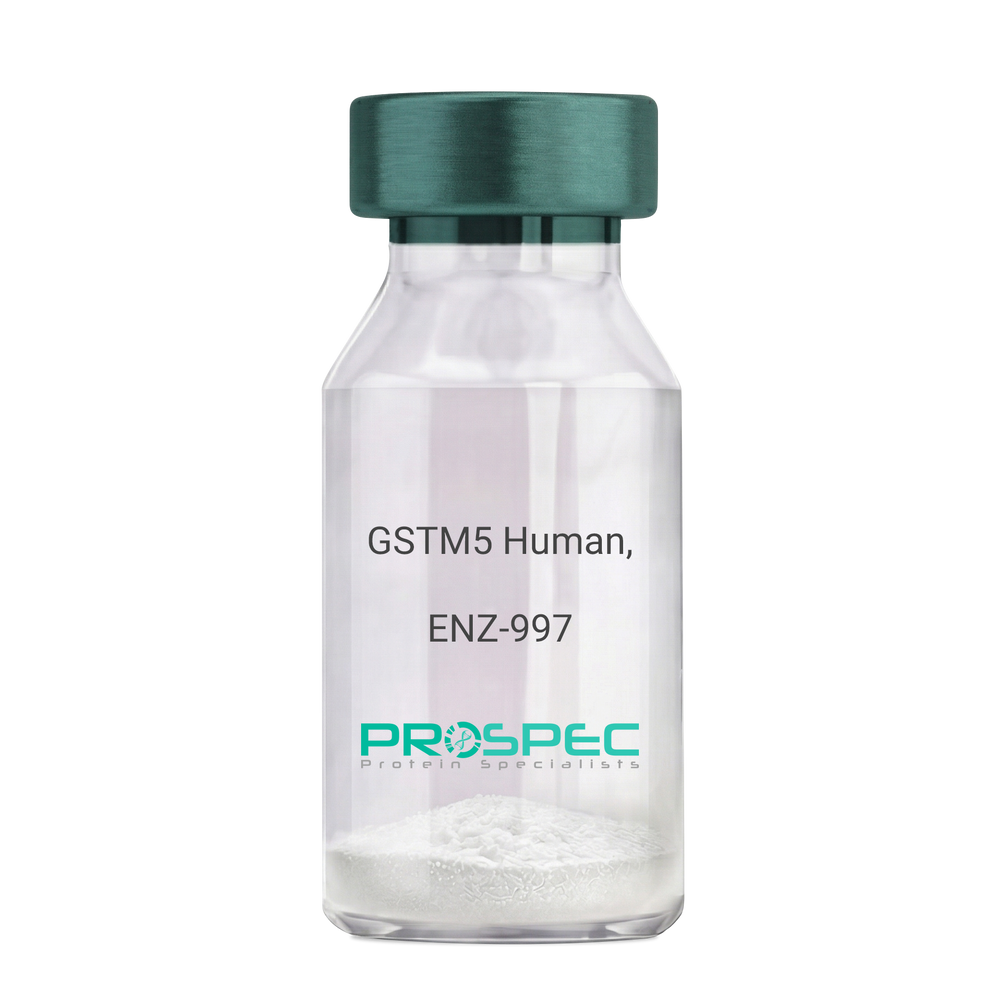
GSTM5 Human, ActiveDescription:
Glutathione S-Transferase MU 5 Human Recombinant, Active
Glutathione S-transferase mu 5, GSTM5-5, GST class-mu 5, GTM5, glutathione S-alkyltransferase M5, S-(hydroxyalkyl) glutathione lyase M5, EC 2.5.1.18.
Product # :
ENZ-997Price :
Quantity :
Shipping Method :
Shipped with Ice Packs
More Info
- description
- source
- formulation
- purity
- biological activity
- More Info
Description
GSTM5 Human Recombinant produced in E. coli is a single polypeptide chain containing 242 amino acids (1-218) and having a molecular mass of 28.2 kDa.GSTM5 is fused to a 24 amino acid His-tag at N-terminus & purified by proprietary chromatographic techniques.
Source
E.coli.
Formulation
The GSTM5 solution (1mg/ml) contains 20mM Tris-HCl buffer (pH 8.0), 0.1M NaCl, 1mM DTT and 10% glycerol.
Purity
Greater than 95% as determined by SDS-PAGE.
Biological Activity
Specific activity is > 90,000 pmol/min/ug, and is defined as the amount of enzyme that conjugate 1.0 u mole of 1-chloro-2,4- dinitrobenzene (CDNB) with reduced glutathione per minute at pH 6.5 at 25C.More Info
-
Introduction
Glutathione S-transferase mu 5 (GSTM5) belongs to the glutathione s-transferase (GST) family of proteins. There are 8 families of GST proteins, specifically alpha, kappa, mu, omega, pi, sigma, theta and zeta, each of which is comprised of proteins which have various functions throughout the cell. GSTM5 belongs to the mu class of enzymes which function in the detoxification of electrophilic compounds, including carcinogens, therapeutic drugs, environmental toxins and products of oxidative stress, by conjugation with glutathione. GSTM5 has an imperative role in detoxification. GSTM5 conjugates reduced glutathione to a large number of exogenous and endogenous hydrophobic electrophiles.
-
Synonyms
Glutathione S-transferase mu 5, GSTM5-5, GST class-mu 5, GTM5, glutathione S-alkyltransferase M5, S-(hydroxyalkyl) glutathione lyase M5, EC 2.5.1.18.
-
Physical Appearance
Sterile Filtered colorless solution.
-
Stability
Store at 4°C if entire vial will be used within 2-4 weeks. Store, frozen at -20°C for longer periods of time. For long term storage it is recommended to add a carrier protein (0.1% HSA or BSA).Avoid multiple freeze-thaw cycles.
-
Amino Acid Sequence
MGSSHHHHHH SSGLVPRGSH MGSHMPMTLG YWDIRGLAHA IRLLLEYTDS SYVEKKYTLG DAPDYDRSQW LNEKFKLGLD FPNLPYLIDG AHKITQSNAI LRYIARKHNL CGETEEEKIR VDILENQVMD NHMELVRLCY DPDFEKLKPK YLEELPEKLK LYSEFLGKRP WFAGDKITFV DFLAYDVLDM KRIFEPKCLD AFLNLKDFIS RFEGLKKISA YMKSSQFLRG LLFGKSATWN SK.
ProSpec's products are furnished for LABORATORY RESEARCH USE ONLY. The product may not be used as drugs, agricultural or pesticidal products, food additives or household chemicals.
- View Data Sheet
Name :
NRG1 (HRG-beta3) AntibodyDescription:
Neuregulin1 isoform HRG-beta3, Mouse Anti Human
Product # :
ANT-458Price :
Quantity :
Shipping Method :
Shipped with Ice Packs
More Info
- formulation
- More Info
Formulation
1mg/ml containing PBS, pH-7.4, & 0.1% Sodium Azide.
More Info
-
Introduction
NRG1 is one of 4 proteins in the neuregulin family which act on the EGFR family of receptors. NRG1 was initially identified as a 44kDa glycoprotein that interacts with the ERBB2 receptor tyrosine kinase to increase its phosphorylation on tyrosine residues. In various brain areas, NRG1 is expressed at synaptic regions and its processing can be regulated by neuronal activity. The HRG-beta3variant of NRG1, along with HRG-alpha, beta1 and beta2 variants, was identified in various normal tissues and cancer cell lines.
-
Immunogen
Anti-human NRG1 mAb is derived from hybridization of mouse F0 myeloma cells with spleen cells from BALB/c mice immunized with recombinant human NRG1 amino acids 20-241 purified from E. coli.
-
Ig Subclass
Mouse IgG1 heavy chain and κ light chain.
-
Clone
Pk1G13AT.
-
Applications
NRG1 antibody has been tested by ELISA and Western blot analysis to assure specificity and reactivity. Since application varies, however, each investigation should be titrated by the reagent to obtain optimal results. Recommended dilution range for Western blot analysis is 1:1,000 ~ 2,000. Recommended starting dilution is 1:1,000.
-
Type
Mouse Anti Human Monoclonal.
-
Storage Procedures
For periods up to 1 month store at 4°C, for longer periods of time, store at -20°C. Prevent freeze thaw cycles.
-
Purification Method
NRG1 antibody was purified from mouse ascitic fluids by protein-G affinity chromatography.
ProSpec's products are furnished for LABORATORY RESEARCH USE ONLY. The product may not be used as drugs, agricultural or pesticidal products, food additives or household chemicals.
- View Data Sheet
Name :
MCP 1 AntibodyDescription:
Macrophage/Monocyte Chemotactic & Activating Factor, Mouse Anti-Human
Small inducible cytokine A2, CCL2, Monocyte chemotactic protein 1, MCP-1, Monocyte chemoattractant protein 1, Monocyte chemotactic and activating factor, MCAF, Monocyte secretory protein JE, HC11, chemokine (C-C motif) ligand 2, MCP1, SCYA2, GDCF-2, SMC-CF, HSMCR30, MGC9434, GDCF-2 HC11.
Product # :
ANT-119Price :
Quantity :
Shipping Method :
Shipped at Room temp
More Info
- formulation
- More Info
Formulation
1mg/ml in PBS (after reconstitution) Shipping ConditionsAntibody is shipped lyophilized at ambient temperature.
More Info
-
Introduction
Chemokine (C-C motif) ligand 2 (CCL2) is a small cytokine belonging to the CC chemokine family that is also known as monocyte chemotactic protein-1 (MCP-1). It is found at the site of tooth eruption and bone degradation. In the bone, CCL2 is expressed by mature osteoclasts and osteoblasts and is under the control of nuclear factor ?B (NF?B). CCL2 recruits immune cells, such as monocytes, to sites of tissue injury and infection. This chemokine is produced as a protein precursor containing signal peptide of 23 amino acids and a mature peptide of 76 amino acids. It is a monomeric polypeptide, with a molecular weight of approximately 13kDa. As with many other CC chemokines, CCL2 is located on chromosome 17 in humans. The cell surface receptors that bind CCL2 are CCR2 and CCR5.
-
Synonyms
Small inducible cytokine A2, CCL2, Monocyte chemotactic protein 1, MCP-1, Monocyte chemoattractant protein 1, Monocyte chemotactic and activating factor, MCAF, Monocyte secretory protein JE, HC11, chemokine (C-C motif) ligand 2, MCP1, SCYA2, GDCF-2, SMC-CF, HSMCR30, MGC9434, GDCF-2 HC11.
-
Solubility
Reconstitute with sterile H20. Mix gently, wash the sides of the vial and wait 30-60 seconds before use.
-
Immunogen
r.Human MCAF.
-
Ig Subclass
Mouse IgG2a.
-
Clone
YNR-HMCAF.
-
Applications
Direct ELISA, Western Blot, Immuneprecipitation.
-
Note
MCAF does not bind to plastic as good as most proteins - use higher concentration for proper binding.
-
Titer
In direct ELISA, using alkaline phosphatase goat anti-mouse Ig (Jackson Laboratories) 1:5,000 dilution will give 0.3 O.D within 10 minutes.
-
Type
Mouse Anti Human Monoclonal.
-
Storage Procedures
In lyophilized form, for long periods, store at 4oC in a dry environment. After reconstitution, if not intended for use within a month, aliquot and store at -20oC.
-
Purification Method
Ion exchange.
ProSpec's products are furnished for LABORATORY RESEARCH USE ONLY. The product may not be used as drugs, agricultural or pesticidal products, food additives or household chemicals.
- View Data Sheet
Name :
TXN2 AntibodyDescription:
Thioredoxin-2, Mouse Anti Human
Thioredoxin mitochondrial, Thioredoxin-2, TXN2, MTRX, TRX2, MT-TRX, TRX-2, TXN-2.
Product # :
ANT-674Price :
Quantity :
Shipping Method :
Shipped with Ice Packs
More Info
- formulation
- More Info
Formulation
1mg/ml containing PBS, pH-7.4, 10% Glycerol and 0.02% Sodium Azide.
More Info
-
Introduction
Thioredoxin-2 is a low molecular weight redox protein. TRX2 contains a redox active disulfide/dithiol group within the conserved Cys-Gly-Pro-Cys active site. The TXN2 is involved in the regulation of the mitochondrial membrane potential and in protection against oxidant-induced apoptosis. Upon stimulation of Fas, TXN2 mediates denitrosylation of mitochondria-associated caspase-3, a process required for caspase-3 activation, and promoted apoptosis.
TRX2 is important at low oxidative stress conditions.
MTRX is involved in the regulation of the mitochondrial membrane potential and cell death. Mitochondrial thioredoxin plays an important roles in protection against oxidant-induced apoptosis. Thioredoxin1 and thioredoxin2 have opposed regulatory functions on hypoxia-inducible factor-1alpha. -
Synonyms
Thioredoxin mitochondrial, Thioredoxin-2, TXN2, MTRX, TRX2, MT-TRX, TRX-2, TXN-2.
-
Physical Appearance
Sterile filtered colorless solution.
-
Immunogen
Anti-human TXN2 mAb, is derived from hybridization of mouse F0 myeloma cells with spleen cells from BALB/c mice immunized with a recombinant human TXN2 protein 60-166 amino acids purified from E. coli.
-
Ig Subclass
Mouse IgG2b heavy chain and k light chain.
-
Clone
PAT1B1AT.
-
Applications
The antibody has been tested by ELISA, Western blot analysis, Flow cytometry and ICC/IF to assure specificity and reactivity. Since application varies, however, each investigation should be titrated by the reagent to obtain optimal results.
-
Type
Mouse Anti Human Monoclonal.
-
Storage Procedures
For periods up to 1 month store at 4°C, for longer periods of time, store at -20°C. Prevent freeze thaw cycles.
-
Purification Method
TXN2 antibody was purified from mouse ascitic fluids by protein-G affinity chromatography.
ProSpec's products are furnished for LABORATORY RESEARCH USE ONLY. The product may not be used as drugs, agricultural or pesticidal products, food additives or household chemicals.
- View Data Sheet
Name :
Calmodulin AntibodyDescription:
Calmodulin, Mouse Anti Human
Calmodulin, CaM, CALM2, PHKD, CAMII, PHKD2, calmodulin 2, phosphorylase kinase delta.
Product # :
ANT-342Price :
Quantity :
Shipping Method :
Shipped with Ice Packs
More Info
- formulation
- More Info
Formulation
1mg/ml containing PBS, pH-7.4, & 0.1% Sodium Azide.
More Info
-
Introduction
Calmodulin (CaM) is an intracellular receptor protein for Ca2+-ions. It activates the myosin light chain kinase which is the catalyst of myosin phosphorylation. CaM participates in the activation of enzymes such as cyclic nucleotide- dependent phosphodiesterase, calcineurin, ATPase, Myosin Light Chain Kinases, and CAM kinase.
-
Synonyms
Calmodulin, CaM, CALM2, PHKD, CAMII, PHKD2, calmodulin 2, phosphorylase kinase delta.
-
Immunogen
Anti-human Calmodulin mAb is derived from hybridization of mouse F0 myeloma cells with spleen cells from BALB/c mice immunized with recombinant human Calmodulin amino acids 1-149 purified from E. coli.
-
Ig Subclass
Mouse IgG2a heavy chain and κ light chain.
-
Clone
PJ4D8AT.
-
Applications
Calmodulin antibody has been tested by ELISA and Western blot analysis to assure specificity and reactivity. Since application varies, however, each investigation should be titrated by the reagent to obtain optimal results. Recommended dilution range for Western blot analysis is 1:500 ~ 1,000. Recommended starting dilution is 1:500.
-
Type
Mouse Anti Human Monoclonal.
-
Storage Procedures
For periods up to 1 month store at 4°C, for longer periods of time, store at -20°C. Prevent freeze thaw cycles.
-
Purification Method
Calmodulin antibody was purified from mouse ascitic fluids by protein-G affinity chromatography.
ProSpec's products are furnished for LABORATORY RESEARCH USE ONLY. The product may not be used as drugs, agricultural or pesticidal products, food additives or household chemicals.
- View Data Sheet
Name :
Vimentin Antibody, FITCDescription:
Vimentin, Mouse Anti Human Antibody, FITC
Product # :
ANT-247Price :
Quantity :
Shipping Method :
Shipped at Room temp
More Info
- formulation
- More Info
Formulation
1mg/ml in PBS (after reconstitution).
More Info
-
Introduction
Vimentin expression in human malignant glioma cells depends on cellular density, algorithms of drug delivery and chemo/radio treatment. Vimentin and detyrosinated microtubules provide structural support for the extensive microtentacles observed in detached tumor cells and a mechanism to promote successful metastatic spread. Primary colorectal carcinomas display aberrant expression of vimentin, and have activated Notch and TGFbeta signaling pathways. Vimentin is a strong arterial substrate for transglutaminases. Transglutaminase-mediated vimentin dimerization results in a novel unifying pathway by which vasodilatory and remodeling responses may be regulated. Ablation of vimentin expression inhibits migration and invasion of colon and breast cancer cell lines.
-
Solubility
Reconstitute with H20. Mix gently, wash the sides of the vial and wait 30-60 seconds before use.
-
Immunogen
Purified Human Vimentin.
-
Ig Subclass
mouse IgG1.
-
Clone
hVimentin.
-
Applications
Immunohistochemistry.
-
Cross reactivity
Vimentin antibody will recognize most mammalian Vimentin except mouse and rat.
-
Titer
For intra-cellular staining, use 5-10µl/1,000,000 cells. Wash twice with PBS/Azide before adding the appropriate amount of secondary fluorescent reagent.
-
Shipping Conditions
Antibody is shipped lyophilized at ambient temperature.
-
Available Conjugates
This antibody is also available conjugated to Biotin and non conjugate form.
-
Type
Mouse Anti Human Monoclonal Antibody.
-
Storage Procedures
In lyophilized form, for long periods, store at 4°C in a dry environment. After reconstitution, if not intended for use within a month, aliquot and store at -20°C.
-
Purification Method
Ion exchange column.
ProSpec's products are furnished for LABORATORY RESEARCH USE ONLY. The product may not be used as drugs, agricultural or pesticidal products, food additives or household chemicals.
- View Data Sheet
Name :
Frataxin AntibodyDescription:
Frataxin, Mouse Anti Human
FXN, Friedreich ataxia protein, Frataxin mitochondrial, FRDA, X25, FA, CyaY, FARR, MGC57199, Frataxin.
Product # :
ANT-671Price :
Quantity :
Shipping Method :
Shipped with Ice Packs
More Info
- formulation
- More Info
Formulation
1mg/ml containing PBS, pH-7.4, 10% Glycerol and 0.02% Sodium Azide.
More Info
-
Introduction
Frataxin is a mitochondrial iron binding protein that is part of the FRATAXIN family. Frataxin functions in regulating mitochondrial iron transport and respiration. The expansion of intronic trinucleotide repeat GAA results in Friedreich ataxia. Frataxin plays a role in iron homeostasis. Frataxin is an anti-apoptotic protein which prevents mitochondrial damage and reactive oxygen species (ROS) production.
-
Synonyms
FXN, Friedreich ataxia protein, Frataxin mitochondrial, FRDA, X25, FA, CyaY, FARR, MGC57199, Frataxin.
-
Physical Appearance
Sterile filtered colorless solution.
-
Immunogen
Anti-human Frataxin mAb, is derived from hybridization of mouse F0 myeloma cells with spleen cells from BALB/c mice immunized with a recombinant human Frataxin protein 42-210 amino acids purified from E. coli.
-
Ig Subclass
Mouse IgG1 heavy chain and k light chain.
-
Clone
PAT10E11AT.
-
Applications
The antibody has been tested by ELISA, Western blot analysis and ICC/IF to assure specificity and reactivity. Since application varies, however, each investigation should be titrated by the reagent to obtain optimal results.
-
Type
Mouse Anti Human Monoclonal.
-
Storage Procedures
For periods up to 1 month store at 4°C, for longer periods of time, store at -20°C. Prevent freeze thaw cycles.
-
Purification Method
Frataxin antibody was purified from mouse ascitic fluids by protein-A affinity chromatography.
ProSpec's products are furnished for LABORATORY RESEARCH USE ONLY. The product may not be used as drugs, agricultural or pesticidal products, food additives or household chemicals.
- View Data Sheet
Name :
Dengue type 2 AntibodyDescription:
Dengue Type 2 (envelop), Mouse antibody
Product # :
ANT-143Price :
Quantity :
Shipping Method :
Shipped at Room temp
More Info
- formulation
- More Info
Formulation
1mg/ml in PBS (after reconstitution).
More Info
-
Introduction
Caused by one of four closely related virusserotypesof the genus Flavivirus, family Flaviviridae, each serotype is sufficiently different that there is no cross-protection and epidemics caused by multiple serotypes (hyperendemicity) can occur. In cell culture experiments and mice Morpholinoantisense oligos have shown specific activity against Dengue virus.
-
Solubility
Reconstitute with H20. Mix gently, wash the sides of the vial and wait 30-60 seconds before use.
-
Immunogen
Dengue-infected cell lysate.
-
Ig Subclass
mouse IgG1.
-
Clone
NYRDeng2.
-
Note
Minimal cross-reactivity with other Dengue subtypes.
-
Titer
By intracellular staining of Dengue-infected cells, 1:1000 dilution.
-
Shipping Conditions
Antibody is shipped lyophilized at ambient temperature.
-
Type
Mouse antibody Monoclonal.
-
Storage Procedures
In lyophilized form, for long periods, store at 4ºC in a dry environment. After reconstitution, if not intended for use within a month, aliquot and store at -20ºC.
-
Purification Method
Ion exchange column.
ProSpec's products are furnished for LABORATORY RESEARCH USE ONLY. The product may not be used as drugs, agricultural or pesticidal products, food additives or household chemicals.
- View Data Sheet
Name :
C-Myc (410-419aa) AntibodyDescription:
c-Myc (410-419aa), Mouse Anti Human
MYC, CMYC, C-MYS, V-MYC, P64.
Product # :
ANT-618Price :
Quantity :
Shipping Method :
Shipped with Ice Packs
More Info
- formulation
- More Info
Formulation
1mg/ml containing PBS, pH-7.4, 10% Glycerol and 0.02% Sodium Azide.
More Info
-
Introduction
c-Myc is a multifunctional, nuclear phosphoprotein that plays a role in cell cycle progression, apoptosis and cellular transformation. It functions as a transcription factor that regulates transcription of specific target genes. Mutations, overexpression, rearrangement and translocation of c-Myc have been associated with a variety of hematopoietic tumors, leukemias and lymphomas, including Burkitt lymphoma. There is evidence to show that alternative translation initiations from an upstream, in-frame non-AUG (CUG) and a downstream AUG start site result in the production of two isoforms with distinct N-termini. The synthesis of non-AUG initiated protein is suppressed in Burkitt's lymphomas, suggesting its importance in the normal function of this gene.
-
Synonyms
MYC, CMYC, C-MYS, V-MYC, P64.
-
Physical Appearance
Sterile filtered colorless solution.
-
Immunogen
Anti-human C-Myc mAb, is derived from hybridization of mouse F0 myeloma cells with spleen cells from BALB/c mice immunized with a recombinant human C-Myc protein 410-419 amino acids purified from E. coli.
-
Ig Subclass
Mouse IgG1 heavy chain and k light chain.
-
Clone
PAT19B4AT.
-
Applications
The antibody has been tested by ELISA, Western blot analysis to assure specificity and reactivity. Since application varies, however, each investigation should be titrated by the reagent to obtain optimal results.
-
Type
Mouse Anti Human Monoclonal.
-
Storage Procedures
For periods up to 1 month store at 4°C, for longer periods of time, store at -20°C. Prevent freeze thaw cycles.
-
Purification Method
C-Myc antibody was purified from mouse ascitic fluids by protein-A affinity chromatography.
ProSpec's products are furnished for LABORATORY RESEARCH USE ONLY. The product may not be used as drugs, agricultural or pesticidal products, food additives or household chemicals.