Search results
1000 results found for “rassf”
Name
Description
Product #
Price
Quantity
Shipping Method
- View Data Sheet
Name :
TFRC NativeDescription:
Transferrin Receptor Human
Transferrin receptor protein 1, TR, TfR, TfR1, Trfr, T9, p90, CD_antigen: CD71, Transferrin receptor, serum form, sTfR, TFRC, CD71
Product # :
PRO-2742Price :
Quantity :
Shipping Method :
Shipped with Ice Packs
More Info
- description
- source
- formulation
- More Info
Description
Human Transferrin Receptor Protein produced in human serum tissue having a molecular mass of 85kDa.
Source
Human serum.
Formulation
TFRC Native solution (0.2µm filtered) contains 250mM TRIS-HCl buffer, 0.15M NaCl, 0.09% NaN3, pH 8.0.
More Info
-
Introduction
Transferrin receptor protein 1 (TFRC) is required for iron delivery from transferring to cells. The TFRC protein is a transmembrane glycoprotein comprised of 2 disulfide-linked monomers joined by 2 disulfide bonds. Each monomer will bind one holo-transferrin molecule producing an iron-Tf-TfR complex which enters the cell by endocytosis.
-
Synonyms
Transferrin receptor protein 1, TR, TfR, TfR1, Trfr, T9, p90, CD_antigen: CD71, Transferrin receptor, serum form, sTfR, TFRC, CD71
-
Physical Appearance
Sterile Filtered brown solution.
-
Stability
Store at 4°C if entire vial will be used within 2-4 weeks. Store, frozen at -20°C for longer periods of time. For long term storage it is recommended to add a carrier protein (0.1% HSA or BSA). Avoid multiple freeze-thaw cycles.
-
Human Virus Test
Starting material donor has been tested and certified negative for antibodies to HIV-1, HIV-2, HCV, HBSAG, Parvovirus B19, Syphilis and HIV/HBV/HCV (PCR).
ProSpec's products are furnished for LABORATORY RESEARCH USE ONLY. The product may not be used as drugs, agricultural or pesticidal products, food additives or household chemicals.
- View Data Sheet
Name :
RBP Human, NativeDescription:
Retinol Binding Protein Native Human
Product # :
CYT-1203Price :
Quantity :
Shipping Method :
Shipped at Room temp
More Info
- description
- source
- formulation
- purity
- More Info
Description
Human Retinol Binding Protein Native produced in urine from the patients with renal tubular proteinuria having a molecular mass of approximately 21kD.
Source
Urine from the patients with renal tubular proteinuria.
Formulation
The protein was lyophilized (0.2 µm filtered) from 20mM NH4HCO3.
Purity
Greater than 96.0%.
More Info
-
Introduction
Human Retinol Binding Protein, also known as RBP, is responsible for transporting and binding vitamin A. Human RBP has a binding site for 1 molecule of retinol and circulates in the plasma together with prealbumin as a protein complex. The prealbumin binding prevents greater glomerular losses of the human RBP. Only the retinol-free form of the RBP, which has no affinity for prealbumin, undergoes glomerular filtration unhindered as a result of its low Mw. Human RBP is re-absorbed by the tubular cells and catabolized there.
-
Physical Appearance
Sterile Filtered White lyophilized (freeze-dried) powder.
-
Stability
RBP Human although stable at room temperature for 3 weeks, should be stored between 2-8°C.
-
Solubility
It is recommended to reconstitute the lyophilized RBP Human in phosphate buffer containing 0.15M NaCl.
-
Human Virus Test
Starting material donor has been tested and certified negative for antibodies to HIV-1, HIV-2, HCV, HBSAG and Syphilis.
ProSpec's products are furnished for LABORATORY RESEARCH USE ONLY. The product may not be used as drugs, agricultural or pesticidal products, food additives or household chemicals.
- View Data Sheet
Name :
GM-CSF K9Description:
Granulocyte Macrophage-Colony Stimulating Factor Canine Recombinant
CSF-2, MGI-1GM, GM-CSF, Pluripoietin-alpha, MGC131935, MGC138897.
Product # :
CYT-724Price :
Quantity :
Shipping Method :
Shipped at Room temp
More Info
- description
- source
- formulation
- purity
- biological activity
- More Info
Description
GMCSF k9 Recombinant produced in E.Coli is a single, non-glycosylated, polypeptide chain containing 128 amino acids and having a molecular mass of 14.2 kDa. GM-CSF is purified by proprietary chromatographic techniques.
Source
Escherichia Coli.
Formulation
GMCSF was lyophilized after extensive dialysis against 1xPBS pH 7.4.
Purity
Greater than 96.0% as determined by
1. Analysis by RP-HPLC.
2. Analysis by SDS-PAGE.Biological Activity
The ED50 as calculated by the dose-dependent stimulation of the proliferation of human TF1 erythroleukemic cells is typically 1-4 ng/ml.More Info
-
Introduction
GMCSF is a cytokine that controls the production, differentiation, and function of granulocytes and macrophages. The active form of the protein is found extracellularly as a homodimer. This gene has been localized to a cluster of related genes at chromosome region 5q31, which is known to be associated with interstitial deletions in the 5q- syndrome and acute myelogenous leukemia. Other genes in the cluster include those encoding interleukins 4, 5, and 13.
GM-CSF stimulates the growth and differentiation of hematopoietic precursor cells from various lineages, including granulocytes, macrophages, eosinophils and erythrocytes. -
Synonyms
CSF-2, MGI-1GM, GM-CSF, Pluripoietin-alpha, MGC131935, MGC138897.
-
Physical Appearance
Sterile Filtered White lyophilized (freeze-dried) powder.
-
Stability
Lyophilized GMCSF although stable at room temperature for 3 weeks, should be stored desiccated below -18°C. Upon reconstitution GMCSF should be stored at 4°C between 2-7 days and for future use below -18°C. For long term storage it is recommended to add a carrier protein (0.1% HSA or BSA).Please prevent freeze-thaw cycles.
-
Solubility
It is recommended to reconstitute the lyophilized GMCSF in sterile 18MΩ-cm H2O not less than 100µg/ml, which can then be further diluted to other aqueous solutions.
-
Amino Acid Sequence
APTRSPTLVT RPSQHVDAIQ EALSLLNNSN DVTAVMNKAV KVVSEVFDPE
GPTCLETRLQ LYKEGLQGSL TSLKNPLTMM ANHYKQHCPP TPESPCATQN
INFKSFKENL KDFLFNIPFD CWKPVKK. -
Background
What is the molecular weight/Mw of GM-CSF K9 Protein?
GM-CSF K9 Protein has a total Mw of 14.2kDa.
What is the source or expression system of GM-CSF K9 Protein?
Escherichia Coli.
What is the Purity of GM-CSF K9 Protein?
GM-CSF K9 Protein is >96% pure as determined by SDS-PAGE.
What is the Biological Activity of GM-CSF K9 Protein?
The ED50 as calculated by the dose-dependent stimulation of the proliferation of human TF1 erythroleukemic cells is typically 1-4 ng/ml.
What is the amino acid sequence of GM-CSF K9 Protein?
APTRSPTLVT RPSQHVDAIQ EALSLLNNSN DVTAVMNKAV KVVSEVFDPE
GPTCLETRLQ LYKEGLQGSL TSLKNPLTMM ANHYKQHCPP TPESPCATQN
INFKSFKENL KDFLFNIPFD CWKPVKK.
What applications can GM-CSF K9 Protein be used in?
GM-CSF K9 Protein can probably be used in western blot, ELISA and Lateral Flow.
What is the endotoxin level for GM-CSF K9 Protein?
The endotoxin level is minimal, GM-CSF K9 Protein was purified using conventional chromatography techniques.
ProSpec's products are furnished for LABORATORY RESEARCH USE ONLY. The product may not be used as drugs, agricultural or pesticidal products, food additives or household chemicals.
- View Data Sheet
Name :
b NGF Human, HEKDescription:
beta Nerve Growth Factor Human Recombinant, HEK
Beta Polypeptide, NGF, NGFB, HSAN5, Beta-NGF, MGC161426, MGC161428.
Product # :
CYT-079Price :
Quantity :
Shipping Method :
Shipped at Room temp
More Info
- description
- source
- formulation
- purity
- biological activity
- More Info
Description
BNGF Human Recombinant produced in HEK293 is a noncovalently disulfide linked homodimer, glycosylated, polypeptide chain (Ser122-Arg239) containing 2 identical 118 amino acids and having a molecular mass of 26.5 kDa.
Source
HEK293 cells.
Formulation
The b-NGF was lyophilized from 1mg/ml in 20mM PB and 0.25M NaCl pH-7.5.
Purity
Greater than 97% as obsereved by SDS-PAGE.
Biological Activity
The specific activity was determined by the dose-dependent stimulation of the proliferation of human TF-1 cells (human erythroleukemic indicator cell line), the ED50 is <0.04-0.4ng/ml.
More Info
-
Introduction
NGF-beta has nerve growth stimulating activity and the complex is involved in the regulation of growth and the differentiation of sympathetic and certain sensory neurons. Mutations in this gene have been associated with hereditary sensory and autonomic neuropathy, type 5 (HSAN5), and dysregulation of this gene's expression is associated with allergic rhinitis.
-
Synonyms
Beta Polypeptide, NGF, NGFB, HSAN5, Beta-NGF, MGC161426, MGC161428.
-
Physical Appearance
Sterile Filtered White lyophilized (freeze-dried) powder.
-
Stability
Lyophilized b-NGF although stable at room temperature for 3 weeks, should be stored desiccated below -18°C. Upon reconstitution b-NGF should be stored at 4°C between 2-7 days and for future use below -18°C.For long term storage it is recommended to add a carrier protein (0.1% HSA or BSA).Please prevent freeze-thaw cycles.
-
Solubility
It is recommended to reconstitute the lyophilized b-NGF in sterile distilled pyrogen free water at a concentration of 0.25mg/ml.
-
Background
What is the molecular weight/Mw of B NGF Protein?
B NGF Protein has a total Mw of 26.5kDa.
What is the source or expression system of B NGF Protein?
HEK293 cells.
What is the Purity of B NGF Protein?
B NGF Protein is >97% pure as determined by SDS-PAGE.
What is the Biological Activity of B NGF Protein?
The specific activity was determined by the dose-dependent stimulation of the proliferation of human TF-1 cells (human erythroleukemic indicator cell line), the ED50 is <0.04-0.4ng/ml.
What is the amino acid sequence of B NGF Protein?
B NGF Protein is composed from 118 amino acids.
What applications can B NGF Protein be used in?
B NGF Protein can probably be used in western blot, ELISA and Lateral Flow.
What is the endotoxin level for B NGF Protein?
The endotoxin level is minimal, B NGF Protein was purified using conventional chromatography techniques.
ProSpec's products are furnished for LABORATORY RESEARCH USE ONLY. The product may not be used as drugs, agricultural or pesticidal products, food additives or household chemicals.
- View Data Sheet
Name :
PDGF AA MouseDescription:
Platelet-derived Growth Factor AA Mouse Recombinant
Glioma-derived growth factor, GDGF, Osteosarcoma-derived Growth Factor, ODGF, PDGF-AA, PDGF-1.
Product # :
CYT-590Price :
Quantity :
Shipping Method :
Shipped at Room temp
More Info
- description
- source
- formulation
- purity
- biological activity
- More Info
Description
PDGF-AA Mouse Recombinant is a disulfide linked homodimeric, non-glycosylated, polypeptide chain containing 2 x 126 amino acids and having a total molecular mass of 28.9 kDa. PDGF-AA is purified by proprietary chromatographic techniques.
Source
Escherichia Coli.
Formulation
The protein was lyophilized with no additives.
Purity
Greater than 97.0% as determined by:
(a) Analysis by RP-HPLC.
(b) Analysis by SDS-PAGE.Biological Activity
Established by the dose-dependent stimulation of Balb/c 3T3 cells proliferation. The expected ED50 for this effect is 8-10 ng/ml corresponding to a specific activity of 100,000-125,000IU/mg.More Info
-
Introduction
PDGF-AA, PDGF-BB and PDGF-AB, are potent mitogens for a variety of cell types including smooth muscle cells, connective tissue cells, bone and cartilage cells, and some blood cells. The PDGF is stored in platelet alpha-granules and released upon platelet activation. The PDGF is involved in a number of biological processes, including hyperplasia, chemotaxis, embryonic neuron development, and respiratory tubule epithelial cell development. Two distinct signaling receptors used by PDGF have been identified and named PDGFR-alpha and PDGFR-beta. PDGFR-alpha is high-affinity receptor for each of the three PDGF forms. On the other hand, PDGFR-beta interacts with only PDGF-BB and PDGF-AB.
-
Synonyms
Glioma-derived growth factor, GDGF, Osteosarcoma-derived Growth Factor, ODGF, PDGF-AA, PDGF-1.
-
Physical Appearance
Sterile Filtered White lyophilized (freeze-dried) powder.
-
Stability
Lyophilized PDGF-AA although stable at room temperature for 3 weeks, should be stored desiccated below -18°C. Upon reconstitution PDGF-AA should be stored at 4°C between 2-7 days and for future use below -18°C.For long term storage it is recommended to add a carrier protein (0.1% HSA or BSA).Please prevent freeze-thaw cycles.
-
Solubility
It is recommended to reconstitute the lyophilized PDGF-AA in sterile 18MΩ-cm H2O at a concentration ranging between 0.1-0.5mg per 1ml, which can then be further diluted to other aqueous solutions.
-
Amino Acid Sequence
MSIEEAVPAV CKTRTVIYEI PRSQVDPTSA NFLIWPPCVE VKRCTGCCNT SSVKCQPSRV HHRSVKVAKV EYVRKKPKLK EVQVRLEEHL ECACATSNLN PDHREEETGR RRESGKNRKR KRLKPT.
ProSpec's products are furnished for LABORATORY RESEARCH USE ONLY. The product may not be used as drugs, agricultural or pesticidal products, food additives or household chemicals.
- View Data Sheet
Name :
SCF Human, HEKDescription:
Stem Cell Factor Human Recombinant, HEK
Kit ligand Precursor, C-kit ligand, SCF, Mast cell growth factor, MGF, SF, KL-1, Kitl, DKFZp686F2250.
Product # :
CYT-111Price :
Quantity :
Shipping Method :
Shipped at Room temp
More Info
- description
- source
- formulation
- purity
- biological activity
- More Info
Description
SCF Human Recombinant produced in HEK cells is a glycosylated monomer, having a molecular weight range of 35-45kDa due to glycosylation.The SCF is purified by proprietary chromatographic techniques.
Source
HEK.
Formulation
SCF was lyophilized from a 0.2µm filtered solution (1mg/ml) containing 1xPBS.
Purity
Greater than 95% as obsereved by SDS-PAGE.
Biological Activity
The specific activity was determined by the dose-dependent stimulation of the proliferation of human TF-1 cells (human erythroleukemic indicator cell line).
The EC50 is 15.25ng/ml.More Info
-
Introduction
Stem cell factor / KIT ligand (SCF) is a cytokine which binds CD117(c-Kit). SCF is also known as "steel factor" or "c-kit ligand". SCF exists in two forms, cell surface bound SCF and soluble (or free) SCF. Soluble SCF is produced by the cleavage of surface bound SCF by metalloproteases. SCF is a growth factor important for the survival, proliferation, and differentiation of hematopoietic stem cells and other hematopoietic progenitor cells. One of its roles is to change the BFU-E (burst-forming unit-erythroid) cells, which are the earliest erythrocyte precursors in the erythrocytic series, into the CFU-E (colony-forming unit-erythroid).
-
Synonyms
Kit ligand Precursor, C-kit ligand, SCF, Mast cell growth factor, MGF, SF, KL-1, Kitl, DKFZp686F2250.
-
Physical Appearance
Sterile Filtered White lyophilized (freeze-dried) powder.
-
Stability
Lyophilized SCF although stable at room temperature for 3 weeks, should be stored desiccated below -18°C. Upon reconstitution SCF should be stored at 4°C between 2-7 days and for future use below -18°C.For long term storage it is recommended to add a carrier protein (0.1% HSA or BSA).Please prevent freeze-thaw cycles.
-
Solubility
It is recommended to reconstitute the lyophilized SCF in sterile PBS containing 0.1% endotoxin-free recombinant HSA.
ProSpec's products are furnished for LABORATORY RESEARCH USE ONLY. The product may not be used as drugs, agricultural or pesticidal products, food additives or household chemicals.
- View Data Sheet
Name :
Bax MouseDescription:
Bax Mouse Recombinant
Apoptosis regulator BAX membrane isoform alpha, Bax, Bcl2-associated X protein.
Product # :
PRO-412Price :
Quantity :
Shipping Method :
Shipped with Ice Packs
More Info
- description
- source
- formulation
- purity
- More Info
Description
Bax Mouse Recombinant amino acid 38-171 produced in E.Coli is a single, non-glycosylated polypeptide chain. The Mouse Bax is purified by proprietary chromatographic techniques.
Source
Escherichia Coli.
Formulation
The protein contains 10mM Tris-HCL pH-8, 1mM EDTA and 250mM NaCl.
Purity
Greater than 95.0% as determined by(a) Analysis by RP-HPLC.
(b) Analysis by SDS-PAGE.More Info
-
Introduction
Bcl-2–associated X protein (Bax) belongs to the Bcl-2 protein family. Bax is a pro-apoptotic Bcl-2 protein containing BH1, BH2 and BH3 domains. Bax accelerates programmed cell death by binding to, and antagonizing the apoptosis repressor bcl2 or its adenovirus homolog e1b 19k protein. Bax induces the release of cytochrome c, activation of casp3, and thereby apoptosis. Bax is expressed in a wide variety of tissues, with highest levels in the testis and ovary. Tumor suppressor protein p53 upregulates the expression of BAX. Bax has been shown to be involved in p53-mediated apoptosis.
-
Synonyms
Apoptosis regulator BAX membrane isoform alpha, Bax, Bcl2-associated X protein.
-
Physical Appearance
Sterile Filtered colorless solution.
-
Stability
Store at 4°C if entire vial will be used within 2-4 weeks. Store, frozen at -20°C for longer periods of time. Avoid multiple freeze-thaw cycles.
ProSpec's products are furnished for LABORATORY RESEARCH USE ONLY. The product may not be used as drugs, agricultural or pesticidal products, food additives or household chemicals.
- View Data Sheet
Name :
CGREF1 HumanDescription:
Cell Growth Regulator With EF-Hand Domain 1 Human Recombinant
Cell Growth Regulator With EF-Hand Domain 1, Cell Growth Regulatory Gene 11 Protein, Hydrophobestin, CGR11, Cell Growth Regulator With EF Hand Domain Protein 1, Cell Growth Regulator With EF Hand Domain 1, Cell growth regulator with EF hand domain protein 1.
Product # :
PRO-2154Price :
Quantity :
Shipping Method :
Shipped with Ice Packs
More Info
- description
- source
- formulation
- purity
- More Info
Description
CGREF1 Human Recombinant produced in E.Coli is a single, non-glycosylated polypeptide chain containing 305 amino acids (20-301 a.a) and having a molecular mass of 32.3kDa. CGREF1 is fused to a 23 amino acid His-tag at N-terminus & purified by proprietary chromatographic techniques.
Source
Escherichia Coli.
Formulation
CGREF1 protein solution (0.5mg/ml) containing Phosphate Buffered Saline (pH7.4).
Purity
Greater than 85% as determined by SDS-PAGE.
More Info
-
Introduction
Cell Growth Regulator with EF-Hand Domain 1, also known as CGREF1 is a secreted calcium ion binding protein. CGREF1 includes two EF-hand domains & both EF-hands are essential for function. CGREF1 is most likely digested extracellularly by an unfamiliar serine protease generating extremely hydrophobic bioactive peptides. CGREF1 mediates cell-cell adhesion in a calcium-dependent manner. In addition, CGREF1 is capable to inhibit growth in more than a few cell lines.
-
Synonyms
Cell Growth Regulator With EF-Hand Domain 1, Cell Growth Regulatory Gene 11 Protein, Hydrophobestin, CGR11, Cell Growth Regulator With EF Hand Domain Protein 1, Cell Growth Regulator With EF Hand Domain 1, Cell growth regulator with EF hand domain protein 1.
-
Physical Appearance
Sterile filtered colorless solution.
-
Stability
Store at 4°C if entire vial will be used within 2-4 weeks. Store, frozen at -20°C for longer periods of time. For long term storage it is recommended to add a carrier protein (0.1% HSA or BSA).Avoid multiple freeze-thaw cycles.
-
Amino Acid Sequence
MGSSHHHHHH SSGLVPRGSH MGSAPKDGVT RPDSEVQHQL LPNPFQPGQE QLGLLQSYLK GLGRTEVQLE HLSREQVLLY LFALHDYDQS GQLDGLELLS MLTAALAPGA ANSPTTNPVI LIVDKVLETQ DLNGDGLMTP AELINFPGVA LRHVEPGEPL APSPQEPQAV GRQSLLAKSP LRQETQEAPG PREEAKGQVE ARRESLDPVQ EPGGQAEADG DVPGPRGEAE GQAEAKGDAP GPRGEAGGQA EAEGDAPGPR GEAGGQAEAR ENGEEAKELP GETLESKNTQ NDFEVHIVQV ENDEI.
ProSpec's products are furnished for LABORATORY RESEARCH USE ONLY. The product may not be used as drugs, agricultural or pesticidal products, food additives or household chemicals.
- View Data Sheet
Name :
RAMP1 HumanDescription:
Receptor Activity-Modifying Protein 1 Human Recombinant
Receptor (G Protein-Coupled) Activity Modifying Protein 1, Receptor (Calcitonin) Activity Modifying Protein 1, Calcitonin-Receptor-Like Receptor Activity-Modifying Protein 1, CRLR Activity-Modifying Protein 1.
Product # :
PRO-1841Price :
Quantity :
Shipping Method :
Shipped with Ice Packs
More Info
- description
- source
- formulation
- purity
- More Info
Description
RAMP1 Human Recombinant produced in E.coli is a single, non-glycosylated polypeptide chain containing 114 amino acids (27-117) and having a molecular mass of 12.9 kDa. RAMP1 is fused to a 23 amino acid His-tag at N-terminus.
Source
Escherichia Coli.
Formulation
The RAMP1 solution (0.25mg/ml) contains 20mM Tris-HCl buffer (pH 8.0), 0.4M Urea and 10% glycerol.
Purity
Greater than 80% as determined by SDS-PAGE.
More Info
-
Introduction
RAMP belongs to the type I transmembrane family of proteins called RAMP (receptor/calcitonin activity modifying proteins) which include an extracellular N terminus and a cytoplasmic C terminus. RAMPs are essential for CRLR transport to the plasma membrane. CRLR (calcitonin-receptor-like receptor) has seven transmembrane domains. Depending on which members of the RAMP family are expressed CRLR receptor functions as either an adrenomedullin receptor or a calcitonin-gene-related peptide (CGRP) receptor. In the presence of RAMP1, CRLR functions as a CGRP receptor. The RAMP1 protein takes part in the terminal glycosylation, maturation, and presentation of the CGRP receptor to the cell surface.
-
Synonyms
Receptor (G Protein-Coupled) Activity Modifying Protein 1, Receptor (Calcitonin) Activity Modifying Protein 1, Calcitonin-Receptor-Like Receptor Activity-Modifying Protein 1, CRLR Activity-Modifying Protein 1.
-
Physical Appearance
Sterile Filtered clear solution.
-
Stability
Store at 4°C if entire vial will be used within 2-4 weeks. Store, frozen at -20°C for longer periods of time. For long term storage it is recommended to add a carrier protein (0.1% HSA or BSA).Avoid multiple freeze-thaw cycles.
-
Amino Acid Sequence
MGSSHHHHHH SSGLVPRGSH MGSCQEANYG ALLRELCLTQ FQVDMEAVGE TLWCDWGRTI RSYRELADCT WHMAEKLGCF WPNAEVDRFF LAVHGRYFRS CPISGRAVRD PPGS
ProSpec's products are furnished for LABORATORY RESEARCH USE ONLY. The product may not be used as drugs, agricultural or pesticidal products, food additives or household chemicals.
- View Data Sheet
Name :
RBKS HumanDescription:
Ribokinase Human Recombinant
Ribokinase, RBKS, RBSK, DKFZp686G13268.
Product # :
PKA-358Price :
Quantity :
Shipping Method :
Shipped with Ice Packs
More Info
- description
- source
- formulation
- purity
- More Info
Description
RBKS Human Recombinant fused with a 20 amino acid His tag at N-terminus produced in E.Coli is a single, non-glycosylated, polypeptide chain containing 342 amino acids (1-322 a.a.) and having a molecular mass of 36.3kDa. The RBKS is purified by proprietary chromatographic techniques.
Source
Escherichia Coli.
Formulation
The RBKS solution (1mg/ml) contains 20mM Tris-HCl buffer (pH 8.0) and 10% glycerol.
Purity
Greater than 95.0% as determined by SDS-PAGE.
More Info
-
Introduction
Ribokinase (RBKS) is a member of the pfkB family of carbohydrate kinases. Ribokinase phosphorylates ribose to form ribose-5-phosphate in the presence of ATP and magnesium as a first step in ribose metabolism.
-
Synonyms
Ribokinase, RBKS, RBSK, DKFZp686G13268.
-
Physical Appearance
Sterile Filtered colorless solution.
-
Stability
Store at 4°C if entire vial will be used within 2-4 weeks. Store, frozen at -20°C for longer periods of time. For long term storage it is recommended to add a carrier protein (0.1% HSA or BSA).Please prevent freeze-thaw cycles.
-
Amino Acid Sequence
MGSSHHHHHH SSGLVPRGSH MAASGEPQRQ WQEEVAAVVV VGSCMTDLVS LTSRLPKTGE TIHGHKFFIG FGGKGANQCV QAARLGAMTS MVCKVGKDSF GNDYIENLKQ NDISTEFTYQ TKDAATGTAS IIVNNEGQNI IVIVAGANLL LNTEDLRAAA NVISRAKVMV CQLEITPATS LEALTMARRS GVKTLFNPAP AIADLDPQFY TLSDVFCCNE SEAEILTGLT VGSAADAGEA ALVLLKRGCQ VVIITLGAEG CVVLSQTEPE PKHIPTEKVK AVDTTGAGDS FVGALAFYLA YYPNLSLEDM LNRSNFIAAV SVQAAGTQSS YPYKKDLPLT LF.
ProSpec's products are furnished for LABORATORY RESEARCH USE ONLY. The product may not be used as drugs, agricultural or pesticidal products, food additives or household chemicals.
- View Data Sheet
Description:
Mouse Anti SARS Nucleocapsid Biotinylated
Product # :
ANT-764Price :
Quantity :
Shipping Method :
Shipped with Ice Packs
More Info
- description
- formulation
- purity
- More Info
Description
SARS Nucleoprotein Biotinylated antibody clone PT3851 is specific for the nucleocapsid SARS-CoV protein and recognizes the nucleoprotein of SARS-CoV-2 by ELISA.
Formulation
1mg/ml in DPBS, 1% BSA and 0.1% Proclin 950.
Purity
Greater than 90% as determined by SDS-PAGE gel.
More Info
-
Introduction
SARS Coronavirus is an enveloped virus containing three outer structural proteins, namely the membrane (M), envelope (E), and spike (S) proteins. Spike (S)-glycoprotein of the virus interacts with a cellular receptor and mediates membrane fusion to allow viral entry into susceptible target cells. Accordingly, S-protein plays an important role in virus infection cycle and is the primary target of neutralizing antibodies.It has recently been shown that SARS is caused by a human coronavirus. Human coronaviruses are the major cause of upper respiratory tract illness in humans, such as the common cold. Coronaviruses are positive-stranded RNA viruses, featuring the largest viral RNA genomes known to date (27-31 kb). The first step in coronavirus infection is binding of the viral spike protein, a 139-kDa protein, to certain receptors on host cells. The spike protein is the main surface antigen of the coronavirus. The most prominent protein in the culture supernatants infected with SARS virus is a 46 kDa nucleocapsid protein. This suggests that the nucleocapsid protein is a major immunogen that may be useful for early diagnostics.
-
Physical Appearance
Sterile Filtered liquid formulation.
-
Stability
Store at 4°C, stable for 2 weeks. For long-term storage, store at -20°C.
-
Immunogen
Recombinant protein fragment 1-49 a.a. of the SARS nucleoprotein.
-
Clone
PT3851
-
Applications
ELISA.
-
Isotype
Mouse IgG2b.
-
Type
Mouse antibody Monoclonal.
-
Purification Method
Protein A affinity purified.
ProSpec's products are furnished for LABORATORY RESEARCH USE ONLY. The product may not be used as drugs, agricultural or pesticidal products, food additives or household chemicals.
- View Data Sheet
Name :
MLF1 HumanDescription:
Myeloid Leukemia Factor 1 Human Recombinant
Myeloid leukemia factor 1, Myelodysplasia-myeloid leukemia factor 1, MLF1.
Product # :
PRO-100Price :
Quantity :
Shipping Method :
Shipped with Ice Packs
More Info
- description
- source
- formulation
- purity
- More Info
Description
MLF1 Human Recombinant fused with a 20 amino acid His tag at N-terminus produced in E.Coli is a single, non-glycosylated, polypeptide chain containing 288 amino acids (1-268 a.a.) and having a molecular mass of 32.8kDa (Molecular weight on SDS-PAGE will appear higher). The MLF1 is purified by proprietary chromatographic techniques.
Source
Escherichia Coli.
Formulation
The MLF1 solution (0.5 mg/ml) contains 20mM Tris-HCl buffer (pH8.0), 40% glycerol, 5mM DTT and 200mM NaCl.
Purity
Greater than 85.0% as determined by SDS-PAGE.
More Info
-
Introduction
Myeloid leukemia factor 1 (MLF1) is a member of the MLF family, and is a widely expressed negative regulator of cell cycle progression functioning upstream of the tumor suppressor p53. MLF1 hinders the erythropoietin-induced erythroid terminal differentiation by averting cells from exiting the cell cycle through suppression of CDKN1B/p27Kip1 levels. MLF1 generally functions in multi-potent progenitor cells, and its dysregulation may be to some extent responsible for leukemogenesis. Translocations between the MLF1 gene and nucleophosmin are linked to myelodysplastic syndrome and acute myeloid leukemia.
-
Synonyms
Myeloid leukemia factor 1, Myelodysplasia-myeloid leukemia factor 1, MLF1.
-
Physical Appearance
Sterile Filtered colorless solution.
-
Stability
Store at 4°C if entire vial will be used within 2-4 weeks. Store, frozen at -20°C for longer periods of time. For long term storage it is recommended to add a carrier protein (0.1% HSA or BSA).Avoid multiple freeze-thaw cycles.
-
Amino Acid Sequence
MGSSHHHHHH SSGLVPRGSH MFRMLNSSFE DDPFFSESIL AHRENMRQMI RSFSEPFGRD LLSISDGRGR AHNRRGHNDG EDSLTHTDVS SFQTMDQMVS NMRNYMQKLE RNFGQLSVDP NGHSFCSSSV MTYSKIGDEP PKVFQASTQT RRAPGGIKET RKAMRDSDSG LEKMAIGHHI HDRAHVIKKS KNKKTGDEEV NQEFINMNES DAHAFDEEWQ SEVLKYKPGR HNLGNTRMRS VGHENPGSRE LKRREKPQQS PAIEHGRRSN VLGDKLHIKG SSVKSNKK.
ProSpec's products are furnished for LABORATORY RESEARCH USE ONLY. The product may not be used as drugs, agricultural or pesticidal products, food additives or household chemicals.
- View Data Sheet
Name :
TFF2 Human, HisDescription:
Trefoil Factor-2 Human Recombinant, His Tag
TFF-2, Spasmolytic polypeptide, Spasmolysin, SML1, Trefoil factor 2, SP, TFF2.
Product # :
CYT-611Price :
Quantity :
Shipping Method :
Shipped at Room temp
More Info
- description
- source
- formulation
- purity
- More Info
Description
TFF-2 Human Recombinant produced in E.Coli is a single, non-glycosylated, polypeptide chain containing 116 amino acids (24-129) which includes a 10 amino acid His Tag fused at N-terminus and having a total molecular mass of 13.2 kDa. TFF2 Human Recombinant is purified by proprietary chromatographic techniques.
Source
Escherichia Coli.
Formulation
The TFF2 protein was lyophilized from 0.4μm filtered solution at a concentration of 0.5mg/ml containing 20mM Tris pH-7.5, and 20mM NaCl.
Purity
Greater than 95.0% as determined by SDS-PAGE.
More Info
-
Introduction
Proteins of the TFF family are characterized by obtaining a minimum of 1 copy of the trefoil motif, a 40-amino acid domain that contains 3 conserved disulfides. Trefoil Factors are stable secretory proteins expressed in gastrointestinal mucosa which protect the mucosa from insults, stabilize the mucus layer and affect healing of the epithelium.TFF2 inhibits gastric acid motility & secretion. TFF2 stabilizes glycoproteins in the mucus gel through interactions with carbohydrate side chains.
-
Synonyms
TFF-2, Spasmolytic polypeptide, Spasmolysin, SML1, Trefoil factor 2, SP, TFF2.
-
Physical Appearance
Filtered White lyophilized (freeze-dried) powder.
-
Stability
Lyophilized TFF2 although stable at room temperature for 3 weeks, should be stored desiccated below -18°C. Upon reconstitution TFF2 should be stored at 4°C between 2-7 days and for future use below -18°C.For long term storage it is recommended to add a carrier protein (0.1% HSA or BSA).Please prevent freeze-thaw cycles.
-
Solubility
It is recommended to add deionized water to prepare a working stock solution of approximately 0.5mg/ml and let the lyophilized pellet dissolve completely. Product is not sterile! Please filter the product by an appropriate sterile filter before using it in the cell culture.
-
Amino Acid Sequence
MKHHHHHHAS EKPSPCQCSR LSPHNRTNCG FPGITSDQCF DNGCCFDSSV TGVPWCFHPL PKQESDQCVM EVSDRRNCGY PGISPEECAS RKCCFSNFIF EVPWCFFPKSVEDCHY.
ProSpec's products are furnished for LABORATORY RESEARCH USE ONLY. The product may not be used as drugs, agricultural or pesticidal products, food additives or household chemicals.
- View Data Sheet
Name :
IMP3 HumanDescription:
IMP3 Human Recombinant
BRMS2, C15orf12, MRPS4, U3 snoRNP protein IMP3.
Product # :
PRO-015Price :
Quantity :
Shipping Method :
Shipped with Ice Packs
More Info
- description
- source
- formulation
- purity
- More Info
Description
IMP3 Human Recombinant produced in E.Coli is a single, non-glycosylated polypeptide chain containing 207 amino acids (1-184 a.a) and having a molecular mass of 24kDa. IMP3 is fused to a 23 amino acid His-tag at N-terminus & purified by proprietary chromatographic techniques.
Source
E.coli.
Formulation
IMP3 protein solution (1mg/ml) containing 20mM Tris-HCl buffer (pH 8.0), 0.4M Urea and 10% glycerol.
Purity
Greater than 90% as determined by SDS-PAGE.
More Info
-
Introduction
IMP3 is the human homolog of the yeast Imp3 protein and Essential for the early cleavages during pre-18S ribosomal RNA processing. IMP3 is a Part of the 60-80S U3 small nucleolar ribonucleoprotein (U3 snoRNP). IMP3 is a member of to the ribosomal protein S4P family and localizes to the nucleoli and interacts with the U3 snoRNP complex. IMP3 contains an S4 domain. U3 small nucleolar ribonucleoprotein protein IMP3 is a protein which in humans is encoded by the IMP3 gene.
-
Synonyms
BRMS2, C15orf12, MRPS4, U3 snoRNP protein IMP3.
-
Physical Appearance
Sterile Filtered colorless solution.
-
Stability
Store at 4°C if entire vial will be used within 2-4 weeks. Store, frozen at -20°C for longer periods of time. For long term storage it is recommended to add a carrier protein (0.1% HSA or BSA).Avoid multiple freeze-thaw cycles.
-
Amino Acid Sequence
MGSSHHHHHH SSGLVPRGSH MGSMVRKLKF HEQKLLKQVD FLNWEVTDHN LHELRVLRRY RLQRREDYTR YNQLSRAVRE LARRLRDLPE RDQFRVRASA ALLDKLYALG LVPTRGSLEL CDFVTASSFC RRRLPTVLLK LRMAQHLQAA VAFVEQGHVR VGPDVVTDPA FLVTRSMEDF VTWVDSSKIK RHVLEYNEER DDFDLEA.
ProSpec's products are furnished for LABORATORY RESEARCH USE ONLY. The product may not be used as drugs, agricultural or pesticidal products, food additives or household chemicals.
- View Data Sheet
Name :
RAET1E HumanDescription:
Retinoic Acid Early Transcript 1E Human Recombinant
bA350J20.7, LETAL, N2DL-4, NKG2DL4, RAET1E2, RL-4, ULBP4, NKG2D ligand 4, Retinoic acid early transcript 1E, RAE-1-like transcript 4, Lymphocyte effector toxicity activation ligand, UNQ1867/PRO4303.
Product # :
PRO-1293Price :
Quantity :
Shipping Method :
Shipped with Ice Packs
More Info
- description
- source
- formulation
- purity
- More Info
Description
RAET1E Human Recombinant produced in E.Coli is a single, non-glycosylated polypeptide chain containing 219 amino acids (31-225 a.a.) and having a molecular mass of 24.9kDa.RAET1E is fused to a 24 amino acid His-tag at N-terminus & purified by proprietary chromatographic techniques.
Source
Escherichia Coli.
Formulation
RAET1E protein solution (1mg/ml) contains 20mM Tris-HCl buffer (pH 8.0), 0.4M UREA and 10% glycerol.
Purity
Greater than 90.0% as determined by SDS-PAGE.
More Info
-
Introduction
RAET1E is a member of the MHC class I family. MHC class I family contains main histocompatibility complex (MHC) class I-related genes positioned in a cluster on chromosome 6q24.2-q25.3. RAET1E and RAET1G protein are different from other RAET1 proteins since they have type I membrane-spanning sequences at their C termini instead glycosylphosphatidylinositol anchor sequences.RAET1E acts as a ligand for NKG2D receptor, expressed on the surface of numerous types of immune cells, involves in innate adaptive immune reactions.RAET1E delivers signals to NK cells and advances tumor immune surveillance by inducing the growth of anti-tumor cytotoxic lymphocyte.
-
Synonyms
bA350J20.7, LETAL, N2DL-4, NKG2DL4, RAET1E2, RL-4, ULBP4, NKG2D ligand 4, Retinoic acid early transcript 1E, RAE-1-like transcript 4, Lymphocyte effector toxicity activation ligand, UNQ1867/PRO4303.
-
Physical Appearance
Sterile Filtered clear solution.
-
Stability
Store at 4°C if entire vial will be used within 2-4 weeks. Store, frozen at -20°C for longer periods of time. For long term storage it is recommended to add a carrier protein (0.1% HSA or BSA).Avoid multiple freeze-thaw cycles.
-
Amino Acid Sequence
MGSSHHHHHH SSGLVPRGSH MGSMHSLCFN FTIKSLSRPG QPWCEAQVFL NKNLFLQYNS DNNMVKPLGL LGKKVYATST WGELTQTLGE VGRDLRMLLC DIKPQIKTSD PSTLQVEMFC QREAERCTGA SWQFATNGEK SLLFDAMNMT WTVINHEASK IKETWKKDRG LEKYFRKLSK GDCDHWLREF LGHWEAMPEP TVSPVNASDI HWSSSSLPD.
ProSpec's products are furnished for LABORATORY RESEARCH USE ONLY. The product may not be used as drugs, agricultural or pesticidal products, food additives or household chemicals.
- View Data Sheet
Name :
ASNA1 AntibodyDescription:
arsA Arsenite Transporter ATP-Binding, Homolog 1, Mouse Anti Human
ArsA Arsenite Transporter ATP-Binding Homolog 1 (Bacterial), Arsenical Pump-Driving ATPase, Transmembrane Domain Recognition Complex 40kDa, TRC40. ASNA-I, ATPase ASNA1, ARSA-I, GET3, Golgi To ER Traffic 3 Homolog, Arsenite-Stimulated ATPase, Transmembrane Domain Recognition Complex 40 KDa ATPase Subunit, HASNA-I, HARSA-I, ARSA, ARSA1, EC 3.6.3.16, EC 3.6.
Product # :
ANT-516Price :
Quantity :
Shipping Method :
Shipped with Ice Packs
More Info
- formulation
- More Info
Formulation
1mg/ml containing PBS, pH-7.4, 10% Glycerol and 0.02% Sodium Azide.
More Info
-
Introduction
ArsA Arsenite Transporter, ATP-Binding, Homolog 1 (ASNA1) is a part of the arsA ATPase family. ASNA1 is the human homolog of the bacterial arsA gene. ArsA ATPase is the catalytic component of a multisubunit oxyanion pump In E.coli, which is in charge for resistance to arsenicals and antimonials. ASNA1 is also a main component of a transmembrane domain (TMD) recognition complex (TRC) which is involved in the post-translational delivery of tail-anchored (TA) proteins from the cytosol to the endoplasmic reticulum (ER).
-
Synonyms
ArsA Arsenite Transporter ATP-Binding Homolog 1 (Bacterial), Arsenical Pump-Driving ATPase, Transmembrane Domain Recognition Complex 40kDa, TRC40. ASNA-I, ATPase ASNA1, ARSA-I, GET3, Golgi To ER Traffic 3 Homolog, Arsenite-Stimulated ATPase, Transmembrane Domain Recognition Complex 40 KDa ATPase Subunit, HASNA-I, HARSA-I, ARSA, ARSA1, EC 3.6.3.16, EC 3.6.
-
Physical Appearance
Sterile filtered colorless solution.
-
Immunogen
Anti-human ASNA1 mAb, clone PAT2A1AT, is derived from hybridization of mouse F0 myeloma cells with spleen cells from BALB/c mice immunized with a recombinant human ASNA1 protein 1-348 amino acids purified from E. coli.
-
Ig Subclass
Mouse IgG2b heavy chain and k light chain.
-
Clone
PAT2A1AT.
-
Applications
The antibody has been tested by ELISA, Western blot analysis to assure specificity and reactivity. Since application varies, however, each investigation should be titrated by the reagent to obtain optimal results. Recommended starting dilution is 1:1000.
-
Type
Mouse Anti Human Monoclonal.
-
Storage Procedures
For periods up to 1 month store at 4°C, for longer periods of time, store at -20°C. Prevent freeze thaw cycles.
-
Purification Method
ASNA1 antibody was purified from mouse ascitic fluids by protein-A affinity chromatography.
ProSpec's products are furnished for LABORATORY RESEARCH USE ONLY. The product may not be used as drugs, agricultural or pesticidal products, food additives or household chemicals.
- View Data Sheet
Name :
EGF RatDescription:
Epidermal Growth Factor Rat Recombinant
Urogastrone, URG, EGF.
Product # :
CYT-669Price :
Quantity :
Shipping Method :
Shipped at Room temp
More Info
- description
- source
- formulation
- purity
- biological activity
- More Info
Description
Epidermal Growth Factor Rat Recombinant produced in E.Coli is a single, non-glycosylated, polypeptide chain containing 53 amino acids and having a molecular mass of 6151 Dalton. The Rat EGF is purified by proprietary chromatographic techniques.
Source
Escherichia Coli.
Formulation
Rat EGF was lyophilized from a 0.2µm filtered concentrated (1.0mg/ml) solution in PBS, pH 7.4.
Purity
Greater than 98.0% as determined by(a) Analysis by RP-HPLC.
(b) Analysis by SDS-PAGE.Biological Activity
The ED50 as calculated by the dose-dependant proliferation of murine BALB/c 3T3 cells is less than 0.1ng/ml, corresponding to a specific activity of > 10,000,000 units/mg.More Info
-
Introduction
Epidermal growth factor has a profound effect on the differentiation of specific cells in vivo and is a potent mitogenic factor for a variety of cultured cells of both ectodermal and mesodermal origin. The EGF precursor is believed to exist as a membrane-bound molecule which is proteolytically cleaved to generate the 53-amino acid peptide hormone that stimulates cells to divide.
EGF stimulates the growth of various epidermal and epithelial tissues in vivo and in vitro and of some fibroblasts in cell culture. -
Synonyms
Urogastrone, URG, EGF.
-
Physical Appearance
Sterile Filtered White lyophilized (freeze-dried) powder.
-
Stability
Lyophilized Rat EGF although stable at room temperature for 3 weeks, should be stored desiccated below -18°C. Upon reconstitution Rat EGF should be stored at 4°C between 2-7 days and for future use below -18°C. For long term storage it is recommended to add a carrier protein (0.1% HSA or BSA).Please prevent freeze-thaw cycles.
-
Solubility
It is recommended to reconstitute the lyophilized Rat EGF in sterile water not less than 100µg/ml, which can then be further diluted to other aqueous solutions.
-
Amino Acid Sequence
NSNTGCPPSY DGYCLNGGVC MYVESVDRYV CNCVIGYIGE RCQHRDLRWW KLR.
-
Background
Pioneering Insights into Epidermal Growth Factor Rat Recombinant: Unraveling Signaling Dynamics and Therapeutic Implications
Abstract:
This research paper delves into the unexplored landscape of Epidermal Growth Factor Rat Recombinant (EGF-RR), delving into its intricate molecular attributes, signaling pathways, and potential therapeutic applications. By employing advanced methodologies encompassing protein expression, receptor binding assays, and bioinformatics analyses, this study sheds light on the complex interplay between EGF-RR and cellular responses, offering a new perspective for therapeutic interventions.
Introduction:
Epidermal Growth Factor (EGF) holds a key role in cellular regulation. This paper navigates the intricacies of Epidermal Growth Factor Rat Recombinant (EGF-RR), focusing on its unique molecular properties and its potential therapeutic implications.
Protein Expression and Purification:
The paper delves into the meticulous engineering of EGF-RR, involving gene optimization for enhanced expression. Protein purification strategies, such as affinity chromatography, are employed to obtain highly purified EGF-RR for subsequent analyses.
Receptor Binding Assays and Ligand Interaction:
Advanced receptor binding assays elucidate the interaction of EGF-RR with its cognate receptor. By quantifying binding affinities and kinetic rates, the study unveils the nuances of EGF-RR's engagement with its receptor, shedding light on potential structural determinants.
Cellular Signaling Pathways and Functional Responses:
Through in vitro cellular assays, the study unravels the intricate signaling pathways initiated by EGF-RR. Quantitative phosphoproteomic analyses expose the dynamic phosphorylation events triggered by EGF-RR, providing insights into its role in cellular proliferation, migration, and differentiation.
Bioinformatics Insights and Molecular Modeling:
Utilizing advanced bioinformatics tools, molecular dynamics simulations provide a deeper understanding of EGF-RR's interactions with its receptor and potential downstream effectors. Structural modeling unveils the conformational changes driving signaling cascades.
Therapeutic Prospects and Novel Avenues:
The molecular insights into EGF-RR's signaling dynamics open avenues for therapeutic exploration. Targeted interventions harnessing EGF-RR's potential in wound healing and tissue regeneration, as well as its role in modulating cancer microenvironments, emerge as promising prospects.
Challenges and Future Directions:
Despite progress, challenges such as deciphering context-dependent signaling responses remain. Future research should focus on unraveling the intricate cross-talk between different signaling pathways and exploring EGF-RR's role in specific disease contexts.
Conclusion:
In a convergence of advanced methodologies and visionary insights, Epidermal Growth Factor Rat Recombinant emerges as a captivating subject. Its distinctive molecular attributes and complex cellular interplay offer potential avenues for therapeutic interventions, ushering in a new era of precision medicine.
What is the molecular weight/Mw of EGF RAT Protein?
EGF RAT Protein has a total Mw of 6.1kDa.
What is the source or expression system of EGF RAT Protein?
Escherichia Coli.
What is the Purity of EGF RAT Protein?
EGF RAT Protein is >98% pure as determined by SDS-PAGE.
What is the Biological Activity of EGF RAT Protein?
The ED50 as calculated by the dose-dependant proliferation of murine BALB/c 3T3 cells is less than 0.1ng/ml, corresponding to a specific activity of > 10,000,000 units/mg.
What is the amino acid sequence of EGF RAT Protein?
NSNTGCPPSY DGYCLNGGVC MYVESVDRYV CNCVIGYIGE RCQHRDLRWW KLR.
What applications can EGF RAT Protein be used in?
EGF RAT Protein can probably be used in western blot, ELISA and Lateral Flow.
What is the endotoxin level for EGF RAT Protein?
The endotoxin level is minimal, EGF RAT Protein was purified using conventional chromatography techniques.
ProSpec's products are furnished for LABORATORY RESEARCH USE ONLY. The product may not be used as drugs, agricultural or pesticidal products, food additives or household chemicals.
- View Data Sheet
Name :
NRAS AntibodyDescription:
Neuroblastoma RAS Viral Oncogene Homolog, Mouse Anti Human
GTPase NRas, HRAS1, ALPS4, N-ras, NRAS1, NS6, Transforming protein N-Ras.
Product # :
ANT-047Price :
Quantity :
Shipping Method :
Shipped with Ice Packs
More Info
- formulation
- More Info
Formulation
1mg/ml containing PBS, pH-7.4, & 0.1% Sodium Azide.
More Info
-
Introduction
NRAS binds GDP/GTP and has intrinsic GTPase activity. NRAS is a membrane protein that shuttles between the Golgi apparatus and the plasma membrane. This transport is regulated through palmitoylation and depalmitoylation by the ZDHHC9-GOLGA7 complex. NRAS is activated to a GTP-bound form by a GTPase activating protein and inactivated to a GDP-bound form by a guanine nucleotide-exchange factor. Defects in this NRAS gene result in juvenile myelomonocytic leukemia.
-
Synonyms
GTPase NRas, HRAS1, ALPS4, N-ras, NRAS1, NS6, Transforming protein N-Ras.
-
Immunogen
Anti-human NRAS mAb, is derived from hybridization of mouse FO myeloma cells with spleen cells from BALB/c mice immunized with recombinant human NRAS amino acids 1-186 purified from E. coli.
-
Ig Subclass
Mouse IgG2a heavy chain and k light chain.
-
Clone
PAT2G9AT.
-
Applications
NRAS antibody has been tested by ELISA and Western blot analysis to assure specificity and reactivity. Since application varies, however, each investigation should be titrated by the reagent to obtain optimal results. Recommended dilution range for Western blot analysis is 1:500 ~ 1:5000.
Recommended starting dilution is 1:1000. -
Type
Mouse Anti Human Monoclonal.
-
Storage Procedures
For periods up to 1 month store at 4°C, for longer periods of time, store at -20°C. Prevent freeze thaw cycles.
-
Purification Method
NRAS antibody was purified from mouse ascitic fluids by protein-G affinity chromatography.
ProSpec's products are furnished for LABORATORY RESEARCH USE ONLY. The product may not be used as drugs, agricultural or pesticidal products, food additives or household chemicals.
- View Data Sheet
Name :
SDF2 Human, sf9Description:
Stromal Cell-Derived Factor 2, Sf9 Human Recombinant
Stromal Cell Derived Factor 2, Stromal Cell-Derived Factor 2, SDF-2
Product # :
CHM-034Price :
Quantity :
Shipping Method :
Shipped with Ice Packs
More Info
- description
- source
- formulation
- purity
- More Info
Description
SDF2 produced in Sf9 Baculovirus cells is a single, glycosylated polypeptide chain containing 202 amino acids (19-211a.a.) and having a molecular mass of 22.3kDa. (Molecular size on SDS-PAGE will appear at approximately 18-28kDa). SDF2 is expressed with an 9 amino acid His tag at C-Terminus and purified by proprietary chromatographic techniques.
Source
Sf9, Baculovirus cells.
Formulation
SDF2 protein solution (0.25mg/ml) contains 50mM Tris-HCl (pH 8.0), 10% glycerol, 0.1M NaCl 0.1mM PMSF and 0.5mM EDTA.
Purity
Greater than 95.0% as determined by SDS-PAGE.
More Info
-
Introduction
Stromal Cell-Derived Factor 2 (SDF2) is a secretory protein which is partly similar to the hydrophilic segments of yeast mannosyltransferases. SDF2 protein’s expression is ubiquitous and the gene is rather conserved among mammals. SDF2 is a protein-coding gene whose alternative splicing results in coding and non-coding variants.
-
Synonyms
Stromal Cell Derived Factor 2, Stromal Cell-Derived Factor 2, SDF-2
-
Physical Appearance
Sterile Filtered colorless solution.
-
Stability
Store at 4°C if entire vial will be used within 2-4 weeks. Store, frozen at -20°C for longer periods of time. For long term storage it is recommended to add a carrier protein (0.1% HSA or BSA). Avoid multiple freeze-thaw cycles.
-
Amino Acid Sequence
ADPSSLGVVT CGSVVKLLNT RHNVRLHSHD VRYGSGSGQQ SVTGVTSVDD SNSYWRIRGK SATVCERGTP IKCGQPIRLT HVNTGRNLHS HHFTSPLSGN QEVSAFGEEG EGDYLDDWTV LCNGPYWVRD GEVRFKHSST EVLLSVTGEQ YGRPISGQKE VHGMAQPSQN NYWKAMEGIF MKPSELLKAE AHHAELHHHH HH
ProSpec's products are furnished for LABORATORY RESEARCH USE ONLY. The product may not be used as drugs, agricultural or pesticidal products, food additives or household chemicals.
- View Data Sheet
Name :
C7ORF49 HumanDescription:
Chromosome 7 Open Reading Frame 49 Human Recombinant
MRI, Modulator of retrovirus infection homolog, C7orf49.
Product # :
PRO-1415Price :
Quantity :
Shipping Method :
Shipped with Ice Packs
More Info
- description
- source
- formulation
- purity
- More Info
Description
C7ORF49 Human Recombinant produced in E. coli is a single polypeptide chain containing 180 amino acids (1-157) and having a molecular mass of 19.2kDa. C7ORF49 is fused to a 23 amino acid His-tag at N-terminus & purified by proprietary chromatographic techniques.
Source
E.coli.
Formulation
The C7ORF49 solution (0.25mg/ml) contains 20mM Tris-HCl buffer (pH8.0), 10% glycerol and 0.1M NaCl.
Purity
Greater than 85% as determined by SDS-PAGE.
More Info
-
Introduction
Chromosome 7 Open Reading Frame 49 (C7ORF49) is affiliated with the lncRNA class. C7ORF49 characterizes the hamster ortholog and suggests that it may modulate the ability of the proteasome to degrade retroviral cores upon cellular infection.
-
Synonyms
MRI, Modulator of retrovirus infection homolog, C7orf49.
-
Physical Appearance
Sterile Filtered colorless solution.
-
Stability
Store at 4°C if entire vial will be used within 2-4 weeks. Store, frozen at -20°C for longer periods of time. For long term storage it is recommended to add a carrier protein (0.1% HSA or BSA).Avoid multiple freeze-thaw cycles.
-
Amino Acid Sequence
MGSSHHHHHH SSGLVPRGSH MGSMETLQSE TKTRVLPSWL TAQVATKNVA PMKAPKRMRM AAVPVAAARL PATRTVYCMN EAEIVDVALG ILIESRKQEK ACEQPALAGA DNPEHSPPCS VSPHTSSGSS SEEEDSGKQA LAPGLSPSQR PGGSSSACSR SPEEEEEEDV LKYVREIFFS.
ProSpec's products are furnished for LABORATORY RESEARCH USE ONLY. The product may not be used as drugs, agricultural or pesticidal products, food additives or household chemicals.
- View Data Sheet
Name :
ARHGDIB HumanDescription:
Rho GDP Dissociation Inhibitor (GDI) Beta Human Recombinant
Rho GDP dissociation inhibitor (GDI) beta, Rho GDI 2, Rho-GDI beta, GDIA2, GDID4, Ly-GDI, RAP1GN1.
Product # :
PRO-1067Price :
Quantity :
Shipping Method :
Shipped with Ice Packs
More Info
- description
- source
- formulation
- purity
- More Info
Description
ARHGDIB Human Recombinant produced in E. coli is a single polypeptide chain containing 428 amino acids (1-201) and having a molecular mass of 49.4kDa.ARHGDIB is fused to a 227 amino acid GST-tag at N-terminus & purified by proprietary chromatographic techniques.
Source
E.coli.
Formulation
The ARHGDIB solution (1mg/1ml) contains phosphate-buffered Saline (pH 7.4) and 10% glycerol.
Purity
Greater than 90% as determined by SDS-PAGE.
More Info
-
Introduction
Members of the Rho (or ARH) protein family and other Ras-related small GTP-binding proteins are involved in various cellular procedures, such as cell cytoskeletal organization, secretion, proliferation, and signaling. The GTPBPs are active only in the GTP-bound state. At least 3 types of proteins firmly regulate cycling between the GTP-bound and GDP-bound states: GDP-dissociation inhibitors (GDIs), GTPase-activating proteins (GAPs) and guanine nucleotide-releasing factors (GRFs). ARHGDIB and the other GDIs lower the level of GDP dissociation from Ras-like GTPases.
-
Synonyms
Rho GDP dissociation inhibitor (GDI) beta, Rho GDI 2, Rho-GDI beta, GDIA2, GDID4, Ly-GDI, RAP1GN1.
-
Physical Appearance
Sterile Filtered colorless solution.
-
Stability
Store at 4°C if entire vial will be used within 2-4 weeks. Store, frozen at -20°C for longer periods of time. For long term storage it is recommended to add a carrier protein (0.1% HSA or BSA).Avoid multiple freeze-thaw cycles.
-
Amino Acid Sequence
MSPILGYWKI KGLVQPTRLL LEYLEEKYEE HLYERDEGDK WRNKKFELGL EFPNLPYYID GDVKLTQSMA IIRYIADKHN MLGGCPKERA EISMLEGAVL DIRYGVSRIA YSKDFETLKV DFLSKLPEML KMFEDRLCHK TYLNGDHVTH PDFMLYDALD VVLYMDPMCL DAFPKLVCFK KRIEAIPQID KYLKSSKYIA WPLQGWQATF GGGDHPPKSD LVPRGSHMTE KAPEPHVEED DDDELDSKLN YKPPPQKSLK ELQEMDKDDE SLIKYKKTLL GDGPVVTDPK APNVVVTRLT LVCESAPGPI TMDLTGDLEA LKKETIVLKE GSEYRVKIHF KVNRDIVSGL KYVQHTYRTG VKVDKATFMV GSYGPRPEEY EFLTPVEEAP KGMLARGTYH NKSFFTDDDK QDHLSWEWNL SIKKEWTE
ProSpec's products are furnished for LABORATORY RESEARCH USE ONLY. The product may not be used as drugs, agricultural or pesticidal products, food additives or household chemicals.
- View Data Sheet
Name :
OX40L HumanDescription:
OX40 Ligand Human Recombinant
Tumor necrosis factor ligand superfamily member 4, OX40 ligand, OX40L, OX-40L, CD252, Tnfsf4, Ox40l, Txgp1l, gp3, OX4, Ath-1, Ath1, CD134L, gp34, Tnlg2b, Txgp1l.
Product # :
CYT-1226Price :
Quantity :
Shipping Method :
Shipped with Ice Packs
More Info
- description
- source
- formulation
- purity
- biological activity
- More Info
Description
OX40L Human Recombinant is a single, glycosylated, polypeptide chain (51-183 a.a) containing a total of139 amino acids and having a molecular mass of 16.2 kDa. OX40L is fused to a 6 a.a his-Tag at C-terminus and is purified by proprietary chromatographic techniques.
Source
HEK293 Cells.
Formulation
The OX40L solution (1mg/ml) contains 10% Glycerol and Phosphate-Buffered Saline (pH 7.4).
Purity
Greater than 90.0% as determined by SDS-PAGE.
Biological Activity
Measured by its binding ability in a functional ELISA with Human OX40/TNFRSF4.
More Info
-
Synonyms
Tumor necrosis factor ligand superfamily member 4, OX40 ligand, OX40L, OX-40L, CD252, Tnfsf4, Ox40l, Txgp1l, gp3, OX4, Ath-1, Ath1, CD134L, gp34, Tnlg2b, Txgp1l.
-
Physical Appearance
Sterile Filtered colorless solution.
-
Stability
Store at 4°C if entire vial will be used within 2-4 weeks. Store, frozen at -20°C for longer periods of time. For long term storage it is recommended to add a carrier protein (0.1% HSA or BSA). Avoid multiple freeze-thaw cycles.
-
Amino Acid Sequence
QVSHRYPRIQ SIKVQFTEYK KEKGFILTSQ KEDEIMKVQN NSVIINCDGF YLISLKGYFS QEVNISLHYQ KDEEPLFQLK KVRSVNSLMV ASLTYKDKVY LNVTTDNTSL DDFHVNGGEL ILIHQNPGEF CVLHHHHHH.
-
Background
OX40 Ligand (OX40L), a member of the tumor necrosis factor (TNF) superfamily, plays a pivotal role in regulating immune responses and orchestrating the delicate balance between activation and tolerance. The human recombinant form of OX40L has emerged as a potent tool in immunology, offering insights into its molecular intricacies and potential applications in therapeutic interventions. This research embarks on a journey to unravel the multifaceted role of OX40L Human Recombinant, shedding light on its structural attributes, signaling pathways, and its promising avenues in immunotherapy. By delving into the properties of OX40L, scientists aim to expand our understanding of immune modulation and open new frontiers in the treatment of immune-related disorders.
Structural Insights into OX40L Human Recombinant:
OX40L, as a trimeric transmembrane protein, exhibits a unique structural configuration that governs its interactions with the OX40 receptor on T cells. The human recombinant form, engineered for controlled study, provides a window into the three-dimensional intricacies of the ligand. Understanding its structure is pivotal for deciphering how OX40L engages with its receptor and modulates immune responses.
Immunomodulatory Signaling Pathways:
OX40L binding to its cognate receptor OX40 on T cells triggers intricate signaling cascades that impact immune cell activation, proliferation, and cytokine production. The OX40-OX40L axis is a crucial regulator of T cell function, influencing both effector and regulatory T cell responses. Unraveling the specific pathways activated by OX40L Human Recombinant provides valuable insights into the modulation of immune responses in health and disease.
Applications in Immunotherapy:
The immunomodulatory properties of OX40L make it an attractive candidate for therapeutic interventions. OX40L Human Recombinant, in preclinical and clinical studies, is being explored for its potential in enhancing antitumor immune responses. By harnessing the ligand's ability to stimulate effector T cells and memory T cell formation, researchers aim to develop novel immunotherapies for cancer and other immune-related disorders.
OX40L in Autoimmune Diseases:
Conversely, OX40L's role in autoimmune diseases has spurred investigations into its inhibition as a therapeutic strategy. Blocking the OX40-OX40L interaction has shown promise in mitigating autoimmune responses, presenting a potential avenue for the development of treatments for conditions such as rheumatoid arthritis and inflammatory bowel disease.
While the potential of OX40L Human Recombinant in immunotherapy is promising, challenges persist. Fine-tuning its applications, understanding potential side effects, and optimizing dosages are critical considerations for translational success. Additionally, comprehending the context-dependent nature of OX40L signaling is essential for tailoring therapeutic strategies to specific diseases and patient profiles.
OX40L Human Recombinant stands at the forefront of immunomodulation research, offering a lens through which we can unravel the complexities of immune responses. Its structural insights, signaling pathways, and therapeutic applications position it as a key player in the evolving landscape of immunotherapy. As researchers continue to dissect the molecular nuances of OX40L, they not only expand our understanding of immune regulation but also pave the way for transformative advancements in the treatment of cancer and autoimmune diseases, shaping the future of precision medicine and immunotherapy.
ProSpec's products are furnished for LABORATORY RESEARCH USE ONLY. The product may not be used as drugs, agricultural or pesticidal products, food additives or household chemicals.
- View Data Sheet
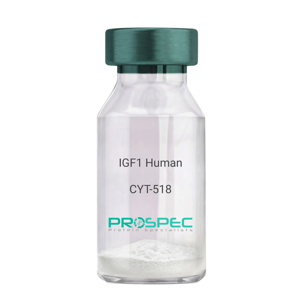
Name :
IGF1 Human Des1-3Description:
Insulin-Like Growth Factor 1 Des (1-3) Human Recombinant
Somatomedin C, IGF-I, IGFI, IGF1, IGF-IA, Mechano growth factor, MGF, Des(1-3), Des1-3, Des 1-3, Des (1-3), IGF-1 (4-70).
Product # :
CYT-518Price :
Quantity :
Shipping Method :
Shipped at Room temp
More Info
- description
- source
- formulation
- purity
- biological activity
- More Info
Description
IGF-I Des(1-3) Human Recombinant produced in E.Coli is a single, non-glycosylated, polypeptide chain containing 67 amino acids (aa 4-70) and having a molecular mass of 7368.5 Dalton. IGF-1 Des1-3 is purified by proprietary chromatographic techniques.
Source
Escherichia Coli.
Formulation
The protein (1mg/ml) was lyophilized was lyophilized from a 0.2µm filtered concentrated solution in 20mM PBS, pH 7.0.
Purity
Greater than 97% as determined by:
(a) Analysis by RP-HPLC.
(b) Analysis by SDS-PAGE.Biological Activity
The ED50 as determined by a cell proliferation assay using FDC-P1 cells is less than 2.0 ng/ml, corresponding to a specific activity of > 5×10⁵ units/mg.More Info
-
Introduction
The somatomedins, or insulin-like growth factors (IGFs), comprise a family of peptides that play important roles in mammalian growth and development. IGF1 mediates many of the growth-promoting effects of growth hormone (GH; MIM 139250). Early studies showed that growth hormone did not directly stimulate the incorporation of sulfate into cartilage, but rather acted through a serum factor, termed 'sulfation factor,' which later became known as 'somatomedin' (Daughaday et al., 1972). Three main somatomedins have been characterized: somatomedin C (IGF1), somatomedin A (IGF2; MIM 147470), and somatomedin B (MIM 193190) (Rotwein, 1986; Rosenfeld, 2003).
-
Synonyms
Somatomedin C, IGF-I, IGFI, IGF1, IGF-IA, Mechano growth factor, MGF, Des(1-3), Des1-3, Des 1-3, Des (1-3), IGF-1 (4-70).
-
Physical Appearance
Sterile Filtered White lyophilized (freeze-dried) powder.
-
Stability
Lyophilized IGF-I des(1-3) although stable at room temperature for 3 weeks, should be stored desiccated below -18°C. Upon reconstitution IGF1 des-1-3 should be stored at 4°C between 2-7 days and for future use below -18°C.For long term storage it is recommended to add a carrier protein (0.1% HSA or BSA).Please prevent freeze-thaw cycles.
-
Solubility
It is recommended to reconstitute the IGF-I des(1-3) in sterile 18M-cm H2O not less than 100µg/ml, which can then be further diluted to other aqueous solutions.
-
Amino Acid Sequence
TLCGAELVDA LQFVCGDRGF YFNKPTGYGS SSRRAPQTGI VDECCFRSCD LRRLEMYCAP LKPAKSA.
-
Background
What is the molecular weight/Mw of IGF1 HUMAN DES1-3 Protein?
IGF1 HUMAN DES1-3 Protein has a total Mw of 7.3kDa.
What is the source or expression system of IGF1 HUMAN DES1-3 Protein?
Escherichia Coli.
What is the Purity of IGF1 HUMAN DES1-3 Protein?
IGF1 HUMAN DES1-3 Protein is >97% pure as determined by SDS-PAGE.
What is the Biological Activity of IGF1 HUMAN DES1-3 Protein?
The ED50 as determined by a cell proliferation assay using FDC-P1 cells is less than 2.0 ng/ml, corresponding to a specific activity of > 5×10⁵ units/mg.
What is the amino acid sequence of IGF1 HUMAN DES1-3 Protein?
TLCGAELVDA LQFVCGDRGF YFNKPTGYGS SSRRAPQTGI VDECCFRSCD LRRLEMYCAP LKPAKSA.
What applications can IGF1 HUMAN DES1-3 Protein be used in?
IGF1 HUMAN DES1-3 Protein can probably be used in western blot, ELISA and Lateral Flow.
What is the endotoxin level for IGF1 HUMAN DES1-3 Protein?
The endotoxin level is minimal, IGF1 HUMAN DES1-3 Protein was purified using conventional chromatography techniques.
ProSpec's products are furnished for LABORATORY RESEARCH USE ONLY. The product may not be used as drugs, agricultural or pesticidal products, food additives or household chemicals.
- View Data Sheet
Name :
GM-CSF MonkeyDescription:
Granulocyte Macrophage-Colony Stimulating Factor Rhesus Macaque Recombinant
CSF-2, MGI-1GM, GM-CSF, Pluripoietin-alpha, MGC131935, MGC138897.
Product # :
CYT-720Price :
Quantity :
Shipping Method :
Shipped at Room temp
More Info
- description
- source
- formulation
- purity
- biological activity
- More Info
Description
Granulocyte Macrophage Colony Stimulating Factor Monkey Recombinant produced in E.Coli is a single, non-glycosylated, polypeptide chain containing 127 amino acids and having a molecular mass of 14.4 kDa.GM-CSF is purified by proprietary chromatographic techniques.
Source
Escherichia Coli.
Formulation
GM-CSF was lyophilized from a concentrated (1mg/ml) solution containing 1x PBS pH 7.4.
Purity
Greater than 98.0% as determined by SDS-PAGE and RP-HPLC.
Biological Activity
The ED50 as determined by a cell proliferation assay using human TF-1 cells is less than 0.1ng/ml, corresponding to a specific activity of > 10,000,000 IU/mg.More Info
-
Introduction
GMCSF is a cytokine that controls the production, differentiation, and function of granulocytes and macrophages. The active form of the protein is found extracellularly as a homodimer. This gene has been localized to a cluster of related genes at chromosome region 5q31, which is known to be associated with interstitial deletions in the 5q- syndrome and acute myelogenous leukemia. Other genes in the cluster include those encoding interleukins 4, 5, and 13. GM-CSF stimulates the growth and differentiation of hematopoietic precursor cells from various lineages, including granulocytes, macrophages, eosinophils and erythrocytes.
-
Synonyms
CSF-2, MGI-1GM, GM-CSF, Pluripoietin-alpha, MGC131935, MGC138897.
-
Physical Appearance
Sterile Filtered White lyophilized (freeze-dried) powder.
-
Stability
Lyophilized GMCSF although stable at room temperature for 3 weeks, should be stored desiccated below -18°C. Upon reconstitution GMCSF should be stored at 4°C between 2-7 days and for future use below -18°C. For long term storage it is recommended to add a carrier protein (0.1% HSA or BSA). Please prevent freeze-thaw cycles.
-
Solubility
It is recommended to reconstitute the lyophilized GMCSF in sterile 18MΩ-cm H2O not less than 100µg/ml, which can then be further diluted to other aqueous solutions.
-
Amino Acid Sequence
APARSPSPGT QPWEHVNAIQ EARRLLNLSR DTAAEMNKTV EVVSEMFDLQ EPSCLQTRLE LYKQGLQGSL TKLKGPLTMM ASHYKQHCPP TPETSCATQI ITFQSFKENL KDFLLVIPFD CWEPVQE.
ProSpec's products are furnished for LABORATORY RESEARCH USE ONLY. The product may not be used as drugs, agricultural or pesticidal products, food additives or household chemicals.