Search results
1000 results found for “peroxisomal biogenesis factor”
Name
Description
Product #
Price
Quantity
Shipping Method
- View Data Sheet
Name :
TNFRSF10A HumanDescription:
TRAIL Receptor-1 Human Recombinant
TNFRSF10A, TNF Receptor Superfamily Member 10a, Tumor Necrosis Factor Receptor Superfamily, Member 10a, TNF-Related Apoptosis-Inducing Ligand Receptor 1, Death Receptor 4, TRAIL Receptor 1, TRAIL-R1, TRAILR1, APO2, DR4, Tumor Necrosis Factor Receptor Superfamily Member 10a Variant 2, Tumor Necrosis Factor Receptor Superfamily Member 10A, Cytotoxic TRAIL Receptor, CD261 Antigen, TRAILR-1, CD261.
Product # :
CYT-1043Price :
Quantity :
Shipping Method :
Shipped with Ice Packs
More Info
- description
- source
- formulation
- purity
- More Info
Description
TNFRSF10A produced in Sf9 Baculovirus cells is a single, glycosylated polypeptide chain containing 224 amino acids (24-239a.a.) and having a molecular mass of 23.9kDa. TNFRSF10A is expressed with an 6 amino acid His tag at C-Terminus and purified by proprietary chromatographic techniques.
Source
Sf9, Baculovirus cells.
Formulation
TNFRSF10A protein solution (0.5mg/ml) contains Phosphate Buffered Saline (pH 7.4) and 10% glycerol.
Purity
Greater than 90.0% as determined by SDS-PAGE.
More Info
-
Introduction
Human TNFRSF10A is a type 1 transmembrane protein in the TNF R superfamily. TNFRSF10A is not expressed in rodents. The trimeric ligand Trail binds TNFRSF10A and induces apoptosis, and recombinant, soluble forms of the receptor inhibit Trail-induced apoptosis. TNFRSF10A is expressed generally in damaged, infected, and malignant cells. TNFRSF10A functions in immune surveillance, inducing apoptosis in cancer cells but not normal cells.
-
Synonyms
TNFRSF10A, TNF Receptor Superfamily Member 10a, Tumor Necrosis Factor Receptor Superfamily, Member 10a, TNF-Related Apoptosis-Inducing Ligand Receptor 1, Death Receptor 4, TRAIL Receptor 1, TRAIL-R1, TRAILR1, APO2, DR4, Tumor Necrosis Factor Receptor Superfamily Member 10a Variant 2, Tumor Necrosis Factor Receptor Superfamily Member 10A, Cytotoxic TRAIL Receptor, CD261 Antigen, TRAILR-1, CD261.
-
Physical Appearance
Sterile Filtered colorless solution.
-
Stability
Store at 4°C if entire vial will be used within 2-4 weeks. Store, frozen at -20°C for longer periods of time. For long term storage it is recommended to add a carrier protein (0.1% HSA or BSA). Avoid multiple freeze-thaw cycles.
-
Amino Acid Sequence
ASGTEAAAAT PSKVWGSSAG RIEPRGGGRG ALPTSMGQHG PSARARAGRA PGPRPAREAS PRLRVHKTFK FVVVGVLLQV VPSSAATIKL HDQSIGTQQW EHSPLGELCP PGSHRSEHPG ACNRCTEGVG YTNASNNLFA CLPCTACKSD EEERSPCTTT RNTACQCKPG TFRNDNSAEM CRKCSRGCPR GMVKVKDCTP WSDIECVHKE SGNGHNLEHH HHHH
ProSpec's products are furnished for LABORATORY RESEARCH USE ONLY. The product may not be used as drugs, agricultural or pesticidal products, food additives or household chemicals.
- View Data Sheet
Name :
WISP2 HumanDescription:
WNT1 Inducible Signaling Pathway Protein 2 Human Recombinant
WNT1 Inducible Signaling Pathway Protein 2, Connective Tissue Growth Factor-Related Protein 58, Connective Tissue Growth Factor-Like Protein, CCN Family Member 5, CTGF-L, CT58, CCN5, WISP-2, CTGFL, WISP2.
Product # :
CYT-970Price :
Quantity :
Shipping Method :
Shipped at Room temp
More Info
- description
- source
- formulation
- purity
- More Info
Description
WISP2 Human Recombinant produced in E.coli is a single, non-glycosylated polypeptide chain containing a total of 228 amino acids and having a molecular mass of 24.4kDa.
Source
Escherichia Coli.
Formulation
Lyophilized from a sterile (0.2µm) filtered aqueous solution containing 0.1% Trifluoroacetic Acid (TFA).
Purity
Greater than 95.0% as determined by SDS-PAGE.
More Info
-
Introduction
WNT1-inducible-signaling pathway protein 2 (WISP2) belongs to the WNT1 inducible signaling pathway (WISP) protein subfamily, which belongs to the connective tissue growth factor (CTGF) family. The CTGF family members are characterized by 4 conserved cysteine-rich domains: insulin-like growth factor-binding domain, von Willebrand factor type C module, thrombospondin domain and C-terminal cystine knot-like (CT) domain. WISP2 protein lacks the CT domain which is implicated in dimerization binding. WISP2 is possibly involved in bone remodeling. WISP2 is expressed in primary osteoblasts and fibroblasts. WISP2 stimulates osteoblast adhesion and inhibits osteocalcin production. WISP2 expression in colon tumors is reduced while the other 2 WISP members are overexpressed in colon tumors. WISP2 may play an imperative role in modulating bone turnover.
-
Synonyms
WNT1 Inducible Signaling Pathway Protein 2, Connective Tissue Growth Factor-Related Protein 58, Connective Tissue Growth Factor-Like Protein, CCN Family Member 5, CTGF-L, CT58, CCN5, WISP-2, CTGFL, WISP2.
-
Physical Appearance
Sterile Filtered White lyophilized (freeze-dried) powder.
-
Stability
Lyophilized WISP2 although stable at room temperature for 3 weeks, should be stored desiccated below -18°C. Upon reconstitution WISP-2 should be stored at 4°C between 2-7 days and for future use below -18°C.For long term storage it is recommended to add a carrier protein (0.1% HSA or BSA).Please prevent freeze-thaw cycles.
-
Solubility
It is recommended to reconstitute the lyophilized WISP-2 in sterile 10mM acetic acidnot less than 100µg/ml, which can then be further diluted to other aqueous solutions.
-
Amino Acid Sequence
MQLCPTPCTC PWPPPRCPLG VPLVLDGCGC CRVCARRLGE PCDQLHVCDA SQGLVCQPGA GPGGRGALCL LAEDDSSCEV NGRLYREGET FQPHCSIRCR CEDGGFTCVP LCSEDVRLPS WDCPHPRRVE VLGKCCPEWV CGQGGGLGTQ PLPAQGPQFS GLVSSLPPGV PCPEWSTAWG PCSTTCGLGM ATRVSNQNRF CRLETQRRLC LSRPCPPSRG RSPQNSAF.
ProSpec's products are furnished for LABORATORY RESEARCH USE ONLY. The product may not be used as drugs, agricultural or pesticidal products, food additives or household chemicals.
- View Data Sheet
Name :
CXCL4 Variant 1 HumanDescription:
Platelet Factor-4 Variant 1 Human Recombinant
CXCL4, PF-4, PF4, Iroplact, Oncostatin-A, SCYB4, MGC138298.
Product # :
CHM-243Price :
Quantity :
Shipping Method :
Shipped at Room temp
More Info
- description
- source
- formulation
- purity
- More Info
Description
CXCL4 Variant-1 Human Recombinant produced in E.Coli is a single, non-glycosylated polypeptide chain containing 77 amino acids and having a molecular mass of 8.7 kDa. The CXCL4 Variant-1 is fused to 6xHis tag at N-Terminus and purified by standard chromatography techniques.
Source
Escherichia Coli.
Formulation
The protein was lyophilized without additives.
Purity
Greater than 95.0% as determined by SDS-PAGE.
More Info
-
Introduction
Platelet factor-4 is a 70-amino acid protein that is released from the alpha-granules of activated platelets . Its major physiologic role appears to be neutralization of molecules on the endothelial surface of blood vessels, thereby inhibiting local antithrombin III activity and promoting coagulation. As a strong chemoattractant for neutrophils and fibroblasts, PF4 probably has a role in inflammation and wound repair. Oncostatin-A is a member of the CXC chemocinfamily. Human PF4 is used for the proof of induced thrombocytopenia. Furthermore it is used as an inhibitor in the angiogenesis during tumor therapy.
-
Synonyms
CXCL4, PF-4, PF4, Iroplact, Oncostatin-A, SCYB4, MGC138298.
-
Physical Appearance
Sterile Filtered white lyophilized powder.
-
Stability
Human CXCL4 although stable at 25°C 1 week, should be stored desiccated below -18°C. Please prevent freeze-thaw cycles.
-
Solubility
It is recommended to reconstitute the lyophilized CXCL4 in sterile 18MΩ-cm H2O not less than 100µg/ml, which can then be further diluted to other aqueous solutions.
-
Amino Acid Sequence
MHHHHHHEAE EDGDLQCLCV KTTSQVRPRH ITSLEVIKAG PHCPTAQLIA TLKNGRKICL DLQALLYKKI IKEHLES.
ProSpec's products are furnished for LABORATORY RESEARCH USE ONLY. The product may not be used as drugs, agricultural or pesticidal products, food additives or household chemicals.
- View Data Sheet
Name :
SDF 1a HumanDescription:
Stromal Cell-Derived Factor-1 Alpha Human Recombinant (CXCL12)
SDF-1, CXCL12, Pre-B cell growth-stimulating factor, PBSF, hIRH, chemokine (C-X-C motif) ligand 12, SDF1, SDF1A, TPAR1, SCYB12, SDF-1a, TLSF-a.
Product # :
CHM-262Price :
Quantity :
Shipping Method :
Shipped at Room temp
More Info
- description
- source
- formulation
- purity
- biological activity
- More Info
Description
Stromal Cell-Derived Factor-1 alpha Human Recombinant produced in E.Coli is a non-glycosylated, Polypeptide chain containing 68 amino acids and having a molecular mass of 8004 Dalton. The SDF-1a is purified by proprietary chromatographic techniques.
Source
Escherichia Coli.
Formulation
The protein was lyophilized from a concentrated (1 mg/ml) sterile solution containing no additives.
Purity
Greater than 98.0% as determined by:
(a) Analysis by RP-HPLC.
(b) Analysis by SDS-PAGE.Biological Activity
The specific activity as determined by its ability to chemoattract human peripheral T cells activated with PHA and IL-2 using a concentation of 20-80 ng/ml corresponding to a Specific Activity of 12,500-50,000IU/mg.More Info
-
Introduction
SDF-1 (stromal cell-derived factor-1) is small cytokine belonging to the chemokine family that is officially designated Chemokine (C-X-C motif) ligand 12 (CXCL12). It is produced in two forms, SDF-1?/CXCL12a and SDF-1?/CXCL12b, by alternate splicing of the same gene. Chemokines are characterized by the presence of four conserved cysteines, which form two disulfide bonds. The CXCL12 proteins belong to the group of CXC chemokines, whose initial pair of cysteines are separated by one intervening amino acid. CXCL12 is strongly chemotactic for lymphocytes and has been implicated as an important cell co-ordinator during development. During embryogenesis it directs the migration of hematopoietic cells from foetal liver to bone marrow. Mice which were knocked-out for CXCL12 gene were lethal before the birth or within just 1 hour of life. As another role, CXCL12a alters also the electrophysiology of neurons. CXCL12 was shown to be expressend in many tissues in mice (including brain, thymus, heart, lung, liver, kidney, spleen and bone marrow).
The receptor for this chemokine is CXCR4, which was previously called fusin. This CXCL12-CXCR4 interaction used to be considered exclusive (unlike for other chemokines and their receptors), but recently it was suggested that CXCL12 is also bound by CXCR7 receptor.
The gene for CXCL12 is located on human chromosome 10. In human and mouse both CXCL12 and CXCR4 show high identity of sequence: 99% and 90%, respectively. -
Synonyms
SDF-1, CXCL12, Pre-B cell growth-stimulating factor, PBSF, hIRH, chemokine (C-X-C motif) ligand 12, SDF1, SDF1A, TPAR1, SCYB12, SDF-1a, TLSF-a.
-
Physical Appearance
Sterile Filtered White lyophilized (freeze-dried) powder.
-
Stability
Lyophilized SDF-1a although stable at room temperature for 3 weeks, should be stored desiccated below -18°C. Upon reconstitution CXCL12 should be stored at 4°C between 2-7 days and for future use below -18°C. For long term storage it is recommended to add a carrier protein (0.1% HSA or BSA).Please prevent freeze-thaw cycles.
-
Solubility
It is recommended to reconstitute the lyophilized Stromal Cell-Derived Factor-1a in sterile 18MΩ-cm H2O not less than 100µg/ml, which can then be further diluted to other aqueous solutions.
-
Amino Acid Sequence
KPVSLSYRCP CRFFESHVAR ANVKHLKILN TPNCALQIVA RLKNNNRQVC IDPKLKWIQE YLEKALNK
ProSpec's products are furnished for LABORATORY RESEARCH USE ONLY. The product may not be used as drugs, agricultural or pesticidal products, food additives or household chemicals.
- View Data Sheet
Name :
HGF B HumanDescription:
Hepatocyte Growth Factor B Chain Human Recombinant
Scatter Factor, SF, Hepatopoietin, HPTA, HGF, HGFB, F-TCF, DFNB39, Hepatocyte growth factor, Hepatocyte growth factor beta chain.
Product # :
CYT-665Price :
Quantity :
Shipping Method :
Shipped with Ice Packs
More Info
- description
- source
- formulation
- purity
- More Info
Description
HGF-B Human Recombinant produced in E.Coli is a single, non-glycosylated, Polypeptide chain containing 234 amino acids fragment (495-728) having a molecular weight of 34kDa and fused with a 4.5kDa amino-terminal hexahistidine tag. The HGF-B is purified by proprietary chromatographic techniques.
Source
Escherichia Coli.
Formulation
HGF-B protein is supplied in 25mM Na. Acetate, pH 4.8, 1mM EDTA and 50% glycerol.
Purity
Greater than 95.0% as determined by SDS-PAGE.
More Info
-
Introduction
Hepatocyte Growth Factor (HGF) is a multifunctional growth factor which regulates both cell growth and cell motility. It exerts a strong mitogenic effect on hepatocytes and primary epithelial cells. HGF synergizes with Interleukin-3 and GM-CSF to stimulate colony formation of hematopoietic progenitor cells in vitro and may, therefore, also modulate hematopoiesis. HGF is secreted as a single inactive polypeptide which is cleaved by serine proteases into a 69kDa Alpha chain and 34kDa Beta chain. A disulfide bond linking the alpha and beta chains produces the active heterodimeric molecule.
-
Synonyms
Scatter Factor, SF, Hepatopoietin, HPTA, HGF, HGFB, F-TCF, DFNB39, Hepatocyte growth factor, Hepatocyte growth factor beta chain.
-
Physical Appearance
Sterile Filtered clear solution.
-
Stability
Store at 4°C if entire vial will be used within 2-4 weeks. Store, frozen at -20°C for longer periods of time. Please avoid freeze thaw cycles.
-
Amino Acid Sequence
VVNGIPTRTNIGWMVSLRYRNKHICGGSLIKESWVLTAR
QCFPSRDLKDYEAWLGIHDVHGRGDEKCKQVLNVSQLV
YGPEGSDLVLMKLARPAVLDDFVSTIDLPNYGCTIPEKTS
CSVYGWGYTGLINYDGLLRVAHLYIMGNEKCSQHHRGK
VTLNESEICAGAEKIGSGPCEGDYGGPLVCEQHKMRMV
LGVIVPGRGCAIPNRPGIFVRVAYYAKWIHKIILTYKVPQS.
ProSpec's products are furnished for LABORATORY RESEARCH USE ONLY. The product may not be used as drugs, agricultural or pesticidal products, food additives or household chemicals.
- View Data Sheet
Name :
PDIA3 AntibodyDescription:
Protein Disulfide Isomerase A3, Mouse Anti Human
ERp57, ERp60, ERp61, GRP57, GRP58, HsT17083, P58, PI-PLC, ER60, Protein disulfide-isomerase A3, Disulfide isomerase ER-60, Endoplasmic reticulum resident protein 60, ER protein 60, 58 kDa microsomal protein, Endoplasmic reticulum resident protein 57, ER protein 57, 58 kDa glucose-regulated protein, PDIA3.
Product # :
ANT-633Price :
Quantity :
Shipping Method :
Shipped with Ice Packs
More Info
- formulation
- More Info
Formulation
1mg/ml containing PBS, pH-7.4, 10% Glycerol and 0.02% Sodium Azide.
More Info
-
Introduction
PDIA3 is an enzyme that belongs to the endoplasmic reticulum and interacts with lectin chaperones calreticulin and calnexin to modulate folding of newly synthesized glycoproteins. PDIA3 has protein disulfide isomerase activity. Complexes of lectins and PDIA3 mediate protein folding by promoting formation of disulfide bonds in their glycoprotein substrates. PDIA3 is expressed in the lumbar spinal cord from rats submitted to peripheral lesion during neonatal period. PDIA3 interacts with thiazide-sensitive sodium-chloride cotransporter in the kidney and is induced by glucose deprivation. PDIA3 is part of the major histocompatibility complex (MHC) class I peptide-loading complex (TAP1), which is important for formation of the final antigen conformation and export from the endoplasmic reticulum to the cell surface.
-
Synonyms
ERp57, ERp60, ERp61, GRP57, GRP58, HsT17083, P58, PI-PLC, ER60, Protein disulfide-isomerase A3, Disulfide isomerase ER-60, Endoplasmic reticulum resident protein 60, ER protein 60, 58 kDa microsomal protein, Endoplasmic reticulum resident protein 57, ER protein 57, 58 kDa glucose-regulated protein, PDIA3.
-
Physical Appearance
Sterile filtered colorless solution.
-
Immunogen
Anti-human PDIA3 mAb, is derived from hybridization of mouse F0 myeloma cells with spleen cells from BALB/c mice immunized with a recombinant human PDIA3 protein 25-505 amino acids purified from E. coli.
-
Ig Subclass
Mouse IgG2a heavy chain and k light chain.
-
Clone
PAT9E9AT.
-
Applications
The antibody has been tested by ELISA, Western blot analysis, Flow cytometry and ICC/IF to assure specificity and reactivity. Since application varies, however, each investigation should be titrated by the reagent to obtain optimal results.
-
Type
Mouse Anti Human Monoclonal.
-
Storage Procedures
For periods up to 1 month store at 4°C, for longer periods of time, store at -20°C. Prevent freeze thaw cycles.
-
Purification Method
PDIA3 antibody was purified from mouse ascitic fluids by protein-A affinity chromatography.
ProSpec's products are furnished for LABORATORY RESEARCH USE ONLY. The product may not be used as drugs, agricultural or pesticidal products, food additives or household chemicals.
- View Data Sheet
Name :
PDPN P56F7AT AntibodyDescription:
Podoplanin clone P56F7AT, Mouse Anti Human
Podoplanin, Glycoprotein 36, PA2.26 antigen, T1A, GP36, GP40, Gp38, OTS8, T1A2, HT1A-1, PA2.26, T1-alpha, PDPN.
Product # :
ANT-641Price :
Quantity :
Shipping Method :
Shipped with Ice Packs
More Info
- formulation
- More Info
Formulation
1mg/ml containing PBS, pH-7.4, 10% Glycerol and 0.02% Sodium Azide.
More Info
-
Introduction
Podoplanin is a small mucin-like type-1 transmembrane protein, typically expressed in various specialized cell types throughout the body. Podoplanin is a type-I integral membrane glycoprotein with diverse distribution in human tissues. PDPN physiological function is related to its mucin-type character. The homologous protein in other species has been described as a differentiation antigen and influenza-virus receptor.
PDPN is expressed in lymphatic progenitor cells and afterwards during mouse development in lymphatic endothelial cells. Podoplanin is a specific marker for lymph vessel endothelial cells. Over-expression of podoplanin significantly elevates endothelial cell adhesion, migration, and tube formation. Inhibition of Podoplanin expression decreases cell adhesion in human dermal lymphatic endothelial cells. Podoplanin is used as a specific marker for lymphatic endothelium in histopathology.
Podoplanin expression is increased in nearly all human colon, rectum, and small intestine tumors. AGGRUS may serve as a diagnostic marker that distinguishes seminomas, the majority of which over express the protein, from embryonal carcinoma in testicular germ cell tumors. -
Synonyms
Podoplanin, Glycoprotein 36, PA2.26 antigen, T1A, GP36, GP40, Gp38, OTS8, T1A2, HT1A-1, PA2.26, T1-alpha, PDPN.
-
Physical Appearance
Sterile filtered colorless solution.
-
Immunogen
Anti-human PDPN mAb, is derived from hybridization of mouse F0 myeloma cells with spleen cells from BALB/c mice immunized with recombinant human PDPN amino acids 99-207 purified from E. coli.
-
Ig Subclass
Mouse IgG1 heavy chain and k light chain.
-
Clone
P56F7AT.
-
Applications
PDPN antibody has been tested by ELISA, Western blot analysis, Flow cytometry and ICC/IF to assure specificity and reactivity. Since application varies, however, each investigation should be titrated by the reagent to obtain optimal results.
-
Type
Mouse Anti Human Monoclonal.
-
Storage Procedures
For periods up to 1 month store at 4°C, for longer periods of time, store at -20°C. Prevent freeze thaw cycles.
-
Purification Method
PDPN antibody was purified from mouse ascitic fluids by protein-A affinity chromatography.
ProSpec's products are furnished for LABORATORY RESEARCH USE ONLY. The product may not be used as drugs, agricultural or pesticidal products, food additives or household chemicals.
- View Data Sheet
Name :
PSMD10 AntibodyDescription:
Gankyrin, Mouse Anti Human
26S proteasome non-ATPase regulatory subunit 10, 26S proteasome regulatory subunit p28, Gankyrin, PSMD10, p28, dJ889N15.2.
Product # :
ANT-609Price :
Quantity :
Shipping Method :
Shipped with Ice Packs
More Info
- formulation
- More Info
Formulation
1mg/ml containing PBS, pH-7.4, 10% Glycerol and 0.02% Sodium Azide.
More Info
-
Introduction
Gankyrin (proteasome 26S subunit) is a multicatalytic proteinase oncoprotein commonly overexpressed in most hepatocellular carcinomas. Proteasomes are found throughout eukaryotic cells at a high concentrations and cleave peptides in an ATP/ubiquitin-dependent process in a non-lysosomal pathway. Gankyrin interacts with S6 ATPase of the 19S regulatory particle of the 26S proteasome. Gankyrin is involved in theregulation of the phosphorylation of the retinoblastoma protein by CDK4, and to enhance the ubiquitinylation of p53 by MDM2. Gankyrin consists of 7 ankyrin repeats and is structurally similar to I kappa Bs. Gankyrin acts as a regulatory subunit of the 26s proteasome which is involved in the atp-dependent degradation of ubiquitinated proteins. Gankyrin is involved in progression of esophageal squamous cell carcinoma. gankyrin plays an oncogenic role especially in early stages of human epatocarcinogenesis. Gankyrin binds to NF-kappaB and suppresses its activity at the transcription level by modulating acetylation through SIRT1. Structural comparison between Gankyrin & p16(INK4A) identified numerous residues of gankyrin that are potentially important for CDK4 binding.
-
Synonyms
26S proteasome non-ATPase regulatory subunit 10, 26S proteasome regulatory subunit p28, Gankyrin, PSMD10, p28, dJ889N15.2.
-
Physical Appearance
Sterile filtered colorless solution.
-
Immunogen
Anti-human PSMD10 mAb, is derived from hybridization of mouse F0 myeloma cells with spleen cells from BALB/c mice immunized with a recombinant human PSMD10 protein 1-226 amino acids purified from E.coli.
-
Ig Subclass
Mouse IgG1 heavy chain and k light chain.
-
Clone
PAT1F4AT.
-
Applications
PSMD10 antibody has been tested by ELISA, Western blot analysis, Flow cytometry and ICC/IF to assure specificity and reactivity. Since application varies, however, each investigation should be titrated by the reagent to obtain optimal results.
-
Type
Mouse Anti Human Monoclonal.
-
Storage Procedures
For periods up to 1 month store at 4°C, for longer periods of time, store at -20°C. Prevent freeze thaw cycles.
-
Purification Method
PSMD10 antibody was purified from mouse ascitic fluids by protein-A affinity chromatography.
ProSpec's products are furnished for LABORATORY RESEARCH USE ONLY. The product may not be used as drugs, agricultural or pesticidal products, food additives or household chemicals.
- View Data Sheet
Name :
MCSF Mouse, Sf9Description:
Macrophage Colony Stimulating Factor Mouse Recombinant, Sf9
M-Csf, Macrophage colony-stimulating factor 1, CSF-1, MCSF, Csf1, C87615, MCSF, op, Processedmacrophage colony-stimulating factor 1.
Product # :
CYT-1141Price :
Quantity :
Shipping Method :
Shipped with Ice Packs
More Info
- description
- source
- formulation
- purity
- biological activity
- More Info
Description
MCSF Mouse produced in Sf9 Baculovirus cells is a single, glycosylated polypeptide chain containing 164 amino acids (33-187 aa) and having a molecular mass of 19.1 kDa.MCSF is fused to a 6 amino acid His tag at C-terminus and purified by proprietary chromatographic techniques.
Source
Sf9, Baculovirus cells.
Formulation
The MCSF solution (1mg/ml) contains 10% Glycerol and Phosphate-Buffered Saline (pH 7.4).
Purity
Greater than 90.0% as determined by SDS-PAGE.
Biological Activity
Determined by cell proliferation assay using M-NFS-60 mouse myelogenous leukemia lymphoblast cells. ED50 range for this effect is ≤ 4ng/ml.
More Info
-
Introduction
Macrophage Colony Stimulating Factor (M CSF) or colony stimulating factor 1, is a cytokine, that promoted differentiation in hematopoietic stem cells to macrophages. Another role of M CSF is to bind to its receptor (colony stimulating factor 1 receptor) and to take part in placenta growth and development.
-
Synonyms
M-Csf, Macrophage colony-stimulating factor 1, CSF-1, MCSF, Csf1, C87615, MCSF, op, Processedmacrophage colony-stimulating factor 1.
-
Physical Appearance
Sterile Filtered colorless solution.
-
Stability
Store at 4°C if entire vial will be used within 2-4 weeks. Store, frozen at -20°C for longer periods of time. For long term storage it is recommended to add a carrier protein (0.1% HSA or BSA).Avoid multiple freeze-thaw cycles.
-
Amino Acid Sequence
ADPKEVSEHC SHMIGNGHLK VLQQLIDSQM ETSCQIAFEF VDQEQLDDPV CYLKKAFFLV
QDIIDETMRF KDNTPNANAT ERLQELSNNL NSCFTKDYEE QNKACVRTFH ETPLQLLEKI
KNFFNETKNL LEKDWNIFTK NCNNSFAKCS SRDVVTKPHH HHHH
ProSpec's products are furnished for LABORATORY RESEARCH USE ONLY. The product may not be used as drugs, agricultural or pesticidal products, food additives or household chemicals.
- View Data Sheet
Name :
TGFB1 RatDescription:
Transforming Growth Factor-Beta 1 Rat Recombinant
Transforming growth factor beta-1, TGF-beta-1, Tgfb, Tgfb-1, TGFbeta1.
Product # :
CYT-1265Price :
Quantity :
Shipping Method :
Shipped at Room temp
More Info
- description
- source
- formulation
- purity
- biological activity
- More Info
Description
Transforming Growth Factor-Beta 1 Rat Recombinant produced in CHO is a homodimer, polypeptide chain containing 2 x 112 amino acids and having a total molecular mass of 25.6kDa.
TGFB1 Rat Recombinant is purified by proprietary chromatographic techniques.
Source
CHO Cells.
Formulation
The protein was lyophilized with 0.1% (v/v) TFA and 35% (v/v) Acetonitrile.
Purity
Greater than 97.0% as determined by SDS-PAGE and SEC-HPLC analyses.
Biological Activity
The biological activity was determined by TGFB1 ability to inhibit the mouse IL-4-dependent proliferation of mouse HT-2 cells. The expected ED50 for this effect is <0.05ng/ml, corresponding to a specific activity of ≥ 2.0 × 10⁷ units/mg.More Info
-
Synonyms
Transforming growth factor beta-1, TGF-beta-1, Tgfb, Tgfb-1, TGFbeta1.
-
Physical Appearance
Sterile Filtered White lyophilized (freeze-dried) powder.
-
Stability
Lyophilized TGFB1 although stable at room temperature for 3 weeks, should be stored desiccated below -18°C. Upon reconstitution Transforming Growth Factor-Beta 1 should be stored at 4°C between 2-7 days and for future use below -18°C.
For long term storage it is recommended to add a carrier protein (0.1% HSA or BSA).
Please prevent freeze-thaw cycles.
-
Solubility
It is recommended to reconstitute the lyophilized Transforming Growth Factor-Beta 1 in sterile 4mM HCl not less than 100µg/ml, which can then be further diluted to other aqueous solutions.
-
Amino Acid Sequence
MGSSHHHHHH SSGLVPRGSH MGSALDTNYC FSSTEKNCCV RQLYIDFRKD LGWKWIHEPK GYHANFCLGP CPYIWSLDTQ YSKVLALYNQ HNPGASASPC CVPQALEPLP IVYYVGRKPK VEQLSNMIVR SCKCS.
-
Background
Mouse TGF-β1 as an inducer of EMT [epithelial–mesenchymal transition ] therefore used in in fibrosis, wound healing, cancer invasion, and metastasis. Mouse TGF-β1 decreases E-cadherin expression and increases N-cadherin, vimentin and fibronectin.
TGF-β1 is produced by T regulatory cells (Tregs), Macrophages and monocytes, Platelets, Fibroblasts, Epithelial cells, Endothelial cells, Smooth muscle cells, Tumor cells, Activated immune cells
What is the source or expression system of Mouse TGFB1 Protein?
CHO Cells
What is the Purity of Mouse TGFB1 Protein?
Mouse TGFB1 Protein is >97% pure as determined by SDS-PAGE and SEC-HPLC analyses.
What is the molecular weight of Mouse TGFB1 Protein?
Mouse TGFB1 Protein having a total Mw of 25.6kDa.
What is the Biological Activity of Mouse TGFB1 Protein?
The biological functionality of Mouse TGFB1 Protein is determined by mouse HT-2 cells.
What is the endotoxin level for Mouse TGFB1 Protein?
The endotoxin level is minimal, Mouse TGFB1 Protein was purified using conventional chromatography techniques.
What is the amino acid sequence of Mouse TGFB1 Protein?
ALDTNYCFSS TEKNCCVRQL YIDFRKDLGW KWIHEPKGYH ANFCLGPCPY IWSLDTQYSK VLALYNQHNP GASASPCCVP QALEPLPIVY YVGRKPKVEQ LSNMIVRSCK CS.
Is TGFB1 a homodimer / homodimeric protein?
Yes, TGFB1 is homo dimer consisting of 2 identical chains.
ProSpec's products are furnished for LABORATORY RESEARCH USE ONLY. The product may not be used as drugs, agricultural or pesticidal products, food additives or household chemicals.
- View Data Sheet
Name :
BAFFR Human, HEKDescription:
BAFF (BLyS) Receptor Human Recombinant, HEK
TNFRSF13C, CD268, BAFF-R, MGC138235, B cell-activating factor receptor.
Product # :
CYT-1224Price :
Quantity :
Shipping Method :
Shipped with Ice Packs
More Info
- description
- source
- formulation
- purity
- biological activity
- More Info
Description
BAFFR Human Recombinant is a single, glycosylated, polypeptide chain (1-78 a.a) containing a total of 314 amino acids and having a molecular mass of 34.4 kDa. BAFFR is fused to 233 a.a hIgG-Tag at C-terminus and is purified by proprietary chromatographic techniques.
Source
HEK293 Cells.
Formulation
The BAFFR solution (1mg/ml) contains 10% Glycerol and Phosphate-Buffered Saline (pH 7.4).
Purity
Greater than 95.0% as determined by SDS-PAGE.
Biological Activity
The ED50 is ≤0.7 ug/ml, measured by its ability in a functional ELISA with BAFF Human.
More Info
-
Synonyms
TNFRSF13C, CD268, BAFF-R, MGC138235, B cell-activating factor receptor.
-
Physical Appearance
Sterile Filtered colorless solution.
-
Stability
Store at 4°C if entire vial will be used within 2-4 weeks. Store, frozen at -20°C for longer periods of time. For long term storage it is recommended to add a carrier protein (0.1% HSA or BSA).Avoid multiple freeze-thaw cycles.
-
Amino Acid Sequence
DGSMRRGPRS LRGRDAPAPT PCVPAECFDL LVRHCVACGL LRTPRPKPAG ASSPAPRTAL QPQESVGAGA GEAALPLPGL LLEPKSCDKT HTCPPCPAPE LLGGPSVFLF PPKPKDTLMI SRTPEVTCVV VDVSHEDPEV KFNWYVDGVE VHNAKTKPRE EQYNSTYRVV SVLTVLHQDW LNGKEYKCKV SNKALPAPIE KTISKAKGQP REPQVYTLPP SRDELTKNQV SLTCLVKGFY PSDIAVEWES NGQPENNYKT TPPVLDSDGS FFLYSKLTVD KSRWQQGNVF SCSVMHEALH NHYTQKSLSL SPGK.
-
Background
B-cell Activating Factor (BAFF) and its corresponding receptor, BAFF-R, are integral components of the immune system, orchestrating crucial processes in B-cell survival, maturation, and differentiation. As we delve into the intricate world of immunology, the study of BAFF and its receptor has unveiled essential pathways that govern the immune responses in health and disease. This research investigates the multifaceted role of BAFF Receptor Protein, shedding light on its structural complexities, signaling mechanisms, and its pivotal contributions to immune regulation. By exploring the interactions between BAFF and its receptor, scientists aim to decipher the delicate balance that underlies immune homeostasis and explore potential therapeutic avenues.
Structural Architecture of BAFF Receptor Protein:
BAFF Receptor, a transmembrane protein predominantly expressed on B cells, belongs to the tumor necrosis factor receptor (TNFR) superfamily. Its intricate structure involves various domains, each playing a unique role in ligand binding, receptor activation, and downstream signaling. Understanding the structural intricacies of BAFF Receptor is paramount to unraveling the molecular events that govern B-cell fate decisions and immune responses.
Physiological Significance in B-Cell Biology:
BAFF Receptor, upon binding with its ligand BAFF, initiates a cascade of events critical for B-cell survival and function. This interaction promotes B-cell maturation, prevents premature apoptosis, and influences the formation of immune synapses. Additionally, BAFF Receptor signaling is tightly regulated to prevent excessive B-cell activation, ensuring immune tolerance and preventing autoimmune responses. Disruptions in these pathways can lead to autoimmune disorders, underscoring the crucial role of BAFF Receptor in maintaining immune equilibrium.
Regulation of Immune Responses:
BAFF Receptor signaling not only affects B-cell development but also has broader implications for immune responses. By modulating antibody production, B-cell activation, and immune memory, BAFF Receptor plays a vital role in shaping adaptive immunity. Its dysregulation has been implicated in various autoimmune conditions, making it an attractive target for therapeutic interventions aimed at restoring immune balance.
BAFF Receptor as a Therapeutic Target:
The intricate involvement of BAFF Receptor in autoimmune diseases, such as rheumatoid arthritis and systemic lupus erythematosus, has positioned it as a promising therapeutic target. Researchers are exploring monoclonal antibodies and other targeted therapies that aim to modulate BAFF Receptor signaling, providing a new frontier in autoimmune disease management. Additionally, understanding the BAFF-BAFF Receptor axis offers potential insights into the development of vaccines and immunotherapies, fostering innovative approaches in the fight against infectious diseases and malignancies.
BAFF Receptor Protein, as a key player in immune regulation, embodies the complexities of immunology. Its interactions with BAFF orchestrate fundamental processes in B-cell biology and adaptive immunity. As scientists unravel the intricate signaling pathways and structural nuances of BAFF Receptor, they pave the way for novel therapeutic strategies and innovative treatments for autoimmune disorders and beyond. This research not only deepens our understanding of immune regulation but also holds the promise of transformative advancements in immunotherapy, ultimately shaping the future of immune-related healthcare.
What is the molecular weight/Mw of BAFF-R Protein?
BAFF-R Protein has a total Mw of 34.4kDa.
What is the source or expression system of BAFF-R Protein?
HEK293 Cells.
What is the Purity of BAFF-R Protein?
BAFF-R Protein is >95% pure as determined by SDS-PAGE.
What is the Biological Activity of BAFF-R Protein?
The ED50 is ≤0.7 ug/ml, measured by its ability in a functional ELISA with BAFF Human.
What is the amino acid sequence of BAFF-R Protein?
DGSMRRGPRS LRGRDAPAPT PCVPAECFDL LVRHCVACGL LRTPRPKPAG ASSPAPRTAL QPQESVGAGA GEAALPLPGL LLEPKSCDKT HTCPPCPAPE LLGGPSVFLF PPKPKDTLMI SRTPEVTCVV VDVSHEDPEV KFNWYVDGVE VHNAKTKPRE EQYNSTYRVV SVLTVLHQDW LNGKEYKCKV SNKALPAPIE KTISKAKGQP REPQVYTLPP SRDELTKNQV SLTCLVKGFY PSDIAVEWES NGQPENNYKT TPPVLDSDGS FFLYSKLTVD KSRWQQGNVF SCSVMHEALH NHYTQKSLSL SPGK.
What applications can BAFF-R Protein be used in?
BAFF-R Protein can probably be used in western blot, ELISA and Lateral Flow.
What is the endotoxin level for BAFF-R Protein?
The endotoxin level is minimal, BAFF-R Protein was purified using conventional chromatography techniques.
ProSpec's products are furnished for LABORATORY RESEARCH USE ONLY. The product may not be used as drugs, agricultural or pesticidal products, food additives or household chemicals.
- View Data Sheet
Name :
BMP8B HumanDescription:
Bone Morphogenetic protein-8b Human Recombinant
Bone morphogenetic protein 8B, BMP-8, BMP-8B, Osteogenic protein 2, OP-2, BMP8B, BMP8, Bone Morphogenetic protein-8b, OP2.
Product # :
CYT-830Price :
Quantity :
Shipping Method :
Shipped with Ice Packs
More Info
- description
- source
- formulation
- purity
- More Info
- sds-page
Description
BMP8B Human Recombinant produced in E.Coli is a single, non-glycosylated polypeptide chain containing 162 amino acids (264-402a.a.) and having a molecular mass of 18.1kDa.BMP8B is fused to a 23 amino acid His-tag at N-terminus & purified by proprietary chromatographic techniques.
Source
E.coli.
Formulation
BMP8B protein solution (1mg/ml) containing 20mM Tris-HCl buffer (pH8.0), 0.4M Urea and 10% glycerol.
Purity
Greater than 90% as determined by SDS-PAGE.
sds-page
More Info
-
Introduction
Bone Morphogenetic protein-8b (BMP8B) belongs to a family of secreted signaling molecules which can induce ectopic bone growth. BMP8B is known for having a possible bone inductive activity as it is related to BMP5 and BMP7. BMP8B is the osteoinductive factor accountable for epithelial osteogenesis.
-
Synonyms
Bone morphogenetic protein 8B, BMP-8, BMP-8B, Osteogenic protein 2, OP-2, BMP8B, BMP8, Bone Morphogenetic protein-8b, OP2.
-
Physical Appearance
Sterile Filtered colorless solution.
-
Stability
Store at 4°C if entire vial will be used within 2-4 weeks. Store, frozen at -20°C for longer periods of time. For long term storage it is recommended to add a carrier protein (0.1% HSA or BSA).Avoid multiple freeze-thaw cycles.
-
Amino Acid Sequence
MGSSHHHHHH SSGLVPRGSH MGSAVRPLRR RQPKKSNELP QANRLPGIFD DVHGSHGRQV CRRHELYVSF QDLGWLDWVI APQGYSAYYC EGECSFPLDS CMNATNHAIL QSLVHLMMPD AVPKACCAPT KLSATSVLYY DSSNNVILRK HRNMVVKACG CH.
-
Background
Bone Morphogenetic Protein-8B Human Recombinant: Unveiling the Potential for Regenerative Medicine and Tissue Engineering
Abstract:
Bone Morphogenetic Protein-8B (BMP-8B) human recombinant is a key member of the bone morphogenetic protein family, renowned for its crucial role in tissue development, regeneration, and repair. This research paper aims to provide a comprehensive analysis of BMP-8B, including its characteristics, signaling pathways, and potential therapeutic applications. Furthermore, innovative methodologies for the production and optimization of BMP-8B human recombinant are proposed, shedding light on its future implications in the field of regenerative medicine and tissue engineering.
Introduction:
Regenerative medicine and tissue engineering offer promising solutions to address the challenges of tissue repair and regeneration. BMP-8B, a prominent member of the BMP family, plays a vital role in orchestrating cellular responses during tissue development and healing. This paper delves into the distinctive features of BMP-8B and presents novel approaches for the production and optimization of BMP-8B human recombinant, aiming to unleash its therapeutic potential in various regenerative contexts.
Characteristics and Signaling Pathways:
BMP-8B is a secreted growth factor belonging to the transforming growth factor-beta (TGF-β) superfamily. It exerts its biological effects by binding to specific cell surface receptors, thereby initiating intricate intracellular signaling cascades. BMP-8B signaling pathways, including Smad-dependent and Smad-independent pathways, regulate crucial processes such as cell differentiation, proliferation, and extracellular matrix synthesis, influencing tissue development and repair.
Production of BMP-8B Human Recombinant:
Efficient production methodologies are essential for harnessing the therapeutic potential of BMP-8B human recombinant. Various recombinant protein expression systems, such as mammalian cells or baculovirus-insect cell systems, have been utilized for the production of functional BMP-8B. Optimization strategies, including codon optimization, signal peptide engineering, and protein folding enhancement, have been employed to improve the yield and bioactivity of BMP-8B recombinant protein.
Potential Therapeutic Applications:
BMP-8B human recombinant holds immense promise in the field of regenerative medicine and tissue engineering. Its involvement in bone and cartilage formation, muscle regeneration, and wound healing makes it a potential candidate for the treatment of skeletal disorders, muscle injuries, and chronic wounds. Furthermore, the ability of BMP-8B to modulate cell behavior and tissue remodeling highlights its broader therapeutic applications in diverse regenerative processes.
Conclusion:
BMP-8B human recombinant emerges as a crucial regulator in regenerative medicine and tissue engineering, offering significant potential for tissue repair and regeneration. Optimizing production methodologies and further unraveling its signaling mechanisms will enhance its therapeutic applications. Given its involvement in bone, cartilage, and muscle formation, as well as wound healing, BMP-8B human recombinant represents a valuable tool for promoting tissue regeneration and addressing the unmet clinical needs in regenerative medicine.
What is the molecular weight/Mw of BMP8B Protein?
BMP8B Protein has a total Mw of 18.1kDa.
What is the source or expression system of BMP8B Protein?
Escherichia Coli.
What is the Purity of BMP8B Protein?
BMP8B Protein is >90% pure as determined by SDS-PAGE.
What is the Biological Activity of BMP8B Protein?
The biological functionality of BMP8B Protein will be determined in the future.
What is the amino acid sequence of BMP8B Protein?
MGSSHHHHHH SSGLVPRGSH MGSAVRPLRR RQPKKSNELP QANRLPGIFD DVHGSHGRQV CRRHELYVSF QDLGWLDWVI APQGYSAYYC EGECSFPLDS CMNATNHAIL QSLVHLMMPD AVPKACCAPT KLSATSVLYY DSSNNVILRK HRNMVVKACG CH.
What applications can BMP8B Protein be used in?
BMP8B Protein can probably be used in western blot, ELISA and Lateral Flow.
What is the endotoxin level for BMP8B Protein?
The endotoxin level is minimal, BMP8B Protein was purified using conventional chromatography techniques.
ProSpec's products are furnished for LABORATORY RESEARCH USE ONLY. The product may not be used as drugs, agricultural or pesticidal products, food additives or household chemicals.
- View Data Sheet
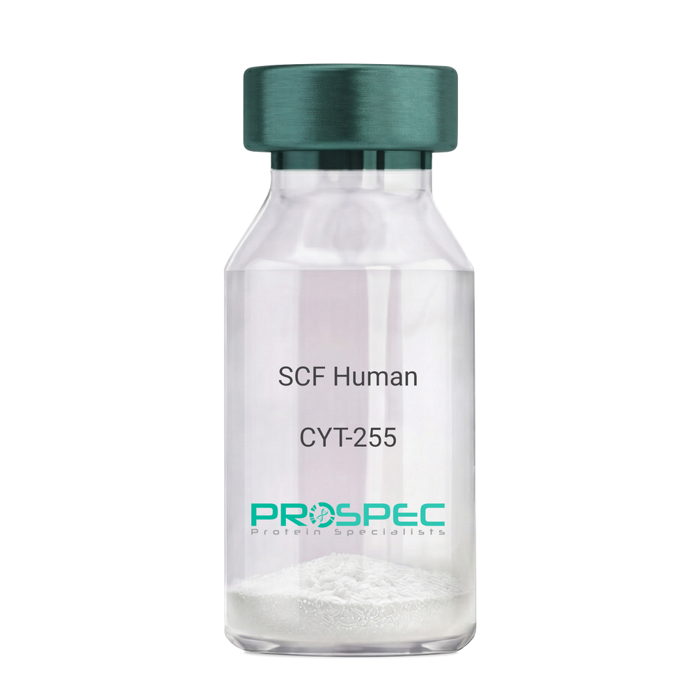
Name :
SCF HumanDescription:
Stem Cell Factor Human Recombinant
Kit ligand Precursor, C-kit ligand, SCF, Mast cell growth factor, MGF, SF, KL-1, Kitl, DKFZp686F2250.
Product # :
CYT-255Price :
Quantity :
Shipping Method :
Shipped at Room temp
More Info
- description
- source
- formulation
- purity
- biological activity
- More Info
Description
Stem Cell Factor Human Recombinant produced in E.Coli is a single, non-glycosylated polypeptide chain containing 165 amino acids and having a molecular mass of 18409 Dalton. The SCF is purified by proprietary chromatographic techniques.
Source
Escherichia Coli.
Formulation
Lyophilized from a concentrated (1mg/ml) solution in water containing 0.02% NaHCO3.
Purity
Greater than 98.0% as determined by:
(a) Analysis by SEC-HPLC.
(b) Analysis by SDS-PAGE.Biological Activity
The ED50 as determined by the dose-dependant stimulation of Human TF-1 cells is < 2 ng/ml, corresponding to a Specific Activity of 500,000 IU/mg.More Info
-
Introduction
Stem cell factor / KIT ligand (SCF) is a cytokine which binds CD117(c-Kit). SCF is also known as "steel factor" or "c-kit ligand". SCF exists in two forms, cell surface bound SCF and soluble (or free) SCF. Soluble SCF is produced by the cleavage of surface bound SCF by metalloproteases. SCF is a growth factor important for the survival, proliferation, and differentiation of hematopoietic stem cells and other hematopoietic progenitor cells. One of its roles is to change the BFU-E (burst-forming unit-erythroid) cells, which are the earliest erythrocyte precursors in the erythrocytic series, into the CFU-E (colony-forming unit-erythroid).
-
Synonyms
Kit ligand Precursor, C-kit ligand, SCF, Mast cell growth factor, MGF, SF, KL-1, Kitl, DKFZp686F2250.
-
Physical Appearance
Sterile Filtered White lyophilized (freeze-dried) powder.
-
Stability
Lyophilized KIT ligand although stable at room temperature for 3 weeks, should be stored desiccated below -18°C. Upon reconstitution SCF should be stored at 4°C between 2-7 days and for future use below -18°C.For long term storage it is recommended to add a carrier protein (0.1% HSA or BSA).Please prevent freeze-thaw cycles.
-
Solubility
It is recommended to reconstitute the lyophilized Stem Cell Factor in sterile 18MΩ-cm H2O not less than 100µg/ml, which can then be further diluted to other aqueous solutions.
-
Amino Acid Sequence
The sequence of the first five N-terminal amino acids was determined and was found to be Met-Glu-Gly-Ile-Cys.
-
Protein content
Protein quantitation was carried out by two independent methods1. UV spectroscopy at 280 nm using the absorbency value of 0.52 as the extinction coefficient for a 0.1% (1mg/ml) solution. This value is calculated by the PC GENE computer analysis program of protein sequences (IntelliGenetics). 2. Analysis by RP-HPLC, using a standard solution of Stem Cell Factor as a Reference Standard.
ProSpec's products are furnished for LABORATORY RESEARCH USE ONLY. The product may not be used as drugs, agricultural or pesticidal products, food additives or household chemicals.
- View Data Sheet
Name :
EGFR Antibody (PAT2H8AT)Description:
Epidermal Growth Factor Receptor Clone PAT2H8AT, Mouse Anti Human
ERBB, ERBB1, Epidermal growth factor receptor, HER1, PIG61, epidermal growth factor receptor, Urogastrone, Proto-oncogene c-ErbB-1, Oncogene ERBB, Cell proliferation inducing protein 61, Cell growth inhibiting protein 40, Avian erythroblastic leukemia viral (verbb) oncogene homolog.
Product # :
ANT-751Price :
Quantity :
Shipping Method :
Shipped with Ice Packs
More Info
- formulation
- More Info
Formulation
1mg/ml containing PBS, pH-7.4, 10% Glycerol and 0.02% Sodium Azide.
More Info
-
Introduction
Epidermal growth factor receptor or EGFR or Erb-1 is a membranal protein that acts as a receptor for the extracellular EGF proteins family. This protein is a part of a family of receptors called ErbB family which are all tyrosine kinases receptors. Mutations in the gene of this receptors can cause cancer.
-
Synonyms
ERBB, ERBB1, Epidermal growth factor receptor, HER1, PIG61, epidermal growth factor receptor, Urogastrone, Proto-oncogene c-ErbB-1, Oncogene ERBB, Cell proliferation inducing protein 61, Cell growth inhibiting protein 40, Avian erythroblastic leukemia viral (verbb) oncogene homolog.
-
Physical Appearance
Sterile filtered colorless solution.
-
Immunogen
Anti-human EGFR mAb, is derived from hybridization of mouse F0 myeloma cells with spleen cells from BALB/c mice immunized with recombinant human EGFR amino acids 424-605 purified from E. coli.
-
Ig Subclass
Mouse IgG2b heavy chain and κ light chain.
-
Clone
PAT2H8AT.
-
Applications
EGFR antibody has been tested by ELISA, Western blot analysis and ICC/IF to assure specificity and reactivity. Since application varies, however, each investigation should be titrated by the reagent to obtain optimal results.
-
Type
Mouse Anti Human Monoclonal.
-
Storage Procedures
For periods up to 1 month store at 4°C, for longer periods of time, store at -20°C. Prevent freeze thaw cycles.
-
Purification Method
EGFR antibody was purified from mouse ascitic fluids by protein-G affinity chromatography.
ProSpec's products are furnished for LABORATORY RESEARCH USE ONLY. The product may not be used as drugs, agricultural or pesticidal products, food additives or household chemicals.
- View Data Sheet
Name :
F9 AntibodyDescription:
Coagulation Factor-IX, Mouse Antibody
Product # :
ANT-229Price :
Quantity :
Shipping Method :
Shipped at Room temp
More Info
- formulation
- More Info
Formulation
1mg/ml in PBS (after reconstitution).
More Info
-
Introduction
Factor IX is a glycoprotein, which is synthesized in the liver. The domain structure of factor IX is similar to that of the other vitamin K dependent coagulation factors. The NH2-terminal region contains 12 g-carboxyglutamic acid (gla) residues, which facilitate the calcium dependent binding of factor IX to negatively charged phospholipid surfaces. Two domains which are homologous to epidermal growth factor (EGF) span the region between the NH2-terminal gla domain and the activation peptide (Ala-146 to Arg-180). Factor IX is activated by either factor XIa or the factor VIIa/tissue factor/phospholipid complex. Cleavage at site A yields the intermediate IXa, which is subsequently converted to the fully active form IXab by cleavage at site B. The NH2-terminal light chain (GLA and EGF domains) remains covalently attached to the COOH-terminal heavy chain by a disulfide bond. The serine protease catalytic triad (Ser-365, His 221, Asp-269) is located in the heavy chain. Factor IXab is the catalytic component of the “intrinsic factor Xase complex” (factor VIIIa/IXa/Ca2+/phospholipid) which proteolytically activates factor X to factor Xa.
-
Solubility
Reconstitute with H20 to get a 1 mg/ml concentration. Mix gently, wash the sides of the vial and wait 30-60 seconds before use.
-
Immunogen
Human Factor-IX.
-
Ig Subclass
Mouse IgG1.
-
Clone
NYRhFIX.
-
Applications
Direct ELISA, Western Blot, imunohistochemistry. For W.B. use 1µg/ml of antibody.
-
Titer
By direct ELISA, 1:10,000 dilution will yield 0.5 O.D using alkaline phosphatase conjugated rabbit anti-mouse Ig (Jackson Laboratories).
-
Shipping Conditions
Antibody is shipped lyophilized at ambient temperature.
-
Type
Mouse Antibody Monoclonal.
-
Storage Procedures
In lyophilized form, for long periods, store at 4 C in a dry environment. After reconstitution, if not intended for use within a month, aliquot and store at -20 C.
-
Purification Method
Protein A.
ProSpec's products are furnished for LABORATORY RESEARCH USE ONLY. The product may not be used as drugs, agricultural or pesticidal products, food additives or household chemicals.
- View Data Sheet
Name :
FGF 2 RatDescription:
Fibroblast Growth Factor-basic Rat Recombinant
HBGH-2, HBGF-2, Prostatropin, FGF-2, FGB-b, Fibroblast Growth Factor-basic, Basic fibroblast growth factor, bFGF, Heparin-binding growth factor 2.
Product # :
CYT-608Price :
Quantity :
Shipping Method :
Shipped at Room temp
More Info
- description
- source
- formulation
- purity
- biological activity
- More Info
Description
bFGF Rat Recombinant (FGF-2) produced in E.Coli is a single, non-glycosylated, polypeptide chain containing 146 amino acids and having a molecular mass of 16.4 kDa.The bFGF2 is purified by proprietary chromatographic techniques.
Source
Escherichia Coli.
Formulation
FGF-b was lyophilized from 1mg/ml solution after extensive dialysis against 10mM Na2PO4, pH 7.5 and 50mM NaCl.
Purity
Greater than 95.0% as determined by SDS-PAGE.
Biological Activity
The ED50 range as determined by the dose-dependant proliferation of NR6R-3T3 cells was found to be 0.903ng/ml, corresponding to a specific activity of 1.1x10⁷ units/mg.
More Info
-
Introduction
FGF-basic is a member of the fibroblast growth factor (FGF) family. FGF family members bind heparin and possess broad mitogenic and angiogenic activities. This protein has been implicated in diverse biological processes, such as limb and nervous system development, wound healing, and tumor growth. The mRNA for this gene contains multiple polyadenylation sites, and is alternatively translated from AUG and non-AUG (CUG) initiation codons resulting in five different isoforms with distinct properties. The CUG-initiated isoforms are localized in the nucleus and are responsible for the intracrine effect, whereas, the AUG-initiated form is mostly cytosolic and is responsible for the paracrine and autocrine effects of this FGF.
The heparin-binding growth factors are angiogenic agents in vivo and are potent mitogens for a variety of cell types in vitro. there are differences in the tissue distribution and concentration of these 2 growth factors. -
Synonyms
HBGH-2, HBGF-2, Prostatropin, FGF-2, FGB-b, Fibroblast Growth Factor-basic, Basic fibroblast growth factor, bFGF, Heparin-binding growth factor 2.
-
Physical Appearance
Sterile Filtered White lyophilized (freeze-dried) powder.
-
Stability
Lyophilized Rat bFGF although stable at room temperature for 3 weeks, should be stored desiccated below -18°C. Upon reconstitution Rat FGF-2 should be stored at 4°C between 2-7 days and for future use below -18°C.For long term storage it is recommended to add a carrier protein (0.1% HSA or BSA).Please prevent freeze-thaw cycles.
-
Solubility
It is recommended to reconstitute the lyophilized Rat bFGF in sterile 18MΩ-cm H2O not less than 100µg/ml, which can then be further diluted to other aqueous solutions.
-
Amino Acid Sequence
MPALPEDGGGA FPPGHFKDPK RLYCKNGGFF LRIHPDGRVD GVREKSDPHV KLQLQAEERG VVSIKGVCAN RYLAMKEDGR LLASKCVTEE CFFFERLESN NYNTYRSRKY SSWYVALKRT GQYKLGSKTG PGQKAILFLP MSAKS.
-
Background
What is the molecular weight/Mw of FGF 2 RAT Protein?
FGF 2 RAT Protein has a total Mw of 16.4kDa.
What is the source or expression system of FGF 2 RAT Protein?
Escherichia Coli.
What is the Purity of FGF 2 RAT Protein?
FGF 2 RAT Protein is >95% pure as determined by SDS-PAGE.
What is the Biological Activity of FGF 2 RAT Protein?
The ED50 range as determined by the dose-dependant proliferation of NR6R-3T3 cells was found to be 0.903ng/ml, corresponding to a specific activity of 1.1x10⁷ units/mg.
What is the amino acid sequence of FGF 2 RAT Protein?
MPALPEDGGGA FPPGHFKDPK RLYCKNGGFF LRIHPDGRVD GVREKSDPHV KLQLQAEERG VVSIKGVCAN RYLAMKEDGR LLASKCVTEE CFFFERLESN NYNTYRSRKY SSWYVALKRT GQYKLGSKTG PGQKAILFLP MSAKS.
What applications can FGF 2 RAT Protein be used in?
FGF 2 RAT Protein can probably be used in western blot, ELISA and Lateral Flow.
What is the endotoxin level for FGF 2 RAT Protein?
The endotoxin level is minimal, FGF 2 RAT Protein was purified using conventional chromatography techniques.
ProSpec's products are furnished for LABORATORY RESEARCH USE ONLY. The product may not be used as drugs, agricultural or pesticidal products, food additives or household chemicals.
- View Data Sheet
Name :
VEGF MouseDescription:
Vascular Endothelial Growth Factor Mouse Recombinant
Vascular endothelial growth factor A, VEGF-A, Vascular permeability factor, VPF, VEGF, MGC70609.
Product # :
CYT-336Price :
Quantity :
Shipping Method :
Shipped at Room temp
More Info
- description
- source
- formulation
- purity
- biological activity
- More Info
Description
Vascular Endothelial Growth Factor Mouse Recombinant produced in E.Coli is a disulfide-linked homodimeric, double polypeptide chains containing 165 amino acids and having a molecular mass of 38.8kDa.
Source
Escherichia Coli.
Formulation
The protein was lyophilized from a 0.2µm filtered solution with PBS, pH 7.4.
Purity
Greater than 95.0% as determined by:
(a) Analysis by RP-HPLC.
(b) Analysis by SDS-PAGE.Biological Activity
The biological activity is determined by the dose-dependent stimulation of the proliferation of human umbilical vein endothelial cells (HUVEC) using a concentration range of 5.0 ng/ml corresponding to a specific activity of 200,000IU/mg.
More Info
-
Introduction
Vascular endothelial growth factor is an important signaling protein involved in both vasculogenesis and angiogenesis. As its name implies, VEGF activity has been mostly studied on cells of the vascular endothelium, although it does have effects on a number of other cell types (e.g. stimulation monocyte/ macrophagemigration, neurons, cancer cells, kidney epithelial cells ). VEGF mediates increased vascular permeability, induces angiogenesis, vasculogenesis and endothelial cell growth, promotes cell migration, and inhibits apoptosis. In vitro, VEGF has been shown to stimulate endothelial cell mitogenesisand cell migration. VEGF is also a vasodilator and increases microvascular permeability and was originally referred to as vascular permeability factor.
Elevated levels of this protein are linked to POEMS syndrome, also known as Crow-Fukase syndrome. Mutations in this gene have been associated with proliferative and nonproliferative diabetic retinopathy. -
Synonyms
Vascular endothelial growth factor A, VEGF-A, Vascular permeability factor, VPF, VEGF, MGC70609.
-
Physical Appearance
Sterile Filtered White lyophilized (freeze-dried) powder.
-
Stability
Lyophilized Vascular Endothelial Growth Factor although stable at room temperature for 3 weeks, should be stored desiccated below -18°C. Upon reconstitution VEGF should be stored at 4°C between 2-7 days and for future use below -18°C.For long term storage it is recommended to add a carrier protein (0.1% HSA or BSA).Please prevent freeze-thaw cycles.
-
Solubility
It is recommended to reconstitute the lyophilized Vascular Endothelial Growth Factor in sterile 18MΩ-cm H2O not less than 100µg/ml, which can then be further diluted to other aqueous solutions.
-
Amino Acid Sequence
MAPTTEGEQK SHEVIKFMDV YQRSYCRPIE TLVDIFQEYP DEIEYIFKPS CVPLMRCAGC CNDEALECVP TSESNITMQI MRIKPHQSQH IGEMSFLQHS RCECRPKKDR TKPEKHCEPC SERRKHLFVQ DPQTCKCSCK NTDSRCKARQ LELNERTCRC DKPRR.
ProSpec's products are furnished for LABORATORY RESEARCH USE ONLY. The product may not be used as drugs, agricultural or pesticidal products, food additives or household chemicals.
- View Data Sheet
Name :
CCN1 (275-381)Description:
Cysteine-Rich Angiogenic Inducer 61 (275-381 a.a.) Human Recombinant
CYR61, Protein CYR61, Cysteine-rich angiogenic inducer 61, IGF-binding protein 10, IGFBP-10, IBP-10, Protein GIG1, CCN family member 1, CCN1, GIG1, IGFBP10.
Product # :
CYT-1235Price :
Quantity :
Shipping Method :
Shipped at Room temp
More Info
- description
- source
- formulation
- purity
- More Info
Description
The CYR61Human is created as a recombinant protein with a 4kda N-terminal fusion of His Tag. The CYR61His-Tagged Fusion Protein, produced in E. coli, is a 17kDa protein containing 107 amino acid residues of the CYR61Human, 275-381 amino acids.
Source
Escherichia Coli.
Formulation
Each mg was lyophilized with 1xPBS, 0.4% SDS and 4mM DTT.
Purity
Greater than 90% as determined by SDS-PAGE.
More Info
-
Synonyms
CYR61, Protein CYR61, Cysteine-rich angiogenic inducer 61, IGF-binding protein 10, IGFBP-10, IBP-10, Protein GIG1, CCN family member 1, CCN1, GIG1, IGFBP10.
-
Physical Appearance
Filtered White lyophilized (freeze-dried) powder.
-
Stability
Store lyophilized CYR61at -20°C. Aliquot the product after reconstitution to avoid repeated freezing/thawing cycles. Reconstituted protein can be stored at 4°C for a limited period of time; it does not show any change after two weeks at 4°C.
-
Solubility
It is recommended to add deionized water to prepare a working stock solution of approximately 0.5mg/ml and let the lyophilized pellet dissolve completely. Product is not sterile! Please filter the product by an appropriate sterile filter before using it on cell culture.
-
Background
Cysteine-Rich Angiogenic Inducer 61 also known as CYR61 is a growth factor-inducible, immediate-early gene which has many activities in various cancers. CYR61 is a part of to the CCN family a group of secreted extracellular matrix-associated proteins which regulate diverse biological processes including cell adhesion, proliferation, migration, survival and angiogenesis. CYR61 plays a significant role in development, tissue repair, wound healing, and various pathological conditions such as cancer and fibrosis. CYR61 is encoded by a growth factor-inducible immediate-early gene. CYR61 promotes the adhesion of endothelial cells by interaction with integrin and enhances growth factor-induced DNA synthesis in the same cell type.
What is the molecular weight/Mw of CCN1 Protein?
CCN1 Protein has a total Mw of 17kDa.
What is the source or expression system of CCN1 Protein?
Escherichia Coli.
What is the Purity of CCN1 Protein?
CCN1 Protein is >90% pure as determined by SDS-PAGE.
What is the Biological Activity of CCN1 Protein?
The biological functionality of CCN1 Protein will be determined in the future.
What is the amino acid sequence of CCN1 Protein?
CCN1 Protein is composed from 107 amino acids.
What applications can CCN1 Protein be used in?
CCN1 Protein can probably be used in western blot, ELISA and Lateral Flow.
What is the endotoxin level for CCN1 Protein?
The endotoxin level is minimal, CCN1 Protein was purified using conventional chromatography techniques.
ProSpec's products are furnished for LABORATORY RESEARCH USE ONLY. The product may not be used as drugs, agricultural or pesticidal products, food additives or household chemicals.
- View Data Sheet
Name :
MIA2 HumanDescription:
Melanoma Inhibitory Activity 2 Human Recombinant
MIA-2, MIA2, Melanoma Inhibitory Activity 2, FLJ22404.
Product # :
CYT-638Price :
Quantity :
Shipping Method :
Shipped at Room temp
More Info
- description
- source
- formulation
- purity
- More Info
Description
MIA2 Human Recombinant produced in E.Coli is a single, non-glycosylated, polypeptide chain consisting of 101 amino having a total molecular mass of 11.5 kDa.The MIA2 is purified by proprietary chromatographic techniques.
Source
Escherichia Coli.
Formulation
The MIA2 protein was lyophilized from a concentrated (1mg/ml) solution containing 20mM Phosphate buffer pH=7.4 and 150mM NaCl.
Purity
Greater than 97.0% as determined by(a) Analysis by RP-HPLC.
(b) Analysis by SDS-PAGE.More Info
-
Introduction
MIA2 performs as a tumour suppressor in hepatocellular carcinoma. MIA2is a novel acute phase protein which its expression is found in the liver and reacts to liver damage in chronic liver diseases and is considerably higher in severe fibrosis patients . MIA2 is induced by IL6, TGF-B & and conditioned medium from activated hepatic stellate cells. MIA2 is is mainly expressed in hepatocytes.
-
Synonyms
MIA-2, MIA2, Melanoma Inhibitory Activity 2, FLJ22404.
-
Physical Appearance
Sterile Filtered White lyophilized (freeze-dried) powder.
-
Stability
Lyophilized MIA2 although stable at room temperature for 3 weeks, should be stored desiccated below -18°C. Upon reconstitution MIA2 should be stored at 4°C between 2-7 days and for future use below -18°C.Please prevent freeze-thaw cycles.
-
Solubility
It is recommended to reconstitute the lyophilized MIA2 in sterile 18MΩ-cm H2O not less than 100µg/ml, which can then be further diluted to other aqueous solutions.
-
Amino Acid Sequence
MLESTKLLAD LKKCGDLECE ALINRVSAMR DYRGPDCRYL NFTKGEEISV YVKLAGERED LWAGSKGKEF GYFPRDAVQI EEVFISEEIQ MSTKESDFLC L.
ProSpec's products are furnished for LABORATORY RESEARCH USE ONLY. The product may not be used as drugs, agricultural or pesticidal products, food additives or household chemicals.
- View Data Sheet
Name :
PMSGDescription:
Pregnant Mare Serum Gonadotropin
Product # :
HOR-272Price :
Quantity :
Shipping Method :
Shipped at Room temp
More Info
- description
- source
- formulation
- More Info
Description
PMSG is a complex glycoprotein obtained from the serum of pregnant mares. This 43-63 kda protein is capable of supplementing and being substituted for the follicle stimulating and interstitial cell-stimulating hormone of the anterior pituitary gland in both the male and female. Thus PMSG-Intervet stimulates development of the ovarian follicle in the female.
Source
Serum of pregnant mares.
Formulation
The PMSG was lyophilized with no additives.
More Info
-
Introduction
PMSG Hormone is a well know used hormone together with progestogen to increase ovulation just before to artificial insemination. PMSG hormone is a placental glycoprotein produced from the serum of pregnant mares. PMSG comprises of an alfa subunit and a beta subunit. PMSG hormone is secreted from endometrial cups within the pregnant mare uterus aging from 40 to 130 days into their maturation, and once extracted, it can been used to promote artificially estrus in female animals. These assemblies produce PMSG hormone to induce mare's ovarian and repsouctive structures. PMSG can induce the growth of follicles by ovaries and results in ovulatation. PMSG hormone has an about a 4 day half-life of bioactivity in species other than horses. The extended biological activity can cause ovarian stimulation and ovulation. However, PMSG use alone often causes cystic ovarian disease because of the unrestrained ovarian stimulation and due to the sugar molecules which decrease clearance of the hormone. PMSG is more likely to be used than other pituitary hormones due to the extended circulatory half-life. PMSG solely exhibits luteinizing hormone like activity, however in other animal classes it has FSH & LH like activity.
-
Physical Appearance
Sterile Filtered White lyophilized (freeze-dried) powder.
-
Stability
Lyophilized PMSG although stable at room temperature for 3 weeks, should be stored between 2-8°C.
-
Solubility
It is recommended to reconstitute the lyophilized PMSG in sterile 18M-cm H2O at a concentration of 1000 IU/ml, which can then be further diluted to other aqueous solutions.
ProSpec's products are furnished for LABORATORY RESEARCH USE ONLY. The product may not be used as drugs, agricultural or pesticidal products, food additives or household chemicals.
- View Data Sheet
Name :
GTSF1 HumanDescription:
Gametocyte Specific Factor 1 Human Recombinant
Gametocyte Specific Factor 1, FAM112B, Family with Sequence Similarity 112 Member B, Gametocyte-Specific Factor 1, Protein FAM112B, GTSF1.
Product # :
PRO-561Price :
Quantity :
Shipping Method :
Shipped with Ice Packs
More Info
- description
- source
- formulation
- purity
- More Info
Description
GTSF1 Human Recombinant produced in E.coli is a single, non-glycosylated polypeptide chain containing 190 amino acids (1-167) and having a molecular mass of 21.7 kDa.GTSF1 is fused to a 23 amino acid His-tag at N-terminus & purified by proprietary chromatographic techniques.
Source
Escherichia Coli.
Formulation
The GTSF1 solution (0.5mg/ml) contains 20mM Tris-HCl buffer (pH 8.0), 0.15M NaCl and 20% glycerol and 1mM DTT.
Purity
Greater than 90.0% as determined by SDS-PAGE.
More Info
-
Introduction
Gametocyte Specific Factor 1 (GTSF1) is a member of the UPF0224 (FAM112) family and contains 1 CHHC-type zinc finger. A key paralog of the GTSF1 gene is GTSF1L.
-
Synonyms
Gametocyte Specific Factor 1, FAM112B, Family with Sequence Similarity 112 Member B, Gametocyte-Specific Factor 1, Protein FAM112B, GTSF1.
-
Physical Appearance
Sterile Filtered clear solution.
-
Stability
Store at 4°C if entire vial will be used within 2-4 weeks. Store, frozen at -20°C for longer periods of time. For long term storage it is recommended to add a carrier protein (0.1% HSA or BSA).Avoid multiple freeze-thaw cycles.
-
Amino Acid Sequence
MGSSHHHHHH SSGLVPRGSH MGSMEETYTD SLDPEKLLQC PYDKNHQIRA CRFPYHLIKC RKNHPDVASK LATCPFNARH QVPRAEISHH ISSCDDRSCI EQDVVNQTRS LRQETLAEST WQCPPCDEDW DKDLWEQTST PFVWGTTHYS DNNSPASNIV TEHKNNLASG MRVPKSLPYV LPWKNNGNAQ.
ProSpec's products are furnished for LABORATORY RESEARCH USE ONLY. The product may not be used as drugs, agricultural or pesticidal products, food additives or household chemicals.
- View Data Sheet
Name :
PEDF HumanDescription:
Pigment Epithelium-Derived Factor Human Recombinant
Pigment epithelium-derived factor,PEDF, Serpin-F1, SerpinF1, EPC-1, EPC1, PIG35.
Product # :
CYT-580Price :
Quantity :
Shipping Method :
Shipped at Room temp
More Info
- description
- source
- formulation
- purity
- More Info
Description
PEDF Human Recombinant produced in E.Coli is a single, non-glycosylated, polypeptide chain containing 400 amino acids and having a molecular mass of 44.5 kDa. The Human PEDF is purified by proprietary chromatographic techniques.
Source
Escherichia Coli.
Formulation
The sterile filtered concentrated (1mg/ml) protein solution was lyophilized with 20mM sodium phosphate buffer & 150mM NaCl pH-7.4.
Purity
Greater than 95% as determined by SDS-PAGE.
More Info
-
Introduction
PEDF is a noninhibitory serpin with neurotrophic, anti-angiogenic, and anti-tumorigenic properties. PEDF is a 50,000 dalton glycoprotein created and secreted in many tissues all the way through the body. A key component of the anti-angiogenic action of PEDF is the induction of apoptosis in proliferating endothelial cells. Additionally, PEDF is capable to inhibit the activity of angiogenic factors such as VEGF and FGF-2. The neuro-protective effects of PEDF are achieved through suppression of neuronal apoptosis induced by peroxide, glutamate, or other neurotoxins. The recognition of a lipase-linked cell membrane receptor for PEDF (PEDF-R) that binds to PEDF with high affinity should facilitate further elucidation of the underlying mechanisms of this pluripotent serpin. To date, PEDF-R is the only signaling receptor known to be used by a serpin family member. The unique range of PEDF activities associate it as a potential therapeutic agent for the treatment of vasculature related neurodegenerative diseases such as age-related macular degeneration (AMD) and proliferative diabetic retinopathy (PDR). PEDF in addition has the potential to be functional in the treatment of various angiogenesis-related diseases including a number of cancers.
-
Synonyms
Pigment epithelium-derived factor,PEDF, Serpin-F1, SerpinF1, EPC-1, EPC1, PIG35.
-
Physical Appearance
Sterile Filtered White lyophilized (freeze-dried) powder.
-
Stability
Lyophilized PEDF although stable at room temperature for 3 weeks, should be stored desiccated below -18°C. Upon reconstitution PEDF Human should be stored at 4°C between 2-7 days and for future use below -18°C.For long term storage it is recommended to add a carrier protein (0.1% HSA or BSA).Please prevent freeze-thaw cycles.
-
Solubility
It is recommended to reconstitute the lyophilized PEDF in sterile 18M-cm H2O not less than 100µg/ml, which can then be further diluted to other aqueous solutions.
-
Amino Acid Sequence
MQNPASPPEE GSPDPDSTGA LVEEEDPFFK VPVNKLAAAV SNFGYDLYRV RSSMSPTTNV LLSPLSVATA LSALSLGAEQ RTESIIHRAL YYDLISSPDI HGTYKELLDT VTAPQKNLKS ASRIVFEKKL RIKSSFVAPL EKSYGTRPRV LTGNPRLDLQ EINNWVQAQM KGKLARSTKE IPDEISILLL GVAHFKGQWV TKFDSRKTSL EDFYLDEERT VRVPMMSDPK AVLRYGLDSD LSCKIAQLPL TGSMSIIFFL PLKVTQNLTL IEESLTSEFI HDIDRELKTV QAVLTVPKLK LSYEGEVTKS LQEMKLQSLF DSPDFSKITG KPIKLTQVEH RAGFEWNEDG AGTTPSPGLQ PAHLTFPLDY HLNQPFIFVL RDTDTGALLF IGKILDPRGP.
-
Background
About PEDF Human
Also known as “Pigment Epithelium-Derived Factor” or “SERPINF1,” PEDF is a
multifunctional protein found in vertebrates. It has anti-tumorigenic, anti-angiogenic, and
neurotrophic functions. Currently, it’s being researched as a candidate for treatment for
several conditions, including heart disease and cancer.What’s the Function of PEDF Human Recombinant?
PEDF is created/secreted in several tissues across the body. It has a unique anti-angiogenic
action because of its induction of apoptosis in proliferating endothelial cells. Also, PEDF
can inhibit many angiogenic factors, including VEGF and FGF-2. PEDF-R is currently the
only known signaling receptor to be used by a serpin family member.
What’s the Application of PEDF Human Recombinant?This version of PEDF is produced in E. Coli. It’s a single, non-glycosylated, polypeptide
chain that contains 400 amino acids. It has a molecular mass of 44.5 kDa, and it’s purified
by proprietary chromatographic techniques.The main purpose of PEDF human recombinant is for research. It has the potential to
become a potential treatment for different angiogenesis-related diseases, including
various cancers. Moreover, it can become crucial during the recovery from vasculature-
related neurodegenerative illness.Such a discovery could be revolutionary for the world, which is why more research is
needed to determine the effect of this non-inhibitory serpin on the body.
ProSpec's products are furnished for LABORATORY RESEARCH USE ONLY. The product may not be used as drugs, agricultural or pesticidal products, food additives or household chemicals.
- View Data Sheet
Name :
FGF 21 Mouse, HisDescription:
Fibroblast Growth Factor-21 Mouse Recombinant, His Tag
Fibroblast growth factor 21, FGF-21.
Product # :
CYT-516Price :
Quantity :
Shipping Method :
Shipped at Room temp
More Info
- description
- source
- formulation
- purity
- More Info
Description
Fibroblast Growth Factor -21 Mouse Recombinant produced in E.Coli is a single, non-glycosylated, polypeptide chain containing 192 amino acids and having a molecular mass of 21.2 kDa. The amino acid sequence of the recombinant human FGF21 is 100% homologous to the amino acid sequence of the Mouse FGF21 without signal sequence and contains 10 a.a. His tag at N-terminal.The FGF-21 is purified by proprietary chromatographic techniques.
Source
Escherichia Coli.
Formulation
Filtered (0.4 µm) and lyophilized from 0.5 mg/ml in 20mM TRIS, 20mM NaCl, pH 7.5.
Purity
Greater than 95.0% as determined by:
(a) Analysis by RP-HPLC.
(b) Analysis by SDS-PAGE.More Info
-
Introduction
The FGFs are a family of more than 20 small (~17–26 kDa) secreted peptides. The initial characterization of these proteins focused on their ability to stimulate fibroblast proliferation. This mitogenic activity was mediated through FGF receptors (FGFRs) 1, 2, or 3. A fourth closely related tyrosine kinase receptor (FGFR4) was able to bind the FGFs but did not lead to a mitogenic response.
FGFs modulate cellular activity via at least 5 distinct subfamilies of high-affinity FGF receptors (FGFRs): FGFR-1, -2, -3, and -4, all with intrinsic tyrosine kinase activity and, except for FGFR-4, multiple splice isoforms, and FGFR-5, which lacks an intracellular kinase domain. There is growing evidence that FGFRs can be important for regulation of glucose and lipid homeostasis. The overexpression of a dominant negative form of FGFR-1 in ? cells leads to diabetes in mice, which thus implies that proper FGF signaling is required for normal ? cell function and glycemia maintenance. FGFR-2 appears to be a key molecule during pancreatic development. Moreover, FGFR-4 has been implicated in cholesterol metabolism and bile acid synthesis.
FGF-19, has been shown to cause resistance to diet-induced obesity and insulin desensitization and to improve insulin, glucose, and lipid profiles in diabetic rodents. Since these effects, at least in part, are mediated through the observed changes in metabolic rates, FGF-19 can be considered as a regulator of energy expenditure.
FGF-21 is preferentially expressed in liver, but an exact knowledge of FGF-21 bioactivity and its mode of action have been lacking to date. FGF-21 is a potent activator of glucose uptake on adipocytes, protects animals from diet-induced obesity when overexpressed in transgenic mice, and lowers blood glucose and triglyceride levels when therapeutically administered to diabetic rodents. -
Synonyms
Fibroblast growth factor 21, FGF-21.
-
Physical Appearance
Filtered white lyophilized powder.
-
Stability
Lyophilized FGF-21 Mouse Recombinant although stable at room temperature for 3 weeks, should be stored desiccated below -18°C. Upon reconstitution Fibroblast Growth Factor 21 should be stored at 4°C between 2-7 days and for future use below -18°C. For long term storage it is recommended to add a carrier protein (0.1% HSA or BSA).Please prevent freeze-thaw cycles.
-
Solubility
It is recommended to add deionized water to prepare a working stock solution of approximately 0.5 mg/ml and let the lyophilized pellet dissolve completely. Product is not sterile! Please filter the product by an appropriate sterile filter before using it in the cell culture. Add DTT (0.2mM) and NaCl (0.1-0.15M) before freezing to prevent potential aggregation.
-
Amino Acid Sequence
MKHHHHHHAS AYPIPDSSPL LQFGGQVRQR YLYTDDDQDT EAHLEIREDG TVVGAAHRSP ESLLELKALKPGVIQILGVK ASRFLCQQPD GALYGSPHFD PEACSFRELL LEDGYNVYQS EAHGLPLRLP QKDSPNQDATSWGPVRFLPM PGLLHEPQDQ AGFLPPEPPD VGSSDPLSMV EPLQGRSPSY AS.
-
Background
What is the molecular weight/Mw of FGF21 MOUSE,HIS Protein?
FGF21 MOUSE,HIS Protein has a total Mw of 21.2kDa.
What is the source or expression system of FGF21 MOUSE,HIS Protein?
Escherichia Coli.
What is the Purity of FGF21 MOUSE,HIS Protein?
FGF21 MOUSE,HIS Protein is >95% pure as determined by SDS-PAGE.
What is the Biological Activity of FGF21 MOUSE,HIS Protein?
The biological functionality of FGF21 MOUSE,HIS Protein will be determined in the future.
What is the amino acid sequence of FGF21 MOUSE,HIS Protein?
MKHHHHHHAS AYPIPDSSPL LQFGGQVRQR YLYTDDDQDT EAHLEIREDG TVVGAAHRSP ESLLELKALKPGVIQILGVK ASRFLCQQPD GALYGSPHFD PEACSFRELL LEDGYNVYQS EAHGLPLRLP QKDSPNQDATSWGPVRFLPM PGLLHEPQDQ AGFLPPEPPD VGSSDPLSMV EPLQGRSPSY AS.
What applications can FGF21 MOUSE,HIS Protein be used in?
FGF21 MOUSE,HIS Protein can probably be used in western blot, ELISA and Lateral Flow.
What is the endotoxin level for FGF21 MOUSE,HIS Protein?
The endotoxin level is minimal, FGF21 MOUSE,HIS Protein was purified using conventional chromatography techniques.
ProSpec's products are furnished for LABORATORY RESEARCH USE ONLY. The product may not be used as drugs, agricultural or pesticidal products, food additives or household chemicals.
- View Data Sheet
Name :
FGF12 HumanDescription:
Fibroblast Growth Factor 12 Human Recombinant
FGF-12, FGF12, FGF12B, FHF1, Fibroblast growth factor 12, Fibroblast growth factor homologous factor 1, FHF-1, Myocyte-activating factor.
Product # :
CYT-1113Price :
Quantity :
Shipping Method :
Shipped at Room temp
More Info
- description
- source
- formulation
- purity
- biological activity
- More Info
Description
Fibroblast Growth Factor 12 Human Recombinant produced in E.Coli is a single, non-glycosylated polypeptide chain containing 181 amino acids and having a molecular mass of 20.5kDa. The FGF12 is purified by proprietary chromatographic techniques.
Source
Escherichia Coli.
Formulation
Lyophilized from a 0.2μm filtered concentrated solution in PBS, pH7.4 and 1mM DTT.
Purity
Greater than 98.0% as determined by:
(a) Analysis by RP-HPLC.
(b) Analysis by SDS-PAGE.Biological Activity
Determined by FGF12 binding ability in a functional ELISA. Immobilized FGFR4/Fc Chimera at 5 µg/mL (100 µL/well) can bind FGF12 with a linear range of 1.6100 ng/mL.
More Info
-
Introduction
FGF12 is part of the Fibroblast Growth Factor (FGF) family which has a vast mitogenic and cell survival functions, and play a role in a range of biological activities, among them are embryonic development, cell growth, morphogenesis, tissue repair, tumor growth, and invasion. FGF-12 doesn’t obtain the N-terminal signal sequence present in the majority of the FGF family members, but it contains clusters of basic residues that act as a nuclear localization signal. When transfected into mammalian cells, FGF12 accumulated in the nucleus, but was not secreted. FGF12 is involved in nervous system development and function. FGF12 binds to IB2 (islet brain-2), a cellular kinase scaffold, and voltage gated sodium channels and is also involved in intracellular signalling and ion exchange.
-
Synonyms
FGF-12, FGF12, FGF12B, FHF1, Fibroblast growth factor 12, Fibroblast growth factor homologous factor 1, FHF-1, Myocyte-activating factor.
-
Physical Appearance
Sterile Filtered White lyophilized (freeze-dried) powder.
-
Stability
Lyophilized FGF12 although stable at room temperature for 3 weeks, should be stored desiccated below -18°C. Upon reconstitution Fibroblast Growth Factor 12 should be stored at 4°C between 2-7 days and for future use below -18°C. Please prevent freeze-thaw cycles.
-
Solubility
It is recommended to reconstitute the lyophilized Fibroblast Growth Factor 12 in sterile 18MΩ-cm H2O not less than 100µg/ml, which can then be further diluted to other aqueous solutions.
-
Amino Acid Sequence
MESKEPQLKG IVTRLFSQQG YFLQMHPDGT IDGTKDENSD YTLFNLIPVG LRVVAIQGVK ASLYVAMNGE GYLYSSDVFT PECKFKESVF ENYYVIYSST LYRQQESGRA WFLGLNKEGQ IMKGNRVKKT KPSSHFVPKP IEVCMYREQS LHEIGEKQGR SRKSSGTPTM NGGKVVNQDS T.
-
Background
What is the molecular weight/Mw of FGF12 Protein?
FGF12 Protein has a total Mw of 20.5kDa.
What is the source or expression system of FGF12 Protein?
Escherichia Coli.
What is the Purity of FGF12 Protein?
FGF12 Protein is >98% pure as determined by SDS-PAGE.
What is the Biological Activity of FGF12 Protein?
Determined by FGF12 binding ability in a functional ELISA. Immobilized FGFR4/Fc Chimera at 5 µg/mL (100 µL/well) can bind FGF12 with a linear range of 1.6100 ng/mL.
What is the amino acid sequence of FGF12 Protein?
MESKEPQLKG IVTRLFSQQG YFLQMHPDGT IDGTKDENSD YTLFNLIPVG LRVVAIQGVK ASLYVAMNGE GYLYSSDVFT PECKFKESVF ENYYVIYSST LYRQQESGRA WFLGLNKEGQ IMKGNRVKKT KPSSHFVPKP IEVCMYREQS LHEIGEKQGR SRKSSGTPTM NGGKVVNQDS T.
What applications can FGF12 Protein be used in?
FGF12 Protein can probably be used in western blot, ELISA and Lateral Flow.
What is the endotoxin level for FGF12 Protein?
The endotoxin level is minimal, FGF12 Protein was purified using conventional chromatography techniques.
ProSpec's products are furnished for LABORATORY RESEARCH USE ONLY. The product may not be used as drugs, agricultural or pesticidal products, food additives or household chemicals.