Search results
1000 results found for “lactoferrin”
Name
Description
Product #
Price
Quantity
Shipping Method
- View Data Sheet
Name :
CTF1 Human, HisDescription:
Cardiotrophin-1 Human Recombinant, His Tag
CTF1, CT1, CT-1, Cardiophin 1.
Product # :
CYT-436Price :
Quantity :
Shipping Method :
Shipped at Room temp
More Info
- description
- source
- formulation
- purity
- More Info
Description
The Cardiotrophin His-Tagged Fusion Protein Human, produced in E. coli, is 22.5 kDa protein containing 200 amino acid residues of the human Cardiotrophin and 12 additional amino acid residues – His Tag (underlined).
Source
Escherichia Coli.
Formulation
CTF1 was filtered (0.4µm) and lyophilized from 0.5 mg/ml in 0.05M Acetate buffer pH-4.
Purity
Purity of CTF1 Human Recombinant is greater than 90% as determined by SDS-PAGE.
More Info
-
Introduction
Cardiotrophin 1 (CT-1) is a 201 amino acid member of the interleukin-6 superfamily. It was identified by its ability to induce hypertrophic response in cardiac myocytes. CT-1 mRNA levels were found both in cardiac myocytes and in cardiac nonmyocytes. CT 1 was also detected in abundance in normal adult human lung and was expressed in both fetal and adult airway smooth muscle cells. CT 1 activates gp130 dependent signaling and stimulates the Janus kinase/signal transducers and activators of transcription (JAK/STAT) pathway to transduce hypertrophic and cytoprotective signals in cardiac myocytes.
CT 1 has also a neurotrophic function. CTF1 deficiency causes increased motoneuron cell death in spinal cord and brainstem nuclei of mice during a period between embryonic day 14 and the first postnatal week. Moreover, CT-1 is a hepatocyte survival factor that efficiently reduces hepatocellular damage in animal models of acute liver injury. Cardiotrophin 1 expression is augmented after hypoxic stimulation and it can protect cardiac cells when added either prior to simulated ischaemia or at the time of reoxygenation following simulated ischaemia. Cardiotrophin 1 can induce expression of the protective heat shock proteins (hsps) in cardiac cells.
Cardiotrophin-1 increased ventricular expression of ANP, brain natriuretic peptide (BNP) and angiotensinogen mRNA.
Cardiophin 1 levels were significantly elevated in patients with heart failure, patients with dilatative cardiomyopathy, moderate/severe mitral regurgitation, stable and unstable angina and after acute myocardial infarction. -
Synonyms
CTF1, CT1, CT-1, Cardiophin 1.
-
Stability
Store lyophilized protein at -20°C. Aliquot the product after reconstitution to avoid repeated freezing/thawing cycles. Reconstituted protein can be stored at 4°C for a limited period of time; it does not show any change after two weeks at 4°C.
-
Solubility
It is recommended to add 0.1M Acetate buffer pH-4 to prepare a working stock solution of approximately 0.5mg/ml and let the lyophilized pellet dissolve completely. For conversion into higher pH value, we recommend intensive dilution by relevant buffer to a concentration of 10?g/ml. In higher concentrations the solubility of this antigen is limited. Protein is not sterile! Please filter the product by an appropriate sterile filter before using it in the cell culture.
-
Amino Acid Sequence
MRGSHHHHHH GSSRREGSLE DPQTDSSVSL LPHLEAKIRQ THSLAHLLTK YAEQLLQEYV QLQGDPFGLPSFSPPRLPVA GLSAPAPSHA GLPVHERLRL DAAALAALPP LLDAVCRRQA ELNPRAPRLL RRLEDAARQA RALGAAVEAL LAALGAANRG PRAEPPAATA SAASATGVFP AKVLGLRVCG LYREWLSRTE GDLGQLLPGG SA.
-
Background
What is the molecular weight/Mw of CTF1 Protein?
CTF1 Protein has a total Mw of 22.5kDa.
What is the source or expression system of CTF1 Protein?
Escherichia Coli.
What is the Purity of CTF1 Protein?
CTF1 Protein is >90% pure as determined by SDS-PAGE.
What is the Biological Activity of CTF1 Protein?
The biological functionality of CTF1 Protein will be determined in the future.
What is the amino acid sequence of CTF1 Protein?
MRGSHHHHHH GSSRREGSLE DPQTDSSVSL LPHLEAKIRQ THSLAHLLTK YAEQLLQEYV QLQGDPFGLPSFSPPRLPVA GLSAPAPSHA GLPVHERLRL DAAALAALPP LLDAVCRRQA ELNPRAPRLL RRLEDAARQA RALGAAVEAL LAALGAANRG PRAEPPAATA SAASATGVFP AKVLGLRVCG LYREWLSRTE GDLGQLLPGG SA.
What applications can CTF1 Protein be used in?
CTF1 Protein can probably be used in western blot, ELISA and Lateral Flow.
What is the endotoxin level for CTF1 Protein?
The endotoxin level is minimal, CTF1 Protein was purified using conventional chromatography techniques.
ProSpec's products are furnished for LABORATORY RESEARCH USE ONLY. The product may not be used as drugs, agricultural or pesticidal products, food additives or household chemicals.
- View Data Sheet
Name :
NKIRAS1 HumanDescription:
NFKB Inhibitor Interacting Ras-Like 1 Human Recombinant
NF-kappa-B inhibitor-interacting Ras-like protein 1, I-kappa-B-interacting Ras-like protein 1, Kappa B-Ras protein 1, KappaB-Ras1, NKIRAS1, KBRAS1.
Product # :
PRO-1039Price :
Quantity :
Shipping Method :
Shipped with Ice Packs
More Info
- description
- source
- formulation
- purity
- More Info
Description
NKIRAS1 Human Recombinant produced in E.Coli is a single, non-glycosylated polypeptide chain containing 212 amino acids (1-192 a.a) and having a molecular mass of 23.8kDa (Molecular weight on SDS-PAGE will appear higher).NKIRAS1 is fused to a 20 amino acid His-tag at N-terminus & purified by proprietary chromatographic techniques.
Source
Escherichia Coli.
Formulation
NKIRAS1 protein solution (0.25mg/ml) containing 20mM Tris-HCl buffer (pH8.0), 20% glycerol and 1mM DTT.
Purity
Greater than 95.0% as determined by SDS-PAGE.
More Info
-
Introduction
NF-kappa-B inhibitor-interacting Ras-like protein 1 (NKIRAS1) is a member of the small GTPase superfamily. NKIRAS1 is an unusual Ras-like protein which acts as a potent regulator of NF-kappa-B activity by thwarting the degradation of NF-kappa-B inhibitor beta (NFKBIB) by most signals, describing why NFKBIB is more resistant to degradation. NKIRAS1 functions by blocking phosphorylation of NFKBIB and mediating cytoplasmic retention of p65/RELA NF-kappa-B subunit. Both GTP- and GDP-bound forms block phosphorylation of NFKBIB.
-
Synonyms
NF-kappa-B inhibitor-interacting Ras-like protein 1, I-kappa-B-interacting Ras-like protein 1, Kappa B-Ras protein 1, KappaB-Ras1, NKIRAS1, KBRAS1.
-
Physical Appearance
Sterile Filtered clear solution.
-
Stability
Store at 4°C if entire vial will be used within 2-4 weeks. Store, frozen at -20°C for longer periods of time. For long term storage it is recommended to add a carrier protein (0.1% HSA or BSA).Avoid multiple freeze-thaw cycles.
-
Amino Acid Sequence
MGSSHHHHHH SSGLVPRGSH MGKGCKVVVC GLLSVGKTAI LEQLLYGNHT IGMEDCETME DVYMASVETD RGVKEQLHLY DTRGLQEGVE LPKHYFSFAD GFVLVYSVNN LESFQRVELL KKEIDKFKDK KEVAIVVLGN KIDLSEQRQV DAEVAQQWAK SEKVRLWEVT VTDRKTLIEP FTLLASKLSQ PQSKSSFPLP GRKNKGNSNS EN.
ProSpec's products are furnished for LABORATORY RESEARCH USE ONLY. The product may not be used as drugs, agricultural or pesticidal products, food additives or household chemicals.
- View Data Sheet
Name :
KLRB1 Human, Sf9Description:
Killer Cell Lectin-Like Receptor Subfamily B, Member 1 Human Recombinant, Sf9
Killer Cell Lectin Like Receptor B1, Natural Killer Cell Surface Protein P1A, Killer Cell Lectin-Like Receptor Subfamily B, Member 1, C-Type Lectin Domain Family 5 Member B, HNKR-P1A, NKR-P1A, CLEC5B, NKRP1A, Killer Cell Lectin-Like Receptor Subfamily B Member 1, CD161 Antigen, NKR-P1, CD161, NKR.
Product # :
PRO-2488Price :
Quantity :
Shipping Method :
Shipped with Ice Packs
More Info
- description
- source
- formulation
- purity
- More Info
Description
KLRB1 produced in Sf9 Baculovirus cells is a single, glycosylated polypeptide chain containing 401 amino acids (67-225a.a.) and having a molecular mass of 45.7kDa. KLRB1 is expressed with a 239 amino acid hIgG-His-tag at C-Terminus and purified by proprietary chromatographic techniques.
Source
Sf9, Baculovirus cells.
Formulation
KLRB1 protein solution (0.25mg/ml) contains Phosphate Buffered Saline (pH 7.4) and 10% glycerol.
Purity
Greater than 90% as determined by SDS-PAGE.
More Info
-
Introduction
KLRB1 has an inhibitory role on natural killer (NK) cells cytotoxicity which are lymphocytes who facilitate cytotoxicity and secrete cytokines subsequent to immune stimulation. Some genes of the C-type lectin superfamily, like the rodent NKRP1 family of glycoproteins, are expressed by NK cells and take part in NK cell function regulation. KLRB1 holds an extracellular domain with a few characteristic motifs of C-type lectins, a transmembrane domain, and a cytoplasmic domain. Due to its external C terminus KLRB1 is considered to be a type II membrane protein.
-
Synonyms
Killer Cell Lectin Like Receptor B1, Natural Killer Cell Surface Protein P1A, Killer Cell Lectin-Like Receptor Subfamily B, Member 1, C-Type Lectin Domain Family 5 Member B, HNKR-P1A, NKR-P1A, CLEC5B, NKRP1A, Killer Cell Lectin-Like Receptor Subfamily B Member 1, CD161 Antigen, NKR-P1, CD161, NKR.
-
Physical Appearance
Sterile Filtered colorless solution.
-
Stability
Store at 4°C if entire vial will be used within 2-4 weeks. Store, frozen at -20°C for longer periods of time. For long term storage it is recommended to add a carrier protein (0.1% HSA or BSA).Avoid multiple freeze-thaw cycles.
-
Amino Acid Sequence
ADPQKSSIEK CSVDIQQSRN KTTERPGLLN CPIYWQQLRE KCLLFSHTVN PWNNSLADCS TKESSLLLIR DKDELIHTQN LIRDKAILFW IGLNFSLSEK NWKWINGSFL NSNDLEIRGD AKENSCISIS QTSVYSEYCS TEIRWICQKE LTPVRNKVYP DSLEPKSCDK THTCPPCPAP ELLGGPSVFL FPPKPKDTLM ISRTPEVTCV VVDVSHEDPE VKFNWYVDGV EVHNAKTKPR EEQYNSTYRV VSVLTVLHQD WLNGKEYKCK VSNKALPAPI EKTISKAKGQ PREPQVYTLP PSRDELTKNQ VSLTCLVKGF YPSDIAVEWE SNGQPENNYK TTPPVLDSDG SFFLYSKLTV DKSRWQQGNV FSCSVMHEAL HNHYTQKSLS LSPGKHHHHH H.
ProSpec's products are furnished for LABORATORY RESEARCH USE ONLY. The product may not be used as drugs, agricultural or pesticidal products, food additives or household chemicals.
- View Data Sheet
Name :
LAGE3 HumanDescription:
L Antigen Family Member 3 Human Recombinant
CVG5, DXS9879E, DXS9951E, ESO3, ITBA2, EKC/KEOPS complex subunit LAGE3, L antigen family member 3, Protein ESO-3.
Product # :
PRO-1424Price :
Quantity :
Shipping Method :
Shipped with Ice Packs
More Info
- description
- source
- formulation
- purity
- More Info
Description
LAGE3 Human Recombinant produced in E. coli is a single polypeptide chain containing 166 amino acids (1-143) and having a molecular mass of 17.2kDa. LAGE3 is fused to a 23 amino acid His-tag at N-terminus & purified by proprietary chromatographic techniques.
Source
E.coli.
Formulation
The LAGE3 solution (0.25mg/ml) contains 20mM Tris-HCl buffer (pH 8.0), 0.15M NaCl, 50% glycerol and 2mM DTT.
Purity
Greater than 85% as determined by SDS-PAGE.
More Info
-
Introduction
L antigen family member 3 (LAGE3) is a part of the CTAG family. LAGE3 is ubiquitously expressed in somatic tissues unlike the other family members, which are usually expressed only in testis and activated in a broad range of human tumors. LAGE3 is also highly conserved in mouse and rat, suggesting that the encoded protein is functionally significant.
-
Synonyms
CVG5, DXS9879E, DXS9951E, ESO3, ITBA2, EKC/KEOPS complex subunit LAGE3, L antigen family member 3, Protein ESO-3.
-
Physical Appearance
Sterile Filtered colorless solution.
-
Stability
Store at 4°C if entire vial will be used within 2-4 weeks. Store, frozen at -20°C for longer periods of time. For long term storage it is recommended to add a carrier protein (0.1% HSA or BSA).Avoid multiple freeze-thaw cycles.
-
Amino Acid Sequence
MGSSHHHHHH SSGLVPRGSH MGSMRDADAD AGGGADGGDG RGGHSCRGGV DTAAAPAGGA PPAHAPGPGR DAASAARGSR MRPHIFTLSV PFPTPLEAEI AHGSLAPDAE PHQRVVGKDL TVSGRILVVR WKAEDCRLLR ISVINFLDQL SLVVRTMQRF GPPVSR.
ProSpec's products are furnished for LABORATORY RESEARCH USE ONLY. The product may not be used as drugs, agricultural or pesticidal products, food additives or household chemicals.
- View Data Sheet
Name :
HSA ProteinDescription:
HSA Human Protein
HSA, ALB, PRO0883, PRO0903, PRO1341, DKFZp779N1935, GIG20, GIG42, PRO1708, PRO2044, PRO2619, PRO2675, UNQ696, SA, HSA.
Product # :
PRO-354Price :
Quantity :
Shipping Method :
Shipped with Ice Packs
More Info
- description
- source
- formulation
- purity
- More Info
Description
HSA contains 584 amino acid residues derived from the prototypicalHSA sequence.
Source
Human Serum.
Formulation
0.2gr/ml solution containing no additives.
Purity
Greater than 97.0% as determined by SDS-PAGE.
More Info
-
Introduction
HSA is synthesized in the liver as preproalbumin which has an N-terminal peptide that is removed before the nascent protein is released from the rough endoplasmic reticulum. The product, proalbumin, is in turn cleaved in the Golgi vesicles to produce the secreted HSA. HSA is a soluble, monomeric protein which comprises about one-half of the blood serum protein. HSA functions primarily as a carrier protein for steroids, fatty acids, and thyroid hormones and plays a role in stabilizing extracellular fluid volume. Mutations in this gene on chromosome 4 result in various anomalous proteins. HSA is a globular unglycosylated serum protein of molecular weight 65,000. The human HSA gene is 16,961 nucleotides long from the putative 'cap' site to the first poly (A) addition site. It is split into 15 exons which are symmetrically placed within the 3 domains that are thought to have arisen by triplication of a single primordial domain.
HSA is widely used to stabilize blood volume generally from donors but the fear of contamination such as HIV & Hepatitis has enticed great interest in the recombinant form which is identical to the natural blood.
Suitable for use in biochemical, excipient (an inert substance used as a diluent or vehicle for a drug), culture media and chromatographic applications. -
Synonyms
HSA, ALB, PRO0883, PRO0903, PRO1341, DKFZp779N1935, GIG20, GIG42, PRO1708, PRO2044, PRO2619, PRO2675, UNQ696, SA, HSA.
-
Physical Appearance
Sterile Filtered clear yellowish solution.
-
Stability
HSA although stable at room temperature for 2 weeks should be stored at 4°C.
ProSpec's products are furnished for LABORATORY RESEARCH USE ONLY. The product may not be used as drugs, agricultural or pesticidal products, food additives or household chemicals.
- View Data Sheet
Name :
GDNF Human, Sf9Description:
Glial-Derived Neurotrophic Factor Human Recombinant, Sf9
Glial cell line-derived neurotrophic factor, hGDNF, Astrocyte-derived trophic factor, ATF, ATF1, ATF2, HFB1-GDNF, HSCR3.
Product # :
CYT-1162Price :
Quantity :
Shipping Method :
Shipped with Ice Packs
More Info
- description
- source
- formulation
- purity
- More Info
Description
GDNF Human Recombinant produced in Baculovirus is a single glycosylated polypeptide chain containing 113amino acids (109-211 aa) and having a molecular mass of 12.8kDa.GDNF is fused to an 10 amino acid His tag at C-terminus and purified by proprietary chromatographic techniques.
Source
Sf9, Baculovirus cells.
Formulation
GDNF protein (0.25mg/ml) contains 10% glycerol and Phosphate-Buffered Saline (pH 7.4).
Purity
Greater than 90.0% as determined by SDS-PAGE.
More Info
-
Introduction
Glial cell-derived neurotrophic factor or GDNF is part of the GDNF group of ligands proteins. GDNF has a crucial part in numerous cell mechanisms such as neurite outgrowth, cell differentiation, cell survival and migration of cells. GDNF enhances neurons survival via GFRa receptors (mainly GFRa1). The mentioned neurons can die as a result from Parkinson's disease or ALS (amyotrophic lateral sclerosis). This protein takes part in the development of the spermatogenesis & kidney, also, it has a role in alcohol metabolism as ameliorating.
-
Synonyms
Glial cell line-derived neurotrophic factor, hGDNF, Astrocyte-derived trophic factor, ATF, ATF1, ATF2, HFB1-GDNF, HSCR3.
-
Physical Appearance
Sterile Filtered colorless solution.
-
Stability
Store at 4°C if entire vial will be used within 2-4 weeks. Store, frozen at -20°C for longer periods of time. For long term storage it is recommended to add a carrier protein (0.1% HSA or BSA).Avoid multiple freeze-thaw cycles.
-
Amino Acid Sequence
ADPMRGQRGK NRGCVLTAIH LNVTDLGLGY ETKEELIFRY CSGSCDAAET TYDKILKNLS RNRRLVSDKV GQACCRPIAF DDDLSFLDDN LVYHILRKHS AKRCGCIHHH HHH
-
Background
What is the molecular weight/Mw of GDNF HUMAN, SF9 Protein?
GDNF HUMAN, SF9 Protein has a total Mw of 12.8kDa.
What is the source or expression system of GDNF HUMAN, SF9 Protein?
Sf9, Baculovirus cells.
What is the Purity of GDNF HUMAN, SF9 Protein?
GDNF HUMAN, SF9 Protein is >90% pure as determined by SDS-PAGE.
What is the Biological Activity of GDNF HUMAN, SF9 Protein?
The biological functionality of GDNF HUMAN, SF9 Protein will be determined in the future.
What is the amino acid sequence of GDNF HUMAN, SF9 Protein?
ADPMRGQRGK NRGCVLTAIH LNVTDLGLGY ETKEELIFRY CSGSCDAAET TYDKILKNLS RNRRLVSDKV GQACCRPIAF DDDLSFLDDN LVYHILRKHS AKRCGCIHHH HHH
What applications can GDNF HUMAN, SF9 Protein be used in?
GDNF HUMAN, SF9 Protein can probably be used in western blot, ELISA and Lateral Flow.
What is the endotoxin level for GDNF HUMAN, SF9 Protein?
The endotoxin level is minimal, GDNF HUMAN, SF9 Protein was purified using conventional chromatography techniques.
ProSpec's products are furnished for LABORATORY RESEARCH USE ONLY. The product may not be used as drugs, agricultural or pesticidal products, food additives or household chemicals.
- View Data Sheet
Name :
SERF2 HumanDescription:
Small EDRK-Rich Factor 2 Human Recombinant
Small EDRK-rich factor 2, Gastric cancer-related protein VRG107, Protein 4F5-related, 4F5re, h4F5rel, FAM2C, HsT17089.
Product # :
PRO-1730Price :
Quantity :
Shipping Method :
Shipped with Ice Packs
More Info
- description
- source
- formulation
- purity
- More Info
Description
SERF2 Human Recombinant produced in E.Coli is a single, non-glycosylated polypeptide chain containing 82 amino acids (1-59 a.a) and having a molecular mass of 9.3kDa.SERF2 is fused to a 23 amino acid His-tag at N-terminus & purified by proprietary chromatographic techniques.
Source
Escherichia Coli.
Formulation
SERF2 protein solution (0.25mg/ml) containing 20mM Tris-HCl buffer (pH 8.0), 0.15M NaCl and 30% glycerol.
Purity
Greater than 85.0% as determined by SDS-PAGE.
More Info
-
Introduction
SERF2 (small EDRK-rich factor 2) is a member of the SERF family. SERF2 is a protein-coding gene. Among the diseases associated with SERF2 are spinal muscular atrophy, and muscular atrophy.
-
Synonyms
Small EDRK-rich factor 2, Gastric cancer-related protein VRG107, Protein 4F5-related, 4F5re, h4F5rel, FAM2C, HsT17089.
-
Physical Appearance
Sterile Filtered clear solution.
-
Stability
Store at 4°C if entire vial will be used within 2-4 weeks. Store, frozen at -20°C for longer periods of time. For long term storage it is recommended to add a carrier protein (0.1% HSA or BSA).Avoid multiple freeze-thaw cycles.
-
Amino Acid Sequence
MGSSHHHHHH SSGLVPRGSH MGSMTRGNQR ELARQKNMKK QSDSVKGKRR DDGLSAAARK QRDSEIMQQK QKKANEKKEE PK
ProSpec's products are furnished for LABORATORY RESEARCH USE ONLY. The product may not be used as drugs, agricultural or pesticidal products, food additives or household chemicals.
- View Data Sheet
Name :
KLF4 HumanDescription:
Kruppel-Like Factor 4 Human Recombinant
Kruppel-like factor 4 (gut), EZF, GKLF, Epithelial zinc finger protein EZF, Gut-enriched krueppel-like factor, endothelial Kruppel-like zinc finger protein.
Product # :
PRO-891Price :
Quantity :
Shipping Method :
Shipped with Ice Packs
More Info
- description
- source
- formulation
- purity
- More Info
Description
KLF4 Human Recombinant produced in E.coli is a single, non-glycosylated polypeptide chain containing 545 amino acids (11-395) and having a molecular mass of 58.1 kDa.The KLF4 is fused to a 159 amino acid His-CaM Tag at N-terminus and purified by proprietary chromatographic techniques.
Source
E.coli.
Formulation
The KLF4 solution (1mg/ml) contains 20mM Tris-HCl buffer (pH 8.0), 1mM DTT, 0.1M NaCl and 10% glycerol.
Purity
Greater than 80% as determined by SDS-PAGE.
More Info
-
Introduction
KLF4 is a transcription factor that performs as both an activator and repressor. KLF4 is expressed mainly in erythroid tissues and found mostly in gut. KLF4 is takes part in the differentiation of epithelial cells in addition to skeletal and kidney development.
-
Synonyms
Kruppel-like factor 4 (gut), EZF, GKLF, Epithelial zinc finger protein EZF, Gut-enriched krueppel-like factor, endothelial Kruppel-like zinc finger protein.
-
Physical Appearance
Sterile Filtered colorless solution.
-
Stability
Store at 4°C if entire vial will be used within 2-4 weeks. Store, frozen at -20°C for longer periods of time. For long term storage it is recommended to add a carrier protein (0.1% HSA or BSA).Avoid multiple freeze-thaw cycles.
-
Amino Acid Sequence
MAHHHHHHMA DQLTEEQIAE FKEAFSLFDK DGDGTITTKE LGTVMRSLGQ NPTEAELQDM INEVDADGNG TIDFPEFLTM MARKMKDTDS EEEIREAFRV FDKDGNGYIS AAELRHVMTN LGEKLTDEEV DEMIREADID GDGQVNYEEF VQMMTAKGSM AVSDALLPSF STFASGPAGR EKTLRQAGAP NNRWREELSH MKRLPPVLPG RPYDLAAATV ATDLESGGAG AACGGSNLAP LPRRETEEFN DLLDLDFILS NSLTHPPESV AATVSSSASA SSSSSPSSSG PASAPSTCSF TYPIRAGNDP GVAPGGTGGG LLYGRESAPP PTAPFNLADI NDVSPSGGFV AELLRPELDP VYIPPQQPQP PGGGLMGKFV LKASLSAPGS EYGSPSVISV SKGSPDGSHP VVVAPYNGGP PRTCPKIKQE AVSSCTHLGA GPPLSNGHRP AAHDFPLGRQ LPSRTTPTLG LEEVLSSRDC HPALPLPPGF HPHPGPNYPS FLPDQMQPQV PPLHYQELMP PGSCMPEEPK PKRGRRSWPR KRTAT
ProSpec's products are furnished for LABORATORY RESEARCH USE ONLY. The product may not be used as drugs, agricultural or pesticidal products, food additives or household chemicals.
- View Data Sheet
Name :
CTGF Human, HisDescription:
Connective Tissue Growth Factor Human Recombinant, His Tag
CCN2, NOV2, HCS24, IGFBP8, MGC102839, CTGF, Connective Tissue Growth Factor.
Product # :
CYT-438Price :
Quantity :
Shipping Method :
Shipped with Ice Packs
More Info
- description
- source
- formulation
- purity
- More Info
- sds-page
Description
CTGF Human Recombinant produced in E.coli is a single, non-glycosylated polypeptide chain containing 344 amino acids (27-349) and having a molecular mass of 37.7kDa.The CTGF is fused to a 21 amino acid His-Tag at N-terminus and purified by proprietary chromatographic techniques.
Source
Escherichia Coli.
Formulation
CTGF protein (1mg/ml) is supplied in 20mM Tris-HCl, pH-8 and 10% Glycerol.
Purity
Greater than 85.0% as determined by Analysis by SDS-PAGE.
sds-page
More Info
-
Introduction
Connective Tissue Growth Factor belongs to the CCN family of proteins. The CCN family presently consists of six members in human also known as: Cyr61 (Cystein rich 61), CTGF (Connective Tissue Growth Factor), Nov (Nephroblastoma Overexpressed gene), WISP-1, 2 and 3 (Wnt-1 Induced Secreted Proteins). The CCN genes encode secreted proteins associated with the Extracellular Matrix (ECM) and cell membrane. CCN proteins are matricellular proteins which are involved in the regulation of various cellular functions including: proliferation, differentiation, survival, adhesion and migration. They are expressed in derivatives of the three embryonic sheets and are implicated in the development of kidney, nervous system, muscle, bone marrow, cartilage and bone. During adulthood, they are implicated in wound healing, bone fracture repair, and pathologies such as: fibrosis, vascular ailments and tumorigenesis.
Full length secreted CCN proteins can show an antiproliferative activity, whereas truncated isoforms are likely to stimulate proliferation and behave as oncogenes. The full length protein consists of four modulesModule I shares partial identity with the N-terminal part of the Insulin-like Growth Factor Binding Proteins (IGFBPs).
Module II includes a stretch of 70amino acid residues – which shares sequence identity with the Von Willebrand Factor Type C repeat (VWC).
Module III contains sequences sharing identity with the Thrombospondin type 1 repeat (TSP1) (WSXCSXXCG), which is thought to be implicated in the binding of sulfated glycoconjugates and to be important for cell adhesion. Module IV, also designated CT, is encoded by exon5. It is the leasts conserved one of the four domains at the level of nucleotide sequence, but it appears to be critical for several of the biological functions attributed to the CCN proteins. Module IV resembles the CT domain of several extracellular protein including, Von Willebrand's factor and mucins. Sequence similarities to heparin-binding motifs are also found within this domain.
Proteolysis of the secreted full-length CCN proteins that has been reported in the case of CCN2 and CCN3 might result in the production of CCN-derived peptides with high affinity for ligands that full-length CNN proteins bind only poorly. Amino-truncated CCN2 isoforms were biologically active whereas no specific biological activity has been attributed to the truncated CCN3. Although the molecular processes underlying the production of these secreted isoforms is presently unknown, it is important to note that proteolysis occur at the same amino acid residues in both CCN2 and CCN3. An elevated expression of CCN2 has also been detected by Northern blotting in human invasive mammary ductal carcinomas, dermatofibromas, pyogenic granuloma, endothelial cells of angiolipomas and angioleiomyomas, and in pancreatic tumors. A study performed with chondrosarcomas representative of various histological grades established that CCN2 expression was closely correlated with increasing levels of malignancy. In agreement with CCN2 playing a role in brain tumor angiogenesis, immunocytochemistry studies indicated that both glioblastoma tumor cells and proliferating endothelial cells stained positive for CCN2. In astrocytomas, CCN2 expression was particularly elevated in high grade tumors, with a marked effect of CCN2 on cell proliferation. Downregulation of CCN2 expression in these cells was associated with a growth arrest at the G1/S transition while over-expression of CCN2 induced a two-fold increase of the number of cells in the G1 phase. Gene profiling analysis allowed to identify a set of about 50 genes whose expression might account for the proliferative activity of CCN2 in these cells. CCN2 was seen in a higher proportion of mononuclear cells of patients with acute lymphoblastic leukemia. -
Synonyms
CCN2, NOV2, HCS24, IGFBP8, MGC102839, CTGF, Connective Tissue Growth Factor.
-
Physical Appearance
Sterile Filtered clear solution.
-
Stability
Store at 4°C if entire vial will be used within 2-4 weeks. Store, frozen at -20°C for longer periods of time. For long term storage it is recommended to add a carrier protein (0.1% HSA or BSA).Avoid multiple freeze-thaw cycles.
-
Amino Acid Sequence
MGSSHHHHHH SSGLVPRGSH MQNCSGPCRC PDEPAPRCPA GVSLVLDGCG CCRVCAKQLG ELCTERDPCD PHKGLFCDFG SPANRKIGVC TAKDGAPCIF GGTVYRSGES FQSSCKYQCT CLDGAVGCMP LCSMDVRLPS PDCPFPRRVK LPGKCCEEWV CDEPKDQTVV GPALAAYRLE DTFGPDPTMI RANCLVQTTE WSACSKTCGM GISTRVTNDN ASCRLEKQSR LCMVRPCEAD LEENIKKGKK CIRTPKISKP IKFELSGCTS MKTYRAKFCG VCTDGRCCTP HRTTTLPVEF KCPDGEVMKK NMMFIKTCAC HYNCPGDNDI FESLYYRKMY GDMA.
-
Background
What is the molecular weight/Mw of CTGF Protein?
CTGF Protein has a total Mw of 37.7kDa.
What is the source or expression system of CTGF Protein?
Escherichia Coli.
What is the Purity of CTGF Protein?
CTGF Protein is >85% pure as determined by SDS-PAGE.
What is the Biological Activity of CTGF Protein?
The biological functionality of CTGF Protein will be determined in the future.
What is the amino acid sequence of CTGF Protein?
MGSSHHHHHH SSGLVPRGSH MQNCSGPCRC PDEPAPRCPA GVSLVLDGCG CCRVCAKQLG ELCTERDPCD PHKGLFCDFG SPANRKIGVC TAKDGAPCIF GGTVYRSGES FQSSCKYQCT CLDGAVGCMP LCSMDVRLPS PDCPFPRRVK LPGKCCEEWV CDEPKDQTVV GPALAAYRLE DTFGPDPTMI RANCLVQTTE WSACSKTCGM GISTRVTNDN ASCRLEKQSR LCMVRPCEAD LEENIKKGKK CIRTPKISKP IKFELSGCTS MKTYRAKFCG VCTDGRCCTP HRTTTLPVEF KCPDGEVMKK NMMFIKTCAC HYNCPGDNDI FESLYYRKMY GDMA.
What applications can CTGF Protein be used in?
CTGF Protein can probably be used in western blot, ELISA and Lateral Flow.
What is the endotoxin level for CTGF Protein?
The endotoxin level is minimal, CTGF Protein was purified using conventional chromatography techniques.
ProSpec's products are furnished for LABORATORY RESEARCH USE ONLY. The product may not be used as drugs, agricultural or pesticidal products, food additives or household chemicals.
- View Data Sheet
Name :
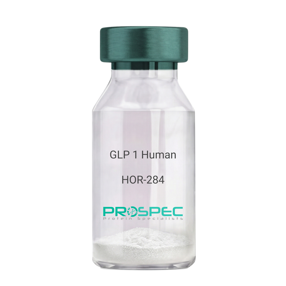
GLP 1 HumanDescription:
Human Glucagon Like Peptide-1
GLP1, Glucagon Like Peptide-1, Incretin hormone.
Product # :
HOR-284Price :
Quantity :
Shipping Method :
Shipped at Room temp
More Info
- description
- formulation
- purity
- More Info
Description
Glucagon Like Peptide-1 is a single, non-glycosylated, polypeptide chain containing 30 amino acids and having a molecular mass of 3297.7 Dalton.The GLP-1 is purified by proprietary chromatographic techniques.
Formulation
The GLP-1 peptide was lyophilized with no additives.
Purity
Greater than 96.0% as determined by RP-HPLC.
More Info
-
Introduction
Glucagon-like peptide-1 (GLP-1) is derived from the transcription product of the proglucagon gene. The major source of GLP-1 in the body is the intestinal L cell that secretes GLP-1 as a guthormone. The biologically active forms of GLP-1 are: GLP-1-(7-37) and GLP-1-(7-36)NH2.
GLP-1 secretion by L cells is dependent on the presence of nutrients in the lumen of the small intestine. The secretagogues (agents that causes or stimulates secretion) of this hormone include major nutrients like carbohydrate, proteinand lipid. Once in the circulation, GLP-1 has a half life of less than 2 minutes, due to rapid degradation by the enzyme dipeptidyl peptidase-4.
GLP-1 possesses several physiological properties that make it a subject of intensive investigation as a potential treatment of diabetes mellitus. The known physiological functions of GLP-1 include: Increases insulin secretion from the pancreas in a glucose-dependent manner, decreases glucagon secretion from the pancreas, increases beta cells mass and insulin gene expression, inhibits acid secretion and gastric emptying in the stomach, decreases food intake by increasing satiety. -
Synonyms
GLP1, Glucagon Like Peptide-1, Incretin hormone.
-
Physical Appearance
Sterile Filtered White lyophilized (freeze-dried) powder.
-
Stability
Lyophilized Glucagon Like Peptide-1 although stable at room temperature for 3 weeks, should be stored desiccated below -18°C. Upon reconstitution GLP-1 should be stored at 4°C between 2-7 days and for future use below -18°C. For long term storage it is recommended to add a carrier protein (0.1% HSA or BSA).Please prevent freeze-thaw cycles.
-
Solubility
It is recommended to reconstitute the lyophilized Glucagon Like Peptide-1 in sterile H2O at 0.5mg/ml, which can then be further diluted to other aqueous solutions.
-
Amino Acid Sequence
H-His-Ala-Glu-Gly-Thr-Phe-Thr-Ser-Asp-Val-Ser-Ser-Tyr-Leu-Glu-Gly-Gln-Ala-Ala-Lys-Glu-Phe-Ile-Ala-Trp-Leu-Val-Lys-Gly-Arg-NH2.
ProSpec's products are furnished for LABORATORY RESEARCH USE ONLY. The product may not be used as drugs, agricultural or pesticidal products, food additives or household chemicals.
- View Data Sheet
Name :
CEA ProteinDescription:
Carcinoembryonic Antigen Human
CEACAM5, Meconium Antigen 100, Carcinoembryonic Antigen, CD66e Antigen, CD66e, Carcinoembryonic Antigen, CEA, oncofetal antigen.
Product # :
PRO-2801Price :
Quantity :
Shipping Method :
Shipped with Ice Packs
More Info
- description
- source
- formulation
- purity
- More Info
Description
CEA produced from patient source colon carcinoma liver metastatic tissue can be used as general marker in screening and monitoring malignant disease states.
Source
Liver tissue.
Formulation
CEA protein solution contains 0.1M PBS, pH 7.4, 0.09 % NaN3 and 2 % methyl-mannoside.
Purity
Greater than 95.0% as determined by SDS-PAGE.
More Info
-
Synonyms
CEACAM5, Meconium Antigen 100, Carcinoembryonic Antigen, CD66e Antigen, CD66e, Carcinoembryonic Antigen, CEA, oncofetal antigen.
-
Physical Appearance
Sterile Filtered colorless solution.
-
Stability
Store at 4°C if entire vial will be used within 2-4 weeks. Store, frozen at -20°C for longer periods of time. For long term storage it is recommended to add a carrier protein (0.1% HSA or BSA). Avoid multiple freeze-thaw cycles.
-
Applications
Blood samples from tissue donors were tested and found to be negative for HBsAg, HIV-1 and HIV-2 antibodies and HCV.
-
Background
Carcinoembryonic Antigen, commonly known as CEA, is a glycoprotein that was initially identified as a tumor marker. Over the years, research into CEA has unveiled its intricate involvement in various physiological processes, not only in cancer but also in the context of normal development and inflammatory conditions. This research aims to delve into the multifaceted roles of CEA, exploring its structural intricacies, regulatory mechanisms, and its implications in health, disease, and beyond.
Structural Complexity of CEA:
CEA, belonging to the immunoglobulin superfamily, is a complex glycoprotein featuring multiple structural domains. Its diverse forms and glycosylation patterns contribute to its functional versatility. CEA is primarily expressed in fetal tissues, but its presence is often detected in adults under pathological conditions, especially in various types of cancer.
CEA in Cancer Biology:
CEA was first recognized as a biomarker for colorectal cancer, but its overexpression is not limited to this context. Elevated CEA levels have been associated with several other malignancies, including breast, lung, and pancreatic cancers. CEA’s involvement in cancer biology ranges from promoting angiogenesis and metastasis to inhibiting immune responses, making it a critical player in tumor progression and evasion.
Beyond Cancer: CEA in Development and Inflammation:
While CEA’s role in cancer is prominent, recent studies have uncovered its participation in normal physiological processes. During embryonic development, CEA is involved in cell adhesion, contributing to tissue organization and morphogenesis. Additionally, CEA expression can be induced in inflammatory conditions, suggesting its involvement in immune responses and tissue repair mechanisms.
CEA as a Diagnostic and Therapeutic Target:
The diverse expression patterns of CEA in various diseases make it a valuable diagnostic tool. CEA assays are widely used for cancer screening, monitoring disease progression, and assessing treatment efficacy. Moreover, CEA’s presence on the surface of cancer cells has made it a target for immunotherapy, enabling the development of targeted therapies aimed at specifically eradicating CEA-positive tumor cells.
ProSpec's products are furnished for LABORATORY RESEARCH USE ONLY. The product may not be used as drugs, agricultural or pesticidal products, food additives or household chemicals.
- View Data Sheet
Name :
ESAT6-CFP10Description:
ESAT6-CFP10 Chimeric Recombinant
Early Secretory Target Mycobacterium Tuberculosis, ESAT-6
Product # :
PRO-2737Price :
Quantity :
Shipping Method :
Shipped with Ice Packs
More Info
- description
- source
- formulation
- purity
- More Info
Description
Recombinant Chimeric ESAT6-CFP10 produced in E. coli is fused to a 6 amino acid his tag at its C-terminus and purified by proprietary chromatographic technique.
Source
Escherichia Coli.
Formulation
ESAT6-CFP10 solution contains 25mM K2CO3, 0.02% NaN3 and PBS.
Purity
Protein is >95% pure as determined by 12% PAGE (coomassie staining).
More Info
-
Introduction
Mycobacterium antigen ESAT6 has been isolated from low molecular weight fractions of the shot-term-culture filtrate (ST-CF) and it can easily be detected in tuberculosis patients. The export of ESAT-6 which is a potent T-cell antigen, and related proteins requires a dedicated secretory apparatus that is encoded by a group of genes, several of which also code for proteins that are recognized strongly by T cells. The ESAT-6 systems can consequently be considered as immunogenicity islands and there is mounting evidence that the equivalent genes are subject to selective pressure imposed by the immune system of the host. This antigen includes many epitopes detectable in the serum of most patients with tuberculosis (more than 90%). By the attempts to obtain the vaccine on the basis of ESAT-6 it was demonstrated that the optimization of adjuvant is very important when using the combination of dioctadecylammonium bromide and monophosphoryllipide. ESAT-6 is very potential as diagnostic for differentiation between the mycobacterial infection and BCG vaccination. The main topic in ESAT-6 using is in antibody production and in test-systems for tuberculosis elaboration. Early secretory antigen 6 (ESAT6) and cell filtrate protein 10 (CFP10) are 2 antigens secreted as a complex by the replicating Mycobacterium tuberculosis complex (MTC).
-
Synonyms
Early Secretory Target Mycobacterium Tuberculosis, ESAT-6
-
Physical Appearance
Sterile Filtered clear solution.
-
Stability
Store at 4°C if entire vial will be used within 2-4 weeks.Store, frozen at -20°C for longer periods of time.For long term storage it is recommended to add a carrier protein (0.1% HSA or BSA).Avoid multiple freeze-thaw cycles.
-
Applications
Immunoassay.
ProSpec's products are furnished for LABORATORY RESEARCH USE ONLY. The product may not be used as drugs, agricultural or pesticidal products, food additives or household chemicals.
- View Data Sheet
Name :
StreptavidinDescription:
Streptavidin Recombinant
Product # :
PRO-791Price :
Quantity :
Shipping Method :
Shipped at Room temp
More Info
- description
- source
- formulation
- purity
- More Info
Description
Streptavidin Streptomyces Avidinii Recombinant produced in E.Coli. The molecular weight per tetramer is approximately 52kDa.
Source
Escherichia Coli.
Formulation
Lyophilized in 10mM potassium phosphate buffer pH 6.5.
Purity
Greater than 98.0% as determined by SDS-PAGE and HPLC.
More Info
-
Introduction
Streptavidin is a tetrameric protein secreted by Streptomyces avidinii which binds firmly to biotin. Streptavidin is widely used in molecular biology through its unique high affinity for the vitamin biotin. The dissociation constant (Kd) of the biotin-streptavidin complex is about ~10-15 mol/L. The strong affinity recognition of biotin and biotinylated molecules has made streptavidin one of the most important components in diagnostics and laboratory kits. The streptavidin/biotin system has one of the biggest free energies of association of yet observed for noncovalent binding of a protein and small ligand in aqueous solution (K_assoc = 10**14). The complexes are also extremely stable over a wide range of temperature and pH.
-
Physical Appearance
Sterile Filtered White lyophilized (freeze-dried) powder.
-
Stability
Streptavidin is shipped at ambient temperature, upon arrival store at -20°C.
-
Solubility
It is recommended to reconstitute the lyophilized Streptavidin in sterile 18MΩ-cm H2O not less than 0.5mg/ml, which can then be further diluted to other aqueous solutions.
-
Amino Acid Sequence
MAEAGITGTWYNQLGSTFIVTAGADGALTGTYESAVGNAESRYVLT
GRYDSAPATDGSGTALGWTVAWKNNYRNAHSATTWSGQYVGGA
EARINTQWLLTSGTTEANAWKSTLVGHDTFTKVKPSAAS. -
Proteolytic Activity
< 10-3 U/mg protein (Azocoll, 25 °C, 24 h, pH 8.0).
-
Specific Activity
> 17U/mg (one unit binds 1 μg D-biotin).
ProSpec's products are furnished for LABORATORY RESEARCH USE ONLY. The product may not be used as drugs, agricultural or pesticidal products, food additives or household chemicals.
- View Data Sheet
Name :
Epoetin HumanDescription:
Erythropoietin-Alpha Human Recombinant
Erythropoietin-Alpha, EPO-a, EPO-alpha, EP, MGC138142.
Product # :
CYT-201Price :
Quantity :
Shipping Method :
Shipped at Room temp
More Info
- description
- source
- formulation
- purity
- biological activity
- More Info
Description
Erythropoietin-alpha Human Recombinant is produced in Chinese hamster ovary (CHO) cells by recombinant DNA technology is a single, polypeptide chain containing 166 amino acids and having a predicted molecular mass of 21,000 Dalton and apparent glycosylated molecular mass of 36-40kDa. EPO-a is purified by proprietary chromatographic techniques.
Source
Chinese Hamster Ovary Cells(CHO).
Formulation
Each mg of lyophilized powder contains 0.59 mg sodium citrate, 0.58 mg sodium chloride and 0.006 mg citric acid.
Purity
Greater than 98.0% as determined by:
(a) Analysis by RP-HPLC.
(b) Analysis by SDS-PAGE.Biological Activity
The Specific Activity was measured by Normocyth -aemic mice and was found to be 150,000 IU/mg.More Info
-
Introduction
This gene is a member of the EPO/TPO family and encodes a secreted, glycosylated cytokine composed of four alpha helical bundles. The protein is found in the plasma and regulates red cell production by promoting erythroid differentiation and initiating hemoglobin synthesis. This protein also has neuroprotective activity against a variety of potential brain injuries and antiapoptotic functions in several tissue types.
-
Synonyms
Erythropoietin-Alpha, EPO-a, EPO-alpha, EP, MGC138142.
-
Physical Appearance
Sterile Filtered White lyophilized (freeze-dried) powder.
-
Stability
Lyophilized Erythropoietin-a although stable at room temperature for 3 weeks, should be stored desiccated below -18°C. Upon reconstitution EPO-alpha should be stored at 4°C between 2-7 days and for future use below -18°C.For long term storage it is recommended to add a carrier protein (0.1% HSA or BSA).Please prevent freeze-thaw cycles.
-
Solubility
It is recommended to reconstitute the lyophilized EPO-alpha in sterile 18MΩ-cm H2O not less than 100µg/ml, which can then be further diluted to other aqueous solutions.
-
Amino Acid Sequence
APPRLICDSR VLERYLLEAK EAENITTGCA EHCSLNENIT VPDTKVNFYA WKRMEVGQQA VEVWQGLALL SEAVLRGQAL LVNSSQPWEP LQLHVDKAVS GLRSLTTLLR ALGAQKEAIS PPDAASAAPL RTITADTFRK LFRVYSNFLR GKLKLYTGEA CRTGDR.
-
Background
What is the molecular weight/Mw of EPOETIN Protein?
EPOETIN Protein has a total Mw of 38kDa.
What is the source or expression system of EPOETIN Protein?
Chinese Hamster Ovary Cells(CHO).
What is the Purity of EPOETIN Protein?
EPOETIN Protein is >98% pure as determined by SDS-PAGE.
What is the Biological Activity of EPOETIN Protein?
The Specific Activity was measured by Normocyth -aemic mice and was found to be 150,000 IU/mg.
What is the amino acid sequence of EPOETIN Protein?
APPRLICDSR VLERYLLEAK EAENITTGCA EHCSLNENIT VPDTKVNFYA WKRMEVGQQA VEVWQGLALL SEAVLRGQAL LVNSSQPWEP LQLHVDKAVS GLRSLTTLLR ALGAQKEAIS PPDAASAAPL RTITADTFRK LFRVYSNFLR GKLKLYTGEA CRTGDR.
What applications can EPOETIN Protein be used in?
EPOETIN Protein can probably be used in western blot, ELISA and Lateral Flow.
What is the endotoxin level for EPOETIN Protein?
The endotoxin level is minimal, EPOETIN Protein was purified using conventional chromatography techniques.
ProSpec's products are furnished for LABORATORY RESEARCH USE ONLY. The product may not be used as drugs, agricultural or pesticidal products, food additives or household chemicals.
- View Data Sheet
Name :
Thyroglobulin HumanDescription:
Thyroglobulin Human Recombinant
Thyroglobulin, TGN, AITD3, TG.
Product # :
PRO-2803Price :
Quantity :
Shipping Method :
Shipped at Room temp
More Info
- description
- source
- formulation
- purity
- More Info
- sds-page
Description
Thyroglobulin Human produced in a mammalian cell line is a single, non-glycosylated polypeptide chain (1-2768 a.a.) and having a molecular mass of 304640 Dalton. Thyroglobulin Human is fused with GlyAlaProGly4SerHis10-tag at C-terminal and purified by proprietary chromatographic techniques.
Source
Mammalian cell line.
Formulation
Thyroglobulin was lyophilized from PBS, pH 7.4 and 5.4 % sucrose.
Purity
Greater than 95.0% as determined by SDS-PAGE.
sds-page
More Info
-
Synonyms
Thyroglobulin, TGN, AITD3, TG.
-
Physical Appearance
Sterile Filtered White lyophilized (freeze-dried) powder.
-
Stability
Lyophilized Thyroglobulin although stable at room temperature for 3 weeks, should be stored desiccated below -18°C. Upon reconstitution Thyroglobulin should be stored at 4°C between 2-7 days and for future use below -18°C. For long term storage it is recommended to add a carrier protein (0.1% HSA or BSA). Please prevent freeze-thaw cycles.
-
Solubility
It is recommended to reconstitute the lyophilized Thyroglobulin in sterile 18MΩ-cm H2O not less than 100µg/ml, which can then be further diluted to other aqueous solutions.
-
Background
Thyroglobulin, a glycoprotein primarily produced in the thyroid gland, stands at the center of thyroid hormone synthesis. Comprising a series of tyrosine residues, thyroglobulin serves as the scaffold upon which thyroid hormones are assembled. Beyond its pivotal role in thyroid physiology, thyroglobulin has garnered significant attention in the realm of thyroid disease diagnostics, offering valuable insights into thyroid function and disorders. This research delves into the intricacies of thyroglobulin human recombinant protein, exploring its biochemical properties, physiological significance, and its crucial applications in both clinical and research settings.
Structural Complexity of Thyroglobulin:
Thyroglobulin is a large, dimeric protein boasting an intricate structure composed of multiple domains. Within its structure lie tyrosine residues crucial for iodine incorporation, a process fundamental for thyroid hormone synthesis. Its size and complexity reflect the sophistication of thyroid hormone production, as thyroglobulin acts as a reservoir for thyroid hormones within the thyroid follicles.
Physiological Significance in Thyroid Function:
Thyroglobulin plays a central role in the synthesis of triiodothyronine (T3) and thyroxine (T4), the thyroid hormones essential for regulating metabolism and overall body homeostasis. During thyroid hormone synthesis, thyroglobulin is secreted into the follicular lumen, where it undergoes iodination and subsequent proteolysis, releasing T3 and T4. This process highlights the indispensable nature of thyroglobulin in thyroid hormone production, making it a key biomolecule in thyroid physiology.
ProSpec's products are furnished for LABORATORY RESEARCH USE ONLY. The product may not be used as drugs, agricultural or pesticidal products, food additives or household chemicals.
- View Data Sheet
Name :
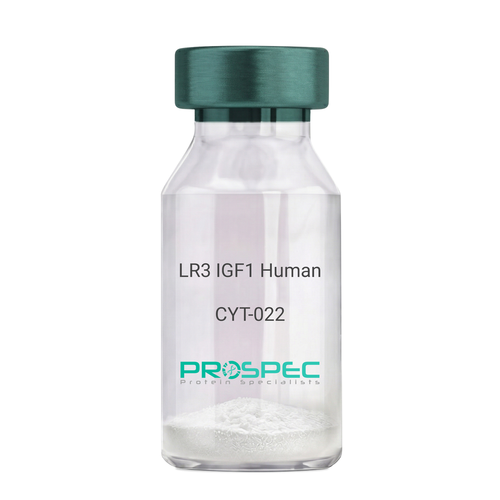
IGF1 LR3 HumanDescription:
LR3 Insulin Like Growth Factor-1 Human Recombinant
R3 IGF1, R3 IGF-1, R3IGF1, R3IGF-1, LONG IGF1, LONG IGF-1, LONG R3 IGF1, LONG R3IGF1, LONG R3 IGF-1, LONG R3IGF-1.
Product # :
CYT-022Price :
Quantity :
Shipping Method :
Shipped at Room temp
More Info
- description
- source
- formulation
- purity
- biological activity
- More Info
- sds-page
Description
The LR3 is a long-term analog of human IGF-1, specifically designed and manufactured for mammalian cell culture to support large-scale manufacturing of recombinant biopharmaceuticals. Recombinant Human LR3 Insulin Like Growth Factor-1 produced in E.Coli is a single, non-glycosylated, polypeptide chain containing 83 amino acids and having a molecular mass of 9.1kDa.
Source
Escherichia Coli.
Formulation
Lyophilized from a 0.2µm filtered concentrated solution in 20mM PB, pH 7.2.
Purity
Greater than 97.0% as determined by SDS-PAGE.
Biological Activity
The ED50 as determined by the stimulation of protein synthesis in L6 myoblasts is less than 10ng/ml, corresponding to a specific activity of 100,000units/mg.sds-page
More Info
-
Introduction
IGF-1 (Insulin-like growth factor-1) is a major hormonal mediator of statural growth. Under regular circumstances, GH (growth hormone) binds to its receptor in the liver, and other tissues, and stimulates the synthesis/secretion of IGF-1. In target tissues, the Type 1 IGF receptor, that is homologous to the insulin receptor, is activated by IGF-1, leading to intracellular signaling which stimulates multiple processes leading to statural growth. IGF-1 metabolic actions are partly directed at stimulating the uptake of glucose, fatty acids, and amino acids so that metabolism supports growing tissues.
-
Synonyms
R3 IGF1, R3 IGF-1, R3IGF1, R3IGF-1, LONG IGF1, LONG IGF-1, LONG R3 IGF1, LONG R3IGF1, LONG R3 IGF-1, LONG R3IGF-1.
-
Physical Appearance
Sterile Filtered White lyophilized (freeze-dried) powder.
-
Stability
Lyophilized LR3 IGF1 although stable at room temperature for 3 weeks, should be stored desiccated below -18°C. Upon reconstitution the LR3 IGF1 should be stored at 4°C between 2-7 days and for future use below -18°C.For long term storage it is recommended to add a carrier protein (0.1% HSA or BSA).Please prevent freeze-thaw cycles.
-
Solubility
It is recommended to reconstitute the lyophilized LR3 IGF1 in sterile 18M-cm H2O at a concentration of 100µg/ml, which can then be further diluted to other aqueous solutions.
-
Amino Acid Sequence
MFPAMPLSSLFVNGPRTLCGAELVDALQFVCGDRGFYFNKPTGYGSSSRRAPQTGIV DECCFRSCDLRRLEMYCAPLKPAKSA.
-
Background
What is the molecular weight/Mw of IGF1 LR3 HUMAN Protein?
IGF1 LR3 HUMAN Protein has a total Mw of 9.1kDa.
What is the source or expression system of IGF1 LR3 HUMAN Protein?
Escherichia Coli.
What is the Purity of IGF1 LR3 HUMAN Protein?
IGF1 LR3 HUMAN Protein is >98% pure as determined by SDS-PAGE.
What is the Biological Activity of IGF1 LR3 HUMAN Protein?
The ED50 as determined by the stimulation of protein synthesis in L6 myoblasts is less than 10ng/ml, corresponding to a specific activity of 100,000units/mg.
What is the amino acid sequence of IGF1 LR3 HUMAN Protein?
MFPAMPLSSLFVNGPRTLCGAELVDALQFVCGDRGFYFNKPTGYGSSSRRAPQTGIV DECCFRSCDLRRLEMYCAPLKPAKSA.
What applications can IGF1 LR3 HUMAN Protein be used in?
IGF1 LR3 HUMAN Protein can probably be used in western blot, ELISA and Lateral Flow.
What is the endotoxin level for IGF1 LR3 HUMAN Protein?
The endotoxin level is minimal, IGF1 LR3 HUMAN Protein was purified using conventional chromatography techniques.
ProSpec's products are furnished for LABORATORY RESEARCH USE ONLY. The product may not be used as drugs, agricultural or pesticidal products, food additives or household chemicals.
- View Data Sheet
Name :
IFNG MouseDescription:
IFN-Gamma Mouse Recombinant
Immune IFN, type II IFN, T cell IFN, MAF, IFNG, IFG, IFI, IFN-gamma.
Product # :
CYT-358Price :
Quantity :
Shipping Method :
Shipped at Room temp
More Info
- description
- source
- formulation
- purity
- biological activity
- More Info
Description
IFN-gamma Mouse Recombinant produced in E.Coli is a single, non-glycosylated, polypeptide chain containing 134 amino acids and having a molecular mass of 15.6kDa.The IFN-gamma is purified by proprietary chromatographic techniques.
Source
Escherichia Coli.
Formulation
Lyophilized from a 0.2µm filtered concentrated (1mg/ml) solution in PBS, pH 7.4 and 5% trehalose.
Purity
Greater than 95.0% as determined by:
(a) Analysis by RP-HPLC.
(b) Analysis by SDS-PAGE.Biological Activity
The specific activity as determined in a viral resistance assay is < 0.1 ng/ml, corresponding to a specific activity of 10,000,000 IU/mg
More Info
-
Introduction
IFN-gamma produced by lymphocytes activated by specific antigens or mitogens.
IFN-gamma, in addition to having antiviral activity, has important immunoregulatory functions, it is a potent activator of macrophages, and has antiproliferative effects on transformed cells and it can potentiate the antiviral and antitumor effects of the type I IFNs. -
Synonyms
Immune IFN, type II IFN, T cell IFN, MAF, IFNG, IFG, IFI, IFN-gamma.
-
Physical Appearance
Sterile Filtered White lyophilized (freeze-dried) powder.
-
Stability
Lyophilized IFN-gamma although stable at room temperature for 3 weeks, should be stored desiccated below -18°C. Upon reconstitution IFN-gamma should be stored at 4°C between 2-7 days and for future use below -18°C.For long term storage it is recommended to add a carrier protein (0.1% HSA or BSA).Please prevent freeze-thaw cycles.
-
Solubility
It is recommended to reconstitute the lyophilized IFN-gamma in sterile distilled water or 20mM AcOH at concentrations ranging between 0.1mg-0.5mg/ml, which can then be further diluted to other aqueous solutions.
-
Amino Acid Sequence
MHGTVIESLE SLNNYFNSSG IDVEEKSLFL DIWRNWQKDG DMKILQSQII SFYLRLFEVL KDNQAISNNI SVIESHLITT FFSNSKAKKD AFMSIAKFEV NNPQVQRQAF NELIRVVHQL LPESSLRKRK RSRC.
-
Background
What is the molecular weight/Mw of IFNG MOUSE Protein?
IFNG MOUSE Protein has a total Mw of 15.6kDa.
What is the source or expression system of IFNG MOUSE Protein?
Escherichia Coli.
What is the Purity of IFNG MOUSE Protein?
IFNG MOUSE Protein is >95% pure as determined by SDS-PAGE.
What is the Biological Activity of IFNG MOUSE Protein?
The specific activity as determined in a viral resistance assay is < 0.1 ng/ml, corresponding to a specific activity of 10,000,000 IU/mg
What is the amino acid sequence of IFNG MOUSE Protein?
MHGTVIESLE SLNNYFNSSG IDVEEKSLFL DIWRNWQKDG DMKILQSQII SFYLRLFEVL KDNQAISNNI SVIESHLITT FFSNSKAKKD AFMSIAKFEV NNPQVQRQAF NELIRVVHQL LPESSLRKRK RSRC.
What applications can IFNG MOUSE Protein be used in?
IFNG MOUSE Protein can probably be used in western blot, ELISA and Lateral Flow.
What is the endotoxin level for IFNG MOUSE Protein?
The endotoxin level is minimal, IFNG MOUSE Protein was purified using conventional chromatography techniques.
ProSpec's products are furnished for LABORATORY RESEARCH USE ONLY. The product may not be used as drugs, agricultural or pesticidal products, food additives or household chemicals.
- View Data Sheet
Name :
MSR1 Human, sf9Description:
Macrophage Scavenger Receptor 1, sf9 Human Recombinant
Macrophage Scavenger Receptor 1, Macrophage Acetylated LDL Receptor I And II, Scavenger Receptor Class A Member 1, SCARA1, Macrophage Scavenger Receptor Type III, CD204 Antigen, CD204, PhSR1, PhSR2, SR-A, SRA.
Product # :
PRO-2318Price :
Quantity :
Shipping Method :
Shipped with Ice Packs
More Info
- description
- source
- formulation
- purity
- More Info
Description
MSR1 produced in Sf9 Baculovirus cells is a single, glycosylated polypeptide chain containing 384 amino acids (77-451 a.a.) and having a molecular mass of 42.4kDa (Migrates at 40-57kDa on SDS-PAGE under reducing conditions). MSR1 is expressed with a 6 amino acid His tag at C-Terminus and purified by proprietary chromatographic techniques.
Source
Sf9, Baculovirus cells.
Formulation
MSR1 protein solution (0.5mg/ml) contains Phosphate Buffered Saline (pH 7.4) and 10% glycerol.
Purity
Greater than 95.0% as determined by SDS-PAGE.
More Info
-
Introduction
Macrophage Scavenger Receptor 1, also known as MSR1 is a member of the class A macrophage scavenger receptors, which comprises three different types 1, 2 and 3 generated by alternative splicing. Furthermore, these receptors or isoforms are macrophage-specific trimeric integral membrane glycoproteins and have been implicated in many macrophage-associated physiological as well as pathological processes which include atherosclerosis, Alzheimer's disease, and host defense.
-
Synonyms
Macrophage Scavenger Receptor 1, Macrophage Acetylated LDL Receptor I And II, Scavenger Receptor Class A Member 1, SCARA1, Macrophage Scavenger Receptor Type III, CD204 Antigen, CD204, PhSR1, PhSR2, SR-A, SRA.
-
Physical Appearance
Sterile Filtered colorless solution.
-
Stability
Store at 4°C if entire vial will be used within 2-4 weeks. Store, frozen at -20°C for longer periods of time. For long term storage it is recommended to add a carrier protein (0.1% HSA or BSA). Avoid multiple freeze-thaw cycles.
-
Amino Acid Sequence
ADPKWETKNC SVSSTNANDI TQSLTGKGND SEEEMRFQEV FMEHMSNMEK RIQHILDMEA NLMDTEHFQN FSMTTDQRFN DILLQLSTLF SSVQGHGNAI DEISKSLISL NTTLLDLQLN IENLNGKIQE NTFKQQEEIS KLEERVYNVS AEIMAMKEEQ VHLEQEIKGE VKVLNNITND LRLKDWEHSQ TLRNITLIQG PPGPPGEKGD RGPTGESGPR GFPGPIGPPG LKGDRGAIGF PGSRGLPGYA GRPGNSGPKG QKGEKGSGNT LTPFTKVRLV GGSGPHEGRV EILHSGQWGT ICDDRWEVRV GQVVCRSLGY PGVQAVHKAA HFGQGTGPIW LNEVFCFGRE SSIEECKIRQ WGTRACSHSE DAGVTCTLHH HHHH.
ProSpec's products are furnished for LABORATORY RESEARCH USE ONLY. The product may not be used as drugs, agricultural or pesticidal products, food additives or household chemicals.
- View Data Sheet
Name :
IFNA7 Human, Sf9Description:
Interferon-alpha 7 Human Recombinant, Sf9
Interferon alpha-7, IFN-alpha-7, Interferon alpha-J, LeIF J, Interferon alpha-J1, IFN-alpha-J1, IFNA7, IFNA-J, IFN-alphaJ.
Product # :
CYT-1075Price :
Quantity :
Shipping Method :
Shipped with Ice Packs
More Info
- description
- source
- formulation
- purity
- More Info
- SDS-PAGE
Description
IFNA7 Human Recombinant produced in Sf9 Baculovirus cells is a single, glycosylated polypeptide chain containing 175 amino acids (24-189) and having a molecular mass of 20.7kDa. IFNA7 is fused to a 6 amino acid His-Tag at C-terminus and purified by proprietary chromatographic techniques.
Source
Sf9, Baculovirus cells.
Formulation
IFNA7 protein solution (0.25mg/ml) containing Phosphate Buffered Saline (pH 7.4) and 10% glycerol.
Purity
Greater than 90.0% as determined by analysis by SDS-PAGE.
SDS-PAGE
More Info
-
Introduction
Interfereon alpha 7 (IFNA7) is a member of the alpha/beta interferon family. IFNA7 is generated by macrophages. IFN-alpha has antiviral functions. Interferon promotes the production of 2 enzymes: a protein kinase and an oligoadenylate synthetase.
-
Synonyms
Interferon alpha-7, IFN-alpha-7, Interferon alpha-J, LeIF J, Interferon alpha-J1, IFN-alpha-J1, IFNA7, IFNA-J, IFN-alphaJ.
-
Physical Appearance
Sterile Filtered clear solution.
-
Stability
Store at 4°C if entire vial will be used within 2-4 weeks. Store, frozen at -20°C for longer periods of time. For long term storage it is recommended to add a carrier protein (0.1% HSA or BSA).Avoid multiple freeze-thaw cycles.
-
Amino Acid Sequence
ADPCDLPQTH SLRNRRALIL LAQMGRISPF SCLKDRHEFR FPEEEFDGHQ FQKTQAISVL HEMIQQTFNL FSTEDSSAAW EQSLLEKFST ELYQQLNDLE ACVIQEVGVE ETPLMNEDFI LAVRKYFQRI TLYLMEKKYS PCAWEVVRAE IMRSFSFSTN LKKGLRRKDH HHHHH.
-
Background
What is the molecular weight/Mw of IFNA7 HUMAN, SF9 Protein?
IFNA7 HUMAN, SF9 Protein has a total Mw of 20.7kDa.
What is the source or expression system of IFNA7 HUMAN, SF9 Protein?
Sf9, Baculovirus cells.
What is the Purity of IFNA7 HUMAN, SF9 Protein?
IFNA7 HUMAN, SF9 Protein is >90% pure as determined by SDS-PAGE.
What is the Biological Activity of IFNA7 HUMAN, SF9 Protein?
The biological functionality of IFNA7 HUMAN, SF9 Protein will be determined in the future.
What is the amino acid sequence of IFNA7 HUMAN, SF9 Protein?
ADPCDLPQTH SLRNRRALIL LAQMGRISPF SCLKDRHEFR FPEEEFDGHQ FQKTQAISVL HEMIQQTFNL FSTEDSSAAW EQSLLEKFST ELYQQLNDLE ACVIQEVGVE ETPLMNEDFI LAVRKYFQRI TLYLMEKKYS PCAWEVVRAE IMRSFSFSTN LKKGLRRKDH HHHHH.
What applications can IFNA7 HUMAN, SF9 Protein be used in?
IFNA7 HUMAN, SF9 Protein can probably be used in western blot, ELISA and Lateral Flow.
What is the endotoxin level for IFNA7 HUMAN, SF9 Protein?
The endotoxin level is minimal, IFNA7 HUMAN, SF9 Protein was purified using conventional chromatography techniques.
ProSpec's products are furnished for LABORATORY RESEARCH USE ONLY. The product may not be used as drugs, agricultural or pesticidal products, food additives or household chemicals.
- View Data Sheet
Name :
Omentin Human, HisDescription:
Omentin Human Recombinant, His Tag
Intelectin-1, HL1, LFR, HL-1, INTL, ITLN, hIntL.
Product # :
CYT-551Price :
Quantity :
Shipping Method :
Shipped at Room temp
More Info
- description
- source
- formulation
- purity
- More Info
Description
Omentin Human Recombinant is produced in E.Coli by recombinant DNA technology is a single, polypeptide chain containing 294 amino acids and having a molecular mass of 32.7 kDa. Recombinant Human Omentin contains His tag fused at N-Terminus.Intelectin is purified by proprietary chromatographic techniques.
Source
Escherichia Coli.
Formulation
Each mg of lyophilized powder contains 20mM Tris & 50mM NaCl pH-8.0.
Purity
Greater than 95.0% as determined by(a) Analysis by RP-HPLC.
(b) Analysis by SDS-PAGE.More Info
-
Introduction
Omentin is a recently recognized gene highly localized to the mental tissue (visceral adipose tissue). Omentin is present in the stromal vascular cells in the adipose tissue rather than in the adipocytes. Omentin is predominantly expressed in the visceral adipose tissue than the subcutaneous tissue, with the omentin mRNA being 150 times higher in the visceral adipose tissue. Omentin has also been detected in human blood using western blot analysis, and seems to increase insulin-stimulated glucose uptake in 3T3-L1 adipocytes in mice. Omentin seems to increase Akt phosphorylation irrespective of insulin presence. Its role in glucose metabolism and obesity remains to be described; an insulin-sensitizing action is possible.Differences in Omentin expression has been noted in adipose tissue from normals and patients with inflammatory bowel disease although its significance is unknown.
-
Synonyms
Intelectin-1, HL1, LFR, HL-1, INTL, ITLN, hIntL.
-
Physical Appearance
Sterile Filtered White lyophilized (freeze-dried) powder.
-
Stability
Lyophilized Intelectin His Tag although stable at room temperature for 3 weeks, should be stored desiccated below -18°C. Upon reconstitution Intelectin should be stored at 4°C between 2-7 days and for future use below -18°C.For long term storage it is recommended to add a carrier protein (0.1% HSA or BSA).Please prevent freeze-thaw cycles.
-
Solubility
It is recommended to reconstitute the lyophilized Omentin His Tag in sterile 18MΩ-cm H2O not less than 100µg/ml, which can then be further diluted to other aqueous solutions.
-
Amino Acid Sequence
MRGSHHHHHH GMASTDEANT YFKEWTCSSS PSLPRSCKEI KDECPSAFDG LYFLRTENGV IYQTFCDMTS GGGGWTLVAS VHENDMRGKC TVGDRWSSQQ GSKAVYPEGD GNWANYNTFG SAEAATSDDY KNPGYYDIQA KDLGIWHVPN KSPMQHWRNS SLLRYRTDTG FLQTLGHNLF GIYQKYPVKY GEGKCWTDNG PVIPVVYDFG DAQKTASYYS PYGQREFTAG FVQFRVFNNE RAANALCAGM RVTGCNTEHH CIGGGGYFPE ASPQQCGDFS GFDWSGYGTH VGYS.
-
Applications
Elisa
Western blot.
ProSpec's products are furnished for LABORATORY RESEARCH USE ONLY. The product may not be used as drugs, agricultural or pesticidal products, food additives or household chemicals.
- View Data Sheet
Name :
ETFB HumanDescription:
Electron-Transfer-Flavoprotein Beta Polypeptide Human Recombinant
Electron-transfer-flavoprotein beta polypeptide, MADD, beta-ETF.
Product # :
PRO-220Price :
Quantity :
Shipping Method :
Shipped with Ice Packs
More Info
- description
- source
- formulation
- purity
- More Info
Description
ETFB Human Recombinant fused with a 20 amino acid His tag at N-terminus produced in E.Coli is a single, non-glycosylated, polypeptide chain containing 275 amino acids (1-255a.a.) and having a molecular mass of 30.0kDa. The ETFB is purified by proprietary chromatographic techniques.
Source
Escherichia Coli.
Formulation
The ETFB solution (0.25mg/ml) contains 20mM Tris-HCl buffer (pH 8.0), 40% glycerol and 0.1M NaCl.
Purity
Greater than 90.0% as determined by SDS-PAGE.
More Info
-
Introduction
ETF is a heterodimer composed of alpha and beta subunit. ETFB protein is electron-transfer-flavoprotein, beta polypeptide that transports electrons between primary flavoprotein dehydrogenases involved in mitochondrial fatty acid and amino acid catabolism and the membrane-bound electron transfer flavoprotein ubiquinone oxidoreductase.
-
Synonyms
Electron-transfer-flavoprotein beta polypeptide, MADD, beta-ETF.
-
Physical Appearance
Sterile Filtered colorless solution.
-
Stability
Store at 4°C if entire vial will be used within 2-4 weeks. Store, frozen at -20°C for longer periods of time. For long term storage it is recommended to add a carrier protein (0.1% HSA or BSA).Avoid multiple freeze-thaw cycles.
-
Amino Acid Sequence
MGSSHHHHHH SSGLVPRGSH MAELRVLVAV KRVIDYAVKI RVKPDRTGVV TDGVKHSMNP FCEIAVEEAV RLKEKKLVKE VIAVSCGPAQ CQETIRTALA MGADRGIHVE VPPAEAERLG PLQVARVLAK LAEKEKVDLV LLGKQAIDDD CNQTGQMTAG FLDWPQGTFA SQVTLEGDKL
KVEREIDGGL ETLRLKLPAV VTADLRLNEP RYATLPNIMK AKKKKIEVIK PGDLGVDLTS KLSVISVEDP PQRTAGVKVE TTEDLVAKLK EIGRI.
ProSpec's products are furnished for LABORATORY RESEARCH USE ONLY. The product may not be used as drugs, agricultural or pesticidal products, food additives or household chemicals.
- View Data Sheet
Name :
CBFB HUmanDescription:
Core Binding Factor Beta Human Recombinant
PEBP2B, polyomavirus enhancer binding protein b, PEA2, CBF-beta.
Product # :
PRO-534Price :
Quantity :
Shipping Method :
Shipped with Ice Packs
More Info
- description
- source
- formulation
- purity
- More Info
Description
CBFB Recombinant Human produced in E.Coli is a single, non-glycosylated polypeptide chain containing 182 amino acids (1-202 a.a.) and having a molecular mass of 23.6 kDa. The CBFB is fused to 20 amino acid His-Tag at N-terminus and purified by proprietary chromatographic techniques.
Source
Escherichia Coli.
Formulation
0.5mg/ml solution containing 20mM MES pH-6, 0.1mM PMSF & 10% glycerol.
Purity
Greater than 85.0% as determined by SDS-PAGE.
More Info
-
Introduction
CBFB beta subunit is a heterodimeric core-binding transcription factor that is part of the PEBP2/CBF transcription factor family which controls a host of genes particulary to hematopoiesis and osteogenesis. CBFB is a non-DNA binding regulatory subunit which increases DNA binding by alpha subunit.
-
Synonyms
PEBP2B, polyomavirus enhancer binding protein b, PEA2, CBF-beta.
-
Physical Appearance
Sterile filtered colorless solution.
-
Stability
CBFB Human although stable at 4C for 1 week, should be stored below -18C. Please prevent freeze thaw cycles.
-
Amino Acid Sequence
MGSSHHHHHH SSGLVPRGSH MPRVVPDQRS KFENEEFFRK LSRECEIKYT GFRDRPHEER QARFQNACRD GRSEIAFVAT GTNLSLQFFP
ASWQGEQRQT PSREYVDLER EAGKVYLKAP MILNGVCVIW KGWIDLQRLD GMGCLEFDEE RAQQEDALAQ QAFEEARRRT REFEDRDRSH
REEMEVRVSQ LLAVTGKKTT RP.
ProSpec's products are furnished for LABORATORY RESEARCH USE ONLY. The product may not be used as drugs, agricultural or pesticidal products, food additives or household chemicals.
- View Data Sheet
Name :
CD68 Human, sf9Description:
CD68 Human Recombinant, sf9
Macrosialin, CD68, Gp110, Macrosialin isoform A, GP110, LAMP4, SCARD1.
Product # :
PRO-2375Price :
Quantity :
Shipping Method :
Shipped with Ice Packs
More Info
- description
- source
- formulation
- purity
- More Info
Description
CD68 Human Recombinant produced in Sf9 Baculovirus cells is a single, glycosylated polypeptide chain containing 307 amino acids (22-319) and having a molecular mass of 32.6kDa (Molecular size on SDS-PAGE will appear at approximately 57-70kDa).CD68 is fused to 6 amino acid His-Tag at C-terminus and purified by proprietary chromatographic techniques.
Source
Sf9, Baculovirus cells.
Formulation
CD68 protein solution (1mg/ml) containing Phosphate Buffered Saline (pH 7.4) and 10% glycerol.
Purity
Greater than 95.0% as determined by analysis by SDS-PAGE.
More Info
-
Introduction
CD68 encodes a 110-kD transmembrane glycoprotein that is highly expressed by human monocytes and tissue macrophages. It is a member of the lysosomal/endosomal-associated membrane glycoprotein (LAMP) family. The protein primarily localizes to lysosomes and endosomes with a smaller fraction circulating to the cell surface. It is a type I integral membrane protein with a heavily glycosylated extracellular domain and binds to tissue- and organ-specific lectins or selectins. The protein is also a member of the scavenger receptor family. Scavenger receptors typically function to clear cellular debris, promote phagocytosis, and mediate the recruitment and activation of macrophages. Alternative splicing results in multiple transcripts encoding different isoforms.
-
Synonyms
Macrosialin, CD68, Gp110, Macrosialin isoform A, GP110, LAMP4, SCARD1.
-
Physical Appearance
Sterile Filtered clear solution.
-
Stability
Store at 4°C if entire vial will be used within 2-4 weeks. Store, frozen at -20°C for longer periods of time. For long term storage it is recommended to add a carrier protein (0.1% HSA or BSA).Avoid multiple freeze-thaw cycles.
-
Amino Acid Sequence
ADPNDCPHKK SATLLPSFTV TPTVTESTGT TSHRTTKSHK TTTHRTTTTG TTSHGPTTAT HNPTTTSHGN VTVHPTSNST ATSQGPSTAT HSPATTSHGN ATVHPTSNST ATSPGFTSSA HPEPPPPSPS PSPTSKETIG DYTWTNGSQP CVHLQAQIQI RVMYTTQGGG EAWGISVLNP NKTKVQGSCE GAHPHLLLSF PYGHLSFGFM QDLQQKVVYL SYMAVEYNVS FPHAAQWTFS AQNASLRDLQ APLGQSFSCS NSSIILSPAV HLDLLSLRLQ AAQLPHTGVF GQSFSCPSDR SHHHHHH.
ProSpec's products are furnished for LABORATORY RESEARCH USE ONLY. The product may not be used as drugs, agricultural or pesticidal products, food additives or household chemicals.
- View Data Sheet
Name :
LDHA AntibodyDescription:
Lactate Dehydrogenase A, Mouse Anti Human
LDH-A, GSD11, LDH1, LDHM, PIG19, EC 1.1.1.27, lactate dehydrogenase M, LDH-M, LDH-1, L-lactate dehydrogenase A chain, LDH muscle subunit, Renal carcinoma antigen NY-REN-59, Cell proliferation-inducing gene 19 protein, LDHA.
Product # :
ANT-004Price :
Quantity :
Shipping Method :
Shipped with Ice Packs
More Info
- formulation
- More Info
Formulation
1mg/ml containing PBS, pH-7.4, & 0.1% Sodium Azide.
More Info
-
Introduction
LDHA catalyzes the conversion of L-lactate and NAD to pyruvate and NADH in the final step of anaerobic glycolysis. LDHA is localized primarily in muscle tissue and is part of the lactate dehydrogenase family. Mutations in LDHA have been linked to exertional myoglobinuria. LDH1 is decreased in essential thrombocythemia. LDHA is induced through a non-genomic pathway of estrogen action. Reduction in LDH-A activity results in stimulation of mitochondrial respiration and decrease of mitochondrial membrane potential.
-
Synonyms
LDH-A, GSD11, LDH1, LDHM, PIG19, EC 1.1.1.27, lactate dehydrogenase M, LDH-M, LDH-1, L-lactate dehydrogenase A chain, LDH muscle subunit, Renal carcinoma antigen NY-REN-59, Cell proliferation-inducing gene 19 protein, LDHA.
-
Immunogen
Anti-human LDHA mAb, is derived from hybridization of mouse F0 myeloma cells with spleen cells from BALB/c mice immunized with recombinant human LDHA amino acids 1-332 purified from E. coli.
-
Ig Subclass
Mouse IgG2b heavy chain and κ light chain.
-
Clone
PAT1A4AT.
-
Applications
LDHA antibody has been tested by ELISA, Western blot and Immunofluorescence analysis to assure specificity and reactivity. Since application varies, however, each investigation should be titrated by the reagent to obtain optimal results. Recommended dilution range for Western blot analysis and Immunofluorescence is 1:500 ~ 8,000.
Recommended starting dilution is 1:1,000. -
Type
Mouse Anti Human Monoclonal.
-
Storage Procedures
For periods up to 1 month store at 4°C, for longer periods of time, store at -20°C. Prevent freeze thaw cycles.
-
Purification Method
LDHA antibody was purified from mouse ascitic fluids by protein-G affinity chromatography.
ProSpec's products are furnished for LABORATORY RESEARCH USE ONLY. The product may not be used as drugs, agricultural or pesticidal products, food additives or household chemicals.