Search results
1000 results found for “beta-ngf”
Name
Description
Product #
Price
Quantity
Shipping Method
- View Data Sheet
Name :
M CSF Human, Sf9 HisDescription:
Macrophage Colony Stimulating Factor Human Recombinant, Sf9
Macrophage colony-stimulating factor 1, CSF-1, M-CSF, MCSF, Lanimostim.
Product # :
CYT-1015Price :
Quantity :
Shipping Method :
Shipped with Ice Packs
More Info
- description
- source
- formulation
- purity
- biological activity
- More Info
Description
MCSF produced in Sf9 Insect cells is a single, glycosylated polypeptide chain containing 231 amino acids (33-255 a.a.) and having a molecular mass of 26.1kDa (Molecular size on SDS-PAGE will appear at approximately 28-40kDa).MCSF is expressed with an 8 amino acid His tag at C-Terminus and purified by proprietary chromatographic techniques.
Source
Sf9, Insect cells.
Formulation
MCSF protein solution (0.25mg/ml) contains Phosphate Buffered Saline (pH 7.4) and 10% glycerol.
Purity
Greater than 95.0% as determined by SDS-PAGE.
Biological Activity
Measured in a cell proliferation assay using M-NFS-60 mouse myelogenous leukemia lymphoblast cell. The ED50 for this effect is less or equal to 3 ng/ml.
More Info
-
Introduction
Granulocyte/Macrophage Colony-Stimulating Factors are cytokines that act in hematopoiesis by controlling the production, differentiation, and function of 2 related white cell populations of the blood, the granulocytes and the monocytes-macrophages. MCSF induces cells of the monocyte/macrophage lineage. MCSF plays a role in immunological defenses, bone metabolism, lipoproteins clearance, fertility and pregnancy.
-
Synonyms
Macrophage colony-stimulating factor 1, CSF-1, M-CSF, MCSF, Lanimostim.
-
Physical Appearance
Sterile filtered colorless solution.
-
Stability
Store at 4°C if entire vial will be used within 2-4 weeks. Store, frozen at -20°C for longer periods of time. For long term storage it is recommended to add a carrier protein (0.1% HSA or BSA).Avoid multiple freeze-thaw cycles.
-
Amino Acid Sequence
EEVSEYCSHM IGSGHLQSLQ RLIDSQMETS CQITFEFVDQ EQLKDPVCYL KKAFLLVQDI MEDTMRFRDN TPNAIAIVQL QELSLRLKSC FTKDYEEHDK ACVRTFYETP LQLLEKVKNV FNETKNLLDK DWNIFSKNCN NSFAECSSQD VVTKPDCNCL YPKAIPSSDP ASVSPHQPLA PSMAPVAGLT WEDSEGTEGS SLLPGEQPLH TVDPGSAKQR PPRLEHHHHH H.
ProSpec's products are furnished for LABORATORY RESEARCH USE ONLY. The product may not be used as drugs, agricultural or pesticidal products, food additives or household chemicals.
- View Data Sheet
Name :
AREG HumanDescription:
Amphiregulin Human Recombinant
Schwannoma-derived growth factor, Colorectum cell-derived growth factor, AR, CRDGF, SDGF, AREGB, MGC13647.
Product # :
CYT-041Price :
Quantity :
Shipping Method :
Shipped at Room temp
More Info
- description
- source
- formulation
- purity
- biological activity
- More Info
Description
Amphiregulin (AREG) Human Recombinant produced in E.Coli is a single, non-glycosylated, polypeptide chain containing 98 amino acids and having a molecular mass of 11.3 KDa.The AREG is purified by proprietary chromatographic techniques.
Source
Escherichia Coli.
Formulation
Lyophilized from a 0.2µm filtered concentrated solution in PBS, pH7.4.
Purity
Greater than 95.0% as determined by:
(a) Analysis by HPLC.
(b) Analysis by SDS-PAGE.Biological Activity
Determined by its ability to stimulate the proliferation of mouse Balb/c 3T3 cells. The expected ED50 for this effect is 5-10 ng/ml, corresponding to a specific activity of 100,000-200,000units/mg.More Info
-
Synonyms
Schwannoma-derived growth factor, Colorectum cell-derived growth factor, AR, CRDGF, SDGF, AREGB, MGC13647.
-
Physical Appearance
Sterile Filtered White lyophilized (freeze-dried) powder.
-
Stability
Lyophilized AREG although stable at room temperature for 3 weeks, should be stored desiccated below -18°C. Upon reconstitution AREG should be stored at 4°C between 2-7 days and for future use below -18°C.For long term storage it is recommended to add a carrier protein (0.1% HSA or BSA).Please prevent freeze-thaw cycles.
-
Solubility
It is recommended to reconstitute the lyophilized AREG in sterile 18M-cm H2O not less than 100µg/ml, which can then be further diluted to other aqueous solutions.
-
Amino Acid Sequence
SVRVEQVVKP PQNKTESENT SDKPKRKKKG GKNGKNRRNR KKKNPCNAEF QNFCIHGECK YIEHLEAVTC KCQQEYFGER CGEKSMKTHS MIDSSLSK.
-
Background
Amphiregulin Human Recombinant: Exploring its Role in Cancer Biology and Therapeutic Applications
Abstract:
Amphiregulin, a member of the epidermal growth factor (EGF) family, has gained significant attention in cancer research. This research paper provides an overview of Amphiregulin human recombinant, highlighting its molecular characteristics, signaling pathways, and therapeutic potential. Understanding the multifaceted role of Amphiregulin opens avenues for targeted cancer therapies. This article provides a concise analysis of Amphiregulin, emphasizing its impact on cancer biology and its therapeutic applications.Introduction:
Cancer continues to be a significant health challenge worldwide, necessitating novel therapeutic approaches. Amphiregulin, an EGF family member, has emerged as a promising target in cancer research. This paper provides an overview of Amphiregulin, shedding light on its structure, function, and therapeutic potential.Amphiregulin Signaling and Mechanisms:
Amphiregulin exerts its effects through the binding and activation of the EGF receptor (EGFR). Upon activation, a cascade of intracellular signaling pathways is triggered, including the MAPK and PI3K/AKT pathways. These pathways regulate critical cellular processes such as cell proliferation, survival, migration, and angiogenesis.Amphiregulin in Cancer Biology:
Amphiregulin has been implicated in various aspects of cancer biology, including tumor growth, metastasis, and resistance to therapy. Its overexpression is observed in several cancer types, and its role in promoting tumor growth and metastasis has been demonstrated in preclinical studies. Targeting Amphiregulin signaling shows promise in inhibiting cancer progression and overcoming therapy resistance.Therapeutic Potential of Amphiregulin Human Recombinant:
Amphiregulin human recombinant holds significant therapeutic potential in cancer treatment. Strategies aimed at blocking Amphiregulin-EGFR interactions or inhibiting downstream signaling pathways are being explored as potential therapeutic interventions. Additionally, Amphiregulin could serve as a predictive biomarker to identify patients who are more likely to respond to targeted therapies.Challenges and Future Directions:
While the therapeutic targeting of Amphiregulin shows promise, several challenges need to be addressed. Further research is required to fully understand the complex interplay between Amphiregulin and other molecular pathways in cancer biology. Additionally, the development of specific and potent inhibitors and the identification of patient selection criteria are important considerations for successful clinical translation.Conclusion:
Amphiregulin human recombinant represents a promising avenue for targeted cancer therapy. Understanding the molecular mechanisms and functional implications of Amphiregulin in cancer biology offers new opportunities for developing innovative treatments. Continued research in this field has the potential to improve patient outcomes and contribute to the advancement of personalized medicine.What is the molecular weight/Mw of AREG Protein?
AREG Protein has a total Mw of 11.3kDa.
What is the source or expression system of AREG Protein?
Escherichia Coli.
What is the Purity of AREG Protein?
AREG Protein is >95% pure as determined by SDS-PAGE.
What is the Biological Activity of AREG Protein?
Determined by its ability to stimulate the proliferation of mouse Balb/c 3T3 cells. The expected ED50 for this effect is 5-10 ng/ml, corresponding to a specific activity of 100,000-200,000units/mg.
What is the amino acid sequence of AREG Protein?
SVRVEQVVKP PQNKTESENT SDKPKRKKKG GKNGKNRRNR KKKNPCNAEF QNFCIHGECK YIEHLEAVTC KCQQEYFGER CGEKSMKTHS MIDSSLSK.
What applications can AREG Protein be used in?
AREG Protein can probably be used in western blot, ELISA and Lateral Flow.
What is the endotoxin level for AREG Protein?
The endotoxin level is minimal, AREG Protein was purified using conventional chromatography techniques.
ProSpec's products are furnished for LABORATORY RESEARCH USE ONLY. The product may not be used as drugs, agricultural or pesticidal products, food additives or household chemicals.
- View Data Sheet
Name :
NAGK AntibodyDescription:
N-Acetylglucosamine Kinase, Mouse Anti Human
N-acetyl-D-glucosamine kinase, N-acetylglucosamine kinase, GlcNAc kinase, NAGK.
Product # :
ANT-092Price :
Quantity :
Shipping Method :
Shipped with Ice Packs
More Info
- formulation
- More Info
Formulation
1mg/ml containing PBS, pH-7.4, 0.2% Sodium Azide and 10% glycerol.
More Info
-
Introduction
N-Acetylglucosamine Kinase (NAGK) is a part of the eukaryotic-type N-acetylglucosamine kinase family. NAGK transforms endogenous N-acetylglucosamine (GlcNAc), a main component of complex carbohydrates, from lysosomal degradation or nutritional sources into GlcNAc 6-phosphate. NAGK is a significant salvage enzyme of amino sugar metabolism in mammals which has ManNAc kinase activity.
-
Synonyms
N-acetyl-D-glucosamine kinase, N-acetylglucosamine kinase, GlcNAc kinase, NAGK.
-
Immunogen
Anti-human NAGK mAb, is derived from hybridization of mouse F0 myeloma cells with spleen cells from BALB/c mice immunized with a recombinant human NAGK amino acids 1-344 purified from E. coli.
-
Ig Subclass
Mouse IgG1 heavy chain and ? light chain.
-
Clone
P4D12AT.
-
Applications
NAGK antibody has been tested by ELISA, Western blot analysis to assure specificity and reactivity. Since application varies, however, each investigation should be titrated by the reagent to obtain optimal results. Recommended dilution range for Western blot analysis is 1:500 ~ 1:5000.
-
Type
Mouse Anti Human Monoclonal.
-
Storage Procedures
For periods up to 1 month store at 4°C, for longer periods of time, store at -20°C. Prevent freeze thaw cycles.
ProSpec's products are furnished for LABORATORY RESEARCH USE ONLY. The product may not be used as drugs, agricultural or pesticidal products, food additives or household chemicals.
- View Data Sheet
Name :
NFATC1 AntibodyDescription:
Nuclear factor of activated T-cells cytoplasmic 1, Mouse Anti Human
Nuclear factor of activated T-cells cytoplasmic 1, NFAT transcription complex cytosolic component, NF-ATc1, NFATc1, NF-ATc, NFATc, NFAT2, MGC138448.
Product # :
ANT-436Price :
Quantity :
Shipping Method :
Shipped with Ice Packs
More Info
- formulation
- More Info
Formulation
1mg/ml containing PBS, pH-7.4, 0.02% Sodium Azide and 10% Glycerol.
More Info
-
Introduction
NFATC1 is a component of the nuclear factor of activated T cells DNA-binding transcription complex. This complex consists of at least 2 components: a preexisting cytosolic component that translocates to the nucleus upon T cell receptor (TCR) stimulation, and an inducible nuclear component. Proteins belonging to this family of transcription factors play an essential role in inducible gene transcription during immune response. NFATC1 is an inducible nuclear component. NFATC1 functions as a major molecular target for the immunosuppressive drugs such as cyclosporin A. Different isoforms of NFATC1 may regulate inducible expression of different cytokine genes.
-
Synonyms
Nuclear factor of activated T-cells cytoplasmic 1, NFAT transcription complex cytosolic component, NF-ATc1, NFATc1, NF-ATc, NFATc, NFAT2, MGC138448.
-
Physical Appearance
Sterile filtered colorless solution.
-
Immunogen
Anti-human NFATC1 mAb is derived from hybridization of mouse F0 myeloma cells with spleen cells from BALB/c mice immunized with recombinant human NFATC1 amino acids 428-716 purified from E. coli.
-
Ig Subclass
Mouse IgG2a heavy chain and κ light chain.
-
Clone
PAT1C3AT.
-
Applications
NFATC1 antibody has been tested by ELISA and Western blot analysis to assure specificity and reactivity. Since application varies, however, each investigation should be titrated by the reagent to obtain optimal results.
-
Type
Mouse Anti Human Monoclonal.
-
Storage Procedures
For periods up to 1 month store at 4°C, for longer periods of time, store at -20°C. Prevent freeze thaw cycles.
-
Purification Method
NFATC1 antibody was purified from mouse ascitic fluids by protein-G affinity chromatography.
ProSpec's products are furnished for LABORATORY RESEARCH USE ONLY. The product may not be used as drugs, agricultural or pesticidal products, food additives or household chemicals.
- View Data Sheet
Name :
LITAF AntibodyDescription:
Lipopolysaccharide-induced TNF factor, Mouse Anti Human
Lipopolysaccharide-induced tumor necrosis factor-alpha factor, LPS-induced TNF-alpha factor, p53-induced gene 7 protein, Small integral membrane protein of lysosome/late endosome, LITAF, PIG7, SIMPLE, TP53I7, FLJ38636, MGC116698, MGC116700, MGC116701, MGC125274, MGC125275, MGC125276.
Product # :
ANT-443Price :
Quantity :
Shipping Method :
Shipped with Ice Packs
More Info
- formulation
- More Info
Formulation
1mg/ml containing PBS, pH-7.4, & 0.02% Sodium Azide and 10% Glycerol.
More Info
-
Introduction
Lipopolysaccharide is a potent stimulator of monocytes and macrophages, causing secretion of tumor necrosis factor-alpha (TNF-alpha) and other inflammatory mediators. LITAF is a lipopolysaccharide-induced TNF-alpha factor, which is a DNA-binding protein and can mediate the TNF-alpha expression by direct binding to the promoter region of the TNF-alpha gene. The transcription of the LITAF gene is induced by tumor suppressor p53 and has been implicated in the p53-induced apoptotic pathway. Mutations in the LITAF gene cause Charcot-Marie-Tooth disease type 1C (CMT1C) and may be involved in the carcinogenesis of extramammary Paget''s disease (EMPD).
-
Synonyms
Lipopolysaccharide-induced tumor necrosis factor-alpha factor, LPS-induced TNF-alpha factor, p53-induced gene 7 protein, Small integral membrane protein of lysosome/late endosome, LITAF, PIG7, SIMPLE, TP53I7, FLJ38636, MGC116698, MGC116700, MGC116701, MGC125274, MGC125275, MGC125276.
-
Physical Appearance
Sterile Filtered clear solution.
-
Immunogen
Anti-human LITAF mAb, is derived from hybridization of mouse F0 myeloma cells with spleen cells from BALB/c mice immunized with recombinant human LITAF amino acids 1-161 purified from E. coli.
-
Ig Subclass
Mouse IgG2b heavy chain and k light chain.
-
Clone
PAT5C10AT.
-
Applications
LITAF antibody has been tested by ELISA and Western blot analysis to assure specificity and reactivity. Since application varies, however, each investigation should be titrated by the reagent to obtain optimal results. Recommended dilution range for Western blot analysis is 1:1000. Recommended starting dilution is 1:1000.
-
Type
Mouse Anti Human Monoclonal.
-
Storage Procedures
For periods up to 1 month store at 4°C, for longer periods of time, store at -20°C. Prevent freeze thaw cycles.
-
Purification Method
LITAF antibody was purified from mouse ascitic fluids by protein-G affinity chromatography.
ProSpec's products are furnished for LABORATORY RESEARCH USE ONLY. The product may not be used as drugs, agricultural or pesticidal products, food additives or household chemicals.
- View Data Sheet
Name :
FGF 18 Human, HisDescription:
Fibroblast Growth Factor-18 Human Recombinant, His Tag
Fibroblast growth factor 18, FGF-18, zFGF5, FGF18.
Product # :
CYT-935Price :
Quantity :
Shipping Method :
Shipped at Room temp
More Info
- description
- source
- formulation
- purity
- More Info
Description
FGF18 Human Recombinant produced in E.Coli is a single, non-glycosylated, polypeptide chain (Glu28-Ala207) containing 190 amino acids including a 10 aa His tag at N-terminus. The total calculated molecular mass is 22.3kDa.
Source
Escherichia Coli.
Formulation
FGF18 was filtered (0.4µm) and lyophilized in phosphate buffered saline and 5% w/v trehalose.
Purity
Purity as determined by densitometric image analysis is greater than 95%.
More Info
-
Introduction
Fibroblast growth factor 18 (FGF18) is a member of the large FGF family which has at least 23 members. FGF18 is a binding growth factor with a core 120 amino acid FGF domain which allows for a common tertiary structure. FGFs are expressed in the course of the embryonic development and in restricted adult tissues. FGF-18 is an indispensable regulator of long bone and calvarial development. FGF-18 signals via FGFR 1c, 2c, 3c, and 4.
-
Synonyms
Fibroblast growth factor 18, FGF-18, zFGF5, FGF18.
-
Physical Appearance
Filtered White lyophilized (freeze-dried) powder.
-
Stability
Store lyophilized protein at -20°C. Aliquot the product after reconstitution to avoid repeated freezing/thawing cycles. Reconstituted protein can be stored at 4°C for a limited period of time; it does not show any change after two weeks at 4°C.
-
Solubility
It is recommended to add deionized water to prepare a working stock solution of approximately 0.5mg/ml and let the lyophilized pellet dissolve completely. FGF18 is not sterile! Please filter the product by an appropriate sterile filter before using it in the cell culture.
-
Amino Acid Sequence
MKHHHHHHASEENVDFRIHV ENQTRARDDV SRKQLRLYQL YSRTSGKHIQ VLGRRISARG EDGDKYAQLL VETDTFGSQV RIKGKETEFY LCMNRKGKLV GKPDGTSKEC VFIEKVLENN YTALMSAKYS GWYVGFTKKG RPRKGPKTRE NQQDVHFMKR YPKGQPELQK PFKYTTVTKR SRRIRPTHPA.
-
Background
What is the molecular weight/Mw of FGF18 HIS Protein?
FGF18 HIS Protein has a total Mw of 22.3kDa.
What is the source or expression system of FGF18 HIS Protein?
Escherichia Coli.
What is the Purity of FGF18 HIS Protein?
FGF18 HIS Protein is >95% pure as determined by SDS-PAGE.
What is the Biological Activity of FGF18 HIS Protein?
The biological functionality of FGF18 HIS Protein will be determined in the future.
What is the amino acid sequence of FGF18 HIS Protein?
MKHHHHHHASEENVDFRIHV ENQTRARDDV SRKQLRLYQL YSRTSGKHIQ VLGRRISARG EDGDKYAQLL VETDTFGSQV RIKGKETEFY LCMNRKGKLV GKPDGTSKEC VFIEKVLENN YTALMSAKYS GWYVGFTKKG RPRKGPKTRE NQQDVHFMKR YPKGQPELQK PFKYTTVTKR SRRIRPTHPA.
What applications can FGF18 HIS Protein be used in?
FGF18 HIS Protein can probably be used in western blot, ELISA and Lateral Flow.
What is the endotoxin level for FGF18 HIS Protein?
The endotoxin level is minimal, FGF18 HIS Protein was purified using conventional chromatography techniques.
ProSpec's products are furnished for LABORATORY RESEARCH USE ONLY. The product may not be used as drugs, agricultural or pesticidal products, food additives or household chemicals.
- View Data Sheet
Name :
IL21R MouseDescription:
Interleukin-21 Receptor Mouse Recombinant
Interleukin-21 receptor, IL-21 receptor, IL-21R, Il21r, Lymphocyte receptor beta, LR-beta, Novel interleukin receptor, Novel cytokine receptor 8, NR8, Novel interleukin receptor, CD360, CD360 antigen, NILR, Nilr.
Product # :
CYT-1204Price :
Quantity :
Shipping Method :
Shipped with Ice Packs
More Info
- description
- source
- formulation
- purity
- biological activity
- More Info
Description
IL21R Mouse Recombinant produced in HEK293 Cells is a single, glycosylated polypeptide chain containing 457amino acids (20-237 a.a) and having a molecular mass of 52 kDa.IL21R is expressed with a 239 amino acid hIgG-His-Tag at C-Terminus and purified by proprietary chromatographic techniques.
Source
HEK293 Cells.
Formulation
The IL21R solution (0.25mg/1ml) contains PBS (pH7.4) and 10% glycerol.
Purity
Greater than 95.0% as determined by SDS-PAGE.
Biological Activity
Measured by its ability to inhibit IFN-g secretion using NK-92 human natural killer cells. The ED50 range ≤ 4 ug/ml with mouse IL-2.
More Info
-
Introduction
IL-21 is produced by CD4+ T cells in response to antigenic stimulation. Its action enhances antigen-specific responses of immune cells. The biological effects of IL-21 include induction of differentiation of T-cells-stimulated B-cells into plasma cells and memory B-cells, stimulation (in conjuction) with IL-4 of IgG production, and induction of apoptotic effects in na?ve B-cells and stimulated B-cells in the absence of T-cell signaling. Additionally, IL-21 promotes the anti-tumor activity of CD8+ T-cells and NK cells. IL-21 exerts its effect through binding to a specific type I cytokine receptor, IL-21R, which also contains the gamma chain (?c) found in other cytokine receptors including IL-2, IL-4, IL-7, IL-9 and IL-15. The IL-21/IL-21R interaction triggers a cascade of events which includes activation of the tyrosine kinases JAK1 and JAK3, followed by activation of the transcription factors STAT1 and STAT3.
-
Synonyms
Interleukin-21 receptor, IL-21 receptor, IL-21R, Il21r, Lymphocyte receptor beta, LR-beta, Novel interleukin receptor, Novel cytokine receptor 8, NR8, Novel interleukin receptor, CD360, CD360 antigen, NILR, Nilr.
-
Physical Appearance
Sterile filtered colorless solution.
-
Stability
Store at 4°C if entire vial will be used within 2-4 weeks.Store, frozen at -20°C for longer periods of time.For long term storage it is recommended to add a carrier protein (0.1% HSA or BSA).Avoid multiple freeze-thaw cycles.
-
Amino Acid Sequence
CLDLTCYTDY LWTITCVLET RSPNPSILSL TWQDEYEELQ DQETFCSLHR SGHNTTHIWY TCHMRLSQFL SDEVFIVNVT DQSGNNSQEC GSFVLAESIK PAPPLNVTVA FSGRYDISWD SAYDEPSNYV LRGKLQYELQ YRNLRDPYAV RPVTKLISVD SRNVSLLPEE FHKDSSYQLQ VRAAPQPGTS FRGTWSEWSD PVIFQTQAGE PEAGWDPHLE PKSCDKTHTC PPCPAPELLG GPSVFLFPPK PKDTLMISRT PEVTCVVVDV SHEDPEVKFN WYVDGVEVHN AKTKPREEQY NSTYRVVSVL TVLHQDWLNG KEYKCKVSNK ALPAPIEKTI SKAKGQPREP QVYTLPPSRD ELTKNQVSLT CLVKGFYPSD IAVEWESNGQ PENNYKTTPP VLDSDGSFFL YSKLTVDKSR WQQGNVFSCS VMHEALHNHY TQKSLSLSPG KHHHHHH
ProSpec's products are furnished for LABORATORY RESEARCH USE ONLY. The product may not be used as drugs, agricultural or pesticidal products, food additives or household chemicals.
- View Data Sheet
Name :
RANK HumanDescription:
RANK Human Recombinant
TNFRSF11A, ODFR, RANK, Tumor Necrosis Factor Receptor Superfamily, Member 11a, Activator Of NFKB, Receptor Activator Of Nuclear Factor-Kappa B, CD265 Antigen, LOH18CR1, TRANCER, CD265
Product # :
CYT-734Price :
Quantity :
Shipping Method :
Shipped at Room temp
More Info
- description
- source
- formulation
- purity
- biological activity
- More Info
Description
RANK Human Recombinant produced in E.Coli is a single, non-glycosylated polypeptide chain containing 174 amino acids and having a molecular mass of 19.1kDa. The RANK is purified by proprietary chromatographic techniques.
Source
Escherichia Coli.
Formulation
Lyophilized from a 0.2 µm filtered concentrated solution in 20mM Tris-HCl, pH 8.0 and 150mM NaCl.
Purity
Greater than 98.0% as determined by:
(a) Analysis by RP-HPLC.
(b) Analysis by SDS-PAGE.Biological Activity
The ED50 as determined by its ability to inhibit sRANK Ligand induced nuclear factor kappa B (NFkappaB) in RAW 264.7 cells is less than 50 ng/ml, corresponding to a specific activity of
> 2.0 × 10⁴ IU/mg in the presence of 15 ng/ml of recombinant sRANK Ligand.More Info
-
Introduction
sRANK Receptor is a part of of the TNF superfamily of ligands and receptors which participates in the regulation of specific immunity and bone turnover. sRANK Receptor was originally acknowledged as a dendritic-cell-membrane protein, which by interacting with RANKL augments the capacity of dendritic cells to stimulate naive T cell proliferation and to endorse the survival of RANK and T cells. The full length human RANK cDNA encodes a type I transmembrane protein of 616 amino acids with a predicted 183 amino acid extracellular domain and a 383 amino acid cytoplasmic domain. sRANK Receptor is also expressed in a various tissues including skeletal muscle, thymus, liver, colon, small intestine and adrenal gland.
-
Synonyms
TNFRSF11A, ODFR, RANK, Tumor Necrosis Factor Receptor Superfamily, Member 11a, Activator Of NFKB, Receptor Activator Of Nuclear Factor-Kappa B, CD265 Antigen, LOH18CR1, TRANCER, CD265
-
Physical Appearance
Sterile Filtered White lyophilized (freeze-dried) powder.
-
Stability
Lyophilized RANK although stable at room temperature for 3 weeks, should be stored desiccated below -18°C. Upon reconstitution sRANK Receptor should be stored at 4°C between 2-7 days and for future use below -18°C. For long term storage it is recommended to add a carrier protein (0.1% HSA or BSA).Please prevent freeze-thaw cycles.
-
Solubility
It is recommended to reconstitute the lyophilized RANK in sterile 18M-cm H2O not less than 100µg/ml, which can then be further diluted to other aqueous solutions.
-
Amino Acid Sequence
QIAPPCTSEK HYEHLGRCCN KCEPGKYMSS KCTTTSDSVC LPCGPDEYLD SWNEEDKCLL HKVCDTGKAL VAVVAGNSTT PRRCACTAGY HWSQDCECCR RNTECAPGLG AQHPLQLNKD TVCKPCLAGY FSDAFSSTDK CRPWTNCTFL GKRVEHHGTE KSDAVCSSSL PARK.
ProSpec's products are furnished for LABORATORY RESEARCH USE ONLY. The product may not be used as drugs, agricultural or pesticidal products, food additives or household chemicals.
- View Data Sheet
Name :
TFRC NativeDescription:
Transferrin Receptor Human
Transferrin receptor protein 1, TR, TfR, TfR1, Trfr, T9, p90, CD_antigen: CD71, Transferrin receptor, serum form, sTfR, TFRC, CD71
Product # :
PRO-2742Price :
Quantity :
Shipping Method :
Shipped with Ice Packs
More Info
- description
- source
- formulation
- More Info
Description
Human Transferrin Receptor Protein produced in human serum tissue having a molecular mass of 85kDa.
Source
Human serum.
Formulation
TFRC Native solution (0.2µm filtered) contains 250mM TRIS-HCl buffer, 0.15M NaCl, 0.09% NaN3, pH 8.0.
More Info
-
Introduction
Transferrin receptor protein 1 (TFRC) is required for iron delivery from transferring to cells. The TFRC protein is a transmembrane glycoprotein comprised of 2 disulfide-linked monomers joined by 2 disulfide bonds. Each monomer will bind one holo-transferrin molecule producing an iron-Tf-TfR complex which enters the cell by endocytosis.
-
Synonyms
Transferrin receptor protein 1, TR, TfR, TfR1, Trfr, T9, p90, CD_antigen: CD71, Transferrin receptor, serum form, sTfR, TFRC, CD71
-
Physical Appearance
Sterile Filtered brown solution.
-
Stability
Store at 4°C if entire vial will be used within 2-4 weeks. Store, frozen at -20°C for longer periods of time. For long term storage it is recommended to add a carrier protein (0.1% HSA or BSA). Avoid multiple freeze-thaw cycles.
-
Human Virus Test
Starting material donor has been tested and certified negative for antibodies to HIV-1, HIV-2, HCV, HBSAG, Parvovirus B19, Syphilis and HIV/HBV/HCV (PCR).
ProSpec's products are furnished for LABORATORY RESEARCH USE ONLY. The product may not be used as drugs, agricultural or pesticidal products, food additives or household chemicals.
- View Data Sheet
Name :
ASF1B AntibodyDescription:
ASF1 Anti-Silencing Function 1 Homolog B, Mouse Anti Human
Histone chaperone ASF1B, Anti-silencing function protein 1 homolog B, hAsf1, hAsf1b, CCG1-interacting factor A-II, CIA-II, hCIA-II, ASF1B.
Product # :
ANT-542Price :
Quantity :
Shipping Method :
Shipped with Ice Packs
More Info
- formulation
- More Info
Formulation
1mg/ml containing PBS, pH-7.4, 10% Glycerol and 0.02% Sodium Azide.
More Info
-
Introduction
ASF1 Anti-Silencing Function 1 Homolog B (ASF1B) belongs to the H3/H4 family of histone chaperone proteins and is similar to the anti-silencing function-1 protein in yeast. ASF1B is the substrate of the tousled-like kinase family of cell cycle-regulated kinases, and may have a crucial role in modulating the nucleosome structure of chromatin by guaranteeing a regular supply of histones at sites of nucleosome assembly. ASF1B cooperates with CAF-1 (chromatin assembly factor 1) to stimulate replication-dependent chromatin assembly. ASF1B is highly expressed in the testis and at lower levels in colon, small intestine and thymus. ASF1B is necessary for spermatogenesis.
-
Synonyms
Histone chaperone ASF1B, Anti-silencing function protein 1 homolog B, hAsf1, hAsf1b, CCG1-interacting factor A-II, CIA-II, hCIA-II, ASF1B.
-
Physical Appearance
Sterile filtered colorless solution.
-
Immunogen
Anti-human ASF1B mAb, clone PAT1D4AT, is derived from hybridization of mouse F0 myeloma cells with spleen cells from BALB/c mice immunized with a recombinant human ASF1B protein (1-202 amino acids) purified from E. coli.
-
Ig Subclass
Mouse IgG2b heavy chain and k light chain.
-
Clone
PAT1D4AT.
-
Applications
The antibody has been tested by ELISA, Western blot analysis to assure specificity and reactivity. Since application varies, however, each investigation should be titrated by the reagent to obtain optimal results. Recommended starting dilution is 1:1000.
-
Type
Mouse Anti Human Monoclonal.
-
Storage Procedures
For periods up to 1 month store at 4°C, for longer periods of time, store at -20°C. Prevent freeze thaw cycles.
-
Purification Method
ASF1B antibody was purified from mouse ascitic fluids by protein-A affinity chromatography.
ProSpec's products are furnished for LABORATORY RESEARCH USE ONLY. The product may not be used as drugs, agricultural or pesticidal products, food additives or household chemicals.
- View Data Sheet
Name :
BAFF AntibodyDescription:
B-cell Activating Factor, Mouse Anti Human
BAFF, BLYS, CD257, TALL1, THANK, ZTNF4, TALL-1, TNFSF20, TNFSF13B.
Product # :
ANT-367Price :
Quantity :
Shipping Method :
Shipped with Ice Packs
More Info
- formulation
- More Info
Formulation
1mg/ml containing PBS, pH-7.4, & 0.1% Sodium Azide.
More Info
-
Introduction
Binds to tnfrsf13b/taci and tnfrsf17/bcma. tnfsf13/april binds to the same 2 receptors, together, they form a 2 ligands -2 receptors pathway involved in the stimulation of b- and t-cell function and the regulation of humoral immunity. A third b-cell specific baff-receptor (baffr/br3) promotes the survival of mature b-cells and the b-cell response. B Lymphocyte Stimulator functions as a potent B-cell growth factor in costimulation assays. Administration of BAFF Human recombinant to mice disrupts splenic B-cell and T-cell zones and results in elevated levels of serum immunoglobulin.
-
Synonyms
BAFF, BLYS, CD257, TALL1, THANK, ZTNF4, TALL-1, TNFSF20, TNFSF13B.
-
Physical Appearance
Sterile Filtered colorless solution.
-
Immunogen
Anti-human BAFF mAb, is derived from hybridization of mouse F0 myeloma cells with spleen cells from BALB/c mice immunized with Recombinant human BAFF amino acids 134-285 purified from E. coli.
-
Ig Subclass
Mouse IgG3 heavy chain and κ light chain.
-
Clone
PH4C7AT.
-
Applications
BAFF antibody has been tested by ELISA and Western blot analysis to assure specificity and reactivity. Since application varies, however, each investigation should be titrated by the reagent to obtain optimal results. Recommended dilution range for Western blot analysis is 1:500 ~ 1,000. Recommended starting dilution is 1:500.
-
Type
Mouse Anti Human Monoclonal.
-
Storage Procedures
For periods up to 1 month store at 4°C, for longer periods of time, store at -20°C. Prevent freeze thaw cycles.
-
Purification Method
BAFF antibody was purified from mouse ascitic fluids by protein-G affinity chromatography.
ProSpec's products are furnished for LABORATORY RESEARCH USE ONLY. The product may not be used as drugs, agricultural or pesticidal products, food additives or household chemicals.
- View Data Sheet
Name :
FAS Blocking AntibodyDescription:
FAS Blocking Antibody (CD95), Mouse anti Human
FASLG receptor, Apoptosis-mediating surface antigen FAS, Apo-1 antigen, CD95, Tumor necrosis factor receptor superfamily member 6 TNR6, APT1, FAS1, TNFRSF6.
Product # :
ANT-205Price :
Quantity :
Shipping Method :
Shipped at Room temp
More Info
- More Info
More Info
-
Introduction
The Fas receptor (CD95) mediates apoptotic signaling by Fas-ligand expressed on the surface of other cells. The Fas-FasL interaction plays an important role in the immune system and lack of this system leads to autoimmunity, indicating that Fas-mediated apoptosis removes self-reactive lymphocytes. Fas signaling is also involved in immune surveillance to remove transformed cells and virus infected cells. Binding of FAS to oligimerized FasL on another cell activates apoptotic signaling through a cytoplasmic domain termed the death domain that interacts with signaling adaptors including FAF, FADD and DAX to activate the caspase proteolytic cascade. Caspase-8 and caspase-10 are first activated, to then cleave and activate downstream caspases, and a variety of cellular substrates that lead to cell death. Caspases cleave nuclear lamins, causing the nucleus to break down and lose its normal structure and another caspase substrate is DFF, inducing cleavage and degradation of the genome. Other caspase substrates are involved in cytoskeletal structure, cell cycle regulation and signaling pathways. Activation of JNK kinase, activation of Jun, and production of ceramide may also play roles in Fas-mediated apoptosis. Activation of fas-mediated apoptosis is opposed by I-FLICE and FAP. Viruses and tumors may escape immune surveillance in part through suppression of fas-mediated apoptosis using similar mechanisms.
-
Synonyms
FASLG receptor, Apoptosis-mediating surface antigen FAS, Apo-1 antigen, CD95, Tumor necrosis factor receptor superfamily member 6 TNR6, APT1, FAS1, TNFRSF6.
-
Solubility
Reconstitute with H20. Mix gently, wash the sides of the vial and wait 30-60 seconds before use.
-
Immunogen
Recombinant Human FAS.
-
Ig Subclass
mouse IgG1.
-
Clone
NYRhFAS.
-
Note
This is a BLOCKING antibody and will block FAS-mediated apoptosis.
-
Titer
By direct ELISA, 1:10,000 dilution will yield 0.5 O.D using alkaline phosphatase conjugated rabbit anti-mouse Ig (Jackson Laboratories).
-
Shipping Conditions
Antibody is shipped lyophilized at ambient temperature.
-
Type
Mouse Anti Human Monoclonal.
-
Storage Procedures
In lyophilized form, for long periods, store at 4°C in a dry environment. After reconstitution, if not intended for use within a month, aliquot and store at -20°C.
-
Purification Method
Ion exchange column Protein concentration1 mg/ml in PBS (after reconstitution).
ProSpec's products are furnished for LABORATORY RESEARCH USE ONLY. The product may not be used as drugs, agricultural or pesticidal products, food additives or household chemicals.
- View Data Sheet
Name :
CTF2P MouseDescription:
Neuropoietin Mouse Recombinant
Cardiotrophin-2, CT-2, Neuropoietin, Np, Ctf2, Gm494.
Product # :
CYT-1128Price :
Quantity :
Shipping Method :
Shipped at Room temp
More Info
- description
- source
- formulation
- purity
- biological activity
- More Info
Description
Neuropoietin Mouse Recombinant produced in E.Coli is a non-glycosylated polypeptide chain containing 182 amino acids and having a molecular mass of approximately 19.7kDa.CTF2P is purified by proprietary chromatographic techniques.
Source
Escherichia Coli.
Formulation
Lyophilized from a 0.2μm filtered concentrated solution in 20mM Tris-HCl, pH 8.0, 0.5mM DTT and 500mM NaCl.
Purity
Greater than 98.0% as determined by:
(a) Analysis by RP-HPLC.
(b) Analysis by SDS-PAGE.Biological Activity
The ED50 as determined by a cell proliferation assay using human TF-1 cells is < 200 ng/ml, corresponding to a specific activity of > 5000 IU/mg.
More Info
-
Introduction
CTF2P, aka Neuropoietinis a part of the IL-6 family of cytokines. CTF2P is the outcome of a gene duplication event involving cardiotrophin-1 (CT-1) and it helps to define a subfamily within the IL-6 family that includes CT-1, CLC and CTNF. CTF2P Increases the platelet count associated with splenomegaly and takes part in neuronal precursor development and maturation.
-
Synonyms
Cardiotrophin-2, CT-2, Neuropoietin, Np, Ctf2, Gm494.
-
Physical Appearance
Sterile Filtered White lyophilized (freeze-dried) powder.
-
Stability
Lyophilized CTF2P although stable at room temperature for 3 weeks, should be stored desiccated below -18°C. Upon reconstitution Neuropoietin should be stored at 4°C between 2-7 days and for future use below -18°C. Please prevent freeze-thaw cycles.
-
Solubility
It is recommended to reconstitute the lyophilized Neuropoietin in sterile PBS not less than 100µg/ml, which can then be further diluted to other aqueous solutions.
-
Amino Acid Sequence
APISPSEPIG QAYSLALYMQ KNTSALLQTY LQHQGSPFSD PGFSAPELQL STLPSAAVSF KTWHAMEDAE RLSRAQGAFL ALTQHLQLVG DDQSYLNPGS PILLAQLGAA RLRAQGLLGN MAAIMTALGL PIPPEEDTLG FVPFGASAFE RKCRGYIVTR EYGHWTDRAV RDLALLKAKY SA.
-
Background
What is the molecular weight/Mw of CTF2P Protein?
CTF2P Protein has a total Mw of 19.7kDa.
What is the source or expression system of CTF2P Protein?
Escherichia Coli.
What is the Purity of CTF2P Protein?
CTF2P Protein is >98% pure as determined by SDS-PAGE.
What is the Biological Activity of CTF2P Protein?
The ED50 as determined by a cell proliferation assay using human TF-1 cells is < 200 ng/ml, corresponding to a specific activity of > 5000 IU/mg.
What is the amino acid sequence of CTF2P Protein?
APISPSEPIG QAYSLALYMQ KNTSALLQTY LQHQGSPFSD PGFSAPELQL STLPSAAVSF KTWHAMEDAE RLSRAQGAFL ALTQHLQLVG DDQSYLNPGS PILLAQLGAA RLRAQGLLGN MAAIMTALGL PIPPEEDTLG FVPFGASAFE RKCRGYIVTR EYGHWTDRAV RDLALLKAKY SA.
What applications can CTF2P Protein be used in?
CTF2P Protein can probably be used in western blot, ELISA and Lateral Flow.
What is the endotoxin level for CTF2P Protein?
The endotoxin level is minimal, CTF2P Protein was purified using conventional chromatography techniques.
ProSpec's products are furnished for LABORATORY RESEARCH USE ONLY. The product may not be used as drugs, agricultural or pesticidal products, food additives or household chemicals.
- View Data Sheet
Name :
ATF4 HumanDescription:
Activating Transcription Factor-4 Human Recombinant
Cyclic AMP-dependent transcription factor ATF-4, cAMP-dependent transcription factor, ATF-4, Activating transcription factor 4, Cyclic AMP-responsive element-binding protein 2, CREB-2, cAMP-responsive element-binding protein 2, DNA-binding protein, TAXREB67, Tax-responsive enhancer element-binding protein 67, TaxREB67, ATF4, CREB2, TXREB.
Product # :
PKA-006Price :
Quantity :
Shipping Method :
Shipped with Ice Packs
More Info
- description
- source
- formulation
- purity
- More Info
Description
ATF4 Human Recombinant produced in E.Coli is a single, non-glycosylated polypeptide chain containing 510 amino acids (1-351 a.a.) and having a molecular mass of 56.6kDa.ATF4 is fused to a 159 amino acid His-Calmodulin-tag at N-terminus & purified by proprietary chromatographic techniques.
Source
Escherichia Coli.
Formulation
ATF4 protein solution (0.5mg/ml) containing 20mM Tris-HCl buffer (pH 8.0), 1mM DTT and 10% glycerol.
Purity
Greater than 90.0% as determined by SDS-PAGE.
More Info
-
Introduction
Activating transcription factor 4 (ATF4) is a member of a family of DNA-binding proteins which includes the AP-1 family of transcription factors, cAMP-response element binding proteins and CREB-like proteins. The ATF4 gene encodes a transcription factor which was initially identified as a widely expressed mammalian DNA binding protein that could bind a tax-responsive enhancer element in the LTR of HTLV-1.
-
Synonyms
Cyclic AMP-dependent transcription factor ATF-4, cAMP-dependent transcription factor, ATF-4, Activating transcription factor 4, Cyclic AMP-responsive element-binding protein 2, CREB-2, cAMP-responsive element-binding protein 2, DNA-binding protein, TAXREB67, Tax-responsive enhancer element-binding protein 67, TaxREB67, ATF4, CREB2, TXREB.
-
Physical Appearance
ATF4 is supplied as a sterile filtered clear solution.
-
Stability
Store at 4°C if entire vial will be used within 2-4 weeks. Store, frozen at -20°C for longer periods of time. For long term storage it is recommended to add a carrier protein (0.1% HSA or BSA).Avoid multiple freeze-thaw cycles.
-
Amino Acid Sequence
MHHHHHHMAD QLTEEQIAEF KEAFSLFDKD GDGTITTKEL GTVMRSLGQN PTEAELQDMI NEVDADGNGT IDFPEFLTMM ARKMKDTDSE EEIREAFRVF DKDGNGYISA AELRHVMTNL GEKLTDEEVD EMIREADIDG DGQVNYEEFV QMMTAKGSHM TEMSFLSSEV LVGDLMSPFD
QSGLGAEESL GLLDDYLEVA KHFKPHGFSS DKAKAGSSEW LAVDGLVSPS NNSKEDAFSG TDWMLEKMDL KEFDLDALLG IDDLETMPDD LLTTLDDTCD LFAPLVQETN KQPPQTVNPI GHLPESLTKP DQVAPFTFLQ PLPLSPGVLS STPDHSFSLE LGSEVDITEG DRKPDYTAYV
AMIPQCIKEE DTPSDNDSGI CMSPESYLGS PQHSPSTRGS PNRSLPSPGV LCGSARPKPY DPPGEKMVAA KVKGEKLDKK LKKMEQNKTA ATRYRQKKRA EQEALTGECK ELEKKNEALK ERADSLAKEI QYLKDLIEEV RKARGKKRVP.
ProSpec's products are furnished for LABORATORY RESEARCH USE ONLY. The product may not be used as drugs, agricultural or pesticidal products, food additives or household chemicals.
- View Data Sheet
Name :
F3 MouseDescription:
Coagulation Factor III Mouse Recombinant
Tissue factor, TF, Coagulation factor III, CD142.
Product # :
PRO-2316Price :
Quantity :
Shipping Method :
Shipped with Ice Packs
More Info
- description
- source
- formulation
- purity
- More Info
Description
F3 produced in Sf9 Baculovirus cells is a single, glycosylated polypeptide chain containing 232 amino acids (29-251 a.a.) and having a molecular mass of 26.4kDa (Migrates at 28-40kDa on SDS-PAGE under reducing conditions). F3 is expressed with a 6 amino acid His tag at C-Terminus and purified by proprietary chromatographic techniques.
Source
Sf9, Baculovirus cells.
Formulation
F3 protein solution (0.5mg/ml) contains Phosphate Buffered Saline (pH 7.4) and 10% glycerol.
Purity
Greater than 95.0% as determined by SDS-PAGE.
More Info
-
Introduction
Tissue factor is well-known as the main cellular initiator of blood coagulation. The Tissue factor gene encodes coagulation factor III which is a cell surface glycoprotein that enables cells to initiate the blood coagulation cascades, and functions as the high-affinity receptor for the coagulation factor VII. Following vessel injury, the Tissue Factor and Factor VIIa complex activates the coagulation protease cascade, which leads to fibrin deposition and activation of platelets. The ensuing complex presents a catalytic event, which is responsible for initiation of the coagulation protease cascades by specific limited proteolysis. Therefore, Tissue factor has a role in normal hemostasis by initiating the cell-surface assembly and propagation of the coagulation protease cascade. Tissue Factor can also be stimulated by the inflammatory mediators interleukin 1 and TNF, as well as by endotoxin, to appear on monocytes and vascular endothelial cells as a component of cellular immune response.
Tissue factor is the only one in the coagulation pathway for which a congenital deficiency has not been described. Certain levels of Tissue Factor are essential for the maintained viability and growth of endothelium and Tissue Factor-expressing tumor cells. Additionally, abnormal Tissue Factor expression inside the vasculature initiates life threatening thrombosis in various diseases, for example sepsis, atherosclerosis, and cancer. Alternative spliced Tissue Factor expression advances tumor growth, and is linked to increased tumor cell proliferation and angiogenesis in pancreatic cancer. -
Synonyms
Tissue factor, TF, Coagulation factor III, CD142.
-
Physical Appearance
Sterile Filtered colorless solution.
-
Stability
Store at 4°C if entire vial will be used within 2-4 weeks. Store, frozen at -20°C for longer periods of time. For long term storage it is recommended to add a carrier protein (0.1% HSA or BSA). Avoid multiple freeze-thaw cycles.
-
Amino Acid Sequence
ADPAGIPEKA FNLTWISTDF KTILEWQPKP TNYTYTVQIS DRSRNWKNKC FSTTDTECDL TDEIVKDVTW AYEAKVLSVP RRNSVHGDGD QLVIHGEEPP FTNAPKFLPY RDTNLGQPVI QQFEQDGRKL NVVVKDSLTL VRKNGTFLTL RQVFGKDLGY IITYRKGSST GKKTNITNTN EFSIDVEEGV SYCFFVQAMI FSRKTNQNSP GSSTVCTEQW KSFLGEHHHH HH.
ProSpec's products are furnished for LABORATORY RESEARCH USE ONLY. The product may not be used as drugs, agricultural or pesticidal products, food additives or household chemicals.
- View Data Sheet
Name :
FGFR1OP HumanDescription:
FGFR1 Oncogene Partner Human Recombinant
FGFR1 oncogene partner, FGFR1OP, FOP.
Product # :
PKA-031Price :
Quantity :
Shipping Method :
Shipped with Ice Packs
More Info
- description
- source
- formulation
- purity
- More Info
Description
FGFR1OP Human Recombinant produced in E.Coli is a single, non-glycosylated polypeptide chain containing 403 amino acids (1-379 a.a) and having a molecular mass of 43.5kDa.FGFR1OP is fused to a 24 amino acid His-tag at N-terminus & purified by proprietary chromatographic techniques.
Source
Escherichia Coli.
Formulation
FGFR1OP protein solution (1mg/ml) containing 20mM Tris-HCl buffer (pH8.0), 10% glycerol and 1mM DTT.
Purity
Greater than 85.0% as determined by SDS-PAGE.
More Info
-
Introduction
FGFR1 oncogene partner (FGFR1OP) is a member of the FGFR1OP family. The FGFR1OP protein is an essentially hydrophilic protein proposed to be a leucine-rich protein family member. A t(6;8)(q27;p11) chromosomal translocation, fusing the FGFR1OP gene and the FGFR1 gene, is seen in cases of myeloproliferative disorder. The ensuing chimeric protein contains the N-terminal leucine-rich region of the FGFR1OP protein fused to the catalytic domain of FGFR1. The FGFR1OP gene is believed to play a significant role in normal proliferation and differentiation of the erythroid lineage.
-
Synonyms
FGFR1 oncogene partner, FGFR1OP, FOP.
-
Physical Appearance
Sterile filtered colorless solution.
-
Stability
Store at 4°C if entire vial will be used within 2-4 weeks. Store, frozen at -20°C for longer periods of time. For long term storage it is recommended to add a carrier protein (0.1% HSA or BSA).Avoid multiple freeze-thaw cycles.
-
Amino Acid Sequence
MGSSHHHHHH SSGLVPRGSH MGSHMAATAA AVVAEEDTEL RDLLVQTLEN SGVLNRIKAE LRAAVFLALE EQEKVENKTP LVNESLKKFL NTKDGRLVAS LVAEFLQFFN LDFTLAVFQP ETSTLQGLEG RENLARDLGI IEAEGTVGGP LLLEVIRRCQ QKEKGPTTGE GALDLSDVHS PPKSPEGKTS AQTTPSKKAN DEANQSDTSV SLSEPKSKSS LHLLSHETKI GSFLSNRTLD GKDKAGLCPD EDDMEGDSFF DDPIPKPEKT YGLRKEPRKQ AGSLASLSDA PPLKSGLSSL AGAPSLKDSE SKRGNTVLKD LKLISDKIGS LGLGTGEDDD YVDDFNSTSH RSEKSEISIG EEIEEDLSVE IDDINTSDKL DDLTQDLTVS QLSDVADYLE DVA.
ProSpec's products are furnished for LABORATORY RESEARCH USE ONLY. The product may not be used as drugs, agricultural or pesticidal products, food additives or household chemicals.
- View Data Sheet
Name :
RANK Human, Sf9Description:
RANK Human Recombinant, Sf9
TNFRSF11A, ODFR, RANK, Tumor Necrosis Factor Receptor Superfamily, Member 11a, Activator Of NFKB, Receptor Activator Of Nuclear Factor-Kappa B, CD265 Antigen, LOH18CR1, TRANCER, CD265
Product # :
CYT-932Price :
Quantity :
Shipping Method :
Shipped with Ice Packs
More Info
- description
- source
- formulation
- purity
- More Info
Description
RANK produced in Sf9 Baculovirus cells is a single, glycosylated polypeptide chain containing 427 amino acids (28-212a.a.) and having a molecular mass of 47.6kDa. (Molecular size on SDS-PAGE will appear at approximately 40-57kDa). RANK is expressed with a 242 amino acid hIgG-His-tag at C-Terminus and purified by proprietary chromatographic techniques.
Source
Sf9, Baculovirus cells.
Formulation
RANK protein solution (1mg/ml) contains Phosphate Buffered Saline (pH 7.4) and 10% glycerol.
Purity
Greater than 90.0% as determined by SDS-PAGE.
More Info
-
Introduction
RANK, is part of the tumor necrosis factor receptor family. RANK is widely expressed with uppermost levels in the skeletal muscle, thymus, liver, colon, small intestine, adrenal gland as well as dendritic cells. Furthermore, in activated human peripheral blood T lymphocytes, RANK expression is induced by IL4 and TGF-b.
-
Synonyms
TNFRSF11A, ODFR, RANK, Tumor Necrosis Factor Receptor Superfamily, Member 11a, Activator Of NFKB, Receptor Activator Of Nuclear Factor-Kappa B, CD265 Antigen, LOH18CR1, TRANCER, CD265
-
Physical Appearance
Sterile Filtered colorless solution.
-
Stability
Store at 4°C if entire vial will be used within 2-4 weeks. Store, frozen at -20°C for longer periods of time.For long term storage it is recommended to add a carrier protein (0.1% HSA or BSA).Avoid multiple freeze-thaw cycles.
-
Amino Acid Sequence
ADPLQIAPPC TSEKHYEHLG RCCNKCEPGK YMSSKCTTTS DSVCLPCGPD EYLDSWNEED KCLLHKVCDT GKALVAVVAG NSTTPRRCAC TAGYHWSQDC ECCRRNTECA PGLGAQHPLQ LNKDTVCKPC LAGYFSDAFS STDKCRPWTN CTFLGKRVEH HGTEKSDAVC SSSLPARKPP NEPHVYLPLE PKSCDKTHTC PPCPAPELLG GPSVFLFPPK PKDTLMISRT PEVTCVVVDV SHEDPEVKFN WYVDGVEVHN AKTKPREEQY NSTYRVVSVL TVLHQDWLNG KEYKCKVSNK ALPAPIEKTI SKAKGQPREP QVYTLPPSRD ELTKNQVSLT CLVKGFYPSD IAVEWESNGQ PENNYKTTPP VLDSDGSFFL YSKLTVDKSR WQQGNVFSCS VMHEALHNHY TQKSLSLSPGKHHHHHH.
ProSpec's products are furnished for LABORATORY RESEARCH USE ONLY. The product may not be used as drugs, agricultural or pesticidal products, food additives or household chemicals.
- View Data Sheet
Name :
OCM HumanDescription:
Oncomodulin-1 Human Recombinant
Oncomodulin-1, OM, Parvalbumin beta, OCM, OCM1, OCMN.
Product # :
PRO-143Price :
Quantity :
Shipping Method :
Shipped at Room temp
More Info
- description
- source
- formulation
- More Info
Description
The Recombinant Human Oncomodulin-1 produced in E.coli has a molecular mass of 13.21kDa containing 117 amino acid residues of the human Oncomodulin-1 and fused to a 9 a.a. His tag at N-terminus.
Source
Escherichia Coli.
Formulation
Oncomodulin-1 was filtered (0.4 µm) and lyophilized from 0.5 mg/ml in 20mM Tris and 50mM NaCl, pH 7.5.
More Info
-
Introduction
Oncomodulin is a member of the superfamily of calmodulin proteins, otherwise known as the EF-hand proteins. It is a high-affinity calcium ion-binding protein. Oncomodulin is an oncodevelopmental protein which is found in early embryonic cells in the placenta and also in tumors.
-
Synonyms
Oncomodulin-1, OM, Parvalbumin beta, OCM, OCM1, OCMN.
-
Stability
Store lyophilized protein at -20°C. Aliquot the product after reconstitution to avoid repeated freezing/thawing cycles. Reconstituted protein can be stored at 4°C for a limited period of time; it does not show any change after two weeks at 4°C.
-
Solubility
It is recommended to add deionized water to prepare a working stock solution of approximately 0.5 mg/ml and let the lyophilized pellet dissolve completely. Product is not sterile! Please filter the product by an appropriate sterile filter before using it in the cell culture.
-
Amino Acid Sequence
MKHHHHHHAS ITDVLSADDI S ITDVLSADDI AAALQECRDP DTFEPQKFFQ TSGLSKMSAN QVKDVFRFID NDQSGYLDEE ELKFFLQKFE SGARELTESE TKSLMAAADN DGDGKIGAEE FQEMVHS.
-
Applications
Western blotting.
ProSpec's products are furnished for LABORATORY RESEARCH USE ONLY. The product may not be used as drugs, agricultural or pesticidal products, food additives or household chemicals.
- View Data Sheet
Name :
FARSB HumanDescription:
Phenylalanyl-TRNA Synthetase Beta Human Recombinant
Phenylalanyl-TRNA Synthetase, Beta Subunit, FARSLB,FRSB, Phenylalanyl-TRNA Synthetase-Like, Beta Subunit, EC 6.1.1.20, PheRS, Phenylalanine-TRNA Synthetase-Like, Beta Subunit, Phenylalanine TRNA Ligase 1, Beta, Cytoplasmic, Phenylalanyl-TRNA Synthetase Beta-Subunit, Phenylalanyl-TRNA Synthetase Beta Subunit, Phenylalanine--TRNA Ligase Beta Subunit, Phenylalanyl-TRNA Synthetase Beta Chain, Phenylalanine--TRNA Ligase Beta Chain, Phenylalanine-TRNA Ligase Beta Chain, Phenylalanine TRNA Ligase 1, Cytoplasmic, EC 6.1.1, HSPC173,PheHB,Beta,FARSB.
Product # :
ENZ-851Price :
Quantity :
Shipping Method :
Shipped with Ice Packs
More Info
- description
- source
- formulation
- purity
- More Info
Description
FARSB Human Recombinant produced in E.Coli is a single, non-glycosylated polypeptide chain containing 612 amino acids (1-589 a.a) and having a molecular mass of 68.5kDa.FARSB is fused to a 23 amino acid His-tag at N-terminus & purified by proprietary chromatographic techniques.
Source
Escherichia Coli.
Formulation
FARSB protein solution (1mg/ml) containing 20mM Tris-HCl buffer (pH 8.0) and 10% glycerol.
Purity
Greater than 85.0% as determined by SDS-PAGE.
More Info
-
Introduction
FARSB, also known as Phenylalanyl-TRNA Synthetase Beta is a member of the phenylalanyl-tRNA synthetase beta subunit family. FARSB is composed tetramer of two alpha and two beta subunits. In the presence of ATP, this tetramer is accountable for attaching L-phenylalanine to the terminal adenosine of the appropriate tRNA.
-
Synonyms
Phenylalanyl-TRNA Synthetase, Beta Subunit, FARSLB,FRSB, Phenylalanyl-TRNA Synthetase-Like, Beta Subunit, EC 6.1.1.20, PheRS, Phenylalanine-TRNA Synthetase-Like, Beta Subunit, Phenylalanine TRNA Ligase 1, Beta, Cytoplasmic, Phenylalanyl-TRNA Synthetase Beta-Subunit, Phenylalanyl-TRNA Synthetase Beta Subunit, Phenylalanine--TRNA Ligase Beta Subunit, Phenylalanyl-TRNA Synthetase Beta Chain, Phenylalanine--TRNA Ligase Beta Chain, Phenylalanine-TRNA Ligase Beta Chain, Phenylalanine TRNA Ligase 1, Cytoplasmic, EC 6.1.1, HSPC173,PheHB,Beta,FARSB.
-
Physical Appearance
Sterile Filtered colorless solution.
-
Stability
Store at 4°C if entire vial will be used within 2-4 weeks. Store, frozen at -20°C for longer periods of time. For long term storage it is recommended to add a carrier protein (0.1% HSA or BSA).Avoid multiple freeze-thaw cycles.
-
Amino Acid Sequence
MGSSHHHHHH SSGLVPRGSH MGSMPTVSVK RDLLFQALGR TYTDEEFDEL CFEFGLELDE ITSEKEIISK EQGNVKAAGA SDVVLYKIDV PANRYDLLCL EGLVRGLQVF KERIKAPVYK RVMPDGKIQK LIITEETAKI RPFAVAAVLR NIKFTKDRYD SFIELQEKLH QNICRKRALV AIGTHDLDTL SGPFTYTAKR PSDIKFKPLN KTKEYTACEL MNIYKTDNHL KHYLHIIENK PLYPVIYDSN GVVLSMPPII NGDHSRITVN TRNIFIECTG TDFTKAKIVL DIIVTMFSEY CENQFTVEAA EVVFPNGKSH TFPELAYRKE MVRADLINKK VGIRETPENL AKLLTRMYLK SEVIGDGNQI EIEIPPTRAD IIHACDIVED AAIAYGYNNI QMTLPKTYTI ANQFPLNKLT ELLRHDMAAA GFTEALTFAL CSQEDIADKL GVDISATKAV HISNPKTAEF QVARTTLLPG LLKTIAANRK MPLPLKLFEI SDIVIKDSNT DVGAKNYRHL CAVYYNKNPG FEIIHGLLDR IMQLLDVPPG EDKGGYVIKA SEGPAFFPGR CAEIFARGQS VGKLGVLHPD VITKFELTMP CSSLEINVGP FL
ProSpec's products are furnished for LABORATORY RESEARCH USE ONLY. The product may not be used as drugs, agricultural or pesticidal products, food additives or household chemicals.
- View Data Sheet
Name :
LGALS3 Mouse, ActiveDescription:
Galectin-3 Mouse Recombinant, BioActive
Lectin galactose binding soluble 3, Lectin, galactose binding, soluble 3, CBP35, GAL3, GALBP, GALIG, LGALS2, MAC2.
Product # :
CYT-1151Price :
Quantity :
Shipping Method :
Shipped with Ice Packs
More Info
- description
- source
- formulation
- purity
- biological activity
- More Info
- sds-page
Description
LGALS3 Mouse Recombinant produced in E.Coli is a single, non-glycosylated polypeptide chain containing 287 amino acids ( 1-264 a.a) and having a molecular mass of 29.8kDa.LGALS3 is fused to a 23 amino acid His-tag at N-terminus & purified by proprietary chromatographic techniques.
Source
E.coli.
Formulation
LGALS3 protein (0.5mg/ml) contains 20mM Tris-HCl buffer (pH 8.0), 0.15M NaCl, 50% glycerol,1mM DTT and 2mM EDTA.
Purity
Greater than 95% as determined by SDS-PAGE.
Biological Activity
Measured by its ability to agglutinate human red blood cells. The ED50 for this effect is ≥ 25ug/ml.
sds-page
More Info
-
Introduction
Galectin 3, or LGALS3, is a protein which belongs to the animal lectins family, that binds betagalactoside residues selectively. LGALS3 is originated and leaves cells through ectocytosis. The protein is capable of inhibition apoptosis and the development of cancer. Galectin 3 is found in epithelial tissues in organisms, it can be located in dendritic cells, Kupffer cells, macrophages etc. LGALS3 levels elevated when inflammation is generating, cell proliferation, trans-activation by viral proteins and cell differentiation.
-
Synonyms
Lectin galactose binding soluble 3, Lectin, galactose binding, soluble 3, CBP35, GAL3, GALBP, GALIG, LGALS2, MAC2.
-
Physical Appearance
Sterile Filtered colorless solution.
-
Stability
Store at 4°C if entire vial will be used within 2-4 weeks. Store, frozen at -20°C for longer periods of time. For long term storage it is recommended to add a carrier protein (0.1% HSA or BSA).Avoid multiple freeze-thaw cycles.
-
Amino Acid Sequence
MGSSHHHHHH SSGLVPRGSH MGSMADSFSL NDALAGSGNP NPQGYPGAWG NQPGAGGYPG AAYPGAYPGQ APPGAYPGQA PPGAYPGQAP PSAYPGPTAP GAYPGPTAPG AYPGSTAPGA FPGQPGAPGA YPSAPGGYPA AGPYGVPAGP LTVPYDLPLP GGVMPRMLIT IMGTVKPNAN RIVLDFRRGN DVAFHFNPRF NENNRRVIVC NTKQDNNWGK EERQSAFPFE SGKPFKIQVL VEADHFKVAV NDAHLLQYNH RMKNLREISQ LGISGDITLT SANHAMI.
-
Background
What is the molecular weight/Mw of LGALS3 MOUSE Protein?
LGALS3 MOUSE Protein has a total Mw of 29.8kDa.
What is the source or expression system of LGALS3 MOUSE Protein?
Escherichia Coli.
What is the Purity of LGALS3 MOUSE Protein?
LGALS3 MOUSE Protein is >95% pure as determined by SDS-PAGE.
What is the Biological Activity of LGALS3 MOUSE Protein?
Measured by its ability to agglutinate human red blood cells. The ED50 for this effect is ≥ 25ug/ml.
What is the amino acid sequence of LGALS3 MOUSE Protein?
MGSSHHHHHH SSGLVPRGSH MGSMADSFSL NDALAGSGNP NPQGYPGAWG NQPGAGGYPG AAYPGAYPGQ APPGAYPGQA PPGAYPGQAP PSAYPGPTAP GAYPGPTAPG AYPGSTAPGA FPGQPGAPGA YPSAPGGYPA AGPYGVPAGP LTVPYDLPLP GGVMPRMLIT IMGTVKPNAN RIVLDFRRGN DVAFHFNPRF NENNRRVIVC NTKQDNNWGK EERQSAFPFE SGKPFKIQVL VEADHFKVAV NDAHLLQYNH RMKNLREISQ LGISGDITLT SANHAMI.
What applications can LGALS3 MOUSE Protein be used in?
LGALS3 MOUSE Protein can probably be used in western blot, ELISA and Lateral Flow.
ProSpec's products are furnished for LABORATORY RESEARCH USE ONLY. The product may not be used as drugs, agricultural or pesticidal products, food additives or household chemicals.
- View Data Sheet
Name :
TNFRSF17 Human, HisDescription:
B-Cell Maturation Antigen Human Recombinant, His Tag
BCMA, CD269, Tumor Necrosis Factor Receptor Superfamily Member 17, BCM, TNFRSF17, B-cell maturation protein, CD269 antigen.
Product # :
CYT-190Price :
Quantity :
Shipping Method :
Shipped with Ice Packs
More Info
- description
- source
- formulation
- purity
- More Info
Description
TNFRSF17 Human Recombinant produced in E.Coli is a single, non-glycosylated polypeptide chain containing 130 amino acids (78-184 a.a) and having a molecular mass of 14.1kDa.TNFRSF17 is fused to a 23 amino acid His-tag at N-terminus & purified by proprietary chromatographic techniques.
Source
Escherichia Coli.
Formulation
TNFRSF17 protein solution (0.5mg/ml) containing 20mM Tris-HCl buffer (pH 8.0), 0.15M NaCl, 10% glycerol and 1mM DTT.
Purity
Greater than 90.0% as determined by SDS-PAGE.
More Info
-
Introduction
TNFRSF17 is a receptor for tnfsf13b/blys/baff and tnfsf13/april. TNFRSF17 promotes b-cell survival and plays a role in the regulation of humoral immunity. TNFRSF17 activates nf-kappa-b and jnk. TNFRSF17 is a member of the TNF-receptor superfamily. TNFRSF17 is expressed in mature B lymphocytes, and is invloved in B cell development and autoimmune response. TNFRSF17 specifically binds to the tumor TNFSF13B/TALL-1/BAFF, which causes NF-kappaB and MAPK8/JNK activation. TNFRSF17 binds to a variety of TRAF family members, and therefore transduces signals for cell survival and proliferation. TNFRSF17 is a type III membrane protein having 1 extracellular cysteine rich domain. Within the TNFRSF, it shares the highest homology with TACI. BCMA and TACI have both been shown to bind to APRIL and BAFF, members of the TNF ligand superfamily. BCMA expression has been found in immune organs. TNFRSF17 appears to be localized to the Golgi compartment. The binding of BCMA to APRIL or BAFF has been shown to stimulate IgM production in peripheral blood B cells and increase the survival of cultured B cells.
-
Synonyms
BCMA, CD269, Tumor Necrosis Factor Receptor Superfamily Member 17, BCM, TNFRSF17, B-cell maturation protein, CD269 antigen.
-
Physical Appearance
Sterile Filtered colorless liquid.
-
Stability
Store at 4°C if entire vial will be used within 2-4 weeks. Store, frozen at -20°C for longer periods of time. For long term storage it is recommended to add a carrier protein (0.1% HSA or BSA).Avoid multiple freeze-thaw cycles.
-
Amino Acid Sequence
MGSSHHHHHH SSGLVPRGSH MGSRKINSEP LKDEFKNTGS GLLGMANIDL EKSRTGDEII LPRGLEYTVE ECTCEDCIKS KPKVDSDHCF PLPAMEEGAT ILVTTKTNDY CKSLPAALSA TEIEKSISAR.
ProSpec's products are furnished for LABORATORY RESEARCH USE ONLY. The product may not be used as drugs, agricultural or pesticidal products, food additives or household chemicals.
- View Data Sheet
Name :
TNFSF12 HumanDescription:
TNF Ligand Superfamily Member 12 Human Recombinant
TWEAK, TNF-related weak inducer of apoptosis, TNFSF12, DR3LG, Apo3-Ligand, APO3L, TNFRSF12A, Tumor necrosis factor ligand superfamily member 12, MGC20669, MGC129581.
Product # :
CYT-699Price :
Quantity :
Shipping Method :
Shipped at Room temp
More Info
- description
- source
- formulation
- purity
- biological activity
- More Info
Description
TNFSF12 Human Recombinant (94-249 a.a.) produced in E.Coli is a single, non-glycosylated, polypeptide chain containing 156 amino acids and having a total molecular mass of 18kDa. The TNFSF12 is fused with an 8 amino acids his tag at N-terminal (M-HHHHHH-R, total 164 a.a.) and purified by proprietary chromatographic techniques.
Source
Escherichia coli.
Formulation
Lyophilized from a 0.2μm filtered solution in PBS.
Purity
Greater than 95% as determined by SDS-PAGE.
Biological Activity
The ED50, as measured in a proliferation assay using HUVECs, is less than 8ng/ml.
More Info
-
Introduction
TNFSF12 is a cytokine that is part of the TNF ligand family. TNFSF12 is a ligand for the FN14/TWEAKR receptor. TNFSF12 has overlapping signaling functions with TNF, but displays a much wider tissue distribution. TNFSF12 induces apoptosis through multiple pathways of cell death in a cell type-specific manner. TNFSF12 promotes proliferation and migration of endothelial cells, and therefore acts as a regulator of angiogenesis. TNFSF12 is expressed in adult heart, pancreas, skeletal muscle, small intestine, spleen and peripheral blood lymphocytes. TWEAK h induces NFkB and chemokine secretion. TNFSF12 exerts an apoptotic activity in HT-29 human adenocarcinoma cells whilst cultured in the presence of IFN-?. TNFSF12 promotes proliferation and migration of endothelial cells.
-
Synonyms
TWEAK, TNF-related weak inducer of apoptosis, TNFSF12, DR3LG, Apo3-Ligand, APO3L, TNFRSF12A, Tumor necrosis factor ligand superfamily member 12, MGC20669, MGC129581.
-
Physical Appearance
Filtered White lyophilized (freeze-dried) powder.
-
Stability
Lyophilized TNFSF12 although stable at room temperature for 3 weeks, should be stored desiccated below -18°C. Upon reconstitution TWEAK should be stored at 4°C between 2-7 days and for future use below -18°C. For long term storage it is recommended to add a carrier protein (0.1% HSA or BSA). Please prevent freeze-thaw cycles.
-
Solubility
It is recommended to reconstitute the lyophilized TNFSF12 in sterile 18MΩ-cm H2O not less than 100µg/ml, which can then be further diluted to other aqueous solutions.
-
Amino Acid Sequence
MHHHHHHRSA PKGRKTRARR AIAAHYEVHP RPGQDGAQAG VDGTVSGWEE ARINSSSPLR YNRQIGEFIV TRAGLYYLYC QVHFDEGKAV YLKLDLLVDG VLALRCLEEF SATAASSLGP QLRLCQVSGL LALRPGSSLR IRTLPWAHLK AAPFLTYFGL FQVH.
ProSpec's products are furnished for LABORATORY RESEARCH USE ONLY. The product may not be used as drugs, agricultural or pesticidal products, food additives or household chemicals.
- View Data Sheet
Name :
CDNF RatDescription:
CDNF Rat Recombinant
Cerebral neurotrophic factor, ARMET-like protein 1, Arginine-rich protein mutated in early stage tumors-like 1, Conserved neurotrophic factor, Cdnf, Armetl1.
Product # :
CYT-730Price :
Quantity :
Shipping Method :
Shipped at Room temp
More Info
- description
- source
- formulation
- purity
- biological activity
- More Info
Description
CDNF Rat Recombinant produced in E.coli is a single, non-glycosylated polypeptide chain containing 163 amino acids and having a molecular mass of 18.8kDa.The CDNF is purified by proprietary chromatographic techniques.
Source
Escherichia Coli.
Formulation
CDNF protein was lyophilized from a 0.2µm filtered concentrated solution in 1xPBS, pH 7.4.
Purity
Greater than 97.0% as determined by:
(a) Analysis by RP-HPLC.
(b) Analysis by SDS-PAGE.Biological Activity
CDNF Rat is able to enhance neurite outgrowth of E16-E18 rat embryonic cortical neurons when immobilized at 5-25 µg/mL on a nitrocellulose-coated microplate.More Info
-
Introduction
CDNF is a member of the ARMET family and acts as a trophic factor for neurons. CDNF inhibits the 6-hydroxy (6-OHDA)-induced degeneration of neurons. When CDNF controlled after 6-OHDA-lesioning, it reestablishes the function and inhibits the degeneration of neurons in substantia nigra. CDNF is universally expressed in neuronal and non-neuronal tissues. The highest levels in the brain are found in the optic nerve and corpus callosum.
-
Synonyms
Cerebral neurotrophic factor, ARMET-like protein 1, Arginine-rich protein mutated in early stage tumors-like 1, Conserved neurotrophic factor, Cdnf, Armetl1.
-
Physical Appearance
Sterile Filtered White lyophilized (freeze-dried) powder.
-
Stability
Lyophilized CDNF although stable at room temperature for 3 weeks, should be stored desiccated below -18°C. Upon reconstitution CDNF should be stored at 4°C between 2-7 days and for future use below -18°C. For long term storage it is recommended to add a carrier protein (0.1% HSA or BSA). Please prevent freeze-thaw cycles.
-
Solubility
It is recommended to reconstitute the lyophilized CDNF in sterile 18M-cm H2O not less than 100µg/ml, which can then be further diluted to other aqueous solutions.
-
Amino Acid Sequence
QGLEAGVRSR ADCEVCKEFL NRFYNSLLTR GIDFSVDTIE EELISFCADT KGKENRLCYY LGATKDSATK ILGEVTRPMS VHMPTVKICE KLKKMDSQIC ELKYEKKLDL ESVDLWKMRV AELKQILHSW GEECRACAEK HDYVNLIKEL APKYVETRPQ TEL.
-
Background
What is the molecular weight/Mw of CDNF Protein?
CDNF Protein has a total Mw of 18.8kDa.
What is the source or expression system of CDNF Protein?
Escherichia Coli.
What is the Purity of CDNF Protein?
CDNF Protein is >97% pure as determined by SDS-PAGE.
What is the Biological Activity of CDNF Protein?
CDNF Rat is able to enhance neurite outgrowth of E16-E18 rat embryonic cortical neurons when immobilized at 5-25 µg/mL on a nitrocellulose-coated microplate.
What is the amino acid sequence of CDNF Protein?
QGLEAGVRSR ADCEVCKEFL NRFYNSLLTR GIDFSVDTIE EELISFCADT KGKENRLCYY LGATKDSATK ILGEVTRPMS VHMPTVKICE KLKKMDSQIC ELKYEKKLDL ESVDLWKMRV AELKQILHSW GEECRACAEK HDYVNLIKEL APKYVETRPQ TEL.
What applications can CDNF Protein be used in?
CDNF Protein can probably be used in western blot, ELISA and Lateral Flow.
What is the endotoxin level for CDNF Protein?
The endotoxin level is minimal, CDNF Protein was purified using conventional chromatography techniques.
ProSpec's products are furnished for LABORATORY RESEARCH USE ONLY. The product may not be used as drugs, agricultural or pesticidal products, food additives or household chemicals.
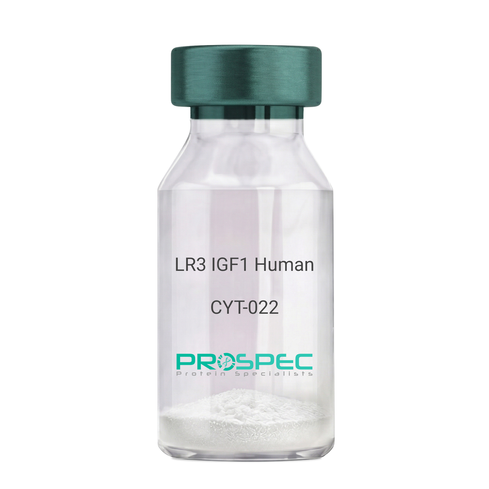
- View Data Sheet
Name :
IGF1 LR3 HumanDescription:
LR3 Insulin Like Growth Factor-1 Human Recombinant
R3 IGF1, R3 IGF-1, R3IGF1, R3IGF-1, LONG IGF1, LONG IGF-1, LONG R3 IGF1, LONG R3IGF1, LONG R3 IGF-1, LONG R3IGF-1.
Product # :
CYT-022Price :
Quantity :
Shipping Method :
Shipped at Room temp
More Info
- description
- source
- formulation
- purity
- biological activity
- More Info
- sds-page
Description
The LR3 is a long-term analog of human IGF-1, specifically designed and manufactured for mammalian cell culture to support large-scale manufacturing of recombinant biopharmaceuticals. Recombinant Human LR3 Insulin Like Growth Factor-1 produced in E.Coli is a single, non-glycosylated, polypeptide chain containing 83 amino acids and having a molecular mass of 9.1kDa.
Source
Escherichia Coli.
Formulation
Lyophilized from a 0.2µm filtered concentrated solution in 20mM PB, pH 7.2.
Purity
Greater than 97.0% as determined by SDS-PAGE.
Biological Activity
The ED50 as determined by the stimulation of protein synthesis in L6 myoblasts is less than 10ng/ml, corresponding to a specific activity of 100,000units/mg.sds-page
More Info
-
Introduction
IGF-1 (Insulin-like growth factor-1) is a major hormonal mediator of statural growth. Under regular circumstances, GH (growth hormone) binds to its receptor in the liver, and other tissues, and stimulates the synthesis/secretion of IGF-1. In target tissues, the Type 1 IGF receptor, that is homologous to the insulin receptor, is activated by IGF-1, leading to intracellular signaling which stimulates multiple processes leading to statural growth. IGF-1 metabolic actions are partly directed at stimulating the uptake of glucose, fatty acids, and amino acids so that metabolism supports growing tissues.
-
Synonyms
R3 IGF1, R3 IGF-1, R3IGF1, R3IGF-1, LONG IGF1, LONG IGF-1, LONG R3 IGF1, LONG R3IGF1, LONG R3 IGF-1, LONG R3IGF-1.
-
Physical Appearance
Sterile Filtered White lyophilized (freeze-dried) powder.
-
Stability
Lyophilized LR3 IGF1 although stable at room temperature for 3 weeks, should be stored desiccated below -18°C. Upon reconstitution the LR3 IGF1 should be stored at 4°C between 2-7 days and for future use below -18°C.For long term storage it is recommended to add a carrier protein (0.1% HSA or BSA).Please prevent freeze-thaw cycles.
-
Solubility
It is recommended to reconstitute the lyophilized LR3 IGF1 in sterile 18M-cm H2O at a concentration of 100µg/ml, which can then be further diluted to other aqueous solutions.
-
Amino Acid Sequence
MFPAMPLSSLFVNGPRTLCGAELVDALQFVCGDRGFYFNKPTGYGSSSRRAPQTGIV DECCFRSCDLRRLEMYCAPLKPAKSA.
-
Background
What is the molecular weight/Mw of IGF1 LR3 HUMAN Protein?
IGF1 LR3 HUMAN Protein has a total Mw of 9.1kDa.
What is the source or expression system of IGF1 LR3 HUMAN Protein?
Escherichia Coli.
What is the Purity of IGF1 LR3 HUMAN Protein?
IGF1 LR3 HUMAN Protein is >98% pure as determined by SDS-PAGE.
What is the Biological Activity of IGF1 LR3 HUMAN Protein?
The ED50 as determined by the stimulation of protein synthesis in L6 myoblasts is less than 10ng/ml, corresponding to a specific activity of 100,000units/mg.
What is the amino acid sequence of IGF1 LR3 HUMAN Protein?
MFPAMPLSSLFVNGPRTLCGAELVDALQFVCGDRGFYFNKPTGYGSSSRRAPQTGIV DECCFRSCDLRRLEMYCAPLKPAKSA.
What applications can IGF1 LR3 HUMAN Protein be used in?
IGF1 LR3 HUMAN Protein can probably be used in western blot, ELISA and Lateral Flow.
What is the endotoxin level for IGF1 LR3 HUMAN Protein?
The endotoxin level is minimal, IGF1 LR3 HUMAN Protein was purified using conventional chromatography techniques.
ProSpec's products are furnished for LABORATORY RESEARCH USE ONLY. The product may not be used as drugs, agricultural or pesticidal products, food additives or household chemicals.