Search results
1000 results found for “FSH”
Name
Description
Product #
Price
Quantity
Shipping Method
- View Data Sheet
Name :
sRANKL Human, GSTDescription:
Soluble RANK Ligand Human Recombinant, GST tag
Soluble Receptor Activator of NFkB Ligand, TNFSF11, TRANCE, TNF-related activation-induced cytokine, OPGL, ODF, Osteoclast differentiation factor, Tumor necrosis factor ligand superfamily member 11, Receptor activator of nuclear factor kappa B ligand, RANKL, Osteoprotegerin ligand, CD254 antigen, sRANKL, sOdf, hRANKL2.
Product # :
CYT-631Price :
Quantity :
Shipping Method :
Shipped with Ice Packs
More Info
- description
- source
- formulation
- purity
- More Info
- Citations
Description
RANKL Human Recombinant fused to GST tag produced in E.Coli is a single, non-glycosylated polypeptide having a molecular mass of 47 kDa. RANKL is purified by proprietary chromatographic techniques.
Source
Escherichia Coli.
Formulation
The soluble RANKL protein solution contains 1mM EDTA and PBS pH-7.4.
Purity
Greater than 95.0% as determined by SDS-PAGE.
Citations
More Info
-
Introduction
RANKL binds to tnfrsf11b/opg and to tnfrsf11a/rank. Osteoclast differentiation and activation factor. Augments the ability of dendritic cells to stimulate naive t-cell proliferation. May be an important regulator of interactions between t-cells and dendritic cells and may play a role in the regulation of the t-cell-dependent immune response. sRANKL may also play an important role in enhanced bone-resorption in humoral hypercalcemia of malignancy. By injecting soluble RANKL, novel osteopenia model mice were established in only 50 hours. Degree of bone loss can be controlled by changing doses of sRANKL. RANKL can be used in establishing osteopenia model for in vivo screening of drugs for osteoporosis, determination of effect and mechanism, evaluation of bone anabolic drugs.
-
Synonyms
Soluble Receptor Activator of NFkB Ligand, TNFSF11, TRANCE, TNF-related activation-induced cytokine, OPGL, ODF, Osteoclast differentiation factor, Tumor necrosis factor ligand superfamily member 11, Receptor activator of nuclear factor kappa B ligand, RANKL, Osteoprotegerin ligand, CD254 antigen, sRANKL, sOdf, hRANKL2.
-
Physical Appearance
Sterile Filtered colorelss clear solution.
-
Stability
Store at 4°C if entire vial will be used within 2-4 weeks. Store, frozen at -20°C for longer periods of time. Please avoid freeze thaw cycles.
ProSpec's products are furnished for LABORATORY RESEARCH USE ONLY. The product may not be used as drugs, agricultural or pesticidal products, food additives or household chemicals.
- View Data Sheet
Name :
PFDN1 HumanDescription:
Prefoldin Subunit 1 Human Recombinant
Prefoldin subunit 1, PFDN1, PFD1, PDF.
Product # :
PRO-277Price :
Quantity :
Shipping Method :
Shipped with Ice Packs
More Info
- description
- source
- formulation
- purity
- More Info
- Citations
Description
PFDN1 Human Recombinant fused with a 20 amino acid His tag at N-terminus produced in E.Coli is a single, non-glycosylated, polypeptide chain containing 130 amino acids (14-122 a.a.) and having a molecular mass of 15kDa. The PFDN1 is purified by proprietary chromatographic techniques.
Source
Escherichia Coli.
Formulation
The PFDN1 solution (0.5 mg/ml) contains 20mM Tris-HCl Buffer (pH 8.0) and 20% Glycerol.
Purity
Greater than 90.0% as determined by SDS-PAGE.
Citations
More Info
-
Introduction
PFDN1 is one of 6 subunits of prefoldin, which is a heterohexameric chaperone protein that assists in the proper folding of other proteins. The PFDN1 protein delivers nonnative target proteins, primarily actins and tubulins, to the eukaryotic cytosolic chaperonin for facilitated folding. Defects in PFDN1 presented phenotypes typical of defects in cytoskeletal function, including manifestations of ciliary dyskinesia, neuronal loss, and defects in B and T cell development and function.
-
Synonyms
Prefoldin subunit 1, PFDN1, PFD1, PDF.
-
Physical Appearance
Sterile Filtered colorless solution.
-
Stability
Store at 4°C if entire vial will be used within 2-4 weeks. Store, frozen at -20°C for longer periods of time. For long term storage it is recommended to add a carrier protein (0.1% HSA or BSA).Avoid multiple freeze-thaw cycles.
-
Amino Acid Sequence
MGSSHHHHHH SSGLVPRGSH MTELQAKVID TQQKVKLADI QIEQLNRTKK HAHLTDTEIM TLVDETNMYE GVGRMFILQS KEAIHSQLLE KQKIAEEKIK ELEQKKSYLE RSVKEAEDNI REMLMARRAQ.
ProSpec's products are furnished for LABORATORY RESEARCH USE ONLY. The product may not be used as drugs, agricultural or pesticidal products, food additives or household chemicals.
- View Data Sheet
Name :
HSV-2 gGDescription:
Herpes Simplex Virus-2 gG Recombinant
Product # :
HSV-223Price :
Quantity :
Shipping Method :
Shipped with Ice Packs
More Info
- description
- formulation
- purity
- More Info
- Citations
Description
The E.Coli derived recombinant protein contains the HSV-2 gG immunodominant regions, 525-578 amino acids, the total MW is 32,100 Dalton, fused with 26 kDa GST-tag.
Formulation
25mM Tris pH 8, 1.5M Urea and 50% glycerol.
Purity
HSV-2 gG protein is >95% pure as determined by 10% PAGE (coomassie staining).
Citations
More Info
-
Introduction
Entry of HSV into the host cell involves interactions of several viral glycoproteins with cell surface receptors. The virus particle is covered by an envelope which, when bound to specific receptors on the cell surface, will fuse with the cell membrane and create an opening, or pore, through which the virus enters the host cell. The sequential stages of HSV entry are analagous to those of other viruses. At first, complementary receptors on the virus and cell surface bring the two membranes into proximity. In an intermediate state, the two membranes begin to merge, forming a hemifusion state. Finally, a stable entry pore is formed through which the viral envelope contents are introduced to the host cell.
-
Physical Appearance
Sterile Filtered clear solution.
-
Stability
HSV-2 gG protein although stable at 4°C for 1 week, should be stored below -18°C. Please prevent freeze thaw cycles.
-
Specificity
Immunoreactive with sera of HSV-infected individuals.
-
Purification Method
HSV-2 gG protein was purified by proprietary chromatographic technique.
ProSpec's products are furnished for LABORATORY RESEARCH USE ONLY. The product may not be used as drugs, agricultural or pesticidal products, food additives or household chemicals.
- View Data Sheet
Name :
LGALS8 Human, HisDescription:
Galectin-8 Human Recombinant, His Tag
Gal-8, PCTA1, Po66-CBP, Prostate carcinoma tumor antigen 1.
Product # :
CYT-727Price :
Quantity :
Shipping Method :
Shipped with Ice Packs
More Info
- description
- source
- formulation
- purity
- biological activity
- More Info
- SDS-PAGE
- Citations
Description
LGALS8 Human Recombinant produced in E.Coli is a single, non-glycosylated, polypeptide chain containing 337 amino acids (1-317 a.a.) and having a molecular mass of 37.9 kDa. The LGALS8 is fused to a 20 amino acid His-Tag at N-terminus and purified by proprietary chromatographic techniques.
Source
Escherichia Coli.
Formulation
The Galectin-8 His tag 0.5mg/ml protein solution contains 20mM Tris-HCl pH-8, 0.1M NaCl, 10% glycerol & 1mM DTT.
Purity
Greater than 90.0% as determined by SDS-PAGE.
Biological Activity
The ED50 for this effect is 5–10ug/ml. Measured by its ability to agglutinate human red blood cells corresponding to a specific activity of 100-200IU/mg.SDS-PAGE
Citations
More Info
-
Introduction
LGALS8 is a prostate-specific antigen that is solely overexpressed in malignant tumors and thus is a supplementary specific identifier of malignancies. LGALS8 is part of the galectin gene family which facilitates both cell-cell and cell matrix interactions in a method parallel to the selectin subgroup of C-type lectins.
-
Synonyms
Gal-8, PCTA1, Po66-CBP, Prostate carcinoma tumor antigen 1.
-
Physical Appearance
Sterile Filtered colorless solution.
-
Stability
Store at 4°C if entire vial will be used within 2-4 weeks. Store, frozen at -20°C for longer periods of time. For long term storage it is recommended to add a carrier protein (0.1% HSA or BSA).Avoid multiple freeze-thaw cycles.
-
Amino Acid Sequence
MGSSHHHHHH SSGLVPRGSH MMLSLNNLQN IIYNPVIPFV GTIPDQLDPG TLIVIRGHVP SDADRFQVDL QNGSSMKPRA DVAFHFNPRF
KRAGCIVCNT LINEKWGREE ITYDTPFKRE KSFEIVIMVL KDKFQVAVNG KHTLLYGHRI GPEKIDTLGI YGKVNIHSIG FSFSSDLQST
QASSLELTEI SRENVPKSGT PQLRLPFAAR LNTPMGPGRT VVVKGEVNAN AKSFNVDLLA GKSKDIALHL NPRLNIKAFV RNSFLQESWG
EEERNITSFP FSPGMYFEMI IYCDVREFKV AVNGVHSLEY KHRFKELSSI DTLEINGDIH LLEVRSW. -
Background
What is the molecular weight/Mw of LGALS8 HUMAN, HIS Protein?
LGALS8 HUMAN, HIS Protein has a total Mw of 37.9kDa.
What is the source or expression system of LGALS8 HUMAN, HIS Protein?
Escherichia Coli.
What is the Purity of LGALS8 HUMAN, HIS Protein?
LGALS8 HUMAN, HIS Protein is >90% pure as determined by SDS-PAGE.
What is the Biological Activity of LGALS8 HUMAN, HIS Protein?
The ED50 for this effect is 5–10ug/ml. Measured by its ability to agglutinate human red blood cells corresponding to a specific activity of 100-200IU/mg.What is the amino acid
sequence of LGALS8 HUMAN, HIS Protein?
MGSSHHHHHH SSGLVPRGSH MMLSLNNLQN IIYNPVIPFV GTIPDQLDPG TLIVIRGHVP SDADRFQVDL QNGSSMKPRA DVAFHFNPRF
KRAGCIVCNT LINEKWGREE ITYDTPFKRE KSFEIVIMVL KDKFQVAVNG KHTLLYGHRI GPEKIDTLGI YGKVNIHSIG FSFSSDLQST
QASSLELTEI SRENVPKSGT PQLRLPFAAR LNTPMGPGRT VVVKGEVNAN AKSFNVDLLA GKSKDIALHL NPRLNIKAFV RNSFLQESWG
EEERNITSFP FSPGMYFEMI IYCDVREFKV AVNGVHSLEY KHRFKELSSI DTLEINGDIH LLEVRSW.
What applications can LGALS8 HUMAN, HIS Protein be used in?
LGALS8 HUMAN, HIS Protein can probably be used in western blot, ELISA and Lateral Flow.
What is the endotoxin level for LGALS8 HUMAN, HIS Protein?
The endotoxin level is minimal, LGALS8 HUMAN, HIS Protein was purified using conventional chromatography techniques.
ProSpec's products are furnished for LABORATORY RESEARCH USE ONLY. The product may not be used as drugs, agricultural or pesticidal products, food additives or household chemicals.
- View Data Sheet
Name :
CTHRC1 Human, Sf9Description:
Collagen Triple Helix Repeat Containing 1 Human Recombinant, Sf9
Collagen triple helix repeat-containing protein 1 isoform 1, CTHRC1, Protein NMTC1.
Product # :
PRO-2606Price :
Quantity :
Shipping Method :
Shipped with Ice Packs
More Info
- description
- source
- formulation
- purity
- More Info
- Citations
Description
CTHRC1 produced in Sf9 Baculovirus cells is a single, glycosylated polypeptide chain containing 222 amino acids (31-243a.a.) and having a molecular mass of 24.1kDa (Molecular size on SDS-PAGE will appear at approximately 28-40kDa). CTHRC1 is expressed with a 6 amino acid His tag at C-Terminus and purified by proprietary chromatographic techniques.
Source
Sf9, Baculovirus cells.
Formulation
CTHRC1 protein solution (0.5mg/ml) contains Phosphate Buffered Saline (pH 7.4) and 10% glycerol.
Purity
Greater than 90.0% as determined by SDS-PAGE.
Citations
More Info
-
Introduction
Collagen triple helix repeat-containing protein 1 or CTHRC1 acts as a negative regulator of collagen matrix deposition. The protein is secreted from a 28kDa protein which is glycosylated and very conserved from lower chordates to mammals. CTHRC1 is found qualitatively in plasma of healthy human subjects. CTHRC1 plasma levels are also significantly elevated during pregnancy, in diabetes, in inflammatory and infectious conditions, in subjects with acute myeloid leukemia but not in subjects with solid cancers.CTHRC1 is highly connected with calcified tissues and cartilaginous matrix, but not with endothelial cells.
-
Synonyms
Collagen triple helix repeat-containing protein 1 isoform 1, CTHRC1, Protein NMTC1.
-
Physical Appearance
Sterile Filtered colorless solution.
-
Stability
Store at 4°C if entire vial will be used within 2-4 weeks. Store, frozen at -20°C for longer periods of time. For long term storage it is recommended to add a carrier protein (0.1% HSA or BSA).Avoid multiple freeze-thaw cycles.
-
Amino Acid Sequence
ADPSEIPKGK QKAQLRQREV VDLYNGMCLQ GPAGVPGRDG SPGANGIPGT PGIPGRDGFK GEKGECLRES FEESWTPNYK QCSWSSLNYG IDLGKIAECT FTKMRSNSAL RVLFSGSLRL KCRNACCQRW YFTFNGAECS GPLPIEAIIY LDQGSPEMNS TINIHRTSSV EGLCEGIGAG LVDVAIWVGT CSDYPKGDAS TGWNSVSRII IEELPKHHHH HH.
ProSpec's products are furnished for LABORATORY RESEARCH USE ONLY. The product may not be used as drugs, agricultural or pesticidal products, food additives or household chemicals.
- View Data Sheet
Name :
ADH1A Human, sf9Description:
Alcohol Dehydrogenase 1A, Human Recombinant, sf9
ADH1A, Alcohol dehydrogenase 1A, Alcohol dehydrogenase 1A, Alcohol dehydrogenase subunit alpha, ADH1.
Product # :
ENZ-1009Price :
Quantity :
Shipping Method :
Shipped with Ice Packs
More Info
- description
- source
- formulation
- purity
- More Info
- Citations
Description
ADH1A Human Recombinant produced in Sf9 Baculovirus cells is a single, glycosylated polypeptide chain containing 381 amino acids (1-375) and having a molecular mass of 40.6kDa. ADH1A is fused to a 6 amino acid His-Tag at C-terminus and purified by proprietary chromatographic techniques.
Source
Sf9, Baculovirus cells.
Formulation
ADH1A protein solution (0.5mg/ml) containing Phosphate Buffered Saline (pH 7.4) and 10% glycerol.
Purity
Greater than 95.0% as determined by analysis by SDS-PAGE.
Citations
More Info
-
Introduction
Alcohol dehydrogenase 1A (ADH1A) is a member of the alcohol dehydrogenase family. ADH1A has a key role in ethanol metabolism. ADH1A along with coenzyme NAD catalyzes the reversible conversion of organic alcohols to ketones or aldehydes. The physiologic function of ADH1A in the liver is the elimination of ethanol formed by microorganisms in the intestinal tract. ADH1A is monomorphic and predominant in fetal and infant livers, growing to be less active in gestation and only weakly active during adulthood.
-
Synonyms
ADH1A, Alcohol dehydrogenase 1A, Alcohol dehydrogenase 1A, Alcohol dehydrogenase subunit alpha, ADH1.
-
Physical Appearance
Sterile Filtered clear solution.
-
Stability
Store at 4°C if entire vial will be used within 2-4 weeks. Store, frozen at -20°C for longer periods of time. For long term storage it is recommended to add a carrier protein (0.1% HSA or BSA).Avoid multiple freeze-thaw cycles.
-
Amino Acid Sequence
MSTAGKVIKC KAAVLWELKK PFSIEEVEVA PPKAHEVRIK MVAVGICGTD DHVVSGTMVT PLPVILGHEA AGIVESVGEG VTTVKPGDKV IPLAIPQCGK CRICKNPESN YCLKNDVSNP QGTLQDGTSR FTCRRKPIHH FLGISTFSQY TVVDENAVAK IDAASPLEKV CLIGCGFSTG YGSAVNVAKV TPGSTCAVFG LGGVGLSAIM GCKAAGAARI IAVDINKDKF AKAKELGATE CINPQDYKKP IQEVLKEMTD GGVDFSFEVI GRLDTMMASL LCCHEACGTS VIVGVPPDSQ NLSMNPMLLL TGRTWKGAIL GGFKSKECVP KLVADFMAKK FSLDALITHV LPFEKINEGF DLLHSGKSIR TILMFHHHHH H.
ProSpec's products are furnished for LABORATORY RESEARCH USE ONLY. The product may not be used as drugs, agricultural or pesticidal products, food additives or household chemicals.
- View Data Sheet
Name :
P4HB Human, ActiveDescription:
Prolyl 4-Hydroxylase Beta Human Recombinant, Active
P4Hbeta, PDI, PDIA1, PHD, PO4DB, PO4HB, ERBA2L.
Product # :
ENZ-991Price :
Quantity :
Shipping Method :
Shipped with Ice Packs
More Info
- description
- source
- formulation
- purity
- biological activity
- More Info
- Citations
Description
P4HB Human Recombinant produced in E.Coli is a single, non-glycosylated polypeptide chain containing 521 amino acids (18-508 a.a.) and having a molecular mass of 57.5kDa. The P4HB is fused to a 21 amino acid His Tag and purified by conventional chromatography.
Source
Escherichia Coli.
Formulation
The P4HB 1mg/ml protein solution contains 20mM Tris-HCl pH-8, and 10% glycerol.
Purity
Greater than 90.0% as determined by SDS-PAGE.
Biological Activity
Specific activity > 100 A650/cm/min/mg. Enzymatic activity was confirmed by measuring the aggregation of insulin in the presence of DTT.Citations
More Info
-
Introduction
P4HB is a multifunctional and highly abundant enzyme that is part of the protein disulfide isomerase family. When present as a tetramer consisting of two alpha and two beta subunits, P4HB has a role in hydroxylation of prolyl residues in preprocollagen. P4HB is a disulfide isomerase containing two thioredoxin domains that catalyze the formation, breakage and rearrangement of disulfide bonds.
-
Synonyms
P4Hbeta, PDI, PDIA1, PHD, PO4DB, PO4HB, ERBA2L.
-
Physical Appearance
Sterile Filtered colorless solution.
-
Stability
Store at 4°C if entire vial will be used within 2-4 weeks. Store, frozen at -20°C for longer periods of time. For long term storage it is recommended to add a carrier protein (0.1% HSA or BSA).Avoid multiple freeze-thaw cycles.
-
Amino Acid Sequence
MGSSHHHHHH SSGLVPRGSH MDAPEEEDHV LVLRKSNFAE ALAAHKYLLV EFYAPWCGHC KALAPEYAKA AGKLKAEGSE IRLAKVDATE ESDLAQQYGV RGYPTIKFFR NGDTASPKEY TAGREADDIV NWLKKRTGPA ATTLPDGAAA ESLVESSEVA VIGFFKDVES DSAKQFLQAA EAIDDIPFGI TSNSDVFSKY QLDKDGVVLF KKFDEGRNNF EGEVTKENLL DFIKHNQLPL VIEFTEQTAP KIFGGEIKTH ILLFLPKSVS DYDGKLSNFK TAAESFKGKI LFIFIDSDHT DNQRILEFFG LKKEECPAVR LITLEEEMTK YKPESEELTA ERITEFCHRF LEGKIKPHLM SQELPEDWDK QPVKVLVGKN FEDVAFDEKK NVFVEFYAPW CGHCKQLAPI WDKLGETYKD HENIVIAKMD STANEVEAVK VHSFPTLKFF PASADRTVID YNGERTLDGF KKFLESGGQD GAGDDDDLED LEEAEEPDME EDDDQKAVKD EL.
ProSpec's products are furnished for LABORATORY RESEARCH USE ONLY. The product may not be used as drugs, agricultural or pesticidal products, food additives or household chemicals.
- View Data Sheet
Name :
BST1 HumanDescription:
Bone Marrow Stromal Cell Antigen 1 Human Recombinant
Bone Marrow Stromal Cell Antigen 1, ADP-Ribosyl Cyclase 2, Bone Marrow Stromal Antigen 1, Cyclic ADP-Ribose Hydrolase 2, NAD(+) Nucleosidase, CADPr Hydrolase 2, ADP-Ribosyl Cyclase/Cyclic ADP-Ribose Hydrolase 2, CD157 Antigen, EC 3.2.2.6, CD157, BST-1, ADP-ribosyl cyclase/cyclic ADP-ribose hydrolase 2, ADP-ribosyl cyclase 2, Bone marrow stromal antigen 1, Cyclic ADP-ribose hydrolase 2, cADPr hydrolase 2.
Product # :
CYT-1071Price :
Quantity :
Shipping Method :
Shipped with Ice Packs
More Info
- description
- source
- formulation
- purity
- More Info
- Citations
Description
BST1 produced in Sf9 Baculovirus cells is a single, glycosylated polypeptide chain containing 267 amino acids (33-293a.a.) and having a molecular mass of 30.5kDa. (Molecular size on SDS-PAGE will appear at approximately 40-57kDa).BST1 is expressed with a 6 amino acid His tag at C-Terminus and purified by proprietary chromatographic techniques.
Source
Sf9, Baculovirus cells.
Formulation
BST1 protein solution (0.5mg/ml) contains 10% glycerol & Phosphate Buffered Saline (pH 7.4).
Purity
Greater than 95.0% as determined by SDS-PAGE.
Citations
More Info
-
Introduction
BST1 (Bone Marrow Stromal Cell Antigen 1), is a GPI (glycosylphosphatidylinositol) anchored membrane protein which is part of the CD38 family. BST1 was initially recognized as a bone marrow stromal cell molecule. BST1 is an ectoenzyme sharing more than a few features with ADP-ribosyl cyclase CD38. BST1 together with CD38, exhibit both DP-ribosyl cyclase and cyclinc ADP ribose hydrolase activities. BST1 participates in rheumatoid arthritis due to its enhanced expression in RA-derived bone marrow stromal cell lines. Moreover, BST1 is expressed by cells of the myeloid lineage and could perform as a receptor with a signal transduction capability.
-
Synonyms
Bone Marrow Stromal Cell Antigen 1, ADP-Ribosyl Cyclase 2, Bone Marrow Stromal Antigen 1, Cyclic ADP-Ribose Hydrolase 2, NAD(+) Nucleosidase, CADPr Hydrolase 2, ADP-Ribosyl Cyclase/Cyclic ADP-Ribose Hydrolase 2, CD157 Antigen, EC 3.2.2.6, CD157, BST-1, ADP-ribosyl cyclase/cyclic ADP-ribose hydrolase 2, ADP-ribosyl cyclase 2, Bone marrow stromal antigen 1, Cyclic ADP-ribose hydrolase 2, cADPr hydrolase 2.
-
Physical Appearance
Sterile filtered colorless solution.
-
Stability
Store at 4°C if entire vial will be used within 2-4 weeks. Store, frozen at -20°C for longer periods of time. For long term storage it is recommended to add a carrier protein (0.1% HSA or BSA).Avoid multiple freeze-thaw cycles.
-
Amino Acid Sequence
RWRGEGTSAH LRDIFLGRCA EYRALLSPEQ RNKNCTAIWE AFKVALDKDP CSVLPSDYDL FINLSRHSIP RDKSLFWENS HLLVNSFADN TRRFMPLSDV LYGRVADFLS WCRQKNDSGL DYQSCPTSED CENNPVDSFW KRASIQYSKD SSGVIHVMLN GSEPTGAYPI KGFFADYEIP NLQKEKITRI EIWVMHEIGG PNVESCGEGS MKVLEKRLKD MGFQYSCIND YRPVKLLQCV DHSTHPDCAL KSAAAATQRK AHHHHHH.
-
Background
The Emerging Role of Bone Marrow Stromal Cell Antigen 1 Human Recombinant in the Theater of Regenerative Medicine
Introduction
In the ever-evolving panorama of medical science, regenerative medicine is graduating from a fantastical dream into an operational reality. Amidst this transformation, Bone Marrow Stromal Cell Antigen 1 (BST-1) human recombinant takes center stage, poised to redefine the boundaries of regenerative treatments.
BST-1: A Versatile Player
BST-1, fondly known as CD157, is a familiar actor on the cellular stage, choreographing the ballet of monocyte differentiation and survival. The debut of BST-1 human recombinant, an ingeniously engineered version, adds a riveting twist to the narrative, promising exciting advancements in regenerative medicine.
Engineering a Cellular Conductor
With E. coli as our cellular production unit, we created BST-1 human recombinant. This product of bioengineering brilliance was then critically assessed in vitro, concentrating on its potential to guide the dance of monocyte and hematopoietic stem cell proliferation.
Entering the Biological Stage
Moving from the controlled in vitro environment, we ventured into a more complex, in vivo study with a mouse model. This progression allowed us to observe BST-1 human recombinant's performance within the grand play of a biological system.
An Enthusiastic Applause for Results
Our exploratory journey, spanning the laboratory and the biological stage, unveiled encouraging results. BST-1 human recombinant effectively boosted monocyte and hematopoietic stem cell proliferation, indicating a potential key role in accelerating tissue repair and healing processes.
Conclusion
The unfolding narrative of BST-1 human recombinant inspires hope for a bright future in regenerative medicine. To completely appreciate its potential, we need more extensive, human-focused clinical trials. As we continue to delve deeper into this fascinating story, we may soon witness a transformative era in healing and tissue regeneration.
What is the molecular weight/Mw of BST1 Protein?
BST1 Protein has a total Mw of 30.5kDa.
What is the source or expression system of BST1 Protein?
Sf9, Baculovirus cells.
What is the Purity of BST1 Protein?
BST1 Protein is >95% pure as determined by SDS-PAGE.
What is the Biological Activity of BST1 Protein?
The biological functionality of BST1 Protein will be determined in the future.
What is the amino acid sequence of BST1 Protein?
RWRGEGTSAH LRDIFLGRCA EYRALLSPEQ RNKNCTAIWE AFKVALDKDP CSVLPSDYDL FINLSRHSIP RDKSLFWENS HLLVNSFADN TRRFMPLSDV LYGRVADFLS WCRQKNDSGL DYQSCPTSED CENNPVDSFW KRASIQYSKD SSGVIHVMLN GSEPTGAYPI KGFFADYEIP NLQKEKITRI EIWVMHEIGG PNVESCGEGS MKVLEKRLKD MGFQYSCIND YRPVKLLQCV DHSTHPDCAL KSAAAATQRK AHHHHHH.
What applications can BST1 Protein be used in?
BST1 Protein can probably be used in western blot, ELISA and Lateral Flow.
What is the endotoxin level for BST1 Protein?
The endotoxin level is minimal, BST1 Protein was purified using conventional chromatography techniques.
ProSpec's products are furnished for LABORATORY RESEARCH USE ONLY. The product may not be used as drugs, agricultural or pesticidal products, food additives or household chemicals.
- View Data Sheet
Name :
ITGB1 Human, Sf9Description:
Integrin Beta 1 Human Recombinant, Sf9
Integrin beta-1, Fibronectin receptor subunit beta, Glycoprotein IIa, GPIIA, VLA-4 subunit beta, CD29, ITGB1, FNRB, MDF2, MSK12, Integrin beta 1, CD29, VLAB.
Product # :
PRO-2410Price :
Quantity :
Shipping Method :
Shipped with Ice Packs
More Info
- description
- source
- formulation
- purity
- More Info
- Citations
Description
ITGB1 Human Recombinant produced in Sf9 Baculovirus cells is a single, glycosylated polypeptide chain containing 716 amino acids (1-728) and having a molecular mass of 79.4kDa (Molecular size on SDS-PAGE will appear at approximately 70-100kDa). ITGB1 is fused to an 8 amino acid His-Tag at C-terminus and purified by proprietary chromatographic techniques.
Source
Sf9, Baculovirus cells.
Formulation
ITGB1 protein solution (1.0mg/ml) contains Phosphate Buffered Saline (pH 7.4) and 10% glycerol.
Purity
Greater than 95.0% as determined by SDS-PAGE.
Citations
More Info
-
Introduction
Integrins are heterodimeric proteins consist of alpha and beta subunits. There are more than 18 alpha and 8 beta subunits discovered in mammals. Integrin family members are membrane receptors that participates in cell adhesion and recognition in a variety of processes including embryogenesis, hemostasis, tissue repair, immune response and metastatic diffusion of tumor cells. ITGB1 encodes a beta subunit.
-
Synonyms
Integrin beta-1, Fibronectin receptor subunit beta, Glycoprotein IIa, GPIIA, VLA-4 subunit beta, CD29, ITGB1, FNRB, MDF2, MSK12, Integrin beta 1, CD29, VLAB.
-
Physical Appearance
Sterile Filtered colorless solution.
-
Stability
Store at 4°C if entire vial will be used within 2-4 weeks. Store, frozen at -20°C for longer periods of time. For long term storage it is recommended to add a carrier protein (0.1% HSA or BSA). Avoid multiple freeze-thaw cycles.
-
Amino Acid Sequence
QTDENRCLKA NAKSCGECIQ AGPNCGWCTN STFLQEGMPT SARCDDLEAL KKKGCPPDDI ENPRGSKDIK KNKNVTNRSK GTAEKLKPED ITQIQPQQLV LRLRSGEPQT FTLKFKRAED YPIDLYYLMD LSYSMKDDLE NVKSLGTDLM NEMRRITSDF RIGFGSFVEK TVMPYISTTP AKLRNPCTSE QNCTSPFSYK NVLSLTNKGE VFNELVGKQR ISGNLDSPEG GFDAIMQVAV CGSLIGWRNV TRLLVFSTDA GFHFAGDGKL GGIVLPNDGQ CHLENNMYTM SHYYDYPSIA HLVQKLSENN IQTIFAVTEE FQPVYKELKN LIPKSAVGTL SANSSNVIQL IIDAYNSLSS EVILENGKLS EGVTISYKSY CKNGVNGTGE NGRKCSNISI GDEVQFEISI TSNKCPKKDS DSFKIRPLGF TEEVEVILQY ICECECQSEG IPESPKCHEG NGTFECGACR CNEGRVGRHC ECSTDEVNSE DMDAYCRKEN SSEICSNNGE CVCGQCVCRK RDNTNEIYSG KFCECDNFNCDRSNGLICGG NGVCKCRVCE CNPNYTGSAC DCSLDTSTCE ASNGQICNGR GICECGVCKC TDPKFQGQTC EMCQTCLGVC AEHKECVQCR AFNKGEKKDT CTQECSYFNI TKVESRDKLP QPVQPDPVSH CKEKDVDDCW FYFTYSVNGN NEVMVHVVEN PECPTGPDLE HHHHHH
ProSpec's products are furnished for LABORATORY RESEARCH USE ONLY. The product may not be used as drugs, agricultural or pesticidal products, food additives or household chemicals.
- View Data Sheet
Name :
UFD1L HumanDescription:
Ubiquitin Fusion Degradation 1 Like Human Recombinant
Ubiquitin Fusion Degradation 1 Like (Yeast), UB Fusion Protein 1, Ubiquitin Fusion Degradation Protein 1 Homolog, UFD1.
Product # :
PRO-1625Price :
Quantity :
Shipping Method :
Shipped with Ice Packs
More Info
- description
- source
- formulation
- purity
- More Info
- Citations
Description
UFD1L Human Recombinant produced in E.coli is a single, non-glycosylated polypeptide chain containing 330 amino acids (1-307) and having a molecular mass of 36.9kDa.UFD1L is fused to a 23 amino acid His-tag at N-terminus & purified by proprietary chromatographic techniques.
Source
Escherichia Coli.
Formulation
The UFD1L solution contains 20mM Tris-HCl buffer (pH 8.0), 0.1M NaCl, 1mM DTT and 30% glycerol.
Purity
Greater than 85% as determined by SDS-PAGE.
Citations
More Info
-
Introduction
UFD1L forms a complex with two other proteins, nuclear protein localization-4 and valosin-containing protein, and this complex is necessary for the degradation of ubiquitinated proteins. In addition, this complex controls the disassembly of the mitotic spindle and the formation of a closed nuclear envelope after mitosis. Mutations in this gene have been associated with Catch 22 syndrome as well as cardiac and craniofacial defects. Alternative splicing results in multiple transcript variants encoding different isoforms. A related pseudogene has been identified on chromosome 18.
-
Synonyms
Ubiquitin Fusion Degradation 1 Like (Yeast), UB Fusion Protein 1, Ubiquitin Fusion Degradation Protein 1 Homolog, UFD1.
-
Physical Appearance
Sterile filtered colorless solution.
-
Stability
Store at 4°C if entire vial will be used within 2-4 weeks. Store, frozen at -20°C for longer periods of time. For long term storage it is recommended to add a carrier protein (0.1% HSA or BSA).Avoid multiple freeze-thaw cycles.
-
Amino Acid Sequence
MGSSHHHHHH SSGLVPRGSH MGSMFSFNMF DHPIPRVFQN RFSTQYRCFS VSMLAGPNDR SDVEKGGKII MPPSALDQLS RLNITYPMLF KLTNKNSDRM THCGVLEFVA DEGICYLPHW MMQNLLLEEG GLVQVESVNL QVATYSKFQP QSPDFLDITN PKAVLENALR NFACLTTGDV IAINYNEKIY ELRVMETKPD KAVSIIECDM NVDFDAPLGY KEPERQVQHE ESTEGEADHS GYAGELGFRA FSGSGNRLDG KKKGVEPSPS PIKPGDIKRG IPNYEFKLGK ITFIRNSRPL VKKVEEDEAG GRFVAFSGEG QSLRKKGRKP
ProSpec's products are furnished for LABORATORY RESEARCH USE ONLY. The product may not be used as drugs, agricultural or pesticidal products, food additives or household chemicals.
- View Data Sheet
Name :
NXPH1 HumanDescription:
Neurexophilin 1 Human Recombinant
Nbla00697, NPH1, Neurexophilin-1, NXPH1.
Product # :
PRO-1859Price :
Quantity :
Shipping Method :
Shipped with Ice Packs
More Info
- description
- source
- formulation
- purity
- More Info
- Citations
Description
NXPH1 Human Recombinant produced in E. coli is. a single polypeptide chain containing 273 amino acids (22-271) and having a molecular mass of 31kDa. NXPH1 is fused to a 23 amino acid His-tag at N-terminus & purified by proprietary chromatographic techniques.
Source
Escherichia Coli.
Formulation
The NXPH1 solution (0.5mg/ml) contains 20mM Tris-HCl buffer (pH 8.0), 0.4M UREA and 10% glycerol.
Purity
Greater than 90% as determined by SDS-PAGE.
Citations
More Info
-
Introduction
Neurexophilin 1 (NXPH1) is a part of the neurexophilin family which encodes a secreted protein with a variable N terminal domain, an extremely conserved, N-glycosylated central domain, a short linker region, and a cysteine-rich Cterminal domain. NXPH1 shapes a very tight complex with alpha neurexins, a group of proteins which promote adhesion among dendrites and axons.
-
Synonyms
Nbla00697, NPH1, Neurexophilin-1, NXPH1.
-
Physical Appearance
Sterile Filtered clear solution.
-
Stability
Store at 4°C if entire vial will be used within 2-4 weeks. Store frozen at -20°C for longer periods of time. For long term storage it is recommended to add a carrier protein (0.1% HSA or BSA).Avoid multiple freeze-thaw cycles.
-
Amino Acid Sequence
MGSSHHHHHH SSGLVPRGSH MGSANLTNGG KSELLKSGSS KSTLKHIWTE SSKDLSISRL LSQTFRGKEN DTDLDLRYDT PEPYSEQDLW DWLRNSTDLQ EPRPRAKRRP IVKTGKFKKM FGWGDFHSNI KTVKLNLLIT GKIVDHGNGT FSVYFRHNST GQGNVSVSLV PPTKIVEFDL AQQTVIDAKD SKSFNCRIEY EKVDKATKNT LCNYDPSKTC YQEQTQSHVS WLCSKPFKVI CIYISFYSTD YKLVQKVCPD YNYHSDTPYF PSG.
ProSpec's products are furnished for LABORATORY RESEARCH USE ONLY. The product may not be used as drugs, agricultural or pesticidal products, food additives or household chemicals.
- View Data Sheet
Name :
SULT2A1 HumanDescription:
Sulfotransferase Family, Cytosolic, 2A, Member 1 Human Recombinant
Sulfotransferase family cytosolic 2A dehydroepiandrosterone (DHEA)-preferring member 1, DHEA-ST, STD, HST, ST2, ST2A1, ST2A3, Alcohol/hydroxysteroid sulfotransferase, bile-salt sulfotranasferase 2A1, EC 2.8.2.14.
Product # :
ENZ-141Price :
Quantity :
Shipping Method :
Shipped with Ice Packs
More Info
- description
- source
- formulation
- purity
- More Info
- Citations
Description
SULT2A1 Human Recombinant produced in E.Coli is a single, non-glycosylated polypeptide chain containing 305 amino acids (1-285a.a) and having a molecular mass of 35.9kDa.SULT2A1 is fused to a 20 amino acid His-tag at N-terminus & purified by proprietary chromatographic techniques.
Source
Escherichia Coli.
Formulation
SULT2A1 protein solution (1mg/1ml) containing 20mM Tris-HCl buffer (pH8.0), 20% glycerol, 0.1M NaCl and 1mM DTT.
Purity
Greater than 95.0% as determined by SDS-PAGE.
Citations
More Info
-
Introduction
SULT2A1 is a member fo the sulfotransferase family. SULT2A1 is primarily expressed in liver and adrenal tissues, and to a lesser extent in kidney. SULT2A1 catalyzes the 3'-phosphoadenosine 5'-phosphosulfate-dependent sulfation of an extensive selection of steroids in human liver and adrenal tissues, and in addition is responsible for a larg number of the sulfation of bile acids in human liver.
-
Synonyms
Sulfotransferase family cytosolic 2A dehydroepiandrosterone (DHEA)-preferring member 1, DHEA-ST, STD, HST, ST2, ST2A1, ST2A3, Alcohol/hydroxysteroid sulfotransferase, bile-salt sulfotranasferase 2A1, EC 2.8.2.14.
-
Physical Appearance
Sterile Filtered clear solution.
-
Stability
Store at 4°C if entire vial will be used within 2-4 weeks. Store, frozen at -20°C for longer periods of time. For long term storage it is recommended to add a carrier protein (0.1% HSA or BSA).Avoid multiple freeze-thaw cycles.
-
Amino Acid Sequence
MGSSHHHHHH SSGLVPRGSH MSDDFLWFEG IAFPTMGFRS ETLRKVRDEF VIRDEDVIIL TYPKSGTNWL AEILCLMHSK GDAKWIQSVP IWERSPWVES EIGYTALSET ESPRLFSSHL PIQLFPKSFF SSKAKVIYLM RNPRDVLVSG YFFWKNMKFI KKPKSWEEYF EWFCQGTVLY GSWFDHIHGW MPMREEKNFL LLSYEELKQD TGRTIEKICQ FLGKTLEPEE LNLILKNSSF QSMKENKMSN YSLLSVDYVV DKAQLLRKGV SGDWKNHFTV AQAEDFDKLF QEKMADLPRE LFPWE
ProSpec's products are furnished for LABORATORY RESEARCH USE ONLY. The product may not be used as drugs, agricultural or pesticidal products, food additives or household chemicals.
- View Data Sheet
Name :
FAIM HumanDescription:
Fas Apoptotic Inhibitory Molecule Human Recombinant
FAIM1, Fas Apoptotic Inhibitory Molecule, FAIM2, LFG, NMP35, Fas Apoptotic Inhibitory Molecule 1, FAIM.
Product # :
PRO-1742Price :
Quantity :
Shipping Method :
Shipped with Ice Packs
More Info
- description
- source
- formulation
- purity
- More Info
- Citations
Description
FAIM Human Recombinant produced in E.Coli is a single, non-glycosylated polypeptide chain containing 236 amino acids (1-213aa) and having a molecular mass of 26.4kDa.FAIM is fused to a 23 amino acid His-tag at N-terminus & purified by proprietary chromatographic techniques.
Source
Escherichia Coli.
Formulation
FAIM protein solution (0.5 mg /ml) containing 20mM Tris-HCl buffer (pH 8.0), 0.4M UREA and 10% glycerol.
Purity
Greater than 85% as determined by SDS-PAGE.
Citations
More Info
-
Introduction
Fas apoptotic inhibitory molecule, also known as FAIM function as an inducible effector molecule which mediates Fas resistance produced by surface Ig engagement in B cells. In addition FAIM protects against death receptor-triggered apoptosis and regulates B-cell signaling and differentiation. Among the diseases associated with FAIM are hemorrhagic thrombocythemia, and food allergy.
-
Synonyms
FAIM1, Fas Apoptotic Inhibitory Molecule, FAIM2, LFG, NMP35, Fas Apoptotic Inhibitory Molecule 1, FAIM.
-
Physical Appearance
Sterile Filtered clear solution.
-
Stability
Store at 4°C if entire vial will be used within 2-4 weeks. Store, frozen at -20°C for longer periods of time.For long term storage it is recommended to add a carrier protein (0.1% HSA or BSA).Please avoid freeze thaw cycles.
-
Amino Acid Sequence
MGSSHHHHHH SSGLVPRGSH MGSMLLPFIR TLPLLCYNHL LVSPDSATLS PPYSLEKMTD LVAVWDVALS DGVHKIEFEH GTTSGKRVVY VDGKEEIRKE WMFKLVGKET FYVGAAKTKA TINIDAISGF AYEYTLEING KSLKKYMEDR SKTTNTWVLH MDGENFRIVL EKDAMDVWCN GKKLETAGEF VDDGTETHFS IGNHDCYIKA VSSGKRKEGI IHTLIVDNRE IPEIAS.
ProSpec's products are furnished for LABORATORY RESEARCH USE ONLY. The product may not be used as drugs, agricultural or pesticidal products, food additives or household chemicals.
- View Data Sheet
Name :
FURIN HumanDescription:
Furin Human Recombinant
Furin (Paired Basic Amino Acid Cleaving Enzyme), PCSK3, PACE, FUR, Paired Basic Amino Acid Residue-Cleaving Enzyme, EC 3.4.21.75, Paired Basic Amino Acid Cleaving Enzyme (Furin, Membrane Associated Receptor Protein), Proprotein Convertase Subtilisin/Kexin Type 3, Furin, Membrane Associated Receptor Protein, Dibasic Processing Enzyme, Dibasic-Processing Enzyme, FES Upstream Region, EC 3.4.21, Furin, SPC1, Dibasic-processing enzyme, Paired basic amino acid residue-cleaving enzyme.
Product # :
PRO-2199Price :
Quantity :
Shipping Method :
Shipped with Ice Packs
More Info
- description
- source
- formulation
- purity
- More Info
- Citations
Description
FURIN Human Recombinant produced in E.Coli is a single, non-glycosylated polypeptide chain containing 645 amino acids (108-715 a.a) and having a molecular mass of 69.8kDa. FURIN is fused to a 37 amino acid His-tag at N-terminus & purified by proprietary chromatographic techniques.
Source
Escherichia Coli.
Formulation
FURIN protein solution (1mg/ml) containing 20mM Tris-HCl (pH8.0) and 10% glycerol.
Purity
Greater than 85.0% as determined by SDS-PAGE.
Citations
More Info
-
Introduction
Furin is a member of the peptidase S8 family. Furin signifies the ubiquitous endoprotease activity within constitutive secretory pathwaysas well as capable of cleavage at the RX (K/R) R consensus motif.Furin is considered to be one of the proteases responsible for the activation of HIV envelope glycoproteins gp160 as well as gp140 and might take part in tumor progression. Among the diseases associated with FURIN are dementia, familial british and plague.
-
Synonyms
Furin (Paired Basic Amino Acid Cleaving Enzyme), PCSK3, PACE, FUR, Paired Basic Amino Acid Residue-Cleaving Enzyme, EC 3.4.21.75, Paired Basic Amino Acid Cleaving Enzyme (Furin, Membrane Associated Receptor Protein), Proprotein Convertase Subtilisin/Kexin Type 3, Furin, Membrane Associated Receptor Protein, Dibasic Processing Enzyme, Dibasic-Processing Enzyme, FES Upstream Region, EC 3.4.21, Furin, SPC1, Dibasic-processing enzyme, Paired basic amino acid residue-cleaving enzyme.
-
Physical Appearance
Sterile Filtered clear solution.
-
Stability
Store at 4°C if entire vial will be used within 2-4 weeks. Store, frozen at -20°C for longer periods of time. For long term storage it is recommended to add a carrier protein (0.1% HSA or BSA).Avoid multiple freeze-thaw cycles.
-
Amino Acid Sequence
MRGSHHHHHH GMASMTGGQQ MGRDLYDDDD KDRWGSMDVY QEPTDPKFPQ QWYLSGVTQR DLNVKAAWAQ GYTGHGIVVS ILDDGIEKNH PDLAGNYDPG ASFDVNDQDP DPQPRYTQMN DNRHGTRCAG EVAAVANNGV CGVGVAYNAR IGGVRMLDGE VTDAVEARSL GLNPNHIHIY SASWGPEDDG KTVDGPARLA EEAFFRGVSQ GRGGLGSIFV WASGNGGREH DSCNCDGYTN SIYTLSISSA TQFGNVPWYS EACSSTLATT YSSGNQNEKQ IVTTDLRQKC TESHTGTSAS APLAAGIIAL TLEANKNLTW RDMQHLVVQT SKPAHLNAND WATNGVGRKV SHSYGYGLLD AGAMVALAQN WTTVAPQRKC IIDILTEPKD IGKRLEVRKT VTACLGEPNH ITRLEHAQAR LTLSYNRRGD LAIHLVSPMG TRSTLLAARP HDYSADGFND WAFMTTHSWD EDPSGEWVLE IENTSEANNY GTLTKFTLVL YGTAPEGLPV PPESSGCKTL TSSQACVVCE EGFSLHQKSC VQHCPPGFAP QVLDTHYSTE NDVETIRASV CAPCHASCAT CQGPALTDCL SCPSHASLDP VEQTCSRQSQ SSRESPPQQQ PPRLPPEVEA GQRLRAGLLP SHLPE.
ProSpec's products are furnished for LABORATORY RESEARCH USE ONLY. The product may not be used as drugs, agricultural or pesticidal products, food additives or household chemicals.
- View Data Sheet
Name :
CHGA HumanDescription:
Chromogranin-A Human Recombinant
CGA, CHGA, Vasostatin-2, Pituitary secretory protein I, SP-I.
Product # :
PRO-692Price :
Quantity :
Shipping Method :
Shipped with Ice Packs
More Info
- description
- source
- formulation
- purity
- More Info
- Citations
Description
Recombinant Human CHGA produced in E.Coli is a single, non-glycosylated polypeptide chain containing 114 amino acids (19-131 a.a) and having a molecular mass of 12.8 kDa. Chromgranin-A is purified by proprietary chromatographic techniques.
Source
Escherichia Coli.
Formulation
The CHGA protein contains 20mM Tris-HCl buffer pH-8, and 10% glycerol.
Purity
Greater than 95.0% as determined by analysis by SDS-PAGE.
Citations
More Info
-
Introduction
Chromgranin-A is part of the neuroendocrine secretory protein family. CHGA is located in secretory vesicles of neurons and endocrine cells. Chromgranin-A is a precursor to three biologically active peptides; vasostatin, pancreastatin, and parastatin. These peptides act as autocrine or paracrine negative modulators of the neuroendocrine system. Other peptides, including chromostatin, beta-granin, WE-14 and GE-25, are also derived from the full-length protein. Chromgranin-A has numerous biological activities on some tissues and organs and exerts a large spectrum of homeostatic actions, including antifungal and antimicrobial effect, modulation of cell adhesion, and inhibition of parathyroid hormone secretion.
-
Synonyms
CGA, CHGA, Vasostatin-2, Pituitary secretory protein I, SP-I.
-
Physical Appearance
Sterile filtered colorless solution.
-
Stability
Store at 4°C if entire vial will be used within 2-4 weeks. Store, frozen at -20°C for longer periods of time. For long term storage it is recommended to add a carrier protein (0.1% HSA or BSA). Avoid multiple freeze-thaw cycles.
-
Amino Acid Sequence
MLPVNSPMNK GDTEVMKCIV EVISDTLSKP SPMPVSQECF ETLRGDERIL SILRHQNLLK ELQDLALQGA KERAHQQKKH SGFEDELSEV LENQSSQAEL KEAVEEPSSK DVME.
ProSpec's products are furnished for LABORATORY RESEARCH USE ONLY. The product may not be used as drugs, agricultural or pesticidal products, food additives or household chemicals.
- View Data Sheet
Name :
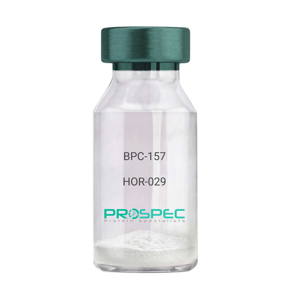
BPC-157Description:
BPC-157 Pentadecapeptide
Product # :
HOR-029Price :
Quantity :
Shipping Method :
Shipped at Room temp
More Info
- description
- formulation
- purity
- More Info
- Citations
Description
BPC-157 Synthetic is a single, non-glycosylated polypeptide chain containing 15 amino acids, having a molecular mass of 1419.55 Dalton and a Molecular formula of C62H98N16O22.
Formulation
The protein was lyophilized with no additives.
Purity
Greater than 97.0% as determined by analysis by RP-HPLC.
Citations
More Info
-
Physical Appearance
Sterile Filtered White lyophilized (freeze-dried) powder.
-
Stability
Lyophilized BPC-157 although stable at room temperature for 3 weeks, should be stored desiccated below -18°C. Upon reconstitution bpc-157 should be stored at 4°C between 2-7 days and for future use below -18°C. For long term storage it is recommended to add a carrier protein (0.1% HSA or BSA). Please prevent freeze-thaw cycles.
-
Solubility
It is recommended to reconstitute the lyophilized BPC-157 in sterile 18MΩ-cm H2O not less than 100 µg/ml, which can then be further diluted to other aqueous solutions.
-
Amino Acid Sequence
H-Gly-Glu-Pro-Pro-Pro-Gly-Lys-Pro-Ala-Asp-Asp-Ala-Gly-Leu-Val-OH
-
Background
BPC-157, short for Body Protection Compound-157, is a synthetic peptide that has garnered significant attention in the field of regenerative medicine and sports science. This peptide, derived from a portion of the human gastric juice protein known as BPC, exhibits remarkable healing and tissue regeneration properties. BPC-157 has shown promise in various preclinical and clinical studies, demonstrating its potential for the treatment of a wide range of injuries and disorders.
The research on BPC-157 encompasses investigations into its mechanisms of action, efficacy, safety, and potential therapeutic applications. Studies have elucidated the peptide's ability to enhance angiogenesis, promote collagen synthesis, modulate inflammatory responses, and protect against oxidative stress. These properties make BPC-157 an intriguing candidate for accelerating tissue healing, reducing inflammation, and improving overall recovery outcomes.
Preclinical studies have revealed the beneficial effects of BPC-157 in several injury models. For instance, BPC-157 has demonstrated its potential in accelerating tendon and ligament healing, mitigating muscle damage, and promoting bone regeneration. These findings suggest that BPC-157 could be a valuable therapeutic tool in orthopedic medicine and sports-related injuries.
Furthermore, BPC-157 has exhibited promising effects on gastrointestinal health. Studies have highlighted its ability to protect and heal the gut lining, reduce ulcer formation, and alleviate symptoms associated with inflammatory bowel disease. These observations open up avenues for BPC-157 as a potential treatment for gastrointestinal disorders.
In addition to its regenerative properties, BPC-157 has shown potential in neurological and psychiatric conditions. Research has indicated its neuroprotective effects, with implications for the treatment of traumatic brain injury, stroke, and neurodegenerative disorders. Preliminary studies also suggest BPC-157's potential as an antidepressant and anxiolytic agent.
Despite the promising findings, further research is needed to fully understand the mechanisms underlying BPC-157's actions and to assess its long-term safety and efficacy. Clinical trials are underway to explore its potential therapeutic applications in humans, including its use in tendon and ligament repair, inflammatory bowel disease, and neurodegenerative disorders.
This comprehensive review aims to summarize the current state of research on BPC-157, providing an overview of its mechanisms of action and therapeutic potential. By examining relevant studies and findings, we aim to shed light on the diverse applications of BPC-157 and its implications for regenerative medicine, sports science, and various disease
ProSpec's products are furnished for LABORATORY RESEARCH USE ONLY. The product may not be used as drugs, agricultural or pesticidal products, food additives or household chemicals.
- View Data Sheet
Name :
SFRP2 MouseDescription:
Secreted Frizzled-Related Protein 2 Mouse Recombinant
Secreted frizzled-related protein 2, sFRP-2, sFRP2, Sfrp2, Protein SDF5, Sdf5, AI851596, sdf, SDF-5, FRP2secreted apoptosis related protein 1, Secreted apoptosis-related protein 1, SARP-1, Sarp1, SARP1.
Product # :
pro-2669Price :
Quantity :
Shipping Method :
Shipped with Ice Packs
More Info
- description
- source
- formulation
- purity
- More Info
- Citations
Description
SFRP2 Mouse Recombinant produced in Sf9 Baculovirus cells is a single, glycosylated polypeptide chain containing 280 amino acids (25-295 a.a) and having a molecular mass of 32.1kDa. SFRP2 is fused to a 6 amino acid His-tag at C-terminus & purified by proprietary chromatographic techniques.
Source
Sf9, Baculovirus cells.
Formulation
The SFRP2 solution (0.25mg/ml) contains 20mM Tris-HCl buffer (pH 8.0), 20% glycerol and 100mM NaCl.
Purity
Greater than 90.0% as determined by SDS-PAGE.
Citations
More Info
-
Introduction
Secreted frizzled-related protein 2 (SFRP2) belongs to the SFRP family which contains a cysteine-rich domain homologous to the putative Wnt-binding site of Frizzled proteins. SFRP2 functions as soluble modulators of Wnt signaling. Methylation of SFRP2 might indicate on colorectal cancer.
-
Synonyms
Secreted frizzled-related protein 2, sFRP-2, sFRP2, Sfrp2, Protein SDF5, Sdf5, AI851596, sdf, SDF-5, FRP2secreted apoptosis related protein 1, Secreted apoptosis-related protein 1, SARP-1, Sarp1, SARP1.
-
Physical Appearance
Sterile Filtered colorless solution.
-
Stability
Store at 4°C if entire vial will be used within 2-4 weeks. Store, frozen at -20°C for longer periods of time. For long term storage it is recommended to add a carrier protein (0.1% HSA or BSA).Avoid multiple freeze-thaw cycles.
-
Amino Acid Sequence
ADPLFLFGQP DFSYKRSNCK PIPANLQLCH GIEYQNMRLP NLLGHETMKE VLEQAGAWIP LVMKQCHPDT KKFLCSLFAP VCLDDLDETI QPCHSLCVQV KDRCAPVMSA FGFPWPDMLE CDRFPQDNDL CIPLASSDHL LPATEEAPKV CEACKTKNED DNDIMETLCK NDFALKIKVK EITYINRDTK IILETKSKTI YKLNGVSERD LKKSVLWLKD SLQCTCEEMN DINAPYLVMG QKQGGELVIT SVKRWQKGQR EFKRISRSIR KLQCHHHHHH
ProSpec's products are furnished for LABORATORY RESEARCH USE ONLY. The product may not be used as drugs, agricultural or pesticidal products, food additives or household chemicals.
- View Data Sheet
Name :
RetatrutideDescription:
Retatrutide
Product # :
HOR-058Price :
Quantity :
Shipping Method :
Shipped at Room temp
More Info
- description
- formulation
- purity
- More Info
- Citations
Description
Retatrutide is a synthetic single, non-glycosylated polypeptide chain containing 39 amino acids, having a molecular mass of 4731 Dalton and a Molecular formula of C221H342N46O68.
Formulation
The protein was lyophilized with no additives.
Purity
Greater than 97.0% as determined by analysis by RP-HPLC.
Citations
More Info
-
Physical Appearance
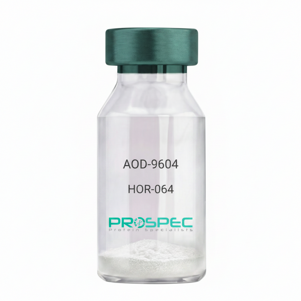
Sterile Filtered White lyophilized (freeze-dried) powder.
-
Stability
Lyophilized Retatrutide although stable at room temperature for 3 weeks, should be stored desiccated below -18°C. Upon reconstitution Retatrutide should be stored at 4°C between 2-7 days and for future use below -18°C.
For long term storage it is recommended to add a carrier protein (0.1% HSA or BSA).
Please prevent freeze-thaw cycles.
-
Solubility
It is recommended to reconstitute the lyophilized Retatrutide in sterile 18MΩ-cm H2O not less than 100 µg/ml, which can then be further diluted to other aqueous solutions.
-
Amino Acid Sequence
H-Tyr-Aib-Gln-Gly-Thr-Phe-Thr-SerAsp-Tyr-Ser-Ile-α-Me-Leu-Leu-Asp-Lys-{diacid-C20-gamma-Glu- (AEEA)-Lys}-Ala-Gln-Aib-Ala-Phe-Ile-Glu-Tyr-Leu-Leu-Glu-Gly-GlyPro-Ser-Ser-Gly-Ala-Pro-Pro-Pro-Ser-NH2.
-
Background
Retatrutide is atriple agonist targeting the GIP, GLP-1, and glucagon receptors which offers a distinct metabolic profile that goes beyond previous dual agonists like Tirzepatide. Retatrutide takes a very important part in metabolic pharmacotherapy, achieving high weight loss of nearly 30% in Phase 3 trials. Retatrutide’s triple-receptor agonism—mainly the inclusion of the glucagon receptor—provides a superior metabolic advantage in clearing liver fat and increasing thermogenesis compared to dual agonists.
ProSpec's products are furnished for LABORATORY RESEARCH USE ONLY. The product may not be used as drugs, agricultural or pesticidal products, food additives or household chemicals.
- View Data Sheet
Name :
FABP1 AntibodyDescription:
Fatty Acid Binding Protein-1, Mouse Anti Human
Fatty acid-binding protein 1 liver, L-FABP, FABPL, FABP-1, FABP1, Z-protein.
Product # :
ANT-346Price :
Quantity :
Shipping Method :
Shipped with Ice Packs
More Info
- formulation
- More Info
- Citations
Formulation
1mg/ml containing PBS, pH-7.4, 10% Glycerol and 0.02% Sodium Azide.
Citations
More Info
-
Introduction
FABP1 (Fatty acid binding protein1) encodes the fatty acid binding protein found in liver. FABP1 is composed of ten antiparallel β strands that form a barrel with a bigger binding pocket than the other FABPs allowing it to accommodate two fatty acids. This protein binds free fatty acids and their coenzyme A derivatives, bilirubin, and some other small molecules in the cytoplasm; it may be involved in intracellular lipid transport and metabolism.
-
Synonyms
Fatty acid-binding protein 1 liver, L-FABP, FABPL, FABP-1, FABP1, Z-protein.
-
Immunogen
Anti-human FABP1 mAb is derived from hybridization of mouse F0 myeloma cells with spleen cells from BALB/c mice immunized with recombinant human FABP1 amino acids 1-127 purified from E. coli.
-
Ig Subclass
Mouse IgG1 heavy chain and k light chain.
-
Clone
P2G4AT.
-
Applications
FABP1 antibody has been tested by ELISA, Western blot and immunohistochemistry analysis to assure specificity and reactivity. Since application varies, however, each investigation should be titrated by the reagent to obtain optimal results. Recommended dilution range for Western blot is 1:1,000 ~ 1:2,000 and immunohistochemistry analysis is 1:100~300. Recommended starting dilution for Western blot is 1:1,000 and Immunohistochemistry is 1:100.
-
Type
Mouse Anti Human Monoclonal.
-
Storage Procedures
For periods up to 1 month store at 4°C, for longer periods of time, store at -20°C. Prevent freeze thaw cycles.
-
Purification Method
FABP1 antibody was purified from mouse ascitic fluids by protein-G affinity chromatography.
ProSpec's products are furnished for LABORATORY RESEARCH USE ONLY. The product may not be used as drugs, agricultural or pesticidal products, food additives or household chemicals.
- View Data Sheet
Name :
FUBP1 AntibodyDescription:
Far Upstream Element Binding Protein, Mouse Anti Human
Far upstream element (FUSE) binding protein 1, FUSE-binding protein 1, DNA helicase V, FUBP, FBP, hDH V.
Product # :
ANT-589Price :
Quantity :
Shipping Method :
Shipped with Ice Packs
More Info
- formulation
- More Info
- Citations
Formulation
1mg/ml containing PBS, pH-7.4, 10% Glycerol and 0.02% Sodium Azide.
Citations
More Info
-
Introduction
FUBP1 is a ssDNA binding protein which stimulates expression of c-myc in undifferentiated cells and activates the far upstream element (FUSE) of c-myc. Regulation of FUSE by FUBP happens by single-strand binding of FUBP to the non-coding strand. FUBP1 performs as an ATP-dependent DNA helicase.
-
Synonyms
Far upstream element (FUSE) binding protein 1, FUSE-binding protein 1, DNA helicase V, FUBP, FBP, hDH V.
-
Physical Appearance
Sterile filtered colorless solution.
-
Immunogen
Anti-human FUBP1 mAb, is derived from hybridization of mouse F0 myeloma cells with spleen cells from BALB/c mice immunized with a recombinant human FUBP1 protein 279-448 amino acids purified from E. coli.
-
Ig Subclass
Mouse IgG2b heavy chain and k light chain.
-
Clone
PAT14F5AT.
-
Applications
FUBP1 antibody has been tested by ELISA, Western blot and ICC/IF analysis to assure specificity and reactivity. Since application varies, however, each investigation should be titrated by the reagent to obtain optimal results. Recommended starting dilution for Western blot analysis is 1:1000.
-
Type
Mouse Anti Human Monoclonal.
-
Storage Procedures
For periods up to 1 month store at 4°C, for longer periods of time, store at -20°C. Prevent freeze thaw cycles.
-
Purification Method
FUBP1 antibody was purified from mouse ascitic fluids by protein-A affinity chromatography.
ProSpec's products are furnished for LABORATORY RESEARCH USE ONLY. The product may not be used as drugs, agricultural or pesticidal products, food additives or household chemicals.
- View Data Sheet
Name :
SDHAF1 HumanDescription:
Succinate Dehydrogenase Complex Assembly Factor 1 Human Recombinant
Succinate Dehydrogenase Complex Assembly Factor 1, LYR Motif-Containing Protein 8, LYR Motif Containing 8, SDH Assembly Factor 1, LYRM8, Succinate Dehydrogenase Assembly Factor 1, Mitochondrial, Succinate dehydrogenase assembly factor 1, mitochondrial.
Product # :
PRO-2123Price :
Quantity :
Shipping Method :
Shipped with Ice Packs
More Info
- description
- source
- formulation
- purity
- More Info
- Citations
Description
SDHAF1 Human Recombinant produced in E.Coli is a single, non-glycosylated polypeptide chain containing 138 amino acids (1-115 a.a) and having a molecular mass of 15.2kDa. SDHAF1 is fused to a 23 amino acid His-tag at N-terminus & purified by proprietary chromatographic techniques.
Source
Escherichia Coli.
Formulation
SDHAF1 protein solution (0.25mg/ml) containing Phosphate buffered saline (pH7.4), 30% glycerol, 2mM DTT and 0.1mM PMSF.
Purity
Greater than 85.0% as determined by SDS-PAGE.
Citations
More Info
-
Introduction
Succinate dehydrogenase assembly factor 1 (SDHAF1) is a member of the complex I LYR family. SDHAF1 plays an important role in succinate dehydrogenase complex (SDH) assembly, a complex which is involved in complex II of the mitochondrial electron transport chain. SDHAF1 functions through taking part in mitochondrial biosynthesis of iron-sulfur centers for complex II.
-
Synonyms
Succinate Dehydrogenase Complex Assembly Factor 1, LYR Motif-Containing Protein 8, LYR Motif Containing 8, SDH Assembly Factor 1, LYRM8, Succinate Dehydrogenase Assembly Factor 1, Mitochondrial, Succinate dehydrogenase assembly factor 1, mitochondrial.
-
Physical Appearance
Sterile Filtered colorless solution.
-
Stability
Store at 4°C if entire vial will be used within 2-4 weeks. Store, frozen at -20°C for longer periods of time. For long term storage it is recommended to add a carrier protein (0.1% HSA or BSA).Avoid multiple freeze-thaw cycles.
-
Amino Acid Sequence
MGSSHHHHHH SSGLVPRGSH MGSMSRHSRL QRQVLSLYRD LLRAGRGKPG AEARVRAEFR QHAGLPRSDV LRIEYLYRRG RRQLQLLRSG HATAMGAFVR PRAPTGEPGG VGSQPDDGDS PRNPHDSTGA PETRPDGR.
ProSpec's products are furnished for LABORATORY RESEARCH USE ONLY. The product may not be used as drugs, agricultural or pesticidal products, food additives or household chemicals.
- View Data Sheet
Name :
DPH2 HumanDescription:
Diphthamide Biosynthesis 2 Human Recombinant
DPH2 Homolog, S-Adenosyl-L-Methionine:L-Histidine 3-Amino-3-Carboxypropyltransferase 2, Diphtheria Toxin Resistance Protein 2, Diphthamide Biosynthesis Protein 2, DPH2L2, Diptheria Toxin Resistance Protein Required For Diphthamide Biosynthesis-Like 2, 2-(3-Amino-3-Carboxypropyl)Histidine Synthase Subunit 2, Diphthamide Biosynthesis Protein 2 Homolog-Like 2, Diphthamide Biosynthesis-Like Protein 2, EC 2.5.1.108, DPH2-Like 2, DPH2.
Product # :
PRO-2421Price :
Quantity :
Shipping Method :
Shipped with Ice Packs
More Info
- description
- source
- formulation
- purity
- More Info
- Citations
Description
DPH2 Human Recombinant produced in Sf9 Baculovirus cells is a single, glycosylated polypeptide chain containing 497 amino acids (1-489a.a.) and having a molecular mass of 53.1kDa (Molecular size on SDS-PAGE will appear at approximately 50-70kDa). DPH2 is expressed with an 8 amino acids His tag at C-Terminus and purified by proprietary chromatographic techniques.
Source
Sf9, Baculovirus cells.
Formulation
DPH2 protein solution (0.5mg/ml) contains Phosphate Buffered Saline (pH 7.4), 1mM DTT and 20% glycerol.
Purity
Greater than 85.0% as determined by SDS-PAGE.
Citations
More Info
-
Introduction
Diphthamide biosynthesis protein 2 (DPH2) is a homodimer and each of its monomers can bind a [4Fe-4S] cluster. DPH2 is the target of ADP ribosylating diphtheria toxin (DT) and Pseudomonas exotoxin A (PE). DPH2 was identified by its ability to complement a diphthamide mutant strain, and thus serves in diphthamide biosynthesis. The loss of DPH2 pre-activates NF-kB and death receptor pathways and renders MCF7 cells hypersensitive to tumor necrosis factor.
-
Synonyms
DPH2 Homolog, S-Adenosyl-L-Methionine:L-Histidine 3-Amino-3-Carboxypropyltransferase 2, Diphtheria Toxin Resistance Protein 2, Diphthamide Biosynthesis Protein 2, DPH2L2, Diptheria Toxin Resistance Protein Required For Diphthamide Biosynthesis-Like 2, 2-(3-Amino-3-Carboxypropyl)Histidine Synthase Subunit 2, Diphthamide Biosynthesis Protein 2 Homolog-Like 2, Diphthamide Biosynthesis-Like Protein 2, EC 2.5.1.108, DPH2-Like 2, DPH2.
-
Physical Appearance
Sterile filtered colorless solution.
-
Stability
Store at 4°C if entire vial will be used within 2-4 weeks. Store, frozen at -20°C for longer periods of time. For long term storage it is recommended to add a carrier protein (0.1% HSA or BSA).Avoid multiple freeze-thaw cycles.
-
Amino Acid Sequence
MESMFSSPAE AALQRETGVP GLLTPLPDLD GVYELERVAG FVRDLGCERV ALQFPDQLLG DAVAVAARLE ETTGSKMFIL GDTAYGSCCV DVLGAEQAGA QALIHFGPAC LSPPARPLPV AFVLRQRSVA LELCVKAFEA QNPDPKAPVV LLSEPACAHA LEALATLLRP RYLDLLVSSP AFPQPVGSLS PEPMPLERFG RRFPLAPGRR LEEYGAFYVG GSKASPDPDL DPDLSRLLLG WAPGQPFSSC CPDTGKTQDE GARAGRLRAR RRYLVERARD ARVVGLLAGT LGVAQHREAL AHLRNLTQAA GKRSYVLALG RPTPAKLANF PEVDVFVLLA CPLGALAPQL SGSFFQPILA PCELEAACNP AWPPPGLAPH LTHYADLLPG SPFHVALPPP ESELWETPDV SLITGDLRPP PAWKSSNDHG SLALTPRPQL ELAESSPAAS FLSSRSWQGL EPRLGQTPVT EAVSGRRGIA IAYEDEGSGL EHHHHHH.
ProSpec's products are furnished for LABORATORY RESEARCH USE ONLY. The product may not be used as drugs, agricultural or pesticidal products, food additives or household chemicals.
- View Data Sheet
Name :
CX3CL1 Human, Sf9Description:
Fractalkine (CX3CL1) Human Recombinant, Sf9
Fractalkine, CX3CL1, Neurotactin, CX3C membrane-anchored chemokine, Small inducible cytokine D1, NTN, NTT, CXC3, CXC3C, SCYD1, ABCD-3, C3Xkine.
Product # :
CHM-042Price :
Quantity :
Shipping Method :
Shipped with Ice Packs
More Info
- description
- source
- formulation
- purity
- More Info
- SDS-PAGE
- Citations
Description
Fractalkine Human Recombinant produced in Sf9 Baculovirus cells is a single, glycosylated polypeptide chain containing 323 amino acids (25-339aa) and having a molecular mass of 34.3kDa.Fractalkine is fused to a 8 amino acid His tag at C-terminus and purified by proprietary chromatographic techniques.
Source
Sf9, Baculovirus cells.
Formulation
The Fractalkine solution (1 mg/ml) contains 10% Glycerol and Phosphate Buffered Saline (pH 7.4).
Purity
Greater than 90.0% as determined by SDS-PAGE.
SDS-PAGE
Citations
More Info
-
Introduction
Fractalkine soluble form is chemotactic for t-cells and monocytes, but not for neutrophils. Fractalkine membrane-bound form promotes adhesion of those leukocytes to endothelial cells. Fractalkine regulates leukocyte adhesion and migration processes at the endothelium and binds to CX3CR1. Fractalkine gene is located on human chromosome 16 along with some CC chemokines known as CCL17 and CCL22.
-
Synonyms
Fractalkine, CX3CL1, Neurotactin, CX3C membrane-anchored chemokine, Small inducible cytokine D1, NTN, NTT, CXC3, CXC3C, SCYD1, ABCD-3, C3Xkine.
-
Physical Appearance
Sterile Filtered colorless solution.
-
Stability
Store at 4°C if entire vial will be used within 2-4 weeks. Store, frozen at -20°C for longer periods of time. For long term storage it is recommended to add a carrier protein (0.1% HSA or BSA).Avoid multiple freeze-thaw cycles.
-
Amino Acid Sequence
QHHGVTKCNI TCSKMTSKIP VALLIHYQQN QASCGKRAII LETRQHRLFC ADPKEQWVKD
AMQHLDRQAA ALTRNGGTFE KQIGEVKPRT TPAAGGMDES VVLEPEATGE SSSLEPTPSS
QEAQRALGTS PELPTGVTGS SGTRLPPTPK AQDGGPVGTE LFRVPPVSTA ATWQSSAPHQ
PGPSLWAEAK TSEAPSTQDP STQASTASSP APEENAPSEG QRVWGQGQSP RPENSLEREE
MGPVPAHTDA FQDWGPGSMA HVSVVPVSSE GTPSREPVAS GSWTPKAEEP IHATMDPQRL GVLITPVPDA QAATRLEHHH HHH -
Background
What is the molecular weight/Mw of CX3CL1 HUMAN, SF9 Protein?
CX3CL1 HUMAN, SF9 Protein has a total Mw of 34.3kDa.
What is the source or expression system of CX3CL1 HUMAN, SF9 Protein?
Sf9, Baculovirus cells.
What is the Purity of CX3CL1 HUMAN, SF9 Protein?
CX3CL1 HUMAN, SF9 Protein is >90% pure as determined by SDS-PAGE.
What is the Biological Activity of CX3CL1 HUMAN, SF9 Protein?
The biological functionality of CX3CL1 HUMAN, SF9 Protein will be determined in the future.
What is the amino acid sequence of CX3CL1 HUMAN, SF9 Protein?
QHHGVTKCNI TCSKMTSKIP VALLIHYQQN QASCGKRAII LETRQHRLFC ADPKEQWVKD
AMQHLDRQAA ALTRNGGTFE KQIGEVKPRT TPAAGGMDES VVLEPEATGE SSSLEPTPSS
QEAQRALGTS PELPTGVTGS SGTRLPPTPK AQDGGPVGTE LFRVPPVSTA ATWQSSAPHQ
PGPSLWAEAK TSEAPSTQDP STQASTASSP APEENAPSEG QRVWGQGQSP RPENSLEREE
MGPVPAHTDA FQDWGPGSMA HVSVVPVSSE GTPSREPVAS GSWTPKAEEP IHATMDPQRL GVLITPVPDA QAATRLEHHH HHH
What applications can CX3CL1 HUMAN, SF9 Protein be used in?
CX3CL1 HUMAN, SF9 Protein can probably be used in western blot, ELISA and Lateral Flow.
What is the endotoxin level for CX3CL1 HUMAN, SF9 Protein?
The endotoxin level is minimal, CX3CL1 HUMAN, SF9 Protein was purified using conventional chromatography techniques.
Who supplies recombinant CX3CL1 HUMAN, SF9 that is in stock and available to order?
ProSpec has the protein in stock and available for immediate delivery.
Where can I order bulk mg quantities of CX3CL1 HUMAN, SF9?
ProSpec supplies bulk mg quantities at affordable prices that meets research and academic budgets.
ProSpec's products are furnished for LABORATORY RESEARCH USE ONLY. The product may not be used as drugs, agricultural or pesticidal products, food additives or household chemicals.
- View Data Sheet
Name :
GFAP HumanDescription:
Glial Fibrillary Acidic Protein Human Recombinant
Glial fibrillary acidic protein, GFAP
Product # :
PRO-2802Price :
Quantity :
Shipping Method :
Shipped at Room temp
More Info
- description
- source
- formulation
- purity
- More Info
- Citations
Description
GFAP Human produced in E.coli is a single, non-glycosylated polypeptide chain (60-383 a.a.) and having a molecular mass of 37906 Dalton.
Source
Escherichia Coli.
Formulation
GFAP was lyophilized from 50mM Tris-HCl pH-7.5, 4M Urea 150mM NaCl.
Purity
Greater than 90.0% as determined by SDS-PAGE.
Citations
More Info
-
Synonyms
Glial fibrillary acidic protein, GFAP
-
Physical Appearance
Sterile Filtered White lyophilized (freeze-dried) powder.
-
Stability
Lyophilized GFAP although stable at room temperature for 3 weeks, should be stored desiccated below -18°C. Upon reconstitution Glial Fibrillary Acidic Protein should be stored at 4°C between 2-7 days and for future use below -18°C. For long term storage it is recommended to add a carrier protein (0.1% HSA or BSA). Please prevent freeze-thaw cycles.
-
Solubility
It is recommended to reconstitute the lyophilized GFAP in sterile 18MΩ-cm H2O not less than 100µg/ml, which can then be further diluted to other aqueous solutions.
-
Background
Glial Fibrillary Acidic Protein (GFAP), a key intermediate filament protein predominantly found in astrocytes, plays a fundamental role in the central nervous system. Initially recognized for its structural functions, GFAP has emerged as a multifaceted molecule with implications in neural development, synaptic plasticity, and various neurological disorders. This research delves into the realm of GFAP human recombinant protein, shedding light on its structural properties, physiological significance, and its diverse roles in both health and disease.
Structural Complexity of GFAP:
GFAP belongs to the family of intermediate filament proteins, conferring structural support to astrocytes. Its unique structure comprises a central α-helical rod domain flanked by non-helical head and tail domains. This structural complexity allows GFAP to form stable filaments, providing structural integrity to astrocytes and contributing to the architecture of the central nervous system.
Physiological Functions in Glial Cells:
Beyond its structural role, GFAP participates in various physiological processes within glial cells. It is involved in the regulation of astrocyte morphology, motility, and migration, crucial for their interactions with neurons and blood vessels. Additionally, GFAP contributes to the formation and maintenance of the blood-brain barrier, highlighting its significance in the brain's homeostasis.
Implications in Neurological Disorders:
Aberrant GFAP expression and aggregation are associated with several neurological disorders. In Alexander disease, a rare neurodegenerative disorder, mutations in the GFAP gene lead to the formation of GFAP aggregates, contributing to disease pathology. Moreover, elevated levels of GFAP in cerebrospinal fluid serve as a biomarker for various neurological conditions, including traumatic brain injury, Alzheimer's disease, and multiple sclerosis, indicating its involvement in the brain's response to injury and neuroinflammation.
GFAP in Neural Regeneration:
Recent studies have unveiled GFAP’s role in neural regeneration and repair processes. In response to brain injury, GFAP-expressing astrocytes become reactive, forming a glial scar that isolates damaged areas. While this scar formation initially limits tissue damage, persistent scar formation can impede neural regeneration. Understanding the dynamics of GFAP expression in reactive astrocytes is crucial for developing therapies that promote neural regeneration following brain injuries or neurodegenerative diseases.
GFAP human recombinant protein, once thought of as a structural element in astrocytes, has proven to be a pivotal player in the complex landscape of glial biology and neurological disorders. Its intricate functions extend beyond providing structural support, encompassing roles in neural development, disease pathology, and tissue repair. As research continues to uncover the nuances of GFAP’s involvement in health and disease, it offers promising avenues for developing targeted therapies and diagnostic tools, emphasizing its significance in the intricate workings of the central nervous system.
ProSpec's products are furnished for LABORATORY RESEARCH USE ONLY. The product may not be used as drugs, agricultural or pesticidal products, food additives or household chemicals.