Search results
1000 results found for “Coagulation Factors”
Name
Description
Product #
Price
Quantity
Shipping Method
- View Data Sheet
Name :
TGFB1 RatDescription:
Transforming Growth Factor-Beta 1 Rat Recombinant
Transforming growth factor beta-1, TGF-beta-1, Tgfb, Tgfb-1, TGFbeta1.
Product # :
CYT-1265Price :
Quantity :
Shipping Method :
Shipped at Room temp
More Info
- description
- source
- formulation
- purity
- biological activity
- More Info
Description
Transforming Growth Factor-Beta 1 Rat Recombinant produced in CHO is a homodimer, polypeptide chain containing 2 x 112 amino acids and having a total molecular mass of 25.6kDa.
TGFB1 Rat Recombinant is purified by proprietary chromatographic techniques.
Source
CHO Cells.
Formulation
The protein was lyophilized with 0.1% (v/v) TFA and 35% (v/v) Acetonitrile.
Purity
Greater than 97.0% as determined by SDS-PAGE and SEC-HPLC analyses.
Biological Activity
The biological activity was determined by TGFB1 ability to inhibit the mouse IL-4-dependent proliferation of mouse HT-2 cells. The expected ED50 for this effect is <0.05ng/ml, corresponding to a specific activity of ≥ 2.0 × 10⁷ units/mg.More Info
-
Synonyms
Transforming growth factor beta-1, TGF-beta-1, Tgfb, Tgfb-1, TGFbeta1.
-
Physical Appearance
Sterile Filtered White lyophilized (freeze-dried) powder.
-
Stability
Lyophilized TGFB1 although stable at room temperature for 3 weeks, should be stored desiccated below -18°C. Upon reconstitution Transforming Growth Factor-Beta 1 should be stored at 4°C between 2-7 days and for future use below -18°C.
For long term storage it is recommended to add a carrier protein (0.1% HSA or BSA).
Please prevent freeze-thaw cycles.
-
Solubility
It is recommended to reconstitute the lyophilized Transforming Growth Factor-Beta 1 in sterile 4mM HCl not less than 100µg/ml, which can then be further diluted to other aqueous solutions.
-
Amino Acid Sequence
MGSSHHHHHH SSGLVPRGSH MGSALDTNYC FSSTEKNCCV RQLYIDFRKD LGWKWIHEPK GYHANFCLGP CPYIWSLDTQ YSKVLALYNQ HNPGASASPC CVPQALEPLP IVYYVGRKPK VEQLSNMIVR SCKCS.
-
Background
Mouse TGF-β1 as an inducer of EMT [epithelial–mesenchymal transition ] therefore used in in fibrosis, wound healing, cancer invasion, and metastasis. Mouse TGF-β1 decreases E-cadherin expression and increases N-cadherin, vimentin and fibronectin.
TGF-β1 is produced by T regulatory cells (Tregs), Macrophages and monocytes, Platelets, Fibroblasts, Epithelial cells, Endothelial cells, Smooth muscle cells, Tumor cells, Activated immune cells
What is the source or expression system of Mouse TGFB1 Protein?
CHO Cells
What is the Purity of Mouse TGFB1 Protein?
Mouse TGFB1 Protein is >97% pure as determined by SDS-PAGE and SEC-HPLC analyses.
What is the molecular weight of Mouse TGFB1 Protein?
Mouse TGFB1 Protein having a total Mw of 25.6kDa.
What is the Biological Activity of Mouse TGFB1 Protein?
The biological functionality of Mouse TGFB1 Protein is determined by mouse HT-2 cells.
What is the endotoxin level for Mouse TGFB1 Protein?
The endotoxin level is minimal, Mouse TGFB1 Protein was purified using conventional chromatography techniques.
What is the amino acid sequence of Mouse TGFB1 Protein?
ALDTNYCFSS TEKNCCVRQL YIDFRKDLGW KWIHEPKGYH ANFCLGPCPY IWSLDTQYSK VLALYNQHNP GASASPCCVP QALEPLPIVY YVGRKPKVEQ LSNMIVRSCK CS.
Is TGFB1 a homodimer / homodimeric protein?
Yes, TGFB1 is homo dimer consisting of 2 identical chains.
ProSpec's products are furnished for LABORATORY RESEARCH USE ONLY. The product may not be used as drugs, agricultural or pesticidal products, food additives or household chemicals.
- View Data Sheet
Name :
TGFBI Human, 182 a.a.Description:
Transforming Growth Factor Beta-Induced (182 a.a.) Human Recombinant
Transforming growth factor-beta-induced protein ig-h3, Beta ig-h3, Kerato-epithelin, RGD-containing collagen-associated protein, RGD-CAP, TGFBI, BIGH3, CSD, CDB1, CDG2, CSD1, CSD2, CSD3, EBMD, LCD1, CDGG1.
Product # :
PRO-672Price :
Quantity :
Shipping Method :
Shipped with Ice Packs
More Info
- description
- source
- formulation
- purity
- More Info
Description
TGFBI Recombinant Human produced in e.Coli is a single, non-glycosylated, polypeptide containing 182 amino acids (502-683) and having a molecular mass of 19.9 kDa (Molecular weight on SDS-PAGE will appear higher). The TGFBI recombinant Human protein is purified by proprietary chromatographic techniques.
Source
Escherichia Coli.
Formulation
The TGFBI 182 a.a. recombinant Human is formulated in 20mM Tris-HCl pH-8, 1mM EDTA, 0.1mM PMSF and 20% glycerol.
Purity
Greater than 95.0% as determined by SDS-PAGE.
More Info
-
Introduction
TGFBI is an extracellular matrix protein induced by transforming growth factor (TGF)-beta 1. TGFBI protein is involved in cell growth, cell differentiation, wound healing and cell adhesion. In addition, some missense mutations of TGFBI were identified in families affected with human autosomal dominant corneal dystrophies. TGFBI gene encodes for a 683 amino-acid protein containing an RGD motif and four internal repeated domains which have highly conserved sequences founded in several species (Fasciclin domain).
-
Synonyms
Transforming growth factor-beta-induced protein ig-h3, Beta ig-h3, Kerato-epithelin, RGD-containing collagen-associated protein, RGD-CAP, TGFBI, BIGH3, CSD, CDB1, CDG2, CSD1, CSD2, CSD3, EBMD, LCD1, CDGG1.
-
Physical Appearance
Sterile filtered liquid formulation.
-
Stability
Store at 4°C if entire vial will be used within 2-4 weeks. Store, frozen at -20°C for longer periods of time. For long term storage it is recommended to add a carrier protein (0.1% HSA or BSA).Avoid multiple freeze-thaw cycles.
-
Amino Acid Sequence
MGTVMDVLKG DNRFSMLVAA IQSAGLTETL NREGVYTVFA PTNEAFRALP PRERSRLLGD AKELANILKY HIGDEILVSG GIGALVRLKS LQGDKLEVSL KNNVVSVNKE PVAEPDIMAT NGVVHVITNV LQPPANRPQE RGDELADSAL EIFKQASAFS RASQRSVRLA PVYQKLLERM KH.
ProSpec's products are furnished for LABORATORY RESEARCH USE ONLY. The product may not be used as drugs, agricultural or pesticidal products, food additives or household chemicals.
- View Data Sheet
Name :
POU2AF1 HumanDescription:
POU class 2 associating factor 1 Human Recombinant
POU domain class 2 associating factor 1, BOB-1, OCA-B, B-cell-specific coactivator OBF-1, OCT-binding factor 1.
Product # :
PRO-880Price :
Quantity :
Shipping Method :
Shipped with Ice Packs
More Info
- description
- source
- formulation
- purity
- More Info
Description
POU2AF1 Human Recombinant produced in E.coli is a single, non-glycosylated polypeptide chain containing 276 amino acids (1-256) and having a molecular mass of 29.6 kDa.The POU2AF1 is fused to a 20 amino acid His-Tag at N-terminus and purified by proprietary chromatographic techniques.
Source
Escherichia Coli.
Formulation
POU2AF1 protein (0.5mg/ml) is supplied in 20mM Tris-HCL, pH-8 and 20% Glycerol.
Purity
Greater than 85.0% as determined by SDS-PAGE.
More Info
-
Introduction
POU2AF1 is a lymphocyte specific transcription coactivator protein. POU2AF1 cooperates only with the Oct1/2 proteins using sub domains in the POU domain of the Oct1/2 proteins, increasing their transcriptional efficiency. Even though POU2AF1 have no basic ability to bind DNA, it links firmly with the octomer motif in the presence of Oct1 and Oct2. POU2AF1 is expressed at maximum levels in spleen and peripheral blood leukocytes.
-
Synonyms
POU domain class 2 associating factor 1, BOB-1, OCA-B, B-cell-specific coactivator OBF-1, OCT-binding factor 1.
-
Physical Appearance
POU2AF1 is supplied as a sterile filtered clear solution.
-
Stability
Store at 4°C if entire vial will be used within 2-4 weeks. Store, frozen at -20°C for longer periods of time. For long term storage it is recommended to add a carrier protein (0.1% HSA or BSA).Avoid multiple freeze-thaw cycles.
-
Amino Acid Sequence
MGSSHHHHHH SSGLVPRGSH MLWQKPTAPE QAPAPARPYQ GVRVKEPVKE LLRRKRGHAS SGAAPAPTAV VLPHQPLATY TTVGPSCLDM EGSVSAVTEE AALCAGWLSQ PTPATLQPLA PWTPYTEYVP HEAVSCPYSA DMYVQPVCPS YTVVGPSSVL TYASPPLITN VTTRSSATPA VGPPLEGPEH QAPLTYFPWP QPLSTLPTST LQYQPPAPAL PGPQFVQLPI SIPEPVLQDM EDPRRAASSL TIDKLLLEEE DSDAYALNHT LSVEGF
ProSpec's products are furnished for LABORATORY RESEARCH USE ONLY. The product may not be used as drugs, agricultural or pesticidal products, food additives or household chemicals.
- View Data Sheet
Name :
LTF Holo HumanDescription:
Lactoferrin Holo Human Recombinant
Lactotransferrin, Lactoferrin, Growth-inhibiting protein 12, Talalactoferrin, LTF, GIG12, LF, HLF2, Neutrophil Lactoferrin.
Product # :
PRO-592Price :
Quantity :
Shipping Method :
Shipped at Room temp
More Info
- description
- source
- formulation
- purity
- More Info
Description
Recombinant Human Holo Lactoferrin produced in Plant is a glycosylated mature polypeptide sequence having an approximate molecular mass of 80 kDa.The Human Holo Lactoferrin is purified by proprietary chromatographic techniques.
Source
Rice Flour.
Formulation
The Human Holo lactoferrin was lyophilized with no additives.
Purity
Greater than 95% as determined by SDS-PAGE.
More Info
-
Introduction
Lactoferrin is a glycoprotein that belongs to the transferrin family of iron binding proteins. It is found in human breast milk as well as most epithelial surface secretions including tears, nasogastric, saliva, and bronchial. Lactoferrin binds 2 molecules of iron with very high affinity. Lactoferrin inhibits bacterial growth by withholding iron, its N-terminal region is an antimicrobial peptide. Lactotransferrin acts synergistically with lysozyme to potentiate the activity of both proteins. The multifunctional protein lactoferrin has many physiological possible roles. It is often referred to as an innate defense protein and frequently serves as the first line of defense in protection against pathogens. It has been shown to have the ability to bind iron, it is a natural anti-bacterial, anti-fungal and anti-viral, it is an antioxidant and it also has immunomodulatory properties. It has many beneficial properties, which make it a good candidate for a number of product applications. Considerable research is currently going on to explain the various suggested biological functions of lactoferrin.
-
Synonyms
Lactotransferrin, Lactoferrin, Growth-inhibiting protein 12, Talalactoferrin, LTF, GIG12, LF, HLF2, Neutrophil Lactoferrin.
-
Physical Appearance
Pink lyophilized powder.
-
Stability
Recombinant Holo Lactoferrin although stable at room temperature for 5 days, should be stored desiccated below -18°C.Please prevent freeze-thaw cycles.
-
Solubility
It is recommended to reconstitute the lyophilized LTF Holo Human in sterile water at 10mg/ml, which can then be further diluted to other aqueous solutions.
ProSpec's products are furnished for LABORATORY RESEARCH USE ONLY. The product may not be used as drugs, agricultural or pesticidal products, food additives or household chemicals.
- View Data Sheet
Name :
KGF Human, PlantDescription:
Keratinocyte Growth Factor Human Recombinant, Plant
HBGF-7, FGF7, FGF-7, KGF.
Product # :
CYT-1212Price :
Quantity :
Shipping Method :
Shipped at Room temp
More Info
- description
- source
- formulation
- purity
- biological activity
- More Info
Description
KGF Human Recombinant produced in Oryza Sativa is a single, polypeptide chain having a molecular mass of approximately 19.2kDa. The KGF is purified by proprietary chromatographic techniques.
Source
Rice Grain
Formulation
Lyophilized KGF from a 0.2μm filtered solution containing no stabilizers.
Purity
Greater than 95% as determined by SDS-PAGE.
Biological Activity
KGF has full biological activity when compared to standards. The activity is determined by the dose-dependent proliferation of HUVECs and is typically 10ng/ml corresponding to a specific activity of 100,000U/mg.
More Info
-
Introduction
KGF is a member of the fibroblast growth factor (FGF) family. FGF family members possess broad mitogenic and cell survival activities, and are involved in a variety of biological processes, including embryonic development, cell growth, morphogenesis, tissue repair, tumor growth and invasion. FGF7 is a potent epithelial cell-specific growth factor, whose mitogenic activity is predominantly exhibited in keratinocytes but not in fibroblasts and endothelial cells. Studies of mouse and rat homologs of this gene implicated roles in morphogenesis of epithelium, reepithelialization of wounds, hair development and early lung organogenesis.
-
Synonyms
HBGF-7, FGF7, FGF-7, KGF.
-
Physical Appearance
Sterile Filtered White lyophilized (freeze-dried) powder.
-
Stability
Lyophilized KGF although stable at room temperature for 3 weeks, should be stored desiccated below -18°C. Upon reconstitution FGF7 should be stored at 4°C between 2-7 days and for future use below -18°C.For long term storage it is recommended to add a carrier protein (0.1% HSA or BSA).Please prevent freeze-thaw cycles.
-
Solubility
It is recommended to reconstitute the lyophilized KGF in sterile 18MΩ-cm at 100µg/ml, which can then be further diluted to other aqueous solutions.
ProSpec's products are furnished for LABORATORY RESEARCH USE ONLY. The product may not be used as drugs, agricultural or pesticidal products, food additives or household chemicals.
- View Data Sheet
Name :
LZTFL1 HumanDescription:
Leucine Zipper Transcription Factor-Like 1 Human Recombinant
Leucine Zipper Transcription Factor-Like 1, BBS17, Leucine Zipper Transcription Factor-Like Protein 1, Leucine zipper transcription factor-like protein 1.
Product # :
PRO-2157Price :
Quantity :
Shipping Method :
Shipped with Ice Packs
More Info
- description
- source
- formulation
- purity
- More Info
Description
LZTFL1 Human Recombinant produced in E.Coli is a single, non-glycosylated polypeptide chain containing 322 amino acids (1-299 a.a) and having a molecular mass of 37.0kDa. LZTFL1 is fused to a 23 amino acid His-tag at N-terminus & purified by proprietary chromatographic techniques.
Source
Escherichia Coli.
Formulation
LZTFL1 protein solution (1mg/ml) containing PBS buffer (pH 7.4), 10% glycerol and 1mM DTT.
Purity
Greater than 90% as determined by SDS-PAGE.
More Info
-
Introduction
Leucine Zipper Transcription Factor-Like 1, also known as LZTFL1 is a ubiquitously expressed protein which localizes to the cytoplasm. LZTFL1 interacts with Bardet-Biedl Syndrome proteins (BBS) and during its interaction with BBS protein complexes, regulates protein trafficking to the ciliary membrane. Nonsense mutations in LZTFL1 cause a form of Bardet-Biedl Syndrome; a ciliopathy characterized partly by polydactyly, obesity, cognitive impairment, hypogonadism, and kidney failure. LZTFL1 also acts as a tumor suppressor; possibly by interacting with E-cadherin as well as the actin cytoskeleton and in this manner regulating the transition of epithelial cells to mesenchymal cells.
-
Synonyms
Leucine Zipper Transcription Factor-Like 1, BBS17, Leucine Zipper Transcription Factor-Like Protein 1, Leucine zipper transcription factor-like protein 1.
-
Physical Appearance
Sterile filtered colorless solution.
-
Stability
Store at 4°C if entire vial will be used within 2-4 weeks. Store, frozen at -20°C for longer periods of time. For long term storage it is recommended to add a carrier protein (0.1% HSA or BSA).Avoid multiple freeze-thaw cycles.
-
Amino Acid Sequence
MGSSHHHHHH SSGLVPRGSH MGSMAELGLN EHHQNEVINY MRFARSKRGL RLKTVDSCFQ DLKESRLVED TFTIDEVSEV LNGLQAVVHS EVESELINTA YTNVLLLRQL FAQAEKWYLK LQTDISELEN RELLEQVAEF EKAEITSSNK KPILDVTKPK LAPLNEGGTA ELLNKEILRL QEENEKLKSR LKTIEIQATN ALDEKSKLEK ALQDLQLDQG NQKDFIKAQD LSNLENTVAA LKSEFQKTLN DKTENQKSLE ENLATAKHDL LRVQEQLHMA EKELEKKFQQ TAAYRNMKEI LTKKNDQIKD LRKRLAQYEP ED.
ProSpec's products are furnished for LABORATORY RESEARCH USE ONLY. The product may not be used as drugs, agricultural or pesticidal products, food additives or household chemicals.
- View Data Sheet
Name :
FAS HumanDescription:
sFas Receptor Human Recombinant
Tumor necrosis factor receptor superfamily member 6, Apo-1 antigen, Apoptosis-mediating surface antigen FAS, FASLG receptor, CD95, FAS, APT1, FAS1, APO-1, FASTM, ALPS1A, TNFRSF6.
Product # :
CYT-125Price :
Quantity :
Shipping Method :
Shipped at Room temp
More Info
- description
- source
- formulation
- purity
- biological activity
- More Info
Description
sFas Receptor Human Recombinant produced in E.coli is a single, non-glycosylated polypeptide chain containing 157 amino acids and having a molecular mass of 17.6kDa.The FAS is purified by proprietary chromatographic techniques.
Source
Escherichia Coli.
Formulation
FAS protein was lyophilized from a 0.2µm filtered concentrated solution in 1×PBS, pH 7.4.
Purity
Greater than 95.0% as determined by:
(a) Analysis by RP-HPLC.
(b) Analysis by SDS-PAGE.Biological Activity
The ED50 was determined by its ability to inhibit the cytotoxicity of Jurkat cells is between 10-15 µg/ml in the presence of 2ng/ml of hFasL.More Info
-
Introduction
Fas and Fas Ligand (FasL) are members of the TNF superfamily and are type I and type II transmembrane proteins, respectively. Binding of FasL to Fas initiates apoptosis in Fas-bearing cells. The apoptosis mechanism involves the recruitment of pro-caspase 8 through an adaptor molecule named FADD followed by processing of the pro-enzyme to active forms. These active caspases subsequently cleave a variety of cellular substrates leading to the eventual cell death. sFasR is able to inhibit FasL-induced apoptosis by acting as a decoy receptor whicht serves as a sink for FasL. The full length Fas Receptor is a 319 a.a type I transmembrane protein, which contains a 157 a.a extracellular domain, a 17 a.a transmembrane domain, and 145 a.a cytoplasmic domain. The mature human Fas ECD shares 55%, 58%, a.a sequence identity with the mouse, rat, Fas, respectively.
-
Synonyms
Tumor necrosis factor receptor superfamily member 6, Apo-1 antigen, Apoptosis-mediating surface antigen FAS, FASLG receptor, CD95, FAS, APT1, FAS1, APO-1, FASTM, ALPS1A, TNFRSF6.
-
Physical Appearance
Sterile Filtered White lyophilized (freeze-dried) powder.
-
Stability
Lyophilized FAS although stable at room temperature for 3 weeks, should be stored desiccated below -18°C. Upon reconstitution FAS should be stored at 4°C between 2-7 days and for future use below -18°C. For long term storage it is recommended to add a carrier protein (0.1% HSA or BSA). Please prevent freeze-thaw cycles.
-
Solubility
It is recommended to reconstitute the lyophilized FAS in sterile 18M-cm H2O not less than 100µg/ml, which can then be further diluted to other aqueous solutions.
-
Amino Acid Sequence
MRLSSKSVNA QVTDINSKGL ELRKTVTTVE TQNLEGLHHD GQFCHKPCPP GERKARDCTV NGDEPDCVPC QEGKEYTDKA HFSSKCRRCR LCDEGHGLEV EINCTRTQNT KCRCKPNFFC NSTVCEHCDP CTKCEHGIIK ECTLTSNTKC KEEGSRS.
-
Background
What is the molecular weight/Mw of FAS Protein?
FAS Protein has a total Mw of 17.6kDa.
What is the source or expression system of FAS Protein?
Escherichia Coli.
What is the Purity of FAS Protein?
FAS Protein is >95% pure as determined by SDS-PAGE.
What is the Biological Activity of FAS Protein?
The ED50 was determined by its ability to inhibit the cytotoxicity of Jurkat cells is between 10-15 µg/ml in the presence of 2ng/ml of hFasL.
What is the amino acid sequence of FAS Protein?
MRLSSKSVNA QVTDINSKGL ELRKTVTTVE TQNLEGLHHD GQFCHKPCPP GERKARDCTV NGDEPDCVPC QEGKEYTDKA HFSSKCRRCR LCDEGHGLEV EINCTRTQNT KCRCKPNFFC NSTVCEHCDP CTKCEHGIIK ECTLTSNTKC KEEGSRS.
What applications can FAS Protein be used in?
FAS Protein can probably be used in western blot, ELISA and Lateral Flow.
What is the endotoxin level for FAS Protein?
The endotoxin level is minimal, FAS Protein was purified using conventional chromatography techniques..
ProSpec's products are furnished for LABORATORY RESEARCH USE ONLY. The product may not be used as drugs, agricultural or pesticidal products, food additives or household chemicals.
- View Data Sheet
Name :
G CSF AntibodyDescription:
Granulocyte Colony Stimulating Factor, Mouse Anti-Human
CSF-3, MGI-1G, GM-CSF beta, Pluripoietin, Filgrastim, Lenograstim, G-CSF, MGC45931, GCSF.
Product # :
ANT-184Price :
Quantity :
Shipping Method :
Shipped at Room temp
More Info
- formulation
- More Info
Formulation
1mg/ml in PBS (after reconstitution).
More Info
-
Introduction
GCSF is a cytokine that controls the production, differentiation, and function of granulocytes. The active protein is found extracellularly. Three transcript variants encoding three different isoforms have been found for this gene.
Granulocyte/macrophage colony-stimulating factors are cytokines that act in hematopoiesis by controlling the production, differentiation, and function of 2 related white cell populations of the blood, the granulocytes and the monocytes-macrophages. This csf induces granulocytes. -
Synonyms
CSF-3, MGI-1G, GM-CSF beta, Pluripoietin, Filgrastim, Lenograstim, G-CSF, MGC45931, GCSF.
-
Solubility
Reconstitute with sterile H20. Mix gently, wash the sides of the vial and wait 30-60 seconds before use.
-
Immunogen
r.Human G-CSF.
-
Ig Subclass
Mouse IgG.
-
Clone
NYRhGCSF.
-
Applications
Direct ELISA, Western Blot, Immuneprecipitation.
-
Titer
By direct ELISA, 1:10,000 dilution will yield 0.4 O.D using alkaline phosphatase conjugated rabbit anti-mouse Ig (Jackson Laboratories).
-
Shipping Conditions
Antibody is shipped lyophilized at ambient temperature.
-
Type
Mouse Anti Human Monoclonal.
-
Storage Procedures
In lyophilized form, for long periods, store at 4oC in a dry environment. After reconstitution, if not intended for use within a month, aliquot and store at -20oC.
-
Purification Method
Protein A.
ProSpec's products are furnished for LABORATORY RESEARCH USE ONLY. The product may not be used as drugs, agricultural or pesticidal products, food additives or household chemicals.
- View Data Sheet
Name :
EGF Mouse, BiotinDescription:
Epidermal Growth Factor Mouse Recombinant, Biotin
Urogastrone, URG, EGF.
Product # :
CYT-841Price :
Quantity :
Shipping Method :
Shipped with Ice Packs
More Info
- description
- source
- formulation
- purity
- biological activity
- More Info
Description
EGF Mouse Recombinant, Biotin produced in E.Coli is a non-glycosylated polypeptide chain containing 61 amino acids and having a total molecular mass of 7.0kDa. This version of EGF has a N terminal leader sequence hosting a biotin conjugation. There are 0.5 biotins for each EGF protein.
Source
Escherichia Coli.
Formulation
The protein (0.5mg/ml) solution contains sterile PBS.
Purity
Greater than 95.0% as determined by analysis by SDS-PAGE.
Biological Activity
The ED50 as determined by the dose-dependent proliferation of mouse BALB/c 3T3 cells is 0.14-0.2ng/ml, corresponding to a specific activity of 7.1x10⁶units/mg.More Info
-
Introduction
Epidermal growth factor has a profound effect on the differentiation of specific cells in vivo and is a potent mitogenic factor for a variety of cultured cells of both ectodermal and mesodermal origin. The EGF precursor is believed to exist as a membrane-bound molecule which is proteolytically cleaved to generate the 53-amino acid peptide hormone that stimulates cells to divide. EGF stimulates the growth of various epidermal and epithelial tissues in vivo and in vitro and of some fibroblasts in cell culture.
-
Synonyms
Urogastrone, URG, EGF.
-
Physical Appearance
Sterile Filtered clear solution.
-
Stability
Should be stored at 4°C.Please do not freeze.
-
Amino Acid Sequence
MKKIDDDKNS YPGCPSSYDG YCLNGGVCMH IESLDSYTCN CVIGYSGDRC QTRDLEWWEL R.
-
Background
Synergistic Explorations: Epidermal Growth Factor Mouse Recombinant and Biotin Conjugation for Enhanced Therapeutic Potential
Abstract:
This research paper delves into the innovative convergence of Epidermal Growth Factor Mouse Recombinant (EGF-MR) and biotin conjugation, unraveling their intricate interplay, molecular attributes, and therapeutic implications. By employing cutting-edge methodologies involving protein engineering, conjugation chemistry, and cellular assays, this study uncovers the augmented cellular responses driven by EGF-MR-biotin complex. The findings highlight a novel avenue for tailored regenerative medicine and targeted therapy.
Introduction:
Epidermal Growth Factor (EGF) governs pivotal cellular processes. This paper navigates the unexplored realm of Epidermal Growth Factor Mouse Recombinant (EGF-MR) in synergy with biotin conjugation, elucidating their combined molecular attributes and therapeutic potential.
Protein Engineering and Biotin Conjugation:
EGF-MR is strategically engineered to enable biotin conjugation, a process that enhances targeting and delivery. This paper delves into site-specific modification approaches, ensuring precise and controlled conjugation of biotin moieties to EGF-MR.
Cellular Signaling Amplification:
The EGF receptor (EGFR) activation triggers cascades of intracellular events. Structural studies and binding kinetics illuminate how the biotin-conjugated EGF-MR modulates EGFR interactions, amplifying downstream signaling pathways like the MAPK and PI3K/Akt cascades.
Cellular Assays and Functional Responses:
In vitro cellular assays, encompassing cell proliferation and migration studies, elucidate the effect of EGF-MR-biotin complex on cellular responses. Live-cell imaging techniques reveal enhanced cell motility and survival, underpinning the potential therapeutic impact.
Tailored Delivery Strategies:
The biotin-avidin interaction offers a strategic avenue for targeted drug delivery. Employing this interaction, EGF-MR-biotin complex can be directed to specific cell types, revolutionizing precision medicine and enabling tailored therapeutic interventions.
Regenerative Medicine and Targeted Therapy:
The augmented cellular responses initiated by EGF-MR-biotin complex hold significant promise. In regenerative medicine, the complex's potential to accelerate tissue regeneration becomes evident. Furthermore, in targeted therapy, the complex's enhanced cellular uptake offers a novel approach to modulate tumor microenvironments.
Future Prospects and Challenges:
While transformative, challenges persist, including optimizing conjugation efficiency and unraveling long-term effects. Future research should focus on refining delivery strategies and conducting comprehensive long-term studies to harness the full therapeutic potential.
Conclusion:
In a convergence of ingenious methodologies and visionary therapeutic approaches, the synergy between Epidermal Growth Factor Mouse Recombinant and biotin emerges as a captivating frontier. The molecular marriage between EGF-MR and biotin not only amplifies cellular responses but also opens doors for targeted interventions and precision therapies, revolutionizing the landscape of medical advancements.
What is the molecular weight/Mw of MEGF, BIOTIN Protein?
MEGF, BIOTIN Protein has a total Mw of 7kDa.
What is the source or expression system of MEGF, BIOTIN Protein?
Escherichia Coli.
What is the Purity of MEGF, BIOTIN Protein?
MEGF, BIOTIN Protein is >95% pure as determined by SDS-PAGE.
What is the Biological Activity of MEGF, BIOTIN Protein?
The ED50 as determined by the dose-dependent proliferation of mouse BALB/c 3T3 cells is 0.14-0.2ng/ml, corresponding to a specific activity of 7.1x10⁶units/mg.
What is the amino acid sequence of MEGF, BIOTIN Protein?
MKKIDDDKNS YPGCPSSYDG YCLNGGVCMH IESLDSYTCN CVIGYSGDRC QTRDLEWWEL R.
What applications can MEGF, BIOTIN Protein be used in?
MEGF, BIOTIN Protein can probably be used in western blot, ELISA and Lateral Flow.
What is the endotoxin level for MEGF, BIOTIN Protein?
The endotoxin level is minimal, MEGF, BIOTIN Protein was purified using conventional chromatography techniques.
ProSpec's products are furnished for LABORATORY RESEARCH USE ONLY. The product may not be used as drugs, agricultural or pesticidal products, food additives or household chemicals.
- View Data Sheet
Name :
FGF 19 HumanDescription:
Fibroblast Growth Factor-19 Human Recombinant
Fibroblast growth factor 19, FGF-19, FGF19.
Product # :
CYT-700Price :
Quantity :
Shipping Method :
Shipped at Room temp
More Info
- description
- source
- formulation
- purity
- biological activity
- More Info
Description
FGF19 Human Recombinant produced in E.Coli is a single, non-glycosylated, polypeptide chain containing 195 amino acids and having a molecular mass of 21.8 kDa.The FGF-19 is purified by proprietary chromatographic techniques.
Source
Escherichia Coli.
Formulation
Filtered (0.2µm) and lyophilized from 1mg/ml in 1xPBS, pH 7.4.
Purity
Greater than 95.0% as determined by:
(a) Analysis by RP-HPLC.
(b) Analysis by SDS-PAGE.Biological Activity
The ED50 as determined by the dose-dependent stimulation of the proliferation of balb/c 3T3 cells is 100-150ng/ml.
More Info
-
Introduction
The FGFs are a family of more than 20 small (~17–26 kDa) secreted peptides. The initial characterization of these proteins focused on their ability to stimulate fibroblast proliferation. This mitogenic activity was mediated through FGF receptors (FGFRs) 1, 2, or 3. A fourth closely related tyrosine kinase receptor (FGFR4) was able to bind the FGFs but did not lead to a mitogenic response.
FGFs modulate cellular activity via at least 5 distinct subfamilies of high-affinity FGF receptors (FGFRs): FGFR-1, -2, -3, and -4, all with intrinsic tyrosine kinase activity and, except for FGFR-4, multiple splice isoforms, and FGFR-5, which lacks an intracellular kinase domain. There is growing evidence that FGFRs can be important for regulation of glucose and lipid homeostasis. The overexpression of a dominant negative form of FGFR-1 in ? cells leads to diabetes in mice, which thus implies that proper FGF signaling is required for normal ? cell function and glycemia maintenance. FGFR-2 appears to be a key molecule during pancreatic development. Moreover, FGFR-4 has been implicated in cholesterol metabolism and bile acid synthesis.
FGF-19, has been shown to cause resistance to diet-induced obesity and INS desensitization and to improve INS, glucose, and lipid profiles in diabetic rodents. Since these effects, at least in part, are mediated through the observed changes in metabolic rates, FGF-19 can be considered as a regulator of energy expenditure.
FGF-21 is preferentially expressed in liver, but an exact knowledge of FGF-21 bioactivity and its mode of action have been lacking to date. FGF-21 is a potent activator of glucose uptake on adipocytes, protects animals from diet-induced obesity when overexpressed in transgenic mice, and lowers blood glucose and triglyceride levels when therapeutically administered to diabetic rodents. -
Synonyms
Fibroblast growth factor 19, FGF-19, FGF19.
-
Physical Appearance
Filtered white lyophilized powder.
-
Stability
Lyophilized FGF-19 Human Recombinant although stable at room temperature for 3 weeks, should be stored desiccated below -18°C. Upon reconstitution Fibroblast Growth Factor-19 should be stored at 4°C between 2-7 days and for future use below -18°C.For long term storage it is recommended to add a carrier protein (0.1% HSA or BSA).Please prevent freeze-thaw cycles.
-
Solubility
It is recommended to reconstitute the lyophilized FGF-19 in sterile 1X PBS not less than 100µg/ml, which can then be further diluted to other aqueous solutions.
-
Amino Acid Sequence
MRPLAFSDAG PHVHYGWGDP IRLRHLYTSG PHGLSSCFLR IRADGVVDCA RGQSAHSLLE IKAVALRTVA IKGVHSVRYL CMGADGKMQG LLQYSEEDCA FEEEIRPDGY NVYRSEKHRL PVSLSSAKQR QLYKNRGFLP LSHFLPMLPM VPEEPEDLRG HLESDMFSSP LETDSMDPFG LVTGLEAVRS PSFEK.
-
Background
What is the molecular weight/Mw of FGF19 Protein?
FGF19 Protein has a total Mw of 21.8kDa.
What is the source or expression system of FGF19 Protein?
Escherichia Coli.
What is the Purity of FGF19 Protein?
FGF19 Protein is >95% pure as determined by SDS-PAGE.
What is the Biological Activity of FGF19 Protein?
The ED50 as determined by the dose-dependent stimulation of the proliferation of balb/c 3T3 cells is 100-150ng/ml.
What is the amino acid sequence of FGF19 Protein?
MRPLAFSDAG PHVHYGWGDP IRLRHLYTSG PHGLSSCFLR IRADGVVDCA RGQSAHSLLE IKAVALRTVA IKGVHSVRYL CMGADGKMQG LLQYSEEDCA FEEEIRPDGY NVYRSEKHRL PVSLSSAKQR QLYKNRGFLP LSHFLPMLPM VPEEPEDLRG HLESDMFSSP LETDSMDPFG LVTGLEAVRS PSFEK.
What applications can FGF19 Protein be used in?
FGF19 Protein can probably be used in western blot, ELISA and Lateral Flow.
What is the endotoxin level for FGF19 Protein?
The endotoxin level is minimal, FGF19 Protein was purified using conventional chromatography techniques.
ProSpec's products are furnished for LABORATORY RESEARCH USE ONLY. The product may not be used as drugs, agricultural or pesticidal products, food additives or household chemicals.
- View Data Sheet
Name :
IGFBP7 HumanDescription:
Insulin-Like Growth Factor Binding Protein-7 Human Recombinant
Insulin-like growth factor-binding protein 7, IBP-7, IGF-binding protein 7, IGFBP-7, IGFBP-rP1, MAC25 protein, PGI2-stimulating factor, Prostacyclin-stimulating factor, Tumor-derived adhesion factor, TAF, IGFBP7, MAC25, PSF, AGM, FSTL2, RAMSVPS, IGFBP-7v, IGFBPRP1.
Product # :
CYT-788Price :
Quantity :
Shipping Method :
Shipped at Room temp
More Info
- description
- source
- formulation
- purity
- More Info
Description
Recombinant Human IGFBP7 produced in E.coli cells is a non-glycosylated, homodimeric protein containing 2x256 amino acid chains and having a molecular mass of 26.4kDa. The IGFBP-7 is purified by proprietary chromatographic techniques.
Source
Escherichia Coli.
Formulation
The IGFBP7 was lyophilized from a 0.2µm filtered concentrated solution in 20mM Tris-HCl, pH 8.5 and 150mM NaCl.
Purity
Greater than 97.0% as determined by:
(a) Analysis by RP-HPLC.
(b) Analysis by SDS-PAGE.More Info
-
Introduction
Insulin-like Growth Factor-Binding Protein 7 (IGFBP7) is a member of the IGFBP family. IGFBP family members are all cysteine rich proteins with conserved cysteine and have an IGFBP domain, a Kazal-like domain and an Ig-like C2-type domain. IGFBP7 is expressed in a broad range of normal human tissues and it mostly shows reduced expression in cancer cell lines of prostate, breast, colon, and lung origin. IGFBP7 has a role in skeletal myogenesis by binding to IGF in a manner which inhibits IGF induced differentiation of skeletal myoblasts, without disturbing IGF induced proliferation. Moreover, IGFBP7 suppresses growth and colony formation of prostate and breast cancer cell lines via an IGF independent mechanism, which triggers a delay in the G1 phase of the cell cycle, and increased apoptosis.
-
Synonyms
Insulin-like growth factor-binding protein 7, IBP-7, IGF-binding protein 7, IGFBP-7, IGFBP-rP1, MAC25 protein, PGI2-stimulating factor, Prostacyclin-stimulating factor, Tumor-derived adhesion factor, TAF, IGFBP7, MAC25, PSF, AGM, FSTL2, RAMSVPS, IGFBP-7v, IGFBPRP1.
-
Physical Appearance
Sterile Filtered White lyophilized (freeze-dried) powder.
-
Stability
Lyophilized IGFBP7 although stable at room temperature for 3 weeks, should be stored desiccated below -18°C. Upon reconstitution IGFBP-7 should be stored at 4°C between 2-7 days and for future use below -18°C. For long term storage it is recommended to add a carrier protein (0.1% HSA or BSA).Please prevent freeze-thaw cycles.
-
Solubility
It is recommended to reconstitute the lyophilized IGFBP-7 in sterile 20mM AcOH (acetic Acid) not less than 100µg/ml, which can then be further diluted to other aqueous solutions.
-
Amino Acid Sequence
SSSDTCGPCE PASCPPLPPL GCLLGETRDA CGCCPMCARG EGEPCGGGGA GRGYCAPGME CVKSRKRRKG KAGAAAGGPG VSGVCVCKSR YPVCGSDGTT YPSGCQLRAA SQRAESRGEK AITQVSKGTC EQGPSIVTPP KDIWNVTGAQ VYLSCEVIGI PTPVLIWNKV KRGHYGVQRT ELLPGDRDNL AIQTRGGPEK HEVTGWVLVS PLSKEDAGEY ECHASNSQGQ ASASAKITVV DALHEIPVKK GEGAEL.
-
Background
What is the molecular weight/Mw of IGFBP7 HUMAN Protein?
IGFBP7 HUMAN Protein has a total Mw of 26.4kDa.
What is the source or expression system of IGFBP7 HUMAN Protein?
Escherichia Coli.
What is the Purity of IGFBP7 HUMAN Protein?
IGFBP7 HUMAN Protein is >97% pure as determined by SDS-PAGE.
What is the Biological Activity of IGFBP7 HUMAN Protein?
The biological functionality of IGFBP7 HUMAN Protein will be determined in the future.
What is the amino acid sequence of IGFBP7 HUMAN Protein?
SSSDTCGPCE PASCPPLPPL GCLLGETRDA CGCCPMCARG EGEPCGGGGA GRGYCAPGME CVKSRKRRKG KAGAAAGGPG VSGVCVCKSR YPVCGSDGTT YPSGCQLRAA SQRAESRGEK AITQVSKGTC EQGPSIVTPP KDIWNVTGAQ VYLSCEVIGI PTPVLIWNKV KRGHYGVQRT ELLPGDRDNL AIQTRGGPEK HEVTGWVLVS PLSKEDAGEY ECHASNSQGQ ASASAKITVV DALHEIPVKK GEGAEL.
What applications can IGFBP7 HUMAN Protein be used in?
IGFBP7 HUMAN Protein can probably be used in western blot, ELISA and Lateral Flow.
What is the endotoxin level for IGFBP7 HUMAN Protein?
The endotoxin level is minimal, IGFBP7 HUMAN Protein was purified using conventional chromatography techniques.
ProSpec's products are furnished for LABORATORY RESEARCH USE ONLY. The product may not be used as drugs, agricultural or pesticidal products, food additives or household chemicals.
- View Data Sheet
Name :
IRF5 HumanDescription:
IFN Regulatory Factor-5 Human Recombinant
IFN Regulatory Factor 5, IRF-5, SLEB10, IBD14.
Product # :
CYT-816Price :
Quantity :
Shipping Method :
Shipped with Ice Packs
More Info
- description
- source
- formulation
- purity
- More Info
- SDS-PAGE
Description
IRF5 Human Recombinant produced in E.Coli is a single, non-glycosylated polypeptide chain containing 101 amino acids (176-240a.a) and having a molecular mass of 10.7kDa.IRF5 is fused to a 36 amino acid His-tag at N-terminus & purified by proprietary chromatographic techniques.
Source
Escherichia Coli.
Formulation
IRF5 protein solution (0.25mg/ml) containing 20mM Tris-HCl buffer (pH 8.0), 0.15M NaCl, 30% glycerol and 1mM DTT.
Purity
Greater than 85% as determined by SDS-PAGE.
SDS-PAGE
More Info
-
Introduction
IFN Regulatory Factor-5, also known as IRF5 belongs to the IFN regulatory factor (IRF) family, a group of transcription factors with various functions, including virus-mediated activation of IFN, and modulation of cell growth, differentiation, apoptosis, and immune system activity. Members of the IRF family are characterized by a conserved N-terminal DNA-binding domain containing tryptophan (W) repeats. Multiple transcript variants encoding different isoforms have been found for this gene. In addition, IRF5 is implicated in the induction of IFNs IFNA and INFB and inflammatory cytokines upon virus infection. IRF5 is activated by TLR7 or TLR8 signaling.
-
Synonyms
IFN Regulatory Factor 5, IRF-5, SLEB10, IBD14.
-
Physical Appearance
Sterile Filtered clear solution.
-
Stability
Store at 4°C if entire vial will be used within 2-4 weeks. Store, frozen at -20°C for longer periods of time.For long term storage it is recommended to add a carrier protein (0.1% HSA or BSA).Please avoid freeze thaw cycles.
-
Amino Acid Sequence
MRGSHHHHHH GMASMTGGQQ MGRDLYDDDD KDRWGSPPTL QPPTLQPPVV LGPPAPDPSP LAPPPGNPAG FRELLSEVLE PGPLPASLPP AGEQLLPDLL I
-
Background
What is the molecular weight/Mw of IRF5 HUMAN Protein?
IRF5 HUMAN Protein has a total Mw of 10.7kDa.
What is the source or expression system of IRF5 HUMAN Protein?
Escherichia Coli.
What is the Purity of IRF5 HUMAN Protein?
IRF5 HUMAN Protein is >85% pure as determined by SDS-PAGE.
What is the Biological Activity of IRF5 HUMAN Protein?
The biological functionality of IRF5 HUMAN Protein will be determined in the future.
What is the amino acid sequence of IRF5 HUMAN Protein?
MRGSHHHHHH GMASMTGGQQ MGRDLYDDDD KDRWGSPPTL QPPTLQPPVV LGPPAPDPSP LAPPPGNPAG FRELLSEVLE PGPLPASLPP AGEQLLPDLL I
What applications can IRF5 HUMAN Protein be used in?
IRF5 HUMAN Protein can probably be used in western blot, ELISA and Lateral Flow.
What is the endotoxin level for IRF5 HUMAN Protein?
The endotoxin level is minimal, IRF5 HUMAN Protein was purified using conventional chromatography techniques.
ProSpec's products are furnished for LABORATORY RESEARCH USE ONLY. The product may not be used as drugs, agricultural or pesticidal products, food additives or household chemicals.
- View Data Sheet
Name :
TNFRSF14 HumanDescription:
HVEM-Fc Human Recombinant
Tumor Necrosis Factor Receptor Superfamily Member 14, HVEM, TR2, Herpes Virus Entry Mediator A, Tumor Necrosis Factor Receptor-Like 2, Herpesvirus Entry Mediator, HVEA, ATAR, CD270, LIGHTR, CD40-Like Protein, Tumor Necrosis Factor Receptor-Like Gene2.
Product # :
CYT-821Price :
Quantity :
Shipping Method :
Shipped at Room temp
More Info
- description
- source
- formulation
- purity
- biological activity
- More Info
Description
TNFRSF14 Human Recombinant produced in Pichia Pastoris is a single, glycosylated, polypeptide chain containing 396 amino acids and having a molecular mass of 58.0kDa. However, TNFRSF14 migrates with an apparent molecular mass of 70 kDa in SDS-PAGE under reducing conditions.
Source
Pichia Pastoris.
Formulation
TNFRSF14 protein was lyophilized from a 0.2 µm filtered concentrated solution in PBS, pH7.4, with 3 % Trehalose.
Purity
Greater than 95.0% as determined by: (a) Analysis by RP-HPLC.(b) Analysis by SDS-PAGE.
Biological Activity
Fully biologically active when compared to standard. The biologically active as determined by its ability to inhibit TNF-beta -mediated cytotoxicity using murine L929 cells.More Info
-
Introduction
TNFRSF14, a member of the TNF receptor superfamily, is a type I transmembrane protein. TNFRSF14 is expressed in peripheral blood T cells, B cells, monocytes and in various tissues enriched in lymphoid cells. TNFRSF14 operates as a co-stimulatory factor for the activation of lymphoid cells and as a deterrent to infection by herpesvirus. Additionally, TNFRSF14 encourages the proliferation of T cells, and triggers apoptosis of various tumor cells.
-
Synonyms
Tumor Necrosis Factor Receptor Superfamily Member 14, HVEM, TR2, Herpes Virus Entry Mediator A, Tumor Necrosis Factor Receptor-Like 2, Herpesvirus Entry Mediator, HVEA, ATAR, CD270, LIGHTR, CD40-Like Protein, Tumor Necrosis Factor Receptor-Like Gene2.
-
Physical Appearance
Sterile Filtered White lyophilized (freeze-dried) powder.
-
Stability
Lyophilized TNFRSF14 although stable at room temperature for 3 weeks, should be stored desiccated below -18°C. Upon reconstitution TNFRSF14 should be stored at 4°C between 2-7 days and for future use below -18°C. For long term storage it is recommended to add a carrier protein (0.1% HSA or BSA). Please prevent freeze-thaw cycles.
-
Solubility
It is recommended to reconstitute the lyophilized TNFRSF14 in sterile 18MΩ-cm H2O not less than 100µg/ml, which can then be further diluted to other aqueous solutions.
-
Amino Acid Sequence
LPSCKEDEYP VGSECCPKCS PGYRVKEACG ELTGTVCEPC PPGTYIAHLN GLSKCLQCQM CDPAMGLRAS RNCSRTENAV CGCSPGHFCI VQDGDHCAAC RAYATSSPGQ RVQKGGTESQ DTLCQNCPPG TFSPNGTLEE CQHQTKCSWL VTKAGAGTSS SHWVEPKSSD KTHTCPPCPA PEFEGAPSVF LFPPKPKDTL MISRTPEVTC VVVDVSHEDP EVKFNWYVDG VEVHNAKTKP REEQYNSTYR VVSVLTVLHQ DWLNGKEYKC KVSNKALPTP IEKTISKAKG QPREPQVYTL PPSRDELTKN QVSLTCLVKG FYPSDIAVEW ESNGQPENNY KTTPPVLDSD GSFFLYSKLT VDKSRWQQGN VFSCSVMHEA LHNHYTQKSL SLSPGK
ProSpec's products are furnished for LABORATORY RESEARCH USE ONLY. The product may not be used as drugs, agricultural or pesticidal products, food additives or household chemicals.
- View Data Sheet
Name :
EGF MouseDescription:
Epidermal Growth Factor Mouse
Urogastrone, URG, EGF.
Product # :
CYT-554Price :
Quantity :
Shipping Method :
Shipped at Room temp
More Info
- description
- source
- formulation
- purity
- biological activity
- More Info
Description
Epidermal Growth Factor Mouse purified from submaxillary gland is a single, glycosylated, polypeptide chain having a molecular mass of 6.1 kDa.The EGF is purified by proprietary chromatographic techniques.
Source
Mouse Submaxillary Gland.
Formulation
The protein was lyophilized from a concentrated (1mg/ml) solution containing 0.01M sodium acetate buffer.
Purity
Greater than 95.0% as determined by:
(a) Analysis by RP-HPLC.
(b) Analysis by SDS-PAGE.Biological Activity
The biological activity is measured in a proliferation assay using BALB/MK cells.More Info
-
Introduction
Epidermal growth factor has a profound effect on the differentiation of specific cells in vivo and is a potent mitogenic factor for a variety of cultured cells of both ectodermal and mesodermal origin. The EGF precursor is believed to exist as a membrane-bound molecule which is proteolytically cleaved to generate the 53-amino acid peptide hormone that stimulates cells to divide.
EGF stimulates the growth of various epidermal and epithelial tissues in vivo and in vitro and of some fibroblasts in cell culture. -
Synonyms
Urogastrone, URG, EGF.
-
Physical Appearance
Sterile Filtered White lyophilized (freeze-dried) powder.
-
Stability
Lyophilized Epidermal Growth Factor Mouse although stable at room temperature for 3 weeks, should be stored desiccated below -18°C. Upon reconstitution EGF should be stored at 4°C between 2-7 days and for future use below -18°C.For long term storage it is recommended to add a carrier protein (0.1% HSA or BSA).Please prevent freeze-thaw cycles.
-
Solubility
It is recommended to reconstitute the lyophilized Epidermal Growth Factor in sterile 18MΩ-cm H2O not less than 100µg/ml, which can then be further diluted to other aqueous solutions.
-
Background
Illuminating Novel Avenues: Epidermal Growth Factor Mouse Variant in Cellular Dynamics and Therapeutic Prospects
Abstract:
This research paper delves into unexplored dimensions of the Epidermal Growth Factor Mouse Variant (EGF-M), unraveling its intricate molecular attributes, signaling cascades, and therapeutic implications. Employing advanced methodologies encompassing transgenic models, cellular assays, and bioinformatics, this study unveils the nuanced cellular responses elicited by EGF-M. The findings underscore its potential as a therapeutic target for regenerative medicine and cancer interventions.
Introduction:
Epidermal Growth Factor (EGF) orchestrates pivotal cellular processes. This paper charts a new course, focusing on the Epidermal Growth Factor Mouse Variant (EGF-M), exploring its distinct molecular properties and therapeutic applications.
Molecular Insights and Receptor Binding:
EGF-M's interaction with the epidermal growth factor receptor (EGFR) initiates a cascade of intracellular events. Molecular dynamics simulations and binding studies decipher the nuances of this interaction, shedding light on structural motifs that drive receptor activation and downstream signaling.
Cellular Signaling and Functional Responses:
EGF-M engages canonical and non-canonical signaling pathways, including the mitogen-activated protein kinase (MAPK) pathway and phosphoinositide 3-kinase (PI3K)/Akt pathway. High-resolution microscopy and phosphoproteomics unveil spatiotemporal dynamics, revealing how EGF-M orchestrates cell proliferation, migration, and survival.
Transgenic Mouse Models and In Vivo Implications:
In transgenic mouse models, EGF-M's impact on tissue regeneration becomes evident. Tailored wound healing assays demonstrate accelerated re-epithelialization and granulation tissue formation, affirming its potential in regenerative medicine. Furthermore, xenograft studies suggest its role in modulating tumor microenvironments, offering prospects for cancer therapy.
Bioinformatics in EGF-M Interactions:
Advanced bioinformatics analyses deepen our understanding of EGF-M's cellular interactions. Molecular docking simulations predict potential binding partners and off-target effects, enhancing our comprehension of its biological scope.
Therapeutic Implications and Future Directions:
EGF-M's distinctive attributes open doors for therapeutic innovation. Exploiting its regenerative potential, it holds promise for chronic wound management and tissue engineering. Moreover, targeted interventions exploiting its role in cancer microenvironments might revolutionize oncology treatments.
Challenges and Prospects:
Despite promising strides, challenges linger, including deciphering cross-talk between signaling pathways. Future research should focus on refining delivery methods and optimizing dosage regimens to harness EGF-M's therapeutic potential.
Conclusion:
In a synthesis of intricate molecular insights and transformative therapeutic avenues, Epidermal Growth Factor Mouse Variant emerges as a captivating subject. Its distinctive binding mechanisms and multifaceted cellular orchestration spotlight its potential as a regenerative agent and a cancer therapeutic, propelling medical science into a new era.
What is the molecular weight/Mw of EGF Protein?
EGF Protein has a total Mw of 6.1Da.
What is the source or expression system of EGF Protein?
Mouse Submaxillary Gland.
What is the Purity of EGF Protein?
EGF Protein is >95% pure as determined by SDS-PAGE.
What is the Biological Activity of EGF Protein?
The biological activity is measured in a proliferation assay using BALB/MK cells.
What is the amino acid sequence of EGF Protein?
EGF Protein is composed from 53 amino acids.
What applications can EGF Protein be used in?
EGF Protein can probably be used in western blot, ELISA and Lateral Flow.
What is the endotoxin level for EGF Protein?
The endotoxin level is minimal, EGF Protein was purified using conventional chromatography techniques.
ProSpec's products are furnished for LABORATORY RESEARCH USE ONLY. The product may not be used as drugs, agricultural or pesticidal products, food additives or household chemicals.
- View Data Sheet
Name :
EIF3I HumanDescription:
Eukaryotic Translation Initiation Factor 3I Human Recombinant
Eukaryotic Translation Initiation Factor 3, Subunit I, EIF3S2, Eukaryotic Translation Initiation Factor 3, Subunit 2 Beta, 36kDa, Eukaryotic Translation Initiation Factor 3 Subunit 2, TRIP-1, eIF-3-beta, EIF3 P36, TGF-Beta Receptor-Interacting Protein 1, PRO2242, eIF3-beta, eIF3-p36, Eukaryotic Translation Initiation Factor 3 Subunit I, Eukaryotic Translation Initiation Factor 3, Subunit 2 (Beta, 36kD), Predicted Protein Of HQ2242, TGFbeta Receptor-Interacting Protein 1, eIF3i.
Product # :
PRO-1740Price :
Quantity :
Shipping Method :
Shipped with Ice Packs
More Info
- description
- source
- formulation
- purity
- More Info
Description
EIF3I Human Recombinant produced in E.Coli is a single, non-glycosylated polypeptide chain containing 348 amino acids (1-325aa) and having a molecular mass of 38.9kDa.EIF3I is fused to a 23 amino acid His-tag at N-terminus & purified by proprietary chromatographic techniques.
Source
Escherichia Coli.
Formulation
APOBEC4 protein solution (1mg/ml) containing 20mM Tris-HCl buffer (pH 8.0), 0.4M UREA and 10% glycerol.
Purity
Greater than 90% as determined by SDS-PAGE.
More Info
-
Introduction
Eukaryotic translation initiation factor 3, subunit I (EIF3I) is part of the eukaryotic translation initiation factor 3 (eIF-3) complex, which is essential for numerous steps in the initiation of protein synthesis. The eIF-3 complex links with the 40S ribosome and facilitates the recruitment of eIF-1, eIF-1A, eIF-2: GTP: methionyl-tRNAi and eIF-5 to form the 43S pre-initiation complex (43S PIC). The eIF-3 complex stimulates mRNA recruitment to the 43S PIC and scanning of the mRNA for AUG recognition. The eIF-3 complex is also essential for disassembly and recycling of post-termination ribosomal complexes and subsequently prevents premature joining of the 40S and 60S ribosomal subunits prior to initiation. Among the diseases associated with EIF3I are clonorchiasis, and tonsillitis.
-
Synonyms
Eukaryotic Translation Initiation Factor 3, Subunit I, EIF3S2, Eukaryotic Translation Initiation Factor 3, Subunit 2 Beta, 36kDa, Eukaryotic Translation Initiation Factor 3 Subunit 2, TRIP-1, eIF-3-beta, EIF3 P36, TGF-Beta Receptor-Interacting Protein 1, PRO2242, eIF3-beta, eIF3-p36, Eukaryotic Translation Initiation Factor 3 Subunit I, Eukaryotic Translation Initiation Factor 3, Subunit 2 (Beta, 36kD), Predicted Protein Of HQ2242, TGFbeta Receptor-Interacting Protein 1, eIF3i.
-
Physical Appearance
Sterile Filtered clear solution.
-
Stability
Store at 4°C if entire vial will be used within 2-4 weeks. Store, frozen at -20°C for longer periods of time.For long term storage it is recommended to add a carrier protein (0.1% HSA or BSA).Please avoid freeze thaw cycles.
-
Amino Acid Sequence
MGSSHHHHHH SSGLVPRGSH MGSMKPILLQ GHERSITQIK YNREGDLLFT VAKDPIVNVW YSVNGERLGT YMGHTGAVWC VDADWDTKHV LTGSADNSCR LWDCETGKQL ALLKTNSAVR TCGFDFGGNI IMFSTDKQMG YQCFVSFFDL RDPSQIDNNE PYMKIPCNDS KITSAVWGPL GECIIAGHES GELNQYSAKS GEVLVNVKEH SRQINDIQLS RDMTMFVTAS KDNTAKLFDS TTLEHQKTFR TERPVNSAAL SPNYDHVVLG GGQEAMDVTT TSTRIGKFEA RFFHLAFEEE FGRVKGHFGP INSVAFHPDG KSYSSGGEDG YVRIHYFDPQ YFEFEFEA
ProSpec's products are furnished for LABORATORY RESEARCH USE ONLY. The product may not be used as drugs, agricultural or pesticidal products, food additives or household chemicals.
- View Data Sheet
Name :
IGF1 E3R HumanDescription:
Insulin Like Growth Factor-1, Mutant E3R Human Recombinant
Insulin-Like Growth Factor-1.
Product # :
CYT-1217Price :
Quantity :
Shipping Method :
Shipped at Room temp
More Info
- description
- source
- formulation
- purity
- More Info
Description
IGF1 E3R Human Recombinant produced in E.Coli is a single, non-glycosylated polypeptide chain containing 77 amino acids (Gly49-Ala118) and having a molecular mass of 8.6kDa. IGF1 E3R Human is fused to a 6 a.a his tag at C-Terminus and is purified by proprietary chromatographic techniques.
Source
Escherichia Coli.
Formulation
The filtered (0.4µm) concentrated protein solution was lyophilized from 0.5mg/ml solution in 50 mM NaCl, 20 mM Tris and 5% (w/v) trehalose, pH 8.0.
Purity
Greater than 95% as determined by SDS-PAGE.
More Info
-
Synonyms
Insulin-Like Growth Factor-1.
-
Physical Appearance
Sterile Filtered White lyophilized (freeze-dried) powder.
-
Stability
Store lyophilized protein at -20°C. Aliquot the product after reconstitution to avoid repeated freezing/thawing cycles. Reconstituted protein can be stored at 4°C for a limited period of time.
-
Solubility
It is recommended to add deionized water to prepare a working stock solution of approximately 0.5mg/ml and let the lyophilized pellet dissolve completely. IGF1 E3R is not sterile! Please filter the product by an appropriate sterile filter before using it in the cell culture.
-
Amino Acid Sequence
MGPRTLCGAE LVDALQFVCG DRGFYFNKPT GYGSSSRRAP QTGIVDECCF RSCDLRRLEM YCAPLKPAKS AHHHHHH.
-
Background
Insulin-like Growth Factor 1 (IGF-1) is a critical peptide hormone that plays a central role in regulating growth and development. Genetic mutations in the IGF-1 gene can result in altered IGF-1 variants, leading to various physiological consequences. Understanding these IGF-1 mutants is of paramount importance as they offer insights into growth disorders, metabolic regulation, and potential therapeutic avenues. This research delves into the world of IGF-1 mutants, shedding light on their diverse functions and implications for human health.
The primary objective of this research is to elucidate the impact of IGF-1 mutations on growth regulation. In vitro and in vivo experiments will be conducted to investigate how these mutants interact with IGF-1 receptors, influence downstream signaling pathways, and modulate growth plate dynamics. Understanding these mechanisms is crucial for unraveling the complexities of growth disorders associated with IGF-1 mutations.
The second objective is to assess the clinical relevance of IGF-1 mutants in growth-related conditions. Clinical studies involving individuals with growth hormone deficiencies or growth disorders linked to IGF-1 mutations will be conducted to evaluate the effects of these mutants on stature and overall health. These investigations may provide valuable insights into potential therapeutic strategies for individuals affected by growth-related disorders.
The third objective is to explore the broader implications of IGF-1 mutants in metabolic regulation and age-related conditions. Research will investigate their roles in metabolic homeostasis, longevity, and susceptibility to age-related diseases. Understanding the multifaceted properties of IGF-1 mutants may open new avenues for therapeutic interventions in various health and aging-related conditions.
By delving into the diverse functions of IGF-1 mutants, this research aims to expand our understanding of their physiological roles and clinical applications. The findings may contribute to the development of targeted interventions for growth disorders, metabolic conditions, and age-related diseases.
What is the molecular weight/Mw of IGF1 E3R HUMAN Protein?
IGF1 E3R HUMAN Protein has a total Mw of 8.6kDa.
What is the source or expression system of IGF1 E3R HUMAN Protein?
Escherichia Coli.
What is the Purity of IGF1 E3R HUMAN Protein?
IGF1 E3R HUMAN Protein is >95% pure as determined by SDS-PAGE.
What is the Biological Activity of IGF1 E3R HUMAN Protein?
The biological functionality of IGF1 E3R HUMAN Protein will be determined in the future.
What is the amino acid sequence of IGF1 E3R HUMAN Protein?
MGPRTLCGAE LVDALQFVCG DRGFYFNKPT GYGSSSRRAP QTGIVDECCF RSCDLRRLEM YCAPLKPAKS AHHHHHH.
What applications can IGF1 E3R HUMAN Protein be used in?
IGF1 E3R HUMAN Protein can probably be used in western blot, ELISA and Lateral Flow.
What is the endotoxin level for IGF1 E3R HUMAN Protein?
The endotoxin level is minimal, IGF1 E3R HUMAN Protein was purified using conventional chromatography techniques.
ProSpec's products are furnished for LABORATORY RESEARCH USE ONLY. The product may not be used as drugs, agricultural or pesticidal products, food additives or household chemicals.
- View Data Sheet
Name :
PDGF CC HumanDescription:
Platelet Derived Growth Factor-CC Human Recombinant
Platelet Derived Growth Factor C, Spinal Cord-Derived Growth Factor, FALLOTEIN, PDGF-C, VEGF-E, SCDGF, Secretory Growth Factor-Like Protein, Platelet-Derived Growth Factor C, PDGFC.
Product # :
CYT-872Price :
Quantity :
Shipping Method :
Shipped at Room temp
More Info
- description
- source
- formulation
- purity
- biological activity
- More Info
Description
PDGF-CC Human Recombinant (235-345) produced in E.Coli is a disulfide-linked homodimer containing 2x118 amino acids and having a total molecular mass of 26.8kDa.The PDGF-CC is fused to a 7 amino acid His tag [M-HHHHHH] at N-terminal and purified by proprietary chromatographic techniques.
Source
Escherichia Coli.
Formulation
Lyophilized from a 0.2µm filtered solution in Acetonitrile and TFA.
Purity
Greater than 97.0% as determined by SDS-PAGE.
Biological Activity
The ED50, as measured in a proliferation assay using mouse NR6R-3T3 cells, is less than 350ng/ml.More Info
-
Introduction
Platelet Derived Growth Factor-CC (PDGF-CC) belongs to the PGDF family of growth factors. PDGF-CC binds with high-affinity to PDGF R-a and activates PDGF R-ab heterodimers. During development, PDGF-CC is involved in ductal morphogenesis, cardiovascular smooth muscle cell proliferation; also PDGF-CC is an angiogenic factor. Furthermore, PDGF-CC is expressed in numerous tumors and tumor cell lines, and may be linked with tumorigenesis.
-
Synonyms
Platelet Derived Growth Factor C, Spinal Cord-Derived Growth Factor, FALLOTEIN, PDGF-C, VEGF-E, SCDGF, Secretory Growth Factor-Like Protein, Platelet-Derived Growth Factor C, PDGFC.
-
Physical Appearance
Sterile Filtered White lyophilized (freeze-dried) powder.
-
Stability
Lyophilized PDGF-CC although stable at room temperature for 3 weeks, should be stored desiccated below -18°C. Upon reconstitution PDGF-CC should be stored at 4°C between 2-7 days and for future use below -18°C.For long term storage it is recommended to add a carrier protein (0.1% HSA or BSA).Please prevent freeze-thaw cycles.
-
Solubility
It is recommended to reconstitute the lyophilized PDGF-CC in sterile 18M-cm H2O not less than 100µg/ml, which can then be further diluted to other aqueous solutions.
-
Amino Acid Sequence
MHHHHHHVVD LNLLTEEVRL YSCTPRNFSV SIREELKRTD TIFWPGCLLV KRCGGNCACC LHNCNECQCV PSKVTKKYHE VLQLRPKTGV RGLHKSLTDV ALEHHEECDC VCRGSTGG.
ProSpec's products are furnished for LABORATORY RESEARCH USE ONLY. The product may not be used as drugs, agricultural or pesticidal products, food additives or household chemicals.
- View Data Sheet
Name :
FGF14 HumanDescription:
Fibroblast Growth Factor 14 Human Recombinant
Fibroblast growth factor 14, FGF-14, Fibroblast growth factor homologous factor 4, FHF-4, FGF14, FHF4, SCA27, bA397O8.2.
Product # :
CYT-756Price :
Quantity :
Shipping Method :
Shipped with Ice Packs
More Info
- description
- source
- formulation
- purity
- More Info
- sds-page
Description
FGF14 Human Recombinant produced in E.Coli is a single, non-glycosylated polypeptide chain containing 271 amino acids (1-247 a.a) and having a molecular mass of 30kDa.FGF14 is fused to a 24 amino acid His-tag at N-terminus & purified by proprietary chromatographic techniques.
Source
E.coli.
Formulation
FGF14 protein solution (0.25mg/ml) contains 20mM Tris-HCl buffer, (pH 8.0), 0.2M NaCl, 50% glycerol and 5mM DTT.
Purity
Greater than 90% as determined by SDS-PAGE.
sds-page
More Info
-
Introduction
Fibroblast Growth Factor 14 (FGF14) belongs to the fibroblast growth factor (FGF) family. FGF family members have broad mitogenic and cell survival activities, and are involved in a various biological processes, including embryonic development, cell growth, morphogenesis, tissue repair, tumor growth and invasion. FGF14 gene mutation is linked with autosomal dominant cerebral ataxia.
-
Synonyms
Fibroblast growth factor 14, FGF-14, Fibroblast growth factor homologous factor 4, FHF-4, FGF14, FHF4, SCA27, bA397O8.2.
-
Physical Appearance
Sterile Filtered colorless solution.
-
Stability
Store at 4°C if entire vial will be used within 2-4 weeks. Store, frozen at -20°C for longer periods of time. For long term storage it is recommended to add a carrier protein (0.1% HSA or BSA).Avoid multiple freeze-thaw cycles.
-
Amino Acid Sequence
MGSSHHHHHH SSGLVPRGSH MGSHMAAAIA SGLIRQKRQA REQHWDRPSA SRRRSSPSKN RGLCNGNLVD IFSKVRIFGL KKRRLRRQDP QLKGIVTRLY CRQGYYLQMH PDGALDGTKD DSTNSTLFNL IPVGLRVVAI QGVKTGLYIA MNGEGYLYPS ELFTPECKFK ESVFENYYVI YSSMLYRQQE SGRAWFLGLN KEGQAMKGNR VKKTKPAAHF LPKPLEVAMY REPSLHDVGE TVPKPGVTPS KSTSASAIMN GGKPVNKSKT T.
-
Background
What is the molecular weight/Mw of FGF14 Protein?
FGF14 Protein has a total Mw of 30kDa.
What is the source or expression system of FGF14 Protein?
Escherichia Coli.
What is the Purity of FGF14 Protein?
FGF14 Protein is >90% pure as determined by SDS-PAGE.
What is the Biological Activity of FGF14 Protein?
The biological functionality of FGF14 Protein will be determined in the future.
What is the amino acid sequence of FGF14 Protein?
MGSSHHHHHH SSGLVPRGSH MGSHMAAAIA SGLIRQKRQA REQHWDRPSA SRRRSSPSKN RGLCNGNLVD IFSKVRIFGL KKRRLRRQDP QLKGIVTRLY CRQGYYLQMH PDGALDGTKD DSTNSTLFNL IPVGLRVVAI QGVKTGLYIA MNGEGYLYPS ELFTPECKFK ESVFENYYVI YSSMLYRQQE SGRAWFLGLN KEGQAMKGNR VKKTKPAAHF LPKPLEVAMY REPSLHDVGE TVPKPGVTPS KSTSASAIMN GGKPVNKSKT T.
What applications can FGF14 Protein be used in?
FGF14 Protein can probably be used in western blot, ELISA and Lateral Flow.
What is the endotoxin level for FGF14 Protein?
The endotoxin level is minimal, FGF14 Protein was purified using conventional chromatography techniques.
ProSpec's products are furnished for LABORATORY RESEARCH USE ONLY. The product may not be used as drugs, agricultural or pesticidal products, food additives or household chemicals.
- View Data Sheet
Name :
IGF1 PAT6F8AT AntibodyDescription:
Insulin-Like Growth Factor-1 Clone PAT6F8AT, Mouse Anti Human
Somatomedin C, IGF-I, IGFI, IGF1, IGF-IA, Mechano growth factor, MGF.
Product # :
ANT-538Price :
Quantity :
Shipping Method :
Shipped with Ice Packs
More Info
- formulation
- More Info
Formulation
1mg/ml containing PBS, pH-7.4, 10% Glycerol and 0.02% Sodium Azide.
More Info
-
Introduction
The somatomedins, or insulin-like growth factors (IGFs), comprise a family of peptides that play important roles in mammalian growth and development. IGF1 mediates many of the growth-promoting effects of growth hormone (GH; MIM 139250). Early studies showed that growth hormone did not directly stimulate the incorporation of sulfate into cartilage, but rather acted through a serum factor, termed 'sulfation factor,' which later became known as 'somatomedin' (Daughaday et al., 1972). Three main somatomedins have been characterized: somatomedin C (IGF1), somatomedin A (IGF2; MIM 147470), and somatomedin B (MIM 193190) (Rotwein, 1986; Rosenfeld, 2003).
-
Synonyms
Somatomedin C, IGF-I, IGFI, IGF1, IGF-IA, Mechano growth factor, MGF.
-
Physical Appearance
Sterile filtered colorless solution.
-
Immunogen
Anti-human IGF1 mAb, clone PAT6F8AT, is derived from hybridization of mouse F0 myeloma cells with spleen cells from BALB/c mice immunized with a recombinant human IGF1 protein 49-118 amino acids purified from E. coli.
-
Ig Subclass
Mouse IgG2b heavy chain and k light chain.
-
Clone
PAT6F8AT.
-
Applications
HSPA5 antibody has been tested by ELISA, Western blot analysis to assure specificity and reactivity. Since application varies, however, each investigation should be titrated by the reagent to obtain optimal results. Recommended starting dilution is 1:1000.
-
Type
Mouse Anti Human Monoclonal.
-
Storage Procedures
For periods up to 1 month store at 4°C, for longer periods of time, store at -20°C. Prevent freeze thaw cycles.
-
Purification Method
IGF1 antibody was purified from mouse ascitic fluids by protein-A affinity chromatography.
ProSpec's products are furnished for LABORATORY RESEARCH USE ONLY. The product may not be used as drugs, agricultural or pesticidal products, food additives or household chemicals.
- View Data Sheet
Name :
Prolactin Ovine, HisDescription:
Ovine Prolactin Recombinant, His Tag
Mammotropin, Luteotropic hormone, Luteotropin, PRL.
Product # :
CYT-1185Price :
Quantity :
Shipping Method :
Shipped with Ice Packs
More Info
- description
- source
- formulation
- purity
- More Info
Description
Prolactin Ovine produced in E.Coli is a single, non-glycosylated polypeptide chain, fused to a 6 His Tag at C-terminus & purified by proprietary chromatographic techniques.
Source
Escherichia Coli.
Formulation
(1mg/ml) in 1 X PBS, pH 7.2 and 50% glycerol.
Purity
Protein is >90% pure as determined by SDS-PAGE.
More Info
-
Introduction
Prolactin is a neuroendocrine hormone synthesized primarily by the pituitary gland but also a variety of other cell types including the placenta, brain and uterus. Prolactin’s primary function is to promote and maintain lactation and also in breast cancer development, regulation of reproductive function and immunoregulation.
-
Synonyms
Mammotropin, Luteotropic hormone, Luteotropin, PRL.
-
Physical Appearance
Sterile Filtered colorless solution.
-
Stability
Store at 4°C if entire vial will be used within 2-4 weeks. Store, frozen at -20°C for longer periods of time. For long term storage it is recommended to add a carrier protein (0.1% HSA or BSA). Avoid multiple freeze-thaw cycles.
ProSpec's products are furnished for LABORATORY RESEARCH USE ONLY. The product may not be used as drugs, agricultural or pesticidal products, food additives or household chemicals.
- View Data Sheet
Name :
Prolactin RabbitDescription:
Prolactin Rabbit Recombinant
Mammotropin, Luteotropic hormone, Luteotropin, PRL.
Product # :
CYT-513Price :
Quantity :
Shipping Method :
Shipped at Room temp
More Info
- description
- source
- formulation
- purity
- biological activity
- More Info
Description
Prolactin Rabbit Recombinant produced in E.Coli is a single, non-glycosylated polypeptide chain containing 200 amino acids and having a molecular mass of 23007 Dalton. The Porlactin is purified by proprietary chromatographic techniques.
Source
Escherichia Coli.
Formulation
The protein was lyophilized from a concentrated (1mg/ml) solution with 0.0045mM NaHCO3.
Purity
Greater than 98.0% as determined by:
(a) Analysis by SEC-HPLC.
(b) Analysis by SDS-PAGE.Biological Activity
The ED50 as determined by the dose-dependant stimulation of the proliferation of rat lymphoma, Nb2-11 was found to be < 0.065ng/ml.More Info
-
Introduction
Prolactin is a neuroendocrine hormone synthesized primarily by the pituitary gland but also a variety of other cell types including the placenta, brain and uterus. Its primary function is to promote and maintain lactation but has also been shown to have a role in breast cancer development, regulation of reproductive function and immunoregulation.
-
Synonyms
Mammotropin, Luteotropic hormone, Luteotropin, PRL.
-
Physical Appearance
Sterile Filtered White lyophilized (freeze-dried) powder.
-
Stability
Lyophilized Prolactin although stable at room temperature for 3 weeks, should be stored desiccated below -18°C. Upon reconstitution Prl should be stored at 4°C between 2-7 days and for future use below -18°C.For long term storage it is recommended to add a carrier protein (0.1% HSA or BSA).Please prevent freeze-thaw cycles.
-
Solubility
It is recommended to reconstitute the lyophilized Prolactin in sterile 18MΩ-cm H2O not less than 100µg/ml, which can then be further diluted to other aqueous solutions.
-
Amino Acid Sequence
The sequence of the first five N-terminal amino acids was determined and was found to be Ala-Leu-Pro-Ile-Cys.
-
Protein content
Protein quantitation was carried out by two independent methods1. UV spectroscopy at 280 nm using the absorbency value of 0.87 as the extinction coefficient for a 0.1% (1mg/ml) solution. This value is calculated by the PC GENE computer analysis program of protein sequences (IntelliGenetics). 2. Analysis by RP-HPLC, using a calibrated solution of Prolactinl as a Reference Standard.
ProSpec's products are furnished for LABORATORY RESEARCH USE ONLY. The product may not be used as drugs, agricultural or pesticidal products, food additives or household chemicals.
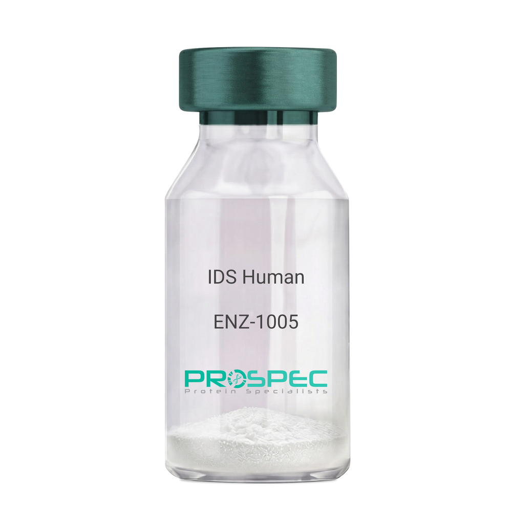
- View Data Sheet
Name :
IDS HumanDescription:
Iduronate 2-Sulfatase Human Recombinant
Iduronate 2-Sulfatase, Alpha-L-Iduronate Sulfate Sulfatase, SIDS, Iduronate 2-Sulfatase 14 KDa Chain, Iduronate 2-Sulfatase 42 KDa Chain, Hunter Syndrome, EC 3.1.6.13, MPS2, Iduronate 2-sulfatase, Alpha-L-iduronate sulfate sulfatase.
Product # :
ENZ-1005Price :
Quantity :
Shipping Method :
Shipped with Ice Packs
More Info
- description
- source
- formulation
- purity
- More Info
Description
IDS Human Recombinant produced in Sf9 Baculovirus cells is a single, glycosylated polypeptide chain containing 533 amino acids (26-550a.a) and having a molecular mass of 60.3kDa. (Molecular size on SDS-PAGE will appear at approximately 35-70kDa). IDS is fused to an 8 amino acid His-tag at C-terminus & purified by proprietary chromatographic techniques.
Source
Sf9, Baculovirus cells.
Formulation
IDS protein solution (0.25mg/ml) containing Phosphate Buffered Saline (pH 7.4) and 10% glycerol.
Purity
Greater than 90% as determined by SDS-PAGE.
More Info
-
Introduction
Iduronate 2-Sulfatase also known as IDS, belongs to the highly-conserved sulfatase family of enzymes which catalyze the hydrolysis of O-sulfate and N-salfate esters from a variety of substrates. IDS is essential for the lysosomal degradation of the glycosaminoglycans (GAG) heparan sulfate as well as dermatan sulfate. Furthermore, IDS hydrolyzes the 2-sulfate group of the IDS units of the GAG.
-
Synonyms
Iduronate 2-Sulfatase, Alpha-L-Iduronate Sulfate Sulfatase, SIDS, Iduronate 2-Sulfatase 14 KDa Chain, Iduronate 2-Sulfatase 42 KDa Chain, Hunter Syndrome, EC 3.1.6.13, MPS2, Iduronate 2-sulfatase, Alpha-L-iduronate sulfate sulfatase.
-
Physical Appearance
Sterile Filtered clear solution.
-
Stability
Store at 4°C if entire vial will be used within 2-4 weeks. Store, frozen at -20°C for longer periods of time. For long term storage it is recommended to add a carrier protein (0.1% HSA or BSA).Avoid multiple freeze-thaw cycles.
-
Amino Acid Sequence
SETQANSTTD ALNVLLIIVD DLRPSLGCYG DKLVRSPNID QLASHSLLFQ NAFAQQAVCA PSRVSFLTGR RPDTTRLYDF NSYWRVHAGN FSTIPQYFKE NGYVTMSVGK VFHPGISSNH TDDSPYSWSF PPYHPSSEKY ENTKTCRGPD GELHANLLCP VDVLDVPEGT LPDKQSTEQA IQLLEKMKTS ASPFFLAVGY HKPHIPFRYP KEFQKLYPLE NITLAPDPEV PDGLPPVAYN PWMDIRQRED VQALNISVPY GPIPVDFQRK IRQSYFASVS YLDTQVGRLL SALDDLQLAN STIIAFTSDH GWALGEHGEW AKYSNFDVAT HVPLIFYVPG RTASLPEAGE KLFPYLDPFD SASQLMEPGR QSMDLVELVS LFPTLAGLAG LQVPPRCPVP SFHVELCREG KNLLKHFRFR DLEEDPYLPG NPRELIAYSQ YPRPSDIPQW NSDKPSLKDI KIMGYSIRTI DYRYTVWVGF NPDEFLANFS DIHAGELYFV DSDPLQDHNM YNDSQGGDLF QLLMPLEHHH HHH.
ProSpec's products are furnished for LABORATORY RESEARCH USE ONLY. The product may not be used as drugs, agricultural or pesticidal products, food additives or household chemicals.
- View Data Sheet
Name :
TPST2 Human, sf9Description:
Tyrosylprotein Sulfotransferase 2, sf9 Human Recombinant
Tyrosylprotein Sulfotransferase 2, EC 2.8.2.20, TPST-2, Transport And Golgi Organization 13 Homolog B (Drosophila), Transport And Golgi Organization 13 Homolog B, Protein-Tyrosine Sulfotransferase 2, Tyrosylprotein Phosphotransferase 2, Tyrosylprotein Sulfotransferase-2, TANGO13B.
Product # :
ENZ-949Price :
Quantity :
Shipping Method :
Shipped with Ice Packs
More Info
- description
- source
- formulation
- purity
- More Info
Description
TPST2 produced in Sf9 Baculovirus cells is a single, glycosylated polypeptide chain containing 361 amino acids (26-377 a.a.) and having a molecular mass of 40.4kDa (Migrates at 40-57kDa on SDS-PAGE under reducing conditions). TPST2 is expressed with a 6 amino acid His tag at C-Terminus and purified by proprietary chromatographic techniques.
Source
Sf9, Baculovirus cells.
Formulation
TPST2 protein solution (0.25mg/ml) contains Phosphate Buffered Saline (pH 7.4) and 10% glycerol.
Purity
Greater than 95.0% as determined by SDS-PAGE.
More Info
-
Introduction
Tyrosylprotein Sulfotransferase 2 (TPST2) is a member of the protein sulfotransferase family. TPST2 is a widely expressed protein, which catalyzes the O-sulfation of tyrosine residues within acidic regions of proteins. The TPST2 protein is a type II integral membrane protein located in the Golgi body.
-
Synonyms
Tyrosylprotein Sulfotransferase 2, EC 2.8.2.20, TPST-2, Transport And Golgi Organization 13 Homolog B (Drosophila), Transport And Golgi Organization 13 Homolog B, Protein-Tyrosine Sulfotransferase 2, Tyrosylprotein Phosphotransferase 2, Tyrosylprotein Sulfotransferase-2, TANGO13B.
-
Physical Appearance
Sterile Filtered colorless solution.
-
Stability
Store at 4°C if entire vial will be used within 2-4 weeks. Store, frozen at -20°C for longer periods of time. For long term storage it is recommended to add a carrier protein (0.1% HSA or BSA). Avoid multiple freeze-thaw cycles.
-
Amino Acid Sequence
ADPQQVLECR AVLAGLRSPR GAMRPEQEEL VMVGTNHVEY RYGKAMPLIF VGGVPRSGTT LMRAMLDAHP EVRCGEETRI IPRVLAMRQA WSKSGREKLR LDEAGVTDEV LDAAMQAFIL EVIAKHGEPA RVLCNKDPFT LKSSVYLSRL FPNSKFLLMV RDGRASVHSM ITRKVTIAGF DLSSYRDCLT KWNKAIEVMY AQCMEVGKEK CLPVYYEQLV LHPRRSLKLI LDFLGIAWSD AVLHHEDLIG KPGGVSLSKI ERSTDQVIKP VNLEALSKWT GHIPGDVVRD MAQIAPMLAQ LGYDPYANPP NYGNPDPFVI NNTQRVLKGD YKTPANLKGY FQVNQNSTSS HLGSSHHHHH H.
ProSpec's products are furnished for LABORATORY RESEARCH USE ONLY. The product may not be used as drugs, agricultural or pesticidal products, food additives or household chemicals.
- View Data Sheet
Name :
CHST10 HumanDescription:
Carbohydrate Sulfotransferase 10 Human Recombinant
Carbohydrate Sulfotransferase 10, HNK1ST, HNK-1 Sulfotransferase,HuHNK-1ST, HNK-1ST, EC 2.8.2.-, EC 2.8.2, CHST10.
Product # :
ENZ-894Price :
Quantity :
Shipping Method :
Shipped with Ice Packs
More Info
- description
- source
- formulation
- purity
- More Info
Description
CHST10 Human Recombinant produced in E.Coli is a single, non-glycosylated polypeptide chain containing 350 amino acids (28-356 a.a) and having a molecular mass of 41.2kDa.CHST10 is fused to a 21 amino acid His-tag at N-terminus & purified by proprietary chromatographic techniques.
Source
Escherichia Coli.
Formulation
CHST10 protein solution (0.5mg/ml) containing 20mM Tris-HCl (pH8.0) and 10% glycerol.
Purity
Greater than 85.0% as determined by SDS-PAGE.
More Info
-
Introduction
Carbohydrate Sulfotransferase 10, also known as CHST10 is a member of the sulfotransferase 2 family. CHST10 was first recognized as a sulfotransferase which acts on the human natural killer-1 (HNK-1) glycan. Furthermore, CHST10 is a carbohydrate involved in neurodevelopment as well as synaptic plasticity.
-
Synonyms
Carbohydrate Sulfotransferase 10, HNK1ST, HNK-1 Sulfotransferase,HuHNK-1ST, HNK-1ST, EC 2.8.2.-, EC 2.8.2, CHST10.
-
Physical Appearance
Sterile Filtered colorless solution.
-
Stability
Store at 4°C if entire vial will be used within 2-4 weeks. Store, frozen at -20°C for longer periods of time. For long term storage it is recommended to add a carrier protein (0.1% HSA or BSA).Avoid multiple freeze-thaw cycles.
-
Amino Acid Sequence
MGSSHHHHHH SSGLVPRGSH MTFKDPDVYS AKQEFLFLTT MPEVRKLPEE KHIPEELKPT GKELPDSQLV QPLVYMERLE LIRNVCRDDA LKNLSHTPVS KFVLDRIFVC DKHKILFCQT PKVGNTQWKK VLIVLNGAFS SIEEIPENVV HDHEKNGLPR LSSFSDAEIQ KRLKTYFKFF IVRDPFERLI SAFKDKFVHN PRFEPWYRHE IAPGIIRKYR RNRTETRGIQ FEDFVRYLGD PNHRWLDLQF GDHIIHWVTY VELCAPCEIM YSVIGHHETL EDDAPYILKE AGIDHLVSYP TIPPGITVYN RTKVEHYFLG ISKRDIRRLY ARFEGDFKLF GYQKPDFLLN.
ProSpec's products are furnished for LABORATORY RESEARCH USE ONLY. The product may not be used as drugs, agricultural or pesticidal products, food additives or household chemicals.