Search results
521 results found for “methylmalonic aciduria”
Name
Description
Product #
Price
Quantity
Shipping Method
- View Data Sheet
Name :
MAFK AntibodyDescription:
V-maf Musculoaponeurotic Fibrosarcoma Oncogene K, Mouse Anti Human
NFE2U, P18, Erythroid transcription factor NF-E2 p18 subunit, MAFK, Transcription factor MafK.
Product # :
ANT-463Price :
Quantity :
Shipping Method :
Shipped with Ice Packs
More Info
- formulation
- More Info
Formulation
1mg/ml containing PBS, pH-7.4, 10% Glycerol and 0.02% Sodium Azide.
More Info
-
Introduction
MAFK is a member of the bZIP family and Maf subfamily. Members from Maf family of basic region/leucine zipper (bZIP) transcription factors influence transcription in a positive or negative way, depending on their protein associate and the context of the target promoter. Small Mafs act as transcriptional repressors when they dimerize among themselves. Yet, they seem to function as transcriptional activators by dimerizing with other basic-zipper proteins and employing them to specific DNA-binding sites.
-
Synonyms
NFE2U, P18, Erythroid transcription factor NF-E2 p18 subunit, MAFK, Transcription factor MafK.
-
Physical Appearance
Sterile Filtered colorless solution.
-
Immunogen
Anti-human MAFK mAb, clone PAT2F7A, is derived from hybridization of mouse F0 myeloma cells with spleen cells from BALB/c mice immunized with a recombinant human MAFK protein 1-156 amino acids purified from E. coli.
-
Ig Subclass
Mouse IgG2a heavy chain and Kappa light chain.
-
Clone
PAT2F7A.
-
Applications
The antibody has been tested by ELISA, Western blot analysis to assure specificity and reactivity. Since application varies, however, each investigation should be titrated by the reagent to obtain optimal results. Recommended starting dilution is 1:1000.
-
Type
Mouse Anti Human Monoclonal.
-
Storage Procedures
For periods up to 1 month store at 4°C, for longer periods of time, store at -20°C. Prevent freeze thaw cycles.
-
Purification Method
MAFK antibody was purified from mouse ascitic fluids by protein-G affinity chromatography.
ProSpec's products are furnished for LABORATORY RESEARCH USE ONLY. The product may not be used as drugs, agricultural or pesticidal products, food additives or household chemicals.
- View Data Sheet
Name :
MIF AntibodyDescription:
Macrophage Migration Inhibitory Factor, Mouse Anti Human
Phenylpyruvate tautomerase, Glycosylation- inhibiting factor, GIF, MMIF, MIF.
Product # :
ANT-311Price :
Quantity :
Shipping Method :
Shipped with Ice Packs
More Info
- formulation
- More Info
Formulation
1mg/ml containing PBS, pH-7.4, 0.02% Sodium Azide and 10% Glycerol.
More Info
-
Introduction
The cytokine Macrophage migration inhibitory factor (MIF) has been identified to be secreted by the pituitary gland and the monocyte/macrophage and to play an important role in endotoxic shock. MIF has the unique property of being released from macrophages and T cells in response to physiological concentrations of glucocorticoids. The secretion of MIF is tightly regulated and decreases at high, anti-inflammatory steroid concentration.
-
Synonyms
Phenylpyruvate tautomerase, Glycosylation- inhibiting factor, GIF, MMIF, MIF.
-
Immunogen
Anti-human MIF mAb, is derived from hybridization of mouse SP2/0 myeloma cells with spleen cells from BALB/c mice immunized with Recombinant human MIF amino acids 1-114 purified from E. coli.
-
Ig Subclass
Mouse IgG1 heavy chain and κ light chain.
-
Clone
P4E4AT.
-
Applications
MIF antibody has been tested by ELISA and Western blot analysis to assure specificity and reactivity. Since application varies, however, each investigation should be titrated by the reagent to obtain optimal results.
-
Type
Mouse Anti Human Monoclonal.
-
Storage Procedures
For periods up to 1 month store at 4°C, for longer periods of time, store at -20°C. Prevent freeze thaw cycles.
-
Purification Method
MIF antibody was purified from mouse ascitic fluids by protein-G affinity chromatography.
ProSpec's products are furnished for LABORATORY RESEARCH USE ONLY. The product may not be used as drugs, agricultural or pesticidal products, food additives or household chemicals.
- View Data Sheet
Name :
MYD88 AntibodyDescription:
Myeloid Differentiation Primary Response 88, Mouse Anti Human
Myeloid differentiation primary response protein MyD88, MYD88, MYD88D.
Product # :
ANT-464Price :
Quantity :
Shipping Method :
Shipped with Ice Packs
More Info
- formulation
- More Info
Formulation
1mg/ml containing PBS, pH-7.4, 10% Glycerol and 0.01% Sodium Azide.
More Info
-
Introduction
Myeloid differentiation primary response gene 88 (MYD88) is a cytosolic adapter protein, which has a central role in the innate and adaptive immune response. MYD88 functions as a vital signal transducer in the interleukin-1 and Toll-like receptor signaling pathways. MYD88 acts via IRAK1, IRAK2, IRF7 and TRAF6, leading to NF-kappa-B activation, cytokine secretion and the inflammatory response. The MYD88 protein increases IL-8 transcription. MYD88 is involved in IL-18-mediated signaling pathway. MYD88 activates IRF1, resulting in its rapid migration into the nucleus to mediate an efficient induction of IFN-beta, NOS2/INOS, and IL12A genes. MYD88 is comprised of an N-terminal death domain and a C-terminal Toll-interleukin1 receptor domain. Patients with defects in the MYD88 gene have an increased susceptibility to pyogenic bacterial infections.
-
Synonyms
Myeloid differentiation primary response protein MyD88, MYD88, MYD88D.
-
Physical Appearance
Sterile Filtered colorless solution.
-
Immunogen
Anti-human MYD88 mAb, clone PAT22F11A, is derived from hybridization of mouse F0 myeloma cells with spleen cells from BALB/c mice immunized with a recombinant human MYD88 protein 1-309 amino acids purified from E. coli.
-
Ig Subclass
Mouse IgG2b heavy chain and Kappa light chain.
-
Clone
PAT22F11A.
-
Applications
The antibody has been tested by ELISA, Western blot analysis to assure specificity and reactivity. Since application varies, however, each investigation should be titrated by the reagent to obtain optimal results. Recommended starting dilution is 1:1000.
-
Type
Mouse Anti Human Monoclonal.
-
Storage Procedures
For periods up to 1 month store at 4°C, for longer periods of time, store at -20°C. Prevent freeze thaw cycles.
-
Purification Method
MYD88 antibody was purified from mouse ascitic fluids by protein-G affinity chromatography.
ProSpec's products are furnished for LABORATORY RESEARCH USE ONLY. The product may not be used as drugs, agricultural or pesticidal products, food additives or household chemicals.
- View Data Sheet
Name :
MAPKSP1 antibodyDescription:
MAPK Scaffold Protein 1, Mouse Anti Human
MAPBP, MP1, MAPK Scaffold Protein 1, Interacting Protein 1, MEK binding partner 1, Mitogen-Activated Protein Kinase kinase 1.
Product # :
ANT-734Price :
Quantity :
Shipping Method :
Shipped with Ice Packs
More Info
- formulation
- More Info
Formulation
1mg/ml containing PBS, pH-7.4, 10% Glycerol and 0.02% Sodium Azide.
More Info
-
Introduction
MAPKSP1 is a scaffold protein which operates in the extracellular signal-regulated kinase (ERK) cascade. MAPKSP1 protein is restricted to late endosomes by the mitogen-activated protein-binding protein-interacting protein, and binds specifically to MAP kinase kinase MAP2K1/MEK1, MAP kinase MAPK3/ERK1, and MAP kinase MAPK1/ERK2.
-
Synonyms
MAPBP, MP1, MAPK Scaffold Protein 1, Interacting Protein 1, MEK binding partner 1, Mitogen-Activated Protein Kinase kinase 1.
-
Physical Appearance
Sterile filtered colorless solution.
-
Immunogen
Anti-human MAPKSP1 mAb, is derived from hybridization of mouse F0 myeloma cells with spleen cells from BALB/c mice immunized with a recombinant human MAPKSP1 protein 1-124 amino acids purified from E. coli.
-
Ig Subclass
Mouse IgG2b heavy chain and k light chain.
-
Clone
PAT1F10AT.
-
Applications
The antibody has been tested by ELISA, Western blot analysis, Flow cytometry and ICC/IF to assure specificity and reactivity. Since application varies, however, each investigation should be titrated by the reagent to obtain optimal results.
-
Type
Mouse Anti Human Monoclonal.
-
Storage Procedures
For periods up to 1 month store at 4°C, for longer periods of time, store at -20°C. Prevent freeze thaw cycles.
-
Purification Method
MAPKSP1 antibody was purified from mouse ascitic fluids by protein-A affinity chromatography.
ProSpec's products are furnished for LABORATORY RESEARCH USE ONLY. The product may not be used as drugs, agricultural or pesticidal products, food additives or household chemicals.
- View Data Sheet
Name :
DDAVPDescription:
Desmopressin
Product # :
HOR-270Price :
Quantity :
Shipping Method :
Shipped at Room temp
More Info
- description
- formulation
- purity
- More Info
Description
Desmopressin also called ADH (Anti-Diuretic Hormone) has a molecular formula of C46H64N14O12S2 , Mpr-Tyr-Phe-Gln-Asn-Cys-Pro-D-Arg-Gly-NH2 having a Mw of 1069.23 Dalton.
Formulation
The Desmopressin peptide was lyophilized with no additives.
Purity
Greater than 98.0% as determined by:
(a) Analysis by RP-HPLC.
(b) Analysis by SDS-PAGE.More Info
-
Introduction
Desmopressin is the first vasopressin analog with a very high and very specific antidiuretic effect, has been widely used for different therapeutic purposes and is believed to be partly responsible for the formation of memories learning and memory processes. Desmopressin increases urine concentration and decreases urine production. Desmopressin is used to prevent and control excessive thirst, urination, and dehydration.
-
Physical Appearance
Sterile Filtered White lyophilized (freeze-dried) powder.
-
Stability
Lyophilized Desmopressin although stable at room temperature for 3 weeks, should be stored desiccated below -18°C. Upon reconstitution DDAVP should be stored at 4°C between 2-7 days and for future use below -18°C. For long term storage it is recommended to add a carrier protein (0.1% HSA or BSA).Please prevent freeze-thaw cycles.
-
Solubility
It is recommended to reconstitute the lyophilized DDAVP in sterile 18MΩ-cm H2O not less than 100 µg/ml, which can then be further diluted to other aqueous solutions.
ProSpec's products are furnished for LABORATORY RESEARCH USE ONLY. The product may not be used as drugs, agricultural or pesticidal products, food additives or household chemicals.
- View Data Sheet
Name :
FTCD HumanDescription:
Formiminotransferase Cyclodeaminase Human Recombinant
Formiminotransferase-cyclodeaminase, FTCD, LCHC1, LC-1.
Product # :
ENZ-302Price :
Quantity :
Shipping Method :
Shipped with Ice Packs
More Info
- description
- source
- formulation
- purity
- More Info
Description
Formiminotransferase Cyclodeaminase Human Recombinant (also called liver cytosol type 1) produced in SF9, is a glycosylated, polypeptide chain having a molecular mass of 59,749 Dalton.The FTCD is expressed with a -6xHis tag and purified by proprietary chromatographic techniques.
Source
Sf9 insect cells.
Formulation
FTCD is supplied in 16mM HEPES buffer pH-7.6, 240mM sodium chloride, and 20% glycerol.
Purity
Greater than 95% as determined by SDS-PAGE.
More Info
-
Introduction
Formiminotransferase cyclodeaminase is an enzyme which catalyzes the conversion of formiminoglutamateand tetrahydrofolateinto formiminotetrahydrofolateand glutamate.
Formiminotransferase cyclodeaminase, a bifunctional enzyme of tetrahydrofolate synthesis, is the target antigen of anti-LC1 (liver cytosol antigen type 1) autoantibodies. Presence of LC1 autoantibodies is a marker for type 2 autoimmune hepatitis (for which anti-LKM 1/cytochrome P450 2D6 autoantibodies are a further marker).
Serum LC1 autoantibody concentrations appear to fluctuate in parallel with aminotransferase levels, a particularly intriguing observation that suggests a possible role of LC1 autoreactivity in the pathogenic mechanism leading to hepatocyte injury. -
Synonyms
Formiminotransferase-cyclodeaminase, FTCD, LCHC1, LC-1.
-
Stability
Store at 4°C if entire vial will be used within 2-4 weeks. Store, frozen at -20°Cfor longer periods of time. Avoid multiple freeze-thaw cycles.
-
Immunological Functions
1. Binds IgG-type human auto-antibodies.2. Standard ELISA test (checker-board analysis of positive/negative sera panels, immuno-dot test).
-
coating concentration
0.35-0.7 µg/ml (depending on the type of ELISA plate and coating buffer).Suitable for biotinylation and iodination.
ProSpec's products are furnished for LABORATORY RESEARCH USE ONLY. The product may not be used as drugs, agricultural or pesticidal products, food additives or household chemicals.
- View Data Sheet
Name :
GAL HumanDescription:
Galanin Prepropeptide Human Recombinant
GALN, GLNN, GMAP, GAL, GAL1.
Product # :
PRO-1433Price :
Quantity :
Shipping Method :
Shipped with Ice Packs
More Info
- description
- source
- formulation
- purity
- More Info
Description
GAL Human Recombinant produced in E. coli is a single polypeptide chain containing 127 amino acids (20-123) and having a molecular mass of 13.9kDa. GAL is fused to a 23 amino acid His-tag at N-terminus & purified by proprietary chromatographic techniques.
Source
E.coli.
Formulation
The GAL solution (0.25mg/ml) contains 20mM Tris-HCl buffer (pH 8.0), 0.2M NaCl, 50% glycerol and 2mM DTT.
Purity
Greater than 90% as determined by SDS-PAGE.
More Info
-
Introduction
Galanin Prepropeptide (GAL) which is localized in brain pathways is involved in both cognition and affect, and also inhibits learning and memory by inhibiting neurotransmitter release and neuronal firing rate. GAL is a part of the galanin family and modulates a variety of physiological processed including cognition/memory, sensory/pain processing, neurotransmitter/hormone secretion, and feeding behavior. Galanin Prepropeptide is upregulated in primary afferent and sympathetic neurones and is required for the development of sympathetic perineuronal baskets subsequent to nerve injury.
-
Synonyms
GALN, GLNN, GMAP, GAL, GAL1.
-
Physical Appearance
Sterile Filtered colorless solution.
-
Stability
Store at 4°C if entire vial will be used within 2-4 weeks. Store, frozen at -20°C for longer periods of time. For long term storage it is recommended to add a carrier protein (0.1% HSA or BSA).Avoid multiple freeze-thaw cycles.
-
Amino Acid Sequence
MGSSHHHHHH SSGLVPRGSH MGSASAGLWS PAKEKRGWTL NSAGYLLGPH AVGNHRSFSD KNGLTSKREL RPEDDMKPGS FDRSIPENNI MRTIIEFLSF LHLKEAGALD RLLDLPAAASSEDIERS.
ProSpec's products are furnished for LABORATORY RESEARCH USE ONLY. The product may not be used as drugs, agricultural or pesticidal products, food additives or household chemicals.
- View Data Sheet
Name :
CRABP1 HumanDescription:
Cellular Retinoic Acid binding Protein 1 Human Recombinant
Cellular retinoic acid-binding protein 1, Cellular retinoic acid-binding protein I, CRABP-I, CRABP1, RBP5, CRABP, CRABPI.
Product # :
PRO-710Price :
Quantity :
Shipping Method :
Shipped with Ice Packs
More Info
- description
- source
- formulation
- purity
- More Info
Description
CRABP1 Human Recombinant produced in E.Coli is a single, non-glycosylated polypeptide chain containing 137 amino acids and having a molecular mass of 15.5kDa. The CRABP1 is purified by proprietary chromatographic techniques.
Source
Escherichia Coli.
Formulation
The CRABP1 (1mg/ml) protein solution contains 20mM Tris-HCl buffer (pH8.0) and 10% glycerol.
Purity
Greater than 95.0% as determined by SDS-PAGE.
More Info
-
Introduction
CRABP1 is a member of special carrier proteins for members of the vitamin A family. It is believed that CRABP1 has an essential role in retinoic acid-mediated differentiation and proliferation processes. Though, CRABP1 is structurally similar to the cellular retinol-binding proteins, it binds only retinoic acid at specific sites within the nucleus, which may contribute to vitamin A-directed differentiation in epithelial tissue. CRABP1 is constitutively expressed and is thought to have different functions in the cell than the related CRABP2. CRABP1 forms a beta-barrel structure which accommodates hydrophobic ligands in its interior.
Loss of CRABP1 function as a result of hypermethylation of its promoter leads to pathogenesis of papillary thyroid carcinoma. Furthermore, frequent methylation-associated silencing of CRABP1 is linked to esophageal squamous-cell carcinoma. -
Synonyms
Cellular retinoic acid-binding protein 1, Cellular retinoic acid-binding protein I, CRABP-I, CRABP1, RBP5, CRABP, CRABPI.
-
Physical Appearance
Sterile Filtered colorless solution.
-
Stability
Store at 4°C if entire vial will be used within 2-4 weeks. Store, frozen at -20°C for longer periods of time. For long term storage it is recommended to add a carrier protein (0.1% HSA or BSA).Avoid multiple freeze-thaw cycles.
-
Amino Acid Sequence
MPNFAGTWKM RSSENFDELL KALGVNAMLR KVAVAAASKP HVEIRQDGDQ FYIKTSTTVR TTEINFKVGE GFEEETVDGR KCRSLATWEN ENKIHCTQTL LEGDGPKTYW TRELANDELI LTFGADDVVC TRIYVRE.
ProSpec's products are furnished for LABORATORY RESEARCH USE ONLY. The product may not be used as drugs, agricultural or pesticidal products, food additives or household chemicals.
- View Data Sheet
Name :
TRAIL Human (114-281 a.a.)Description:
TRAIL/APO 2 Ligand (114-281 a.a.) Human Recombinant
Tumor necrosis factor ligand superfamily member 10, TNF-related apoptosis-inducing ligand, Protein TRAIL, Apo-2 ligand, Apo-2L, CD253 antigen, TL2, APO2L, TNFSF10.
Product # :
CYT-546Price :
Quantity :
Shipping Method :
Shipped with Ice Packs
More Info
- description
- source
- formulation
- purity
- More Info
Description
Soluble TNF-related apoptosis-inducing ligand Human Recombinant produced in E.Coli is a single, non-glycosylated polypeptide chain containing 169 amino acids (114-281) and having a molecular mass of 19.6 kDa. The sTRAIL is purified by proprietary chromatographic techniques.
Source
Escherichia Coli.
Formulation
1mg/ml in 20mM Tris-HCl pH-7.5, 300mM NaCl, 0.1mM DTT & 10% glycerol.
Purity
Greater than 95.0% as determined by SDS-PAGE.
More Info
-
Introduction
TNF-related apoptosis-inducing ligand (TRAIL) is a ligand molecule which induces apoptosis. It is a type II transmembrane protein with homology to other members of the tumor necrosis factor family.
In humans, the gene that encodes for TRAIL is located at chromosome 3q26.
TRAIL binds to the death receptors, DR4 and DR5. The process of apoptosis is caspase-8-dependent. This protein preferentially induces apoptosis in transformed and tumor cells, but does not appear to kill normal cells although it is expressed at a significant level in most normal tissues. -
Synonyms
Tumor necrosis factor ligand superfamily member 10, TNF-related apoptosis-inducing ligand, Protein TRAIL, Apo-2 ligand, Apo-2L, CD253 antigen, TL2, APO2L, TNFSF10.
-
Physical Appearance
Sterile Filtered colorless liquid.
-
Stability
Store at 4°C if entire vial will be used within 2-4 weeks. Store, frozen at -20°C for longer periods of time. For long term storage it is recommended to add a carrier protein (0.1% HSA or BSA).Avoid multiple freeze-thaw cycles.
-
Amino Acid Sequence
MVRERGPQRV AAHITGTRGR SNTLSSPNSK NEKALGRKIN SWESSRSGHS FLSNLHLRNGELVIHEKGFY YIYSQTYFRF QEEIKENTKN DKQMVQYIYK YTSYPDPILL MKSARNSCWSKDAEYGLYSI YQGGIFELKE NDRIFVSVTN EHLIDMDHEA SFFGAFLVG.
ProSpec's products are furnished for LABORATORY RESEARCH USE ONLY. The product may not be used as drugs, agricultural or pesticidal products, food additives or household chemicals.
- View Data Sheet
Name :
DTD2 HumanDescription:
D-Tyrosyl-tRNA Deacylase 2 Human Recombinant
D-Tyrosyl-TRNA Deacylase 2 (Putative), Chromosome 14 Open Reading Frame 126, Probable D-Tyrosyl-TRNA(Tyr) Deacylase 2, EC 3.1.-.-,C14orf126
Product # :
ENZ-763Price :
Quantity :
Shipping Method :
Shipped with Ice Packs
More Info
- description
- source
- formulation
- purity
- More Info
Description
DTD2 Human Recombinant produced in E.coli is a single, non-glycosylated polypeptide chain containing 191 amino acids (1-168) and having a molecular mass of 21.0 kDa. DTD2 is fused to a 23 amino acid His-tag at N-terminus.
Source
Escherichia Coli.
Formulation
The DTD2 solution (1mg/ml) contains 20mM Tris-HCl buffer (pH 8.0), 150mM NaCl, 1mM DTT and 20% glycerol.
Purity
Greater than 90% as determined by SDS-PAGE.
More Info
-
Introduction
DTD2 is e member of the DTD family. DTD2 protein hydrolyzes D-tyrosyl-tRNA(Tyr) into D-tyrosine and free tRNA(Tyr) as a defense mechanism against a harmful effect of D-tyrosine.
-
Synonyms
D-Tyrosyl-TRNA Deacylase 2 (Putative), Chromosome 14 Open Reading Frame 126, Probable D-Tyrosyl-TRNA(Tyr) Deacylase 2, EC 3.1.-.-,C14orf126
-
Physical Appearance
Sterile Filtered clear solution.
-
Stability
Store at 4°C if entire vial will be used within 2-4 weeks. Store, frozen at -20°C for longer periods of time. For long term storage it is recommended to add a carrier protein (0.1% HSA or BSA).Avoid multiple freeze-thaw cycles.
-
Amino Acid Sequence
MGSSHHHHHH SSGLVPRGSH MGSMAEGSRI PQARALLQQC LHARLQIRPA DGDVAAQWVE VQRGLVIYVC FFKGADKELL PKMVNTLLNV KLSETENGKH VSILDLPGNI LIIPQATLGG RLKGRNMQYH SNSGKEEGFE LYSQFVTLCE KEVAANSKCA EARVVVEHGT YGNRQVLKLD TNGPFTHLIE F
ProSpec's products are furnished for LABORATORY RESEARCH USE ONLY. The product may not be used as drugs, agricultural or pesticidal products, food additives or household chemicals.
- View Data Sheet
Name :
TGFB3 (24-412 a.a.) HumanDescription:
Transforming Growth Factor-Beta 3 (24-412 a.a.) Human Recombinant
Transforming Growth Factor, Beta 3, Prepro-Transforming Growth Factor Beta-3, TGF-Beta-3, ARVD1, RNHF, Arrhythmogenic Right Ventricular Dysplasia 1, Transforming Growth Factor Beta-3, TGF-Beta3, ARVD, Transforming growth factor beta-3, TGF-beta-3.
Product # :
CYT-886Price :
Quantity :
Shipping Method :
Shipped with Ice Packs
More Info
- description
- source
- formulation
- purity
- More Info
Description
TGFB3 Human Recombinant produced in E.Coli is a single, non-glycosylated polypeptide chain containing 412 amino acids (24-412 a.a) and having a molecular mass of 47.2kDa. TGFB3 is fused to a 23 amino acid His-tag at N-terminus & purified by proprietary chromatographic techniques.
Source
Escherichia Coli.
Formulation
TGFB3 protein solution (0.5mg/ml) containing 20mM Tris-HCl (pH8.0) and 10% glycerol.
Purity
Greater than 85.0% as determined by SDS-PAGE.
More Info
-
Introduction
Transforming growth factor betas (TGF Betas) mediate many cell-cell interactions that occur during embryonic development. Three TGF Betas have been identified in mammals. TGF Beta 1, TGF Beta 2 and TGF Beta 3 are each synthesized as precursor proteins that are very similar in that each is cleaved to yield a 112 amino acid polypeptide that remains associated with the latent portion of the molecule.
-
Synonyms
Transforming Growth Factor, Beta 3, Prepro-Transforming Growth Factor Beta-3, TGF-Beta-3, ARVD1, RNHF, Arrhythmogenic Right Ventricular Dysplasia 1, Transforming Growth Factor Beta-3, TGF-Beta3, ARVD, Transforming growth factor beta-3, TGF-beta-3.
-
Physical Appearance
Sterile Filtered colorless solution.
-
Stability
Store at 4°C if entire vial will be used within 2-4 weeks. Store, frozen at -20°C for longer periods of time. For long term storage it is recommended to add a carrier protein (0.1% HSA or BSA).Avoid multiple freeze-thaw cycles.
-
Amino Acid Sequence
MGSSHHHHHH SSGLVPRGSH MGSLSTCTTL DFGHIKKKRV EAIRGQILSK LRLTSPPEPT VMTHVPYQVL ALYNSTRELL EEMHGEREEG CTQENTESEY YAKEIHKFDM IQGLAEHNEL AVCPKGITSK VFRFNVSSVE KNRTNLFRAE FRVLRVPNPS SKRNEQRIEL FQILRPDEHI AKQRYIGGKN LPTRGTAEWL SFDVTDTVRE WLLRRESNLG LEISIHCPCH TFQPNGDILE NIHEVMEIKF KGVDNEDDHG RGDLGRLKKQ KDHHNPHLIL MMIPPHRLDN PGQGGQRKKR ALDTNYCFRN LEENCCVRPL YIDFRQDLGW KWVHEPKGYY ANFCSGPCPY LRSADTTHST VLGLYNTLNP EASASPCCVP QDLEPLTILY YVGRTPKVEQ LSNMVVKSCK CS.
ProSpec's products are furnished for LABORATORY RESEARCH USE ONLY. The product may not be used as drugs, agricultural or pesticidal products, food additives or household chemicals.
- View Data Sheet
Name :
TXNRD1 Human 161-649 a.a.Description:
Thioredoxin Reductase 1 161-649 a.a. Human Recombinant
Thioredoxin reductase 1 cytoplasmic, TR, Gene associated with retinoic and interferon-induced mortality 12 protein, GRIM-12, Gene associated with retinoic and IFN-induced mortality 12 protein, KM-102-derived reductase-like factor, Thioredoxin reductase TR1, TXNRD1, GRIM12, KDRF, TR1, TXNR, TRXR1, MGC9145.
Product # :
ENZ-042Price :
Quantity :
Shipping Method :
Shipped with Ice Packs
More Info
- description
- source
- formulation
- purity
- More Info
Description
TXNRD1 Human Recombinant fused with a 21 amino acid His tag at N-terminus produced in E.Coli is a single, non-glycosylated, polypeptide chain containing 510 amino acids (161-649 a.a.) and having a molecular mass of 55.9kDa. The TXNRD1 is purified by proprietary chromatographic techniques.
Source
Escherichia Coli.
Formulation
The TXNRD1 solution (1 mg/ml) contains 20mM Tris-HCl buffer (pH8.0) and 10% Glycerol.
Purity
Greater than 90.0% as determined by SDS-PAGE.
More Info
-
Introduction
TXNRD1 belongs to the selenium-containing pyridine nucleotide-disulphide oxidoreductase family, which has a conserved catalytic site of Cys-Val-Asn-Val-Gly-Cys. TXNRD1 decreases thioredoxins as well as other substrates, and participates in selenium metabolism and protection against oxidative stress. Inhibition of TXNRD1 activity serves as a potential treatment for cancer, AIDS and other autoimmune diseases as well as bacterial infections and parasitic diseases.
-
Synonyms
Thioredoxin reductase 1 cytoplasmic, TR, Gene associated with retinoic and interferon-induced mortality 12 protein, GRIM-12, Gene associated with retinoic and IFN-induced mortality 12 protein, KM-102-derived reductase-like factor, Thioredoxin reductase TR1, TXNRD1, GRIM12, KDRF, TR1, TXNR, TRXR1, MGC9145.
-
Physical Appearance
Sterile Filtered colorless solution.
-
Stability
Store at 4°C if entire vial will be used within 2-4 weeks. Store, frozen at -20°C for longer periods of time. For long term storage it is recommended to add a carrier protein (0.1% HSA or BSA).Avoid multiple freeze-thaw cycles.
-
Amino Acid Sequence
MGSSHHHHHH SSGLVPRGSH MYDYDLIIIG GGSGGLAAAK EAAQYGKKVM VLDFVTPTPL GTRWGLGGTC VNVGCIPKKL MHQAALLGQA LQDSRNYGWK VEETVKHDWD RMIEAVQNHI GSLNWGYRVA LREKKVVYEN AYGQFIGPHR IKATNNKGKE KIYSAERFLI ATGERPRYLG IPGDKEYCIS SDDLFSLPYC PGKTLVVGAS YVALECAGFL AGIGLDVTVM VRSILLRGFD QDMANKIGEH MEEHGIKFIR QFVPIKVEQI EAGTPGRLRV VAQSTNSEEI IEGEYNTVML AIGRDACTRK IGLETVGVKI NEKTGKIPVT DEEQTNVPYI YAIGDILEDK VELTPVAIQA GRLLAQRLYA GSTVKCDYEN VPTTVFTPLE YGACGLSEEK AVEKFGEENI EVYHSYFWPL EWTIPSRDNN KCYAKIICNT KDNERVVGFH VLGPNAGEVT QGFAAALKCG LTKKQLDSTI GIHPVCAEVF TTLSVTKRSG ASILQAGCCG.
ProSpec's products are furnished for LABORATORY RESEARCH USE ONLY. The product may not be used as drugs, agricultural or pesticidal products, food additives or household chemicals.
- View Data Sheet
Name :
BCAT1 HumanDescription:
Branched Chain Amino-Acid Transaminase 1 Human Recombinant
Branched chain amino-acid transaminase 1 cytosolic, BCT1, BCATC, Protein ECA39, placental protein 18, PP18, PNAS121, MECA39, EC 2.6.1.42.
Product # :
ENZ-597Price :
Quantity :
Shipping Method :
Shipped with Ice Packs
More Info
- description
- source
- formulation
- purity
- More Info
Description
BCAT1 Recombinant produced in E. coli is a single polypeptide chain containing 409 amino acids (1-386) and having a molecular mass of 45.4kDa.BCAT1 is fused to a 23 amino acid His-tag at N-terminus & purified by proprietary chromatographic techniques.
Source
E.coli.
Formulation
The BCAT1 solution (1mg/ml) contains 20mM Tris-HCl buffer (pH 8.0), 100mM Nacl, 1mM DTT and 20% glycerol.
Purity
Greater than 90% as determined by SDS-PAGE.
More Info
-
Introduction
BCAT1 catalyzes the leading reaction in the catabolism of the indispensable branched chain amino acids isoleucine, leucine and valine.
-
Synonyms
Branched chain amino-acid transaminase 1 cytosolic, BCT1, BCATC, Protein ECA39, placental protein 18, PP18, PNAS121, MECA39, EC 2.6.1.42.
-
Physical Appearance
Sterile Filtered colorless solution.
-
Stability
Store at 4°C if entire vial will be used within 2-4 weeks. Store, frozen at -20°C for longer periods of time. For long term storage it is recommended to add a carrier protein (0.1% HSA or BSA).Avoid multiple freeze-thaw cycles.
-
Amino Acid Sequence
MGSSHHHHHH SSGLVPRGSH MGSMKDCSNG CSAECTGEGG SKEVVGTFKA KDLIVTPATI LKEKPDPNNL VFGTVFTDHM LTVEWSSEFG WEKPHIKPLQ NLSLHPGSSA LHYAVELFEG LKAFRGVDNK IRLFQPNLNM DRMYRSAVRA TLPVFDKEEL LECIQQLVKL DQEWVPYSTS ASLYIRPTFI GTEPSLGVKK PTKALLFVLL SPVGPYFSSG TFNPVSLWAN PKYVRAWKGG TGDCKMGGNY GSSLFAQCEA VDNGCQQVLW LYGEDHQITE VGTMNLFLYW INEDGEEELA TPPLDGIILP GVTRRCILDL AHQWGEFKVS ERYLTMDDLT TALEGNRVRE MFGSGTACVV CPVSDILYKG ETIHIPTMEN GPKLASRILS KLTDIQYGRE ESDWTIVLS.
ProSpec's products are furnished for LABORATORY RESEARCH USE ONLY. The product may not be used as drugs, agricultural or pesticidal products, food additives or household chemicals.
- View Data Sheet
Name :
MSP1 AntibodyDescription:
Malaria Falciparum MSP1 , antibody
Product # :
ANT-792Price :
Quantity :
Shipping Method :
Shipped with Ice Packs
More Info
- description
- formulation
- purity
- More Info
Description
Merozoite surface antigen is a protein located on the outside of the merozoite, playing an imperative role in immune reaction. About 55% cases of malaria are infected by Plasmodium falciparum (Pf). Pf MSP1 has to be used with Plasmodium vivax (Pv) together for ELISA and rapid diagnostic test, Plasmodium falciparum and vivax infection takes about 95% of Plasmodium caused infection.
Purification Method: MSP1 Antibody is the purified IgG of mouse monoclonal antibody targeting to merozoite surface protein 1 of Plasmodium falciparum for research.
Formulation
PBS, 0.05% sodium azide and 25mM K2CO3.
Purity
Protein is >90% pure.
Purified by protein A chromatography.
More Info
-
Physical Appearance
Sterile Filtered solution.
-
Stability
Pf. MSP1 although stable at 4°C for 1 week, should be stored below -18°C. Please prevent freeze thaw cycles.
-
Applications
ELISA.
-
Background
Mab-Pf-MSP1 is a monoclonal antibody that recognizes the Plasmodium falciparum Merozoite Surface Protein 1 (MSP1), a major surface antigen expressed during the blood stage of malaria infection. It is widely used for immunological assays such as ELISA, Western blotting, immunofluorescence, and studies investigating parasite invasion, MSP1 processing, and malaria vaccine development.
1. What is MSP1 antibody?
Mab-Pf-MSP1 is a purified mouse monoclonal IgG antibody that specifically targets the merozoite surface protein 1 (MSP1) of Plasmodium falciparum.
2. What species is MSP1 antibody derived from?
Mab-Pf-MSP1 is produced in mouse and is supplied as a purified mouse IgG monoclonal antibody.
3. What antigen does Pf-MSP1 antibody recognize?
The antibody specifically recognizes merozoite surface protein 1 (MSP1) from Plasmodium falciparum.
4. Does MSP1 antibody cross-react with Plasmodium vivax MSP1?
No. Mab-Pf-MSP1 has no cross-reactivity with merozoite surface protein 1 (MSP1) from Plasmodium vivax.
5. What is the format of MSP1 antibody?
The antibody is supplied as purified IgG in liquid form, making it ready for use or further dilution.
6. How is MSP1 antibody purified?
The antibody is purified from mouse ascites fluid using Protein A chromatography and has a purity greater than 90%.
7. What is the concentration of MSP1 antibody?
Pf-MSP1 antibody is supplied at a concentration that ranges between 0.5mg-2mg/mL
8. What buffer is MSP1 antibody supplied in?
The antibody is provided in PBS containing 25 mM potassium carbonate (K₂CO₃).
9. Does MSP1 antibody contain a preservative?
Yes. The formulation contains 0.05% sodium azide as a preservative.
10. What applications is MSP1 antibody recommended for?
MSP1 antibody is recommended for ELISA applications and is suggested to be diluted more than 1:750 for indirect ELISA.
11. What dilution is recommended for indirect ELISA?
For indirect ELISA, a dilution greater than 1:750 is recommended. Users may optimize the dilution depending on their assay conditions.
12. How should MSP1 antibody be stored?
Store the antibody at 4°C for short-term use. For long-term storage, keep it at −20°C.
13. Why should the vial be centrifuged before opening?
The vial should be centrifuged before opening to ensure complete recovery of the contents and minimize product loss.
14. What is the purity of MSP1 Antibody?
The antibody has a purity of greater than 90%, as determined following Protein A chromatography purification.
15. Is MSP1 antibody supplied in liquid or lyophilized form?
Mab-Pf-MSP1 is supplied in liquid form as purified IgG.
16. Can MSP1 antibody be used to distinguish Plasmodium falciparum from Plasmodium vivax?
Yes. Since MSP1 antibody specifically recognizes P. falciparum MSP1 and does not cross-react with P. vivax MSP1, it can be useful in studies requiring species-specific detection.
ProSpec's products are furnished for LABORATORY RESEARCH USE ONLY. The product may not be used as drugs, agricultural or pesticidal products, food additives or household chemicals.
- View Data Sheet
Name :
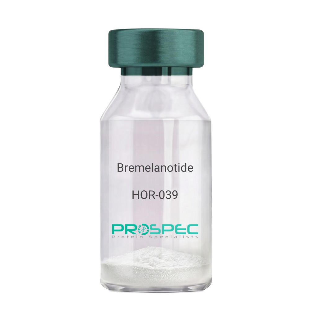
BremelanotideDescription:
Bremelanotide
PT-141, Rekynda, bremelanotide acetate.
Product # :
HOR-039Price :
Quantity :
Shipping Method :
Shipped at Room temp
More Info
- description
- formulation
- purity
- More Info
Description
Bremelanotide Synthetic is a single, non-glycosylated polypeptide chain containing 6 amino acids, having a molecular mass of 1025.16 Dalton and a Molecular formula of C50H68N40O10.
Formulation
The protein was lyophilized with no additives.
Purity
Greater than 97.0% as determined by analysis by RP-HPLC.
More Info
-
Synonyms
PT-141, Rekynda, bremelanotide acetate.
-
Physical Appearance
Sterile Filtered White lyophilized (freeze-dried) powder.
-
Stability
Lyophilized Bremelanotide although stable at room temperature for 3 weeks, should be stored desiccated below -18°C. Upon reconstitution Bremelanotide should be stored at 4°C between 2-7 days and for future use below -18°C. For long term storage it is recommended to add a carrier protein (0.1% HSA or BSA). Please prevent freeze-thaw cycles.
-
Solubility
It is recommended to reconstitute the lyophilized Bremelanotide in sterile 18MΩ-cm H2O not less than 100 µg/ml, which can then be further diluted to other aqueous solutions.
-
Amino Acid Sequence
Ac-Nle-cyclo(-Asp-His-D-Phe-Arg-Trp-Lys)-OH.
-
Background
Bremelanotide (PT-141), a synthetic heptapeptide, is a novel therapeutic agent primarily utilized for the treatment of sexual dysfunction. This paper provides an exhaustive review of Bremelanotide, detailing its biochemical structure, mechanism of action, therapeutic applications, and future prospects in clinical medicine.
Bremelanotide (PT-141), derived from Melanotan II, is a synthetic peptide developed for its role in treating sexual dysfunction in both men and women (Clayton et al., 2016). This paper presents a comprehensive examination of Bremelanotide, its biological properties, and potential therapeutic uses.
Bremelanotide acts as a non-selective agonist of the melanocortin receptors, primarily MC3R and MC4R, present in the central nervous system. The peptide initiates its action in the hypothalamus, leading to downstream effects on sexual desire and arousal (King et al., 2007).
The US Food and Drug Administration approved Bremelanotide for the treatment of premenopausal women with hypoactive sexual desire disorder (HSDD) in 2019. In clinical trials, it demonstrated efficacy in enhancing sexual desire and reducing distress associated with HSDD (Kingsberg et al., 2019).
While the current focus of Bremelanotide is on treating sexual dysfunction, its future potential may extend to other areas due to its unique mechanism of action. Further research is needed to explore the full therapeutic potential of this intriguing peptide. In conclusion, Bremelanotide represents a significant advance in sexual dysfunction therapeutics, with potential implications extending beyond this realm.
ProSpec's products are furnished for LABORATORY RESEARCH USE ONLY. The product may not be used as drugs, agricultural or pesticidal products, food additives or household chemicals.
- View Data Sheet
Name :
ARFIP1 341 a.a. HumanDescription:
ADP-Ribosylation Factor Interacting Protein 1 341 a.a Human Recombinant
HSU52521, ADP-ribosylation factor-interacting protein 1, Arfaptin-1.
Product # :
PRO-2089Price :
Quantity :
Shipping Method :
Shipped with Ice Packs
More Info
- description
- source
- formulation
- purity
- More Info
Description
ARFIP1 Human Recombinant produced in E.Coli is a single, non-glycosylated polypeptide chain containing 364 amino acids (1-341 a.a) and having a molecular mass of 41.0kDa.ARFIP1 is fused to a 23 amino acid His-tag at N-terminus & purified by proprietary chromatographic techniques.
Source
Escherichia Coli.
Formulation
ARFIP1 protein solution (0.25mg/ml) contains Phosphate buffered saline (pH7.4), 30% glycerol and 1mM DTT.
Purity
Greater than 90.0% as determined by SDS-PAGE.
More Info
-
Introduction
ADP-Ribosylation Factor Interacting Protein 1 (ARFIP1) contains 1 AH domain and is a putative target protein of ADP-ribosylation factor.
-
Synonyms
HSU52521, ADP-ribosylation factor-interacting protein 1, Arfaptin-1.
-
Physical Appearance
Sterile Filtered clear solution.
-
Stability
Store at 4°C if entire vial will be used within 2-4 weeks. Store, frozen at -20°C for longer periods of time. For long term storage it is recommended to add a carrier protein (0.1% HSA or BSA).Avoid multiple freeze-thaw cycles.
-
Amino Acid Sequence
MGSSHHHHHH SSGLVPRGSH MGSMAQESPK NSAAEIPVTS NGEVDDSREH SFNRDLKHSL PSGLGLSETQ ITSHGFDNTK EGVIEAGAFQ GGQRTQTKSG PVILADEIKN PAMEKLELVR KWSLNTYKCT RQIISEKLGR GSRTVDLELE AQIDILRDNK KKYENILKLA QTLSTQLFQM VHTQRQLGDA FADLSLKSLE LHEEFGYNAD TQKLLAKNGE TLLGAINFFI ASVNTLVNKT IEDTLMTVKQ YESARIEYDA YRTDLEELNL GPRDANTLPK IEQSQHLFQA HKEKYDKMRN DVSVKLKFLE ENKVKVLHNQ LVLFHNAIAA YFAGNQKQLE QTLKQFHIKL KTPGVDAPSW LEEQ.
ProSpec's products are furnished for LABORATORY RESEARCH USE ONLY. The product may not be used as drugs, agricultural or pesticidal products, food additives or household chemicals.
- View Data Sheet
Name :
NABP1 HumanDescription:
Nucleic Acid Binding Protein 1 Human Recombinant
Nucleic Acid Binding Protein 1, OBFC2A, Sensor Of Single-Strand DNA Complex Subunit B2, Single-Stranded DNA-Binding Protein 2, SSB2, Oligonucleotide/Oligosaccharide-Binding Fold Containing 2A, Nucleic Acid-Binding Protein 1, Oligonucleotide/Oligosaccharide-Binding Fold-Containing Protein 2A, Sensor Of SsDNA Subunit B2, SOSS-B2, SOSS Complex Subunit B2, hSSB2.
Product # :
PRO-1718Price :
Quantity :
Shipping Method :
Shipped with Ice Packs
More Info
- description
- source
- formulation
- purity
- More Info
Description
NABP1 Human Recombinant produced in E.Coli is a single, non-glycosylated polypeptide chain containing 227 amino acids (1-204 a.a) and having a molecular mass of 24.8kDa.NABP1 is fused to a 23 amino acid His-tag at N-terminus & purified by proprietary chromatographic techniques.
Source
Escherichia Coli.
Formulation
NABP1 protein solution (0.5mg/ml) containing 20mM Tris-HCl buffer (pH 8.0) 0.2M, NaCl, 50% glycerol and 2mM DTT.
Purity
Greater than 95.0% as determined by SDS-PAGE.
More Info
-
Introduction
Nucleic acid binding protein 1 is a part of the SOSS complex, amultiprotein complex which functions downstream of the MRN complex to promote DNA repair and G2/M checkpoint.In the SOSS complex, NABP1 acts as a sensor of single-stranded DNA which binds to single-stranded DNA, inparticular to polypyrimidines. The SOSS complex links with DNA lesions and affects diverse endpoints in the cellular DNA damage response including cell-cycle checkpoint activation, recombinational repair andmaintenance of genomic stability. NABP1 is essential for efficient homologous recombination-dependent repairof double-strand breaks (DSBs) and ATM-dependent signaling pathways.
-
Synonyms
Nucleic Acid Binding Protein 1, OBFC2A, Sensor Of Single-Strand DNA Complex Subunit B2, Single-Stranded DNA-Binding Protein 2, SSB2, Oligonucleotide/Oligosaccharide-Binding Fold Containing 2A, Nucleic Acid-Binding Protein 1, Oligonucleotide/Oligosaccharide-Binding Fold-Containing Protein 2A, Sensor Of SsDNA Subunit B2, SOSS-B2, SOSS Complex Subunit B2, hSSB2.
-
Physical Appearance
Sterile Filtered clear solution.
-
Stability
Store at 4°C if entire vial will be used within 2-4 weeks. Store, frozen at -20°C for longer periods of time. For long term storage it is recommended to add a carrier protein (0.1% HSA or BSA).Avoid multiple freeze-thaw cycles.
-
Amino Acid Sequence
MGSSHHHHHH SSGLVPRGSH MGSMNRVNDP LIFIRDIKPG LKNLNVVFIV LEIGRVTKTK DGHEVRSCKV ADKTGSITIS VWDEIGGLIQ PGDIIRLTRG YASMWKGCLT LYTGRGGELQ KIGEFCMVYS EVPNFSEPNP DYRGQQNKGA QSEQKNNSMN SNMGTGTFGP VGNGVHTGPE SREHQFSHAG RSNGRGLINP QLQGTASNQT VMTTISNGRD PRRAFKR.
ProSpec's products are furnished for LABORATORY RESEARCH USE ONLY. The product may not be used as drugs, agricultural or pesticidal products, food additives or household chemicals.
- View Data Sheet
Name :
TGFB3 (207 a.a.) HumanDescription:
Transforming Growth Factor-Beta 3 (207 a.a.) Human Recombinant
Transforming Growth Factor-beta3, TGFB3, ARVD, FLJ16571, TGF-beta3.
Product # :
CYT-319Price :
Quantity :
Shipping Method :
Shipped with Ice Packs
More Info
- description
- source
- formulation
- More Info
Description
TGFB3 Human Recombinant protein encoding amino acids 644-850 and total molecular mass of 50 kDa (Including GST Tag).
Source
Escherichia Coli.
Formulation
100µl purified human TGF Beta 3protein at 100µg/ml in 50mM Tris-Acetate, pH-7.5, 1mM EDTA and 20% Glycerol.
More Info
-
Introduction
Transforming growth factor betas (TGF Betas) mediate many cell-cell interactions that occur during embryonic development. Three TGF Betas have been identified in mammals. TGF Beta 1, TGF Beta 2 and TGF Beta 3 are each synthesized as precursor proteins that are very similar in that each is cleaved to yield a 112 amino acid polypeptide that remains associated with the latent portion of the molecule.
-
Synonyms
Transforming Growth Factor-beta3, TGFB3, ARVD, FLJ16571, TGF-beta3.
-
Physical Appearance
Sterile Filtered clear solution.
-
Stability
TGF-beta 3 Human Recombinant although stable at 4°C for 1 week, should be stored at -20°C. For long term storage it is recommended to add a carrier protein (0.1% HSA or BSA).Please avoid freeze-thaw cycles.
-
Applications
(a) ELISA.
(b) Western Blotting.
(c) Inhibition Assays.
ProSpec's products are furnished for LABORATORY RESEARCH USE ONLY. The product may not be used as drugs, agricultural or pesticidal products, food additives or household chemicals.
- View Data Sheet
Name :
SPP1 MouseDescription:
Osteopontin 1 Mouse Recombinant
Secreted Phosphoprotein-1, OPN, BNSP, BSPI, ETA-1, MGC110940, SPP-1, Osteopontin, Bone sialoprotein 1, Urinary stone protein, Nephropontin, Uropontin, SPP1
Product # :
CYT-1201Price :
Quantity :
Shipping Method :
Shipped with Ice Packs
More Info
- description
- source
- formulation
- purity
- biological activity
- More Info
Description
SPP1 Mouse Recombinant produced in HEK293 Cells is a single, glycosylated polypeptide chain containing 287 amino acids (17-294 a.a) and having a molecular mass of 31.8kDa.SPP1 is expressed with a 6 amino acid His tag at C-Terminus and purified by proprietary chromatographic techniques.
Source
HEK293 Cells.
Formulation
The SPP1 solution (0.25mg/1ml) contains phosphate buffered saline (pH7.4) and 10% glycerol.
Purity
Greater than 90.0% as determined by SDS-PAGE.
Biological Activity
Measured by the ability of the immobilized protein to support the adhesion of HEK293 HEK cells when the cells are added to mouse SPP1 coated plates. The ED50 range is ≤ 1.5 ug/ml.
More Info
-
Introduction
Osteopontin is a glycoprotein that was first identified in osteoblasts and is involved in bone remodeling, immune functions in fibroblasts, macrophages, and lymphocytes during inflammation and wound healing. SPP1 binds tightly to hydroxyapatite. SPP1 forms an integral part of the mineralized matrix. SPP1 is vital to cell-matrix interaction. Secreted Phosphoprotein-1 protects against cardiac ischemia-reperfusion injury via late preconditioning. Expression of both Ostepontin and CD44 in hepatocellular carcinoma is linked with advanced tumor stage and contributes to prognosis information. SPP1 is the most over-expressed gene in intrahepatic cholangiocarcinoma. Secreted Phosphoprotein-1 overexpression is related with interstitial lung diseases.
-
Synonyms
Secreted Phosphoprotein-1, OPN, BNSP, BSPI, ETA-1, MGC110940, SPP-1, Osteopontin, Bone sialoprotein 1, Urinary stone protein, Nephropontin, Uropontin, SPP1
-
Physical Appearance
Sterile Filtered colorless solution.
-
Stability
Store at 4°C if entire vial will be used within 2-4 weeks.Store, frozen at -20°C for longer periods of time.For long term storage it is recommended to add a carrier protein (0.1% HSA or BSA).Avoid multiple freeze-thaw cycles.
-
Amino Acid Sequence
DGSLPVKVTD SGSSEEKLYS LHPDPIATWL VPDPSQKQNL LAPQNAVSSE EKDDFKQETL PSNSNESHDH MDDDDDDDDD DGDHAESEDS VDSDESDESH HSDESDETVT ASTQADTFTP IVPTVDVPNG RGDSLAYGLR SKSRSFQVSD EQYPDATDED LTSHMKSGES KESLDVIPVA QLLSMPSDQD NNGKGSHESS QLDEPSLETH RLEHSKESQE SADQSDVIDS QASSKASLEH QSHKFHSHKD KLVLDPKSKE DDRYLKFRIS HELESSSSEV NHHHHHH
ProSpec's products are furnished for LABORATORY RESEARCH USE ONLY. The product may not be used as drugs, agricultural or pesticidal products, food additives or household chemicals.
- View Data Sheet
Name :
Eotaxin MouseDescription:
Eotaxin Mouse Recombinant (CCL11)
Small inducible cytokine A11, CCL11, Eosinophil chemotactic protein, chemokine (C-C motif) ligand 11, SCYA11.
Product # :
CHM-308Price :
Quantity :
Shipping Method :
Shipped at Room temp
More Info
- description
- source
- formulation
- purity
- biological activity
- More Info
Description
Eotaxin Mouse Recombinant produced in E.Coli is a single, non-glycosylated polypeptide chain containing 74 amino acids and having a molecular mass of 8403.2 Dalton. The CCL11 is purified by proprietary chromatographic techniques.
Source
Escherichia Coli.
Formulation
Lyophilized from a concentrated (1mg/ml) solution in water containing no additives.
Purity
Greater than 95.0% as determined by:
(a) Analysis by RP-HPLC.
(b) Analysis by SDS-PAGE.Biological Activity
The Biological activity was determined by measuring the dose dependent phosphorylation of ERK1 and ERK2 in CCR3 transfected 293 cells. Significant ERK phosphorylation is observed with >100 ng/ml (corresponding to a Specific Activity of 10,000IU/mg) of recombinant mouse eotaxin.More Info
-
Introduction
Chemokine (C-C motif) ligand 11 (CCL11) is a small cytokine belonging to the CC chemokine family that is also known as eotaxin. CCL11 selectively recruits eosinophils by inducing their chemotaxis, and therefore, is implicated in allergic responses. The effects of CCL11 are mediated by its binding to a G-protein-linked receptor known as a chemokine receptor. Chemokine receptors for which CCL11 is a ligand include CCR2, CCR3 and CCR5. The gene for human CCL11 (scya11) is encoded on three exons and is located on chromosome 17.
-
Synonyms
Small inducible cytokine A11, CCL11, Eosinophil chemotactic protein, chemokine (C-C motif) ligand 11, SCYA11.
-
Physical Appearance
Sterile Filtered White lyophilized (freeze-dried) powder.
-
Stability
Lyophilized Eotaxin although stable at room temperature for 3 weeks, should be stored desiccated below -18°C. Upon reconstitution CCL11 should be stored at 4°C between 2-7 days and for future use below -18°C. For long term storage it is recommended to add a carrier protein (0.1% HSA or BSA).Please prevent freeze-thaw cycles.
-
Solubility
It is recommended to reconstitute the lyophilized Eotaxin in sterile 18MΩ-cm H2O not less than 100µg/ml, which can then be further diluted to other aqueous solutions.
-
Amino Acid Sequence
The sequence of the first five N-terminal amino acids was determined and was found to be His-Pro-Gly-Ser-Ile.
-
Background
What is the molecular weight/Mw of EOTAXIN MOUSE Protein?
EOTAXIN MOUSE Protein has a total Mw of 8.4kDa.
What is the source or expression system of EOTAXIN MOUSE Protein?
Escherichia Coli.
What is the Purity of EOTAXIN MOUSE Protein?
EOTAXIN MOUSE Protein is >95% pure as determined by SDS-PAGE.
What is the Biological Activity of EOTAXIN MOUSE Protein?
The Biological activity was determined by measuring the dose dependent phosphorylation of ERK1 and ERK2 in CCR3 transfected 293 cells. Significant ERK phosphorylation is observed with >100 ng/ml (corresponding to a Specific Activity of 10,000IU/mg) of recombinant mouse eotaxin.
What is the amino acid sequence of EOTAXIN MOUSE Protein?
The sequence of the first five N-terminal amino acids was determined and was found to be His-Pro-Gly-Ser-Ile.
What applications can EOTAXIN MOUSE Protein be used in?
EOTAXIN MOUSE Protein can probably be used in western blot, ELISA and Lateral Flow.
What is the endotoxin level for EOTAXIN MOUSE Protein?
The endotoxin level is minimal, EOTAXIN MOUSE Protein was purified using conventional chromatography techniques.
ProSpec's products are furnished for LABORATORY RESEARCH USE ONLY. The product may not be used as drugs, agricultural or pesticidal products, food additives or household chemicals.
- View Data Sheet
Name :
Ebola Zaire ProteinDescription:
Ebola Zaire Recombinant Protein
Product # :
EVD-078Price :
Quantity :
Shipping Method :
Shipped with Ice Packs
More Info
- description
- source
- formulation
- purity
- More Info
Description
Nucleoprotein (NP) of Ebola virus has strong antigenicity in immune reactions. C-terminal of EBO-Z nucleoprotein was expressed and purified from E. coli, it migrated at 15kDa on SDS-PAGE and pI is 4.87. Ebola Zaire is fused to a c-terminal his tag and purified by a proprietary chromatographic technique.
Source
Escherichia Coli.
Formulation
Ebola Zaire protein solution is supplied in Phosphate buffer and 0.02% sodium azide.
Purity
>95% pure as determined by 12% SDS-PAGE (Coomassie blue stain).
More Info
-
Introduction
Ebolavirus (EVD) belongs to the Filoviridae family of proteins which is comprised of a single-strand, non-infectious RNA genome. EVD genome is about 19,000 base pairs long and covers 7 genes in the order 3'-UTR-NP-VP35-VP40-GP-VP30-VP24-L-5'-UTR. There are 4 different ebolaviruses such as: Zaire (EBO-Z), Sudan (EBO-S), Cote d’Ivoire (EBO-CI) and Reston (EBO-R) that differ in amino acid sequence and location of where the gene overlaps. Similar to filoviruses and ebolavirions, EVD is a filamentous element that could appear in 3 forms: shepherd's crook, U shape or a 6 shape. Ebolaviruses may be coiled, toroid, or branched. Most ebolavirions are 80nm wideand 14,000nm long.
-
Stability
Store at 4°C if entire vial will be used within 2-4 weeks. Store, frozen at -20°C for longer periods of time. For long term storage it is recommended to add a carrier protein (0.1% HSA or BSA).Avoid multiple freeze-thaw cycles.
-
Applications
Immunoassay.
ProSpec's products are furnished for LABORATORY RESEARCH USE ONLY. The product may not be used as drugs, agricultural or pesticidal products, food additives or household chemicals.
- View Data Sheet
Name :
HAV P3CDescription:
Hepatitis A Virus P3C Recombinant
Product # :
HAV-231Price :
Quantity :
Shipping Method :
Shipped with Ice Packs
More Info
- description
- source
- formulation
- purity
- More Info
Description
The E.Coli derived 40 kDa recombinant protein contains the P3C immunodominant regions, amino acids 1643-1743.
Source
Escherichia Coli.
Formulation
10mM CBB, pH9.6, 0.1% SDS and 50% glycerol.
Purity
HAV P3C protein is >90% pure as determined by 10% PAGE(coomassie staining).
More Info
-
Introduction
Forty-two antigenic domains were identified across the hepatitis A virus (HAV) polyprotein by using a set of 237 overlapping 20-mer synthetic peptides spanning the entire HAV polyprotein. Nineteen antigenic domains were found within the structural proteins, and 22 were found within the nonstructural proteins, with 1 domain spanning the junction of VP1 and P2A proteins. Five of these domains were considered immunodominant, as judged by both the breadth and the strength of their immunoreactivity. One domain is located within the VP2 protein at position 57-90 aa. A second domain, located at position 767-842 aa, contains the C-terminal part of the VP1 protein and the entire P2A protein. A third domain, located at position 1403-1456 aa, comprises the C-terminal part of the P2C protein and the N-terminal half of the P3A protein. The fourth domain, located at position 1500-1519 aa, includes almost the entire P3B, and the last domain, located at position 1719-1764 aa, contains the C-terminal region of the P3C protein and the N-terminal region of the P3D protein. Four of the five most immunoreactive domains are derived from small HAV proteins and/or encompass protein cleavage sites separating different HAV proteins.
-
Stability
HAV P3C protein although stable at 4°C for 1 week, should be stored below -18°C. Please prevent freeze thaw cycles.
-
Applications
HAV P3C antigen is suitable for ELISA and Western blots, excellent antigen for detection of HAV with minimal specificity problems.
-
Specificity
Immunoreactive with sera HAV-infected individuals.
-
Purification Method
HAV P3C protein was purified by proprietary chromatographic technique.
ProSpec's products are furnished for LABORATORY RESEARCH USE ONLY. The product may not be used as drugs, agricultural or pesticidal products, food additives or household chemicals.
- View Data Sheet
Name :
DCTN2 (1-406) HumanDescription:
Dynactin 2 (1-406 a.a.) Human Recombinant
DCTN2, Dynactin 2 (P50), DCTN50, Dynactin Complex 50 KDa Subunit, 50 KDa Dynein-Associated Polypeptide, P50 Dynamitin, 50 KD Dynein-Associated Polypeptide, DYNAMITIN, HEL-S-77, RBP50, Dynactin Complex 50 KD Subunit, Dynactin Subunit 2, Epididymis Secretory Protein Li 77, DCTN-50.
Product # :
PRO-1820Price :
Quantity :
Shipping Method :
Shipped with Ice Packs
More Info
- description
- source
- formulation
- purity
- More Info
Description
Dynactin 2 Human Recombinant produced in E.Coli is a single, non-glycosylated polypeptide chain containing 429 amino acids (1-406 a.a) and having a molecular mass of 47.2kDa.DCTN2 is fused to a 23 amino acid His-tag at N-terminus & purified by proprietary chromatographic techniques.
Source
Escherichia Coli.
Formulation
DCTN2 protein solution (0.5mg/ml) containing 20mM Tris-HCl (pH8.0), 20% glycerol, 0.15M NaCl and 1mM DTT.
Purity
Greater than 90.0% as determined by SDS-PAGE.
More Info
-
Introduction
DCTN2 is a 50kDa subunit of dynactin, which is a macromolecular complex consisting of 10-11 subunits ranging in size from 22 to 150 kDa. Dynactin binds to both microtubules and cytoplasmic dynein. Dynactin is involved in a various cellular functions, including ER-to-Golgi transport, the centripetal movement of lysosomes and endosomes, spindle formation, chromosome movement, nuclear positioning, and axonogenesis. The DCTN2 subunit is present in 4-5 copies per dynactin molecule. DCTN2 is comprised of 3 short alpha-helical coiled-coil domains which mediate association with self or other dynactin subunits. DCTN2 interacts directly with the largest subunit (p150) of dynactin and is able to affix p150 in place. DCTN2 modulates cytoplasmic dynein binding to an organelle, and plays a part in prometaphase chromosome alignment and spindle organization during mitosis. DCTN2 is involved in anchoring microtubules to centrosomes. DCTN2 has a role in synapse formation during brain development.
-
Synonyms
DCTN2, Dynactin 2 (P50), DCTN50, Dynactin Complex 50 KDa Subunit, 50 KDa Dynein-Associated Polypeptide, P50 Dynamitin, 50 KD Dynein-Associated Polypeptide, DYNAMITIN, HEL-S-77, RBP50, Dynactin Complex 50 KD Subunit, Dynactin Subunit 2, Epididymis Secretory Protein Li 77, DCTN-50.
-
Physical Appearance
Sterile Filtered colorless solution.
-
Stability
Store at 4°C if entire vial will be used within 2-4 weeks. Store, frozen at -20°C for longer periods of time. For long term storage it is recommended to add a carrier protein (0.1% HSA or BSA).Avoid multiple freeze-thaw cycles.
-
Amino Acid Sequence
MGSSHHHHHH SSGLVPRGSH MGSMADPKYA DLPGIARNEP DVYETSDLPE DDQAEFDAFA QELEELTSTS VEHIIVNPNA AYDKFKDKRV GTKGLDFSDR IGKTKRTGYE SGEYEMLGEG LGVKETPQQK YQRLLHEVQE LTTEVEKIKT TVKESATEEK LTPVLLAKQL AALKQQLVAS HLEKLLGPDA AINLTDPDGA LAKRLLLQLE ATKNSKGGSG GKTTGTPPDS SLVTYELHSR PEQDKFSQAA KVAELEKRLT ELETAVRCDQ DAQNPLSAGL QGACLMETVE LLQAKVSALD LAVLDQVEAR LQSVLGKVNE IAKHKASVED ADTQSKVHQL YETIQRWSPI ASTLPELVQR LVTIKQLHEQ AMQFGQLLTH LDTTQQMIAN SLKDNTTLLT QVQTTMRENL ATVEGNFASI DERMKKLGK.
ProSpec's products are furnished for LABORATORY RESEARCH USE ONLY. The product may not be used as drugs, agricultural or pesticidal products, food additives or household chemicals.
- View Data Sheet
Name :
NAPA HumanDescription:
N-Ethylmaleimide-Sensitive Factor Attachment Protein, Alpha Human Recombinant
SNAPA, SNAP-alpha.
Product # :
PRO-250Price :
Quantity :
Shipping Method :
Shipped with Ice Packs
More Info
- description
- source
- formulation
- purity
- More Info
Description
NAPA Human Recombinant produced in E.Coli is a single, non-glycosylated polypeptide chain containing 315 amino acids (1-295) and having a molecular mass of 35.3 kDa. NAPA is fused to 20 amino acid His Tag at N-terminus and purified by standard chromatography techniques.
Source
Escherichia Coli.
Formulation
NAPA protein solution (1mg/ml) contains 20mM Tris-HCl pH-7.5 and 10% glycerol.
Purity
Greater than 95.0% as determined by SDS-PAGE.
More Info
-
Introduction
NAPA is part of the SNAP (Soluble NSF Attachment Protein) family. SNAPs, acting together with SNAREs (SNAP receptors) and the N-ethylmaleimide-sensitive fusion protein (NSF), are necessary for the fusion of transport vesicles to their objective membranes in synaptic transmission, intra-Golgi transport, endosome-to-endosome fusion and transcytotic vesicles-to-plasma membrane transport. NAPA is in charge of the binding of NSF and therefore the formation of a 20S fusion particle.
-
Synonyms
SNAPA, SNAP-alpha.
-
Physical Appearance
Sterile Filtered colorless solution.
-
Stability
Store at 4°C if entire vial will be used within 2-4 weeks. Store, frozen at -20°C for longer periods of time. For long term storage it is recommended to add a carrier protein (0.1% HSA or BSA).Avoid multiple freeze-thaw cycles.
-
Amino Acid Sequence
MGSSHHHHHH SSGLVPRGSH MDNSGKEAEA MALLAEAERK VKNSQSFFSG LFGGSSKIEE ACEIYARAAN MFKMAKNWSA AGNAFCQAAQ LHLQLQSKHD AATCFVDAGN AFKKADPQEA INCLMRAIEI YTDMGRFTIA AKHHISIAEI YETELVDIEK AIAHYEQSAD YYKGEESNSS ANKCLLKVAG YAALLEQYQK AIDIYEQVGT NAMDSPLLKY SAKDYFFKAA LCHFCIDMLN AKLAVQKYEE LFPAFSDSRE CKLMKKLLEA
HEEQNVDSYT ESVKEYDSIS RLDQWLTTML LRIKKTIQGD EEDLR.
ProSpec's products are furnished for LABORATORY RESEARCH USE ONLY. The product may not be used as drugs, agricultural or pesticidal products, food additives or household chemicals.