Search results
1000 results found for “ectodysplasin”
Name
Description
Product #
Price
Quantity
Shipping Method
- View Data Sheet
Name :
GDF15 Human, HisDescription:
Growth and Differentiation Factor 15 Human Recombinant, His Tag
GDF-15, MIC1, MIC-1, NAG-1, PDF, PLAB, PTGFB, Growth/differentiation factor 15, Placental bone morphogenetic protein, Placental TGF-beta, Macrophage inhibitory cytokine 1, Prostate differentiation factor, NSAID-activated gene 1 protein, NSAID-regulated gene 1 protein, NRG-1, GDF15.
Product # :
CYT-691Price :
Quantity :
Shipping Method :
Shipped with Ice Packs
More Info
- description
- source
- formulation
- purity
- More Info
Description
Recombinant Human GDF15 produced in E.Coli is a single, non-glycosylated polypeptide chain containing 151 amino acids (195-308) and having a molecular mass of 16.7 kDa(molecular weight on SDS-PAGE will appear higher).GDF15 is expressed with a 36 amino acid His tag fused at N-Terminus and purified by proprietary chromatographic techniques.
Source
Escherichia Coli.
Formulation
The GDF15 protein solution contains 10mM sodium citrate, pH-3.5 & 10% glycerol.
Purity
Greater than 95.0% as determined by SDS-PAGE.
More Info
-
Introduction
GDF15 is part of the TGF-Beta superfamily that is involved in regulating inflammatory and apoptotic pathways in injured tissues and throughout disease processes. GDF15 is most abundant in the liver. Its expression in liver can be considerably up-regulated in during injury of organs such as liver, kidney, heart and lung. GDF-15 promotes proliferation or growth arrest and differentiation due to differences in cellular differentiation. GDF15 prevents apoptosis in cerebellar granule neurons by activating Akt and inhibiting endogenously active ERK. GDF15 is a novel autocrine/endocrine factor that antagonizes the hypertrophic response and loss of ventricular performance.
-
Synonyms
GDF-15, MIC1, MIC-1, NAG-1, PDF, PLAB, PTGFB, Growth/differentiation factor 15, Placental bone morphogenetic protein, Placental TGF-beta, Macrophage inhibitory cytokine 1, Prostate differentiation factor, NSAID-activated gene 1 protein, NSAID-regulated gene 1 protein, NRG-1, GDF15.
-
Physical Appearance
Sterile filtered colorless solution.
-
Stability
Store at 4°C if entire vial will be used within 2-4 weeks. Store, frozen at -20°C for longer periods of time. For long term storage it is recommended to add a carrier protein (0.1% HSA or BSA). Avoid multiple freeze-thaw cycles.
-
Amino Acid Sequence
MRGSHHHHHH GMASMTGGQQ MGRDLYDDDD KDRWGSMARA RNGDHCPLGP GRCCRLHTVR ASLEDLGWAD WVLSPREVQV TMCIGACPSQ FRAANMHAQI KTSLHRLKPD TVPAPCCVPA SYNPMVLIQK TDTGVSLQTY DDLLAKDCHC I.
-
Background
What is the molecular weight/Mw of GDF15 HUMAN, HIS Protein?
GDF15 HUMAN, HIS Protein has a total Mw of 16.7kDa.
What is the source or expression system of GDF15 HUMAN, HIS Protein?
Escherichia Coli.
What is the Purity of GDF15 HUMAN, HIS Protein?
GDF15 HUMAN, HIS Protein is >95% pure as determined by SDS-PAGE.
What is the Biological Activity of GDF15 HUMAN, HIS Protein?
The biological functionality of GDF15 HUMAN, HIS Protein will be determined in the future.
What is the amino acid sequence of GDF15 HUMAN, HIS Protein?
MRGSHHHHHH GMASMTGGQQ MGRDLYDDDD KDRWGSMARA RNGDHCPLGP GRCCRLHTVR ASLEDLGWAD WVLSPREVQV TMCIGACPSQ FRAANMHAQI KTSLHRLKPD TVPAPCCVPA SYNPMVLIQK TDTGVSLQTY DDLLAKDCHC I.
What applications can GDF15 HUMAN, HIS Protein be used in?
GDF15 HUMAN, HIS Protein can probably be used in western blot, ELISA and
Lateral Flow.
What is the endotoxin level for GDF15 HUMAN, HIS Protein?
The endotoxin level is minimal, GDF15 HUMAN, HIS Protein was purified using conventional chromatography techniques.
ProSpec's products are furnished for LABORATORY RESEARCH USE ONLY. The product may not be used as drugs, agricultural or pesticidal products, food additives or household chemicals.
- View Data Sheet
Name :
Adiponectin Human, Sf9Description:
Adiponectin Human Recombinant, Sf9
Adiponectin, C1Q And Collagen Domain Containing, Adipose Most Abundant Gene Transcript 1 Protein, Adipocyte Complement-Related 30 KDa Protein, 30 KDa Adipocyte Complement-Related Protein, ACRP30, APM-1, GBP28, ACDC, APM1, Adipocyte, C1q And Collagen Domain-Containing Protein, Adipocyte, C1Q And Collagen Domain Containing, Adipose Most Abundant Gene Transcript 1, Adipose Specific Collagen-Like Factor, Gelatin-Binding Protein 28, Gelatin-Binding Protein, Adiponectin Precursor, Adiponectin, ADIPQTL1, ADPN, 30 kDa adipocyte complement-related protein, dipocyte complement-related 30 kDa protein, ACRP30, Adipocyte, C1q and collagen domain-containing protein, Adipose most abundant gene transcript 1 protein, apM-1, Gelatin-binding protein.
Product # :
CYT-1002Price :
Quantity :
Shipping Method :
Shipped with Ice Packs
More Info
- description
- source
- formulation
- purity
- More Info
- sds-page
Description
Acrp30 Human Recombinant produced in Sf9 Baculovirus cells is a single, glycosylated polypeptide chain containing 146 amino acids (106-242a.a.) and having a molecular mass of 16.9kDa. (Molecular size on SDS-PAGE will appear at approximately 13.5-18kDa). Acrp30 is expressed with a 6 amino acid His tag at C-Terminus and purified by proprietary chromatographic techniques.
Source
Sf9, Baculovirus cells.
Formulation
Acrp30 protein solution (0.5mg/ml) contains Phosphate Buffered Saline (pH 7.4) and 10% glycerol.
Purity
Greater than 95.0% as determined by SDS-PAGE.
sds-page
More Info
-
Introduction
The adipose tissue exclusively expresses and secretes Adiponectin (Acrp30). Acrp30 is involved in various physiological processes such as energy homeostasis, insulin sensitivity, hormonal processes, fatty acid metabolism and obesity.
Adiponectin circulates in the plasma. Decreased levels of Adiponectin are associated with insulin resistance and hyperinsulinemia, as seen in people with obesity insulin resistance, and diabetes type 2, whose plasma levels of adiponectin are reduced.
The modular structure of Acrp30 is comprised of N-terminal collagenous domain followed by a C-terminal globular domain.
Acrp30 also acts as a significant negative regulator in hematopoiesis and immune systems; it may be involved in ending inflammatory responses through its inhibitory functions. Adiponectin inhibits endothelial NF-kappa-b signaling through a cAMP-dependent pathway, it also inhibits TNF-alpha- induced expression of endothelial adhesion molecules. -
Synonyms
Adiponectin, C1Q And Collagen Domain Containing, Adipose Most Abundant Gene Transcript 1 Protein, Adipocyte Complement-Related 30 KDa Protein, 30 KDa Adipocyte Complement-Related Protein, ACRP30, APM-1, GBP28, ACDC, APM1, Adipocyte, C1q And Collagen Domain-Containing Protein, Adipocyte, C1Q And Collagen Domain Containing, Adipose Most Abundant Gene Transcript 1, Adipose Specific Collagen-Like Factor, Gelatin-Binding Protein 28, Gelatin-Binding Protein, Adiponectin Precursor, Adiponectin, ADIPQTL1, ADPN, 30 kDa adipocyte complement-related protein, dipocyte complement-related 30 kDa protein, ACRP30, Adipocyte, C1q and collagen domain-containing protein, Adipose most abundant gene transcript 1 protein, apM-1, Gelatin-binding protein.
-
Physical Appearance
Sterile Filtered clear solution.
-
Stability
Store at 4°C if entire vial will be used within 2-4 weeks. Store, frozen at -20°C for longer periods of time. For long term storage it is recommended to add a carrier protein (0.1% HSA or BSA).Avoid multiple freeze-thaw cycles.
-
Amino Acid Sequence
ADPEGAYVYR SAFSVGLETY VTIPNMPIRF TKIFYNQQNH YDGSTGKFHC NIPGLYYFAY HITVYMKDVK VSLFKKDKAM LFTYDQYQEN NVDQASGSVL LHLEVGDQVW LQVYGEGERN GLYADNDNDS TFTGFLLYHD HHHHHH.
-
Background
What is the molecular weight/Mw of ADIPONECTIN Protein?
ADIPONECTIN Protein has a total Mw of 16.9kDa.
What is the source or expression system of ADIPONECTIN Protein?
Sf9, Baculovirus cells.
What is the Purity of ADIPONECTIN Protein?
ADIPONECTIN Protein is >95% pure as determined by SDS-PAGE.
What is the Biological Activity of ADIPONECTIN Protein?
The biological functionality of ADIPONECTIN Protein will be determined in the future.
What is the amino acid sequence of ADIPONECTIN Protein?
ADPEGAYVYR SAFSVGLETY VTIPNMPIRF TKIFYNQQNH YDGSTGKFHC NIPGLYYFAY HITVYMKDVK VSLFKKDKAM LFTYDQYQEN NVDQASGSVL LHLEVGDQVW LQVYGEGERN GLYADNDNDS TFTGFLLYHD HHHHHH.
What applications can ADIPONECTIN Protein be used in?
ADIPONECTIN Protein can probably be used in western blot, ELISA and Lateral Flow.
What is the endotoxin level for ADIPONECTIN Protein?
The endotoxin level is minimal, ADIPONECTIN Protein was purified using conventional chromatography techniques.
ProSpec's products are furnished for LABORATORY RESEARCH USE ONLY. The product may not be used as drugs, agricultural or pesticidal products, food additives or household chemicals.
- View Data Sheet
Name :
PSMD11 PAT2C7AT AntibodyDescription:
26S proteasome non-ATPase regulatory subunit 11 clone PAT2C7AT Mouse Anti Human
26S proteasome non-ATPase regulatory subunit 11, 26S proteasome regulatory subunit S9, 26S proteasome regulatory subunit RPN6, 26S proteasome regulatory subunit p44.5, PSMD11, S9, Rpn6, p44.5, MGC3844.
Product # :
ANT-675Price :
Quantity :
Shipping Method :
Shipped with Ice Packs
More Info
- formulation
- More Info
Formulation
1mg/ml containing PBS, pH-7.4, 10% Glycerol and 0.02% Sodium Azide.
More Info
-
Introduction
The 26S proteasome is a multicatalytic proteinase complex with a well ordered structure composed of two complexes- a 20S core and a 19S regulator. PSMD11 is a non-ATPase subunit of the 19S regulator. The 20S core is composed of four rings of 28 non-identical subunits; two rings are composed of 7 alpha subunits and two rings are composed of 7 beta subunits. The 19S regulator is composed of a base that contains six ATPase subunits and two non-ATPase subunits, and a lid that contains up to ten non-ATPase subunits. Proteasomes are spread throughout eukaryotic cells at a high concentration and cleave peptides in an ATP/ubiquitin-dependent process in a non-lysosomal pathway. An indispensable function of a modified proteasome, the immunoproteasome, is the processing of class I MHC peptides.
-
Synonyms
26S proteasome non-ATPase regulatory subunit 11, 26S proteasome regulatory subunit S9, 26S proteasome regulatory subunit RPN6, 26S proteasome regulatory subunit p44.5, PSMD11, S9, Rpn6, p44.5, MGC3844.
-
Physical Appearance
Sterile filtered colorless solution.
-
Immunogen
Anti-human PSMD11 mAb, is derived from hybridization of mouse F0 myeloma cells with spleen cells from BALB/c mice immunized with recombinant human PSMD11 PAT2C7AT amino acids 1-422 purified from E. coli.
-
Ig Subclass
Mouse IgG2b heavy chain and ? light chain.
-
Clone
PAT2C7AT.
-
Applications
PSMD11 antibody has been tested by ELISA, Western blot analysis, Flow cytometry and ICC/IF to assure specificity and reactivity. Since application varies, however, each investigation should be titrated by the reagent to obtain optimal results.
-
Type
Mouse Anti Human Monoclonal.
-
Storage Procedures
For periods up to 1 month store at 4°C, for longer periods of time, store at -20°C. Prevent freeze thaw cycles.
-
Purification Method
PSMD11 antibody was purified from mouse ascitic fluids by protein-A affinity chromatography.
ProSpec's products are furnished for LABORATORY RESEARCH USE ONLY. The product may not be used as drugs, agricultural or pesticidal products, food additives or household chemicals.
- View Data Sheet
Name :
ECHS1 HumanDescription:
Enoyl CoA Hydratase, Short chain, 1, Mitochondrial Human Recombinant
Enoyl-CoA hydratase 1, SCEH.
Product # :
ENZ-556Price :
Quantity :
Shipping Method :
Shipped with Ice Packs
More Info
- description
- source
- formulation
- purity
- More Info
Description
ECHS1 Human Recombinant produced in E.Coli is a single, non-glycosylated polypeptide chain containing 284 amino acids (28-290 a.a.) and having a molecular mass of 30.6kDa.ECHS1 is fused to a 21 amino acid His-tag at N-terminus & purified by proprietary chromatographic techniques.
Source
Escherichia Coli.
Formulation
The ECHS1 protein solution (1mg/ml) contains 20mM Tris-HCl buffer (pH-8), 1mM DTT,0.1M NaCl and 20% glycerol.
Purity
Greater than 95.0% as determined by SDS-PAGE.
More Info
-
Introduction
ECHS1 is part of the hydratase/isomerase superfamily. ECHS1 is localized in the mitochondrial matrix and Expressed in muscle, liver and fibroblasts, with low expression in kidney and spleen, ECHS1 exists as a homohexamer that takes part in the second phase of the mitochondrial fatty acid β-oxidation pathway.
-
Synonyms
Enoyl-CoA hydratase 1, SCEH.
-
Physical Appearance
Sterile filtered colorless solution.
-
Stability
Store at 4°C if entire vial will be used within 2-4 weeks. Store, frozen at -20°C for longer periods of time. For long term storage it is recommended to add a carrier protein (0.1% HSA or BSA).Avoid multiple freeze-thaw cycles.
-
Amino Acid Sequence
MGSSHHHHHH SSGLVPRGSH MASGANFEYI IAEKRGKNNT VGLIQLNRPK ALNALCDGLI DELNQALKIF EEDPAVGAIV LTGGDKAFAA GADIKEMQNL SFQDCYSSKF LKHWDHLTQV KKPVIAAVNG YAFGGGCELA MMCDIIYAGE KAQFAQPEIL IGTIPGAGGT QRLTRAVGKS LAMEMVLTGD RISAQDAKQA GLVSKICPVE TLVEEAIQCA EKIASNSKIV VAMAKESVNA AFEMTLTEGS KLEKKLFYST FATDDRKEGM TAFVEKRKAN FKDQ.
ProSpec's products are furnished for LABORATORY RESEARCH USE ONLY. The product may not be used as drugs, agricultural or pesticidal products, food additives or household chemicals.
- View Data Sheet
Name :
VEGF MouseDescription:
Vascular Endothelial Growth Factor Mouse Recombinant
Vascular endothelial growth factor A, VEGF-A, Vascular permeability factor, VPF, VEGF, MGC70609.
Product # :
CYT-336Price :
Quantity :
Shipping Method :
Shipped at Room temp
More Info
- description
- source
- formulation
- purity
- biological activity
- More Info
Description
Vascular Endothelial Growth Factor Mouse Recombinant produced in E.Coli is a disulfide-linked homodimeric, double polypeptide chains containing 165 amino acids and having a molecular mass of 38.8kDa.
Source
Escherichia Coli.
Formulation
The protein was lyophilized from a 0.2µm filtered solution with PBS, pH 7.4.
Purity
Greater than 95.0% as determined by:
(a) Analysis by RP-HPLC.
(b) Analysis by SDS-PAGE.Biological Activity
The biological activity is determined by the dose-dependent stimulation of the proliferation of human umbilical vein endothelial cells (HUVEC) using a concentration range of 5.0 ng/ml corresponding to a specific activity of 200,000IU/mg.
More Info
-
Introduction
Vascular endothelial growth factor is an important signaling protein involved in both vasculogenesis and angiogenesis. As its name implies, VEGF activity has been mostly studied on cells of the vascular endothelium, although it does have effects on a number of other cell types (e.g. stimulation monocyte/ macrophagemigration, neurons, cancer cells, kidney epithelial cells ). VEGF mediates increased vascular permeability, induces angiogenesis, vasculogenesis and endothelial cell growth, promotes cell migration, and inhibits apoptosis. In vitro, VEGF has been shown to stimulate endothelial cell mitogenesisand cell migration. VEGF is also a vasodilator and increases microvascular permeability and was originally referred to as vascular permeability factor.
Elevated levels of this protein are linked to POEMS syndrome, also known as Crow-Fukase syndrome. Mutations in this gene have been associated with proliferative and nonproliferative diabetic retinopathy. -
Synonyms
Vascular endothelial growth factor A, VEGF-A, Vascular permeability factor, VPF, VEGF, MGC70609.
-
Physical Appearance
Sterile Filtered White lyophilized (freeze-dried) powder.
-
Stability
Lyophilized Vascular Endothelial Growth Factor although stable at room temperature for 3 weeks, should be stored desiccated below -18°C. Upon reconstitution VEGF should be stored at 4°C between 2-7 days and for future use below -18°C.For long term storage it is recommended to add a carrier protein (0.1% HSA or BSA).Please prevent freeze-thaw cycles.
-
Solubility
It is recommended to reconstitute the lyophilized Vascular Endothelial Growth Factor in sterile 18MΩ-cm H2O not less than 100µg/ml, which can then be further diluted to other aqueous solutions.
-
Amino Acid Sequence
MAPTTEGEQK SHEVIKFMDV YQRSYCRPIE TLVDIFQEYP DEIEYIFKPS CVPLMRCAGC CNDEALECVP TSESNITMQI MRIKPHQSQH IGEMSFLQHS RCECRPKKDR TKPEKHCEPC SERRKHLFVQ DPQTCKCSCK NTDSRCKARQ LELNERTCRC DKPRR.
ProSpec's products are furnished for LABORATORY RESEARCH USE ONLY. The product may not be used as drugs, agricultural or pesticidal products, food additives or household chemicals.
- View Data Sheet
Name :
VEGF RatDescription:
Vascular Endothelial Growth Factor Rat Recombinant
Vascular endothelial growth factor A, VEGF-A, Vascular permeability factor, VPF, VEGF, MGC70609.
Product # :
CYT-392Price :
Quantity :
Shipping Method :
Shipped at Room temp
More Info
- description
- source
- formulation
- purity
- biological activity
- More Info
Description
Vascular Endothelial Growth Factor Rat Recombinant produced in E.Coli is a double, non-glycosylated, polypeptide chain containing 165 amino acids and having a molecular mass of 38,750 Dalton. The VEGF is purified by proprietary chromatographic techniques.
Source
Escherichia Coli.
Formulation
The protein was lyophilized from 10mM NaP, pH 7.5.
Purity
Greater than 97.0% as determined by:
(a) Analysis by RP-HPLC.
(b) Analysis by SDS-PAGE.Biological Activity
Determined by the dose-dependent stimulation of the proliferation of human umbilical vein endothelial cells (HUVEC) using a concentration range of 4.0-8.0 ng/ml.More Info
-
Introduction
Vascular endothelial growth factor is an important signaling protein involved in both vasculogenesis and angiogenesis. As its name implies, VEGF activity has been mostly studied on cells of the vascular endothelium, although it does have effects on a number of other cell types (e.g. stimulation monocyte/ macrophagemigration, neurons, cancer cells, kidney epithelial cells ).VEGF mediates increased vascular permeability, induces angiogenesis, vasculogenesis and endothelial cell growth, promotes cell migration, and inhibits apoptosis. In vitro, VEGF has been shown to stimulate endothelial cell mitogenesisand cell migration. VEGF is also a vasodilator and increases microvascular permeability and was originally referred to as vascular permeability factor.
Elevated levels of this protein are linked to POEMS syndrome, also known as Crow-Fukase syndrome. Mutations in this gene have been associated with proliferative and nonproliferative diabetic retinopathy. -
Synonyms
Vascular endothelial growth factor A, VEGF-A, Vascular permeability factor, VPF, VEGF, MGC70609.
-
Physical Appearance
Sterile Filtered White lyophilized (freeze-dried) powder.
-
Stability
Lyophilized Vascular Endothelial Growth Factor although stable at room temperature for 3 weeks, should be stored desiccated below -18°C. Upon reconstitution VEGF should be stored at 4°C between 2-7 days and for future use below -18°C.For long term storage it is recommended to add a carrier protein (0.1% HSA or BSA).Please prevent freeze-thaw cycles.
-
Solubility
It is recommended to reconstitute the lyophilized Vascular Endothelial Growth Factor in sterile 18MΩ-cm H2O not less than 100µg/ml, which can then be further diluted to other aqueous solutions.
-
Amino Acid Sequence
MAPTTEGEQK AHEVVKFMDV YQRSYCRPIE TLVDIFQEYP DEIEYIFKPS CVPLMRCAGC CNDEALECVP TSESNVTMQI MRIKPHQSQH IGEMSFLQHS RCECRPKKDR TKPEKHCEPC SERRKHLFVQ DPQTCKCSCK NTDSRCKARQ LELNERTCRC DKPRR.
ProSpec's products are furnished for LABORATORY RESEARCH USE ONLY. The product may not be used as drugs, agricultural or pesticidal products, food additives or household chemicals.
- View Data Sheet
Name :
T.pallidum p47Description:
Treponema pallidum p47 Recombinant
Product # :
TRP-243Price :
Quantity :
Shipping Method :
Shipped with Ice Packs
More Info
- description
- source
- formulation
- purity
- More Info
Description
The E.Coli derived recombinant protein contains the T.Pallidum p47 immunodominant regions. The protein contains beta-galactosidase (114 kDa) fused at the N- terminus.
Source
Escherichia Coli.
Formulation
8M urea, 10mM Tris-HCl pH 8.0, 1mM EDTA and 1mM DTT.
Purity
Treponema Pallidum protein is >95% pure as determined by SDS-PAGE.
More Info
-
Introduction
Treponema pallidum is a gram-negative spirochaete bacterium and is considered to be metabolically crippled. There are at least four known subspecies: T. pallidum pallidum, T. pallidum pertenue, T. pallidum carateum and T. pallidum endemicum. The helical structure of T. pallidum pallidum allows it to move in a corkscrew motion through viscous mediums such as mucus. Treponema pallidum subsp. pallidum has one of the smallest bacterial genomes at 1.14 million base pairs (Mb) and has limited metabolic capabilities, reflecting its adaptation through genome reduction to the rich environment of mammalian tissue.
-
Stability
Treponema Pallidum although stable at 4°C for 1 week, should be stored below -18°C. Please prevent freeze thaw cycles.
-
Applications
Treponema Pallidum protein is suitable for ELISA and Western blots, excellent antigen for detection of T.Pallidum with minimal specificity problems.
-
Specificity
Immunoreactive with sera of T.Pallidum infected individuals.
-
Purification Method
Treponema Pallidum proteinwas purified by proprietary chromatographic technique.
ProSpec's products are furnished for LABORATORY RESEARCH USE ONLY. The product may not be used as drugs, agricultural or pesticidal products, food additives or household chemicals.
- View Data Sheet
Name :
SERPINA9 MouseDescription:
Serpin Peptidase Inhibitor, Clade A Mouse Recombinant
Serpin A9, Serpina9, SERPINA9.
Product # :
PRO-2366Price :
Quantity :
Shipping Method :
Shipped with Ice Packs
More Info
- description
- source
- formulation
- purity
- More Info
Description
SERPINA9 Mouse Recombinant produced in Sf9 Baculovirus cells is a single, glycosylated polypeptide chain containing 401 amino acids (26-418 a.a) and having a molecular mass of 45.2kDa (Molecular size on SDS-PAGE will appear at approximately 50-70kDa).SERPINA9 is fused to an 8 amino acid His-tag at C-terminus & purified by proprietary chromatographic techniques.
Source
Sf9, Baculovirus cells.
Formulation
SERPINA9 protein solution (0.25mg/ml) contains Phosphate Buffered Saline (pH 7.4) and 10% glycerol.
Purity
Greater than 85.0% as determined by SDS-PAGE.
More Info
-
Introduction
Serpin Peptidase Inhibitor, Clade A Member 9, also known as serpin A9, belongs to the Serpin superfamily of serine protease inhibitors. Serpins are the most extensively distributed superfamily of protease inhibitors which use a conformational modification to inhibit target enzymes. Serpins are known to inhibit serine proteases as well as inhibiting caspases in addition to papain-like cysteine proteases. Serpins are conformational labile and numerous of the disease-linked mutations of serpins outcome in misfolding or in pathogenic, inactive polymers. serpin A9 demonstrates inhibition towards trypsin, thrombin, as well as plasmin and binds DNA and heparin.
-
Synonyms
Serpin A9, Serpina9, SERPINA9.
-
Physical Appearance
Sterile Filtered colorless solution.
-
Stability
Store at 4°C if entire vial will be used within 2-4 weeks. Store, frozen at -20°C for longer periods of time. For long term storage it is recommended to add a carrier protein (0.1% HSA or BSA). Avoid multiple freeze-thaw cycles.
-
Amino Acid Sequence
NPYNQESSHL PSMKKNPASQ VSPSNTRFSF LLYQRLAQEN PGQNILFSPV SISTSLAMLS LGARSATKTQ ILRTLGFNFT WVSEPTIHMG FEYLVRSLNK CHQGRELRMG SVLFIRKELQ LQATFLDRVK KLYGAKVFSE DFSNAATAQA QINSYVEKET KGKVVDVIQD LDSQTAMVLV NHIFFKANWT QPFSTANTNK SFPFLLSKGT TVHVPMMHQT ESFAFGVDKE LGCSILQMDY RGDAVAFFVL PGKGKMRQLE KSLSARRLRK WSRSLQKRWI KVFIPKFSIS ASYNLETILP KMGIRDAFNS NADFSGITKT HFLQVSKAAH KAVLDVSEEG TEAAAATTTK LIVRSRDTPS SIIAFKEPFL ILLLDKNTES VLFLGKVENP RKMLEHHHHH H
ProSpec's products are furnished for LABORATORY RESEARCH USE ONLY. The product may not be used as drugs, agricultural or pesticidal products, food additives or household chemicals.
- View Data Sheet
Name :
EFNB2 MouseDescription:
Ephrin- B2 Mouse Recombinant
Ephrin-B2, ELF-2, EPH-related receptor tyrosine kinase ligand 5, LERK-5, HTK ligand, HTK-L, Elf2, Eplg5, Htkl, Lerk5, Efnb2, ELF-2, Epl5, Eplg5, Htk-L, Lerk5, NLERK-1, EFNB2.
Product # :
PRO-2246Price :
Quantity :
Shipping Method :
Shipped with Ice Packs
More Info
- description
- source
- formulation
- purity
- More Info
Description
EFNB2 Mouse Recombinant produced in Sf9 Baculovirus cells is a single polypeptide chain containing 212 amino acids (29-232) and having a molecular mass of 23.4kDa.(Molecular size on SDS-PAGE will appear at approximately 28-40KDa).EFNB2 is fused to 8 amino acid His-tag at C-terminus & purified by proprietary chromatographic techniques.
Source
Sf9, Baculovirus cells.
Formulation
The EFNB2 solution (0.5mg/1ml) contains phosphate buffered saline (pH7.4) and 10% glycerol.
Purity
Greater than 90.0% as determined by SDS-PAGE.
More Info
-
Introduction
Ephrin-B2 belongs to the ephrin (EPH) family. The ephrins and EPH-related receptors contain the largest subfamily of receptor protein-tyrosine kinases and have been associated with mediating developmental events, particularly in the nervous system and in erythropoiesis. Based upon their structures and sequence relationships, ephrins are allocated into the ephrin-A (EFNA) class, which are anchored to the membrane by a glycosylphosphatidylinositol linkage, and the ephrin-B (EFNB) class, which are transmembrane proteins. Ephrin-B2 binds to the EPHB4 and EPHA3 receptors.
-
Synonyms
Ephrin-B2, ELF-2, EPH-related receptor tyrosine kinase ligand 5, LERK-5, HTK ligand, HTK-L, Elf2, Eplg5, Htkl, Lerk5, Efnb2, ELF-2, Epl5, Eplg5, Htk-L, Lerk5, NLERK-1, EFNB2.
-
Physical Appearance
Sterile Filtered clear solution.
-
Stability
Store at 4°C if entire vial will be used within 2-4 weeks. Store, frozen at -20°C for longer periods of time. For long term storage it is recommended to add a carrier protein (0.1% HSA or BSA).Avoid multiple freeze-thaw cycles.
-
Amino Acid Sequence
RSIVLEPIYW NSSNSKFLPG QGLVLYPQIG DKLDIICPKV DSKTVGQYEY YKVYMVDKDQ ADRCTIKKEN TPLLNCARPD QDVKFTIKFQ EFSPNLWGLE FQKNKDYYII STSNGSLEGL DNQEGGVCQT RAMKILMKVG QDASSAGSAR NHGPTRRPEL EAGTNGRSST TSPFVKPNPG SSTDGNSAGH SGNNLLGSEV ALFALEHHHH HH.
ProSpec's products are furnished for LABORATORY RESEARCH USE ONLY. The product may not be used as drugs, agricultural or pesticidal products, food additives or household chemicals.
- View Data Sheet
Name :
STX12 HumanDescription:
Syntaxin-12 Human Recombinant
Syntaxin 12, STX13, STX14.
Product # :
PRO-1099Price :
Quantity :
Shipping Method :
Shipped with Ice Packs
More Info
- description
- source
- formulation
- purity
- More Info
Description
STX12 Human Recombinant produced in E. coli is a single polypeptide chain containing 272 amino acids (1-248) and having a molecular mass of 31.0kDa.STX12 is fused to a 24 amino acid His-tag at N-terminus & purified by proprietary chromatographic techniques.
Source
E.coli.
Formulation
The STX12 solution (0.5mg/1ml) contains 20mM Tris-HCl buffer (pH 8.0), 200mM NaCl, 1mM DTT and 20% glycerol.
Purity
Greater than 85% as determined by SDS-PAGE.
More Info
-
Introduction
STX12 is a member of the syntaxin family which are cellular receptors for transport of vesicles that take part in exocytosis in neutrophils. STX12 regulates protein transport from late endosomes and the trans-Golgi network and back. Additionally, STX12 cooperates with ABC1 (ATP-binding cassette transporter A1), which enables cellular release of cholesterol and choline-phospholipids to apoA-I (apolipoprotein A-I).
-
Synonyms
Syntaxin 12, STX13, STX14.
-
Physical Appearance
Sterile Filtered colorless solution.
-
Stability
Store at 4°C if entire vial will be used within 2-4 weeks. Store, frozen at -20°C for longer periods of time. For long term storage it is recommended to add a carrier protein (0.1% HSA or BSA).Avoid multiple freeze-thaw cycles.
-
Amino Acid Sequence
MGSSHHHHHH SSGLVPRGSH MGSHMSYGPL DMYRNPGPSG PQLRDFSSII QTCSGNIQRI SQATAQIKNL MSQLGTKQDS SKLQENLQQL QHSTNQLAKE TNELLKELGS LPLPLSTSEQ RQQRLQKERL MNDFSAALNN FQAVQRRVSE KEKESIARAR AGSRLSAEER QREEQLVSFD SHEEWNQMQS QEDEVAITEQ DLELIKERET AIRQLEADIL DVNQIFKDLA MMIHDQGDLI DSIEANVESS EVHVERATEQ LQRAAYYQKK SR
ProSpec's products are furnished for LABORATORY RESEARCH USE ONLY. The product may not be used as drugs, agricultural or pesticidal products, food additives or household chemicals.
- View Data Sheet
Name :
ITGB1BP3 HumanDescription:
Integrin Beta 1 Binding Protein 3 Human Recombinant
Nicotinamide riboside kinase 2, Ribosylnicotinamide kinase 2, Ribosylnicotinic acid kinase 2, ITGB1BP3, MIBP, NRK2, NmR-K 2.
Product # :
PRO-1177Price :
Quantity :
Shipping Method :
Shipped with Ice Packs
More Info
- description
- source
- formulation
- purity
- More Info
Description
ITGB1BP3 Human Recombinant produced in E. coli is a single polypeptide chain containing 253 amino acids (1-230) and having a molecular mass of 28.4 kDa.ITGB1BP3 is fused to a 23 amino acid His-tag at N-terminus & purified by proprietary chromatographic techniques.
Source
E.coli.
Formulation
The ITGB1BP3 solution (0.25mg/1ml) contains 20mM Tris-HCl buffer (pH 8.0), 100mM NaCl, 1mM DTT and 40% glycerol.
Purity
Greater than 90% as determined by SDS-PAGE.
More Info
-
Introduction
Nicotinamide riboside kinase 2 (ITGB1BP3) is a member of the uridine kinase family and NRK subfamily. ITGB1BP3 catalyzes the phosphorylation of nicotinic acid riboside and nicotinamide riboside to create nicotinic acid mononucleotide and nicotinamide mononucleotide. ITGB1BP3 reduces laminin matrix deposition and cell adhesion to laminin, but not to fibronectin. ITGB1BP3 is involved in the regulation of PXN at the protein level and of PXN tyrosine phosphorylation. ITGB1BP3 also has a role in the regulation of terminal myogenesis.
-
Synonyms
Nicotinamide riboside kinase 2, Ribosylnicotinamide kinase 2, Ribosylnicotinic acid kinase 2, ITGB1BP3, MIBP, NRK2, NmR-K 2.
-
Physical Appearance
Sterile Filtered colorless solution.
-
Stability
Store at 4°C if entire vial will be used within 2-4 weeks. Store, frozen at -20°C for longer periods of time. For long term storage it is recommended to add a carrier protein (0.1% HSA or BSA).Avoid multiple freeze-thaw cycles.
-
Amino Acid Sequence
MGSSHHHHHH SSGLVPRGSH MGSMKLIVGI GGMTNGGKTT LTNSLLRALP NCCVIHQDDF FKPQDQIAVG EDGFKQWDVL ESLDMEAMLD TVQAWLSSPQ KFARAHGVSV QPEASDTHIL LLEGFLLYSY KPLVDLYSRR YFLTVPYEEC KWRRSTRNYT VPDPPGLFDG HVWPMYQKYR QEMEANGVEV VYLDGMKSRE ELFREVLEDI QNSLLNRSQE SAPSPARPAR TQGPGRGCGH RTARPAASQQ DSM
ProSpec's products are furnished for LABORATORY RESEARCH USE ONLY. The product may not be used as drugs, agricultural or pesticidal products, food additives or household chemicals.
- View Data Sheet
Name :
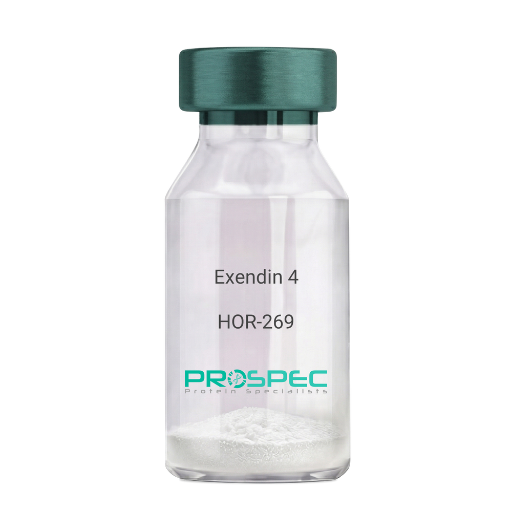
Exendin 4Description:
Exendin-4 Recombinant
Product # :
HOR-269Price :
Quantity :
Shipping Method :
Shipped at Room temp
More Info
- description
- source
- formulation
- purity
- biological activity
- More Info
Description
Exendin-4 Recombinant produced in E.Coli is a non-glycosylated polypeptide chain containing 39 amino acids and having a molecular mass of approximately 4.2kDa. Exendin-4 is purified by proprietary chromatographic techniques.
Source
Escherichia Coli.
Formulation
Lyophilized from a 0.2μm filtered concentrated solution in PBS, pH 7.4.
Purity
Greater than 96.0% as determined by:
(a) Analysis by RP-HPLC.
(b) Analysis by SDS-PAGE.Biological Activity
1. Regulates Glucose levels rapidly
2. Reduces Insulin resistance
3. Reduces Glucagon4. Stimulates beta cell growth production.
More Info
-
Introduction
Exedin-4 Recombinant is a like peptide-1 receptor agonist. The native hormone is produced in the gut of Gila monster Heloderma suspectrum (a type of reptile found in the desert) that stimulates production without causing threateningly low blood sugar, which can occur after using some anti-diabetes products. Recently, researchers used extracted saliva from gila monsters to create an unprecedented breakthrough in Diabetes Type 2 treatment. Unlike other products taken for type 2 diabetes, Exedin-4 has not been linked with weight gain and actually resulted in weight loss, according to the researchers. Exedin-4 enhances glucose-dependant secretion, suppresses inappropriately elevated secretion and slows gastric emptying in vivo. It also promotes B-cell proliferation and neogenesis in vitro and in animal models. Exedin-4 stimulates an increase in acinar cAMP, without stimulating the release of amylase.
-
Physical Appearance
Sterile Filtered White lyophilized (freeze-dried) powder.
-
Stability
Lyophilized Exendin Recombinant although stable at room temperature for 3 weeks, should be stored desiccated below -18°C. Upon reconstitution Exendin-4 should be stored at 4°C between 2-7 days and for future use below -18°C. For long term storage it is recommended to add a carrier protein (0.1% HSA or BSA).Please prevent freeze-thaw cycles.
-
Solubility
It is recommended to reconstitute the lyophilized Exendin Recombinant in sterile 18MΩ-cm H2O not less than 100 µg/ml, which can then be further diluted to other aqueous solutions.
-
Amino Acid Sequence
HGEGTFTSDL SKQMEEEAVR LFIEWLKNGG PSSGAPPPS.
ProSpec's products are furnished for LABORATORY RESEARCH USE ONLY. The product may not be used as drugs, agricultural or pesticidal products, food additives or household chemicals.
- View Data Sheet
Name :
Adipsin HumanDescription:
Complement Factor D Human Recombinant
Complement factor D, EC 3.4.21.46, Adipsin, C3 convertase activator, Properdin factor D, CFD, DF, PFD, ADN.
Product # :
PRO-1360Price :
Quantity :
Shipping Method :
Shipped with Ice Packs
More Info
- description
- source
- formulation
- purity
- More Info
Description
Adipsin Human Recombinant produced in E.Coli is a single, non-glycosylated polypeptide chain containing 249 amino acids (26-253 a.a) and having a molecular mass of 26.6kDa.Adipsin is fused to a 21 amino acid His-tag at N-terminus.
Source
Escherichia Coli.
Formulation
Adipsin protein solution (1mg/ml) containing 20mM Tris-HCl buffer (pH8.0), 10% glycerol and 0.4M Urea.
Purity
Greater than 85.0% as determined by SDS-PAGE.
More Info
-
Introduction
Complement Factor D (Adipsin), which belongs to the trypsin family of peptidases, is involved in the alternative complement pathway of the complement system where it cleaves factor B. In the alternative complement pathway, Adipsin is best known for its role in humoral suppression of infectious agents. In addition, Adipsin is a serine protease which is secreted by adipocytes into the bloodstream. Ultimately, Adipsin has a high level of expression in fat, proposing a role for adipose tissue in immune system biology.
-
Synonyms
Complement factor D, EC 3.4.21.46, Adipsin, C3 convertase activator, Properdin factor D, CFD, DF, PFD, ADN.
-
Physical Appearance
Sterile Filtered colorless solution.
-
Stability
Store at 4°C if entire vial will be used within 2-4 weeks. Store, frozen at -20°C for longer periods of time. For long term storage it is recommended to add a carrier protein (0.1% HSA or BSA).Avoid multiple freeze-thaw cycles.
-
Amino Acid Sequence
MGSSHHHHHH SSGLVPRGSH MILGGREAEA HARPYMASVQ LNGAHLCGGV LVAEQWVLSA AHCLEDAADG KVQVLLGAHS LSQPEPSKRL YDVLRAVPHP DSQPDTIDHD LLLLQLSEKA TLGPAVRPLP WQRVDRDVAP GTLCDVAGWG IVNHAGRRPD SLQHVLLPVL DRATCNRRTH HDGAITERLM CAESNRRDSC KGDSGGPLVC GGVLEGVVTS GSRVCGNRKK PGIYTRVASY AAWIDSVLA.
ProSpec's products are furnished for LABORATORY RESEARCH USE ONLY. The product may not be used as drugs, agricultural or pesticidal products, food additives or household chemicals.
- View Data Sheet
Name :
Dnak SBD C-terminus E.ColiDescription:
Dnak Substrate Binding Domain C-terminal E.Coli Recombinant
HSP-70, HSP70, DnaK, Chaperone protein dnaK, Heat shock protein 70, Heat shock 70 kDa protein, groP, grpF, seg, b0014, JW0013.
Product # :
HSP-009Price :
Quantity :
Shipping Method :
Shipped with Ice Packs
More Info
- description
- source
- formulation
- purity
- More Info
Description
Recombinant Dnak Substrate Binding Domain C-terminal produced in E.Coli is a single, non-glycosylated polypeptide chain containing 255 amino acids and having a molecular mass of 27.7 kDa.
Source
Escherichia Coli.
Formulation
The DnaK protein contains 25mM Tris-HCl, pH7.5, 100mM NaCl, 5mM DTT and 10% Glycerol.
Purity
Greater than 95.0% as determined by SDS-PAGE.
More Info
-
Introduction
DnaK, originally identified for its DNA replication by bacteriophage l in E. coli is the bacterial hsp70 chaperone. This protein is involved in the folding and assembly of newly synthesized polypeptide chains and in preventing the aggregation of stress-denatured proteins.The protein coding region of the substrate binding domain of DnaK (amino acids 385-638) was amplified by PCR and cloned into an E. coli expression vector. The substrate binding domain of DNAK was purified to apparent homogeneity by using conventional column chromatography techniques. Additional amino acid (Met) is attached at N- terminus.
-
Synonyms
HSP-70, HSP70, DnaK, Chaperone protein dnaK, Heat shock protein 70, Heat shock 70 kDa protein, groP, grpF, seg, b0014, JW0013.
-
Physical Appearance
Sterile filtered colorless solution.
-
Stability
Store at 4°C if entire vial will be used within 2-4 weeks. Store, frozen at -20°C for longer periods of time. For long term storage it is recommended to add a carrier protein (0.1% HSA or BSA).Avoid multiple freeze-thaw cycles.
-
Amino Acid Sequence
MDVKDVLLLD VTPLSLGIET MGGVMTTLIA KNTTIPTKHS QVFSTAEDNQ SAVTIHVLQGERKRAADNKS LGQFNLDGIN PAPRGMPQIE VTFDIDADGI LHVSAKDKNS GKEQKITIKA SSGLNEDEIQ KMVRDAEANA EADRKFEELV QTRNQGDHLL HSTRKQVEEA GDKLPADDKTAIESALTALE TALKGEDKAA IEAKMQELAQ VSQKLMEIAQ QQHAQQQTAG ADASANNAKD DDVVDAEFEE VKDKK.
ProSpec's products are furnished for LABORATORY RESEARCH USE ONLY. The product may not be used as drugs, agricultural or pesticidal products, food additives or household chemicals.
- View Data Sheet
Name :
Vimentin PAT5H8AT AntibodyDescription:
Vimentin PAT5H8AT, Mouse Anti Human
Vimentin, Vim, FLJ36605.
Product # :
ANT-682Price :
Quantity :
Shipping Method :
Shipped with Ice Packs
More Info
- formulation
- More Info
Formulation
1mg/ml containing PBS, pH-7.4, 10% Glycerol and 0.02% Sodium Azide.
More Info
-
Introduction
Vimentin expression in human malignant glioma cells depends on cellular density, algorithms of drug delivery and chemo/radio treatment. Vimentin and detyrosinated microtubules provide structural support for the extensive microtentacles observed in detached tumor cells and a mechanism to promote successful metastatic spread. Primary colorectal carcinomas display aberrant expression of vimentin, and have activated Notch and TGFbeta signaling pathways. Vimentin is a strong arterial substrate for transglutaminases. Transglutaminase-mediated vimentin dimerization results in a novel unifying pathway by which vasodilatory and remodeling responses may be regulated. Ablation of vimentin expression inhibits migration and invasion of colon and breast cancer cell lines. Vimentin is the main intermediate filament protein in mesenchymal cells and is therefore of value in the differential diagnosis of undifferentiated neoplasms.
-
Synonyms
Vimentin, Vim, FLJ36605.
-
Physical Appearance
Sterile filtered colorless solution.
-
Immunogen
Anti-human Vimentin mAb, is derived from hybridization of mouse F0 myeloma cells with spleen cells from BALB/c mice immunized with a recombinant human Vimentin protein 1-466 amino acids purified from E. coli.
-
Ig Subclass
Mouse IgG1 heavy chain and k light chain.
-
Clone
PAT5H8AT.
-
Applications
Vimentin antibody has been tested by ELISA, Western blot analysis, Flow cytometry and ICC/IF to assure specificity and reactivity. Since application varies, however, each investigation should be titrated by the reagent to obtain optimal results.
-
Type
Mouse Anti Human Monoclonal.
-
Storage Procedures
For periods up to 1 month store at 4°C, for longer periods of time, store at -20°C. Prevent freeze thaw cycles.
-
Purification Method
Vimentin antibody was purified from mouse ascitic fluids by protein-A affinity chromatography.
ProSpec's products are furnished for LABORATORY RESEARCH USE ONLY. The product may not be used as drugs, agricultural or pesticidal products, food additives or household chemicals.
- View Data Sheet
Name :
EPHX1 Human, Sf9Description:
Epoxide Hydrolase 1 Microsomal Human Recombinant, sf9
Epoxide hydrolase 1, Epoxide hydratase, Microsomal epoxide hydrolase, Meh, EPHX1, EPHX, EPOX, Epoxide Hydrolase 1 Microsomal, Microsomal Epoxide Hydrolase, EC 3.3.2.9, HYL1
Product # :
ENZ-1076Price :
Quantity :
Shipping Method :
Shipped with Ice Packs
More Info
- description
- source
- formulation
- purity
- More Info
Description
EPHX1 produced in Sf9 Insect cells is a single, glycosylated polypeptide chain containing 442 amino acids (21-455 a.a.) and having a molecular mass of 51.5kDaEPHX1 is expressed with an 6 amino acid His tag at C-Terminus and purified by proprietary chromatographic techniques.
Source
Sf9, Insect cells.
Formulation
EPHX1 protein solution (0.25mg/ml) contains 20mM Tris-HCl buffer (pH 8.0) 50% glycerol,1mM DTT and 0.1M NaCl.
Purity
Greater than 85% as determined by SDS-PAGE.
More Info
-
Introduction
Epoxide Hydrolase 1 Microsomal (EPHX1) is a vital biotransformation enzyme which transfers epoxides from the degradation of aromatic compounds to trans-dihydrodiols that can be conjugated and excreted from the body. Epoxide hydrolase plays a role in both activation and detoxification of epoxides. Mutations in EPHX1 trigger preeclampsia, epoxide hydrolase deficiency or increased epoxide hydrolase activity.
-
Synonyms
Epoxide hydrolase 1, Epoxide hydratase, Microsomal epoxide hydrolase, Meh, EPHX1, EPHX, EPOX, Epoxide Hydrolase 1 Microsomal, Microsomal Epoxide Hydrolase,
EC 3.3.2.9, HYL1 -
Physical Appearance
Sterile Filtered colorless solution.
-
Stability
Store at 4°C if entire vial will be used within 2-4 weeks. Store, frozen at -20°C for longer periods of time. For long term storage it is recommended to add a carrier protein (0.1% HSA or BSA). Avoid multiple freeze-thaw cycles.
-
Amino Acid Sequence
MRDKEETLPL EDGWWGPGTR SAAREDDSIR PFKVETSDEE IHDLHQRIDK FRFTPPLEDS CFHYGFNSNY LKKVISYWRN EFDWKKQVEI LNRYPHFKTK IEGLDIHFIH VKPPQLPAGH TPKPLLMVHG WPGSFYEFYK IIPLLTDPKN HGLSDEHVFE VICPSIPGYG FSEASSKKGF NSVATARIFY KLMLRLGFQE FYIQGGDWGS LICTNMAQLV PSHVKGLHLN MALVLSNFST LTLLLGQRFG RFLGLTERDV ELLYPVKEKV FYSLMRESGY MHIQCTKPDT VGSALNDSPV GLAAYILEKF STWTNTEFRY LEDGGLERKF SLDDLLTNVM LYWTTGTIIS SQRFYKENLG QGWMTQKHER MKVYVPTGFS AFPFELLHTP EKWVRFKYPK LISYSYMVRG GHFAAFEEPE LLAQDIRKFL SVLERQHHHH HH.
ProSpec's products are furnished for LABORATORY RESEARCH USE ONLY. The product may not be used as drugs, agricultural or pesticidal products, food additives or household chemicals.
- View Data Sheet
Name :
CNDP1 Human, ActiveDescription:
CNDP Dipeptidase 1 Human Recombinant, Active
Carnosine Dipeptidase 1 (Metallopeptidase M20 Family), Glutamate Carboxypeptidase-Like Protein 2, CNDP Dipeptidase 1, Serum Carnosinase, Carnosinase 1, CPGL2, CN1, Carnosine Dipeptidase 1, EC 3.4.13.20, HsT2308.
Product # :
ENZ-1022Price :
Quantity :
Shipping Method :
Shipped with Ice Packs
More Info
- description
- source
- formulation
- purity
- biological activity
- More Info
Description
CNDP1 Human Recombinant produced in Sf9 Baculovirus cells is a single, glycosylated polypeptide chain containing 489 amino acids (27-507 a.a.) and having a molecular mass of 54.9kDa (Migrates at 50-70kDa on SDS-PAGE under reducing conditions).CNDP1 is expressed with a 8 amino acid His tag at C-Terminus and purified by proprietary chromatographic techniques.
Source
Sf9, Insect cells.
Formulation
CNDP1 protein solution (0.25mg/ml) contains Phosphate Buffered Saline (pH 7.4) and 10% glycerol.
Purity
Greater than 95.0% as determined by SDS-PAGE.
Biological Activity
Specific activity is >3,000 pmol/min/ug, and as measured by the hydrolysis of carnosine per minute at pH6.8 at 25°C.More Info
-
Introduction
CNDP Dipeptidase 1, also known as CNDP1 is a member of the peptidase M20A family. CNDP1 Mannheim which is the shortest allelic form has been more common in the absence of nephropathy in addition to being associated with lower serum carnosinase levels. Furthermore, Carnosine inhibited the increased production of fibronectin as well as collagen type VI in podocytes and the increased production of TGF-beta in mesangial cells. Diabetic patients with the CNDP1 Mannheim variant are less at risk for nephropathy. In addition, on renal cells carnosine protects against the adverse effects of high glucose levels.
-
Synonyms
Carnosine Dipeptidase 1 (Metallopeptidase M20 Family), Glutamate Carboxypeptidase-Like Protein 2, CNDP Dipeptidase 1, Serum Carnosinase, Carnosinase 1, CPGL2, CN1, Carnosine Dipeptidase 1, EC 3.4.13.20, HsT2308.
-
Physical Appearance
Sterile filtered colorless solution.
-
Stability
Store at 4°C if entire vial will be used within 2-4 weeks.Store, frozen at -20°C for longer periods of time. For long term storage it is recommended to add a carrier protein (0.1% HSA or BSA).Avoid multiple freeze-thaw cycles.
-
Amino Acid Sequence
SPSPPPALLE KVFQYIDLHQ DEFVQTLKEW VAIESDSVQP VPRFRQELFR MMAVAADTLQ RLGARVASVD MGPQQLPDGQ SLPIPPVILA ELGSDPTKGT VCFYGHLDVQ PADRGDGWLT DPYVLTEVDG KLYGRGATDN KGPVLAWINA VSAFRALEQD LPVNIKFIIE GMEEAGSVAL EELVEKEKDR FFSGVDYIVI SDNLWISQRK PAITYGTRGN SYFMVEVKCR DQDFHSGTFG GILHEPMADL VALLGSLVDS SGHILVPGIY DEVVPLTEEE INTYKAIHLD LEEYRNSSRV EKFLFDTKEE ILMHLWRYPS LSIHGIEGAF DEPGTKTVIP GRVIGKFSIR LVPHMNVSAV EKQVTRHLED VFSKRNSSNK MVVSMTLGLH PWIANIDDTQ YLAAKRAIRT VFGTEPDMIR DGSTIPIAKM FQEIVHKSVV LIPLGAVDDG EHSQNEKINR WNYIEGTKLF AAFFLEMAQL HLEHHHHHH.
ProSpec's products are furnished for LABORATORY RESEARCH USE ONLY. The product may not be used as drugs, agricultural or pesticidal products, food additives or household chemicals.
- View Data Sheet
Name :
RPL35 HumanDescription:
Ribosomal Protein L35 Human Recombinant
Ribosomal Protein L35, 60S Ribosomal Protein L35, RPL35A, DBA5, L35, 60S ribosomal protein L35.
Product # :
PRO-2097Price :
Quantity :
Shipping Method :
Shipped with Ice Packs
More Info
- description
- source
- formulation
- purity
- More Info
Description
RPL35 Human Recombinant produced in E.Coli is a single, non-glycosylated polypeptide chain containing 146 amino acids (1-123 a.a) and having a molecular mass of 16.9kDa. RPL35 is fused to a 23 amino acid His-tag at N-terminus & purified by proprietary chromatographic techniques.
Source
Escherichia Coli.
Formulation
RPL35 protein solution (0.25 mg/ml) containing 20mM Tris-HCl buffer (pH 8.0) and 10% glycerol.
Purity
Greater than 80% as determined by SDS-PAGE.
More Info
-
Introduction
Ribosomal Protein L35, also known as RPL35 is a ribosomal protein which is a component of the 60S subunit. Ribosomes are the organelles which catalyze protein synthesis, they consist of a small 40S subunit and a large 60S subunit. Jointly these subunits are composed of 4 RNA species and about 80 structurally different proteins. RPL35 is a member of the L29P family of ribosomal proteins. RPL35 is located in the cytoplasm. As typicaly for genes encoding ribosomal proteins, there are multiple processed pseudogenes of RPL35 dispersed through the genome.
-
Synonyms
Ribosomal Protein L35, 60S Ribosomal Protein L35, RPL35A, DBA5, L35, 60S ribosomal protein L35.
-
Physical Appearance
Sterile Filtered clear solution.
-
Stability
Store at 4°C if entire vial will be used within 2-4 weeks. Store, frozen at -20°C for longer periods of time. For long term storage it is recommended to add a carrier protein (0.1% HSA or BSA).Avoid multiple freeze-thaw cycles.
-
Amino Acid Sequence
MGSSHHHHHH SSGLVPRGSH MGSMAKIKAR DLRGKKKEEL LKQLDDLKVE LSQLRVAKVT GGAASKLSKI RVVRKSIARV LTVINQTQKE NLRKFYKGKK YKPLDLRPKK TRAMRRRLNK HEENLKTKKQ QRKERLYPLR KYAVKA.
ProSpec's products are furnished for LABORATORY RESEARCH USE ONLY. The product may not be used as drugs, agricultural or pesticidal products, food additives or household chemicals.
- View Data Sheet
Name :
ARHGDIB HumanDescription:
Rho GDP Dissociation Inhibitor (GDI) Beta Human Recombinant
Rho GDP dissociation inhibitor (GDI) beta, Rho GDI 2, Rho-GDI beta, GDIA2, GDID4, Ly-GDI, RAP1GN1.
Product # :
PRO-1067Price :
Quantity :
Shipping Method :
Shipped with Ice Packs
More Info
- description
- source
- formulation
- purity
- More Info
Description
ARHGDIB Human Recombinant produced in E. coli is a single polypeptide chain containing 428 amino acids (1-201) and having a molecular mass of 49.4kDa.ARHGDIB is fused to a 227 amino acid GST-tag at N-terminus & purified by proprietary chromatographic techniques.
Source
E.coli.
Formulation
The ARHGDIB solution (1mg/1ml) contains phosphate-buffered Saline (pH 7.4) and 10% glycerol.
Purity
Greater than 90% as determined by SDS-PAGE.
More Info
-
Introduction
Members of the Rho (or ARH) protein family and other Ras-related small GTP-binding proteins are involved in various cellular procedures, such as cell cytoskeletal organization, secretion, proliferation, and signaling. The GTPBPs are active only in the GTP-bound state. At least 3 types of proteins firmly regulate cycling between the GTP-bound and GDP-bound states: GDP-dissociation inhibitors (GDIs), GTPase-activating proteins (GAPs) and guanine nucleotide-releasing factors (GRFs). ARHGDIB and the other GDIs lower the level of GDP dissociation from Ras-like GTPases.
-
Synonyms
Rho GDP dissociation inhibitor (GDI) beta, Rho GDI 2, Rho-GDI beta, GDIA2, GDID4, Ly-GDI, RAP1GN1.
-
Physical Appearance
Sterile Filtered colorless solution.
-
Stability
Store at 4°C if entire vial will be used within 2-4 weeks. Store, frozen at -20°C for longer periods of time. For long term storage it is recommended to add a carrier protein (0.1% HSA or BSA).Avoid multiple freeze-thaw cycles.
-
Amino Acid Sequence
MSPILGYWKI KGLVQPTRLL LEYLEEKYEE HLYERDEGDK WRNKKFELGL EFPNLPYYID GDVKLTQSMA IIRYIADKHN MLGGCPKERA EISMLEGAVL DIRYGVSRIA YSKDFETLKV DFLSKLPEML KMFEDRLCHK TYLNGDHVTH PDFMLYDALD VVLYMDPMCL DAFPKLVCFK KRIEAIPQID KYLKSSKYIA WPLQGWQATF GGGDHPPKSD LVPRGSHMTE KAPEPHVEED DDDELDSKLN YKPPPQKSLK ELQEMDKDDE SLIKYKKTLL GDGPVVTDPK APNVVVTRLT LVCESAPGPI TMDLTGDLEA LKKETIVLKE GSEYRVKIHF KVNRDIVSGL KYVQHTYRTG VKVDKATFMV GSYGPRPEEY EFLTPVEEAP KGMLARGTYH NKSFFTDDDK QDHLSWEWNL SIKKEWTE
ProSpec's products are furnished for LABORATORY RESEARCH USE ONLY. The product may not be used as drugs, agricultural or pesticidal products, food additives or household chemicals.
- View Data Sheet
Name :
VEGF HumanDescription:
Vascular Endothelial Growth Factor Human Recombinant
Vascular endothelial growth factor A, VEGF-A, Vascular permeability factor, VPF, VEGF, MGC70609.
Product # :
CYT-241Price :
Quantity :
Shipping Method :
Shipped at Room temp
More Info
- description
- source
- formulation
- purity
- biological activity
- More Info
Description
Vascular Endothelial Growth Factor Human Recombinant produced in E.Coli is a double, non-glycosylated, polypeptide chain containing 165 amino acids and having a molecular mass of 38.2kDa.The VEGF is purified by proprietary chromatographic techniques.
Source
Escherichia Coli.
Formulation
The VEGF protein was lyophilized from a concentrated (1mg/ml) solution with no additives.
Purity
Greater than 95.0% as determined by:
(a) Analysis by RP-HPLC.
(b) Analysis by SDS-PAGE.Biological Activity
Determined by the dose-dependent stimulation of the proliferation of human umbilical vein endothelial cells (HUVEC) using a concentration range of 3.7-5.6 ng/ml, corresponding to a Specific Activity of 178,570-270,270IU/mg.More Info
-
Introduction
Vascular endothelial growth factor (VEGF) is an important signaling protein involved in vessel formation As its name implies, VEGF activity has been mostly studied on cells of the vascular endothelium, although it does have effects on a number of other cell types (e.g. stimulation monocyte/macrophage migration, neurons, cancer cells, kidney epithelial cells ). VEGF mediates increased vascular permeability, induces vasculogenesis and endothelial cell production, promotes cell migration, and inhibits apoptosis. In vitro, VEGF has been shown to stimulate endothelial cell mitogenesis and cell migration. VEGF is also a vasodilator and increases microvascular permeability and was originally referred to as vascular permeability factor. VEGF is located in normal cartilage though only osteoarthritic cartilage expresses the VEGF receptors, NP1, VEGFR1 and VEGFR2. The VEGF level in the culture media from OA chondrocytes was more than 3 folds higher than in media from normal chondrocytes
-
Synonyms
Vascular endothelial growth factor A, VEGF-A, Vascular permeability factor, VPF, VEGF, MGC70609.
-
Physical Appearance
Sterile Filtered White lyophilized (freeze-dried) powder.
-
Stability
Lyophilized VEGF although stable at room temperature for 3 weeks, should be stored desiccated below -18°C. Upon reconstitution VEGF should be stored at 4°C between 2-7 days and for future use below -18°C.For long term storage it is recommended to add a carrier protein (0.1% HSA or BSA).Please prevent freeze-thaw cycles.
-
Solubility
It is recommended to reconstitute the lyophilized VEGF in sterile 18M-cm H2O not less than 100µg/ml, which can then be further diluted to other aqueous solutions.
-
Amino Acid Sequence
APMAEGGGQN HHEVVKFMDV YQRSYCHPIE TLVDIFQEYP DEIEYIFKPS CVPLMRCGGC CNDEGLECVP TEESNITMQI MRIKPHQGQH IGEMSFLQHN KCECRPKKDR ARQENPCGPC SERRKHLFVQ DPQTCKCSCK NTDSRCKARQ LELNERTCRC DKPRR
-
Protein content
VEGF protein quantitation was carried out by two independent methods:1. UV spectroscopy at 280 nm using the absorbency value of 0.2875 as the extinction coefficient for a 0.1% (1mg/ml) solution. This value is calculated by the PC GENE computer analysis program of protein sequences (IntelliGenetics). 2. Analysis by RP-HPLC, using a calibrated solution of VEGF as a Reference Standard.
ProSpec's products are furnished for LABORATORY RESEARCH USE ONLY. The product may not be used as drugs, agricultural or pesticidal products, food additives or household chemicals.
- View Data Sheet
Name :
M CSF MouseDescription:
Macrophage-Colony Stimulating Factor Mouse Recombinant
CSF-1, Lanimostim, MCSF, M-CSF.
Product # :
CYT-439Price :
Quantity :
Shipping Method :
Shipped at Room temp
More Info
- description
- source
- formulation
- purity
- biological activity
- More Info
Description
Macrophage Colony Stimulating Factor Mouse Recombinant produced in E.coli is a disulfide linked homodimer, non-glycosylated, polypeptide chain containing 2 x 156 amino acids and having a total molecular mass of 36.4 KD.MCSF is purified by proprietary chromatographic techniques.
Source
Escherichia Coli.
Formulation
The protein was lyophilized from a sterile (0.2µm) filtered solution containing 10mM sodium phosphate, 50mM sodium chloride, pH 7.5.
Purity
Greater than 95.0% as determined by SDS-PAGE.
Biological Activity
The ED50, as calculated by the dose-dependant stimulation of the proliferation of murine M-NFS-60 indicator cells is 1.33ng/ml corresponding to a specific activity of 7.5x10⁵ units/mg.
More Info
-
Introduction
Granulocyte/Macrophage Colony-Stimulating Factors are cytokines that act in hematopoiesis by controlling the production, differentiation, and function of 2 related white cell populations of the blood, the granulocytes and the monocytes-macrophages. CSF-1 induces cells of the monocyte/macrophage lineage. It plays a role in immunological defenses, bone metabolism, lipoproteins clearance, fertility and pregnancy.
-
Synonyms
CSF-1, Lanimostim, MCSF, M-CSF.
-
Physical Appearance
Sterile Filtered White lyophilized (freeze-dried) powder.
-
Stability
Lyophilized M-CSF although stable at room temperature for 3 weeks, should be stored desiccated below -18°C. Upon reconstitution MCSF should be stored at 4°C between 2-7 days and for future use below -18°C.Please prevent freeze-thaw cycles.
-
Solubility
It is recommended to reconstitute the lyophilized M-CSF in sterile 18MΩ-cm H2O not less than 100µg/ml, which can then be further diluted to other aqueous solutions.
-
Amino Acid Sequence
MKEVSEHCSH MIGNGHLKVL QQLIDSQMET SCQIAFEFVD QEQLDDPVCY LKKAFFLVQD IIDETMRFKD NTPNANATER LQELSNNLNS CFTKDYEEQN KACVRTFHET PLQLLEKIKN FFNETKNLLE KDWNIFTKNC NNSFAKCSSR DVVTKP.
-
Background
Macrophage-Colony Stimulating Factor Mouse Recombinant: An In-Depth Analysis
Abstract:
Macrophage-Colony Stimulating Factor (M-CSF) is a crucial cytokine involved in the regulation of macrophage biology, including their differentiation, survival, and function. This research paper provides an in-depth analysis of M-CSF Mouse Recombinant, focusing on its structure, signaling pathways, and diverse functions in the context of human research. Additionally, the paper explores the therapeutic potential of M-CSF modulation in various diseases.
Introduction:
M-CSF plays a vital role in the development and maintenance of macrophages, key immune cells involved in innate immunity and tissue homeostasis. This paper aims to provide a comprehensive analysis of M-CSF Mouse Recombinant, highlighting its importance in human macrophage biology and its potential therapeutic applications.
Structure and Function of M-CSF:
M-CSF is a homodimeric protein that binds to its receptor, CSF-1R, leading to the activation of downstream signaling pathways. It regulates the proliferation, survival, and activation of macrophages, influencing immune responses and tissue remodeling processes.
Signaling Pathways:
Upon binding to CSF-1R, M-CSF triggers various intracellular signaling pathways, including the MAPK pathway, PI3K/Akt pathway, and JAK/STAT pathway. These pathways regulate gene expression and mediate cellular responses, impacting macrophage functions.
Role in Macrophage Development and Function:
M-CSF is essential for the differentiation and maturation of macrophages from hematopoietic progenitor cells. It promotes the survival, proliferation, and activation of macrophages, enhancing their phagocytic activity, cytokine production, and antigen presentation capabilities.
Therapeutic Potential:
Given its crucial role in macrophage biology, M-CSF modulation has emerged as a potential therapeutic strategy. M-CSF inhibitors and CSF-1R antagonists have shown promise in the treatment of inflammatory and autoimmune diseases, as well as certain cancers. Targeting M-CSF signaling can modulate immune responses and affect disease progression.
Clinical Applications and Future Directions:
The therapeutic potential of M-CSF modulation is being explored in various clinical settings. Clinical trials investigating M-CSF inhibitors as monotherapy or combination therapy are underway in diseases such as rheumatoid arthritis and cancer. Future research should focus on understanding the intricate mechanisms of M-CSF signaling, optimizing therapeutic strategies, and developing personalized treatment approaches.
ProSpec's products are furnished for LABORATORY RESEARCH USE ONLY. The product may not be used as drugs, agricultural or pesticidal products, food additives or household chemicals.
- View Data Sheet
Name :
SERPINA7 HumanDescription:
Serpin Peptidase Inhibitor, Clade A Member 7 Human
Product # :
PRO-2739Price :
Quantity :
Shipping Method :
Shipped at Room temp
More Info
- description
- source
- formulation
- purity
- More Info
Description
Human Serpin Peptidase Inhibitor, Clade A Member 7 Protein produced in Human plasma having a molecular mass of approximately 55kD.
Source
Human serum.
Formulation
The protein was lyophilized (0.2 µm filtered) from 20mM NH4HCO3.
Purity
Greater than 98.0%.
More Info
-
Introduction
Thyroxine binding globulin, also known as SERPINA7, is synthesized in the liver and carried in the blood.
SERPINA7 may be decreased or increased in a number of diseases. Its main value in diagnosis is to assess thyroid function by determining the amount of free T3/T4.
Thyroxine binding globulin, binds and transports thyroid hormones in the circulation. SERPINA7 carries the majority of the T3/T4 in the blood. SERPINA7 has only one binding site for T3/T4 and about 25% of Thyroxine binding globulin is usually saturated. -
Physical Appearance
Sterile Filtered White lyophilized (freeze-dried) powder.
-
Stability
SERPINA7 Human although stable at room temperature for 3 weeks, should be stored between 2-8°C.
-
Solubility
It is recommended to reconstitute the lyophilized SERPINA7 Human in phosphate buffer containing 0.15M NaCl.
-
Human Virus Test
Starting material donor has been tested and certified negative for antibodies to HIV-1, HIV-2, HCV, HBSAG, Parvovirus B19, Syphilis and HIV/HBV/HCV (PCR).
ProSpec's products are furnished for LABORATORY RESEARCH USE ONLY. The product may not be used as drugs, agricultural or pesticidal products, food additives or household chemicals.
- View Data Sheet
Name :
CLEC7A HumanDescription:
C-Type Lectin Domain Family 7, Member A Human Recombinant
BGR, Dendritic Cell-Associated C-Type Lectin-1, Dendritic Cell-Associated C-Type Lectin 1, C-Type Lectin Domain Family 7, Member A, Lectin-Like Receptor 1, CD369 Antigen, CANDF4, SCARE2, CD369, C-Type Lectin Domain Containing 7A, C-Type Lectin Domain Family 7 Member A, C-Type (Calcium Dependent, Carbohydrate-Recognition Domain) Lectin, Superfamily Member 12, C-Type Lectin Superfamily Member 12, DC-Associated C-Type Lectin 1, Beta-Glucan Receptor, Dectin-1, CLECSF12, DECTIN1.
Product # :
PRO-2634Price :
Quantity :
Shipping Method :
Shipped with Ice Packs
More Info
- description
- source
- formulation
- purity
- More Info
Description
CLEC7A Human Recombinant produced in Sf9 Baculovirus cells is a single, glycosylated polypeptide chain containing 183 amino acids (71-244 a.a) and having a molecular mass of 21kDa. CLEC7A is fused to a 9 amino acid His-tag at C-terminus & purified by proprietary chromatographic techniques.
Source
Sf9, Baculovirus cells.
Formulation
The CLEC7A solution (0.5mg/1ml) contains phosphate buffered saline (pH7.4) and 10% glycerol.
Purity
Greater than 95.0% as determined by SDS-PAGE.
More Info
-
Introduction
C-type lectin domain family 7 member A 1 or CLEC7A is a protein, that in the innate immune system, acts against fungal pathogens. CLEC7A can be found in the immune system response cells such as monocytes, macrophages & neutrophils, or in dendritic and T cells. The protein is enhanced by macrophages by using GM-CSF, IL-4, or IL-13, or diminishes by dexamethasone, IL-10 and LPS.
-
Synonyms
BGR, Dendritic Cell-Associated C-Type Lectin-1, Dendritic Cell-Associated C-Type Lectin 1, C-Type Lectin Domain Family 7, Member A, Lectin-Like Receptor 1, CD369 Antigen, CANDF4, SCARE2, CD369, C-Type Lectin Domain Containing 7A, C-Type Lectin Domain Family 7 Member A, C-Type (Calcium Dependent, Carbohydrate-Recognition Domain) Lectin, Superfamily Member 12, C-Type Lectin Superfamily Member 12, DC-Associated C-Type Lectin 1, Beta-Glucan Receptor, Dectin-1, CLECSF12, DECTIN1.
-
Physical Appearance
Sterile Filtered clear solution.
-
Stability
Store at 4°C if entire vial will be used within 2-4 weeks. Store, frozen at -20°C for longer periods of time. For long term storage it is recommended to add a carrier protein (0.1% HSA or BSA).Avoid multiple freeze-thaw cycles.
-
Amino Acid Sequence
ADPRHNSGRN PEEKDNFLSR NKENHKPTES SLDEKVAPSK ASQTTGGFSQ SCLPNWIMHG KSCYLFSFSG NSWYGSKRHC SQLGAHLLKI DNSKEFEFIE SQTSSHRINA FWIGLSRNQS EGPWFWEDGS AFFPNSFQVR NTVPQESLLH NCVWIHGSEV YNQICNTSSY SICEKELHHH HHH.
ProSpec's products are furnished for LABORATORY RESEARCH USE ONLY. The product may not be used as drugs, agricultural or pesticidal products, food additives or household chemicals.
- View Data Sheet
Name :
SF20 Mouse, HisDescription:
MYDGF Mouse Recombinant, His Tag
D17Wsu104e, Il25, Ly6elg, MYDGF, Interleukin-25, IL-25, Stromal cell-derived growth factor SF20.
Product # :
CYT-1040Price :
Quantity :
Shipping Method :
Shipped with Ice Packs
More Info
- description
- source
- formulation
- purity
- More Info
- sds-page
Description
MYDGF Mouse Recombinant produced in E.Coli is a single, non-glycosylated polypeptide chain containing 165 amino acids (25-166 a.a) and having a molecular mass of 18.1kDa. MYDGF is fused to a 23 amino acid His-tag at N-terminus & purified by proprietary chromatographic techniques.
Source
Escherichia Coli.
Formulation
MYDGF protein solution (0.5mg/ml) containing 20mM Tris-HCl (pH8.0), 0.1M NaCl and 10% glycerol.
Purity
Greater than 95.0% as determined by SDS-PAGE.
sds-page
More Info
-
Introduction
Myeloid-derived growth factor (Mydgf) is a paracrine-acting protein and a bone marrow-derived monocyte which stimulates cardiac myocyte survival and adaptive angiogenesis for cardiac protection and repair after myocardial infarction. Mydgf induces endothelial cell proliferation through a MAPK1/3-, STAT3- and CCND1-mediated signaling lane. When comparing wild-type mice to mice with a Mydgf-deficiency, the later develop larger infarct scars and more acute contractile dysfunction.
-
Synonyms
D17Wsu104e, Il25, Ly6elg, MYDGF, Interleukin-25, IL-25, Stromal cell-derived growth factor SF20.
-
Physical Appearance
Sterile Filtered clear solution.
-
Stability
Store at 4°C if entire vial will be used within 2-4 weeks. Store, frozen at -20°C for longer periods of time. For long term storage it is recommended to add a carrier protein (0.1% HSA or BSA).Avoid multiple freeze-thaw cycles.
-
Amino Acid Sequence
MGSSHHHHHH SSGLVPRGSH MGSVSEPTTV PFDVRPGGVV HSFSQDVGPG NKFTCTFTYA SQGGTNEQWQ MSLGTSEDSQ HFTCTIWRPQ GKSYLYFTQF KAELRGAEIE YAMAYSKAAF ERESDVPLKS EEFEVTKTAV SHRPGAFKAE LSKLVIVAKA ARSEL.
ProSpec's products are furnished for LABORATORY RESEARCH USE ONLY. The product may not be used as drugs, agricultural or pesticidal products, food additives or household chemicals.