Search results
577 results found for “Superoxide Dismutase”
Name
Description
Product #
Price
Quantity
Shipping Method
- View Data Sheet
Name :
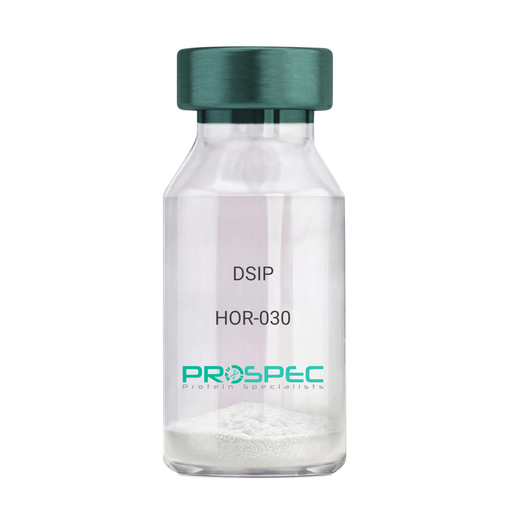
DSIPDescription:
Delta Sleep Inducing Peptide
Product # :
HOR-030Price :
Quantity :
Shipping Method :
Shipped at Room temp
More Info
- description
- formulation
- purity
- More Info
Description
DSIP Synthetic is a single, non-glycosylated polypeptide chain containing 15 amino acids, having a molecular mass of 1419.55 Dalton and a Molecular formula of C62H98N16O22.
Formulation
The protein was lyophilized with no additives.
Purity
Greater than 97.0% as determined by analysis by RP-HPLC.
More Info
-
Physical Appearance
Sterile Filtered White lyophilized (freeze-dried) powder.
-
Stability
Lyophilized DSIP although stable at room temperature for 3 weeks, should be stored desiccated below -18°C. Upon reconstitution DSIP should be stored at 4°C between 2-7 days and for future use below -18°C. For long term storage it is recommended to add a carrier protein (0.1% HSA or BSA). Please prevent freeze-thaw cycles.
-
Solubility
It is recommended to reconstitute the lyophilized DSIP in sterile 18MΩ-cm H2O not less than 100 µg/ml, which can then be further diluted to other aqueous solutions.
-
Amino Acid Sequence
H-Gly-Glu-Pro-Pro-Pro-Gly-Lys-Pro-Ala-Asp-Asp-Ala-Gly-Leu-Val-OH
-
Background
Delta Sleep-Inducing Peptide (DSIP), also known as Sleep-Promoting Peptide, is a neuropeptide that has been the subject of extensive research due to its potential role in sleep regulation, stress response, and neuroprotection. This nonapeptide, first isolated from the cerebral venous blood of rabbits during sleep, has been shown to induce slow-wave sleep, modulate pain perception, and exhibit potential antioxidant and immunomodulatory properties.
DSIP's primary function is its interaction with the sleep regulatory system. By modulating the release of certain neurotransmitters, DSIP can influence sleep patterns, particularly promoting slow-wave sleep, the most restorative stage of sleep. Studies by Kovalzon et al. (2011) have demonstrated that DSIP can enhance sleep quality in rats, suggesting potential applications in sleep disorders and the promotion of healthy sleep patterns.
In addition to its sleep-inducing effects, DSIP has been shown to possess neuroprotective properties. Research by Zolotarev et al. (2014) found that DSIP could protect neurons from oxidative stress, suggesting potential applications in the treatment of neurodegenerative diseases such as Alzheimer's and Parkinson's disease.
Given its sleep-inducing and neuroprotective effects, DSIP has been proposed as a potential therapeutic agent for a variety of conditions, including sleep disorders, chronic pain, and neurodegenerative diseases. For instance, a study by Spong et al. (2016) found that DSIP could improve sleep quality in patients with chronic insomnia, indicating its potential as a therapeutic agent in the treatment of sleep disorders.
While research on DSIP is promising, it is important to note that most studies have been conducted in animals or in vitro. More research is needed to fully understand the potential effects and applications of DSIP in humans. However, the existing body of research suggests that DSIP could be a promising tool in the treatment of sleep disorders, chronic pain, and neurodegenerative diseases.
ProSpec's products are furnished for LABORATORY RESEARCH USE ONLY. The product may not be used as drugs, agricultural or pesticidal products, food additives or household chemicals.
- View Data Sheet
Name :
CoV-2 NucleocapsidDescription:
Coronavirus 2019 Nucleocapsid Recombinant
Product # :
SARS-013Price :
Quantity :
Shipping Method :
Shipped with Ice Packs
More Info
- description
- source
- formulation
- purity
- More Info
Description
The E.Coli derived recombinant protein contains the Coronavirus 2019 full-length nucleocapsid: Gene bank- MN908947, fused to 6xHis tag at C-terminal and having a Mw. Of 48 kDa as appears on SDS-PAGE.
Source
Escherichia Coli.
Formulation
CoV 2019 protein solution is supplied in PBS and 25mM K2CO3.
Purity
CoV 2019 Nucleocapsid protein is >95% pure as determined SDS-PAGE.
More Info
-
Introduction
A human infecting coronavirus (viral pneumonia) called 2019 novel coronavirus, 2019-nCoV was found in the fish market at the city of Wuhan, Hubei province of China on December 2019.
The 2019-nCoV shares an 87% identity to the 2 bat-derived severe acute respiratory syndrome 2018 SARS-CoV-2 located in Zhoushan of eastern China. 2019-nCoV has an analogous receptor-BD-structure to that of 2018 SARS-CoV, even though there is a.a. diversity so thus the 2019-nCoV might bind to ACE2 receptor protein (angiotensin-converting enzyme 2) in humans.
While bats are possibly the host of 2019-nCoV, researchers suspect that animal from the ocean sold at the seafood market was an intermediate host. RSCU analysis proposes that the 2019-nCoV is a recombinant within the viral spike glycoprotein between the bat coronavirus and an unknown coronavirus.
-
Physical Appearance
Sterile Filtered clear solution.
-
Stability
CoV 2019 Nucleocapsid protein is shipped on ice packs. Upon arrival, Store at -20°C.
-
Amino Acid Sequence
MSDNGPQNQRNAPRITFGGPSDSTGSNQNGERSGARSKQRRPQGLPNNTASWFTALTQHGKED
LKFPRGQGVPINTNSSPDDQIGYYRRATRRIRGGDGKMKDLSPRWYFYYLGTGPEAGLPYGAN
KDGIIWVATEGALNTPKDHIGTRNPANNAAIVLQLPQGTTLPKGFYAEGSRGGSQASSRSSSR
SRNSSRNSTPGSSRGTSPARMAGNGGDAALALLLLDRLNQLESKMSGKGQQQQGQTVTKKSAA
EASKKPRQKRTATKAYNVTQAFGRRGPEQTQGNFGDQELIRQGTDYKHWPQIAQFAPSASAFF
GMSRIGMEVTPSGTWLTYTGAIKLDDKDPNFKDQVILLNKHIDAYKTFPPTEPKKDKKKKADE
TQALPQRQKKQQTVTLLPAADLDDFSKQLQQSMSSADSTQA
ProSpec's products are furnished for LABORATORY RESEARCH USE ONLY. The product may not be used as drugs, agricultural or pesticidal products, food additives or household chemicals.
- View Data Sheet
Name :
FTCD HumanDescription:
Formiminotransferase Cyclodeaminase Human Recombinant
Formiminotransferase-cyclodeaminase, FTCD, LCHC1, LC-1.
Product # :
ENZ-302Price :
Quantity :
Shipping Method :
Shipped with Ice Packs
More Info
- description
- source
- formulation
- purity
- More Info
Description
Formiminotransferase Cyclodeaminase Human Recombinant (also called liver cytosol type 1) produced in SF9, is a glycosylated, polypeptide chain having a molecular mass of 59,749 Dalton.The FTCD is expressed with a -6xHis tag and purified by proprietary chromatographic techniques.
Source
Sf9 insect cells.
Formulation
FTCD is supplied in 16mM HEPES buffer pH-7.6, 240mM sodium chloride, and 20% glycerol.
Purity
Greater than 95% as determined by SDS-PAGE.
More Info
-
Introduction
Formiminotransferase cyclodeaminase is an enzyme which catalyzes the conversion of formiminoglutamateand tetrahydrofolateinto formiminotetrahydrofolateand glutamate.
Formiminotransferase cyclodeaminase, a bifunctional enzyme of tetrahydrofolate synthesis, is the target antigen of anti-LC1 (liver cytosol antigen type 1) autoantibodies. Presence of LC1 autoantibodies is a marker for type 2 autoimmune hepatitis (for which anti-LKM 1/cytochrome P450 2D6 autoantibodies are a further marker).
Serum LC1 autoantibody concentrations appear to fluctuate in parallel with aminotransferase levels, a particularly intriguing observation that suggests a possible role of LC1 autoreactivity in the pathogenic mechanism leading to hepatocyte injury. -
Synonyms
Formiminotransferase-cyclodeaminase, FTCD, LCHC1, LC-1.
-
Stability
Store at 4°C if entire vial will be used within 2-4 weeks. Store, frozen at -20°Cfor longer periods of time. Avoid multiple freeze-thaw cycles.
-
Immunological Functions
1. Binds IgG-type human auto-antibodies.2. Standard ELISA test (checker-board analysis of positive/negative sera panels, immuno-dot test).
-
coating concentration
0.35-0.7 µg/ml (depending on the type of ELISA plate and coating buffer).Suitable for biotinylation and iodination.
ProSpec's products are furnished for LABORATORY RESEARCH USE ONLY. The product may not be used as drugs, agricultural or pesticidal products, food additives or household chemicals.
- View Data Sheet
Name :
sCD4 AntibodyDescription:
Rat Anti-Mouse CD4
gp55, HLA-2, L3 / T4, Ly-4, T cell antigen T4/LEU3, T4, sCD4, CD4mut.
Product # :
ANT-165Price :
Quantity :
Shipping Method :
Shipped at Room temp
More Info
- formulation
- More Info
Formulation
1mg/ml in PBS (after reconstitution).
More Info
-
Introduction
CD4 is a cell-surface glycoprotein found on the mature helper T cells and immature thymocytes, as well as on monocytes and macrophages. (Some cytotoxic T cells have CD4 protein as well.) Normally, about 65% of T cells in the blood are CD4+ (have CD4 protein protruding from their membrane). A mature T cell with either have CD4 or CD8, but not both. During one stage of development T cells develop CD4 and CD8 receptors, but they eventually are differentiated in the thymus and become more specialized.
-
Synonyms
gp55, HLA-2, L3 / T4, Ly-4, T cell antigen T4/LEU3, T4, sCD4, CD4mut.
-
Solubility
Reconstitute with of H20. Mix gently, wash the sides of the vial and wait 30-60 seconds before use.
-
Immunogen
Purified mouse LN CD4+ T cells.
-
Ig Subclass
Rat IgG.
-
Clone
mCD4.
-
Applications
Cytotoxic and staining antibody. For staining, use 10µl/1,000,000 cells. Titer for cytotoxicity should be determined by the investigator Available ConjugatesThis antibody is also available conjugated to biotin and FITC.
-
Type
Rat Anti Mouse Monoclonal.
-
Storage Procedures
Lyophilized: store at 4oC. After reconstitution, if not intended for use within a month, aliquot and store at -20oC.
-
Purification Method
Ion exchange column.
ProSpec's products are furnished for LABORATORY RESEARCH USE ONLY. The product may not be used as drugs, agricultural or pesticidal products, food additives or household chemicals.
- View Data Sheet
Name :
CoV-2 Spike (800-1000)Description:
Coronavirus 2019 Spike (800-1000 a.a.) Recombinant
Product # :
SARS-016Price :
Quantity :
Shipping Method :
Shipped with Ice Packs
More Info
- description
- source
- formulation
- purity
- More Info
Description
The E.Coli derived recombinant protein contains the Coronavirus 2019 Spike (800-1000 a.a.) immunodominant regions, fused to 6xHis tag at C-terminal.
Source
Escherichia Coli.
Formulation
CoV 2019 Spike Protein 1mg/ml solution is supplied in 1x PBS.
Purity
CoV 2019 Spike Protein is >90% pure as determined SDS-PAGE.
More Info
-
Introduction
A human infecting coronavirus (viral pneumonia) called 2019 novel coronavirus, 2019-nCoV was found in the fish market at the city of Wuhan, Hubei province of China on December 2019.
The 2019-nCoV shares an 87% identity to the 2 bat-derived severe acute respiratory syndrome 2018 SARS-CoV-2 located in Zhoushan of eastern China. 2019-nCoV has an analogous receptor-BD-structure to that of 2018 SARS-CoV, even though there is a.a. diversity so thus the 2019-nCoV might bind to ACE2 receptor protein (angiotensin-converting enzyme 2) in humans.
While bats are possibly the host of 2019-nCoV, researchers suspect that animal from the ocean sold at the seafood market was an intermediate host. RSCU analysis proposes that the 2019-nCoV is a recombinant within the viral spike glycoprotein between the bat coronavirus and an unknown coronavirus.
-
Physical Appearance
Sterile Filtered clear solution.
-
Stability
CoV 2019 Spike Protein is shipped on ice packs. Upon arrival, Store at -20°C.
ProSpec's products are furnished for LABORATORY RESEARCH USE ONLY. The product may not be used as drugs, agricultural or pesticidal products, food additives or household chemicals.
- View Data Sheet
Name :
CoV-2-S1 (319-541), BiotinDescription:
Coronavirus 2019 Spike Glycoprotein-S1 Receptor Binding Domain (319-541 a.a), Biotinylated Recombinant
Product # :
SARS-030Price :
Quantity :
Shipping Method :
Shipped at Room temp
More Info
- description
- source
- formulation
- purity
- More Info
Description
The HEK293 derived Biotinylated recombinant protein contains the Coronavirus 2019 Spike Glycoprotein S1 Receptor Binding Domain [ RBD ], Wuhan-Hu-1 strain, amino acids 319-541 fused to His tag & AVI tag at C-terminal and having a molecular mass of 28.7kDa.
Source
HEK293 Cells.
Formulation
CoV-2 S1 RBD protein is lyophilized from 1x PBS pH-7.4 + 10% trehalose.
Purity
Protein is >90% pure as determined SDS-PAGE.
More Info
-
Introduction
A human infecting coronavirus (viral pneumonia) called 2019 novel coronavirus, 2019-nCoV was found in the fish market at the city of Wuhan, Hubei province of China on December 2019.
The 2019-nCoV shares an 87% identity to the 2 bat-derived severe acute respiratory syndrome 2018 SARS-CoV-2 located in Zhoushan of eastern China. 2019-nCoV has an analogous receptor-BD-structure to that of 2018 SARS-CoV, even though there is a.a. diversity so thus the 2019-nCoV might bind to ACE2 receptor protein (angiotensin-converting enzyme 2) in humans.
While bats are possibly the host of 2019-nCoV, researchers suspect that animal from the ocean sold at the seafood market was an intermediate host. RSCU analysis proposes that the 2019-nCoV is a recombinant within the viral spike glycoprotein between the bat coronavirus and an unknown coronavirus.
-
Physical Appearance
Lyophilized freezed dried powder.
-
Stability
Lyophilized Cov-2 S1 Glycoprotein RBD although stable at room temperature for 3 weeks, should be stored desiccated below -18°C. Upon reconstitution CoV2 Spike protein should be stored at 4°C between 2-7 days and for future use below -18°C.For long term storage it is recommended to add a carrier protein (0.1% HSA or BSA).Please prevent freeze-thaw cycles.
-
Solubility
It is recommended to reconstitute the lyophilized CoV-2 S1 protein in sterile 18M-cm H2O at aconcentration of 0.2mg/ml and not less than 0.1mg/ml, which can then be further diluted to other aqueous solutions.
-
Purification Method
Purified by Metal-Afinity chromatographic technique.
ProSpec's products are furnished for LABORATORY RESEARCH USE ONLY. The product may not be used as drugs, agricultural or pesticidal products, food additives or household chemicals.
- View Data Sheet
Name :
PMSGDescription:
Pregnant Mare Serum Gonadotropin
Product # :
HOR-272Price :
Quantity :
Shipping Method :
Shipped at Room temp
More Info
- description
- source
- formulation
- More Info
Description
PMSG is a complex glycoprotein obtained from the serum of pregnant mares. This 43-63 kda protein is capable of supplementing and being substituted for the follicle stimulating and interstitial cell-stimulating hormone of the anterior pituitary gland in both the male and female. Thus PMSG-Intervet stimulates development of the ovarian follicle in the female.
Source
Serum of pregnant mares.
Formulation
The PMSG was lyophilized with no additives.
More Info
-
Introduction
PMSG Hormone is a well know used hormone together with progestogen to increase ovulation just before to artificial insemination. PMSG hormone is a placental glycoprotein produced from the serum of pregnant mares. PMSG comprises of an alfa subunit and a beta subunit. PMSG hormone is secreted from endometrial cups within the pregnant mare uterus aging from 40 to 130 days into their maturation, and once extracted, it can been used to promote artificially estrus in female animals. These assemblies produce PMSG hormone to induce mare's ovarian and repsouctive structures. PMSG can induce the growth of follicles by ovaries and results in ovulatation. PMSG hormone has an about a 4 day half-life of bioactivity in species other than horses. The extended biological activity can cause ovarian stimulation and ovulation. However, PMSG use alone often causes cystic ovarian disease because of the unrestrained ovarian stimulation and due to the sugar molecules which decrease clearance of the hormone. PMSG is more likely to be used than other pituitary hormones due to the extended circulatory half-life. PMSG solely exhibits luteinizing hormone like activity, however in other animal classes it has FSH & LH like activity.
-
Physical Appearance
Sterile Filtered White lyophilized (freeze-dried) powder.
-
Stability
Lyophilized PMSG although stable at room temperature for 3 weeks, should be stored between 2-8°C.
-
Solubility
It is recommended to reconstitute the lyophilized PMSG in sterile 18M-cm H2O at a concentration of 1000 IU/ml, which can then be further diluted to other aqueous solutions.
ProSpec's products are furnished for LABORATORY RESEARCH USE ONLY. The product may not be used as drugs, agricultural or pesticidal products, food additives or household chemicals.
- View Data Sheet
Name :
CA1 E.ColiDescription:
Carbonic Anhydrase-1 E.Coli Recombinant
Carbonate dehydratase, CAN, ECK0125, JW0122, yadF, CA 1, CA I, CA1, CAI, Car 1, Car1, Carbonate dehydratase I, Carbonic anhydrase 1, Carbonic anhydrase B.
Product # :
ENZ-1138Price :
Quantity :
Shipping Method :
Shipped with Ice Packs
More Info
- description
- source
- formulation
- purity
- More Info
Description
CA1 E.Coli Recombinant produced in E.Coli is a single, non-glycosylated polypeptide chain containing 240 amino acids (1-220) and having a molecular mass of 27.0 kDa. CA1 E.Coli is fused to a 20 amino acid His-Tag at N-terminus and purified by proprietary chromatographic techniques.
Source
Escherichia Coli.
Formulation
CA1 E.Coli protein (1mg/ml) is formulated in 20mM Tris-HCl buffer (pH 8.0) containing 1mM DTT, 10% glycerol.
Purity
Greater than 95.0% as determined by SDS-PAGE.
More Info
-
Introduction
CA, also known as carbonic anhydrase is an enzyme that functions around the involvement of CO2 + H2O to HCO3- + H+ (conversion of CO2 to bicarbonate & protons). This enzyme holds a Zn ion as part of its active site. The central function of CA is to preserve the acid-base homeostasis within the blood and various organs. carbonic anhydrase assists Carbonic Anhydrase I to move CO2 to and from tissues.
-
Synonyms
Carbonate dehydratase, CAN, ECK0125, JW0122, yadF, CA 1, CA I, CA1, CAI, Car 1, Car1, Carbonate dehydratase I, Carbonic anhydrase 1, Carbonic anhydrase B.
-
Physical Appearance
Sterile Filtered clear solution.
-
Stability
Store at 4°C if entire vial will be used within 2-4 weeks. Store, frozen at -20°C for longer periods of time. For long term storage it is recommended to add a carrier protein (0.1% HSA or BSA). Avoid multiple freeze-thaw cycles.
-
Amino Acid Sequence
MGSSHHHHHH SSGLVPRGSH MKDIDTLISN NALWSKMLVE EDPGFFEKLA QAQKPRFLWI GCSDSRVPAE RLTGLEPGEL FVHRNVANLV IHTDLNCLSV VQYAVDVLEV EHIIICGHYG CGGVQAAVEN PELGLINNWL LHIRDIWFKH SSLLGEMPQE RRLDTLCELN VMEQVYNLGH STIMQSAWKR GQKVTIHGWA YGIHDGLLRD LDVTATNRET LEQRYRHGIS NLKLKHANHK.
ProSpec's products are furnished for LABORATORY RESEARCH USE ONLY. The product may not be used as drugs, agricultural or pesticidal products, food additives or household chemicals.
- View Data Sheet
Name :
FAS Blocking AntibodyDescription:
FAS Blocking Antibody (CD95), Mouse anti Human
FASLG receptor, Apoptosis-mediating surface antigen FAS, Apo-1 antigen, CD95, Tumor necrosis factor receptor superfamily member 6 TNR6, APT1, FAS1, TNFRSF6.
Product # :
ANT-205Price :
Quantity :
Shipping Method :
Shipped at Room temp
More Info
- More Info
More Info
-
Introduction
The Fas receptor (CD95) mediates apoptotic signaling by Fas-ligand expressed on the surface of other cells. The Fas-FasL interaction plays an important role in the immune system and lack of this system leads to autoimmunity, indicating that Fas-mediated apoptosis removes self-reactive lymphocytes. Fas signaling is also involved in immune surveillance to remove transformed cells and virus infected cells. Binding of FAS to oligimerized FasL on another cell activates apoptotic signaling through a cytoplasmic domain termed the death domain that interacts with signaling adaptors including FAF, FADD and DAX to activate the caspase proteolytic cascade. Caspase-8 and caspase-10 are first activated, to then cleave and activate downstream caspases, and a variety of cellular substrates that lead to cell death. Caspases cleave nuclear lamins, causing the nucleus to break down and lose its normal structure and another caspase substrate is DFF, inducing cleavage and degradation of the genome. Other caspase substrates are involved in cytoskeletal structure, cell cycle regulation and signaling pathways. Activation of JNK kinase, activation of Jun, and production of ceramide may also play roles in Fas-mediated apoptosis. Activation of fas-mediated apoptosis is opposed by I-FLICE and FAP. Viruses and tumors may escape immune surveillance in part through suppression of fas-mediated apoptosis using similar mechanisms.
-
Synonyms
FASLG receptor, Apoptosis-mediating surface antigen FAS, Apo-1 antigen, CD95, Tumor necrosis factor receptor superfamily member 6 TNR6, APT1, FAS1, TNFRSF6.
-
Solubility
Reconstitute with H20. Mix gently, wash the sides of the vial and wait 30-60 seconds before use.
-
Immunogen
Recombinant Human FAS.
-
Ig Subclass
mouse IgG1.
-
Clone
NYRhFAS.
-
Note
This is a BLOCKING antibody and will block FAS-mediated apoptosis.
-
Titer
By direct ELISA, 1:10,000 dilution will yield 0.5 O.D using alkaline phosphatase conjugated rabbit anti-mouse Ig (Jackson Laboratories).
-
Shipping Conditions
Antibody is shipped lyophilized at ambient temperature.
-
Type
Mouse Anti Human Monoclonal.
-
Storage Procedures
In lyophilized form, for long periods, store at 4°C in a dry environment. After reconstitution, if not intended for use within a month, aliquot and store at -20°C.
-
Purification Method
Ion exchange column Protein concentration1 mg/ml in PBS (after reconstitution).
ProSpec's products are furnished for LABORATORY RESEARCH USE ONLY. The product may not be used as drugs, agricultural or pesticidal products, food additives or household chemicals.
- View Data Sheet
Name :
YWHAG Human, HisDescription:
Tyr-3/Trp-5 Monooxygenase Activation Protein Gamma Human Recombinant, His Tag
14-3-3 protein gamma, Protein kinase C inhibitor protein 1, KCIP-1, YWHAG, Tyrosine 3-monooxygenase/tryptophan 5-monooxygenase activation protein, gamma polypeptide.
Product # :
PKA-262Price :
Quantity :
Shipping Method :
Shipped with Ice Packs
More Info
- description
- source
- formulation
- purity
- More Info
Description
YWHAG Human Recombinant produced in E.Coli is a single, non-glycosylated, polypeptide chain containing 246 amino acids (2-247) and having a molecular mass of 36 kDa. YWHAG is fused to His Tag and purified by proprietary chromatographic techniques.
Source
Escherichia Coli.
Formulation
YWHAG solution containing 1x PBS and 50% glycerol.
Purity
Greater than 95.0% as determined by SDS-PAGE.
More Info
-
Introduction
The 14-3-3 family of proteins plays a key regulatory role in signal transduction, checkpoint control, apoptotic and nutrient-sensing pathways. 14-3-3 proteins are highly conserved and ubiquitously expressed. There are at least seven isoforms that have been identified in mammals. The 14-3-3gamma, a subtype of the 14-3-3 family of proteins, was thought to be brain and neuron-specific. It has been shown to interact with RAF1 and protein kinase C, proteins involved in various signal transduction pathways.
-
Synonyms
14-3-3 protein gamma, Protein kinase C inhibitor protein 1, KCIP-1, YWHAG, Tyrosine 3-monooxygenase/tryptophan 5-monooxygenase activation protein, gamma polypeptide.
-
Physical Appearance
Sterile filtered colorless solution.
-
Stability
Store at 4°C if entire vial will be used within 2-4 weeks. Store, frozen at -20°C for longer periods of time. Avoid multiple freeze-thaw cycles.
ProSpec's products are furnished for LABORATORY RESEARCH USE ONLY. The product may not be used as drugs, agricultural or pesticidal products, food additives or household chemicals.
- View Data Sheet
Name :
S100A9 MouseDescription:
S100 Calcium Binding Protein A9 Mouse Recombinant
Calgranulin B, 60B8AG, CAGB, CFAG, CGLB, L1AG, LIAG, MAC387, MIF, MRP14, NIF, P14, Protein S100-A9, S100 calcium-binding protein A9.
Product # :
PRO-878Price :
Quantity :
Shipping Method :
Shipped with Ice Packs
More Info
- description
- source
- formulation
- purity
- More Info
Description
S100A9 Mouse Recombinant produced in E.coli is a single, non-glycosylated polypeptide chain containing 133 amino acids (1-113) and having a molecular mass of 15.2 kDa.The S100A9 is fused to a 20 amino acid His-Tag at N-terminus and purified by proprietary chromatographic techniques.
Source
Escherichia Coli.
Formulation
S100A9 protein (0.5mg/ml) is supplied in 20mM Tris-HCL, pH-8, 1mM DTT and 20% Glycerol.
Purity
Greater than 90.0% as determined by SDS-PAGE.
More Info
-
Introduction
S100A9 is part of the S100 family of proteins containing 2 EF-hand calcium-binding motifs. S100A9 protein is localized in the cytoplasm and/or nucleus of a wide range of cells, and participates in the regulation of several cellular processes such as cell cycle progression and differentiation. S100 genes include no less than 13 proteins which are localized as a cluster on chromosome 1q21. S100A9 is involved in the inhibition of casein kinase and altered expression of this protein is associated with the disease cystic fibrosis.
-
Synonyms
Calgranulin B, 60B8AG, CAGB, CFAG, CGLB, L1AG, LIAG, MAC387, MIF, MRP14, NIF, P14, Protein S100-A9, S100 calcium-binding protein A9.
-
Physical Appearance
S100A9 is supplied as a sterile filtered yellowish solution.
-
Stability
Store at 4°C if entire vial will be used within 2-4 weeks. Store, frozen at -20°C for longer periods of time. For long term storage it is recommended to add a carrier protein (0.1% HSA or BSA).Avoid multiple freeze-thaw cycles.
-
Amino Acid Sequence
MGSSHHHHHH SSGLVPRGSH MANKAPSQME RSITTIIDTF HQYSRKEGHP DTLSKKEFRQ MVEAQLATFM KKEKRNEALI NDIMEDLDTN QDNQLSFEEC MMLMAKLIFA CHEKLHENNP RGHGHSHGKG CGK
ProSpec's products are furnished for LABORATORY RESEARCH USE ONLY. The product may not be used as drugs, agricultural or pesticidal products, food additives or household chemicals.
- View Data Sheet
Name :
CTSZ MouseDescription:
Cathepsin-Z Mouse Recombinant
Cathepsin Z, CTSZ.
Product # :
ENZ-934Price :
Quantity :
Shipping Method :
Shipped with Ice Packs
More Info
- description
- source
- formulation
- purity
- More Info
Description
CTSZ produced in Sf9 Baculovirus cells is a single, glycosylated polypeptide chain containing 292 amino acids (23-306a.a.) and having a molecular mass of 32.8kDa. (Molecular size on SDS-PAGE will appear at approximately 28-40kDa). CTSZ is expressed with an 8 amino acid His tag at C-Terminus and purified by proprietary chromatographic techniques.
Source
Sf9, Baculovirus cells.
Formulation
CTSZ protein solution (0.5mg/ml) contains Phosphate Buffered Saline (pH 7.4) and 10% glycerol.
Purity
Greater than 95% as determined by SDS-PAGE.
More Info
-
Introduction
Cathepsin-Z (CTSZ) is a lysosomal cysteine proteinase and member of the peptidase C1 family. CTSZ, which has been also known as cathepsin X and cathepsin P, exhibits carboxy-monopeptidase and carboxy-dipeptidase activities. CTSZ is expressed ubiquitously in cancer cell lines and primary tumors and, similar to other members of this family, takes part in tumorigenesis.
-
Synonyms
Cathepsin Z, CTSZ.
-
Physical Appearance
Sterile Filtered clear solution.
-
Stability
Store at 4°C if entire vial will be used within 2-4 weeks. Store, frozen at -20°C for longer periods of time.For long term storage it is recommended to add a carrier protein (0.1% HSA or BSA).Please avoid freeze thaw cycles.
-
Amino Acid Sequence
ARARLYFRSG QTCYHPIRGD QLALLGRRTY PRPHEYLSPA DLPKNWDWRN VNGVNYASVT RNQHIPQYCG SCWAHGSTSA MADRINIKRK GAWPSILLSV QNVIDCGNAG SCEGGNDLPV WEYAHKHGIP DETCNNYQAK DQDCDKFNQC GTCTEFKECH TIQNYTLWRV GDYGSLSGRE KMMAEIYANG PISCGIMATE MMSNYTGGIY AEHQDQAVIN HIISVAGWGV SNDGIEYWIV RNSWGEPWGE KGWMRIVTST YKGGTGDSYN LAIESACTFG DPIVLEHHHH HH.
ProSpec's products are furnished for LABORATORY RESEARCH USE ONLY. The product may not be used as drugs, agricultural or pesticidal products, food additives or household chemicals.
- View Data Sheet
Name :
ECHS1 Human, ActiveDescription:
Enoyl CoA Hydratase, Short chain, 1, Mitochondrial, Human Recombinant, Active
Enoyl-CoA Hydratase, Short Chain1, Enoyl Coenzyme A Hydratase, Short Chain, 1, Mitochondrial, Enoyl-CoA Hydratase, Short Chain, 1, Mitochondrial, Short Chain Enoyl-CoA Hydratase, EC 4.2.1.17, SCEH, Enoyl-CoA Hydratase, Mitochondrial, Short-Chain Enoyl-CoA Hydratase, Enoyl-CoA Hydratase 1, EC 4.2.1, ECHS1D, ECHS1 .
Product # :
ENZ-1046Price :
Quantity :
Shipping Method :
Shipped with Ice Packs
More Info
- description
- source
- formulation
- purity
- biological activity
- More Info
Description
ECHS1 Human Recombinant produced in E.Coli is a single, non-glycosylated polypeptide chain containing 284 amino acids (28-290 a.a) and having a molecular mass of 30.6kDa. ECHS1 is fused to a 21 amino acid His-tag at N-terminus & purified by proprietary chromatographic techniques.
Source
Escherichia Coli.
Formulation
ECHS1 protein solution (1mg/ml) containing 20mM Tris-HCl buffer (pH 8.0), 1mM DTT, 20% Glycerol and 100mM NaCl.
Purity
Greater than 95.0% as determined by SDS-PAGE.
Biological Activity
Specific activity is > 150 units/mg, and is defined as the amount of enzyme that hydrolyzes 1.0 umole of crotonoyl-CoA to hydroxybutyryl-CoA per minute per minute at pH 7.5 at 25°C.
More Info
-
Introduction
ECHS1 is part of the hydratase/isomerase superfamily. ECHS1 is localized in the mitochondrial matrix and Expressed in muscle, liver and fibroblasts, with low expression in kidney and spleen, ECHS1 exists as a homohexamer that takes part in the second phase of the mitochondrial fatty acid β-oxidation pathway.
-
Synonyms
Enoyl-CoA Hydratase, Short Chain1, Enoyl Coenzyme A Hydratase, Short Chain, 1, Mitochondrial, Enoyl-CoA Hydratase, Short Chain, 1, Mitochondrial, Short Chain Enoyl-CoA Hydratase, EC 4.2.1.17, SCEH, Enoyl-CoA Hydratase, Mitochondrial, Short-Chain Enoyl-CoA Hydratase, Enoyl-CoA Hydratase 1, EC 4.2.1, ECHS1D, ECHS1 .
-
Physical Appearance
Sterile Filtered clear solution.
-
Stability
Store at 4°C if entire vial will be used within 2-4 weeks. Store, frozen at -20°C for longer periods of time. For long term storage it is recommended to add a carrier protein (0.1% HSA or BSA).Avoid multiple freeze-thaw cycles.
-
Amino Acid Sequence
MGSSHHHHHH SSGLVPRGSH MASGANFEYI IAEKRGKNNT VGLIQLNRPK ALNALCDGLI DELNQALKIF EEDPAVGAIV LTGGDKAFAA GADIKEMQNL SFQDCYSSKF LKHWDHLTQV KKPVIAAVNG YAFGGGCELA MMCDIIYAGE KAQFAQPEIL IGTIPGAGGT QRLTRAVGKS LAMEMVLTGD RISAQDAKQA GLVSKICPVE TLVEEAIQCA EKIASNSKIV VAMAKESVNA AFEMTLTEGS KLEKKLFYST FATDDRKEGM TAFVEKRKAN FKDQ.
ProSpec's products are furnished for LABORATORY RESEARCH USE ONLY. The product may not be used as drugs, agricultural or pesticidal products, food additives or household chemicals.
- View Data Sheet
Name :
Borrelia OspADescription:
Borrelia Burgdorferi Outer Surface Protein A Recombinant
Product # :
BOR-013Price :
Quantity :
Shipping Method :
Shipped with Ice Packs
More Info
- description
- source
- formulation
- purity
- More Info
Description
Recombinant Borrelia Burgdorferi (strain B31) OspA produced in E.coli is a non-glycosylated, full-length polypeptide chain having a calculated molecular mass of 27kDa. Borrelia OspA is expressed with a -6x His tag at N-terminus and purified by proprietary chromatographic techniques.
Source
Escherichia Coli.
Formulation
Borrelia OspA is supplied in 20mM HEPES buffer pH-8.0, 200mM NaCl and 20% glycerol.
Purity
Greater than 95.0% as determined by SDS-PAGE.
More Info
-
Introduction
Borrelia belongs to a genus of bacteria of the spirochete phylum. Borrelia causes borreliosis, which is a zoonotic, vector-borne disease transmitted mainly by ticks and some by lice, depending on the species. Of the 36 known species of Borrelia, 12 are distinguished to cause Lyme disease or borreliosis and are transmitted by ticks. The main Borrelia species causing Lyme disease are Borrelia burgdorferi, Borrelia afzelii, and Borrelia garinii. The Borrelia genus members have a linear chromosome which is about 900 kbp in length as well as an excess of both linear and circular plasmids in the 5-220 kbp size range. The plasmids are atypical, as compared to most bacterial plasmids, since they contain many paralogous sequences, a large number of pseudogenes and, in some cases, essential genes. Moreover, a number of the plasmids have features suggesting that they are prophages.
-
Physical Appearance
Sterile Filtered clear solution.
-
Stability
Store at 4°C if entire vial will be used within 2-4 weeks. Store, frozen at -20°C for longer periods of time. Avoid multiple freeze-thaw cycles.
ProSpec's products are furnished for LABORATORY RESEARCH USE ONLY. The product may not be used as drugs, agricultural or pesticidal products, food additives or household chemicals.
- View Data Sheet
Name :
GDF11 HumanDescription:
Growth and Differentiation factor 11 Human Recombinant
Growth Differentiation Factor 11, GDF-11, Bone Morphogenetic Protein 11, BMP11.
Product # :
CYT-402Price :
Quantity :
Shipping Method :
Shipped at Room temp
More Info
- description
- source
- formulation
- purity
- biological activity
- More Info
Description
GDF11 Human Recombinant produced in E.Coli is a non-glycosylated homodimer containing 2x10⁹ amino acids and having a total molecular mass of 25kDa.
Source
Escherichia Coli.
Formulation
Lyophilized from a concentrated (1mg/ml) solution containing 0.1% Trifluoroacetic Acid (TFA).
Purity
Greater than 95.0% as determined by SDS-PAGE.
Biological Activity
The ED50, determined by the ability to inhibit alkaline phosphatase activity in ATDC5 cells, is typically less than 1 ng/mL. This corresponds to a specific activity of 1x10⁶ units/mg.More Info
-
Introduction
GDF-11 belongs to the bone morphogenetic protein (BMP) family and the TGF-beta superfamily. GDF-11 is a central developmental factor which controls muscular and neural development. In adults, GDF-11 encourages cardiac hypertrophy reverse by the revival of cardiomyocytes.
-
Synonyms
Growth Differentiation Factor 11, GDF-11, Bone Morphogenetic Protein 11, BMP11.
-
Physical Appearance
Sterile Filtered White lyophilized (freeze-dried) powder.
-
Stability
Lyophilized GDF11 although stable at room temperature for 3 weeks, should be stored desiccated below -18°C. Upon reconstitution GDF11 should be stored at 4°C between 2-7 days and for future use below -18°C.For long term storage it is recommended to add a carrier protein (0.1% HSA or BSA).Please prevent freeze-thaw cycles.
-
Solubility
It is recommended to reconstitute the lyophilized GDF11 in sterile 18M-cm H2O not less than 100µg/ml, which can then be further diluted to other aqueous solutions.
-
Amino Acid Sequence
NLGLDCDEHS SESRCCRYPL TVDFEAFGWD WIIAPKRYKA NYCSGQCEYM FMQKYPHTHLVQQANPRGSA GPCCTPTKMS PINMLYFNDK QQIIYGKIPG MVVDRCGCS
-
Background
What is the molecular weight/Mw of GDF11 HUMAN Protein?
GDF11 HUMAN Protein has a total Mw of 25kDa.
What is the source or expression system of GDF11 HUMAN Protein?
Escherichia Coli.
What is the Purity of GDF11 HUMAN Protein?
GDF11 HUMAN Protein is >95% pure as determined by SDS-PAGE.
What is the Biological Activity of GDF11 HUMAN Protein?
The ED50, determined by the ability to inhibit alkaline phosphatase activity in ATDC5 cells, is typically less than 1 ng/mL. This corresponds to a specific activity of 1x10⁶ units/mg.
What is the amino acid sequence of GDF11 HUMAN Protein?
NLGLDCDEHS SESRCCRYPL TVDFEAFGWD WIIAPKRYKA NYCSGQCEYM FMQKYPHTHL
VQQANPRGSA GPCCTPTKMS PINMLYFNDK QQIIYGKIPG MVVDRCGCS.
What applications can GDF11 HUMAN Protein be used in?
GDF11 HUMAN Protein can probably be used in western blot, ELISA and Lateral Flow.
What is the endotoxin level for GDF11 HUMAN Protein?
The endotoxin level is minimal, GDF11 HUMAN Protein was purified using conventional chromatography techniques.
ProSpec's products are furnished for LABORATORY RESEARCH USE ONLY. The product may not be used as drugs, agricultural or pesticidal products, food additives or household chemicals.
- View Data Sheet
Name :
HK2 AntibodyDescription:
Hexokinase-2, Mouse Anti Human
Hexokinase-2, EC 2.7.1.1, HK2, Hexokinase type II, HK II, Muscle form hexokinase, HXK2, DKFZp686M1669.
Product # :
ANT-378Price :
Quantity :
Shipping Method :
Shipped with Ice Packs
More Info
- formulation
- More Info
Formulation
1mg/ml containing PBS, pH-7.4, & 0.1% Sodium Azide.
More Info
-
Introduction
Hexokinases phosphorylate glucose to produce glucose-6-phosphate, thus committing glucose to the glycolytic pathway. Hexokinase 2 is the predominant form found in skeletal muscle. It localizes to the outer membrane of mitochondria. Expression of this gene is insulin-responsive, and studies in rat suggest that it is involved in the increased rate of glycolysis seen in rapidly growing cancer cells.
-
Synonyms
Hexokinase-2, EC 2.7.1.1, HK2, Hexokinase type II, HK II, Muscle form hexokinase, HXK2, DKFZp686M1669.
-
Immunogen
Anti-human Hexokinase-2 mAb is derived from hybridization of mouse F0 myeloma cells with spleen cells from BALB/c mice immunized with recombinant human Hexokinase-2 amino acids 1-917 purified from E. coli.
-
Ig Subclass
Mouse IgG1 heavy chain and κ light chain.
-
Clone
P1A7AT.
-
Applications
Hexokinase-2 antibody has been tested by ELISA and Western blot analysis to assure specificity and reactivity. Since application varies, however, each investigation should be titrated by the reagent to obtain optimal results. Recommended dilution range for Western blot analysis is 1:1,000 ~ 3,000. Recommended starting dilution is 1:2,000.
-
Type
Mouse Anti Human Monoclonal.
-
Storage Procedures
For periods up to 1 month store at 4°C, for longer periods of time, store at -20°C. Prevent freeze thaw cycles.
-
Purification Method
Hexokinase-2 antibody was purified from mouse ascitic fluids by protein-G affinity chromatography.
ProSpec's products are furnished for LABORATORY RESEARCH USE ONLY. The product may not be used as drugs, agricultural or pesticidal products, food additives or household chemicals.
- View Data Sheet
Name :
NUDT1 AntibodyDescription:
Nudix Type Motif 1, Mouse Anti Human
7,8-dihydro-8-oxoguanine triphosphatase, 8-oxo-dGTPase, Nucleoside diphosphate-linked moiety X motif 1, Nudix motif 1, NUDT1, MTH1, Nudix Type Motif 1.
Product # :
ANT-595Price :
Quantity :
Shipping Method :
Shipped with Ice Packs
More Info
- formulation
- More Info
Formulation
1mg/ml containing PBS, pH-7.4, 10% Glycerol and 0.02% Sodium Azide.
More Info
-
Introduction
NUDT1 is an enzyme that hydrolyzes oxidized purine nucleoside triphosphates, such as 8-oxo-dGTP, 8-oxo-dATP, 2-hydroxy-dATP, and 2-hydroxy rATP, to monophosphates, thereby preventing misincorporation of 8-oxo-dGTP into DNA thus preventing A:T to C:G transversions. NUDT1 is found mostly in the cytoplasm, with some in the mitochondria, suggesting that it is involved in the sanitization of nucleotide pools both for nuclear and mitochondrial genomes. Moreover, NUDT1 is expressed at much higher levels in proliferating cells than in resting cells. Misincorporation of oxidized nucleoside triphosphates into DNA/RNA during replication and transcription can cause mutations which may result in carcinogenesis or neurodegeneration.
-
Synonyms
7,8-dihydro-8-oxoguanine triphosphatase, 8-oxo-dGTPase, Nucleoside diphosphate-linked moiety X motif 1, Nudix motif 1, NUDT1, MTH1, Nudix Type Motif 1.
-
Physical Appearance
Sterile filtered colorless solution.
-
Immunogen
Anti-human NUDT1 mAb, is derived from hybridization of mouse F0 myeloma cells with spleen cells from BALB/c mice immunized with recombinant human NUDT1 amino acids 1-156 purified from E. coli.
-
Ig Subclass
Mouse IgG1 heavy chain and k light chain.
-
Clone
PAT3B3AT.
-
Applications
NUDT1 antibody has been tested by ELISA, Western blot analysis, Flow cytometry and ICC/IF to assure specificity and reactivity. Since application varies, however, each investigation should be titrated by the reagent to obtain optimal results.
-
Type
Mouse Anti Human Monoclonal.
-
Storage Procedures
For periods up to 1 month store at 4°C, for longer periods of time, store at -20°C. Prevent freeze thaw cycles.
-
Purification Method
NUDT1 antibody was purified from mouse ascitic fluids by protein-A affinity chromatography.
ProSpec's products are furnished for LABORATORY RESEARCH USE ONLY. The product may not be used as drugs, agricultural or pesticidal products, food additives or household chemicals.
- View Data Sheet
Name :
SAA1 MonkeyDescription:
Serum Amyloid A (APO-SAA1) Rhesus Macaque Recombinant
Serum amyloid A protein, SAA, Amyloid protein A, Amyloid fibril protein AA, SAA1, SAA2, PIG4, TP53I4, MGC111216.
Product # :
CYT-719Price :
Quantity :
Shipping Method :
Shipped at Room temp
More Info
- description
- source
- formulation
- purity
- biological activity
- More Info
Description
SAA1 monkey Recombinant produced in E.Coli is a single, non-glycosylated, polypeptide chain containing 104 amino acids and having a total molecular mass of 11.8 kDa. SAA1 is purified by proprietary chromatographic techniques.
Source
Escherichia Coli.
Formulation
The protein was lyophilized with 1x PBS pH-7.4
Purity
Greater than 97.0% as determined by: (a) Analysis by RP-HPLC. (b) Analysis by SDS-PAGE.
Biological Activity
Determined by its ability to chemoattract human monocytes using a concentration range of 1.0-10.0 ng/ml.More Info
-
Introduction
SAA1 protein is an acute phase apolipoprotein reactant which is produced mostly by hepatocytes and under regulation of inflammatory cytokines. SAA1 (Serum amyloid A1) protein is produced mainly in the liver and circulates in low levels in the blood. The SAA1 seems to have a role in the immune system. SAA1 protein levels increase in the blood and other tissues under conditions of inflammation. SAA1 may facilitate the repair of injured tissues; it also acts as an antibacterial agent, and signals the migration of germ-fighting cells to sites of infection. SAA1 also functions as an apolipoprotein of the HDL complex.
Elevated levels of SAA1 ultimately affect secondary amyloidosis, extracellular amassing of amyloid fibrils, resulting from a circulating precursor, in a variety of tissues and organs. The most widespread type of amyloidosis appears secondary to chronic inflammatory disease, mainly rheumatoid arthritis. The SAA1 cleavage product a designated amyloid protein A is deposited systemically as amyloid in vital organs such as the liver, spleen, and kidneys in chronic inflammatory diseases patients. These deposits are extremely insoluble and resistant to proteolysis; they disrupt tissue structure and compromise performance. -
Synonyms
Serum amyloid A protein, SAA, Amyloid protein A, Amyloid fibril protein AA, SAA1, SAA2, PIG4, TP53I4, MGC111216.
-
Physical Appearance
Sterile Filtered White lyophilized (freeze-dried) powder.
-
Stability
Lyophilized SAA1 although stable at room temperature for 3 weeks, should be stored desiccated below -18°C. Upon reconstitution SAA1 should be stored at 4°C between 2-7 days and for future use below -18°C. For long term storage it is recommended to add a carrier protein (0.1% HSA or BSA). Please prevent freeze-thaw cycles.
-
Solubility
It is recommended to reconstitute the lyophilized SAA1 in sterile 18MΩ-cm H2O not less than 100µg/ml, which can then be further diluted to other aqueous solutions.
-
Amino Acid Sequence
RSWFSFLGEA YDGARDMWRA YSDMKEANYK NSDKYFHARG NYDAAQRGPG GVWAAEVISD ARENIQKLLG RGAEDTLADQ AANEWGRSGK DPNHFRPAGL PEKY.
ProSpec's products are furnished for LABORATORY RESEARCH USE ONLY. The product may not be used as drugs, agricultural or pesticidal products, food additives or household chemicals.
- View Data Sheet
Name :
DBI MouseDescription:
Diazepam Binding Inhibitor Mouse Recombinant
Acyl-CoA-binding protein, ACBP, Diazepam-binding inhibitor, DBI, Endozepine, EP, Dbi.
Product # :
PRO-2527Price :
Quantity :
Shipping Method :
Shipped with Ice Packs
More Info
- description
- source
- formulation
- purity
- More Info
Description
DBI Mouse Recombinant produced in E.Coli is a single, non-glycosylated polypeptide chain containing 110 amino acids (1-87 a.a) and having a molecular mass of 12.4kDa. DBI is fused to a 23 amino acid His-tag at N-terminus & purified by proprietary chromatographic techniques.
Source
Escherichia Coli.
Formulation
DBI protein solution (1mg/ml) contains Phosphate buffer saline (pH7.4).
Purity
Greater than 95.0% as determined by SDS-PAGE.
More Info
-
Introduction
DBI, also known as Acyl-CoA-binding protein isoform 2, is a diazepam binding inhibitor, which is regulated by hormones. DBI participates in lipid metabolism and in the displacement of beta-carbolines & benzodiazepines, which modulate signal transduction at type A gamma-aminobutyric acid receptors located in brain synapses. Moreover, during adipocyte differentiation the expression of DBI is significantly induced. DBI preforms as an acyl-CoA pool former and regulates LCFA (long-chain fatty acids) metabolism in peripheral tissues.
-
Synonyms
Acyl-CoA-binding protein, ACBP, Diazepam-binding inhibitor, DBI, Endozepine, EP, Dbi.
-
Physical Appearance
Sterile Filtered clear solution.
-
Stability
Store at 4°C if entire vial will be used within 2-4 weeks. Store, frozen at -20°C for longer periods of time. For long term storage it is recommended to add a carrier protein (0.1% HSA or BSA).Avoid multiple freeze-thaw cycles.
-
Amino Acid Sequence
MGSSHHHHHH SSGLVPRGSH MGSMSQAEFD KAAEEVKRLK TQPTDEEMLF IYSHFKQATV GDVNTDRPGL LDLKGKAKWD SWNKLKGTSK ESAMKTYVEK VDELKKKYGI
ProSpec's products are furnished for LABORATORY RESEARCH USE ONLY. The product may not be used as drugs, agricultural or pesticidal products, food additives or household chemicals.
- View Data Sheet
Name :
PDGF AA MouseDescription:
Platelet-derived Growth Factor AA Mouse Recombinant
Glioma-derived growth factor, GDGF, Osteosarcoma-derived Growth Factor, ODGF, PDGF-AA, PDGF-1.
Product # :
CYT-590Price :
Quantity :
Shipping Method :
Shipped at Room temp
More Info
- description
- source
- formulation
- purity
- biological activity
- More Info
Description
PDGF-AA Mouse Recombinant is a disulfide linked homodimeric, non-glycosylated, polypeptide chain containing 2 x 126 amino acids and having a total molecular mass of 28.9 kDa. PDGF-AA is purified by proprietary chromatographic techniques.
Source
Escherichia Coli.
Formulation
The protein was lyophilized with no additives.
Purity
Greater than 97.0% as determined by:
(a) Analysis by RP-HPLC.
(b) Analysis by SDS-PAGE.Biological Activity
Established by the dose-dependent stimulation of Balb/c 3T3 cells proliferation. The expected ED50 for this effect is 8-10 ng/ml corresponding to a specific activity of 100,000-125,000IU/mg.More Info
-
Introduction
PDGF-AA, PDGF-BB and PDGF-AB, are potent mitogens for a variety of cell types including smooth muscle cells, connective tissue cells, bone and cartilage cells, and some blood cells. The PDGF is stored in platelet alpha-granules and released upon platelet activation. The PDGF is involved in a number of biological processes, including hyperplasia, chemotaxis, embryonic neuron development, and respiratory tubule epithelial cell development. Two distinct signaling receptors used by PDGF have been identified and named PDGFR-alpha and PDGFR-beta. PDGFR-alpha is high-affinity receptor for each of the three PDGF forms. On the other hand, PDGFR-beta interacts with only PDGF-BB and PDGF-AB.
-
Synonyms
Glioma-derived growth factor, GDGF, Osteosarcoma-derived Growth Factor, ODGF, PDGF-AA, PDGF-1.
-
Physical Appearance
Sterile Filtered White lyophilized (freeze-dried) powder.
-
Stability
Lyophilized PDGF-AA although stable at room temperature for 3 weeks, should be stored desiccated below -18°C. Upon reconstitution PDGF-AA should be stored at 4°C between 2-7 days and for future use below -18°C.For long term storage it is recommended to add a carrier protein (0.1% HSA or BSA).Please prevent freeze-thaw cycles.
-
Solubility
It is recommended to reconstitute the lyophilized PDGF-AA in sterile 18MΩ-cm H2O at a concentration ranging between 0.1-0.5mg per 1ml, which can then be further diluted to other aqueous solutions.
-
Amino Acid Sequence
MSIEEAVPAV CKTRTVIYEI PRSQVDPTSA NFLIWPPCVE VKRCTGCCNT SSVKCQPSRV HHRSVKVAKV EYVRKKPKLK EVQVRLEEHL ECACATSNLN PDHREEETGR RRESGKNRKR KRLKPT.
ProSpec's products are furnished for LABORATORY RESEARCH USE ONLY. The product may not be used as drugs, agricultural or pesticidal products, food additives or household chemicals.
- View Data Sheet
Name :
UBE2G2 HumanDescription:
Ubiquitin-Conjugating Enzyme E2G2 Human Recombinant
Ubiquitin-conjugating enzyme E2 G2, E2 ubiquitin-conjugating enzyme G2, Ubiquitin carrier protein G2, Ubiquitin-protein ligase G2, UBE2G2, Ubiquitin-Conjugating Enzyme E2G2, Ubiquitin-conjugating enzyme E2 G2 isoform 1, UBC7.
Product # :
ENZ-884Price :
Quantity :
Shipping Method :
Shipped with Ice Packs
More Info
- description
- source
- formulation
- purity
- More Info
Description
UBE2G2 Human Recombinant produced in E.Coli is a single, non-glycosylated polypeptide chain containing 188 amino acids (1-165a.a.) and having a molecular mass of 21kDa.UBE2G2 is fused to a 23 amino acid His-tag at N-terminus & purified by proprietary chromatographic techniques.
Source
Escherichia Coli.
Formulation
UBE2G2 protein solution (1mg/ml) containing Phosphate buffered saline (pH7.4), 10% glycerol and 1mM DTT.
Purity
Greater than 95.0% as determined by SDS-PAGE.
More Info
-
Introduction
Ubiquitin-Conjugating Enzyme E2G2 (UBE2G2) is a protein coding gene which is a part of the E2 ubiquitin-conjugating enzyme family. UBE2G2 accepts ubiquitin from the E1 complex and catalyzes its covalent attachment to various proteins. UBE2G2 is ubiquitously expressed mainly in adult muscle. UBE2G2 also takes part in endoplasmic reticulum-associated degradation (ERAD).
-
Synonyms
Ubiquitin-conjugating enzyme E2 G2, E2 ubiquitin-conjugating enzyme G2, Ubiquitin carrier protein G2, Ubiquitin-protein ligase G2, UBE2G2, Ubiquitin-Conjugating Enzyme E2G2, Ubiquitin-conjugating enzyme E2 G2 isoform 1, UBC7.
-
Physical Appearance
Sterile Filtered clear solution.
-
Stability
Store at 4°C if entire vial will be used within 2-4 weeks. Store, frozen at -20°C for longer periods of time. For long term storage it is recommended to add a carrier protein (0.1% HSA or BSA).Avoid multiple freeze-thaw cycles.
-
Amino Acid Sequence
MGSSHHHHHH SSGLVPRGSH MGSMAGTALK RLMAEYKQLT LNPPEGIVAG PMNEENFFEW EALIMGPEDT CFEFGVFPAI LSFPLDYPLS PPKMRFTCEM FHPNIYPDGR VCISILHAPG DDPMGYESSA ERWSPVQSVE KILLSVVSML AEPNDESGAN VDASKMWRDD REQFYKIAKQ IVQKSLGL.
ProSpec's products are furnished for LABORATORY RESEARCH USE ONLY. The product may not be used as drugs, agricultural or pesticidal products, food additives or household chemicals.
- View Data Sheet
Name :
UBE2N HumanDescription:
Ubiquitin Conjugating Enzyme E2N Human Recombinant
Ubiquitin-conjugating enzyme E2 N, Bendless-like ubiquitin-conjugating enzyme, Ubc13, Ubiquitin carrier protein N, Ubiquitin-protein ligase N, UBE2N, BLU, MGC8489, UbcH-ben, MGC131857.
Product # :
ENZ-104Price :
Quantity :
Shipping Method :
Shipped with Ice Packs
More Info
- description
- source
- formulation
- purity
- More Info
Description
UBE2N produced in E.Coli is a single, non-glycosylated polypeptide chain containing 172 amino acids (1-152a.a.) and having a molecular mass of 19.3kDa.UBE2N is fused to a 20 amino acid His-tag at N-terminus & purified by proprietary chromatographic techniques.
Source
Escherichia Coli.
Formulation
UBE2N solution (0.5mg/ml) containing 20mM Tris-HCl buffer (pH8.0), 20% glycerol, 0.1M NaCl and 1mM DTT.
Purity
Greater than 95.0% as determined by SDS-PAGE.
More Info
-
Introduction
UBE2N belongs to the E2 ubiquitin-conjugating enzyme family. UBE2N catalyzes the ATP-dependent synthesis of non-canonical polyubiquitin chains, a process which doesn’t set in motion proteasomal degradation. UBE2N mediates the transcription of some target genes and is believed to have a role in cell cycle progression; cellular differentiation and DNA repair mechanisms which ensure cell survival after DNA damage.
-
Synonyms
Ubiquitin-conjugating enzyme E2 N, Bendless-like ubiquitin-conjugating enzyme, Ubc13, Ubiquitin carrier protein N, Ubiquitin-protein ligase N, UBE2N, BLU, MGC8489, UbcH-ben, MGC131857.
-
Physical Appearance
Sterile filtered colorless solution.
-
Stability
Store at 4°C if entire vial will be used within 2-4 weeks. Store, frozen at -20°C for longer periods of time. For long term storage it is recommended to add a carrier protein (0.1% HSA or BSA).Avoid multiple freeze-thaw cycles.
-
Amino Acid Sequence
MGSSHHHHHH SSGLVPRGSH MAGLPRRIIK ETQRLLAEPV PGIKAEPDES NARYFHVVIA GPQDSPFEGG TFKLELFLPE EYPMAAPKVR FMTKIYHPNV DKLGRICLDI LKDKWSPALQ IRTVLLSIQA LLSAPNPDDP LANDVAEQWK TNEAQAIETA RAWTRLYAMN NI.
ProSpec's products are furnished for LABORATORY RESEARCH USE ONLY. The product may not be used as drugs, agricultural or pesticidal products, food additives or household chemicals.
- View Data Sheet
Name :
CoV-2 S1 (319-541), Sf9Description:
Coronavirus 2019-nCoV Spike Glycoprotein-S1 Receptor Binding Domain Recombinant,SF9
Severe acute respiratory syndrome coronavirus 2, COVID-19, COVID-19 virus, COVID19, HCoV-19, Human coronavirus 2019, SARS-2, SARS-CoV2, SARS2, Wuhan coronavirus, Wuhan seafood market pneumonia virus, SARS-CoV-2 SP RBD, 2019-nCoV SP RBD, 2019-nCoV, 2019-nCoV; Spike RBD Protein.
Product # :
SARS-049Price :
Quantity :
Shipping Method :
Shipped with Ice Packs
More Info
- description
- source
- formulation
- purity
- biological activity
- More Info
Description
Recombinant Coronavirus 2019-nCoV Spike Glycoprotein-S1 Receptor Binding Domain is a single, glycosylated polypeptide chain containing a total of 232 amino acids (319-541) and having a calculated Mw of 26.2 kDa. CoV-2 S1 (319-541) is fused to a 6 amino acid His-tag at C-terminus,and is purified by proprietary chromatographic techniques.
Source
Sf9, Baculovirus cells.
Formulation
CoV-2 S1 (319-541) solution (0.25mg/ml) contains Phosphate-Buffered Saline (pH 7.4) containing 10% glycerol.
Purity
Greater than 95.0% as determined by SDS-PAGE.
Biological Activity
Measured by its binding ability in a functional ELISA with Human ACE-2 (CAT# enz-1159).
More Info
-
Introduction
A human infecting coronavirus (viral pneumonia) called 2019 novel coronavirus, 2019-nCoV was found in the fish market at the city of Wuhan, Hubei province of China on December 2019.The 2019-nCoV shares an 87% identity to the 2 bat-derived severe acute respiratory syndrome 2018 SARS-CoV-2 located in Zhoushan of eastern China. 2019-nCoV has an analogous receptor-BD-structure to that of 2018 SARS-CoV, even though there is a.a. diversity so thus the 2019-nCoV might bind to ACE2 receptor protein (angiotensin-converting enzyme 2) in humans.While bats are probably the host of 2019-nCoV, researchers suspect that animal from the ocean sold at the seafood market was an intermediate host. RSCU analysis proposes that the 2019-nCoV is a recombinant within the viral spike glycoprotein between the bat coronavirus and an unknown coronavirus.
-
Synonyms
Severe acute respiratory syndrome coronavirus 2, COVID-19, COVID-19 virus, COVID19, HCoV-19, Human coronavirus 2019, SARS-2, SARS-CoV2, SARS2, Wuhan coronavirus, Wuhan seafood market pneumonia virus, SARS-CoV-2 SP RBD, 2019-nCoV SP RBD, 2019-nCoV, 2019-nCoV; Spike RBD Protein.
-
Physical Appearance
Sterile Filtered colorless solution.
-
Stability
Store at 4°C if entire vial will be used within 2-4 weeks. Store, frozen at -20°C for longer periods of time. For long term storage it is recommended to add a carrier protein (0.1% HSA or BSA). Avoid multiple freeze-thaw cycles.
-
Amino Acid Sequence
ADPRVQPTES IVRFPNITNL CPFGEVFNAT RFASVYAWNR KRISNCVADY SVLYNSASFS TFKCYGVSPT KLNDLCFTNV YADSFVIRGD EVRQIAPGQT GKIADYNYKL PDDFTGCVIA WNSNNLDSKV GGNYNYLYRL FRKSNLKPFE RDISTEIYQA GSTPCNGVEG FNCYFPLQSY GFQPTNGVGY QPYRVVVLSF ELLHAPATVC GPKKSTNLVK NKCVNFHHHH HH
ProSpec's products are furnished for LABORATORY RESEARCH USE ONLY. The product may not be used as drugs, agricultural or pesticidal products, food additives or household chemicals.
- View Data Sheet
Name :
Borrelia p100Description:
Borrelia Burgdorferi p100 Recombinant
Product # :
BOR-003Price :
Quantity :
Shipping Method :
Shipped with Ice Packs
More Info
- description
- source
- formulation
- purity
- More Info
Description
Recombinant Borrelia Burgdorferi p100 (p100/p83) produced in SF9 is a glycosylated, polypeptide chain having a calculated molecular mass of 77,813 Dalton. Borrelia p100 is expressed with a -6x His tag at N-terminus and purified by proprietary chromatographic techniques.
Source
Sf9 insect cells.
Formulation
Borrelia p100 is supplied in 20mM HEPES buffer pH-8.0, 200mM NaCl and 20% glycerol.
Purity
Greater than 95% as determined by SDS-PAGE.
More Info
-
Introduction
Borrelia belongs to a genus of bacteria of the spirochete phylum. Borrelia causes borreliosis, which is a zoonotic, vector-borne disease transmitted mainly by ticks and some by lice, depending on the species. Of the 36 known species of Borrelia, 12 are distinguished to cause Lyme disease or borreliosis and are transmitted by ticks. The main Borrelia species causing Lyme disease are Borrelia burgdorferi, Borrelia afzelii, and Borrelia garinii. The Borrelia genus members have a linear chromosome which is about 900 kbp in length as well as an excess of both linear and circular plasmids in the 5-220 kbp size range. The plasmids are atypical, as compared to most bacterial plasmids, since they contain many paralogous sequences, a large number of pseudogenes and, in some cases, essential genes. Moreover, a number of the plasmids have features suggesting that they are prophages.
-
Physical Appearance
Sterile Filtered clear solution.
-
Stability
Store at 4°C if entire vial will be used within 2-4 weeks. Store, frozen at -20°C for longer periods of time. Avoid multiple freeze-thaw cycles.
-
Applications
Western blot with Lyme positive plasma.
ProSpec's products are furnished for LABORATORY RESEARCH USE ONLY. The product may not be used as drugs, agricultural or pesticidal products, food additives or household chemicals.