Skip to content

High-quality research proteins.

  • 1-800-805-6531

  • Mon - Sat 8.00-18.00, Sun - Closed

  • info@prospecbio.com

  • PO Box 6591 East Brunswick NJ 08816

  • Products
    • Cytokines
    • Growth factors
    • Chemokines
    • Cd antigens
    • Neurotrophins
    • Hormones
    • Enzymes
    • Viral antigens
    • Recombinant proteins
    • Natural proteins
    • Monoclonal antibodies
    • Polyclonal antibodies
    • Thermal Packaging
  • About
    • Company
    • Founder
    • Contribution
  • Customers
    • Eastern Asia
    • Eastern Europe
    • North America
    • South America
    • South East Asia
    • Southern Asia
    • Western Europe
  • Ordering
    • Ordering Method
    • Ordering - Credit Card
    • Ordering - PO
    • Shipping Fees
    • FAQs
  • Services
    • DNA Cloning
    • Protein Expression
    • Monoclonal Antibody
    • Fermentation
    • Technology License
  • Contact us
Log in

Country/region

  • USD $ | Albania
  • USD $ | Andorra
  • USD $ | Argentina
  • USD $ | Armenia
  • USD $ | Australia
  • USD $ | Austria
  • USD $ | Azerbaijan
  • USD $ | Belarus
  • USD $ | Belgium
  • USD $ | Bolivia
  • USD $ | Brazil
  • USD $ | Bulgaria
  • USD $ | Cambodia
  • USD $ | Canada
  • USD $ | Chile
  • USD $ | China
  • USD $ | Colombia
  • USD $ | Costa Rica
  • USD $ | Croatia
  • USD $ | Cyprus
  • USD $ | Czechia
  • USD $ | Denmark
  • USD $ | Estonia
  • USD $ | Finland
  • USD $ | France
  • USD $ | Georgia
  • USD $ | Germany
  • USD $ | Greece
  • USD $ | Hong Kong SAR
  • USD $ | Hungary
  • USD $ | Iceland
  • USD $ | India
  • USD $ | Ireland
  • USD $ | Israel
  • USD $ | Italy
  • USD $ | Japan
  • USD $ | Kazakhstan
  • USD $ | Latvia
  • USD $ | Liechtenstein
  • USD $ | Lithuania
  • USD $ | Madagascar
  • USD $ | Malta
  • USD $ | Mexico
  • USD $ | Moldova
  • USD $ | Monaco
  • USD $ | Netherlands
  • USD $ | New Zealand
  • USD $ | North Macedonia
  • USD $ | Norway
  • USD $ | Panama
  • USD $ | Paraguay
  • USD $ | Peru
  • USD $ | Philippines
  • USD $ | Poland
  • USD $ | Portugal
  • USD $ | Romania
  • USD $ | Singapore
  • USD $ | Slovakia
  • USD $ | Slovenia
  • USD $ | South Africa
  • USD $ | South Korea
  • USD $ | Spain
  • USD $ | Sri Lanka
  • USD $ | Sweden
  • USD $ | Switzerland
  • USD $ | Taiwan
  • USD $ | Thailand
  • USD $ | Türkiye
  • USD $ | Ukraine
  • USD $ | United Kingdom
  • USD $ | United States
  • USD $ | Uruguay
  • USD $ | Vatican City
  • USD $ | Venezuela
  • USD $ | Vietnam
  • Facebook
  • Instagram
logoProSpec - Recombinant Protein Manufacturer for academic and R&D industry
Become a distributor
Account Log in
Wishlist
Cart Cart(0)
  • Products
    +
    • Cytokines
    • Growth factors
    • Chemokines
    • Cd antigens
    • Neurotrophins
    • Hormones
    • Enzymes
    • Viral antigens
    • Recombinant proteins
    • Natural proteins
    • Monoclonal antibodies
    • Polyclonal antibodies
    • Thermal Packaging
  • About
    +
    • Company
    • Founder
    • Contribution
  • Customers
    +
    • Eastern Asia
    • Eastern Europe
    • North America
    • South America
    • South East Asia
    • Southern Asia
    • Western Europe
  • Ordering
    +
    • Ordering Method
    • Ordering - Credit Card
    • Ordering - PO
    • Shipping Fees
    • FAQs
  • Services
    +
    • DNA Cloning
    • Protein Expression
    • Monoclonal Antibody
    • Fermentation
    • Technology License
  • Contact us

Item added to your cart

View cart
View All
  • Cytokines
  • Growth factors
  • Chemokines
  • Cd antigens
  • Neurotrophins
  • Hormones
  • Enzymes
  • Viral antigens
  • Recombinant proteins
  • Natural proteins
  • Monoclonal antibodies
  • Polyclonal antibodies
  • Thermal Packaging
  • Tumor Necrosis Factor

    Tumor Necrosis Factor

    View All
  • 4-1BB

    4-1BB

    View All
  • Adiponectin

    Adiponectin

    View All
  • AIF1

    AIF1

    View All
  • Other Cytokines

    Other Cytokines

    View All
  • AITRL

    AITRL

    View All
  • Angiopoietin

    Angiopoietin

    View All
  • Apolipoprotein

    Apolipoprotein

    View All
  • B-Cell Activating Factor

    B-Cell Activating Factor

    View All
  • Beta Defensin

    Beta Defensin

    View All
  • Bone Morphogenetic Protein

    Bone Morphogenetic Protein

    View All
  • B type Natriuretic Peptide

    B type Natriuretic Peptide

    View All
  • BST

    BST

    View All
  • Betacellulin

    Betacellulin

    View All
  • Cardiotrophin

    Cardiotrophin

    View All
  • Activin

    Activin

    View All
  • Fibroblast Growth Factor

    Fibroblast Growth Factor

    View All
  • Other Growth Factors

    Other Growth Factors

    View All
  • CSF

    CSF

    View All
  • CTGF

    CTGF

    View All
  • IGFBP

    IGFBP

    View All
  • VEGF Protein

    VEGF Protein

    View All
  • EGF

    EGF

    View All
  • Epigen

    Epigen

    View All
  • Erythropoietin

    Erythropoietin

    View All
  • Insulin-Like Growth Factor

    Insulin-Like Growth Factor

    View All
  • Growth Hormone

    Growth Hormone

    View All
  • HDGF

    HDGF

    View All
  • Hepatocyte Growth Factor

    Hepatocyte Growth Factor

    View All
  • Insulin

    Insulin

    View All
  • BCA-1/ BLC (CXCL13)

    BCA-1/ BLC (CXCL13)

    View All
  • BRAK (CXCL14)

    BRAK (CXCL14)

    View All
  • C-10 (CCL6)

    C-10 (CCL6)

    View All
  • Other Chemokines

    Other Chemokines

    View All
  • MEC (CCL28)

    MEC (CCL28)

    View All
  • LD78-beta (CCL3L1)

    LD78-beta (CCL3L1)

    View All
  • CTACK (CCL27)

    CTACK (CCL27)

    View All
  • CXCL16

    CXCL16

    View All
  • CXCL17

    CXCL17

    View All
  • Platelet Factor-4 (CXCL4)

    Platelet Factor-4 (CXCL4)

    View All
  • ENA-78 (CXCL5)

    ENA-78 (CXCL5)

    View All
  • Eotaxin (CCL11,24,26)

    Eotaxin (CCL11,24,26)

    View All
  • Exodus-2 (CCL21)

    Exodus-2 (CCL21)

    View All
  • Fractalkine (CX3CL1)

    Fractalkine (CX3CL1)

    View All
  • CXCL6

    CXCL6

    View All
  • Other CD Antigens

    Other CD Antigens

    View All
  • CD14

    CD14

    View All
  • CD164

    CD164

    View All
  • CD1

    CD1

    View All
  • CD2

    CD2

    View All
  • CD200

    CD200

    View All
  • CD207

    CD207

    View All
  • CD226

    CD226

    View All
  • CD23

    CD23

    View All
  • CD244

    CD244

    View All
  • CD247

    CD247

    View All
  • CD27

    CD27

    View All
  • CD274

    CD274

    View All
  • CD300

    CD300

    View All
  • CD33

    CD33

    View All
  • Other Neurotrophins

    Other Neurotrophins

    View All
  • Beta-NGF

    Beta-NGF

    View All
  • BDNF

    BDNF

    View All
  • CDNF

    CDNF

    View All
  • CNTF

    CNTF

    View All
  • GDNF

    GDNF

    View All
  • Glia Maturation Factor

    Glia Maturation Factor

    View All
  • MANF

    MANF

    View All
  • Midkine

    Midkine

    View All
  • Neuroglobin

    Neuroglobin

    View All
  • Neurotrophic factors

    Neurotrophic factors

    View All
  • Neuregulin

    Neuregulin

    View All
  • Neuropilin

    Neuropilin

    View All
  • Pigment Epithelium-Derived Factor

    Pigment Epithelium-Derived Factor

    View All
  • Pleiotrophin

    Pleiotrophin

    View All
  • Peptide Hormones

    Peptide Hormones

    View All
  • Other Hormones

    Other Hormones

    View All
  • HCG

    HCG

    View All
  • Endothelin

    Endothelin

    View All
  • Exendin

    Exendin

    View All
  • FSH

    FSH

    View All
  • Glucagon

    Glucagon

    View All
  • GHRP

    GHRP

    View All
  • GLP

    GLP

    View All
  • Thyrostimulin

    Thyrostimulin

    View All
  • Inhibin A

    Inhibin A

    View All
  • LHRH

    LHRH

    View All
  • Melanotan

    Melanotan

    View All
  • Procalcitonin

    Procalcitonin

    View All
  • PTH

    PTH

    View All
  • Peroxiredoxin

    Peroxiredoxin

    View All
  • Peptidase

    Peptidase

    View All
  • Synthetase

    Synthetase

    View All
  • Transferase

    Transferase

    View All
  • Hydrolase

    Hydrolase

    View All
  • Dehydrogenase

    Dehydrogenase

    View All
  • ACE2

    ACE2

    View All
  • Esterase

    Esterase

    View All
  • Protein Kinases

    Protein Kinases

    View All
  • Other Enzymes

    Other Enzymes

    View All
  • Phosphatase

    Phosphatase

    View All
  • Deaminase

    Deaminase

    View All
  • Oxygenase

    Oxygenase

    View All
  • Adenylate Kinase

    Adenylate Kinase

    View All
  • Lyase

    Lyase

    View All
  • Chagas

    Chagas

    View All
  • SARS

    SARS

    View All
  • Influenza

    Influenza

    View All
  • ASFV

    ASFV

    View All
  • Astrovirus

    Astrovirus

    View All
  • Borrelia

    Borrelia

    View All
  • Helicobacter Pylori

    Helicobacter Pylori

    View All
  • Chikungunya

    Chikungunya

    View All
  • Chlamydia

    Chlamydia

    View All
  • Cytomegalo

    Cytomegalo

    View All
  • Dengue

    Dengue

    View All
  • EBV

    EBV

    View All
  • Ebola

    Ebola

    View All
  • Feline Leukemia Virus

    Feline Leukemia Virus

    View All
  • Hepatitis A

    Hepatitis A

    View All
  • Other

    Other

    View All
  • Actin

    Actin

    View All
  • ADAM

    ADAM

    View All
  • Complement Component

    Complement Component

    View All
  • Ag85

    Ag85

    View All
  • Eukaryotic Translation Initiation Factor

    Eukaryotic Translation Initiation Factor

    View All
  • Anterior Gradient Protein

    Anterior Gradient Protein

    View All
  • Heat Shock Protein

    Heat Shock Protein

    View All
  • Alpha-2-HS-Glycoprotein

    Alpha-2-HS-Glycoprotein

    View All
  • Hemoglobin

    Hemoglobin

    View All
  • Allergy

    Allergy

    View All
  • Anaplasma

    Anaplasma

    View All
  • Angiogenin

    Angiogenin

    View All
  • Ankyrin Repeat Domain

    Ankyrin Repeat Domain

    View All
  • Annexin

    Annexin

    View All
  • Other Natural Proteins

    Other Natural Proteins

    View All
  • Aprotinin

    Aprotinin

    View All
  • Transferrin

    Transferrin

    View All
  • Avidin

    Avidin

    View All
  • Anti Coagulation Factors

    Anti Coagulation Factors

    View All
  • Natural Albumin

    Natural Albumin

    View All
  • Natural Coagulation Factors

    Natural Coagulation Factors

    View All
  • Fibronectin

    Fibronectin

    View All
  • Thrombin

    Thrombin

    View All
  • Anti Human Enzyme

    Anti Human Enzyme

    View All
  • Other Monoclonal Antibodies

    Other Monoclonal Antibodies

    View All
  • Anti Human Cytokine

    Anti Human Cytokine

    View All
  • Anti Human Heat Shock Protein

    Anti Human Heat Shock Protein

    View All
  • Anti Mouse Lymphocyte

    Anti Mouse Lymphocyte

    View All
  • Anti Human Chemokine

    Anti Human Chemokine

    View All
  • Anti Human Lymphocyte

    Anti Human Lymphocyte

    View All
  • Anti Viral Monoclonal

    Anti Viral Monoclonal

    View All
  • Anti Mouse Cytokine

    Anti Mouse Cytokine

    View All
  • Anti-GST Monoclonal

    Anti-GST Monoclonal

    View All
  • Other Polyclonal Antibodies

    Other Polyclonal Antibodies

    View All
  • Anti Viral

    Anti Viral

    View All
  • Anti-GST Monoclonal

    Anti-GST Monoclonal

    View All
Back

Search results

616 results found for “Secreted Frizzled-Related Protein”

Name

Description

Product #

Price

Quantity

Shipping Method

  • View Data Sheet

    Name :

    PKM2 Mouse

    Description:

    Tumour Type M2 Pyruvate Kinase Mouse Recombinant

    Pyruvate kinase isozymes M1/M2, EC 2.7.1.40, Pyruvate kinase muscle isozyme, Pyruvate kinase 2/3, Cytosolic thyroid hormone-binding protein, CTHBP, THBP1, M2PK, PKM2, PK3, PK2, PKM, TCB, OIP3, MGC3932, Tumor Type M2 Pyruvate Kinase.

    Product # :

    PKA-097

    Price :

    Quantity :

    Shipping Method :

    Ice Icon

    Shipped with Ice Packs

    Add To Cart

    More Info

    • description
    • source
    • formulation
    • purity
    • biological activity
    • More Info

    Description

    PKM2 Mouse Recombinant produced in E.Coli is a single, non-glycosylated polypeptide chain containing 554 amino acids (1-531 a.a) and having a molecular mass of 60.2kDa. PKM2 is fused to a 23 amino acid His-tag at N-terminus & purified by proprietary chromatographic techniques.

    Source

    Escherichia Coli.

    Formulation

    PKM2 protein solution (0.5mg/ml) containing 20mM Tris-HCl (pH 8.5), 0.2M NaCl, 1mM DTT and 30% glycerol.

    Purity

    Greater than 85% as determined by SDS-PAGE.

    Biological Activity

    Specific activity: > 500 units/mg. One unit will convert 1 pmole of phospho(enol)pyruvate to pyruvate per minute at pH 7.5 at 37°C.

    More Info

    • Introduction

      Pyruvate kinase is a key enzyme in the glycolytic pathway. The M2 isoenzyme of pyruvate kinase is specifically expressed at high levels in tumor cells, and can be measured in plasma of patients with advanced breast cancer. The marker is useful for measuring disease activity, sensitivity to chemotherapy and recurrence.

    • Synonyms

      Pyruvate kinase isozymes M1/M2, EC 2.7.1.40, Pyruvate kinase muscle isozyme, Pyruvate kinase 2/3, Cytosolic thyroid hormone-binding protein, CTHBP, THBP1, M2PK, PKM2, PK3, PK2, PKM, TCB, OIP3, MGC3932, Tumor Type M2 Pyruvate Kinase.

    • Physical Appearance

      Sterile Filtered clear solution.

    • Stability

      Store at 4°C if entire vial will be used within 2-4 weeks. Store, frozen at -20°C for longer periods of time.For long term storage it is recommended to add a carrier protein (0.1% HSA or BSA).Please avoid freeze thaw cycles.

    • Amino Acid Sequence

      MGSSHHHHHH SSGLVPRGSH MGSMPKPHSE AGTAFIQTQQ LHAAMADTFL EHMCRLDIDS APITARNTGI ICTIGPASRS VEMLKEMIKS GMNVARLNFS HGTHEYHAET IKNVREATES FASDPILYRP VAVALDTKGP EIRTGLIKGS GTAEVELKKG ATLKITLDNA YMEKCDENIL WLDYKNICKV VEVGSKIYVD DGLISLQVKE KGADFLVTEV ENGGSLGSKK GVNLPGAAVD LPAVSEKDIQ DLKFGVEQDV DMVFASFIRK AADVHEVRKV LGEKGKNIKI ISKIENHEGV RRFDEILEAS DGIMVARGDL GIEIPAEKVF LAQKMMIGRC NRAGKPVICA TQMLESMIKK PRPTRAEGSD VANAVLDGAD CIMLSGETAK GDYPLEAVRM QHLIAREAEA AIYHLQLFEE LRRLAPITSD PTEAAAVGAV EASFKCCSGA IIVLTKSGRS AHQVARYRPR APIIAVTRNP QTARQAHLYR GIFPVLCKDA VLNAWAEDVD LRVNLAMDVG KARGFFKKGD VVIVLTGWRP GSGFTNTMRV VPVP.

    ProSpec's products are furnished for LABORATORY RESEARCH USE ONLY. The product may not be used as drugs, agricultural or pesticidal products, food additives or household chemicals.

    Pkm2 Mouse
  • View Data Sheet

    Name :

    PYCR2 Human

    Description:

    Pyrroline-5-Carboxylate Reductase 2 Human Recombinant

    P5CR2, Pyrroline-5-carboxylate reductase 2 isoform 1, P5C reductase 2.

    Product # :

    ENZ-118

    Price :

    Quantity :

    Shipping Method :

    Ice Icon

    Shipped with Ice Packs

    Add To Cart

    More Info

    • description
    • source
    • formulation
    • purity
    • More Info

    Description

    PYCR2 Human Recombinant produced in E.Coli is a single, non-glycosylated polypeptide chain containing 343 amino acids (1-320 a.a) and having a molecular mass of 36kDa.PYCR2 is fused to a 23 amino acid His-tag at N-terminus & purified by proprietary chromatographic techniques.

    Source

    Escherichia Coli.

    Formulation

    PYCR2 protein solution (0.25mg/ml) in phosphate buffered saline (pH7.4), 50% glycerol, 5mM DTT and 1mM EDTA.

    Purity

    Greater than 85.0% as determined by SDS-PAGE.

    More Info

    • Introduction

      Pyrroline-5-carboxylate reductase 2 isoform 1 (PYCR2) is a member of the pyrroline-5-carboxylate reductase family. PYCR2 protein catalyzes the conversion of pyrroline-5-carboxylate to proline, which is the last step in proline biosynthesis. The three substrates of the PYCR2 enzyme are L-proline, NAD+, and NADP+, while its four products are 1-pyrroline-5-carboxylate, NADH, NADPH, and H+.

    • Synonyms

      P5CR2, Pyrroline-5-carboxylate reductase 2 isoform 1, P5C reductase 2.

    • Physical Appearance

      Sterile Filtered colorless solution.

    • Stability

      Store at 4°C if entire vial will be used within 2-4 weeks. Store, frozen at -20°C for longer periods of time. For long term storage it is recommended to add a carrier protein (0.1% HSA or BSA).Avoid multiple freeze-thaw cycles.

    • Amino Acid Sequence

      MGSSHHHHHH SSGLVPRGSH MGSMSVGFIG AGQLAYALAR GFTAAGILSA HKIIASSPEM NLPTVSALRK MGVNLTRSNK ETVKHSDVLF LAVKPHIIPF ILDEIGADVQ ARHIVVSCAA GVTISSVEKK LMAFQPAPKV IRCMTNTPVV VQEGATVYAT GTHALVEDGQ LLEQLMSSVG FCTEVEEDLI DAVTGLSGSG PAYAFMALDA LADGGVKMGL PRRLAIQLGA QALLGAAKML LDSEQHPCQL KDNVCSPGGA TIHALHFLES GGFRSLLINA VEASCIRTRE LQSMADQEKI SPAALKKTLL DRVKLESPTV STLTPSSPGK LLTRSLALGG KKD.

    ProSpec's products are furnished for LABORATORY RESEARCH USE ONLY. The product may not be used as drugs, agricultural or pesticidal products, food additives or household chemicals.

    Pycr2 Human
  • View Data Sheet

    Name :

    RAC3 Human

    Description:

    Ras-Related C3 Botulinum Toxin Substrate 3 Human Recombinant

    Ras-related C3 substrate 3, p21-Rac3, RAC3.

    Product # :

    PRO-073

    Price :

    Quantity :

    Shipping Method :

    Ice Icon

    Shipped with Ice Packs

    Add To Cart

    More Info

    • description
    • source
    • formulation
    • purity
    • More Info

    Description

    RAC3 Human Recombinant produced in E.Coli is a single, non-glycosylated, polypeptide chain containing 189 amino acids (1-189 a.a.) and having a total molecular mass of 21kDa.RAC3 is purified by proprietary chromatographic techniques.

    Source

    Escherichia Coli.

    Formulation

    The RAC3 solution (0.5mg/ml) contains 20mM Tris-HCl buffer (pH8.0), 10% glycerol, 2mM DTT, 200mM NaCl, 0.1mM PMSF and 1mM EDTA.

    Purity

    Greater than 95.0% as determined by SDS-PAGE.

    More Info

    • Introduction

      RAC3 belongs to the Rac subfamily of the Rho small G proteins. RAC3 is a small (approximately 21kDa) monomeric GTP-binding protein, which is an important component of intracellular signaling pathway, including the control of cell growth, cytoskeletal reorganization, and the activation of protein kinases.

    • Synonyms

      Ras-related C3 substrate 3, p21-Rac3, RAC3.

    • Physical Appearance

      Sterile Filtered colorless solution.

    • Stability

      Store at 4°C if entire vial will be used within 2-4 weeks. Store, frozen at -20°C for longer periods of time. For long term storage it is recommended to add a carrier protein (0.1% HSA or BSA). Avoid multiple freeze-thaw cycles.

    • Amino Acid Sequence

      MQAIKCVVVG DGAVGKTCLL ISYTTNAFPG EYIPTVFDNY SANVMVDGKP VNLGLWDTAG QEDYDRLRPL SYPQTDVFLI CFSLVSPASF ENVRAKWYPE VRHHCPHTPI LLVGTKLDLR DDKDTIERLR DKKLAPITYP QGLAMAREIG SVKYLECSAL TQRGLKTVFD EAIRAVLCPP PVKKPGKKC.

    ProSpec's products are furnished for LABORATORY RESEARCH USE ONLY. The product may not be used as drugs, agricultural or pesticidal products, food additives or household chemicals.

    Rac3 Human
  • View Data Sheet

    Name :

    PLA2G2E Human

    Description:

    Secreted Phospholipase A2-IIE Human Recombinant

    Group IIE secretory phospholipase A2, EC 3.1.1.4, Phosphatidylcholine 2-acylhydrolase GIIE, GIIE sPLA2, sPLA(2)-IIE, sPLA2-IIE, PLA2G2E.

    Product # :

    ENZ-327

    Price :

    Quantity :

    Shipping Method :

    Room Temp Icon

    Shipped at Room temp

    Add To Cart

    More Info

    • description
    • source
    • formulation
    • purity
    • More Info

    Description

    Secreted Phospholipase A2-IIE Human Recombinant manufactured with N-terminal His-Tag. PLA2G2E His-Tagged Fusion Protein is 15.8 kDa protein containing 123 amino acid residues of the human secreted phospholipase A2-IIE and 16 additional amino acid residues – His-Tag (underlined).

    Source

    Escherichia Coli.

    Formulation

    Sterile filtered and lyophilized from 0.5 mg/ml in 0.05M Acetate buffer pH-4.

    Purity

    Greater than 95% as determined by SDS PAGE.

    More Info

    • Introduction

      Phospholipase A2 (PLA2) catalyzes the hydrolysis of the sn-2 position of membrane glycerophospholipids to liberate arachidonic acid (AA), a precursor of eicosanoids including prostaglandins and leukotrienes. The same reaction also produces lysophosholipids, which represent another class of lipid mediators.
      The secretory PLA2 (sPLA2) family, in which 10 isozymes have been identified, consists of low molecular weight, Ca2+-requiring secretory enzymes that have been implicated in a number of biological processes, such as modification of eicosanoid generation, inflammation, and host defense.This enzyme has been proposed to hydrolyze phosphatidylcholine (PC) in lipoproteins to liberate lyso-PC and free fatty acids in the arterial wall, thereby facilitating the accumulation of bioactive lipids and modified lipoproteins in atherosclerotic foci.
      In mice, sPLA2 expression significantly influences HDL particle size and composition and demonstrate that an induction of sPLA2 is required for the decrease in plasma HDL cholesterol in response to inflammatory stimuli. Instillation of bacteria into the bronchi was associated with surfactant degradation and a decrease in large:small ratio of surfactant aggregates in rats.

    • Synonyms

      Group IIE secretory phospholipase A2, EC 3.1.1.4, Phosphatidylcholine 2-acylhydrolase GIIE, GIIE sPLA2, sPLA(2)-IIE, sPLA2-IIE, PLA2G2E.

    • Physical Appearance

      Sterile Filtered lyophilized (freeze-dried) powder.

    • Stability

      Store lyophilized protein at -20°C. Aliquot the product after reconstitution to avoid repeated freezing/thawing cycles. Reconstituted protein can be stored at 4°C for a limited period of time; it does not show any change after two weeks at 4°C.

    • Solubility

      Add 0.2 ml of 0.1M Acetate buffer pH-4 and let the lyophilized pellet dissolve completely. For conversion into higher pH value, we recommend intensive dilution by relevant buffer to a concentration of 10 μg/ml. In higher concentrations the solubility of this antigen is limited.

    • Amino Acid Sequence

      MRGSHHHHHH GMASHMNLVQ FGVMIEKMTG KSALQYNDYG CYCGIGGSHW PVDQTDWCCH AHDCCYGRLE KLGCEPKLEK YLFSVSERGI FCAGRTTCQR LTCECDKRAA LCFRRNLGTY NRKYAHYPNK LCTGPTPPC

    ProSpec's products are furnished for LABORATORY RESEARCH USE ONLY. The product may not be used as drugs, agricultural or pesticidal products, food additives or household chemicals.

    Pla2G2E Human
  • View Data Sheet

    Name :

    ACVR1 Human

    Description:

    Activin A Receptor Type 1 Human Recombinant

    ACVR1A, ALK2, ACVR1, ACTRI, ACTR-I, ACVRLK2, FOP, SKR1, TSRI, Activin receptor type I, Activin receptor-like kinase 2, ALK-2, TSR-I, Serine/threonine-protein kinase receptor R1, TGF-B superfamily receptor type I.

    Product # :

    CYT-1140

    Price :

    Quantity :

    Shipping Method :

    Ice Icon

    Shipped with Ice Packs

    Add To Cart

    More Info

    • description
    • source
    • formulation
    • purity
    • More Info
    • sds-page

    Description

    ACVR1 produced in Sf9 Baculovirus cells is a single, glycosylated polypeptide chain containing 342 amino acids (21-123a.a.) and having a molecular mass of 38.4kDa. ACVR1 is expressed with a 239 amino acid hIgG-His-Tag at C-Terminus and purified by proprietary chromatographic techniques.

    Source

    Sf9, Baculovirus cells.

    Formulation

    ACVR1 protein solution (0.25mg/ml) contains Phosphate Buffered Saline (pH 7.4) and 10% glycerol.

    Purity

    Greater than 90.0% as determined by SDS-PAGE.

    sds-page

    ACVR1 Human sds-page - Product image 1

    More Info

    • Introduction

      Activin A Receptor Type 1 (ACVR1) is a member of TGF-beta serine/threonine kinase receptor family. ACVR1 forms a receptor complex contains2 type II and 2 type I transmembrane serine/threonine kinases. Type II receptors phosphorylate and activate type I receptors which autophosphorylate,bind and activate SMAD transcriptional regulators. ACVR1 takes part in left-right pattern formation during embryogenesis and is also essential in the BMP pathway which is responsible for the development and repair of the skeletal system.ACVR1 is linked to Fibrodysplasia Ossificans Progressiva which isknown for the formation of heterotopic bone throughout the body.

    • Synonyms

      ACVR1A, ALK2, ACVR1, ACTRI, ACTR-I, ACVRLK2, FOP, SKR1, TSRI, Activin receptor type I, Activin receptor-like kinase 2, ALK-2, TSR-I, Serine/threonine-protein kinase receptor R1, TGF-B superfamily receptor type I.

    • Physical Appearance

      Sterile filtered colorless solution.

    • Stability

      Store at 4°C if entire vial will be used within 2-4 weeks.Store, frozen at -20°C for longer periods of time. For long term storage it is recommended to add a carrier protein (0.1% HSA or BSA).Avoid multiple freeze-thaw cycles.

    • Amino Acid Sequence

      MEDEKPKVNP KLYMCVCEGL SCGNEDHCEG QQCFSSLSIN DGFHVYQKGC FQVYEQGKMT
      CKTPPSPGQA VECCQGDWCN RNITAQLPTK GKSFPGTQNF HLELEPKSCD KTHTCPPCPA
      PELLGGPSVF LFPPKPKDTL MISRTPEVTC VVVDVSHEDP EVKFNWYVDG VEVHNAKTKP
      REEQYNSTYR VVSVLTVLHQ DWLNGKEYKC KVSNKALPAP IEKTISKAKG QPREPQVYTL
      PPSRDELTKN QVSLTCLVKG FYPSDIAVEW ESNGQPENNY KTTPPVLDSD GSFFLYSKLT
      VDKSRWQQGN VFSCSVMHEA LHNHYTQKSL SLSPGKHHHH HH.

    • Background

      Functional Implications and Therapeutic Prospects of Activin A Receptor Type 1 Human Recombinant

      1. Abstract

      This study illuminates the functional roles and potential therapeutic applications of Activin A Receptor Type 1 Human Recombinant (ACVR1), a crucial protein in the TGF-beta superfamily signaling pathway. Through a comprehensive review of its structure, signaling mechanism, biological functions, and disease associations, this paper aims to elucidate the current understanding of ACVR1 and its potential therapeutic implications in various disease states.

      2. Introduction

      The Activin A Receptor Type 1 Human Recombinant, abbreviated as ACVR1, is a receptor protein vital for transmitting cellular signals in the Transforming Growth Factor-beta (TGF-beta) superfamily pathway. Known to play pivotal roles in organogenesis, bone growth, and cell differentiation, the ACVR1 and its functions present vast therapeutic potential.

      3. Structure and Signaling of ACVR1

      ACVR1 is a transmembrane serine/threonine kinase receptor, characterized by an extracellular ligand-binding domain and an intracellular kinase domain for signal transduction. Binding of ligands such as Activin A leads to the formation of heteromeric complexes with type II receptors, triggering phosphorylation events that activate downstream signaling pathways.

      4. Biological Functions of ACVR1

      Being a part of the TGF-beta superfamily signaling pathway, ACVR1 is implicated in a broad spectrum of biological processes. It is crucial for embryonic development, cellular proliferation, differentiation, apoptosis, and homeostasis. It also plays a significant role in bone morphogenesis, contributing to skeletal patterning and growth.

      5. ACVR1 in Disease Pathology

      The dysregulation of ACVR1 has been associated with various pathological conditions, including Fibrodysplasia Ossificans Progressiva (FOP), a rare genetic disorder characterized by progressive ossification of soft tissues. Mutations in ACVR1 lead to enhanced BMP signaling, causing aberrant bone formation. This highlights the critical role of ACVR1 in skeletal homeostasis and disease.

      6. Therapeutic Potential of ACVR1

      Given the central role of ACVR1 in cellular signaling and its association with disease, it presents a promising target for therapeutic intervention. Strategies to modulate ACVR1 signaling could potentially ameliorate symptoms of diseases like FOP, offering promising avenues for novel therapeutic approaches.

      7. Conclusion and Future Perspectives

      While our understanding of ACVR1's functional roles has expanded significantly over the years, much remains to be elucidated. Further research into the precise molecular mechanisms of ACVR1 and its pathway will pave the way for therapeutic advances, enhancing our capability to combat various diseases.

      What is the molecular weight / Mw of ACVR1 Protein?
      ACVR1 Protein has a total Mw of 38.4kDa.

      What is the source or expression system of ACVR1 Protein?
      Sf9, Baculovirus cells.

      What is the Purity of ACVR1 Protein?
      ACVR1 Protein is >90% pure as determined by SDS-PAGE.

      What is the Biological Activity of ACVR1 Protein?
      The biological functionality of ACVR1 Protein will be determined in the future.

      What is the endotoxin level for ACVR1 Protein?
      The endotoxin level is minimal, ACVR1 Protein was purified using conventional chromatography techniques.

      What is the amino acid sequence of ACVR1 Protein?
      MEDEKPKVNP KLYMCVCEGL SCGNEDHCEG QQCFSSLSIN DGFHVYQKGC FQVYEQGKMT
      CKTPPSPGQA VECCQGDWCN RNITAQLPTK GKSFPGTQNF HLELEPKSCD KTHTCPPCPA
      PELLGGPSVF LFPPKPKDTL MISRTPEVTC VVVDVSHEDP EVKFNWYVDG VEVHNAKTKP
      REEQYNSTYR VVSVLTVLHQ DWLNGKEYKC KVSNKALPAP IEKTISKAKG QPREPQVYTL
      PPSRDELTKN QVSLTCLVKG FYPSDIAVEW ESNGQPENNY KTTPPVLDSD GSFFLYSKLT
      VDKSRWQQGN VFSCSVMHEA LHNHYTQKSL SLSPGKHHHH HH.

      What applications can ACVR1 Protein be used in?
      ACVR1 Protein can probably be used in western blot, ELISA and Lateral Flow

    ProSpec's products are furnished for LABORATORY RESEARCH USE ONLY. The product may not be used as drugs, agricultural or pesticidal products, food additives or household chemicals.

    Acvr1 Human
  • View Data Sheet

    Name :

    BST2 Human

    Description:

    Bone Marrow Stromal Cell Antigen 2 Human Recombinant

    Bone marrow stromal cell antigen 2, CD317 antigen, BST-2, HM1.24 antigen, Tetherin, NPC-A-7.

    Product # :

    CYT-059

    Price :

    Quantity :

    Shipping Method :

    Ice Icon

    Shipped with Ice Packs

    Add To Cart

    More Info

    • description
    • source
    • formulation
    • purity
    • More Info
    • sds-page

    Description

    BST2 Human Recombinant produced in E.coli is a single, non-glycosylated polypeptide chain containing 133 amino acids (50-161) and having a molecular mass of 14.8 kDa.The BST2 is fused to a 21 amino acid His-Tag at N-terminus and purified by proprietary chromatographic techniques.

    Source

    Escherichia Coli.

    Formulation

    BST2 protein 0.5mg/ml is supplied in 20mM Tris-HCl, pH-8, 0.1M NaCl, 1mM DTT and 20% Glycerol.

    Purity

    Greater than 80.0% as determined by SDS-PAGE.

    sds-page

    BST2-sds-page - Product image 1

    More Info

    • Introduction

      BST2 takes part in the growth and development of B-cells. The human cellular protein BST2 inhibits retrovirus infection by maintaining the diffusion of virus particles after budding from infected cells. BST2 was originally discovered as an inhibitor to HIV-1 infection in the absence of Vpu, but it is also known to inhibit the release of other viruses such as the Lassa and Marburg virions. In addition, BST2 has a part in B-cell activation in rheumatoid arthritis.

    • Synonyms

      Bone marrow stromal cell antigen 2, CD317 antigen, BST-2, HM1.24 antigen, Tetherin,
      NPC-A-7.

    • Physical Appearance

      BST2 is supplied as a sterile filtered clear solution.

    • Stability

      Store at 4°C if entire vial will be used within 2-4 weeks. Store, frozen at -20°C for longer periods of time. For long term storage it is recommended to add a carrier protein (0.1% HSA or BSA).Avoid multiple freeze-thaw cycles.

    • Amino Acid Sequence

      MGSSHHHHHH SSGLVPRGSH MSEACRDGLR AVMECRNVTH LLQQELTEAQ KGFQDVEAQA ATCNHTVMAL MASLDAEKAQ GQKKVEELEG EITTLNHKLQ DASAEVERLR RENQVLSVRI ADKKYYPSSQ DSS

    • Background

      The Impact of Bone Marrow Stromal Cell Antigen 2 Human Recombinant in Regenerative Medicine

      Introduction

      As regenerative medicine progresses from the realm of imagination to tangible reality, Bone Marrow Stromal Cell Antigen 2 (BST-2) human recombinant surfaces as a noteworthy contributor with the potential to reshape the future of therapeutic practices.

      BST-2: The Cellular Virtuoso

      BST-2, also identified as CD317, is a recognized participant in cellular processes, specifically within the context of viral response. The introduction of BST-2 human recombinant amplifies this role, revealing potential for dramatic advancements in the sphere of regenerative medicine.

      Engineering a Cellular Maestro

      Capitalizing on the production capacity of E. coli, we successfully synthesized BST-2 human recombinant. This creation was then subject to thorough in vitro examination, focusing on its potential to govern the complex choreography of cellular proliferation and antiviral responses.

      Stepping into the Biological Arena

      Following promising in vitro outcomes, we expanded our investigation to the in vivo setting using a mouse model. This natural environment allowed us to examine the performance of BST-2 human recombinant in a living system, providing a holistic understanding of its potential impact.

      A Standing Ovation for Results

      Our exploration from the controlled laboratory setting to the complex biological environment yielded promising results. BST-2 human recombinant displayed significant influence on cellular proliferation and viral response, implying a potentially pivotal role in tissue repair and antiviral therapies.

      Conclusion

      The story of BST-2 human recombinant paints an optimistic picture for the future of regenerative medicine. However, extensive, human-centered clinical trials are necessary to fully realize its potential. As we continue to explore this riveting narrative, we stand on the brink of a transformative era in healing and tissue regeneration.

      What is the molecular weight/Mw of BST2 Protein?
      BST2 Protein has a total Mw of 14.8kDa.

      What is the source or expression system of BST2 Protein?
      Escherichia Coli.

      What is the Purity of BST2 Protein?
      BST2 Protein is >80% pure as determined by SDS-PAGE.

      What is the Biological Activity of BST2 Protein?
      The biological functionality of BST2 Protein will be determined in the future.

      What is the amino acid sequence of BST2 Protein?
      MGSSHHHHHH SSGLVPRGSH MSEACRDGLR AVMECRNVTH LLQQELTEAQ KGFQDVEAQA ATCNHTVMAL MASLDAEKAQ GQKKVEELEG EITTLNHKLQ DASAEVERLR RENQVLSVRI ADKKYYPSSQ DSS

      What applications can BST2 Protein be used in?
      BST2 Protein can probably be used in western blot, ELISA and Lateral Flow.

      What is the endotoxin level for BST2 Protein?
      The endotoxin level is minimal, BST2 Protein was purified using conventional chromatography techniques.

    ProSpec's products are furnished for LABORATORY RESEARCH USE ONLY. The product may not be used as drugs, agricultural or pesticidal products, food additives or household chemicals.

    Bst2 Human
  • View Data Sheet

    Name :

    RB1 Human

    Description:

    Retinoblastoma Associated Protein Human Recombinant

    RB, OSRC, RB-1, RB1, p105-Rb, OSTEOSARCOMA, RETINOBLASTOMA-RELATED,PP110, Retinoblastoma-associated protein.

    Product # :

    PRO-584

    Price :

    Quantity :

    Shipping Method :

    Room Temp Icon

    Shipped at Room temp

    Add To Cart

    More Info

    • description
    • source
    • formulation
    • purity
    • More Info

    Description

    Retinoblastoma Human Recombinant fused with 6X His tag produced in E.Coli is a single, non-glycosylated, polypeptide chain containing 146 amino acids and having a molecular mass of 16.5 kDa.The Retinoblastoma is purified by proprietary chromatographic techniques.

    Source

    Escherichia Coli.

    Formulation

    The RB1 was lyophilized from 1xPBS pH-7.4.

    Purity

    Greater than 95.0% as determined by:
    (a) Analysis by RP-HPLC.
    (b) Analysis by SDS-PAGE.

    More Info

    • Introduction

      Retinoblastoma (RB) is an embryonic malignant neoplasm of retinal origin. It almost always presents in early childhood and is often bilateral. Spontaneous regression ('cure') occurs in some cases. Retinoblastoma acts as a regulator of other genes and forms a complex with adenovirus e1a and with sv40 large t antigen. Retinoblastoma acts as a tumor suppressor and modulats functionally certain cellular proteins with which t and e1a compete for pocket binding. Retinoblastoma is potent inhibitor of e2f-mediated trans-activation, recruits and targets histone methyltransferase suv39h1 leading to epigenetic transcriptional repression, inhibits the intrinsic kinase activity of taf1.

    • Synonyms

      RB, OSRC, RB-1, RB1, p105-Rb, OSTEOSARCOMA, RETINOBLASTOMA-RELATED,PP110, Retinoblastoma-associated protein.

    • Physical Appearance

      Sterile Filtered White lyophilized (freeze-dried) powder.

    • Stability

      Lyophilized Retinoblastoma although stable at room temperature for 3 weeks, should be stored desiccated below -18°C. Upon reconstitution Retinoblastoma should be stored at 4°C between 2-7 days and for future use below -18°C.For long term storage it is recommended to add a carrier protein (0.1% HSA or BSA).Please prevent freeze-thaw cycles.

    • Solubility

      It is recommended to reconstitute the lyophilized Retinoblastoma in sterile 18MΩ-cm H2O not less than 100 µg/ml, which can then be further diluted to other aqueous solutions.

    • Amino Acid Sequence

      MASFPSSPLRIPGGNIYISPLKSPYKISEGLPTPTKMTPRSRILVSIGESFG
      TSEKFQKINQMVCNSDRVLKRSAEGSNPPKPLKKLRFDIEGSDEADGSK
      HLPGESKFQQKLAEMTSTRTRMQKQKMNDSMDTSNKEEKHHHHHH.

    ProSpec's products are furnished for LABORATORY RESEARCH USE ONLY. The product may not be used as drugs, agricultural or pesticidal products, food additives or household chemicals.

    Rb1 Human
  • View Data Sheet

    Name :

    PRDX2 Mouse

    Description:

    Eukaryotic Translation Initiation Factor 4E Mouse Recombinant

    PRDX2, Peroxiredoxin-2 (EC:1.11.1.15), TSA, Thioredoxin peroxidase 1, Thioredoxin-dependent peroxide reductase 1, Thiol-specific antioxidant protein, Prdx2, Tdpx1, Tpx.  

    Product # :

    ENZ-1061

    Price :

    Quantity :

    Shipping Method :

    Ice Icon

    Shipped with Ice Packs

    Add To Cart

    More Info

    • description
    • source
    • formulation
    • purity
    • biological activity
    • More Info

    Description

    PRDX2 Mouse Recombinant produced in E.Coli is a single, non-glycosylated polypeptide chain containing 222 amino acids (1-198 a.a) and having a molecular mass of 24.3kDa. PRDX2 is fused to a 24 amino acid His-tag at N-terminus & purified by proprietary chromatographic techniques.

    Source

    Escherichia Coli.

    Formulation

    PRDX2 protein solution (1mg/ml) containing 20mM Tris-HCl buffer (pH8.0) and 10% Glycerol 1mM DTT.

    Purity

    Greater than 90.0% as determined by SDS-PAGE.

    Biological Activity

    Specific activity is > 700 pmol/min/ug, Activity is defined as the amount of hydroperoxide that 1ug of enzyme can reduce at 25C for minute. 

    More Info

    • Introduction

      PRDX2 is part of the peroxiredoxin family of antioxidant enzymes, which reduce hydrogen peroxide and alkyl hydroperoxides. PRDX2 takes part as an antioxidant protective role in cells, and contributes to the antiviral activity of CD8(+) T-cells. PRDX2 has proliferative effect in cancer development or progression.
      If PRDX2 protection is insufficient against peroxidases, the DNA damage results in neurological disease such as Alzheimer's or DNA damage leading to cancer.

    • Synonyms

      PRDX2, Peroxiredoxin-2 (EC:1.11.1.15), TSA, Thioredoxin peroxidase 1, Thioredoxin-dependent peroxide reductase 1, Thiol-specific antioxidant protein, Prdx2, Tdpx1, Tpx.

    • Physical Appearance

      Sterile Filtered clear solution.

    • Stability

      Store at 4°C if entire vial will be used within 2-4 weeks. Store, frozen at -20°C for longer periods of time. For long term storage it is recommended to add a carrier protein (0.1% HSA or BSA).Avoid multiple freeze-thaw cycles.

    • Amino Acid Sequence

      MGSSHHHHHH SSGLVPRGSH MGSHMASGNA QIGKSAPDFT ATAVVDGAFK EIKLSDYRGK YVVLFFYPLD FTFVCPTEII AFSDHAEDFR KLGCEVLGVS VDSQFTHLAW INTPRKEGGL GPLNIPLLAD VTKSLSQNYG VLKNDEGIAY RGLFIIDAKG VLRQITVNDL PVGRSVDEAL RLVQAFQYTD EHGEVCPAGW KPGSDTIKPN VDDSKEYFSK HN

    ProSpec's products are furnished for LABORATORY RESEARCH USE ONLY. The product may not be used as drugs, agricultural or pesticidal products, food additives or household chemicals.

    PRDX2 Mouse
  • View Data Sheet

    Name :

    RANK Human

    Description:

    RANK Human Recombinant

    TNFRSF11A, ODFR, RANK, Tumor Necrosis Factor Receptor Superfamily, Member 11a, Activator Of NFKB, Receptor Activator Of Nuclear Factor-Kappa B, CD265 Antigen, LOH18CR1, TRANCER, CD265

    Product # :

    CYT-734

    Price :

    Quantity :

    Shipping Method :

    Room Temp Icon

    Shipped at Room temp

    Add To Cart

    More Info

    • description
    • source
    • formulation
    • purity
    • biological activity
    • More Info

    Description

    RANK Human Recombinant produced in E.Coli is a single, non-glycosylated polypeptide chain containing 174 amino acids and having a molecular mass of 19.1kDa. The RANK is purified by proprietary chromatographic techniques.

    Source

    Escherichia Coli.

    Formulation

    Lyophilized from a 0.2 µm filtered concentrated solution in 20mM Tris-HCl, pH 8.0 and 150mM NaCl.

    Purity

    Greater than 98.0% as determined by:
    (a) Analysis by RP-HPLC.
    (b) Analysis by SDS-PAGE.

    Biological Activity

    The ED50 as determined by its ability to inhibit sRANK Ligand induced nuclear factor kappa B (NFkappaB) in RAW 264.7 cells is less than 50 ng/ml, corresponding to a specific activity of
    > 2.0 × 10⁴ IU/mg in the presence of 15 ng/ml of recombinant sRANK Ligand.

    More Info

    • Introduction

      sRANK Receptor is a part of of the TNF superfamily of ligands and receptors which participates in the regulation of specific immunity and bone turnover. sRANK Receptor was originally acknowledged as a dendritic-cell-membrane protein, which by interacting with RANKL augments the capacity of dendritic cells to stimulate naive T cell proliferation and to endorse the survival of RANK and T cells. The full length human RANK cDNA encodes a type I transmembrane protein of 616 amino acids with a predicted 183 amino acid extracellular domain and a 383 amino acid cytoplasmic domain. sRANK Receptor is also expressed in a various tissues including skeletal muscle, thymus, liver, colon, small intestine and adrenal gland.

    • Synonyms

      TNFRSF11A, ODFR, RANK, Tumor Necrosis Factor Receptor Superfamily, Member 11a, Activator Of NFKB, Receptor Activator Of Nuclear Factor-Kappa B, CD265 Antigen, LOH18CR1, TRANCER, CD265

    • Physical Appearance

      Sterile Filtered White lyophilized (freeze-dried) powder.

    • Stability

      Lyophilized RANK although stable at room temperature for 3 weeks, should be stored desiccated below -18°C. Upon reconstitution sRANK Receptor should be stored at 4°C between 2-7 days and for future use below -18°C. For long term storage it is recommended to add a carrier protein (0.1% HSA or BSA).Please prevent freeze-thaw cycles.

    • Solubility

      It is recommended to reconstitute the lyophilized RANK in sterile 18M-cm H2O not less than 100µg/ml, which can then be further diluted to other aqueous solutions.

    • Amino Acid Sequence

      QIAPPCTSEK HYEHLGRCCN KCEPGKYMSS KCTTTSDSVC LPCGPDEYLD SWNEEDKCLL HKVCDTGKAL VAVVAGNSTT PRRCACTAGY HWSQDCECCR RNTECAPGLG AQHPLQLNKD TVCKPCLAGY FSDAFSSTDK CRPWTNCTFL GKRVEHHGTE KSDAVCSSSL PARK.

    ProSpec's products are furnished for LABORATORY RESEARCH USE ONLY. The product may not be used as drugs, agricultural or pesticidal products, food additives or household chemicals.

    Tnfrsf11A Human
  • View Data Sheet

    Name :

    DUSP22 Human

    Description:

    Dual Specificity Phosphatase 22 Human Recombinant

    Dual specificity protein phosphatase 22, DUSP22, JNK-stimulatory phosphatase-1, JSP-1, Low molecular weight dual specificity phosphatase 2, LMW-DSP2, Mitogen-activated protein kinase phosphatase x, MAP kinase phosphatase x, MKP-x, LMWDSP2, MKPX, JKAP, JSP1, LMWDSP2, MKPX, VHX.

    Product # :

    ENZ-800

    Price :

    Quantity :

    Shipping Method :

    Ice Icon

    Shipped with Ice Packs

    Add To Cart

    More Info

    • description
    • source
    • formulation
    • purity
    • More Info

    Description

    DUSP22 Human Recombinant produced in E.Coli is a single, non-glycosylated polypeptide chain containing 207 amino acids (1-184 a.a.) and having a molecular mass of 23.3kDa.DUSP22 is fused to a 23 amino acid His-tag at N-terminus & purified by proprietary chromatographic techniques.

    Source

    Escherichia Coli.

    Formulation

    DUSP22 protein solution (0.5mg/ml) contains 20mM Tris-HCl buffer (pH 8.0), 0.4M Urea and 10% glycerol.

    Purity

    Greater than 85.0% as determined by SDS-PAGE.

    More Info

    • Introduction

      Dual Specificity Phosphatase 22, also known as DUSP22, is a part of the protein-tyrosine phosphatase family which holds 1 tyrosine-protein phosphatase domain. DUSP22 activates the Jnk signaling pathway and dephosphorylates and deactivates p38 and stress-activated protein kinase/c-Jun N-terminal kinase (SAPK/JNK). DUSP22 interacts with MAPK1 and MAPK8.

    • Synonyms

      Dual specificity protein phosphatase 22, DUSP22, JNK-stimulatory phosphatase-1, JSP-1, Low molecular weight dual specificity phosphatase 2, LMW-DSP2, Mitogen-activated protein kinase phosphatase x, MAP kinase phosphatase x, MKP-x, LMWDSP2, MKPX, JKAP, JSP1, LMWDSP2, MKPX, VHX.

    • Physical Appearance

      Sterile Filtered colorless solution.

    • Stability

      Store at 4°C if entire vial will be used within 2-4 weeks. Store, frozen at -20°C for longer periods of time. For long term storage it is recommended to add a carrier protein (0.1% HSA or BSA).Avoid multiple freeze-thaw cycles.

    • Amino Acid Sequence

      MGSSHHHHHH SSGLVPRGSH MGSMGNGMNK ILPGLYIGNF KDARDAEQLS KNKVTHILSV HDSARPMLEG VKYLCIPAAD SPSQNLTRHF KESIKFIHEC RLRGESCLVH CLAGVSRSVT LVIAYIMTVT DFGWEDALHT VRAGRSCANP NVGFQRQLQE FEKHEVHQYR QWLKEEYGES PLQDAEEAKN ILAAPGILKF WAFLRRL.

    ProSpec's products are furnished for LABORATORY RESEARCH USE ONLY. The product may not be used as drugs, agricultural or pesticidal products, food additives or household chemicals.

    Dusp22 Human
  • View Data Sheet

    Name :

    STC 2 Human

    Description:

    Stanniocalcin-2 Human Recombinant

    Stanniocalcin-2, STC, STC2, STCRP, STC-2, Stanniocalcin-related protein, STC-related protein.

    Product # :

    HOR-296

    Price :

    Quantity :

    Shipping Method :

    Room Temp Icon

    Shipped at Room temp

    Add To Cart

    More Info

    • description
    • source
    • formulation
    • purity
    • More Info

    Description

    Stanniocalcin-2 Human Recombinant produced in HEK 293 cell line is a single, glycosylated, polypeptide chain containing 289 amino acids and having a total molecular mass of 31.9kDa (calculated). The Stanniocalcin contains four extra residues which were used as a spacer and 8 residues form the C-Terminal Flag- tag.Stanniocalcin is purified by proprietary chromatographic techniques.The amino acid sequence of the recombinant human Stanniocalcin-2 is 100% homologous to the amino acid sequence AA 25-302 of the human mature Human Stanniocalcin-2.

    Source

    HEK 293 cell line (Human embryonic kidney).

    Formulation

    Filtered (0.4µm) and lyophilized in 0.5mg/ml in 20mM Tris buffer, 20mM NaCl, pH 7.5.

    Purity

    Greater than 90.0% as determined by SDS-PAGE.

    More Info

    • Introduction

      STC2 is a homodimeric glycoprotein that is expressed in a wide variety of tissues and may have autocrine or paracrine functions. The encoded protein has 10 of its 15 cysteine residues conserved among stanniocalcin family members and is phosphorylated by casein kinase 2 exclusively on its serine residues. Its C-terminus contains a cluster of histidine residues which may interact with metal ions. The protein may play a role in the regulation of renal and intestinal calcium and phosphate transport, cell metabolism, or cellular calcium/phosphate homeostasis. Constitutive over expression of human stanniocalcin 2 in mice resulted in pre- and postnatal growth restriction, reduced bone and skeletal muscle growth, and organomegaly. Expression of this gene is induced by estrogen and altered in some breast cancers.

    • Synonyms

      Stanniocalcin-2, STC, STC2, STCRP, STC-2, Stanniocalcin-related protein, STC-related protein.

    • Physical Appearance

      White lyophilized (freeze-dried) powder.

    • Stability

      Lyophilized STC-2 although stable at room temperature for 3 weeks, should be stored desiccated below -18°C. Upon reconstitution Stanniocalcin should be stored at 4°C between 2-7 days and for future use below -18°C.For long term storage it is recommended to add a carrier protein (0.1% HSA or BSA).Please prevent freeze-thaw cycles.

    • Solubility

      Add deionized water to a working concentration approximately 0.5 mg/ml and let the lyophilized pellet dissolve completely. Product is not sterile! Please filter the product by appropriate sterile filter before using it in the cell culture.

    • Amino Acid Sequence

      TDATNPPEGP QDRSSQQKGR LSLQNTAEIQ HCLVNAGDVG CGVFECFENN SCEIRGLHGI CMTFLHNAGKFDAQGKSFIK DALKCKAHAL RHRFGCISRK CPAIREMVSQ LQRECYLKHD LCAAAQENTR VIVEMIHFKDLLLHEPYVDL VNLLLTCGEE VKEAITHSVQ VQCEQNWGSL CSILSFCTSA IQKPPTAPPE RQPQVDRTKLSRAHHGEAGH HLPEPSSRET GRGAKGERGS KSHPNAHARG RVGGLGAQGP SGSSEWEDEQ SEYSDIRRAAADYKDDDDK.

    ProSpec's products are furnished for LABORATORY RESEARCH USE ONLY. The product may not be used as drugs, agricultural or pesticidal products, food additives or household chemicals.

    Stc2 Human
  • View Data Sheet

    Name :

    ACE2 (18-740) Human, Fc

    Description:

    Angiotensin Converting Enzyme 2 (18-740 a.a.), Fc Human Recombinant

    Product # :

    ENZ-1126

    Price :

    Quantity :

    Shipping Method :

    Ice Icon

    Shipped with Ice Packs

    Add To Cart

    More Info

    • description
    • source
    • formulation
    • purity
    • biological activity
    • More Info

    Description

    The HEK293 derived ACE2 Human recombinant protein contains the amino acids 18-740 fused to Fc tag at C-terminal. ACE2 Protein binds to SARS Coronavirus-2 [ CoV-2019 ] Spike receptor binding domain.

    Source

    HEK293 Cells

    Formulation

    ACE2 Human protein solution is supplied in 50mM Tris-HCl, pH7.5, and 150mM NaCl and glycerol.

    Purity

    ACE-2 Protein is >95% pure as determined SDS-PAGE.

    Biological Activity

    ACE2 activity was measured by its binding ability in a functional ELISA.

    The immobilized Recombinant Human ACE2 protein binds to SARS CoV2 Spike protein Receptor Binding Domain at 2ug per ml.

    More Info

    • Introduction

      ACE-2 (Angiotensin converting enzyme 2) an enzyme bound to cell membranes in various organs such as intestines arteries , lungs, heart & kidney. ACE2 an entry receptor of SARS coronaviruses as well as SARS-CoV-2,.The coronavirus spike (S) glycoprotein is a class I viral fusion antigen located on the external envelope of the virion that takes part in a critical part in viral infection by identifying host cell receptors and facilitating fusion of the viral and cellular membranes. 2 main domains in coronavirus S1 have been recognized, the N-terminal domain and C-terminal domain. One or the other and/or both S1 domains function as a receptor-binding domain. SARS-CoV + MERS-CoV equally use C-domain to attach their receptors.ACE2 is a type I transmembrane antigen with an extracellular N-terminal domain having the catalytic site and an intracellular C-terminal tail. ACE2 obtains a signal peptide, a transmembrane domain, and a single metalloproteinase active site containing an HEXXH zinc-binding domain. ACE-2 plays a role as a mono-carboxypeptidase which degrades Ang I to produce the nonapeptide Ang 1–9 and Ang II to create the heptapeptide Ang 1–7.

    • Physical Appearance

      Sterile Filtered clear solution.

    • Stability

      ACE-2 Human Recombinant Protein is shipped on ice packs. Upon arrival, Store at -20°C.

    • Purification Method

      Purified by Protein-G chromatographic technique.

    ProSpec's products are furnished for LABORATORY RESEARCH USE ONLY. The product may not be used as drugs, agricultural or pesticidal products, food additives or household chemicals.

    Ace2 Protein
  • View Data Sheet

    Name :

    IL4R Human

    Description:

    Interleukin-4 Receptor Human Recombinant

    Interleukin 4 Receptor, IL-4 Receptor Subunit Alpha, Interleukin 13 Receptor, IL-4RA, IL4RA, Interleukin-4 Receptor Subunit Alpha, Interleukin-4 Receptor Alpha Chain, IL4R Nirs Variant 1, IL-4R Subunit Alpha, CD124 Antigen, IL-4R-Alpha, CD124, IL4R.

    Product # :

    CYT-1047

    Price :

    Quantity :

    Shipping Method :

    Ice Icon

    Shipped with Ice Packs

    Add To Cart

    More Info

    • description
    • source
    • formulation
    • purity
    • More Info

    Description

    IL4R produced in Sf9 Baculovirus cells is a single, glycosylated polypeptide chain (26-232 a.a.) and fused to an 8 aa His Tag at C-terminus containing a total of 215 amino acids and having a molecular mass of 24.7kDa.IL4R shows multiple bands between 28-40kDa on SDS-PAGE, reducing conditions and purified by proprietary chromatographic techniques.

    Source

    Sf9, Baculovirus cells.

    Formulation

    IL4R protein solution (0.25mg/ml) contains Phosphate buffered saline (pH7.4).

    Purity

    Greater than 95.0% as determined by SDS-PAGE.

    More Info

    • Introduction

      IL4 is a pleiotropic cytokine produced by activated T cells. IL4 is a ligand for interleukin 4 receptor. The interleukin 4 receptor also binds to IL13, which may contribute to many overlapping functions of this cytokine and IL13. STAT6, a signal transducer and activator of transcription, has been shown to play a central role in mediating the immune regulatory signal of this cytokine. This gene, IL3, IL5, IL13, and CSF2 form a cytokine gene cluster on chromosome 5q, with this gene particularly close to IL13. IL4, IL13 and IL5 are found to be regulated coordinately by several long-range regulatory elements in an over 120 kilobase range on the chromosome. Two alternatively spliced transcript variants of this gene encoding distinct isoforms have been reported.

    • Synonyms

      Interleukin 4 Receptor, IL-4 Receptor Subunit Alpha, Interleukin 13 Receptor, IL-4RA, IL4RA, Interleukin-4 Receptor Subunit Alpha, Interleukin-4 Receptor Alpha Chain, IL4R Nirs Variant 1, IL-4R Subunit Alpha, CD124 Antigen, IL-4R-Alpha, CD124, IL4R.

    • Physical Appearance

      Sterile filtered colorless solution.

    • Stability

      Store at 4°C if entire vial will be used within 2-4 weeks. Store, frozen at -20°C for longer periods of time. For long term storage it is recommended to add a carrier protein (0.1% HSA or BSA).Avoid multiple freeze-thaw cycles.

    • Amino Acid Sequence

      MKVLQEPTCV SDYMSISTCE WKMNGPTNCS TELRLLYQLV FLLSEAHTCI PENNGGAGCV CHLLMDDVVS ADNYTLDLWA GQQLLWKGSF KPSEHVKPRA PGNLTVHTNV SDTLLLTWSN PYPPDNYLYN HLTYAVNIWS ENDPADFRIY NVTYLEPSLR IAASTLKSGI SYRARVRAWA
      QCYNTTWSEW SPSTKWHNSY REPFEQHLEH HHHHH

    ProSpec's products are furnished for LABORATORY RESEARCH USE ONLY. The product may not be used as drugs, agricultural or pesticidal products, food additives or household chemicals.

    Il4R Human
  • View Data Sheet

    Name :

    PECR Human

    Description:

    Peroxisomal Trans-2-enoyl-CoA Reductase Human Recombinant

    Peroxisomal trans-2-enoyl-CoA reductase, TERP, HSA250303, SDR29C1, 2,4-dienoyl-CoA reductase-related protein, DCR-RP, pVI-ARL, EC 1.3.1.38, HPDHASE, putative short chain alcohol dehydrogenase, short chain dehydrogenase/reductase family 29C member 1.

    Product # :

    ENZ-177

    Price :

    Quantity :

    Shipping Method :

    Ice Icon

    Shipped with Ice Packs

    Add To Cart

    More Info

    • description
    • source
    • formulation
    • purity
    • More Info

    Description

    PECR Human Recombinant produced in E.coli is a single, non-glycosylated polypeptide chain containing 327 amino acids (1-303) and having a molecular mass of 35.1 kDa.PECR is fused to a 24 amino acid His-tag at N-terminus & purified by proprietary chromatographic techniques.

    Source

    E.coli.

    Formulation

    The PECR solution (1mg/ml) contains 20mM Tris-HCl buffer (pH 8.0), 0.15M NaCl, 2mM DTT and 10% glycerol.

    Purity

    Greater than 95% as determined by SDS-PAGE.

    More Info

    • Introduction

      PECR is the main enzyme for a projected peroxisomal chain elongation pathway and is primarily expressed in kidney and liver.

    • Synonyms

      Peroxisomal trans-2-enoyl-CoA reductase, TERP, HSA250303, SDR29C1, 2,4-dienoyl-CoA reductase-related protein, DCR-RP, pVI-ARL, EC 1.3.1.38, HPDHASE, putative short chain alcohol dehydrogenase, short chain dehydrogenase/reductase family 29C member 1.

    • Physical Appearance

      Sterile Filtered colorless solution.

    • Stability

      Store at 4°C if entire vial will be used within 2-4 weeks. Store, frozen at -20°C for longer periods of time. For long term storage it is recommended to add a carrier protein (0.1% HSA or BSA).Avoid multiple freeze-thaw cycles.

    • Amino Acid Sequence

      MGSSHHHHHH SSGLVPRGSH MGSHMASWAK GRSYLAPGLL QGQVAIVTGG ATGIGKAIVK ELLELGSNVV IASRKLERLK SAADELQANL PPTKQARVIP IQCNIRNEEE VNNLVKSTLD TFGKINFLVN NGGGQFLSPA EHISSKGWHA VLETNLTGTF YMCKAVYSSW MKEHGGSIVN IIVPTKAGFP LAVHSGAARA GVYNLTKSLA LEWACSGIRI NCVAPGVIYS QTAVENYGSW GQSFFEGSFQ KIPAKRIGVP EEVSSVVCFL LSPAASFITG QSVDVDGGRS LYTHSYEVPD HDNWPKGAGD LSVVKKMKET FKEKAKL

    ProSpec's products are furnished for LABORATORY RESEARCH USE ONLY. The product may not be used as drugs, agricultural or pesticidal products, food additives or household chemicals.

    Pecr Human
  • View Data Sheet

    Name :

    NRP1 Mouse

    Description:

    Neuropilin 1 Mouse Recombinant

    Neuropilin-1, A5 protein, CD304, Nrp1, Nrp.

    Product # :

    CYT-971

    Price :

    Quantity :

    Shipping Method :

    Ice Icon

    Shipped with Ice Packs

    Add To Cart

    More Info

    • description
    • source
    • formulation
    • purity
    • More Info

    Description

    NRP1 Mouse Recombinant produced in Sf9 Baculovirus cells is a single, glycosylated polypeptide chain containing 843 amino acids (22-856 a.a.) and having a molecular mass of 94.7kDa (Migrates at 100-150kDa on SDS-PAGE under reducing conditions).NRP1 is expressed with an 8 amino acid His tag at C-Terminus and purified by proprietary chromatographic techniques.

    Source

    Sf9, Baculovirus cells.

    Formulation

    NRP1 protein solution (0.25mg/ml) containing Phosphate Buffered Saline (pH 7.4) and 20% glycerol.

    Purity

    Greater than 90.0% as determined by SDS-PAGE.

    More Info

    • Introduction

      Neuropilin 1 (Nrp1) is a transmembrane glycoprotein which functions as a co-receptor for several extracellular ligands including class III/IV semaphorins, some isoforms of vascular endothelial growth factor and transforming growth factor beta. Nrp1 binds vascular endothelial growth factor (VEGF)-A and is believed to serve as a coreceptor for kinase insert domain-containing receptor (KDR) by connecting with KDR and enhancing VEGF signaling. Nrp1 is a marker of regulatory T cells.

    • Synonyms

      Neuropilin-1, A5 protein, CD304, Nrp1, Nrp.

    • Physical Appearance

      Sterile Filtered colorless solution.

    • Stability

      Store at 4°C if entire vial will be used within 2-4 weeks. Store, frozen at -20°C for longer periods of time. For long term storage it is recommended to add a carrier protein (0.1% HSA or BSA). Avoid multiple freeze-thaw cycles.

    • Amino Acid Sequence

      FRSDKCGGTI KIENPGYLTS PGYPHSYHPS EKCEWLIQAP EPYQRIMINF NPHFDLEDRD CKYDYVEVID GENEGGRLWG KFCGKIAPSP VVSSGPFLFI KFVSDYETHG AGFSIRYEIF KRGPECSQNY TAPTGVIKSP GFPEKYPNSL ECTYIIFAPK MSEIILEFES FDLEQDSNPP GGMFCRYDRL EIWDGFPEVG PHIGRYCGQK TPGRIRSSSG VLSMVFYTDS AIAKEGFSAN YSVLQSSISE DFKCMEALGM ESGEIHSDQITASSQYGTNW SVERSRLNYP ENGWTPGEDS YKEWIQVDLG LLRFVTAVGT QGAISKETKK KYYVKTYRVD ISSNGEDWIS LKEGNKAIIF QGNTNPTDVV LGVFSKPLIT RFVRIKPVSW ETGISMRFEV YGCKITDYPC SGMLGMVSGL ISDSQITASN QADRNWMPEN IRLVTSRTGW ALPPSPHPYT NEWLQVDLGD EKIVRGVIIQ GGKHRENKVF MRKFKIAYSN NGSDWKTIMD DSKRKAKSFE GNNNYDTPEL RTFSPLSTRF IRIYPERATH SGLGLRMELL GCEVEAPTAG PTTPNGNPVD ECDDDQANCH SGTGDDFQLT GGTTVLATEK PTIIDSTIQS EFPTYGFNCE FGWGSHKTFC HWEHDSHAQL RWSVLTSKTG PIQDHTGDGN FIYSQADENQ KGKVARLVSP VVYSQSSAHC MTFWYHMSGS HVGTLRVKLR YQKPEEYDQL VWMVVGHQGD HWKEGRVLLH KSLKLYQVIF EGEIGKGNLG GIAVDDISIN NHISQEDCAK PTDLDKKNTE IKIDETGSTP GYEGEGEGDK NISRKPGNVL KTLDPLEHHH HHH

    ProSpec's products are furnished for LABORATORY RESEARCH USE ONLY. The product may not be used as drugs, agricultural or pesticidal products, food additives or household chemicals.

    Nrp1 Mouse
  • View Data Sheet

    Name :

    CYP2D6

    Description:

    Cytochrome P450 2D6 Human Recombinant

    Cytochrome P450 2D6, CYPIID6, P450-DB1, Debrisoquine 4-hydroxylase, CYP2D6, CPD6, CYP2D, CYP2D@, CYP2DL1, P450C2D, MGC120389, MGC120390, LKM1, liver/kidney microsomal antigen 1.

    Product # :

    ENZ-316

    Price :

    Quantity :

    Shipping Method :

    Ice Icon

    Shipped with Ice Packs

    Add To Cart

    More Info

    • description
    • source
    • formulation
    • purity
    • More Info

    Description

    Cytochrome P450 2D6 Human Recombinant (also called liver/kidney microsomal antigen 1) produced in SF9, is a glycosylated, polypeptide chain having a molecular mass of 60 kDa. The LKM1 is purified by proprietary chromatographic techniques.

    Source

    Sf9 insect cells.

    Formulation

    LKM-1 is supplied in 17mM HEPES pH-7.5, 425mM NaCl, 1mM DTT, 1% Triton X-100 and 15% glycerol.

    Purity

    Greater than 95% as determined by SDS-PAGE.

    More Info

    • Introduction

      Cytochrome P450 2D6 is a member of a complex family of microsomal enzymes (mono-oxygenases) present in the endoplasmic reticulum membrane, which perform detoxification reaction on xenobiotic compounds. Cytochrome P450 2D6 is the molecular target of autoantibodies against the "liver kidney microsomal antigen 1" (LKM 1) which has been classically defined by immunofluorescence microscopy. The presence of these autoantibodies is considered indicative of Autoimmune Hepatitis Type 2; LKM 1 antibodies have also been detected in patients with hepatitis C viral infection. The International Autoimmune Hepatitis Group therefore has subdivided the AIH type 2 into two subgroups: 2a with HCV and 2b without HCV. AIH 2a patients are often over 40 and predominantly male.
      The use of a purified recombinant Cytochrome P450 2D6 antigen allows the differentiation of autoimmune hepatitis from drug-induced hepatitis where transient autoantibodies to other P450 family members occur which cannot be differentiated by immunofluorescence techniques.

    • Synonyms

      Cytochrome P450 2D6, CYPIID6, P450-DB1, Debrisoquine 4-hydroxylase, CYP2D6, CPD6, CYP2D, CYP2D@, CYP2DL1, P450C2D, MGC120389, MGC120390, LKM1, liver/kidney microsomal antigen 1.

    • Physical Appearance

      Sterile filtered liquid formulation.

    • Stability

      Store at 4°C if entire vial will be used within 2-4 weeks. Store, frozen at -20°C for longer periods of time. Avoid multiple freeze-thaw cycles.

    • Immunological Functions

      1. Binds IgG-type human auto-antibodies.2. Standard ELISA test (checker-board analysis of positive/negative samples). 3. Immunodot analysis with positive/negative samples.

    • coating concentration

      0.35-0.6 µg/ml (depending on the type of ELISA plate and coating buffer). Suitable for labeling of functional groups.

    • Applications

      Western-Blot with monoclonal anti-LKM-1 antibody and anti-LKM-1 autoantibody- positive sample.

    ProSpec's products are furnished for LABORATORY RESEARCH USE ONLY. The product may not be used as drugs, agricultural or pesticidal products, food additives or household chemicals.

    Cyp2D6 Human
  • View Data Sheet

    Name :

    NRGN Human

    Description:

    Neurogranin Human Recombinant

    hng, RC3, Neurogranin, Ng, NRGN.

    Product # :

    CYT-293

    Price :

    Quantity :

    Shipping Method :

    Ice Icon

    Shipped with Ice Packs

    Add To Cart

    More Info

    • description
    • source
    • formulation
    • purity
    • More Info

    Description

    NRGN Human Recombinant produced in E.Coli is a single, non-glycosylated polypeptide chain containing 101 amino acids (1-78 a.a.) and having a molecular mass of 10.0kDa (molecular size on SDS-PAGE will appear higher). NRGN is fused to a 23 amino acid His-tag at N-terminus & purified by proprietary chromatographic techniques.

    Source

    Escherichia Coli.

    Formulation

    NRGN protein solution (0.5mg/ml) contains 20mM Tris-HCl buffer (pH7.0), 30% glycerol 0.1mM PMSF and 1mM EDTA.

    Purity

    Greater than 90% as determined by SDS-PAGE.

    More Info

    • Introduction

      Neurogranin (NRGN) is a calmodulin-binding protein which is expressed solely in the brain, mainly in dendritic spines. NRGN is also taking part in the protein kinase C signaling pathway by being a "third messenger" substrate of protein kinase C-mediated molecular cascades during synaptic development and remodeling. NRGN binds to calmodulin in the absence of calcium. NRGN protein’s phosphorylation by protein kinase C lowers its binding ability.

    • Synonyms

      hng, RC3, Neurogranin, Ng, NRGN.

    • Physical Appearance

      Sterile filtered colorless solution.

    • Stability

      Store at 4°C if entire vial will be used within 2-4 weeks. Store, frozen at -20°C for longer periods of time. For long term storage it is recommended to add a carrier protein (0.1% HSA or BSA).Avoid multiple freeze-thaw cycles.

    • Amino Acid Sequence

      MGSSHHHHHH SSGLVPRGSH MGSMDCCTEN ACSKPDDDIL DIPLDDPGAN AAAAKIQASF RGHMARKKIK SGERGRKGPG PGGPGGAGVA RGGAGGGPSG D.

    ProSpec's products are furnished for LABORATORY RESEARCH USE ONLY. The product may not be used as drugs, agricultural or pesticidal products, food additives or household chemicals.

    Nrgn Human
  • View Data Sheet

    Name :

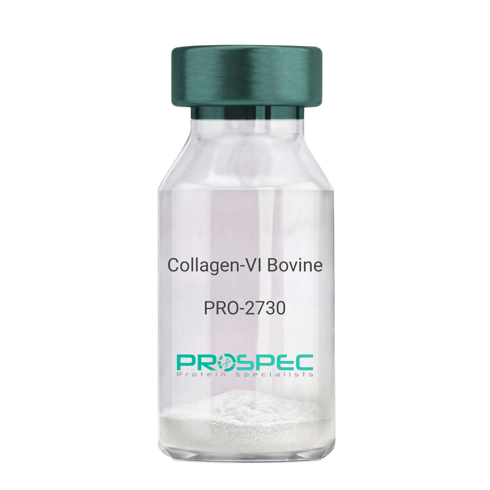
    Collagen-VI Bovine

    Description:

    Bovine Collagen-VI

    Product # :

    PRO-2730

    Price :

    Quantity :

    Shipping Method :

    Room Temp Icon

    Shipped at Room temp

    Add To Cart

    More Info

    • description
    • source
    • formulation
    • purity
    • More Info

    Description

    Bovine Collagen-VI is a natural protein purified from Bovine placenta. Bovine Collagen-VI is purified by proprietary chromatographic techniques.

    Source

    Bovine placenta.

    Formulation

    Collagen-VI was lyophilized without additives.

    Purity

    > 90.0%.

    More Info

    • Introduction

      Collagen, a major component of the extracellular matrix, is a fibrous protein that provides tensile strength to tissues giving them structural integrity. Collagen and its derivative, gelatin, have been widely used in medical, pharmaceutical and consumer products for more than 100 years. The supply of these materials, created from animal remains, is both abundant and inexpensive. However, most formulations are not highly purified and have the potential to cause an inflammatory reaction in some product users. In addition, concerns have been raised over the last several years about the potential for contamination of bovine products with the agent that causes mad cow disease and its human variant, Creutzfeldt-Jakob Disease. Animal collagens are subject to extensive modifications that continue over the life of the molecule in the extracellular space. These differences influence both the extractability of collagens from tissue and the biophysical characteristics of these collagens. As a result, collagens isolated from tissues exhibit significant lot-to-lot variability and, as bulk materials, are often analytically intractable. Products that contain animal-derived collagen can induce potentially harmful inflammatory or immune responses in humans and pose risk of contamination with viruses or prions, potentially life-threatening pathogens. Recombinant collagens are essentially identical to the native collagen protein thereby reducing the risk of inflammation, immune response, and disease as compared to animal-sourced collagen.

    • Physical Appearance

      Filtered White lyophilized (freeze-dried) powder.

    • Stability

      Lyophilized Collagen-VI although stable at room temperature for 3 weeks, should be stored desiccated below -18C. Upon reconstitution Collagen-VI should be stored at 4C between 2-7 days and for future use below -18C.For long term storage it is recommended to add a carrier protein (0.1% HSA or BSA).Please prevent freeze-thaw cycles.

    • Solubility

      It is recommended to Add 0.5 M acetic acid, pH 2.5 to prepare a working stock solution not less than 100µg/ml, which can then be further diluted to other aqueous solutions.

    ProSpec's products are furnished for LABORATORY RESEARCH USE ONLY. The product may not be used as drugs, agricultural or pesticidal products, food additives or household chemicals.

    Collagen Vi Bovine
  • View Data Sheet

    Name :

    AKT1 Antibody

    Description:

    Protein Kinase B Alpha, Mouse Anti Human

    V-Akt Murine Thymoma Viral Oncogene Homolog 1, Protein Kinase B Alpha, Proto-Oncogene C-Akt, RAC-PK-Alpha, EC 2.7.11.1, PKB Alpha, CWS6, PKB, RAC, RAC-Alpha Serine/Threonine-Protein Kinase, Rac Protein Kinase Alpha, Protein Kinase B, PKB-ALPHA, RAC-ALPHA, EC 2.7.11, AKT1m, PRKBA, AKT, RAC-alpha serine/threonine-protein kinase.

    Product # :

    ANT-710

    Price :

    Quantity :

    Shipping Method :

    Ice Icon

    Shipped with Ice Packs

    Add To Cart

    More Info

    • formulation
    • More Info

    Formulation

    1mg/ml containing PBS, pH-7.4, 10% Glycerol and 0.02% Sodium Azide.

    More Info

    • Introduction

      Akt1, also known as Akt or else protein kinase B (PKB) is an important molecule in mammaliancellular signaling. In humans, there are three genes in the "Akt family": Akt1, Akt2, and Akt3. Moreover, these enzymes are members of the serine/threonine-specific protein kinase family (EC2.7.11.1). Akt1 is involved in cellular survival pathways, by inhibiting apoptoticprocesses. Akt1 is also able to induce protein synthesis pathways, and is therefore a key signaling protein in the cellular pathways which lead to skeletal muscle hypertrophy, and general tissue growth. Since it can block apoptosis, and thereby promote cell survival, Akt1 has been implicated as a most important factor in numerous types of cancer. Akt (now also called Akt1) was at first identified as the oncogenein the transforming retrovirus, AKT8.

    • Synonyms

      V-Akt Murine Thymoma Viral Oncogene Homolog 1, Protein Kinase B Alpha, Proto-Oncogene C-Akt, RAC-PK-Alpha, EC 2.7.11.1, PKB Alpha, CWS6, PKB, RAC, RAC-Alpha Serine/Threonine-Protein Kinase, Rac Protein Kinase Alpha, Protein Kinase B, PKB-ALPHA, RAC-ALPHA, EC 2.7.11, AKT1m, PRKBA, AKT, RAC-alpha serine/threonine-protein kinase.

    • Physical Appearance

      Sterile filtered colorless solution.

    • Immunogen

      Anti-human AKT1 mAb, is derived from hybridization of mouse F0 myeloma cells with spleen cells from BALB/c mice immunized with recombinant human AKT1 amino acids 1-480 purified from E. coli.

    • Ig Subclass

      Mouse IgG2a heavy chain and κ light chain.

    • Clone

      PAT6D5AT.

    • Applications

      AKT1 antibody has been tested by ELISA and Western blot analysis to assure specificity and reactivity. Since application varies, however, each investigation should be titrated by the reagent to obtain optimal results.

    • Type

      Mouse Anti Human Monoclonal.

    • Storage Procedures

      For periods up to 1 month store at 4°C, for longer periods of time, store at -20°C. Prevent freeze thaw cycles.

    • Purification Method

      AKT1 antibody was purified from mouse ascetic fluids by protein-A affinity chromatography.

    ProSpec's products are furnished for LABORATORY RESEARCH USE ONLY. The product may not be used as drugs, agricultural or pesticidal products, food additives or household chemicals.

    Akt1 Antibody
  • View Data Sheet

    Name :

    AITR Human

    Description:

    AITR Human Recombinant

    TNFRSF18, AITR, CD357, GITR, GITR-D, Tumor necrosis factor receptor superfamily member 18, Activation-inducible TNFR family receptor, Glucocorticoid-induced TNFR-related protein, CD357, UNQ319/PRO364.

    Product # :

    CYT-925

    Price :

    Quantity :

    Shipping Method :

    Ice Icon

    Shipped with Ice Packs

    Add To Cart

    More Info

    • description
    • source
    • formulation
    • purity
    • More Info
    • sds-page

    Description

    AITR Human Recombinant produced in Sf9 Baculovirus is a single, glycosylated polypeptide chain containing 145 amino acids (26-162a.a.) and having a molecular mass of 15.6kDa (Migrates at 18-28kDa on SDS-PAGE under reducing conditions).AITR is fused to an 8 amino acid His-tag at C-terminus & purified by proprietary chromatographic techniques.

    Source

    Escherichia Coli.

    Formulation

    AITR protein solution (0.5mg/ml) containing Phosphate Buffered Saline (pH 7.4) and 10% glycerol.

    Purity

    Greater than 95.0% as determined by SDS-PAGE.

    sds-page

    AITR-sds-page - Product image 1

    More Info

    • Synonyms

      TNFRSF18, AITR, CD357, GITR, GITR-D, Tumor necrosis factor receptor superfamily member 18, Activation-inducible TNFR family receptor, Glucocorticoid-induced TNFR-related protein, CD357, UNQ319/PRO364.

    • Physical Appearance

      Sterile filtered colorless solution.

    • Stability

      Store at 4°C if entire vial will be used within 2-4 weeks. Store, frozen at -20°C for longer periods of time. For long term storage it is recommended to add a carrier protein (0.1% HSA or BSA).Avoid multiple freeze-thaw cycles.

    • Amino Acid Sequence

      QRPTGGPGCG PGRLLLGTGT DARCCRVHTT RCCRDYPGEE CCSEWDCMCV QPEFHCGDPC CTTCRHHPCP PGQGVQSQGK FSFGFQCIDC ASGTFSGGHE GHCKPWTDCT QFGFLTVFPG NKTHNAVCVP GSPPAEPLEH HHHHH.

    • Background

      AITR Human Recombinant: Unveiling its Role in Immune Regulation and Therapeutic Potential

      1. Abstract

      This research paper aims to provide a comprehensive exploration of the AITR Human Recombinant, a crucial receptor involved in immune regulation. By examining its structure, signaling pathways, biological functions, and implications in disease, we unravel the potential therapeutic applications of AITR in immune-related disorders.

      2. Introduction

      AITR, also known as TNFRSF18, is a receptor protein that plays a vital role in immune regulation. With its involvement in T-cell responses and immune tolerance, AITR has emerged as an intriguing target for therapeutic interventions in various immune-mediated conditions.

      3. Structure and Signaling of AITR

      AITR is a transmembrane receptor protein belonging to the tumor necrosis factor receptor superfamily. Its extracellular domain interacts with its ligand, glucocorticoid-induced TNFR-related protein (GITR) ligand, leading to downstream signaling events that modulate immune cell function.

      4. Biological Functions of AITR

      AITR activation influences T-cell responses by regulating T-cell activation, proliferation, and cytokine production. Additionally, AITR signaling can modulate the balance between effector and regulatory T-cell populations, thereby playing a role in immune tolerance and immune homeostasis.

      5. AITR in Disease Pathology

      AITR dysregulation has been associated with various immune-related disorders, including autoimmune diseases, cancer, and transplant rejection. Understanding the role of AITR in these pathologies may provide insights into potential therapeutic strategies targeting AITR signaling.

      6. Therapeutic Potential of AITR

      The unique role of AITR in immune regulation makes it an appealing target for therapeutic interventions. Modulation of AITR signaling holds promise for manipulating immune responses in the context of autoimmune diseases, cancer immunotherapy, and transplantation.

      7. Conclusion and Future Perspectives

      While our understanding of AITR and its functions has advanced significantly, further research is warranted to unravel its complex signaling pathways and therapeutic potential. Continued investigations into AITR biology will enhance our ability to develop targeted therapies for immune-related disorders.

      What is the molecular weight/Mw of AITR Protein?
      AITR Protein has a total Mw of 15.6kDa.

      What is the source or expression system of AITR Protein?
      Escherichia Coli.

      What is the Purity of AITR Protein?
      AITR Protein is >95% pure as determined by SDS-PAGE.

      What is the Biological Activity of AITR Protein?
      The biological functionality of AITR Protein will be determined in the future.

      What is the amino acid sequence of AITR Protein?
      QRPTGGPGCG PGRLLLGTGT DARCCRVHTT RCCRDYPGEE CCSEWDCMCV QPEFHCGDPC CTTCRHHPCP PGQGVQSQGK FSFGFQCIDC ASGTFSGGHE GHCKPWTDCT QFGFLTVFPG NKTHNAVCVP GSPPAEPLEH HHHHH.

      What applications can AITR Protein be used in?
      AITR Protein can probably be used in western blot, ELISA and Lateral Flow.

      What is the endotoxin level for AITR Protein?
      The endotoxin level is minimal, AITR Protein was purified using conventional chromatography techniques.

    ProSpec's products are furnished for LABORATORY RESEARCH USE ONLY. The product may not be used as drugs, agricultural or pesticidal products, food additives or household chemicals.

    Aitr Human
  • View Data Sheet

    Name :

    Zika NS1

    Description:

    Zika NS1 Protein Recombinant

    Product # :

    ZKV-002

    Price :

    Quantity :

    Shipping Method :

    Ice Icon

    Shipped with Ice Packs

    Add To Cart

    More Info

    • description
    • source
    • formulation
    • purity
    • More Info

    Description

    The E.Coli derived Recombinant Zika NS1 protein (Strain: ZikaSPH2015) having an Mw of 45kDa is derived from the full length Zika NS1 protein. The Zika NS1 protein is fused to a 6xHis tag at C-terminus and purified by proprietary chromatographic technique.

    Source

    Escherichia Coli.

    Formulation

    Zika NS1 protein solution contains PBS and 100mM arginine.

    Purity

    Zika NS1 protein is >95% pure as determined by SDS-PAGE.

    More Info

    • Introduction

      Zika virus (ZIKV) belongs to the family Flaviviridae and the genus Flavivirus, it is transmitted by daytime-active Aedes mosquitoes, such as A. aegypti and A. albopictus. The Zika virus is related to the dengue, yellow fever, Japanese encephalitis, and West Nile viruses. Much like the other flaviviruses, Zika virus is enveloped and icosahedral and has a nonsegmented, single-stranded, positive-sense RNA genome. Zika fever is an infection, which often causes no symptoms or only mild ones, like a mild form of dengue fever, and it is treated by rest. As of February 2016, there has been mounting evidence that Zika fever in pregnant women can cause abnormal brain development in their fetuses by mother-to-child transmission, which may result in miscarriage or microcephaly, however it is not yet known whether Zika virus causes microcephaly. Furthermore, a connection has been established with neurologic conditions in infected adults, including Guillain–Barre syndrome. Since the 1950s, Zika virus has been detected only within a narrow equatorial belt from Africa to Asia. Between the years 2013 and 2014, Zika virus has spread eastward across the Pacific Ocean to French Polynesia, New Caledonia, the Cook Islands, and Easter Island, and in 2015 to Mexico, Central America, the Caribbean, and South America, where the Zika outbreak has reached pandemic levels.

    • Physical Appearance

      Sterile Filtered solution.

    • Stability

      Zika NS1 protein although stable at 4°C for 1 week, should be stored below -18°C. Please prevent freeze thaw cycles.

    • Applications

      Each laboratory should determine an optimum working titer for use in its particular application.

    ProSpec's products are furnished for LABORATORY RESEARCH USE ONLY. The product may not be used as drugs, agricultural or pesticidal products, food additives or household chemicals.

    Zika Ns1
  • View Data Sheet

    Name :

    PARK7 P1E12AT Antibody

    Description:

    Parkinson Disease Protein 7 Clone P1E12AT, Mouse Anti Human

    PARK7, Parkinson disease protein 7, Oncogene DJ-1, Parkinson disease (autosomal recessive, early onset) 7.

    Product # :

    ANT-515

    Price :

    Quantity :

    Shipping Method :

    Ice Icon

    Shipped with Ice Packs

    Add To Cart

    More Info

    • formulation
    • More Info

    Formulation

    1mg/ml containing PBS, pH-7.4, 10% Glycerol and 0.02% Sodium Azide.

    More Info

    • Introduction

      The PARK7 is a ubiquitously expressed protein involved in various cellular processes including spermatogenesis and fertilization, cancer, RNA-binding, androgen-receptor signaling and oxidative stress. Mutations in the PARK7 are the cause of autosomal recessive early-onset Parkinson’s disease 7 (Park7).

    • Synonyms

      PARK7, Parkinson disease protein 7, Oncogene DJ-1, Parkinson disease (autosomal recessive, early onset) 7.

    • Physical Appearance

      Sterile filtered colorless solution.

    • Immunogen

      Anti-human PARK7 mAb, clone P1E12AT, is derived from hybridization of mouse F0 myeloma cells with spleen cells from BALB/c mice immunized with a recombinant human PARK7 protein 1-189 amino acids purified from E. coli

    • Ig Subclass

      Mouse IgG2b heavy chain and k light chain.

    • Clone

      P1E12AT.

    • Applications

      The antibody has been tested by ELISA, Western blot analysis to assure specificity and reactivity. Since application varies, however, each investigation should be titrated by the reagent to obtain optimal results. Recommended starting dilution is 1:1000.

    • Type

      Mouse Anti Human Monoclonal.

    • Storage Procedures

      For periods up to 1 month store at 4°C, for longer periods of time, store at -20°C. Prevent freeze thaw cycles.

    • Purification Method

      PARK7 antibody was purified from mouse ascitic fluids by protein-A affinity chromatography.

    ProSpec's products are furnished for LABORATORY RESEARCH USE ONLY. The product may not be used as drugs, agricultural or pesticidal products, food additives or household chemicals.

    Park7 P1E12At Antibody
  • View Data Sheet

    Name :

    sCD40L Mouse, sf9

    Description:

    soluble CD40 Ligand/TRAPMouse Recombinant, sf9

    CD40 ligand, T-cell antigen Gp39, TNF-related activation protein, TRAP, Tumor necrosis factor ligand superfamily member 5, CD154, CD40lg, CD154, CD40-L, Cd40l, gp39, HIGM1, IGM, IMD3, Ly-62, Ly62, T-BAM, Tnfsf5.

    Product # :

    CYT-917

    Price :

    Quantity :

    Shipping Method :

    Ice Icon

    Shipped with Ice Packs

    Add To Cart

    More Info

    • description
    • source
    • formulation
    • purity
    • More Info

    Description

    sCD40 Mouse Recombinant produced in Sf9 Baculovirus cells is a single, glycosylated polypeptide chain containing 155 amino acids (112-260 a.a.) and having a molecular mass of 17.2kDa (Migrates at 18-28kDa on SDS-PAGE under reducing conditions).sCD40 is expressed with a 6 amino acid His tag at C-Terminus and purified by proprietary chromatographic techniques.

    Source

    Sf9, Baculovirus cells.

    Formulation

    sCD40 protein solution (0.25mg/ml) contains Phosphate Buffered Saline (pH 7.4) and 10% glycerol.

    Purity

    Greater than 95.0% as determined by SDS-PAGE.

    More Info

    • Introduction

      CD40L or CD154 is a membrane glycoprotein and differentiation antigen expressed on the surface of T-cells. The CD40 ligand stimulates B-cell proliferation and secretion of all immunoglobulin isotypes in the presence of cytokines . CD40 ligand has been shown to induce cytokine production and tumoricidal activity in peripheral blood monocytes. It also costimulates proliferation of activated T-cells and this is accompanied by the production of IFN-gamma , TNF-alpha , and IL2.

    • Synonyms

      CD40 ligand, T-cell antigen Gp39, TNF-related activation protein, TRAP, Tumor necrosis factor ligand superfamily member 5, CD154, CD40lg, CD154, CD40-L, Cd40l, gp39, HIGM1, IGM, IMD3, Ly-62, Ly62, T-BAM, Tnfsf5.

    • Physical Appearance

      Sterile Filtered colorless solution.

    • Stability

      Store at 4°C if entire vial will be used within 2-4 weeks. Store, frozen at -20°C for longer periods of time. For long term storage it is recommended to add a carrier protein (0.1% HSA or BSA). Avoid multiple freeze-thaw cycles.

    • Amino Acid Sequence

      MQRGDEDPQI AAHVVSEANS NAASVLQWAK KGYYTMKSNL VMLENGKQLT VKREGLYYVY TQVTFCSNRE PSSQRPFIVG LWLKPSSGSE RILLKAANTH SSSQLCEQQS VHLGGVFELQ AGASVFVNVT EASQVIHRVG FSSFGLLKLH HHHHH.

    ProSpec's products are furnished for LABORATORY RESEARCH USE ONLY. The product may not be used as drugs, agricultural or pesticidal products, food additives or household chemicals.

    Scd40L Mouse Sf9
  • View Data Sheet

    Name :

    L1CAM Human

    Description:

    L1 Cell Adhesion Molecule Human Recombinant

    L1 Cell Adhesion Molecule, Antigen Identified By Monoclonal Antibody R1, N-CAM-L1, NCAM-L1, CAML1, MIC5, Neural Cell Adhesion Molecule L1, CD171 Antigen, N-CAML1, CD171, HSAS1, MASA, HSAS, SPG1, S10.

    Product # :

    PRO-2446

    Price :

    Quantity :

    Shipping Method :

    Ice Icon

    Shipped with Ice Packs

    Add To Cart

    More Info

    • description
    • source
    • formulation
    • purity
    • biological activity
    • More Info

    Description

    L1CAM Human Recombinant produced in Sf9 Baculovirus cells is a single, glycosylated polypeptide chain containing 1104 amino acids (20-1115a.a.) and having a molecular mass of 123.6kDa (Molecular size on SDS-PAGE will appear at approximately 100-150kDa). L1CAM is expressed with a 8 amino acids His tag at C-Terminus and purified by proprietary chromatographic techniques.

    Source

    Sf9, Baculovirus cells.

    Formulation

    L1CAM protein solution (0.25mg/ml) contains Phosphate Buffered Saline (pH 7.4) and 10% glycerol.

    Purity

    Greater than 90.0% as determined by SDS-PAGE.

    Biological Activity

    More to 30% measured by the ability of the immobilized protein to support the adhesion of Neuro-2a mouse neuroblastoma cells. When cells are added to human L1CAM coated plates 1 ug/ml.

    More Info

    • Introduction

      L1 Cell Adhesion Molecule (L1CAM) which is a cell adhesion receptor of the immunoglobulin superfamily takes part in nerve cell function. L1CAM is a neural cell adhesion molecule involved in the dynamics of cell adhesion and in the generation of transmembrane signals at tyrosine kinase receptors. L1CAM takes part in cell migration, neurite outgrowth and myelination. Furthermore, L1CAM plays an important role in the dynamics of neuronal structure and function in the mature brain.

    • Synonyms

      L1 Cell Adhesion Molecule, Antigen Identified By Monoclonal Antibody R1, N-CAM-L1, NCAM-L1, CAML1, MIC5, Neural Cell Adhesion Molecule L1, CD171 Antigen, N-CAML1, CD171, HSAS1, MASA, HSAS, SPG1, S10.

    • Physical Appearance

      Sterile filtered colorless solution.

    • Stability

      Store at 4°C if entire vial will be used within 2-4 weeks. Store, frozen at -20°C for longer periods of time. For long term storage it is recommended to add a carrier protein (0.1% HSA or BSA).Avoid multiple freeze-thaw cycles.

    • Amino Acid Sequence

      IQIPEELMEP PVITEQSPRR LVVFPTDDIS LKCEASGKPE VQFRWTRDGV HFKPKEELGV TVYQSPHSGS FTITGNNSNF AQRFQGIYRC FASNKLGTAM SHEIRLMAEG APKWPKETVK PVEVEEGESV VLPCNPPPSA EPLRIYWMNS KILHIKQDER VTMGQNGNLY FANVLTSDNH SDYICHAHFP GTRTIIQKEP IDLRVKATNS MIDRKPRLLF PTNSSSHLVA LQGQPLVLEC IAEGFPTPTI KWLRPSGPMP ADRVTYQNHN KTLQLLKVGE EDDGEYRCLA ENSLGSARHA YYVTVEAAPY WLHKPQSHLY GPGETARLDC QVQGRPQPEV TWRINGIPVE ELAKDQKYRI QRGALILSNV QPSDTMVTQC EARNRHGLLL ANAYIYVVQL PAKILTADNQ TYMAVQGSTA YLLCKAFGAP VPSVQWLDED GTTVLQDERF FPYANGTLGI RDLQANDTGR YFCLAANDQN NVTIMANLKV KDATQITQGP RSTIEKKGSR VTFTCQASFD PSLQPSITWR GDGRDLQELG DSDKYFIEDG RLVIHSLDYS DQGNYSCVAS TELDVVESRA QLLVVGSPGP VPRLVLSDLH LLTQSQVRVS WSPAEDHNAP IEKYDIEFED KEMAPEKWYS LGKVPGNQTS TTLKLSPYVH YTFRVTAINK YGPGEPSPVS ETVVTPEAAP EKNPVDVKGE GNETTNMVIT WKPLRWMDWN APQVQYRVQW RPQGTRGPWQ EQIVSDPFLV VSNTSTFVPY EIKVQAVNSQ GKGPEPQVTI GYSGEDYPQA IPELEGIEIL NSSAVLVKWR PVDLAQVKGH LRGYNVTYWR EGSQRKHSKR HIHKDHVVVP ANTTSVILSG LRPYSSYHLE VQAFNGRGSG PASEFTFSTP EGVPGHPEAL HLECQSNTSL LLRWQPPLSH NGVLTGYVLS YHPLDEGGKG QLSFNLRDPE LRTHNLTDLS PHLRYRFQLQ ATTKEGPGEA IVREGGTMAL SGISDFGNIS ATAGENYSVV SWVPKEGQCN FRFHILFKAL GEEKGGASLS PQYVSYNQSS YTQWDLQPDT DYEIHLFKER MFRHQMAVKT NGTGRVRLPP AGFATELEHH HHHH.

    ProSpec's products are furnished for LABORATORY RESEARCH USE ONLY. The product may not be used as drugs, agricultural or pesticidal products, food additives or household chemicals.

    L1Cam Human
  • 1
  • …
  • 19
  • 20
  • 21
  • 22
  • 23
  • …
  • 26
Your browser does not support the video tag.

Products

  • Cytokines
  • Growth Factors
  • Chemokines
  • CD Antigens
  • Neurotrophins
  • Hormones
  • Enzymes
  • Viral Antigens
  • Recombinant Proteins
  • Natural Proteins
  • Monoclonal Antibodies
  • Polyclonal Antibodies
  • Thermal Packaging

About

  • Company
  • Founder
  • Contribution
  • Contact Us
  • My Account

Ordering

  • Ordering Method
  • Ordering - Credit Card
  • Ordering PO
  • Shipping Fees
  • FAQ's

Legal

  • Terms & Conditions
  • Privacy
  • Site Map
  • info@prospecbio.com
  • Facebook
  • Instagram
  • Linkedin Profile

© 2026 ProSpec-Tany TechnoGene Ltd. All Rights Reserved.

  • Choosing a selection results in a full page refresh.
  • Opens in a new window.