Search results
569 results found for “SERTA Domain Containing”
Name
Description
Product #
Price
Quantity
Shipping Method
- View Data Sheet
Name :
FGB HumanDescription:
Fibrinogen Beta Chain Human Recombinant
Fibrinogen beta chain, Fibrinopeptide B, Fibrinogen, B Beta Polypeptide, Epididymis Secretory Sperm Binding Protein Li 78p, HEL-S-78p.
Product # :
PRO-2210Price :
Quantity :
Shipping Method :
Shipped with Ice Packs
More Info
- description
- source
- formulation
- purity
- More Info
- Citations
Description
FGB Human Recombinant produced in E.Coli is a single, non-glycosylated polypeptide chain containing 351 amino acids (164-491a.a) and having a molecular mass of 40kDa. FGB is fused to a 23 amino acid His-tag at N-terminus & purified by proprietary chromatographic techniques.
Source
Escherichia Coli.
Formulation
FGB protein solution (1mg/ml) containing 20mM Tris-HCl buffer (pH8.0) and 10% glycerol.
Purity
Greater than 85.0% as determined by SDS-PAGE.
Citations
More Info
-
Introduction
Fibrinogen Beta Chain 9 (FGB) is the beta component of fibrinogen, a blood-borne glycoprotein composed of 3 pairs of nonidentical polypeptide chains. When vascular injury accrues, fibrinogen is cleaved by thrombin to form fibrin which is found in abundance in blood clots.
-
Synonyms
Fibrinogen beta chain, Fibrinopeptide B, Fibrinogen, B Beta Polypeptide, Epididymis Secretory Sperm Binding Protein Li 78p, HEL-S-78p.
-
Physical Appearance
Sterile Filtered colorless solution.
-
Stability
Store at 4°C if entire vial will be used within 2-4 weeks. Store, frozen at -20°C for longer periods of time. For long term storage it is recommended to add a carrier protein (0.1% HSA or BSA). Avoid multiple freeze-thaw cycles.
-
Amino Acid Sequence
MGSSHHHHHH SSGLVPRGSH MGSDNENVVN EYSSELEKHQ LYIDETVNSN IPTNLRVLRS ILENLRSKIQ KLESDVSAQM EYCRTPCTVS CNIPVVSGKE CEEIIRKGGE TSEMYLIQPD SSVKPYRVYC DMNTENGGWT VIQNRQDGSV DFGRKWDPYK QGFGNVATNT DGKNYCGLPG EYWLGNDKIS QLTRMGPTEL LIEMEDWKGD KVKAHYGGFT VQNEANKYQI SVNKYRGTAG NALMDGASQL MGENRTMTIH NGMFFSTYDR DNDGWLTSDP RKQCSKEDGG GWWYNRCHAA NPNGRYYWGG QYTWDMAKHG TDDGVVWMNW KGSWYSMRKM SMKIRPFFPQ Q.
ProSpec's products are furnished for LABORATORY RESEARCH USE ONLY. The product may not be used as drugs, agricultural or pesticidal products, food additives or household chemicals.
- View Data Sheet
Name :
SETD7 AntibodyDescription:
Set7/9 Histone Methyltransferase, Mouse Anti Human
Histone-lysine N-methyltransferase, H3 lysine-4 specific SET7, EC 2.1.1.43, Histone H3-K4 methyltransferase, H3-K4-HMTase, SET domain-containing protein 7, Set9, SET7/9, SETD7.
Product # :
ANT-308Price :
Quantity :
Shipping Method :
Shipped with Ice Packs
More Info
- formulation
- More Info
- Citations
Formulation
1mg/ml containing PBS, pH-7.4, 10% Glycerol & 0.02% Sodium Azide.
Citations
More Info
-
Introduction
Set 7/9 is a histone methyltransferase (HMTase) that transfers methyl groups to Lys4 of histone H3, in complex with S-adenosyl-L-methionine (AdoMet). The methylation of lysine residues of histones plays a critical role in the regulation of chromatin structure and gene expression. Acetylation, phosphorylation and methylation of the amino-terminal tails of histone are thought to be involved in the regulation of chromatin structure and function. The enzymes identified in the methylation of specific lysine residue on histones belong to the SET family with just one exception. Set7/9, unlike most other SET proteins, is exclusively a mono-methylase.
-
Synonyms
Histone-lysine N-methyltransferase, H3 lysine-4 specific SET7, EC 2.1.1.43, Histone H3-K4 methyltransferase, H3-K4-HMTase, SET domain-containing protein 7, Set9, SET7/9, SETD7.
-
Physical Appearance
Sterile filtered colorless solution.
-
Immunogen
Anti-human SETD7 mAb is derived from hybridization of mouse F0 myeloma cells with spleen cells from BALB/c mice immunized with recombinant human SETD7 amino acids 1-366 purified from E. coli.
-
Ig Subclass
Mouse IgG2b heavy chain and κ light chain.
-
Clone
Ps4E5AT.
-
Applications
SETD7 antibody has been tested by ELISA and Western blot analysis to assure specificity and reactivity. Since application varies, however, each investigation should be titrated by the reagent to obtain optimal results. Recommended dilution range for Western blot analysis is 1:1,000 ~ 2,000. Recommended starting dilution is 1:1,000.
-
Type
Mouse Anti Human Monoclonal.
-
Storage Procedures
For periods up to 1 month store at 4°C, for longer periods of time, store at -20°C. Prevent freeze thaw cycles.
-
Purification Method
SETD7 antibody was purified from hybridoma cell supernatant by protein-A affinity chromatography.
ProSpec's products are furnished for LABORATORY RESEARCH USE ONLY. The product may not be used as drugs, agricultural or pesticidal products, food additives or household chemicals.
- View Data Sheet
Name :
DYNLRB2 HumanDescription:
Dynein Light Chain Roadblock-Type 2 Human Recombinant
Dynein light chain roadblock-type 2, Dynein light chain 2B cytoplasmic, Roadblock domain-containing protein 2, DYNLRB2, DNCL2B, DNLC2B, ROBLD2, MGC62033.
Product # :
PRO-174Price :
Quantity :
Shipping Method :
Shipped with Ice Packs
More Info
- description
- source
- formulation
- purity
- More Info
- Citations
Description
DYNLRB2 Human Recombinant fused with a 20 amino acid His tag at N-terminus produced in E.Coli is a single, non-glycosylated, polypeptide chain containing 116 amino acids (1-96 a.a.) and having a molecular mass of 13kDa. The DYNLRB2 is purified by proprietary chromatographic techniques.
Source
Escherichia Coli.
Formulation
The DYNLRB2 solution (0.5 mg/ml) contains 20mM Tris-HCl buffer (pH8.0), 20% glycerol, 0.1M NaCl and 1mM DTT.
Purity
Greater than 90.0% as determined by SDS-PAGE.
Citations
More Info
-
Introduction
Dynein light chain roadblock-type 2 (DYNLRB2) functions as one of several non-catalytic accessory components of the cytoplasmic dynein complex which are considered to be involved in linking dynein to cargos and to adapter proteins that regulate dynein function. Cytoplasmic dynein functions as a motor for the intracellular retrograde motility of vesicles and organelles along microtubules.
-
Synonyms
Dynein light chain roadblock-type 2, Dynein light chain 2B cytoplasmic, Roadblock domain-containing protein 2, DYNLRB2, DNCL2B, DNLC2B, ROBLD2, MGC62033.
-
Physical Appearance
Sterile Filtered colorless solution.
-
Stability
Store at 4°C if entire vial will be used within 2-4 weeks. Store, frozen at -20°C for longer periods of time. For long term storage it is recommended to add a carrier protein (0.1% HSA or BSA).Avoid multiple freeze-thaw cycles.
-
Amino Acid Sequence
MGSSHHHHHH SSGLVPRGSH MAEVEETLKR IQSHKGVIGT MVVNAEGIPI RTTLDNSTTV QYAGLLHHLT MKAKSTVRDI DPQNDLTFLR IRSKKHEIMV APDKEYLLIV IQNPCE.
ProSpec's products are furnished for LABORATORY RESEARCH USE ONLY. The product may not be used as drugs, agricultural or pesticidal products, food additives or household chemicals.
- View Data Sheet
Name :
HSV-1 gGDescription:
Herpes Simplex Virus-1 gG Recombinant
Product # :
HSV-224Price :
Quantity :
Shipping Method :
Shipped with Ice Packs
More Info
- description
- formulation
- purity
- More Info
- Citations
Description
The E.Coli derived recombinant protein contains the HSV-1 gG immunodominant regions, 84-175 amino acids and fused to a GST-Tag at C-terminus.
Formulation
25mM Tris-HCl pH 7.2, 1mM EDTA, and 50% glycerol.
Purity
HSV-1 gG protein is >95% pure as determined by 10% PAGE (coomassie staining).
Citations
More Info
-
Introduction
Entry of HSV into the host cell involves interactions of several viral glycoproteins with cell surface receptors. The virus particle is covered by an envelope which, when bound to specific receptors on the cell surface, will fuse with the cell membrane and create an opening, or pore, through which the virus enters the host cell. The sequential stages of HSV entry are analagous to those of other viruses. At first, complementary receptors on the virus and cell surface bring the two membranes into proximity. In an intermediate state, the two membranes begin to merge, forming a hemifusion state. Finally, a stable entry pore is formed through which the viral envelope contents are introduced to the host cell.
-
Physical Appearance
Sterile Filtered clear solution.
-
Stability
HSV-1 gG protein although stable at 4°C for 1 week, should be stored below -18°C. Please prevent freeze thaw cycles.
-
Specificity
Immunoreactive with sera of HSV-infected individuals.
-
Purification Method
HSV-1 gG was purified by Sepharose-Derived Purification.
ProSpec's products are furnished for LABORATORY RESEARCH USE ONLY. The product may not be used as drugs, agricultural or pesticidal products, food additives or household chemicals.
- View Data Sheet
Name :
TNFSF8 Human, Sf9Description:
CD30 Ligand Human Recombinant, Sf9
Tumor Necrosis Factor Superfamily Member 8, Tumor Necrosis Factor (Ligand) Superfamily, Member 8, CD153 Antigen, CD30 Ligand, CD30LG, CD30-L, CD30L, Tumor Necrosis Factor (Ligand) Superfamily Member 8, Tumor Necrosis Factor Ligand 3A, CD30 Antigen Ligand, TNLG3A, CD153, Tumor necrosis factor ligand superfamily member 8, TNFSF8, CD30 ligand.
Product # :
CYT-954Price :
Quantity :
Shipping Method :
Shipped with Ice Packs
More Info
- description
- source
- formulation
- purity
- More Info
- Citations
Description
TNFSF8 produced in Sf9 Baculovirus cells is a single, glycosylated polypeptide chain containing 181 amino acids (63-234a.a.) and having a molecular mass of 20.7kDa (Molecular size on SDS-PAGE will appear at approximately 18-40kDa). TNFSF8 is expressed with a 6 amino acid His tag at C-Terminus and purified by proprietary chromatographic techniques.
Source
Sf9, Baculovirus cells.
Formulation
TNFSF8 protein solution (0.5mg/ml) contains Phosphate Buffered Saline (pH 7.4) and 10% glycerol.
Purity
Greater than 85% as determined by SDS-PAGE.
Citations
More Info
-
Introduction
CD30 Ligand (TNFSF8) is a cytokine which is a member of the tumor necrosis factor (TNF) ligand family. The TNFSF8 cytokine is a ligand for TNFRSF8/CD30, which is a cell surface antigen and a marker for Hodgkin lymphoma and related hematologic malignancies. The employment of the TNFSF8 cytokine expressed on B cell surface has an inhibitory role in modulating Ig class switch. TNFSF8 enhances cell proliferation of some lymphoma cell lines, while inducing cell death and reducing cell proliferation of other lymphoma cell lines. The pleiotropic biological activities of the TNFSF8 cytokine on different CD30+ lymphoma cell lines has a pathophysiologic role in Hodgkin's and some non-Hodgkin's lymphomas.
-
Synonyms
Tumor Necrosis Factor Superfamily Member 8, Tumor Necrosis Factor (Ligand) Superfamily, Member 8, CD153 Antigen, CD30 Ligand, CD30LG, CD30-L, CD30L, Tumor Necrosis Factor (Ligand) Superfamily Member 8, Tumor Necrosis Factor Ligand 3A, CD30 Antigen Ligand, TNLG3A, CD153, Tumor necrosis factor ligand superfamily member 8, TNFSF8, CD30 ligand.
-
Physical Appearance
Sterile filtered colorless solution.
-
Stability
Store at 4°C if entire vial will be used within 2-4 weeks. Store, frozen at -20°C for longer periods of time. For long term storage it is recommended to add a carrier protein (0.1% HSA or BSA).Avoid multiple freeze-thaw cycles.
-
Amino Acid Sequence
ADPQRTDSIP NSPDNVPLKG GNCSEDLLCI LKRAPFKKSW AYLQVAKHLN KTKLSWNKDG ILHGVRYQDG NLVIQFPGLY FIICQLQFLV QCPNNSVDLK LELLINKHIK KQALVTVCES GMQTKHVYQN LSQFLLDYLQ VNTTISVNVD TFQYIDTSTF PLENVLSIFL YSNSDHHHHH H.
ProSpec's products are furnished for LABORATORY RESEARCH USE ONLY. The product may not be used as drugs, agricultural or pesticidal products, food additives or household chemicals.
- View Data Sheet
Name :
Zika NS1Description:
Zika NS1 Protein Recombinant
Product # :
ZKV-002Price :
Quantity :
Shipping Method :
Shipped with Ice Packs
More Info
- description
- source
- formulation
- purity
- More Info
- Citations
Description
The E.Coli derived Recombinant Zika NS1 protein (Strain: ZikaSPH2015) having an Mw of 45kDa is derived from the full length Zika NS1 protein. The Zika NS1 protein is fused to a 6xHis tag at C-terminus and purified by proprietary chromatographic technique.
Source
Escherichia Coli.
Formulation
Zika NS1 protein solution contains PBS and 100mM arginine.
Purity
Zika NS1 protein is >95% pure as determined by SDS-PAGE.
Citations
More Info
-
Introduction
Zika virus (ZIKV) belongs to the family Flaviviridae and the genus Flavivirus, it is transmitted by daytime-active Aedes mosquitoes, such as A. aegypti and A. albopictus. The Zika virus is related to the dengue, yellow fever, Japanese encephalitis, and West Nile viruses. Much like the other flaviviruses, Zika virus is enveloped and icosahedral and has a nonsegmented, single-stranded, positive-sense RNA genome. Zika fever is an infection, which often causes no symptoms or only mild ones, like a mild form of dengue fever, and it is treated by rest. As of February 2016, there has been mounting evidence that Zika fever in pregnant women can cause abnormal brain development in their fetuses by mother-to-child transmission, which may result in miscarriage or microcephaly, however it is not yet known whether Zika virus causes microcephaly. Furthermore, a connection has been established with neurologic conditions in infected adults, including Guillain–Barre syndrome. Since the 1950s, Zika virus has been detected only within a narrow equatorial belt from Africa to Asia. Between the years 2013 and 2014, Zika virus has spread eastward across the Pacific Ocean to French Polynesia, New Caledonia, the Cook Islands, and Easter Island, and in 2015 to Mexico, Central America, the Caribbean, and South America, where the Zika outbreak has reached pandemic levels.
-
Physical Appearance
Sterile Filtered solution.
-
Stability
Zika NS1 protein although stable at 4°C for 1 week, should be stored below -18°C. Please prevent freeze thaw cycles.
-
Applications
Each laboratory should determine an optimum working titer for use in its particular application.
ProSpec's products are furnished for LABORATORY RESEARCH USE ONLY. The product may not be used as drugs, agricultural or pesticidal products, food additives or household chemicals.
- View Data Sheet
Name :
GM-CSF Poricne, HisDescription:
Granulocyte Macrophage-Colony Stimulating Factor Porcine Recombinant, His Tag
CSF-2, MGI-1GM, GM-CSF, Pluripoietin-alpha, Molgramostin, Sargramostim
Product # :
CYT-1160Price :
Quantity :
Shipping Method :
Shipped with Ice Packs
More Info
- description
- source
- formulation
- purity
- biological activity
- More Info
- Citations
Description
GMCSF Poricne Recombinant produced in E.Coli is a single, non-glycosylated polypeptide chain containing 148 amino acids (18-144a.a.) and having a molecular mass of 16.6kDa.GMCSF is fused to a 21 amino acid His tag at N-Terminus and purified by proprietary chromatographic techniques.
Source
Escherichia Coli.
Formulation
GMCSF protein solution (0.5mg/ml) containing Phosphate-Buffered Saline (pH7.4).
Purity
Greater than 95.0% as determined by SDS-PAGE.
Biological Activity
The ED50 range is ≤ 40 ng/ml, and is measured in a cell proliferation assay using TF-1 human erythroleukemic cells.
Citations
More Info
-
Introduction
The hematopoietic growth factor GM-CSF or granulocyte macrophage colony-stimulating factor, stimulates the development of neutrophils & macrophages, enhance proliferation and development of early erythroid megakaryocytic & eosinophilic progenitor cells. GM-CSF is secreted from the fibroblasts, monocytes, T-lymphocytes & endothelial cells. This protein blocks the migration of neutrophils & induces the biological activity of mature end-cells.
-
Synonyms
CSF-2, MGI-1GM, GM-CSF, Pluripoietin-alpha, Molgramostin, Sargramostim
-
Physical Appearance
Sterile Filtered colorless solution.
-
Stability
Store at 4°C if entire vial will be used within 2-4 weeks. Store, frozen at -20°C for longer periods of time. For long term storage it is recommended to add a carrier protein (0.1% HSA or BSA).Avoid multiple freeze-thaw cycles.
-
Amino Acid Sequence
MGSSHHHHHH SSGLVPRGSH MAPTRPPSPV TRPWQHVDAI KEALSLLNNS NDTAAVMNET VDVVCEMFDP QEPTCVQTRL NLYKQGLRGS LTRLKSPLTL LAKHYEQHCP LTEETSCETQ SITFKSFKDS LNKFLFTIPF DCWGPVKK
-
Background
What is the molecular weight/Mw of GM-CSF PORCINE, HIS Protein?
GM-CSF PORCINE, HIS Protein has a total Mw of 16.6kDa.
What is the source or expression system of GM-CSF PORCINE, HIS Protein?
Escherichia Coli.
What is the Purity of GM-CSF PORCINE, HIS Protein?
GM-CSF PORCINE, HIS Protein is >95% pure as determined by SDS-PAGE.
What is the Biological Activity of GM-CSF PORCINE, HIS Protein?
The ED50 range is ≤ 40 ng/ml, and is measured in a cell proliferation assay using TF-1 human erythroleukemic cells.
What is the amino acid sequence of GM-CSF PORCINE, HIS Protein?
MGSSHHHHHH SSGLVPRGSH MAPTRPPSPV TRPWQHVDAI KEALSLLNNS NDTAAVMNET VDVVCEMFDP QEPTCVQTRL NLYKQGLRGS LTRLKSPLTL LAKHYEQHCP LTEETSCETQ SITFKSFKDS LNKFLFTIPF DCWGPVKK
What applications can GM-CSF PORCINE, HIS Protein be used in?
GM-CSF PORCINE, HIS Protein can probably be used in western blot, ELISA and Lateral Flow.
What is the endotoxin level for GM-CSF PORCINE, HIS Protein?
The endotoxin level is minimal, GM-CSF PORCINE, HIS Protein was purified using conventional chromatography techniques.
ProSpec's products are furnished for LABORATORY RESEARCH USE ONLY. The product may not be used as drugs, agricultural or pesticidal products, food additives or household chemicals.
- View Data Sheet
Name :
UBA5 HumanDescription:
Ubiquitin-Like Modifier Activating Enzyme 5 Human Recombinant
Ubiquitin-like modifier-activating enzyme 5, Ubiquitin-activating enzyme 5, ThiFP1, UFM1-activating enzyme, Ubiquitin-activating enzyme E1 domain-containing protein 1, UBA5, UBE1DC1.
Product # :
ENZ-602Price :
Quantity :
Shipping Method :
Shipped with Ice Packs
More Info
- description
- source
- formulation
- purity
- More Info
- Citations
Description
UBA5 Human Recombinant produced in E.coli is a single, non-glycosylated polypeptide chain containing 428 amino acids (1-404) and having a molecular mass of 47.4kDa.UBA5 is fused to a 24 amino acid His-tag at N-terminus & purified by proprietary chromatographic techniques.
Source
Escherichia Coli.
Formulation
The UBA5 solution (0.5mg/ml) contains 20mM Tris-HCl buffer (pH 8.0), 1mM DTT, 10% glycerol and 50mM NaCl.
Purity
Greater than 90.0% as determined by SDS-PAGE.
Citations
More Info
-
Introduction
Ubiquitin-like modifier activating enzyme 5 (UBA5) is a member of the ubiquitin-activating E1 family and UBA5 subfamily. Ubiquitin and ubiquitin-like proteins are recognized as covalently conjugated to various cellular substrates by a three-step enzymatic pathway. The ubiquitin-activating enzyme (E1) has a vital role in the first step of ubiquitination pathway to activate ubiquitin or ubiquitin-like proteins. UBA5 activates an ubiquitin-like protein, ubiquitin-fold modifier 1 (Ufm1), by forming a high-energy thioester bond. UBA5 is located primarily in cytoplasm, while it generally localizes to the nucleus in presence of SUMO2.
-
Synonyms
Ubiquitin-like modifier-activating enzyme 5, Ubiquitin-activating enzyme 5, ThiFP1, UFM1-activating enzyme, Ubiquitin-activating enzyme E1 domain-containing protein 1, UBA5, UBE1DC1.
-
Physical Appearance
Sterile filtered colorless solution.
-
Stability
Store at 4°C if entire vial will be used within 2-4 weeks. Store, frozen at -20°C for longer periods of time. For long term storage it is recommended to add a carrier protein (0.1% HSA or BSA).Avoid multiple freeze-thaw cycles.
-
Amino Acid Sequence
MGSSHHHHHH SSGLVPRGSH MGSHMAESVE RLQQRVQELE RELAQERSLQ VPRSGDGGGG RVRIEKMSSE VVDSNPYSRL MALKRMGIVS DYEKIRTFAV AIVGVGGVGS VTAEMLTRCG IGKLLLFDYD KVELANMNRL FFQPHQAGLS KVQAAEHTLR NINPDVLFEV HNYNITTVEN
FQHFMDRISN GGLEEGKPVD LVLSCVDNFE ARMTINTACN ELGQTWMESG VSENAVSGHI QLIIPGESAC FACAPPLVVA ANIDEKTLKR EGVCAASLPT TMGVVAGILV QNVLKFLLNF GTVSFYLGYN AMQDFFPTMS MKPNPQCDDR NCRKQQEEYK KKVAALPKQE VIQEEEEIIH
EDNEWGIELV SEVSEEELKN FSGPVPDLPE GITVAYTIPK KQEDSVTELT VEDSGESLED LMAKMKNM.
ProSpec's products are furnished for LABORATORY RESEARCH USE ONLY. The product may not be used as drugs, agricultural or pesticidal products, food additives or household chemicals.
- View Data Sheet
Name :
NCF4 HumanDescription:
Neutrophil Cytosolic Factor 4 Human Recombinant
Neutrophil cytosol factor 4, NCF-4, Neutrophil NADPH oxidase factor 4, SH3 and PX domain-containing protein 4, p40-phox, p40phox, NCF4, SH3PXD4, NCF, SH3PXD4.
Product # :
PRO-1258Price :
Quantity :
Shipping Method :
Shipped with Ice Packs
More Info
- description
- source
- formulation
- purity
- More Info
- Citations
Description
NCF4 Human Recombinant produced in E.Coli is a single, non-glycosylated polypeptide chain containing 359 amino acids (1-339 a.a) and having a molecular mass of 41.1kDa.NCF4 is fused to a 20 amino acid His-tag at N-terminus & purified by proprietary chromatographic techniques.
Source
Escherichia Coli.
Formulation
NCF4 protein solution (1mg/ml) containing 20mM Tris-HCl buffer (pH 8.0), 0.1M NaCl, 10% glycerol and 1mM DTT.
Purity
Greater than 90.0% as determined by SDS-PAGE.
Citations
More Info
-
Introduction
Neutrophil Cytosolic Factor 4 (NCF4) is a cytosolic regulatory factor of the superoxide-producing phagocyte NADPH-oxidase, which is a multicomponent enzyme system imperative for host defense. The NCF4 protein is preferentially expressed in cells of myeloid lineage. NCF4 interacts mainly with neutrophil cytosolic factor 2 (NCF2/p67-phox) to create a complex with neutrophil cytosolic factor (NCF1/p47-phox), which further interacts with the small G protein RAC1 and translocates to the membrane upon cell stimulation. This complex subsequently activates flavocytochrome b, the membrane-integratedcatalytic core of the enzyme system. The PX domain of the NCF4 protein can bind phospholipid products of the PI(3) kinase, suggesting its part in PI(3) kinase-mediated signaling events. The phosphorylation of the NCF4 protein negatively regulates the enzyme activity.
-
Synonyms
Neutrophil cytosol factor 4, NCF-4, Neutrophil NADPH oxidase factor 4, SH3 and PX domain-containing protein 4, p40-phox, p40phox, NCF4, SH3PXD4, NCF, SH3PXD4.
-
Physical Appearance
Sterile Filtered colorless solution.
-
Stability
Store at 4°C if entire vial will be used within 2-4 weeks. Store, frozen at -20°C for longer periods of time. For long term storage it is recommended to add a carrier protein (0.1% HSA or BSA).Avoid multiple freeze-thaw cycles.
-
Amino Acid Sequence
MGSSHHHHHH SSGLVPRGSH MAVAQQLRAE SDFEQLPDDV AISANIADIE EKRGFTSHFV FVIEVKTKGG SKYLIYRRYR QFHALQSKLE ERFGPDSKSS ALACTLPTLP AKVYVGVKQE IAEMRIPALN AYMKSLLSLP VWVLMDEDVR IFFYQSPYDS EQVPQALRRL RPRTRKVKSV SPQGNSVDRM AAPRAEALFD FTGNSKLELN FKAGDVIFLL SRINKDWLEG TVRGATGIFP LSFVKILKDF PEEDDPTNWL RCYYYEDTIS TIKDIAVEED LSSTPLLKDL LELTRREFQR EDIALNYRDA EGDLVRLLSD EDVALMVRQA RGLPSQKRLF PWKLHITQKD NYRVYNTMP.
ProSpec's products are furnished for LABORATORY RESEARCH USE ONLY. The product may not be used as drugs, agricultural or pesticidal products, food additives or household chemicals.
- View Data Sheet
Name :
GIPC2 AntibodyDescription:
GIPC PDZ Domain Member 2, Mouse Anti Human
GIPC PDZ domain containing family member 2, SEMCAP-2, PDZ domain-containing protein GIPC2, semaphorin cytoplasmic domain associated protein 2, semaF cytoplasmic domain associated protein 2, SEMCAP2, FLJ20075.
Product # :
ANT-079Price :
Quantity :
Shipping Method :
Shipped with Ice Packs
More Info
- formulation
- More Info
- Citations
Formulation
1mg/ml containing PBS, pH-7.4, & 0.1% Sodium Azide.
Citations
More Info
-
Introduction
GIPC2 is a 315 aa protein which is restricted to the cytoplasm and holds one PDZ domain. GIPC2 is expressed in large quantities in colon and kidney and in small quantities in adult liver. GIPC2 has a vital part in human gastric cancer by control of growth factor signaling or cell adhesion.
-
Synonyms
GIPC PDZ domain containing family member 2, SEMCAP-2, PDZ domain-containing protein GIPC2, semaphorin cytoplasmic domain associated protein 2, semaF cytoplasmic domain associated protein 2, SEMCAP2, FLJ20075.
-
Immunogen
Anti-human GIPC2 mAb, is derived from hybridization of mouse FO myeloma cells with spleen cells from BALB/c mice immunized with recombinant human GIPC2 amino acids 1-315 purified from E. coli.
-
Ig Subclass
Mouse IgG1 heavy chain and κ light chain.
-
Clone
PAT1G10AT.
-
Applications
GIPC2 antibody has been tested by ELISA, Western blot analysis to assure specificity and reactivity. Since application varies, however, each investigation should be titrated by the reagent to obtain optimal results. Recommended dilution range for Western blot analysis is 1:1000.
-
Type
Mouse Anti Human Monoclonal.
-
Storage Procedures
For periods up to 1 month store at 4°C, for longer periods of time, store at -20°C. Prevent freeze thaw cycles.
-
Purification Method
GIPC2 antibody was purified from mouse ascitic fluids by protein-G affinity chromatography.
ProSpec's products are furnished for LABORATORY RESEARCH USE ONLY. The product may not be used as drugs, agricultural or pesticidal products, food additives or household chemicals.
- View Data Sheet
Name :
IL17RA HumanDescription:
Interleukin-17 Receptor Alpha Human Recombinant
Interleukin 17 Receptor A, IL-17 Receptor A, IL-17RA, CDw217, IL17R, Interleukin-17 Receptor A, Interleukin 17 Receptor, CD217 Antigen, HIL-17R, CANDF5, CD217, IMD51.
Product # :
CYT-1023Price :
Quantity :
Shipping Method :
Shipped with Ice Packs
More Info
- description
- source
- formulation
- purity
- More Info
- Citations
Description
IL17RA Human Recombinant produced in Sf9 Baculovirus cells is a single, glycosylated polypeptide chain containing 527 amino acids (33-320) and having a molecular mass of 60.4kDa (Molecular size on SDS-PAGE will appear at approximately 70-100kDa). IL17RA is fused to a 239 amino acids hIgG-His-Tag at C-terminus and purified by proprietary chromatographic techniques.
Source
Sf9, Baculovirus cells.
Formulation
IL17RA protein solution (0.5mg/ml) containing Phosphate Buffered Saline (pH 7.4) and 10% glycerol.
Purity
Greater than 90% as determined by SDS-PAGE.
Citations
More Info
-
Introduction
Interleukin-17 receptor A isoform 1 (IL17RA) is a cytokine receptor which binds interleukin 17. IL17RA is a potent inducer of the maturation of CD34-positive hematopoietic precursors into neutrophils. IL17RA is a type 1 membrane glycoprotein which binds with low affinity to interleukin 17A. The IL17RA protein and Interleukin 17A has a pathogenic role in many inflammatory and autoimmune diseases such as rheumatoid arthritis.
-
Synonyms
Interleukin 17 Receptor A, IL-17 Receptor A, IL-17RA, CDw217, IL17R, Interleukin-17 Receptor A, Interleukin 17 Receptor, CD217 Antigen, HIL-17R, CANDF5, CD217, IMD51.
-
Physical Appearance
Sterile Filtered colorless solution.
-
Stability
Store at 4°C if entire vial will be used within 2-4 weeks. Store, frozen at -20°C for longer periods of time. For long term storage it is recommended to add a carrier protein (0.1% HSA or BSA).Avoid multiple freeze-thaw cycles.
-
Amino Acid Sequence
LRLLDHRALV CSQPGLNCTV KNSTCLDDSW IHPRNLTPSS PKDLQIQLHF AHTQQGDLFP VAHIEWTLQT DASILYLEGA ELSVLQLNTN ERLCVRFEFL SKLRHHHRRW RFTFSHFVVD PDQEYEVTVH HLPKPIPDGD PNHQSKNFLV PDCEHARMKV TTPCMSSGSL WDPNITVETL EAHQLRVSFT LWNESTHYQI LLTSFPHMEN HSCFEHMHHI PAPRPEEFHQ RSNVTLTLRN LKGCCRHQVQ IQPFFSSCLN DCLRHSATVS CPEMPDTPEP IPDYMPLWVE PKSCDKTHTC PPCPAPELLG GPSVFLFPPK PKDTLMISRT PEVTCVVVDV SHEDPEVKFN WYVDGVEVHN AKTKPREEQY NSTYRVVSVL TVLHQDWLNG KEYKCKVSNK ALPAPIEKTI SKAKGQPREP QVYTLPPSRD ELTKNQVSLT CLVKGFYPSD IAVEWESNGQ PENNYKTTPP VLDSDGSFFL YSKLTVDKSR WQQGNVFSCS VMHEALHNHY TQKSLSLSPG KHHHHHH.
ProSpec's products are furnished for LABORATORY RESEARCH USE ONLY. The product may not be used as drugs, agricultural or pesticidal products, food additives or household chemicals.
- View Data Sheet
Name :
CoV-2 3CLDescription:
Coronavirus 2019 3CL Protease Recombinant
Product # :
SARS-058Price :
Quantity :
Shipping Method :
Shipped at Room temp
More Info
- description
- source
- formulation
- purity
- biological activity
- More Info
- Citations
Description
Recombinant Coronavirus 2019 3CL Protease having a Mw of approximately 33.8 kDa was purified from E. coli. The CoV-2 3CL contains 306 amino acids and purified by proprietary chromatographic technique.
Source
Escherichia Coli.
Formulation
CoV-2 3CL solution contains 5 % Trehalose, 0.02 % Tween-20 and PBS, pH 7.4.
Purity
Protein is >98% pure as determined by SDS-PAGE and HPLC analyses.
Biological Activity
CoV-2 3CL is a cysteine protease that cleaves proteins with sequences including LQ(S/A/G). The cleavage takes c-terminal to the glutamine residue.
Citations
More Info
-
Introduction
The SARS CoV-2 3CL protease (C30 Endopeptidase) derived from human SARS-CoV-2 is a C30-type cysteine protease situated in the NS3 segment. SARS CoV-2 3CL protease biological functionality is needed to process viral polyproteins to functional complete units. SARS CoV-2 3CL protease (main protease) is the core protease protein in CoV and corresponds to non-structural protein 5. SARS CoV-2 3CL cleaves the CoV polyprotein at 11 locations. SARS CoV-2 3CL protease has a cysteine-histidine catalytic dyad at its active site and cleaves a Gln–(Ser/Ala/Gly) peptide bond. SARS CoV-2 3CL protease at position P1 cleaves the bond between the glutamine and serine/alanine/glycine. SARS CoV-2 3CL protease is necessary in the processing of the CoV replicase polyprotein.
-
Physical Appearance
Filtered White lyophilized (freeze-dried) powder.
-
Stability
Store lyophilized protein at -20°C. Aliquot the product after reconstitution to avoid repeated freezing/thawing cycles. Reconstituted protein can be stored at 4°C for a limited period of time; it does not show any change after two weeks at 4°C.
-
Solubility
It is recommended to add deionized water to prepare a working stock solution of approximately 0.5mg/ml and let the lyophilized pellet dissolve completely. CoV-2 3CL is not sterile! Please filter the product by an appropriate sterile filter before using it in the cell culture.
-
Amino Acid Sequence
SGFRKMAFPS GKVEGCMVQV TCGTTTLNGL WLDDVVYCPR HVICTSEDML NPNYEDLLIR KSNHNFLVQA GNVQLRVIGH SMQNCVLKLK VDTANPKTPK YKFVRIQPGQ TFSVLACYNG SPSGVYQCAM RPNFTIKGSF LNGSCGSVGF NIDYDCVSFC YMHHMELPTG VHAGTDLEGN FYGPFVDRQT AQAAGTDTTI TVNVLAWLYA AVINGDRWFL NRFTTTLNDF NLVAMKYNYE PLTQDHVDIL GPLSAQTGIA VLDMCASLKE LLQNGMNGRT ILGSALLEDE FTPFDVVRQC SGVTFQ
ProSpec's products are furnished for LABORATORY RESEARCH USE ONLY. The product may not be used as drugs, agricultural or pesticidal products, food additives or household chemicals.
- View Data Sheet
Name :
IL1B Mouse, His ActiveDescription:
Interleukin-1 beta Human Recombinant, His Tag BioActive
Interleukin 1 beta, IL-1b, IL-1beta, Catabolin, H1, IL 1,IL 1 beta,IL-1 beta, IL1 BETA,IL1B,IL1B_HUMAN,IL1F2, Interleukin 1 beta, Interleukin-1 beta, OAF,OTTHUMP00000162031, Preinterleukin 1 beta,Pro interleukin 1 beta.
Product # :
CYT-1149Price :
Quantity :
Shipping Method :
Shipped with Ice Packs
More Info
- description
- source
- formulation
- purity
- biological activity
- More Info
- SDS-PAGE
- Citations
Description
IL1B Mouse Recombinant produced in E.Coli is a single, non-glycosylated polypeptide chain containing 189 amino acids (118-269 a.a) and having a molecular mass of 21kDa.IL1B is fused to a 37 amino acid His-tag at N-terminus & purified by proprietary chromatographic techniques.
Source
E.coli.
Formulation
IL1B protein (1mg/ml) contains 20 mM Tris-HCl buffer (pH 8.0) and 10% glycerol.
Purity
Greater than 90% as determined by SDS-PAGE.
Biological Activity
Measured in a cell proliferation assay using D10.G4.1 mouse helper T cells. The ED50 range < 0.1 ng/ml.
SDS-PAGE
Citations
More Info
-
Introduction
Interleukin-1 beta is a cytokine that causes inflammation and can regulate angiogenesis through interaction with vascular endothelial cells or promoting the creation of proangiogenic modulators through the paracrine system. The cytokine causes migration and proliferation of endothelial cells, creation of mediators to inflammation, expression of adhesion-molecules & recruit of leukocyte cells. Interleukin-1 beta was found crucial for the process of tumors in various organism models.
-
Synonyms
Interleukin 1 beta, IL-1b, IL-1beta, Catabolin, H1, IL 1,IL 1 beta,IL-1 beta, IL1 BETA,IL1B,IL1B_HUMAN,IL1F2, Interleukin 1 beta, Interleukin-1 beta, OAF,OTTHUMP00000162031, Preinterleukin 1 beta,Pro interleukin 1 beta.
-
Physical Appearance
Sterile Filtered colorless solution.
-
Stability
Store at 4°C if entire vial will be used within 2-4 weeks. Store, frozen at -20°C for longer periods of time. For long term storage it is recommended to add a carrier protein (0.1% HSA or BSA).Avoid multiple freeze-thaw cycles.
-
Amino Acid Sequence
MRGSHHHHHH GMASMTGGQQ MGRDLYDDDD KDRWGSMVPI RQLHYRLRDE QQKSLVLSDP YELKALHLNG QNINQQVIFS MSFVQGEPSN DKIPVALGLK GKNLYLSCVM KDGTPTLQLE SVDPKQYPKK KMEKRFVFNK IEVKSKVEFE SAEFPNWYIS TSQAEHKPVF LGNNSGQDII DFTMESVSS.
-
Background
What is the molecular weight/Mw of IL1B MOUSE, HIS ACTIVE Protein?
IL1B MOUSE, HIS ACTIVE Protein has a total Mw of 21kDa.
What is the source or expression system of IL1B MOUSE, HIS ACTIVE Protein?
Escherichia Coli.
What is the Purity of IL1B MOUSE, HIS ACTIVE Protein?
IL1B MOUSE, HIS ACTIVE Protein is >90% pure as determined by SDS-PAGE.
What is the Biological Activity of IL1B MOUSE, HIS ACTIVE Protein?
Measured in a cell proliferation assay using D10.G4.1 mouse helper T cells. The ED50 range < 0.1 ng/ml.
What is the amino acid sequence of IL1B MOUSE, HIS ACTIVE Protein?
MRGSHHHHHH GMASMTGGQQ MGRDLYDDDD KDRWGSMVPI RQLHYRLRDE QQKSLVLSDP YELKALHLNG QNINQQVIFS MSFVQGEPSN DKIPVALGLK GKNLYLSCVM KDGTPTLQLE SVDPKQYPKK KMEKRFVFNK IEVKSKVEFE SAEFPNWYIS TSQAEHKPVF LGNNSGQDII DFTMESVSS.
What applications can IL1B MOUSE, HIS ACTIVE Protein be used in?
IL1B MOUSE, HIS ACTIVE Protein can probably be used in western blot, ELISA and Lateral Flow.
What is the endotoxin level for IL1B MOUSE, HIS ACTIVE Protein?
The endotoxin level is minimal, IL1B MOUSE, HIS ACTIVE Protein was purified using conventional chromatography techniques.
ProSpec's products are furnished for LABORATORY RESEARCH USE ONLY. The product may not be used as drugs, agricultural or pesticidal products, food additives or household chemicals.
- View Data Sheet
Name :
IL 1 Beta AntibodyDescription:
Interleukin-1b, Mouse Anti-Human
Catabolin, Lymphocyte-activating factor (LAF), Endogenous Pyrogen (EP), Leukocyte Endogenous Mediator (LEM), Mononuclear Cell Factor (MCF), IL1F2, IL-1 beta.
Product # :
ANT-248Price :
Quantity :
Shipping Method :
Shipped at Room temp
More Info
- formulation
- More Info
- Citations
Formulation
1 mg/ml in PBS (after reconstitution).
Citations
More Info
-
Introduction
Interleukin-1b is produced by activated macrophages, IL-1B stimulates thymocyte proliferation by inducing il-2 release, b-cell maturation and proliferation, and fibroblast growth factor activity. IL1B proteins are involved in the inflammatory response, being identified as endogenous pyrogens, and are reported to stimulate the release of prostaglandin and collagenase from synovial cells.
-
Synonyms
Catabolin, Lymphocyte-activating factor (LAF), Endogenous Pyrogen (EP), Leukocyte Endogenous Mediator (LEM), Mononuclear Cell Factor (MCF), IL1F2, IL-1 beta.
-
Solubility
Reconstitute with sterile H20. Mix gently, wash the sides of the vial and wait 30-60 seconds before use.
-
Immunogen
r.Human IL-1b.
-
Ig Subclass
Mouse IgG2b.
-
Clone
NYR-hIL1b.
-
Applications
Direct ELISA, Western Blot, Immuneprecipitation, immunohistochemistry.
-
Note
The antibody was produced in BALB/c mice.
-
Titer
By direct ELISA, 1:20,000 dilution will yield >1.0 O.D using alkaline phosphatase conjugated rabbit anti-mouse Ig (Jackson Laboratories). 1:2,000 dilution will completely inhibit a thymocyte co-stimulation assay using suboptimal doses of concanavalin A.
-
Shipping Conditions
Antibody is shipped lyophilized at ambient temperature.
-
Type
Mouse Anti Human Monoclonal.
-
Storage Procedures
In lyophilized form, for long periods, store at 4°C in a dry environment. After reconstitution, if not intended for use within a month, aliquot and store at -20°C.
-
Purification Method
Ion Exchange.
ProSpec's products are furnished for LABORATORY RESEARCH USE ONLY. The product may not be used as drugs, agricultural or pesticidal products, food additives or household chemicals.
- View Data Sheet
Name :
IMPAD1 Mouse BioactiveDescription:
Inositol Monophosphatase Domain Containing 1 Mouse Recombinant Bioactive
Inositol monophosphatase 3, IMP 3, IMPase 3, Golgi 3-prime phosphoadenosine 5-prime phosphate 3-prime phosphatase, Golgi-resident PAP phosphatase, gPAPP, Inositol monophosphatase domain-containing protein 1, Inositol-1(or 4)-monophosphatase 3, Myo-inositol monophosphatase A3.
Product # :
PRO-2380Price :
Quantity :
Shipping Method :
Shipped with Ice Packs
More Info
- description
- source
- formulation
- purity
- biological activity
- More Info
- Citations
Description
IMPAD1 Mouse Recombinant produced in Sf9 Baculovirus cells is a single, glycosylated polypeptide chain containing 332 amino acids (34-356a.a.) and having a molecular mass of 36.2kDa. (Molecular size on SDS-PAGE will appear at approximately 28-40kDa). IMPAD1 is expressed with a 6 amino acid His tag at C-Terminus and purified by proprietary chromatographic techniques.
Source
Sf9, Baculovirus cells.
Formulation
IMPAD1 protein solution (0.5mg/ml) containing Phosphate Buffered Saline (pH 7.4) and 10% glycerol.
Purity
Greater than 95.0% as determined by SDS-PAGE.
Biological Activity
Specific activity is > 5,000 pmol/min/ug and is defined as the amount of enzyme that hydrolyze Adenosine 3, 5-diphosphate per minute at pH 7.5 at 25C.Citations
More Info
-
Introduction
Inositol monophosphatase 3 (IMPAD1) belongs to the inositol monophosphatase family. IMPAD1 is restricted to the Golgi apparatus and catalyzes the hydrolysis of phosphoadenosine phosphate (PAP) to adenosine monophosphate (AMP). IMPAD1 gene mutations cause the GRAPP type chondrodysplasia with joint dislocations, and a pseudogene of the IMPAD1 gene is located on the long arm of chromosome 1.
-
Synonyms
Inositol monophosphatase 3, IMP 3, IMPase 3, Golgi 3-prime phosphoadenosine 5-prime phosphate 3-prime phosphatase, Golgi-resident PAP phosphatase, gPAPP, Inositol monophosphatase domain-containing protein 1, Inositol-1(or 4)-monophosphatase 3, Myo-inositol monophosphatase A3.
-
Physical Appearance
Sterile Filtered colorless solution.
-
Stability
Store at 4°C if entire vial will be used within 2-4 weeks. Store, frozen at -20°C for longer periods of time. For long term storage it is recommended to add a carrier protein (0.1% HSA or BSA).Avoid multiple freeze-thaw cycles.
-
Amino Acid Sequence
ADPGRFSLFG LGSEPAAGEA EVASDGGTVD LREMLAVAVL AAERGGDEVR RVRESNVLHE KSKGKTREGA DDKMTSGDVL SNRKMFYLLK TAFPNVQINT EEHVDASDKE VIVWNRKIPE DILKEIAAPK EVPAESVTVW IDPLDATQEY TEDLRKYVTT MVCVAVNGKP VLGVIHKPFS EYTAWAMVDG GSNVKARSSY NEKTPKIIVS RSHAGMVKQV ALQTFGNQTS IIPAGGAGYK VLALLDVPDM TQEKADLYIH VTYIKKWDIC AGNAILKALG GHMTTLNGEE ISYTGSDGIE GGLLASIRMN HQALVRKLPD LEKSGHHHHH HH.
ProSpec's products are furnished for LABORATORY RESEARCH USE ONLY. The product may not be used as drugs, agricultural or pesticidal products, food additives or household chemicals.
- View Data Sheet
Name :
CoV-2 N (1-419), HEKDescription:
Coronavirus 2019 Nucleocapsid (1-419 a.a.), Recombinant, HEK
Product # :
SARS-035Price :
Quantity :
Shipping Method :
Shipped at Room temp
More Info
- description
- source
- formulation
- purity
- More Info
- Citations
Description
The HEK293 derived recombinant protein contains the Coronavirus 2019 CoV-2 Nucleocapsid, Wuhan-Hu-1 strain, amino acids 1-419 fused to His tag at C-terminal having a calculated Mw of 47.3 kDa and migrating between 60-65 due to glycosylation.
Source
HEK293 Cells.
Formulation
CoV-2 Nucleocapsid protein is supplied in 1x PBS pH-7.4 & 10% trehalose.
Purity
Protein is >95% pure as determined SDS-PAGE.
Citations
More Info
-
Introduction
A human infecting coronavirus (viral pneumonia) called 2019 novel coronavirus, 2019-nCoV was found in the fish market at the city of Wuhan, Hubei province of China on December 2019.
The 2019-nCoV shares an 87% identity to the 2 bat-derived severe acute respiratory syndrome 2018 SARS-CoV-2 located in Zhoushan of eastern China. 2019-nCoV has an analogous receptor-BD-structure to that of 2018 SARS-CoV, even though there is a.a. diversity so thus the 2019-nCoV might bind to ACE2 receptor protein (angiotensin-converting enzyme 2) in humans.
While bats are possibly the host of 2019-nCoV, researchers suspect that animal from the ocean sold at the seafood market was an intermediate host. RSCU analysis proposes that the 2019-nCoV is a recombinant within the viral spike glycoprotein between the bat coronavirus and an unknown coronavirus.
-
Physical Appearance
Sterile Filtered White lyophilized (freeze-dried) powder.
-
Stability
Lyophilized Cov-2 Nucleocapsid protein although stable at room temperature for 3 weeks, should be stored desiccated below -18°C. Upon reconstitution CoV2 Nucleocapsid protein should be stored at 4°C between 2-7 days and for future use below -18°C.For long term storage it is recommended to add a carrier protein (0.1% HSA or BSA).Please prevent freeze-thaw cycles.
-
Solubility
It is recommended to add deionized water to prepare a working stock solution of approximately 0.5mg/ml and let the lyophilized pellet dissolve completely.
-
Purification Method
Purified by Metal-Afinity chromatographic technique.
ProSpec's products are furnished for LABORATORY RESEARCH USE ONLY. The product may not be used as drugs, agricultural or pesticidal products, food additives or household chemicals.
- View Data Sheet
Name :
HEV ORF2 (403-461 a.a.)Description:
Hepatitis E Virus ORF2 (403-461 a.a.) Recombinant
Product # :
HEV-272Price :
Quantity :
Shipping Method :
Shipped with Ice Packs
More Info
- description
- formulation
- purity
- More Info
- Citations
Description
The e.coli derived HEV protein is fused with beta-galactosidase at the N-Terminus and contains the HEV immunodominant regions from ORF2 403-461 a.a.
Formulation
20mM Tris-HCl pH-8, 8M urea and 10mM B-ME.
Purity
HEV ORF2 Protein is >95% pure as determined by 10% PAGE (coomassie staining).
Citations
More Info
-
Introduction
Hepatitis E virus (HEV), the major etiologic agent of enterically transmitted non-A, non-B hepatitis worldwide, is a spherical, non-enveloped, single stranded RNA virus that is approximately 32 to 34 nm in diameter. HEV belongs to a genus of HEV-like viruses (unassigned genus). HEV has a single-stranded polyadenylated RNA genome of approximately 8 kb. Based on its physicochemical properties it is presumed to be a calici-like virus.
-
Stability
HEV ORF2 protein although stable at 4°C for 1 week, should be stored below -18°C. Please prevent freeze thaw cycles.
-
Applications
HEV ORF2 antigen is suitable for ELISA and Western blots, excellent antigen for detection of HEV with minimal specificity problems.
-
Specificity
Immunoreactive with sera HEV-infected individuals.
-
Purification Method
HEV ORF2 was purified by proprietary chromatographic technique.
ProSpec's products are furnished for LABORATORY RESEARCH USE ONLY. The product may not be used as drugs, agricultural or pesticidal products, food additives or household chemicals.
- View Data Sheet
Name :
CDKN1A Human (20-149)Description:
Cyclin-Dependent Kinase Inhibitor 1A (20-149 a.a.) Human Recombinant
Cyclin-Dependent Kinase Inhibitor 1A (P21, Cip1), Melanoma Differentiation Associated Protein 6, Wild-Type P53-Activated Fragment 1, CDK-Interaction Protein 1, DNA Synthesis Inhibitor, CDK-Interacting Protein 1, CDKN1, P21Cip1/Waf1, MDA6, P21CIP1, CAP20, WAF1, CIP1, PIC1, P21, SDI1.
Product # :
PKA-137Price :
Quantity :
Shipping Method :
Shipped at Room temp
More Info
- description
- source
- formulation
- purity
- More Info
- Citations
Description
The CDKN1A Human is created as a recombinant protein with a 4kda N-terminal fusion of His Tag. The CDKN1A His-Tagged Fusion Protein, produced in E. coli, is a 20kDa protein containing 130 amino acid residues of the CDKN1A Human, 20-149 amino acids.
Source
Escherichia Coli.
Formulation
Each mg was lyophilized with 1xPBS, 0.4% SDS and 4mM DTT.
Purity
Greater than 90% as determined by SDS-PAGE.
Citations
More Info
-
Synonyms
Cyclin-Dependent Kinase Inhibitor 1A (P21, Cip1), Melanoma Differentiation Associated Protein 6, Wild-Type P53-Activated Fragment 1, CDK-Interaction Protein 1, DNA Synthesis Inhibitor, CDK-Interacting Protein 1, CDKN1, P21Cip1/Waf1, MDA6, P21CIP1, CAP20, WAF1, CIP1, PIC1, P21, SDI1.
-
Physical Appearance
Filtered White lyophilized (freeze-dried) powder.
-
Stability
Store lyophilized CDKN1A at -20°C. Aliquot the product after reconstitution to avoid repeated freezing/thawing cycles. Reconstituted protein can be stored at 4°C for a limited period of time; it does not show any change after two weeks at 4°C.
-
Solubility
It is recommended to add deionized water to prepare a working stock solution of approximately 0.5mg/ml and let the lyophilized pellet dissolve completely. Product is not sterile! Please filter the product by an appropriate sterile filter before using it on cell culture.
-
Amino Acid Sequence
LFGPVDSEQL SRDCDALMAG CIQEARERWN FDFVTETPLEGDFAWERVRG LGLPKLYLPT GPRRGRDELG GGRRPGTSPA LLQGTAEEDH VDLSLSCTLVPRSGEQAEGS PGGPGDSQGR KRRQTSMTDF
-
Background
CDKN1A gene in humans plays an important role in maintaining genomic stability which is critical to cancer prevention and proper function of regular cellular processes. CDKN1A plays a main role in the cellular response to DNA mutation. CDKN1A overexpression causes cell cycle to stop. CDKN1A, along with CDK2 complexes aids to prevent kinase activity and inhibits progression through G1/S. CDKN1A can additionally increase assembly and activity in complexes of CDK4 or CDK6 and cyclin D.
ProSpec's products are furnished for LABORATORY RESEARCH USE ONLY. The product may not be used as drugs, agricultural or pesticidal products, food additives or household chemicals.
- View Data Sheet
Name :
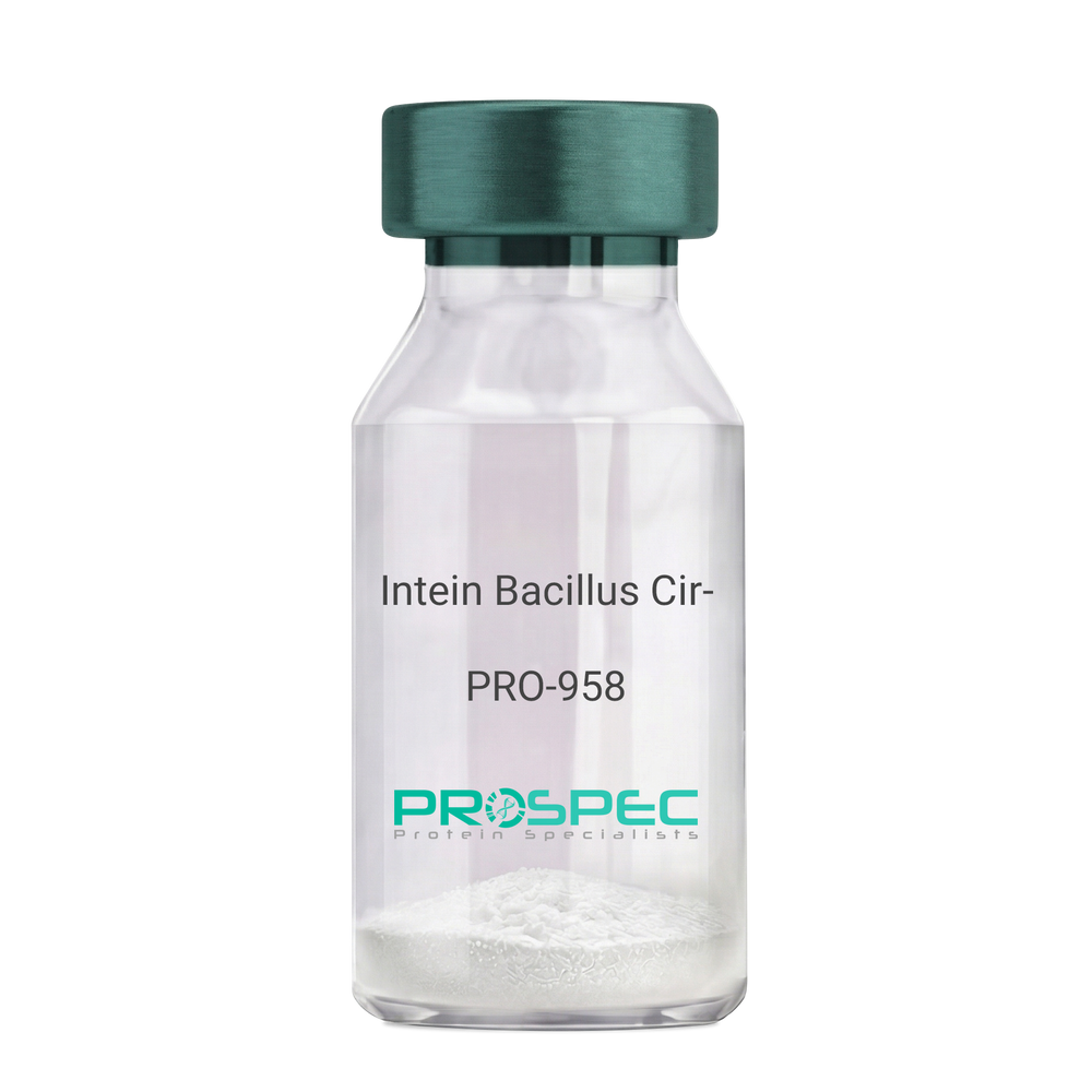
Intein Bacillus CirculansDescription:
Intein Bacillus Circulans Recombinant
Intein-CBD
Product # :
PRO-958Price :
Quantity :
Shipping Method :
Shipped with Ice Packs
More Info
- description
- source
- formulation
- purity
- More Info
- Citations
Description
Intein Bacillus Recombinant produced in E.coli is a single, non-glycosylated polypeptide chain containing 533 amino acids (3-518) and having a molecular mass of 59.4 kDa.Intein is fused to a 16 amino acid His-tag at C-terminus & purified by proprietary chromatographic techniques.
Source
E.coli.
Formulation
The Intein solution (1mg/ml) contains 20mM Tris-HCl buffer (pH 8.0), 1mM DTT, 0.1M NaCl and 10% glycerol.
Purity
Greater than 95% as determined by SDS-PAGE.
Citations
More Info
-
Introduction
Intein is a section of a protein which can remove itself and return the remaining segment with a peptide bond. In addition, Inteins hold an endonuclease domain which takes part in Intein proliferation. Actually, various genes have unrelated intein-coding segments inserted at altered positions and they were found in all three domains of life (eukaryotes, bacteria, and archaea) and in viruses.
-
Synonyms
Intein-CBD
-
Physical Appearance
Sterile Filtered colorless solution.
-
Stability
Store at 4°C if entire vial will be used within 2-4 weeks. Store, frozen at -20°C for longer periods of time. For long term storage it is recommended to add a carrier protein (0.1% HSA or BSA).Avoid multiple freeze-thaw cycles.
-
Amino Acid Sequence
MKIEEGKLVI GSLEGCFAKG TNVLMADGSI ECIENIEVGN KVMGKDGRPR EVIKLPRGRE TMYSVVQKSQ HRAHKSDSSR EVPELLKFTC NATHELVVRT PRSVRRLSRT IKGVEYFEVI TFEMGQKKAP DGRIVELVKE VSKSYPISEG PERANELVES YRKASNKAYF EWTIEARDLS LLGSHVRKAT YQTYAPILYE NDHFFDYMQK SKFHLTIEGP KVLAYLLGLW IGDGLSDRAT FSVDSRDTSL MERVTEYAEK LNLCAEYKDR KEPQVAKTVN LYSKVVRGAS TNPGVSAWQV NTAYTAGQLV TYNGKTYKCL QPHTSLAGWE PSNVPALWQL QGGHGGIRNN LNTENPLWDA IVGLGFLKDG VKNIPSFLST DNIGTRETFL AGLIDSDGYV TDEHGIKATI KTIHTSVRDG LVSLARSLGL VVSVNAEPAK VDMNVTKHKI SYAIYMSGGD VLLNVLSKCA GSKKFRPAPA AAFARECRGF YFELQELKED DYYGITLSDD SDHQFLLGSQ VVVQNLEHHH HHH
ProSpec's products are furnished for LABORATORY RESEARCH USE ONLY. The product may not be used as drugs, agricultural or pesticidal products, food additives or household chemicals.
- View Data Sheet
Name :
RELM b MouseDescription:
RELM-Beta Mouse Recombinant
Resistin-like beta, RELM beta, Cysteine-rich secreted protein FIZZ2, Colon and small intestine-specific cysteine-rich protein, Cysteine-rich secreted protein A12-alpha-like 1, Colon carcinoma-related gene protein, RELM-b, XCP2, HXCP2.
Product # :
CYT-413Price :
Quantity :
Shipping Method :
Shipped at Room temp
More Info
- description
- source
- formulation
- purity
- More Info
- Citations
Description
Mouse RELM-b Recombinant produced in E.Coli is a monomeric, non-glycosylated, polypeptide chain containing 83 amino acids and having a molecular mass of 8.9kDa. The Mouse RETNLB is purified by proprietary chromatographic techniques.
Source
Escherichia Coli.
Formulation
The protein was lyophilized from a concentrated (1mg/ml) protein solution containing 10mM Acetic Acid with 2:1 mannitol to protein.
Purity
Greater than 97% as determined by SDS-PAGE.
Citations
More Info
-
Introduction
RELM-beta (Resistin-Like Molecule-beta) is a member of a recently identified family of secreted proteins containing a conserved cystein-rich C-terminus. The RELM family consists of resistin (also called FIZZ3), RELM-alfa (FIZZ1), RELM-beta (FIZZ2) and RELM-gamma. Only resisistin and RELM-beta were found in humans whereas all four RELM family members were identified in rodents.
RELM-beta appears to be produced as a homodimer exclusively by intestinal goblet cells and can be found in high quantities in stool. Remarkably, stool of germ-free mice displaying sterile intestinal tract does not contain RELM-beta until bacterial colonization takes place after pathogen-free mice entered natural environment. Some, but not all, colon carcinoma cell lines secrete RELM-beta into the cell culture supernatant.
The physiological function of RELM-beta is not known. High doses of recombinant RELM-beta showed hyperglycemic effects including lowered glucose disposal and increased hepatic glucose production in mice. -
Synonyms
Resistin-like beta, RELM beta, Cysteine-rich secreted protein FIZZ2, Colon and small intestine-specific cysteine-rich protein, Cysteine-rich secreted protein A12-alpha-like 1, Colon carcinoma-related gene protein, RELM-b, XCP2, HXCP2.
-
Physical Appearance
Brownish lyophilized powder.
-
Stability
Lyophilized RETNLB is stable at -20°C. After reconstitution the protein should be kept at all times at -20°C. It is recommended to add a carrier protein (0.1% HSA or BSA) for long term storage.
-
Solubility
Reconstitute at 0.1 mg/ml with sterile pyrogen free water.
-
Amino Acid Sequence
MQCSFESLVD QRIKEALSRQ EPKTISCTSV TSSGRLASCP AGMVVTGCAC GYGCGSWDIR NGNTCHCQCS VMDWASARCC RMA.
ProSpec's products are furnished for LABORATORY RESEARCH USE ONLY. The product may not be used as drugs, agricultural or pesticidal products, food additives or household chemicals.
- View Data Sheet
Name :
Rabbit SARS CoV-2 IgG S1Description:
Recombinant Anti Rabbit SARS CoV-2 IgG Spike S1
Product # :
ANT-762Price :
Quantity :
Shipping Method :
Shipped with Ice Packs
More Info
- description
- source
- formulation
- More Info
- Citations
Description
Recombinant Anti Rabbit SARS CoV-2 IgG Kappa Spike S1 is a recombinant monoclonal antibody derived from HEK293 cells which recognizes the SARS-CoV and SARS-CoV-2 Spike glycoprotein. CoV-2 IgG S1 Rabbit Antibody binds to both SARS-CoV and SARS-CoV-2 with high affinity at amino acids 318-510 (RBD, Receptor Binding Domain) in the S1 subunit of the Spike protein. This chimeric rabbit antibody was made using the variable domain sequences of the original Human IgG1 format.
Source
HEK293 Cells.
Formulation
1mg/ml in PBS and 0.02% Proclin 300.
Citations
More Info
-
Introduction
SARS Coronavirus is an enveloped virus containing three outer structural proteins, namely the membrane (M), envelope (E), and spike (S) proteins. Spike (S)-glycoprotein of the virus interacts with a cellular receptor and mediates membrane fusion to allow viral entry into susceptible target cells. Accordingly, S-protein plays an important role in virus infection cycle and is the primary target of neutralizing antibodies.It has recently been shown that SARS is caused by a human coronavirus. Human coronaviruses are the major cause of upper respiratory tract illness in humans, such as the common cold. Coronaviruses are positive-stranded RNA viruses, featuring the largest viral RNA genomes known to date (27-31 kb). The first step in coronavirus infection is binding of the viral spike protein, a 139-kDa protein, to certain receptors on host cells. The spike protein is the main surface antigen of the coronavirus. The most prominent protein in the culture supernatants infected with SARS virus is a 46 kDa nucleocapsid protein. This suggests that the nucleocapsid protein is a major immunogen that may be useful for early diagnostics.
-
Physical Appearance
Sterile Filtered liquid formulation.
-
Stability
Store at 4°C, stable for 2 weeks. For long-term storage, store at -20°C.
-
Immunogen
The native monoclonal antibody was generated by sequencing peripheral blood lymphocytes of a patient exposed to the SARS-CoV
-
Applications
ELISA.
-
Type
Mouse antibody Monoclonal.
-
Purification Method
Protein A affinity purified.
ProSpec's products are furnished for LABORATORY RESEARCH USE ONLY. The product may not be used as drugs, agricultural or pesticidal products, food additives or household chemicals.
- View Data Sheet
Name :
HIV-1 p24, BiotinDescription:
HIV-1 p24 Recombinant, Biotin Labeled
Product # :
HIV-106Price :
Quantity :
Shipping Method :
Shipped with Ice Packs
More Info
- description
- source
- formulation
- purity
- More Info
- Citations
Description
The E.coli derived 39 kDa biotin labeled recombinant protein is a non-glycosylated polypeptide chain, containing the HIV-1 p24 immunodominant regions, amino acids 77-436.The HIV-1 p24 Biotinylated is fused to beta-galactosidase (114 kDa) at the N-terminus.
Source
Escherichia Coli.
Formulation
8M Urea, 20mM Tris-HCl pH 8.0, 10mM b-mercaptoethanol.
Purity
Greater than 95.0% as determined by SDS-PAGE.
Citations
More Info
-
Introduction
Human immunodeficiency virus (HIV) is a retrovirusthat can lead to a condition in which the immune systembegins to fail, leading to opportunistic infections. HIV primarily infects vital cells in the humanimmune systemsuch as helper T cells(specifically CD4+ T cells), macrophagesand dendritic cells. HIV infection leads to low levels of CD4+ T cells through three main mechanisms: firstly, direct viral killing of infected cells; secondly, increased rates of apoptosisin infected cells; and thirdly, killing of infected CD4+ T cells by CD8 cytotoxic lymphocytesthat recognize infected cells. When CD4+ T cell numbers decline below a critical level, cell-mediated immunityis lost, and the body becomes progressively more susceptible to opportunistic infections. HIV was classified as a member of the genus Lentivirus, part of the family of Retroviridae. Lentiviruses have many common morphologies and biological properties. Many species are infected by lentiviruses, which are characteristically responsible for long-duration illnesses with a long incubation period. Lentiviruses are transmitted as single-stranded, positive-sense, enveloped RNA viruses. Upon entry of the target cell, the viral RNA genomeis converted to double-stranded DNAby a virally encoded reverse transcriptasethat is present in the virus particle. This viral DNA is then integrated into the cellular DNA by a virally encoded integraseso that the genome can be transcribed. Once the virus has infected the cell, two pathways are possible: either the virus becomes latentand the infected cell continues to function, or the virus becomes active and replicates, and a large number of virus particles are liberated that can then infect other cells.
-
Physical Appearance
Sterile filtered colorless clear solution.
-
Stability
HIV-1 p24 although stable at 4°C for 1 week, should be stored below -18°C. Please prevent freeze thaw cycles.
-
Applications
HIV-1 p24 antigen is suitable for ELISA and Western blots, excellent antigen for early detection of HIV seroconvertors with minimal specificity problems.
-
Specificity
Immunoreactive with all sera of HIV-1 infected individuals.
ProSpec's products are furnished for LABORATORY RESEARCH USE ONLY. The product may not be used as drugs, agricultural or pesticidal products, food additives or household chemicals.
- View Data Sheet
Name :
DCUN1D4 HumanDescription:
DCN1 Defective in Cullin Neddylation 1 Domain Containing 4 Human Recombinant
DCN1 Defective In Cullin Neddylation 1 Domain Containing 4, Defective In Cullin Neddylation Protein 1-Like Protein 4, DCUN1 Domain-Containing Protein 4, KIAA0276, DCN1-Like Protein 4.
Product # :
PRO-1715Price :
Quantity :
Shipping Method :
Shipped with Ice Packs
More Info
- description
- source
- formulation
- purity
- More Info
- Citations
Description
DCUN1D4 Human Recombinant produced in E.coli is a single, non-glycosylated polypeptide chain containing 315 amino acids (1-292) and having a molecular mass of 36.5kDa.DCUN1D4 is fused to a 23 amino acid His-tag at N-terminus & purified by proprietary chromatographic techniques.
Source
Escherichia Coli.
Formulation
The DCUN1D4 solution (0.25mg/ml) contains 20mM Tris-HCl buffer (pH8.0), 0.2M NaCl, 2mM DTT, 2mM EDTA and 50% glycerol.
Purity
Greater than 85.0% as determined by SDS-PAGE.
Citations
More Info
-
Introduction
DCUN1D4 holds 1 DCUN1 domain.
-
Synonyms
DCN1 Defective In Cullin Neddylation 1 Domain Containing 4, Defective In Cullin Neddylation Protein 1-Like Protein 4, DCUN1 Domain-Containing Protein 4, KIAA0276, DCN1-Like Protein 4.
-
Physical Appearance
Sterile Filtered clear solution.
-
Stability
Store at 4°C if entire vial will be used within 2-4 weeks. Store, frozen at -20°C for longer periods of time. For long term storage it is recommended to add a carrier protein (0.1% HSA or BSA).Avoid multiple freeze-thaw cycles.
-
Amino Acid Sequence
MGSSHHHHHH SSGLVPRGSH MGSMHSDAAA VNFQLNSHLS TLANIHKIYH TLNKLNLTED IGQDDHQTGS LRSCSSSDCF NKVMPPRKKR RPASGDDLSA KKSRHDSMYR KYDSTRIKTE EEAFSSKRCL EWFYEYAGTD DVVGPEGMEK FCEDIGVEPE NVVMLVLAWK LDAQNMGYFT LQEWLKGMTS LQCDTTEKLR NTLDYLRSFL NDSTNFKLIY RYAFDFAREK DQRSLDINTA KCMLGLLLGK IWPLFPVFHQ FLEQSKYKVI NKDQWCNVLE FSRTINLDLS NYDEDGAWPV LLDEFVEWYK DKQMS
ProSpec's products are furnished for LABORATORY RESEARCH USE ONLY. The product may not be used as drugs, agricultural or pesticidal products, food additives or household chemicals.
- View Data Sheet
Name :
IFN b Mouse, HisDescription:
IFN Beta Mouse Recombinant, His Tag
Leukocyte IFN, B cell IFN, Type I IFN, IFNB1, IFB, IFF, IFNB, IFN-b 1b, MGC96956.
Product # :
CYT-651Price :
Quantity :
Shipping Method :
Shipped with Ice Packs
More Info
- description
- source
- formulation
- purity
- More Info
- SDS-PAGE
- Citations
Description
IFN beta Mouse Recombinant produced in E.Coli is a single, non-glycosylated polypeptide chain containing 182 amino acids (22-182 a.a.) and having a molecular mass of 22 kDa. Mouse IFN beta is fused to 21 amino acid at N-terminus and purified by proprietary chromatographic techniques.
Source
Escherichia Coli.
Formulation
The IFN-Beta (0.25mg/ml) contains 20mM HEPES (pH6.0), 0.5M NaCl, 10% glycerol.
Purity
Greater than 95% as determined by Analysis by SDS-PAGE.
SDS-PAGE
Citations
More Info
-
Introduction
IFN-beta 1b has antiviral, antibacterial and anticancer activities.
Influenza A viruses not only inhibit IFN-beta gene induction but also supress Type-I IFN signaling via mechanism involving induction of the SOCS-3 protein.
Intracellular bacteria and cytosolic poly (dA-dT) trigger IFN-beta responses in different human cells without requiring human ZBP1. -
Synonyms
Leukocyte IFN, B cell IFN, Type I IFN, IFNB1, IFB, IFF, IFNB, IFN-b 1b, MGC96956.
-
Physical Appearance
Sterile filtered colorless solution.
-
Stability
Store at 4°C if entire vial will be used within 2-4 weeks. Store, frozen at -20°C for longer periods of time. For long term storage it is recommended to add a carrier protein (0.1% HSA or BSA). Avoid multiple freeze-thaw cycles.
-
Amino Acid Sequence
MGSSHHHHHH SSGLVPRGSH MINYKQLQLQ ERTNIRKCQE LLEQLNGKIN LTYRADFKIP MEMTEKMQKS YTAFAIQEML QNVFLVFRNN FSSTGWNETI VVRLLDELHQ QTVFLKTVLE EKQEERLTWE MSSTALHLKS YYWRVQRYLK LMKYNSYAWM VVRAEIFRNF LIIRRLTRNF QN.
-
Background
What is the molecular weight/Mw of IFN B MOUSE, HIS Protein?
IFN B MOUSE, HIS Protein has a total Mw of 22kDa.
What is the source or expression system of IFN B MOUSE, HIS Protein?
Escherichia Coli.
What is the Purity of IFN B MOUSE, HIS Protein?
IFN B MOUSE, HIS Protein is >95% pure as determined by SDS-PAGE.
What is the Biological Activity of IFN B MOUSE, HIS Protein?
The biological functionality of IFN B MOUSE, HIS Protein will be determined in the future.
What is the amino acid sequence of IFN B MOUSE, HIS Protein?
MGSSHHHHHH SSGLVPRGSH MINYKQLQLQ ERTNIRKCQE LLEQLNGKIN LTYRADFKIP MEMTEKMQKS YTAFAIQEML QNVFLVFRNN FSSTGWNETI VVRLLDELHQ QTVFLKTVLE EKQEERLTWE MSSTALHLKS YYWRVQRYLK LMKYNSYAWM VVRAEIFRNF LIIRRLTRNF QN.
What applications can IFN B MOUSE, HIS Protein be used in?
IFN B MOUSE, HIS Protein can probably be used in western blot, ELISA and Lateral Flow.
What is the endotoxin level for IFN B MOUSE, HIS Protein?
The endotoxin level is minimal, IFN B MOUSE, HIS Protein was purified using conventional chromatography techniques.
ProSpec's products are furnished for LABORATORY RESEARCH USE ONLY. The product may not be used as drugs, agricultural or pesticidal products, food additives or household chemicals.