Search results
1000 results found for “Anti Human Lymphocyte”
Name
Description
Product #
Price
Quantity
Shipping Method
- View Data Sheet
Name :
IgA 1+2 antibodyDescription:
IgA 1&2, Mouse Anti-Human
Product # :
ANT-201Price :
Quantity :
Shipping Method :
Shipped at Room temp
More Info
- formulation
- More Info
Formulation
1mg/ml in PBS (after reconstitution).
More Info
-
Solubility
Reconstitute with H20 to get a 1 mg/ml concentration. Mix gently, wash the sides of the vial and wait 30-60 seconds before use.
-
Immunogen
Purified hIgA.
-
Ig Subclass
Mouse IgG1.
-
Note
This antibody recognizes both human IgA1 and human IgA2 subtypes.
-
Titer
By direct ELISA, 1:20,000 dilution will yield >1.0 O.D using alkaline phosphatase conjugated rabbit anti-mouse Ig (Jackson Laboratories).
-
Shipping Conditions
Antibody is shipped lyophilized at ambient temperature.
-
Type
Mouse Anti Human Monoclonal.
-
Storage Procedures
In lyophilized form, for long periods, store at 4°C in a dry environment. After reconstitution, if not intended for use within a month, aliquot and store at -20°C.Please prevent freeze-thaw cycles.
-
Purification Method
Ion exchange column.
ProSpec's products are furnished for LABORATORY RESEARCH USE ONLY. The product may not be used as drugs, agricultural or pesticidal products, food additives or household chemicals.
- View Data Sheet
Name :
CD27 Human, sf9Description:
CD27 Human Recombinant, sf9
Tumor Necrosis Factor Receptor Superfamily Member 7, T-Cell Activation Antigen CD27, CD27 Molecule, CD27 Antigen, T Cell Activation Antigen S152, CD27L Receptor, TNFRSF7, S152, Tp55, T14.
Product # :
PRO-2345Price :
Quantity :
Shipping Method :
Shipped with Ice Packs
More Info
- description
- source
- formulation
- purity
- More Info
Description
CD27 Human Recombinant produced in Sf9 Baculovirus cells is a single, glycosylated polypeptide chain containing 413 amino acids (21-191a.a.) and having a molecular mass of 46.4kDa (Molecular size on SDS-PAGE will appear at approximately 28-57kDa).CD27 is fused with a 239 amino acids hIgG-His tag at C-Terminus and purified by proprietary chromatographic techniques.
Source
Sf9, Baculovirus cells.
Formulation
CD27 protein solution (0.5mg/ml) contains Phosphate Buffered Saline (pH7.4) and 10% glycerol.
Purity
Greater than 85.0% as determined by SDS-PAGE.
More Info
-
Introduction
CD27 belongs to the TNF-receptor superfamily. CD27 is necessary for initiation and long-term maintenance of T cell immunity. CD27 binds to ligand CD70, and has a crucial role in regulating B-cell activation and immunoglobulin synthesis. The CD27 receptor transduces signals which result in the activation of NF-kappaB and MAPK8/JNK. Adaptor proteins TRAF2 and TRAF5 mediate the signaling process of CD27. CD27-binding protein (SIVA), which is a proapoptotic protein, can bind to the CD27 receptor and is believed to have a significant role in the apoptosis induced by CD27.
-
Synonyms
Tumor Necrosis Factor Receptor Superfamily Member 7, T-Cell Activation Antigen CD27, CD27 Molecule, CD27 Antigen, T Cell Activation Antigen S152, CD27L Receptor, TNFRSF7, S152, Tp55, T14.
-
Physical Appearance
Sterile Filtered colorless solution.
-
Stability
Store at 4°C if entire vial will be used within 2-4 weeks. Store, frozen at -20°C for longer periods of time. For long term storage it is recommended to add a carrier protein (0.1% HSA or BSA). Avoid multiple freeze-thaw cycles.
-
Amino Acid Sequence
ADPTPAPKSC PERHYWAQGK LCCQMCEPGT FLVKDCDQHR KAAQCDPCIP GVSFSPDHHT RPHCESCRHC NSGLLVRNCT ITANAECACR NGWQCRDKEC TECDPLPNPS LTARSSQALS PHPQPTHLPY VSEMLEARTA GHMQTLADFR QLPARTLSTH WPPQRSLCSS DFIRLEPKSC DKTHTCPPCP APELLGGPSV FLFPPKPKDT LMISRTPEVT CVVVDVSHED PEVKFNWYVD GVEVHNAKTK PREEQYNSTY RVVSVLTVLH QDWLNGKEYK CKVSNKALPA PIEKTISKAK GQPREPQVYT LPPSRDELTK NQVSLTCLVK GFYPSDIAVE WESNGQPENN YKTTPPVLDS DGSFFLYSKL TVDKSRWQQG NVFSCSVMHE ALHNHYTQKS LSLSPGKHHH HHH.
ProSpec's products are furnished for LABORATORY RESEARCH USE ONLY. The product may not be used as drugs, agricultural or pesticidal products, food additives or household chemicals.
- View Data Sheet
Name :
ACSF2 AntibodyDescription:
Acyl-CoA Synthetase Family Member 2, Mouse Anti Human
Acyl-CoA Synthetase Family Member 2, PPARG Binding, Long Chain Fatty Acid Acyl Co-A Ligase Like, Acyl-CoA Synthetase Family Member 2, Mitochondrial, EC 6.2.1.26, EC 6.2.1.-, FLJ20920, EC 6.2.1, AVYV493, ACSMW, Acyl-CoA synthetase family member 2, mitochondrial.
Product # :
ANT-694Price :
Quantity :
Shipping Method :
Shipped with Ice Packs
More Info
- formulation
- More Info
Formulation
1mg/ml containing PBS, pH-7.4, 10% Glycerol and 0.02% Sodium Azide.
More Info
-
Introduction
Acyl-CoA synthetase family member 2, also known as ACSF2 is a member of the ATP-dependent AMP-binding enzyme family. Acyl-CoA synthetases are a family of enzymes which catalyze the thioesterification of fatty acids with coenzymeA to form activated intermediates, which play a basic part in lipid metabolism as well as homeostasis of lipid-related processes. ACSF2 is required for the complex of lipid synthesis, energy production via beta-oxidation, protein acylation and fatty-acid dependent transcriptional regulation. Moreover, ACSF2 is required for fatty acid import into cells by the process of vectorial acylation.
-
Synonyms
Acyl-CoA Synthetase Family Member 2, PPARG Binding, Long Chain Fatty Acid Acyl Co-A Ligase Like, Acyl-CoA Synthetase Family Member 2, Mitochondrial, EC 6.2.1.26, EC 6.2.1.-, FLJ20920, EC 6.2.1, AVYV493, ACSMW, Acyl-CoA synthetase family member 2, mitochondrial.
-
Physical Appearance
Sterile filtered colorless solution.
-
Immunogen
Anti-human ACSF2 mAb, is derived from hybridization of mouse F0 myeloma cells with spleen cells from BALB/c mice immunized with recombinant human ACSF2 amino acids 42-615 purified from E. coli.
-
Ig Subclass
Mouse IgG1 heavy chain and κ light chain.
-
Clone
PAT18B4AT.
-
Applications
ACSF2 antibody has been tested by ELISA, Western blot analysis and ICC/IF to assure specificity and reactivity. Since application varies, however, each investigation should be titrated by the reagent to obtain optimal results.
-
Type
Mouse Anti Human Monoclonal.
-
Storage Procedures
For periods up to 1 month store at 4°C, for longer periods of time, store at -20°C. Prevent freeze thaw cycles.
-
Purification Method
ACSF2 antibody was purified from mouse ascitic fluids by protein-A affinity chromatography.
ProSpec's products are furnished for LABORATORY RESEARCH USE ONLY. The product may not be used as drugs, agricultural or pesticidal products, food additives or household chemicals.
- View Data Sheet
Name :
Epo (NYRhEPO) AntibodyDescription:
Erythropoietin Mouse Anti Human, (NYRhEPO)
EPO-a, EPO-alpha, Epoetin, EP, MGC138142.
Product # :
ANT-196Price :
Quantity :
Shipping Method :
Shipped at Room temp
More Info
- formulation
- More Info
Formulation
1mg/ml in PBS (after reconstitution).
More Info
-
Introduction
This gene is a member of the EPO/TPO family and encodes a secreted, glycosylated cytokine composed of four alpha helical bundles. The protein is found in the plasma and regulates red cell production by promoting erythroid differentiation and initiating hemoglobin synthesis. This protein also has neuroprotective activity against a variety of potential brain injuries and antiapoptotic functions in several tissue types.
-
Synonyms
EPO-a, EPO-alpha, Epoetin, EP, MGC138142.
-
Solubility
Reconstitute with sterile H20. Mix gently, wash the sides of the vial and wait 30-60 seconds before use.
-
Immunogen
r.Human erythropoietin.
-
Ig Subclass
Mouse IgG1.
-
Clone
NYRhEPO.
-
Applications
Direct ELISA, Western Blot, Immuneprecipitation.
-
Titer
By direct ELISA, 1:10,000 dilution will yield 0.5 O.D using alkaline phosphatase conjugated rabbit anti-mouse Ig (Jackson Laboratories).
-
Shipping Conditions
Antibody is shipped lyophilized at ambient temperature.
-
Type
Mouse Anti Human Monoclonal.
-
Storage Procedures
In lyophilized form, for long periods, store at 4°C in a dry environment. After reconstitution, if not intended for use within a month, aliquot and store at -20°C.
-
Purification Method
Ion exchange.
ProSpec's products are furnished for LABORATORY RESEARCH USE ONLY. The product may not be used as drugs, agricultural or pesticidal products, food additives or household chemicals.
- View Data Sheet
Name :
BAFFR Human, HEKDescription:
BAFF (BLyS) Receptor Human Recombinant, HEK
TNFRSF13C, CD268, BAFF-R, MGC138235, B cell-activating factor receptor.
Product # :
CYT-1224Price :
Quantity :
Shipping Method :
Shipped with Ice Packs
More Info
- description
- source
- formulation
- purity
- biological activity
- More Info
Description
BAFFR Human Recombinant is a single, glycosylated, polypeptide chain (1-78 a.a) containing a total of 314 amino acids and having a molecular mass of 34.4 kDa. BAFFR is fused to 233 a.a hIgG-Tag at C-terminus and is purified by proprietary chromatographic techniques.
Source
HEK293 Cells.
Formulation
The BAFFR solution (1mg/ml) contains 10% Glycerol and Phosphate-Buffered Saline (pH 7.4).
Purity
Greater than 95.0% as determined by SDS-PAGE.
Biological Activity
The ED50 is ≤0.7 ug/ml, measured by its ability in a functional ELISA with BAFF Human.
More Info
-
Synonyms
TNFRSF13C, CD268, BAFF-R, MGC138235, B cell-activating factor receptor.
-
Physical Appearance
Sterile Filtered colorless solution.
-
Stability
Store at 4°C if entire vial will be used within 2-4 weeks. Store, frozen at -20°C for longer periods of time. For long term storage it is recommended to add a carrier protein (0.1% HSA or BSA).Avoid multiple freeze-thaw cycles.
-
Amino Acid Sequence
DGSMRRGPRS LRGRDAPAPT PCVPAECFDL LVRHCVACGL LRTPRPKPAG ASSPAPRTAL QPQESVGAGA GEAALPLPGL LLEPKSCDKT HTCPPCPAPE LLGGPSVFLF PPKPKDTLMI SRTPEVTCVV VDVSHEDPEV KFNWYVDGVE VHNAKTKPRE EQYNSTYRVV SVLTVLHQDW LNGKEYKCKV SNKALPAPIE KTISKAKGQP REPQVYTLPP SRDELTKNQV SLTCLVKGFY PSDIAVEWES NGQPENNYKT TPPVLDSDGS FFLYSKLTVD KSRWQQGNVF SCSVMHEALH NHYTQKSLSL SPGK.
-
Background
B-cell Activating Factor (BAFF) and its corresponding receptor, BAFF-R, are integral components of the immune system, orchestrating crucial processes in B-cell survival, maturation, and differentiation. As we delve into the intricate world of immunology, the study of BAFF and its receptor has unveiled essential pathways that govern the immune responses in health and disease. This research investigates the multifaceted role of BAFF Receptor Protein, shedding light on its structural complexities, signaling mechanisms, and its pivotal contributions to immune regulation. By exploring the interactions between BAFF and its receptor, scientists aim to decipher the delicate balance that underlies immune homeostasis and explore potential therapeutic avenues.
Structural Architecture of BAFF Receptor Protein:
BAFF Receptor, a transmembrane protein predominantly expressed on B cells, belongs to the tumor necrosis factor receptor (TNFR) superfamily. Its intricate structure involves various domains, each playing a unique role in ligand binding, receptor activation, and downstream signaling. Understanding the structural intricacies of BAFF Receptor is paramount to unraveling the molecular events that govern B-cell fate decisions and immune responses.
Physiological Significance in B-Cell Biology:
BAFF Receptor, upon binding with its ligand BAFF, initiates a cascade of events critical for B-cell survival and function. This interaction promotes B-cell maturation, prevents premature apoptosis, and influences the formation of immune synapses. Additionally, BAFF Receptor signaling is tightly regulated to prevent excessive B-cell activation, ensuring immune tolerance and preventing autoimmune responses. Disruptions in these pathways can lead to autoimmune disorders, underscoring the crucial role of BAFF Receptor in maintaining immune equilibrium.
Regulation of Immune Responses:
BAFF Receptor signaling not only affects B-cell development but also has broader implications for immune responses. By modulating antibody production, B-cell activation, and immune memory, BAFF Receptor plays a vital role in shaping adaptive immunity. Its dysregulation has been implicated in various autoimmune conditions, making it an attractive target for therapeutic interventions aimed at restoring immune balance.
BAFF Receptor as a Therapeutic Target:
The intricate involvement of BAFF Receptor in autoimmune diseases, such as rheumatoid arthritis and systemic lupus erythematosus, has positioned it as a promising therapeutic target. Researchers are exploring monoclonal antibodies and other targeted therapies that aim to modulate BAFF Receptor signaling, providing a new frontier in autoimmune disease management. Additionally, understanding the BAFF-BAFF Receptor axis offers potential insights into the development of vaccines and immunotherapies, fostering innovative approaches in the fight against infectious diseases and malignancies.
BAFF Receptor Protein, as a key player in immune regulation, embodies the complexities of immunology. Its interactions with BAFF orchestrate fundamental processes in B-cell biology and adaptive immunity. As scientists unravel the intricate signaling pathways and structural nuances of BAFF Receptor, they pave the way for novel therapeutic strategies and innovative treatments for autoimmune disorders and beyond. This research not only deepens our understanding of immune regulation but also holds the promise of transformative advancements in immunotherapy, ultimately shaping the future of immune-related healthcare.
What is the molecular weight/Mw of BAFF-R Protein?
BAFF-R Protein has a total Mw of 34.4kDa.
What is the source or expression system of BAFF-R Protein?
HEK293 Cells.
What is the Purity of BAFF-R Protein?
BAFF-R Protein is >95% pure as determined by SDS-PAGE.
What is the Biological Activity of BAFF-R Protein?
The ED50 is ≤0.7 ug/ml, measured by its ability in a functional ELISA with BAFF Human.
What is the amino acid sequence of BAFF-R Protein?
DGSMRRGPRS LRGRDAPAPT PCVPAECFDL LVRHCVACGL LRTPRPKPAG ASSPAPRTAL QPQESVGAGA GEAALPLPGL LLEPKSCDKT HTCPPCPAPE LLGGPSVFLF PPKPKDTLMI SRTPEVTCVV VDVSHEDPEV KFNWYVDGVE VHNAKTKPRE EQYNSTYRVV SVLTVLHQDW LNGKEYKCKV SNKALPAPIE KTISKAKGQP REPQVYTLPP SRDELTKNQV SLTCLVKGFY PSDIAVEWES NGQPENNYKT TPPVLDSDGS FFLYSKLTVD KSRWQQGNVF SCSVMHEALH NHYTQKSLSL SPGK.
What applications can BAFF-R Protein be used in?
BAFF-R Protein can probably be used in western blot, ELISA and Lateral Flow.
What is the endotoxin level for BAFF-R Protein?
The endotoxin level is minimal, BAFF-R Protein was purified using conventional chromatography techniques.
ProSpec's products are furnished for LABORATORY RESEARCH USE ONLY. The product may not be used as drugs, agricultural or pesticidal products, food additives or household chemicals.
- View Data Sheet
Name :
IL 17 HumanDescription:
Interleukin-17 Human Recombinant
CTLA-8, IL-17, IL-17A, Cytotoxic T-lymphocyte-associated antigen 8.
Product # :
CYT-250Price :
Quantity :
Shipping Method :
Shipped at Room temp
More Info
- description
- source
- formulation
- purity
- biological activity
- More Info
- sds-page
Description
Interleukin-17A Human Recombinant produced in E.Coli is a homodimeric, non-glycosylated polypeptide chain containing a total of 264 amino acids (2 chains of 132 aa) and having a molecular mass of 31kDa. The IL-17 is purified by proprietary chromatographic techniques.
Source
Escherichia Coli.
Formulation
Sterile filtered lyophilized (freeze-dried) powder, with 20mM Citrate, 0.1M NaCl, pH4.5.
Purity
Greater than 95.0% as determined by SDS-PAGE.
Biological Activity
The ED50 as determined by the dose-dependent induction of IL-6 in Hs68 cell line was found to be approximately 2ng/ml corresponding to a Specific Activity of 500,000IU/mg.sds-page
More Info
-
Introduction
IL17 is a proinflammatory cytokine produced by activated T cells. IL-17 regulates the activities of NF-kappaB and mitogen-activated protein kinases. Interleukin-17 can stimulate the expression of IL6 and cyclooxygenase-2 (PTGS2/COX-2), as well as enhance the production of nitric oxide (NO). High levels of IL-17 are associated with several chronic inflammatory diseases including rheumatoid arthritis, psoriasis and multiple sclerosis.
-
Synonyms
CTLA-8, IL-17, IL-17A, Cytotoxic T-lymphocyte-associated antigen 8.
-
Physical Appearance
Sterile Filtered White lyophilized (freeze-dried) powder.
-
Stability
Lyophilized Interleukin 17A although stable at room temperature for 3 weeks, should be stored desiccated below -18°C. Upon reconstitution IL17 should be stored at 4°C between 2-7 days and for future use below -18°C.For long term storage it is recommended to add a carrier protein (0.1% HSA or BSA).Please prevent freeze-thaw cycles.
-
Solubility
It is recommended to reconstitute the lyophilized Interleukin 17 in sterile 18MΩ-cm H2O not less than 100µg/ml, which can then be further diluted to other aqueous solutions.
-
Protein content
Protein quantitation was carried out by two independent methods1. UV spectroscopy at 280 nm using the absorbency value of 1.13 as the extinction coefficient for a 0.1% (1mg/ml) solution. This value is calculated by the PC GENE computer analysis program of protein sequences (IntelliGenetics). 2. Analysis by RP-HPLC, using a standard solution of IL-17 as a Reference Standard.
ProSpec's products are furnished for LABORATORY RESEARCH USE ONLY. The product may not be used as drugs, agricultural or pesticidal products, food additives or household chemicals.
- View Data Sheet
Name :
LIF Human, HisDescription:
Leukemia Inhibitory Factor Human Recombinant, His tag
CDF, HILDA, D-FACTOR, Differentiation- stimulating factor, Melanoma-derived LPL inhibitor, MLPLI, Emfilermin, Leukemia inhibitory factor, LIF, DIA.
Product # :
CYT-1082Price :
Quantity :
Shipping Method :
Shipped at Room temp
More Info
- description
- source
- formulation
- purity
- More Info
Description
LIF Human Recombinant produced in E.Coli is a single, non-glycosylated, polypeptide chain (a.a 23-202) containing 189 amino acids including a 9 a.a N-terminal His tag. The total molecular mass is 20.9kDa (calculated).
Source
Escherichia Coli.
Formulation
LIF filtered (0.4 µm) and lyophilized from 0.5mg/ml solution in 20 mM Tris buffer, 20 mM NaCl and 5% w/v trehalose, pH 7.5.
Purity
Greater than 95.0% as determined by SDS-PAGE.
More Info
-
Introduction
Leukemia Inhibitory Factor also called LIF is a lymphoid factor that promotes long-term maintenance of embryonic stem cells by suppressing spontaneous differentiation. Leukemia Inhibitory Factor has several functions such as cholinergic neuron differentiation, control of stem cell pluripotency, bone & fat metabolism, mitogenesis of factor dependent cell lines & promotion of megakaryocyte production in vivo. Human and mouse LIF exhibit a 78% identity in its amino acid sequence.
-
Synonyms
CDF, HILDA, D-FACTOR, Differentiation- stimulating factor, Melanoma-derived LPL inhibitor, MLPLI, Emfilermin, Leukemia inhibitory factor, LIF, DIA.
-
Physical Appearance
Filtered White lyophilized (freeze-dried) powder.
-
Stability
Store lyophilized protein at -20°C. Aliquot the product after reconstitution to avoid repeated freezing/thawing cycles. Reconstituted protein can be stored at 4°C for a limited period of time; it does not show any change after two weeks at 4°C.
-
Solubility
It is recommended to add deionized water to prepare a working stock solution of approximately 0.5mg/ml and let the lyophilized pellet dissolve completely.
-
Amino Acid Sequence
MKHHHHHHAS PLPITPVNAT CAIRHPCHNN LMNQIRSQLA QLNGSANALF ILYYTAQGEP FPNNLDKLCG PNVTDFPPFH ANGTEKAKLV ELYRIVVYLG TSLGNITRDQ KILNPSALSL HSKLNATADI LRGLLSNVLC RLCSKYHVGH VDVTYGPDTS GKDVFQKKKL GCQLLGKYKQ IIAVLAQAF.
ProSpec's products are furnished for LABORATORY RESEARCH USE ONLY. The product may not be used as drugs, agricultural or pesticidal products, food additives or household chemicals.
- View Data Sheet
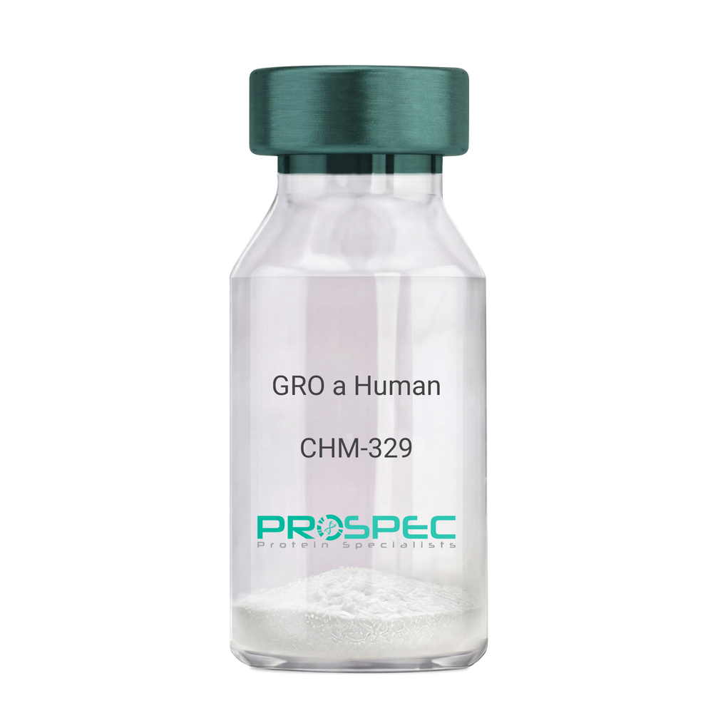
Name :
GRO a HumanDescription:
GRO-Alpha Human Recombinant (CXCL1)
Growth-regulated protein alpha, CXCL1, Melanoma growth stimulatory activity, MGSA, Neutrophil-activating protein 3, NAP-3, GRO-alpha (1-73), chemokine (C-X-C motif) ligand 1, GRO1, GROa, SCYB1, MGSA-a, MGSA alpha.
Product # :
CHM-329Price :
Quantity :
Shipping Method :
Shipped at Room temp
More Info
- description
- source
- formulation
- purity
- biological activity
- More Info
Description
GRO Alpha Human Recombinant produced in E.Coli is a single,non-glycosylated, polypeptide chain containing 73 amino acids and having a molecular mass of 7811 Dalton. The GRO-alpha is purified by proprietary chromatographic techniques.
Source
Escherichia Coli.
Formulation
Lyophilized from a 0.2µm filtered concentrated (1mg/ml) solution in 20mM PB, pH 7.4, 50mM NaCl.
Purity
Greater than 97.0% as determined by:
(a) Analysis by RP-HPLC.
(b) Analysis by SDS-PAGE.Biological Activity
Determined by its ability to chemoattract human peripheral blood neutrophils using a concentration range of 10.0-100.0 ng/ml.
More Info
-
Introduction
Chemokine (C-X-C motif) ligand 1 (CXCL1) is a small cytokine belonging to the CXC chemokine family that was previously called GRO1 oncogene, Neutrophil-activating protein 3 (NAP-3) and melanoma growth stimulating activity, alpha (MSGA-a). It is secreted by human melanoma cells, has mitogenic properties and is implicated in melanoma pathogenesis. CXCL1 is expressed by macrophages, neutrophilsand epithelial cells, and has neutrophil chemoattractant activity. CXCL1 plays a role in spinal cord development by inhibiting the migration of oligodendrocyte precursors and is involved in the processes of angiogenesis, inflammation, wound healing, and tumorigenesis. This chemokine elicits its effects by signaling through the chemokine receptor CXCR2. The gene for CXCL1 is located on human chromosome 4 amongst genes for other CXC chemokines.
-
Synonyms
Growth-regulated protein alpha, CXCL1, Melanoma growth stimulatory activity, MGSA, Neutrophil-activating protein 3, NAP-3, GRO-alpha (1-73), chemokine (C-X-C motif) ligand 1, GRO1, GROa, SCYB1, MGSA-a, MGSA alpha.
-
Physical Appearance
Sterile Filtered White lyophilized (freeze-dried) powder.
-
Stability
Lyophilized GRO-alpha although stable at room temperature for 3 weeks, should be stored desiccated below -18°C. Upon reconstitution CXCL1 should be stored at 4°C between 2-7 days and for future use below -18°C. For long term storage it is recommended to add a carrier protein (0.1% HSA or BSA).Please prevent freeze-thaw cycles.
-
Solubility
It is recommended to reconstitute the lyophilized CXCL1 in sterile 18MΩ-cm H2O not less than 100µg/ml, which can then be further diluted to other aqueous solutions.
-
Amino Acid Sequence
The sequence of the first five N-terminal amino acids was determined and was found to be Ala-Ser-Val-Ala-Thr.
ProSpec's products are furnished for LABORATORY RESEARCH USE ONLY. The product may not be used as drugs, agricultural or pesticidal products, food additives or household chemicals.
- View Data Sheet
Name :
RSV Paired AntibodyDescription:
Mouse Anti Human Respiratory Syncytial Virus
Product # :
ANT-653Price :
Quantity :
Shipping Method :
Shipped with Ice Packs
More Info
- description
- formulation
- purity
- More Info
Description
RSV gold conjugation antibody and RSV capture antibody are used to develop rapid test for RSV rapid test. Please note that when ordering for example: 100µg antibody we ship 50µg from each of the antibodies (100µg in total).
Formulation
* RSV gold conjugation antibody in PBS, PH 7.4.
* RSV capture antibody in PBS, PH 7.4.Purity
Greater than 95%.
More Info
-
Physical Appearance
2 vials of sterile Filtered clear colorless solution.
-
Stability
RSV Paired Antibody although stable at 4°C for 1 week, should be stored below -18°C. Please prevent freeze thaw cycles.
-
Applications
Lateral flow rapid test.
-
Type
Mouse antibody Monoclonal.
-
Purification Method
Purified monoclonal IgG2a by protein A chromatography.
ProSpec's products are furnished for LABORATORY RESEARCH USE ONLY. The product may not be used as drugs, agricultural or pesticidal products, food additives or household chemicals.
- View Data Sheet
Name :
BMP 2 AntibodyDescription:
Bone Morphogenetic protein-2, Mouse Anti-Human
BMP-2, BMP2A.
Product # :
ANT-183Price :
Quantity :
Shipping Method :
Shipped at Room temp
More Info
- source
- formulation
- purity
- More Info
Source
The BMP-2 antibody was prepared by affinity chromatography from ascites of BALB/c mouseinoculated by a hybridoma cell that secreted anti- BMP-2 monoclonal antibodies.
Formulation
Samples are lyophilized in sterile PBS buffers.
Purity
Greater than 90%, analysed by reducing SDS-PAGE.
More Info
-
Introduction
BMP2 belongs to the transforming growth factor-beta (TGFB) superfamily. Bone morphogenic protein induces bone formation. BMP2 is a candidate gene for the autosomal dominant disease of fibrodysplasia (myositis) ossificans progressiva.
-
Synonyms
BMP-2, BMP2A.
-
Physical Appearance
Sterile Filtered White lyophilized (freeze-dried) powder.
-
Stability
For long-term storage, keep aliquot samples at -70°C. Once reconstituted, samples are stable at 4°C for several weeks.
-
Solubility
Reconstitute with sterile H20. Mix gently, wash the sides of the vial and wait 30-60 seconds before use.
-
Applications
Applied in detections of antigen or antibody: ELISA or Western blot. For indirect ELISA, dilute antibody solution by 1: 250.
-
Type
Mouse Anti Human Monoclonal.
ProSpec's products are furnished for LABORATORY RESEARCH USE ONLY. The product may not be used as drugs, agricultural or pesticidal products, food additives or household chemicals.
- View Data Sheet
Name :
ADK AntibodyDescription:
Adenosine Kinase, Mouse Anti Human
Adenosine 5''-phosphotransferase, EC 2.7.1.20, AK, ADK.
Product # :
ANT-057Price :
Quantity :
Shipping Method :
Shipped with Ice Packs
More Info
- formulation
- More Info
Formulation
1mg/ml containing PBS, pH-7.4, & 0.1% Sodium Azide.
More Info
-
Introduction
Adenosine Kinase is an abundant enzyme in mammalian tissues which catalyzes the transfer of the gamma-phosphate from ATP to adenosine, thus is as a regulator of concentrations of both extracellular adenosine and intracellular adenine nucleotides. Adenosine has extensive effects on the cardiovascular, nervous, respiratory, and immune systems and inhibitors of the enzyme take a crucial pharmacological part in growing intravascular adenosine concentrations and acting as anti-inflammatory agents.
-
Synonyms
Adenosine 5''-phosphotransferase, EC 2.7.1.20, AK, ADK.
-
Immunogen
Anti-human ADK mAb, is derived from hybridization of mouse FO myeloma cells with spleen cells from BALB/c mice immunized with recombinant human ADK amino acids 22-362 purified from E. coli.
-
Ig Subclass
Mouse IgG1 heavy chain and k light chain.
-
Clone
PAT4F8AT.
-
Applications
ADK antibody has been tested by ELISA and Western blot analysis to assure specificity and reactivity. Since application varies, however, each investigation should be titrated by the reagent to obtain optimal results. Recommended dilution range for Western blot analysis is 1:1000.
Recommended starting dilution is 1:1000. -
Type
Mouse Anti Human Monoclonal.
-
Storage Procedures
For periods up to 1 month store at 4°C, for longer periods of time, store at -20°C. Prevent freeze thaw cycles.
-
Purification Method
ADK antibody was purified from mouse ascitic fluids by protein-G affinity chromatography.
ProSpec's products are furnished for LABORATORY RESEARCH USE ONLY. The product may not be used as drugs, agricultural or pesticidal products, food additives or household chemicals.
- View Data Sheet
Name :
LSM2 AntibodyDescription:
LSM2 Homolog, U6 Small Nuclear RNA Associated, Mouse Anti Human
LSM2 homolog U6 small nuclear RNA associated (S. cerevisiae), Small nuclear ribonuclear protein D homolog, chromosome 6 open reading frame 28, snRNP core Sm-like protein Sm-x5, C6orf28, YBL026W, Protein G7b, snRNP.
Product # :
ANT-082Price :
Quantity :
Shipping Method :
Shipped with Ice Packs
More Info
- formulation
- More Info
Formulation
1mg/ml containing PBS, pH-7.4, & 0.1% Sodium Azide.
More Info
-
Introduction
LSM2 is a member of the snRNP Sm proteins family. Sm-like proteins are branded in several organisms based on sequence homology with the Sm protein family. Sm-like proteins hold the Sm sequence motif that is made up of 2 regions separated by a linker of variable length which folds as a loop. The Sm-like proteins are believed to create a steady heteromer present in tri-snRNP particles, which are vital for pre-mRNA splicing. LSM2 binds specifically to the 3'-terminal U-tract of U6 snRNA nad takes part in pre-mRNA splicing.
-
Synonyms
LSM2 homolog U6 small nuclear RNA associated (S. cerevisiae), Small nuclear ribonuclear protein D homolog, chromosome 6 open reading frame 28, snRNP core Sm-like protein Sm-x5, C6orf28, YBL026W, Protein G7b, snRNP.
-
Immunogen
Anti-human LSM2 mAb, is derived from hybridization of mouse FO myeloma cells with spleen cells from BALB/c mice immunized with recombinant human LSM2 amino acids 1-95 purified from E. coli.
-
Ig Subclass
Mouse IgG2a heavy chain and k light chain.
-
Clone
PAT2B2AT.
-
Applications
LSM2 antibody has been tested by ELISA, Western blot analysis to assure specificity and reactivity. Since application varies, however, each investigation should be titrated by the reagent to obtain optimal results. Recommended dilution range for Western blot analysis is 1:1,000~1:3,000.
-
Type
Mouse Anti Human Monoclonal.
-
Storage Procedures
For periods up to 1 month store at 4°C, for longer periods of time, store at -20°C. Prevent freeze thaw cycles.
-
Purification Method
LSM2 antibody was purified from mouse ascitic fluids by protein-G affinity chromatography.
ProSpec's products are furnished for LABORATORY RESEARCH USE ONLY. The product may not be used as drugs, agricultural or pesticidal products, food additives or household chemicals.
- View Data Sheet
Name :
ALK/P80 AntibodyDescription:
ALK/P80 Protein, Mouse Anti Human
ALK/P80 Protein, ALK/P80, p80 protein, anaplastic lymphoma kinase, ALK, nucleophosmin, NPM.
Product # :
ANT-331Price :
Quantity :
Shipping Method :
Shipped with Ice Packs
More Info
- formulation
- More Info
Formulation
1mg/ml containing PBS, pH-7.4, & 0.1% Sodium Azide.
More Info
-
Introduction
ALK/p80 is a human p80 protein, identified as a hybrid of the anaplastic lymphoma kinase (ALK) gene and the nucleophosmin (NPM) gene resulting from the t(2;5)(p23;q35) translocation found in a third of large cell lymphomas.
-
Synonyms
ALK/P80 Protein, ALK/P80, p80 protein, anaplastic lymphoma kinase, ALK, nucleophosmin, NPM.
-
Immunogen
Anti-human ALK/P80 mAb is derived from hybridization of mouse SP2/0 myeloma cells with spleen cells from BALB/c mice immunized with recombinant human ALK/P80 amino acids 81-294 purified from E. coli.
-
Ig Subclass
Mouse IgG2b heavy chain and κ light chain.
-
Clone
P5E3AT.
-
Applications
ALK/P80 antibody has been tested by ELISA, Western blot and Immunohistochemical analysis to assure specificity and reactivity. Since application varies, however, each investigation should be titrated by the reagent to obtain optimal results. Recommended dilution range for Western blot analysis is 1:1,000 ~ 2,000. Recommended starting dilution is 1:1,000.
-
Type
Mouse Anti Human Monoclonal.
-
Storage Procedures
For periods up to 1 month store at 4°C, for longer periods of time, store at -20°C. Prevent freeze thaw cycles.
-
Purification Method
ALK/P80 antibody was purified from mouse ascitic fluids by protein-G affinity chromatography.
ProSpec's products are furnished for LABORATORY RESEARCH USE ONLY. The product may not be used as drugs, agricultural or pesticidal products, food additives or household chemicals.
- View Data Sheet
Name :
4-1BBR Human, Sf9Description:
4-1BB Receptor Human Recombinant, Sf9
Tumor Necrosis Factor Receptor Superfamily, Member 9, T-Cell Antigen 4-1BB Homolog, 4-1BB Ligand Receptor, T-Cell Antigen ILA, CD137 Antigen, CDw137, CD137, ILA, Interleukin-Activated Receptor, Homolog Of Mouse Ly63, Induced By Lymphocyte Activation (ILA), Homolog Of Mouse 4-1BB, Receptor Protein 4-1BB, T Cell Antigen ILA, 4-1BB, Tumor necrosis factor receptor superfamily member 9.
Product # :
CYT-931Price :
Quantity :
Shipping Method :
Shipped with Ice Packs
More Info
- description
- source
- formulation
- purity
- More Info
Description
4-1BBR produced in Sf9 Baculovirus cells is a single, glycosylated polypeptide chain containing 411 amino acids (18-186a.a.) and having a molecular mass of 45.3kDa. (Molecular size on SDS-PAGE will appear at approximately 40-57kDa). 4-1BBR is expressed with a 242 amino acid hIgG-His-tag at C-Terminus and purified by proprietary chromatographic techniques.
Source
Sf9, Baculovirus cells.
Formulation
4-1BBR protein solution (0.5mg/ml) contains Phosphate Buffered Saline (pH 7.4) and 10% glycerol.
Purity
Greater than 90.0% as determined by SDS-PAGE
More Info
-
Introduction
4-1BBR is a member of the TNF-receptor superfamily. 4-1BB receptor contributes to the clonal expansion, survival, and development of T cells.4-1BBRcan also induce proliferation in peripheral monocytes, enhance T cell apoptosis induced by TCR/CD3 triggered activation, and regulate CD28 co-stimulation to promote Th1 cell responses. The expression of this receptor is induced by lymphocyte activation. TRAF adaptor proteins have been shown to bind to this receptor and transduce the signals leading to activation of NF-kappaB.
-
Synonyms
Tumor Necrosis Factor Receptor Superfamily, Member 9, T-Cell Antigen 4-1BB Homolog, 4-1BB Ligand Receptor, T-Cell Antigen ILA, CD137 Antigen, CDw137, CD137, ILA, Interleukin-Activated Receptor, Homolog Of Mouse Ly63, Induced By Lymphocyte Activation (ILA), Homolog Of Mouse 4-1BB, Receptor Protein 4-1BB, T Cell Antigen ILA, 4-1BB, Tumor necrosis factor receptor superfamily member 9.
-
Physical Appearance
Sterile Filtered colorless solution.
-
Stability
Store at 4°C if entire vial will be used within 2-4 weeks. Store, frozen at -20°C for longer periods of time. For long term storage it is recommended to add a carrier protein (0.1% HSA or BSA).Avoid multiple freeze-thaw cycles.
-
Amino Acid Sequence
ADLFERTRSL QDPCSNCPAG TFCDNNRNQI CSPCPPNSFS SAGGQRTCDI CRQCKGVFRT RKECSSTSNA ECDCTPGFHC LGAGCSMCEQ DCKQGQELTK KGCKDCCFGT FNDQKRGICR PWTNCSLDGK SVLVNGTKER DVVCGPSPAD LSPGASSVTP PAPAREPGHS PQLEPKSCDK THTCPPCPAP ELLGGPSVFL FPPKPKDTLM ISRTPEVTCV VVDVSHEDPE VKFNWYVDGV EVHNAKTKPR EEQYNSTYRV VSVLTVLHQD WLNGKEYKCK VSNKALPAPI EKTISKAKGQ PREPQVYTLP PSRDELTKNQ VSLTCLVKGF YPSDIAVEWE SNGQPENNYK TTPPVLDSDG SFFLYSKLTV DKSRWQQGNV FSCSVMHEAL HNHYTQKSLS LSPGKHHHHH H.
ProSpec's products are furnished for LABORATORY RESEARCH USE ONLY. The product may not be used as drugs, agricultural or pesticidal products, food additives or household chemicals.
- View Data Sheet
Name :
Recombinant HER2 AntibodyDescription:
Recombinant Human Anti HER2
Product # :
ANT-602Price :
Quantity :
Shipping Method :
Shipped with Ice Packs
More Info
- description
- source
- formulation
- purity
- biological activity
- More Info
Description
Recombinant Human Anti HER2 is produced by recombinant DNA technology in a Chinese Hamster Ovary mammalian cell expression system in a serum-free medium and has a molecular weight of approximately 148 kDa.
Source
CHO.
Formulation
Each ml of Recombinant HER2 Antibody solution (32.1mg/ml) contains 0.56mg histidine-HCl, 0.36mg histidine and 0.1mg polysorbate-20, pH-6.
Purity
Should be not less than 95.0% as determined by:
(a) Analysis by SEC-HPLC.
(b) Analysis by SDS-PAGE.Biological Activity
The ED50 as determined by the proliferation inhibition of BT474 cell, Perform a comparison of a dilution series of the Sample solution with a dilution series of the Standard solution, measured potency was found to be 0.9 x 10⁴EU/mg.More Info
-
Introduction
HER-2/neu (erbB-2) encodes an 185-kDa orphan receptor tyrosine kinase that is constitutively active as a dimer and displays potent oncogenic activity when overexpressed. Herstatin, as the product of alternative HER-2 transcript, retains intron 8. The herstatin mRNA is expressed in normal human fetal kidney and liver, but is at reduced levels relative to p185HER-2 mRNA in carcinoma cells that contain an amplified HER-2 gene. Herstatin appears to be an inhibitor of p185HER-2, because it disrupts dimers, reduces tyrosine phosphorylation of p185, and inhibits the anchorage-independent growth of transformed cells that overexpress HER-2.
-
Physical Appearance
Sterile filtered colorless liquid formulation.
-
Stability
Recombinant Human Anti HER2 should be stored between 2-8°C.
-
Amino Acid Sequence
LIGHT CHAIN
DIQMTQSPSSLSASVGDRVTITCRASQDVNTAVAWYQQ
KPGKAPKLLIYSASFLYSGVPSRFSGSRSGTDFTLTISSL
QPEDFATYYCQQHYTTPPTFGQGTKVEIKRTVAAPSVFI
FPPSDEQLKSGTASVVCLLNNFYPREAKVQWKVDNALQ
SGNSQESVTEQDSKDSTYSLSSTLTLSKADYEKHKVYAC
EVTHQGLSSPVTKSFNRGEC.
HEAVY CHAIN
EVQLVESGGGLVQPGGSLRLSCAASGFNIKDTYIHWVR
QAPGKGLEWVARIYPTNGYTRYADSVKGRFTISADTSK
NTAYLQMNSLRAEDTAVYYCSRWGGDGFYAMDYWGQ
GTLVTVSSASTKGPSVFPLAPSSKSTSGGTAALGCLVKD
YFPEPVTVSWNSGALTSGVHTFPAVLQSSGLYSLSSVVT
VPSSSLGTQTYICNVNHKPSNTKVDKKVEPKSCDKTHTCP
PCPAPELLGGPSVFLFPPKPKDTLMISRTPEVTCVVVDVS
HEDPEVKFNWYVDGVEVHNAKTKPREEQYNSTYRVVSV
LTVLHQDWLNGKEYKCKVSNKALPAPIEKTISKAKGQPR
EPQVYTLPPSREEMTKNQVSLTCLVKGFYPSDIAVEWE
SNGQPENNYKTTPPVLDSDGSFFLYSKLTVDKSRWQQ
GNVFSCSVMHEALHNHYTQKSLSLSPGK.
ProSpec's products are furnished for LABORATORY RESEARCH USE ONLY. The product may not be used as drugs, agricultural or pesticidal products, food additives or household chemicals.
- View Data Sheet
Name :
CD3G HumanDescription:
CD3G Human Recombinant
T-cell surface glycoprotein CD3 gamma chain, CD3-GAMMA, T3G, CD3G.
Product # :
PRO-1392Price :
Quantity :
Shipping Method :
Shipped with Ice Packs
More Info
- description
- source
- formulation
- purity
- More Info
Description
CD3G Human Recombinant produced in E.Coli is a single, non-glycosylated polypeptide chain containing 117 amino acids (23-116a.a.) and having a molecular mass of 13.1kDa. CD3G is fused to a 23 amino acid His-tag at N-terminus & purified by proprietary chromatographic techniques.
Source
E.coli.
Formulation
CD3G protein solution (1mg/ml) containing 20mM Tris-HCl buffer (pH 8.0), 0.4M urea and 10% glycerol.
Purity
Greater than 90% as determined by SDS-PAGE.
More Info
-
Introduction
CD3-gamma polypeptide is encoded by CD3 Gamma (CD3G) which together with CD3-epsilon, -delta and -zeta, and the T-cell receptor alpha/beta and gamma/delta heterodimers, configures the T-cell receptor-CD3 complex. The T-cell receptor-CD3 complex takes part in coupling antigen recognition to numerous intracellular signal-transduction pathways. The genes encoding the epsilon, gamma and delta polypeptides are located in the same cluster on chromosome 11. Deficiencies in this gene are associated with T cell immunodeficiency.
-
Synonyms
T-cell surface glycoprotein CD3 gamma chain, CD3-GAMMA, T3G, CD3G.
-
Physical Appearance
Sterile Filtered colorless solution.
-
Stability
Store at 4°C if entire vial will be used within 2-4 weeks. Store, frozen at -20°C for longer periods of time. For long term storage it is recommended to add a carrier protein (0.1% HSA or BSA).Avoid multiple freeze-thaw cycles.
-
Amino Acid Sequence
MGSSHHHHHH SSGLVPRGSH MGSQSIKGNH LVKVYDYQED GSVLLTCDAE AKNITWFKDG KMIGFLTEDK KKWNLGSNAK DPRGMYQCKG SQNKSKPLQV YYRMCQNCIE LNAATIS.
ProSpec's products are furnished for LABORATORY RESEARCH USE ONLY. The product may not be used as drugs, agricultural or pesticidal products, food additives or household chemicals.
- View Data Sheet
Name :
CD21 HumanDescription:
CD21 Human Recombinant
Complement receptor type 2, Cr2, Complement C3d receptor, Epstein-Barr virus receptor, EBV receptor, CD21 antigen, CR2, C3DR, CD21, SLEB9.
Product # :
PRO-2615Price :
Quantity :
Shipping Method :
Shipped with Ice Packs
More Info
- description
- source
- formulation
- purity
- More Info
Description
CD21 Human Recombinant produced in Sf9 Baculovirus cells is a single, glycosylated polypeptide chain containing 959 amino acids (21-971aa) and having a molecular mass of 105.2kDa.CD21 is fused to an 8 amino acid His-Tag at C-terminus and purified by proprietary chromatographic techniques.
Source
Sf9, Baculovirus cells.
Formulation
The CD21 solution (0.5mg/ml) contains 10% Glycerol and Phosphate Buffered Saline (pH 7.4).
Purity
Greater than 95.0% as determined by SDS-PAGE.
More Info
-
Introduction
CD21 is expressed strongly on mature B cells, follicular dentritic cells and weakly on immature thymocytes and T lymphocytes. In B-cell ontogeny, CD21 appears after the pre-B-stage, is maintained during peripheral B-cell development and is lost upon terminal differentiation into plasma cells. CD21 expression is also gradually lost after stimulation of B cells in vitro. CD21 functions as receptor for C3d, C3dg and iC3b Complement components, for EBV and for IFNalpha. CD21 binds to CD23 and associates with CD19, CD81 and Leu13 to form a large signal-transduction complex involved in B cell activation.
-
Synonyms
Complement receptor type 2, Cr2, Complement C3d receptor, Epstein-Barr virus receptor, EBV receptor, CD21 antigen, CR2, C3DR, CD21, SLEB9.
-
Physical Appearance
Sterile Filtered colorless solution.
-
Stability
Store at 4°C if entire vial will be used within 2-4 weeks. Store, frozen at -20°C for longer periods of time. For long term storage it is recommended to add a carrier protein (0.1% HSA or BSA).Avoid multiple freeze-thaw cycles.
-
Amino Acid Sequence
ISCGSPPPIL NGRISYYSTP IAVGTVIRYS CSGTFRLIGE KSLLCITKDK VDGTWDKPAP
KCEYFNKYSS CPEPIVPGGY KIRGSTPYRH GDSVTFACKT NFSMNGNKSV WCQANNMWGP
TRLPTCVSVF PLECPALPMI HNGHHTSENV GSIAPGLSVT YSCESGYLLV GEKIINCLSS
GKWSAVPPTC EEARCKSLGR FPNGKVKEPP ILRVGVTANF FCDEGYRLQG PPSSRCVIAG
QGVAWTKMPV CEEIFCPSPP PILNGRHIGN SLANVSYGSI VTYTCDPDPE EGVNFILIGE
STLRCTVDSQ KTGTWSGPAP RCELSTSAVQ CPHPQILRGR MVSGQKDRYT YNDTVIFACM
FGFTLKGSKQ IRCNAQGTWE PSAPVCEKEC QAPPNILNGQ KEDRHMVRFD PGTSIKYSCN
PGYVLVGEES IQCTSEGVWT PPVPQCKVAA CEATGRQLLT KPQHQFVRPD VNSSCGEGYK
LSGSVYQECQ GTIPWFMEIR LCKEITCPPP PVIYNGAHTG SSLEDFPYGT TVTYTCNPGP
ERGVEFSLIG ESTIRCTSND QERGTWSGPA PLCKLSLLAV QCSHVHIANG YKISGKEAPY
FYNDTVTFKC YSGFTLKGSS QIRCKADNTW DPEIPVCEKE TCQHVRQSLQ ELPAGSRVEL
VNTSCQDGYQ LTGHAYQMCQ DAENGIWFKK IPLCKVIHCH PPPVIVNGKH TGMMAENFLY
GNEVSYECDQ GFYLLGEKKL QCRSDSKGHG SWSGPSPQCL RSPPVTRCPN PEVKHGYKLN
KTHSAYSHND IVYVDCNPGF IMNGSRVIRC HTDNTWVPGV PTCIKKAFIG CPPPPKTPNG
NHTGGNIARF SPGMSILYSC DQGYLLVGEA LLLCTHEGTW SQPAPHCKEV NCSSPADMDG
IQKGLEPRKM YQYGAVVTLE CEDGYMLEGS PQSQCQSDHQ WNPPLAVCRS RVEHHHHHH
ProSpec's products are furnished for LABORATORY RESEARCH USE ONLY. The product may not be used as drugs, agricultural or pesticidal products, food additives or household chemicals.
- View Data Sheet
Name :
NRG1 (HRG-beta3) AntibodyDescription:
Neuregulin1 isoform HRG-beta3, Mouse Anti Human
Product # :
ANT-458Price :
Quantity :
Shipping Method :
Shipped with Ice Packs
More Info
- formulation
- More Info
Formulation
1mg/ml containing PBS, pH-7.4, & 0.1% Sodium Azide.
More Info
-
Introduction
NRG1 is one of 4 proteins in the neuregulin family which act on the EGFR family of receptors. NRG1 was initially identified as a 44kDa glycoprotein that interacts with the ERBB2 receptor tyrosine kinase to increase its phosphorylation on tyrosine residues. In various brain areas, NRG1 is expressed at synaptic regions and its processing can be regulated by neuronal activity. The HRG-beta3variant of NRG1, along with HRG-alpha, beta1 and beta2 variants, was identified in various normal tissues and cancer cell lines.
-
Immunogen
Anti-human NRG1 mAb is derived from hybridization of mouse F0 myeloma cells with spleen cells from BALB/c mice immunized with recombinant human NRG1 amino acids 20-241 purified from E. coli.
-
Ig Subclass
Mouse IgG1 heavy chain and κ light chain.
-
Clone
Pk1G13AT.
-
Applications
NRG1 antibody has been tested by ELISA and Western blot analysis to assure specificity and reactivity. Since application varies, however, each investigation should be titrated by the reagent to obtain optimal results. Recommended dilution range for Western blot analysis is 1:1,000 ~ 2,000. Recommended starting dilution is 1:1,000.
-
Type
Mouse Anti Human Monoclonal.
-
Storage Procedures
For periods up to 1 month store at 4°C, for longer periods of time, store at -20°C. Prevent freeze thaw cycles.
-
Purification Method
NRG1 antibody was purified from mouse ascitic fluids by protein-G affinity chromatography.
ProSpec's products are furnished for LABORATORY RESEARCH USE ONLY. The product may not be used as drugs, agricultural or pesticidal products, food additives or household chemicals.
- View Data Sheet
Name :
IL 10 Human, HisDescription:
Interleukin-10 Human Recombinant, His
B-TCGF, CSIF, TGIF, IL-10, IL10A, MGC126450, MGC126451, Cytokine synthesis inhibitory factor.
Product # :
CYT-486Price :
Quantity :
Shipping Method :
Shipped with Ice Packs
More Info
- description
- source
- formulation
- purity
- More Info
Description
Interleukin-10 Human Recombinant produced in E.Coli is single, a non-glycosylated, Polypeptide chain containing 181 amino acids fragment (19-178) and having a total molecular mass of 20.94kDa with a 20 amino acids N-Terminal His tag. The IL-10 His-Tag protein is purified by proprietary chromatographic techniques.
Source
Escherichia Coli.
Formulation
Interleukin-10 His is supplied in 20mM Tris-HCl pH-8 and 20% glycerol.
Purity
Greater than 95.0% by SDS-PAGE.
More Info
-
Introduction
Interleukin-10 is a multifunctional cytokine produced by a variety of cell types including T helper cells activated B cells and activated macrophages. Interleukin-10 regulates immune-mediated inflammation and modulates the function of cells such as lymphocytes, monocytes, natural killer cells and dendritic cells.
-
Synonyms
B-TCGF, CSIF, TGIF, IL-10, IL10A, MGC126450, MGC126451, Cytokine synthesis inhibitory factor.
-
Physical Appearance
Sterile Filtered clear solution.
-
Stability
Store at 4°C if entire vial will be used within 2-4 weeks. Store, frozen at -20°C for longer periods of time. For long term storage it is recommended to add a carrier protein (0.1% HSA or BSA).Please avoid freeze thaw cycles.
-
Amino Acid Sequence
MGSSHHHHHH SSGLVPRGSH MSPGQGTQSE NSCTHFPGNL PNMLRDLRDA FSRVKTFFQM KDQLDNLLLK ESLLEDFKGY LGCQALSEMI QFYLEEVMPQ AENQDPDIKA HVNSLGENLK TLRLRLRRCH RFLPCENKSK AVEQVKNAFN KLQEKGIYKA MSEFDIFINY IEAYMTMKIR N.
ProSpec's products are furnished for LABORATORY RESEARCH USE ONLY. The product may not be used as drugs, agricultural or pesticidal products, food additives or household chemicals.
- View Data Sheet
Name :
BMP7 AntibodyDescription:
Bone Morphogenetic Protein-7, Mouse Anti Human
Osteogenic Protein 1, OP-1, BMP-7.
Product # :
ANT-312Price :
Quantity :
Shipping Method :
Shipped with Ice Packs
More Info
- formulation
- More Info
Formulation
1mg/ml containing PBS, pH-7.4, & 0.1% Sodium Azide.
More Info
-
Introduction
The bone morphogenetic proteins (BMPs) are a family of secreted signaling molecules that can induce ectopic bone growth. Many BMPs are part of the transforming growth factor-beta (TGFB) superfamily. BMPs were originally identified by an ability of demineralized bone extract to induce endochondral osteogenesis in vivo in an extraskeletal site. Based on its expression early in embryogenesis, the BMP encoded by this gene has a proposed role in early development. In addition, the fact that this BMP is closely related to BMP5 and BMP7 has lead to speculation of possible bone inductive activity.
-
Synonyms
Osteogenic Protein 1, OP-1, BMP-7.
-
Physical Appearance
Sterile Filtered colorless solution.
-
Immunogen
Anti-human BMP7 mAb, is derived from hybridization of mouse SP2/O myeloma cells with spleen cells from BALB/c mice immunized with Recombinant human BMP7 amino acids 293-431 purified from E. coli.
-
Ig Subclass
Mouse IgG1 heavy chain and κ light chain.
-
Clone
P4E7AT.
-
Applications
BMP7 antibody has been tested by ELISA and Western blot analysis to assure specificity and reactivity. Since application varies, however, each investigation should be titrated by the reagent to obtain optimal results. Recommended dilution range for Western blot analysis is 1:500 ~ 2,000. Recommended starting dilution is 1:1,000.
-
Type
Mouse Anti Human Monoclonal.
-
Storage Procedures
For periods up to 1 month store at 4°C, for longer periods of time, store at -20°C. Prevent freeze thaw cycles.
-
Purification Method
BMP7 antibody was purified from mouse ascitic fluids by protein-G affinity chromatography.
ProSpec's products are furnished for LABORATORY RESEARCH USE ONLY. The product may not be used as drugs, agricultural or pesticidal products, food additives or household chemicals.
- View Data Sheet
Name :
AKR1C1 AntibodyDescription:
Aldo-Keto Reductase Family 1 Member C1, Mouse Anti Human
DDH1, DDH, HAKRC, 20-alpha-HSD, DD1/DD2, HBAB, C9, DD1, H-37, MBAB, MGC8954, 2-ALPHA-HSD, AKR1C1, Aldo-keto reductase family 1 member C1, 20-alpha-hydroxysteroid dehydrogenase, Trans-1,2-dihydrobenzene-1,2-diol dehydrogenase, Indanol dehydrogenase, Dihydrodiol dehydrogenase 1/2, Chlordecone reductase homolog HAKRC, High-affinity hepatic bile acid-binding protein.
Product # :
ANT-044Price :
Quantity :
Shipping Method :
Shipped with Ice Packs
More Info
- formulation
- More Info
Formulation
1mg/ml containing PBS, pH-7.4, & 0.1% Sodium Azide.
More Info
-
Introduction
AKR1C1 transfers progesterone to its inactive state or in other words catalyzes the reaction of 20-alpha-hydroxy progesterone (20-alpha-OHP). In the liver and intestine. AKR1C1 transfers bile and monitors the intrahepatic bile acid concentration though it has a low bile-binding ability. AKR1C1 participates in myelin formation. AKR1C1 is part of the aldo/keto reductase superfamily, which has over 40 known enzymes which catalyze the conversion of aldehydes and ketones to their corresponding alcohols by utilizing NADH and/or NADPH as cofactors thus display overlapping but distinct substrate specificity.
-
Synonyms
DDH1, DDH, HAKRC, 20-alpha-HSD, DD1/DD2, HBAB, C9, DD1, H-37, MBAB, MGC8954, 2-ALPHA-HSD, AKR1C1, Aldo-keto reductase family 1 member C1, 20-alpha-hydroxysteroid dehydrogenase, Trans-1,2-dihydrobenzene-1,2-diol dehydrogenase, Indanol dehydrogenase, Dihydrodiol dehydrogenase 1/2, Chlordecone reductase homolog HAKRC, High-affinity hepatic bile acid-binding protein.
-
Immunogen
Anti-human AKR1C1 mAb, is derived from hybridization of mouse FO myeloma cells with spleen cells from BALB/c mice immunized with recombinant human AKR1C1 amino acids 1-323 purified from E. coli.
-
Ig Subclass
Mouse IgG1 heavy chain and k light chain.
-
Clone
PAT6D10AT.
-
Applications
AKR1C1 antibody has been tested by ELISA and Western blot analysis to assure specificity and reactivity. Since application varies, however, each investigation should be titrated by the reagent to obtain optimal results. Recommended dilution range for Western blot analysis is 1:1000.
Recommended starting dilution is 1:1000. -
Type
Mouse Anti Human Monoclonal.
-
Storage Procedures
For periods up to 1 month store at 4°C, for longer periods of time, store at -20°C. Prevent freeze thaw cycles.
-
Purification Method
AKR1C1 antibody was purified from mouse ascitic fluids by protein-G affinity chromatography.
ProSpec's products are furnished for LABORATORY RESEARCH USE ONLY. The product may not be used as drugs, agricultural or pesticidal products, food additives or household chemicals.
- View Data Sheet
Name :
PPIF AntibodyDescription:
Cyclophilin-F, Mouse Anti Human
Oeptidylprolyl Isomerase F, PPIF, CYP-D, CYP3, SnuCyp-20, Peptidyl-prolyl cis-trans isomerase mitochondrial, Cyclophilin F, FLJ90798, MGC117207, peptidylprolyl isomerase F.
Product # :
ANT-389Price :
Quantity :
Shipping Method :
Shipped with Ice Packs
More Info
- formulation
- More Info
Formulation
1mg/ml containing PBS, pH-7.4, & 0.1% Sodium Azide.
More Info
-
Introduction
PPIF is a part of the peptidyl-prolyl cis-trans isomerase (PPIase) family. PPIF accelerates the folding of proteins. It catalyzes the cis-trans isomerization of proline imidic peptide bonds in oligopeptides. PPIF is key component of the mitochondrial permeability transition pore in the inner mitochondrial membrane. Activation of this pore is thought to be involved in the induction of apoptotic and necrotic cell death.
-
Synonyms
Oeptidylprolyl Isomerase F, PPIF, CYP-D, CYP3, SnuCyp-20, Peptidyl-prolyl cis-trans isomerase mitochondrial, Cyclophilin F, FLJ90798, MGC117207, peptidylprolyl isomerase F.
-
Immunogen
Anti-human Cyclophilin-F mAb is derived from hybridization of mouse F0 myeloma cells with spleen cells from BALB/c mice immunized with recombinant human Cyclophilin-F amino acids 30-207 purified from E. coli.
-
Ig Subclass
Mouse IgG2b heavy chain and κ light chain.
-
Clone
PAT1F5AT.
-
Applications
Cyclophilin-F antibody has been tested by ELISA, Western blot and immunohistochemistry analysis to assure specificity and reactivity. Since application varies, however, each investigation should be titrated by the reagent to obtain optimal results. Recommended dilution range for Western blot is 1:500~1:1,000 and immunohistochemistry analysis is 1:50~100. Recommended starting dilution for Western blot is 1:500 and Immunohistochemistry is 1:50.
-
Type
Mouse Anti Human Monoclonal.
-
Storage Procedures
For periods up to 1 month store at 4°C, for longer periods of time, store at -20°C. Prevent freeze thaw cycles.
-
Purification Method
Cyclophilin-F antibody was purified from mouse ascitic fluids by protein-G affinity chromatography.
ProSpec's products are furnished for LABORATORY RESEARCH USE ONLY. The product may not be used as drugs, agricultural or pesticidal products, food additives or household chemicals.
- View Data Sheet
Name :
CD7 HumanDescription:
CD7 Human Recombinant
T-cell antigen CD7, GP40, LEU-9, Tp40, TP41, CD7, T-cell leukemia antigen, T-cell surface antigen Leu-9.
Product # :
PRO-1673Price :
Quantity :
Shipping Method :
Shipped with Ice Packs
More Info
- description
- source
- formulation
- purity
- More Info
Description
CD7 Human Recombinant produced in E.Coli is a single, non-glycosylated polypeptide chain containing 178 amino acids (26-180 a.a) and having a molecular mass of 18.8 kDa.CD7 is fused to a 23 amino acid His-tag at N-terminus & purified by proprietary chromatographic techniques. 800x600
Source
E.coli.
Formulation
CD7 protein solution (1mg/ml) containing 20mM Tris-HCl buffer (pH 8.0), 0.4M Urea and 10% glycerol.
Purity
Greater than 90% as determined by SDS-PAGE.
More Info
-
Introduction
T-cell antigen CD7 (CD7) is a type I transmembrane glycoprotein which is expressed on pluripotential hemapoietic cells, nearly all human thymocytes and some peripheral blood T cells. The CD7 molecule is missing in a subset of human T cells under some physiologic conditions. An increase in CD7 negative T cells can be observed in a number of inflammatory skin diseases.
-
Synonyms
T-cell antigen CD7, GP40, LEU-9, Tp40, TP41, CD7, T-cell leukemia antigen, T-cell surface antigen Leu-9.
-
Physical Appearance
Sterile Filtered colorless solution.
-
Stability
Store at 4°C if entire vial will be used within 2-4 weeks. Store, frozen at -20°C for longer periods of time. For long term storage it is recommended to add a carrier protein (0.1% HSA or BSA).Avoid multiple freeze-thaw cycles.
-
Amino Acid Sequence
MGSSHHHHHH SSGLVPRGSH MGSAQEVQQS PHCTTVPVGA SVNITCSTSG GLRGIYLRQL GPQPQDIIYY EDGVVPTTDR RFRGRIDFSG SQDNLTITMH RLQLSDTGTY TCQAITEVNV YGSGTLVLVT EEQSQGWHRC SDAPPRASAL PAPPTGSALP DPQTASALPD PPAASALP.
ProSpec's products are furnished for LABORATORY RESEARCH USE ONLY. The product may not be used as drugs, agricultural or pesticidal products, food additives or household chemicals.
- View Data Sheet
Name :
Bcl XL Human, GSTDescription:
B-Cell Lymphoma Extra Large Human Recombinant, GST
BclXL, Bcl-X(L), Bcl-XL.
Product # :
PRO-640Price :
Quantity :
Shipping Method :
Shipped at Room temp
More Info
- description
- source
- formulation
- purity
- More Info
Description
Bcl-XL Human Recombinant produced in E.Coli is a single, non-glycosylated polypeptide chain containing amino acids 1-210.The Bcl-XL is expressed as GST-Tag fusion protein and purified by proprietary chromatographic techniques.
Source
Escherichia Coli.
Formulation
The protein contains 10mM Tris-HCL pH-8, 1mM EDTA and 250mM NaCl.
Purity
Greater than 95.0% as determined by SDS-PAGE.
More Info
-
Introduction
Bcl-XL is a transmembrane protein located in the mitochondrial membranes of cells that are long-lived and postmitotic, such as adult brain cells. It plays arole in the signal transduction pathway of the FAS-Ligand. Bcl-XL is an anti-apoptotic protein which is a member of the Bcl-2 family which are able to form heterodimers, and this is an significant event in the regulation of apoptosis. BCL-XL is involved in the survival of cancer cells.
Bcl-xL is the leading monitor of apoptosis/active cell suicide. Bcl-xL has cell death repressor activity and therefore acts as a survival protein. -
Synonyms
BclXL, Bcl-X(L), Bcl-XL.
-
Physical Appearance
Sterile Filtered White lyophilized (freeze-dried) powder.
-
Stability
Lyophilized Bcl-XL although stable at room temperature for 3 weeks, should be stored desiccated below -18°C. Upon reconstitution Bcl-XL should be stored at 4°C between 2-7 days and for future use below -18°C.For long term storage it is recommended to add a carrier protein (0.1% HSA or BSA).Please prevent freeze-thaw cycles.
-
Solubility
Suspend BclXL in 100?l of 0.5M Acetic acid, over night at 4°C.
Dilute 10 fold into selected buffer system.
Bcl-XL has tendency to form intramolecular disulfide bond, 5mM DTT is recommended in assay buffer. When running SDS-PAGE gel, 10mM DTT is recommended.
ProSpec's products are furnished for LABORATORY RESEARCH USE ONLY. The product may not be used as drugs, agricultural or pesticidal products, food additives or household chemicals.