Search results
1000 results found for “resistin”
Name
Description
Product #
Price
Quantity
Shipping Method
- View Data Sheet
Name :
S100A6 HumanDescription:
S100 Calcium Binding Protein A6 Human Recombinant
Protein S100-A6, Calcyclin, Growth factor-inducible protein 2A9, MLN 4, Prolactin receptor-associated protein, PRA, S100 calcium-binding protein A6, S100A6, CACY, 2A9, 5B10, CABP.
Product # :
PRO-148Price :
Quantity :
Shipping Method :
Shipped with Ice Packs
More Info
- description
- source
- formulation
- purity
- More Info
- Citations
Description
S100A6 Human Recombinant fused with a 20 amino acid His tag at N-terminus produced in E.Coli is a single, non-glycosylated, polypeptide chain containing 110 amino acids (1-90 a.a.) and having a molecular mass of 12.3kDa. The S100A6 is purified by proprietary chromatographic techniques.
Source
Escherichia Coli.
Formulation
The S100A6 solution (1 mg/ml) contains 20mM Tris-HCl buffer (pH8.0), 0.1M NaCl, 1mM DTT and 30% glycerol.
Purity
Greater than 95.0% as determined by SDS-PAGE.
Citations
More Info
-
Introduction
S100A6 is a member of the S100 family of proteins containing 2 EF-hand calcium-binding motifs. S100 proteins are localized either in the cytoplasm or the nucleus of a wide range of cells. S100 proteins are involved in the regulation of a number of cellular processes such as cell cycle progression and differentiation. There are at least 13 members in the S100 gene family, which are located as a cluster on chromosome 1q21. S100A6 function in stimulation of prolactin secretion and exocytosis. Chromosomal rearrangements and altered expression of the S100A6 gene are implicated in melanoma.
-
Synonyms
Protein S100-A6, Calcyclin, Growth factor-inducible protein 2A9, MLN 4, Prolactin receptor-associated protein, PRA, S100 calcium-binding protein A6, S100A6, CACY, 2A9, 5B10, CABP.
-
Physical Appearance
Sterile Filtered colorless solution.
-
Stability
Store at 4°C if entire vial will be used within 2-4 weeks. Store, frozen at -20°C for longer periods of time. For long term storage it is recommended to add a carrier protein (0.1% HSA or BSA).Avoid multiple freeze-thaw cycles.
-
Amino Acid Sequence
MGSSHHHHHH SSGLVPRGSH MACPLDQAIG LLVAIFHKYS GREGDKHTLS KKELKELIQK ELTIGSKLQD AEIARLMEDL DRNKDQEVNF QEYVTFLGAL ALIYNEALKG.
ProSpec's products are furnished for LABORATORY RESEARCH USE ONLY. The product may not be used as drugs, agricultural or pesticidal products, food additives or household chemicals.
- View Data Sheet
Name :
S100P Human, HisDescription:
S100 Calcium Binding Protein P Human Recombinant, His Tag
Protein S100-E, S100E, Migration-Inducing Gene 9, MIG9.
Product # :
PRO-830Price :
Quantity :
Shipping Method :
Shipped with Ice Packs
More Info
- description
- source
- formulation
- purity
- More Info
- Citations
Description
S100P Human Recombinant produced in E.Coli is a single, non-glycosylated, polypeptide chain containing 115 amino acids (1-95a.a.) and having a molecular mass of 12.6 kDa. S100P protein is fused to a 20 amino acid His tag at N-terminus and is purified by standard chromatography.
Source
Escherichia Coli.
Formulation
S100P Human solution (1mg/ml) containing 20mM Tris-HCl pH-8, 1mM DTT, 0.05M NaCl & 20% glycerol.
Purity
Greater than 90% as determined by SDS-PAGE.
Citations
More Info
-
Introduction
S100P is a Ca2+ binding protein that is part of the S100 family of proteins containing 2 EF-hand calcium-binding motifs. S100 proteins are found in the cytoplasm and/or nucleus of a large range of cells, and participates in the regulation of a number of cellular function such as cell cycle progression and differentiation. S100P participates in numerous biological functions but the exact functions or mechanism of its action is still mostly unknown. Once S100P binds calcium ions it goes through a conformational change that results in an exposure of a hydrophobic surface which permits the interaction with specific target proteins.
-
Synonyms
Protein S100-E, S100E, Migration-Inducing Gene 9, MIG9.
-
Physical Appearance
Sterile filtered colorless solution.
-
Stability
Store at 4°C if entire vial will be used within 2-4 weeks. Store, frozen at -20°C for longer periods of time. For long term storage it is recommended to add a carrier protein (0.1% HSA or BSA). Avoid multiple freeze-thaw cycles.
-
Amino Acid Sequence
MGSSHHHHHH SSGLVPRGSH MTELETAMGM IIDVFSRYSG SEGSTQTLTK GELKVLMEKE LPGFLQSGKD KDAVDKLLKD LDANGDAQVD FSEFIVFVAA ITSACHKYFE KAGLK.
ProSpec's products are furnished for LABORATORY RESEARCH USE ONLY. The product may not be used as drugs, agricultural or pesticidal products, food additives or household chemicals.
- View Data Sheet
Name :
Noggin MouseDescription:
Noggin Mouse Recombinant
Noggin, SYM1, SYNS1, NOG.
Product # :
CYT-600Price :
Quantity :
Shipping Method :
Shipped at Room temp
More Info
- description
- source
- formulation
- purity
- biological activity
- More Info
- Citations
Description
Noggin Mouse Recombinant produced in E.Coli is a non-glycosylated, disulfide-linked protein consisting of two 206 amino acid polypeptide chains, having a total molecular mass of approximately 46.4 kDa (each chain 23.2 kDa).
Source
Escherichia Coli.
Formulation
Lyophilized from a 0.2μm filtered solution in 30% acetonitrile, 0.1% TFA.
Purity
Greater than 95.0% as determined by SDS-PAGE.
Biological Activity
The ED50 as determined by inhibiting BMP-4-induced alkaline phosphatase production of murine ATDC5 cells is less than 2ng/ml, corresponding to a specific activity of > 5.0 × 10⁵ IU/mg in the presence of 5ng/ml BMP-4.Citations
More Info
-
Introduction
The secreted polypeptide noggin, encoded by the NOG gene, binds and inactivates members of the transforming growth factor-beta (TGF-beta) superfamily signaling proteins, such as bone morphogenetic protein-4 (BMP4). By diffusing through extracellular matrices more efficiently than members of the TGF-beta superfamily, noggin may have a principal role in creating morphogenic gradients. Noggin appears to have pleiotropic effect, both early in development as well as in later stages. It was originally isolated from Xenopus based on its ability to restore normal dorsal-ventral body axis in embryos that had been artificially ventralized by UV treatment. The results of the mouse knockout of noggin suggest that it is involved in numerous developmental processes, such as neural tube fusion and joint formation. Recently, several dominant human NOG mutations in unrelated families with proximal symphalangism (SYM1) and multiple synostoses syndrome (SYNS1) were identified; both SYM1 and SYNS1 have multiple joint fusion as their principal feature, and map to the same region (17q22) as NOG. All NOG mutations altered evolutionarily conserved amino acid residues. The amino acid sequence of human noggin is highly homologous to that of Xenopus, rat and mouse.
-
Synonyms
Noggin, SYM1, SYNS1, NOG.
-
Physical Appearance
Sterile Filtered White lyophilized (freeze-dried) powder.
-
Stability
Lyophilized Mouse Noggin although stable at room temperature for 3 weeks, should be stored desiccated below -18°C. Upon reconstitution Mouse Noggin should be stored at 4°C between 2-7 days and for future use below -18°C.For long term storage it is recommended to add a carrier protein (0.1% HSA or BSA).Please prevent freeze-thaw cycles.
-
Solubility
It is recommended to be briefly centrifuged prior to opening to bring the contents to the bottom. Reconstitute in 10mM HAc to a concentration of 0.1-1.0 mg/ml. Further dilutions should be made in appropriate buffered solutions.
-
Amino Acid Sequence
MQHYLHIRPAPSDNLPLVDLIEHPDPIFDPKEKDLNETLLRSLLGGHYD
PGFMATSPPEDRPGGGGGPAGGAEDLAELDQLLRQRPSGAMPSEIKG
LEFSEGLAQGKKQRLSKKLRRKLQMWLWSQTFCPVLYAWNDLGSRF
WPRYVKVGSCFSKRSCSVPEGMVCKPSKSVHLTVLRWRCQRRGQR
CGWIPIQYPIISECKCSC.
ProSpec's products are furnished for LABORATORY RESEARCH USE ONLY. The product may not be used as drugs, agricultural or pesticidal products, food additives or household chemicals.
- View Data Sheet
Name :
FTH1 HumanDescription:
Ferritin Human Recombinant, Heavy Chain
Ferritin heavy chain, Cell proliferation-inducing gene 15 protein, FTH1, FHC, FTH, PLIF, FTHL6, PIG15, MGC104426.
Product # :
PRO-658Price :
Quantity :
Shipping Method :
Shipped with Ice Packs
More Info
- description
- source
- formulation
- purity
- More Info
- Citations
Description
FTH1 Human Recombinant produced in E.Coli is a single, non-glycosylated polypeptide chain containing 183 amino acids and having a molecular mass of 21 kDa.
Source
Escherichia Coli.
Formulation
The FTH1 protein solution contains 20mM Tris-HCl pH-7.5, 1mM DTT and 10% glycerol.
Purity
Greater than 95.0% as determined by SDS-PAGE.
Citations
More Info
-
Introduction
Ferritin is a fairly large, iron-storage heteropolymeric protein composed of 2 subunit types, light Ferritin & heavy Ferritin polypeptides, which is expressed in most kinds of cells and co-assemble in different proportion in a tissue-specific manner. Ferritin is composed of 24 self-assembled polypeptide subunits of the heavy and light ferritin chains and is characterized by the capacity to remove Fe (II) from solution in the presence of oxygen.
Ferritin light polypeptide protein is the main intracellular iron storage protein in prokaryotes and eukaryotes. Variation in ferritin subunit composition influence the rates of iron uptake and release in various tissues. A key function of ferritin is the storage of iron in a soluble and nontoxic state. Defects in this light chain ferritin gene are associated with several neurodegenerative diseases and hyper ferrit anemia-cataract syndrome.
Ferritin stores iron in a soluble, nontoxic, readily accessible form. Ferritin is needed for iron homeostasis. Iron is taken up in the ferrous form and deposited as ferric hydroxides after it has been oxidized. -
Synonyms
Ferritin heavy chain, Cell proliferation-inducing gene 15 protein, FTH1, FHC, FTH, PLIF, FTHL6, PIG15, MGC104426.
-
Physical Appearance
Sterile Filtered colorless solution.
-
Stability
Store at 4°C if entire vial will be used within 2-4 weeks. Store, frozen at -20°C for longer periods of time. For long term storage it is recommended to add a carrier protein (0.1% HSA or BSA).Avoid multiple freeze-thaw cycles.
-
Amino Acid Sequence
MTTASTSQVR QNYHQDSEAA INRQINLELY ASYVYLSMSY YFDRDDVALK NFAKYFLHQS HEEREHAEKL MKLQNQRGGR IFLQDIKKPD CDDWESGLNA MECALHLEKN VNQSLLELHK LATDKNDPHL CDFIETHYLN EQVKAIKELG DHVTNLRKMG APESGLAEYL FDKHTLGDSD NES.
ProSpec's products are furnished for LABORATORY RESEARCH USE ONLY. The product may not be used as drugs, agricultural or pesticidal products, food additives or household chemicals.
- View Data Sheet
Name :
KRT19 Human, HisDescription:
Cytokeratin 19 Human Recombinant , His Tag
Keratin type I cytoskeletal 19, Cytokeratin-19, CK-19, Keratin-19, K19, KRT19, CK19, K1CS, MGC15366.
Product # :
PRO-1347Price :
Quantity :
Shipping Method :
Shipped with Ice Packs
More Info
- description
- source
- formulation
- purity
- More Info
- Citations
Description
KRT19 Human Recombinant produced in E.coli is a single, non-glycosylated polypeptide chain containing 423 amino acids (1-400) and having a molecular mass of 46.5kDa.KRT19 is fused to a 23 amino acid His-Tag at N-terminus and purified by proprietary chromatographic techniques.
Source
Escherichia Coli.
Formulation
The KRT19 solution (0.5mg/ml) contains 20mM Tris-HCl buffer (pH 8.0), 0.4M Urea and 10% glycerol.
Purity
Greater than 90.0% as determined by SDS-PAGE.
Citations
More Info

-
Introduction
CTK-19 is a member of the keratin family. The keratins are intermediate filament proteins responsible for the structural integrity of epithelial cells and are subdivided into cytokeratins and hair keratins. The type I cytokeratins consist of acidic proteins which are arranged in pairs of heterotypic keratin chains. Unlike its related family members, this smallest known acidic cytokeratin is not paired with a basic cytokeratin in epithelial cells. It is specifically expressed in the periderm, the transiently superficial layer that envelopes the developing epidermis. The type I cytokeratins are clustered in a region of chromosome 17q12-q21.
-
Synonyms
Keratin type I cytoskeletal 19, Cytokeratin-19, CK-19, Keratin-19, K19, KRT19, CK19, K1CS, MGC15366.
-
Physical Appearance
Sterile Filtered colorless solution.
-
Stability
Store at 4°C if entire vial will be used within 2-4 weeks. Store, frozen at -20°C for longer periods of time. For long term storage it is recommended to add a carrier protein (0.1% HSA or BSA).Please avoid freeze thaw cycles.
-
Amino Acid Sequence
MGSSHHHHHH SSGLVPRGSH MGSMTSYSYR QSSATSSFGG LGGGSVRFGP GVAFRAPSIH GGSGGRGVSV SSARFVSSSS SGAYGGGYGG VLTASDGLLA GNEKLTMQNL NDRLASYLDK VRALEAANGE LEVKIRDWYQ KQGPGPSRDY SHYYTTIQDL RDKILGATIE NSRIVLQIDN ARLAADDFRT KFETEQALRM SVEADINGLR RVLDELTLAR TDLEMQIEGL KEELAYLKKN HEEEISTLRG QVGGQVSVEV DSAPGTDLAK ILSDMRSQYE VMAEQNRKDA EAWFTSRTEE LNREVAGHTE QLQMSRSEVT DLRRTLQGLE IELQSQLSMK AALEDTLAET EARFGAQLAH IQALISGIEA QLGDVRADSE RQNQEYQRLM DIKSRLEQEI ATYRSLLEGQ EDHYNNLSAS KVL.
ProSpec's products are furnished for LABORATORY RESEARCH USE ONLY. The product may not be used as drugs, agricultural or pesticidal products, food additives or household chemicals.
- View Data Sheet
Name :
LAIR1 HumanDescription:
Leukocyte-Associated Ig-Like Receptor 1 Human Recombinant
Leukocyte-associated immunoglobulin-like receptor 1, LAIR-1, hLAIR1, CD305 antigen, LAIR1, CD305.
Product # :
PRO-431Price :
Quantity :
Shipping Method :
Shipped with Ice Packs
More Info
- description
- source
- formulation
- purity
- More Info
- Citations
Description
LAIR1 Recombinant produced in E.Coli is a single, non-glycosylated polypeptide chain containing amino acids 105 and having a molecular mass of 11.5 kDa. The extracellular domain of LAIR-1 (22-125aa) was overexpressed in E.coli, and purified by using conventional chromatography techniques.
Source
Escherichia Coli.
Formulation
The protein (1mg/ml) contains 20mM Tris-HCl (pH7.5) 50mM NaCl and 1mM EDTA.
Purity
Greater than 95.0% as determined by SDS-PAGE.
Citations
More Info
-
Introduction
LAIR1 is a glycoprotein (~32kDa) expressed on the surface of the majority of human peripheral blood mononuclear leukocytes including T cells, B cells, NK cells, macrophages and dendritic cells. LAIR-1 functions as an inhibitory receptor in NK cells, T cells and B cells. Inhibitory receptors control the immune response to prevent lysis of cells recognized as self. Lair1 consists of a leader sequence, extracellular domain, transmembrane domain, cytoplasmic region.
LAIR1 is a member of both the immunoglobulin superfamily and the leukocyte-associated inhibitory receptor family. LAIR1 maps to a region of 19q13.4 labeled the leukocyte receptor cluster, which contains at least 29 genes encoding leukocyte-expressed receptors of the immunoglobulin superfamily. -
Synonyms
Leukocyte-associated immunoglobulin-like receptor 1, LAIR-1, hLAIR1, CD305 antigen, LAIR1, CD305.
-
Physical Appearance
Sterile filtered colorless solution.
-
Stability
Store at 4°C if entire vial will be used within 2-4 weeks.Store, frozen at -20°C for longer periods of time. For long term storage it is recommended to add a carrier protein (0.1% HSA or BSA).Avoid multiple freeze-thaw cycles.
-
Amino Acid Sequence
MQEEDLPRPS ISAEPGTVIP LGSHVTFVCR GPVGVQTFRL ERESRSTYND TEDVSQASPSESEARFRIDS VSEGNAGPYR CIYYKPPKWS EQSDYLELLV KETSG.
ProSpec's products are furnished for LABORATORY RESEARCH USE ONLY. The product may not be used as drugs, agricultural or pesticidal products, food additives or household chemicals.
- View Data Sheet
Name :
MBL2 HumanDescription:
Mannose-Binding Lectin 2 Human Recombinant
COLEC1, HSMBPC, MBL, MBL2D, MBP, MBP-C, MBP1, Mannose-binding protein C, Collectin-1, Mannan-binding protein, Mannose-binding lectin.
Product # :
PRO-1285Price :
Quantity :
Shipping Method :
Shipped with Ice Packs
More Info
- description
- source
- formulation
- purity
- More Info
- Citations
Description
MBL2 Human Recombinant produced in E. coli is a single polypeptide chain containing 164 amino acids (108-248) and having a molecular mass of 18 kDa. MBL2 is fused to 23 amino acid His-tag at N-terminus & purified by proprietary chromatographic techniques.
Source
E.coli.
Formulation
The MBL2 solution (1mg/1ml) contains 20mM Tris-HCl buffer (pH 8.0), 2M Urea, 20% glycerol and 0.2M Nacl.
Purity
Greater than 90% as determined by SDS-PAGE.
Citations
More Info
-
Introduction
Mannose-binding protein C (MBL2),belongs to the collectin family of patternrecognition molecules and is an important component in the innate immune system. MBL2 is a secreted glycoprotein which recognizes mannose and N-acetylglucosamine on various microorganisms, and is capable of activating the classical complement pathway. Lacking MBL2 has been associated with susceptibility to autoimmune and infectious diseases.
-
Synonyms
COLEC1, HSMBPC, MBL, MBL2D, MBP, MBP-C, MBP1, Mannose-binding protein C, Collectin-1, Mannan-binding protein, Mannose-binding lectin.
-
Physical Appearance
Sterile Filtered colorless solution.
-
Stability
Store at 4°C if entire vial will be used within 2-4 weeks. Store, frozen at -20°C for longer periods of time. For long term storage it is recommended to add a carrier protein (0.1% HSA or BSA).Avoid multiple freeze-thaw cycles.
-
Amino Acid Sequence
MGSSHHHHHH SSGLVPRGSH MGSAASERKA LQTEMARIKK WLTFSLGKQV GNKFFLTNGE IMTFEKVKAL CVKFQASVAT PRNAAENGAI QNLIKEEAFL GITDEKTEGQ FVDLTGNRLT YTNWNEGEPN NAGSDEDCVL LLKNGQWNDV PCSTSHLAVC EFPI.
ProSpec's products are furnished for LABORATORY RESEARCH USE ONLY. The product may not be used as drugs, agricultural or pesticidal products, food additives or household chemicals.
- View Data Sheet
Name :
ZNHIT1 HumanDescription:
Zinc Finger HIT-Type Containing 1 Human Recombinant
Zinc Finger HIT-Type Containing 1, ZNHIT1, ZNFN4A1, Zinc Finger Protein, Subfamily 4A (HIT Domain Containing) Member 1, Zinc Finger HIT Domain Containing 1, Putative Cyclin G1 Interacting Protein, Cyclin-G1-Binding Protein 1, Zinc Finger Protein Subfamily 4A Member 1, P18 Hamlet, CG1I, H_DJ0747G18.14, p18Hamlet, Zinc Finger HIT Domain-Containing Protein 1, Zinc Finger HIT Type 1, CGBP1.
Product # :
PRO-1448Price :
Quantity :
Shipping Method :
Shipped with Ice Packs
More Info
- description
- source
- formulation
- purity
- More Info
- Citations
Description
ZNHIT1 Human Recombinant produced in E.coli is a single, non-glycosylated polypeptide chain containing 177 amino acids (1-154) and having a molecular mass of 19.9kDa. ZNHIT1 is fused to a 23 amino acid His-tag at N-terminus.
Source
Escherichia Coli.
Formulation
The ZNHIT1 solution (1mg/ml) contains 20mM Tris-HCl buffer (pH 8.0), 10% glycerol and 0.4M Urea.
Purity
Greater than 95.0% as determined by SDS-PAGE.
Citations
More Info
-
Introduction
Zinc Finger HIT-Type Containing 1 (ZNHIT1) is a member of the ZNHIT1 family and contains 1 HIT-type zinc finger. The ZNHIT1 protein is induced by DNA damage and appears to have a role in p53-mediated apoptosis induction. ZNHIT1 interacts with MAPK11 and MAPK14 and is a component of the chromatin-remodeling SRCAP complex composed of at least SRCAP, DMAP1, RUVBL1, RUVBL2, ACTL6A, YEATS4, ACTR6 and ZNHIT1. ZNHIT1 binds to NR1D2 and discharges it of its inhibitory effect on the transcription of APOC3 without affecting its DNA-binding activity.
-
Synonyms
Zinc Finger HIT-Type Containing 1, ZNHIT1, ZNFN4A1, Zinc Finger Protein, Subfamily 4A (HIT Domain Containing) Member 1, Zinc Finger HIT Domain Containing 1, Putative Cyclin G1 Interacting Protein, Cyclin-G1-Binding Protein 1, Zinc Finger Protein Subfamily 4A Member 1, P18 Hamlet, CG1I, H_DJ0747G18.14, p18Hamlet, Zinc Finger HIT Domain-Containing Protein 1, Zinc Finger HIT Type 1, CGBP1.
-
Physical Appearance
Sterile Filtered colorless solution.
-
Stability
Store at 4°C if entire vial will be used within 2-4 weeks. Store, frozen at -20°C for longer periods of time. For long term storage it is recommended to add a carrier protein (0.1% HSA or BSA).Avoid multiple freeze-thaw cycles.
-
Amino Acid Sequence
MGSSHHHHHH SSGLVPRGSH MGSMVEKKTS VRSQDPGQRR VLDRAARQRR INRQLEALEN DNFQDDPHAG LPQLGKRLPQ FDDDADTGKK KKKTRGDHFK LRFRKNFQAL LEEQNLSVAE GPNYLTACAG PPSRPQRPFC AVCGFPSPYT CVSCGARYCT VRCLGTHQET RCLKWTV.
ProSpec's products are furnished for LABORATORY RESEARCH USE ONLY. The product may not be used as drugs, agricultural or pesticidal products, food additives or household chemicals.
- View Data Sheet
Name :
Protein-L, HisDescription:
Protein L Recombinant, His Tag
Product # :
PRO-1930Price :
Quantity :
Shipping Method :
Shipped at Room temp
More Info
- description
- source
- formulation
- purity
- More Info
- Citations
Description
Recombinant Protein-L produced in E.Coli is a single non-glycosylated polypeptide chain fused with a 6×His tag at N-terminus. Protein-L is comprised of 5 IgG-binding regions of protein L (B1-B2-B3-B4-B5) containing 372 amino acids in total and having a molecular mass of 41.5kDa, however, it migrates with an apparent molecular mass of 46kDa on SDS-PAGE. Cell wall binding region, cell membrane binding region and albumin binding region have been eliminated from the recombinant Protein-L to guarantee the maximum specific IgG binding.
Source
Escherichia Coli.
Formulation
Protein-L was lyophilized without any additives.
Purity
Greater than 97.0% as determined by:
(a) Analysis by SEC-HPLC.
(b) Analysis by SDS-PAGE.Citations
More Info
-
Introduction
The Recombinant Protein L is comprised of 5 kappa-binding domains. Protein L has the exceptional ability to bind through kappa light chain interactions without hindering with the antibody’s antigen-binding site. This gives Protein L the capacity to bind a broader range of Ig classes and subclasses than other antibody-binding proteins. The recombinant Protein L is perfect for purification of polyclonal or monoclonal IgG antibodies. Protein L binds to IgG from humans, mice, rats and pigs.
-
Physical Appearance
Sterile Filtered White lyophilized (freeze-dried) powder.
-
Stability
Lyophilized Protein-L although stable at room temperature for 3 weeks, should be stored desiccated below -18°C. Upon reconstitution Protein-L should be stored at 4°C between 2-7 days and for future use below -18°C. For long term storage it is recommended to add a carrier protein (0.1% HSA or BSA). Please prevent freeze-thaw cycles.
-
Solubility
It is recommended to reconstitute the lyophilized Protein-L in sterile 18M-cm H2O not less than 0.1mg/ml, which can then be further diluted to other aqueous solutions.
-
Amino Acid Sequence
MHHHHHHKEE TPETPETDSE EEVTIKANLI FANGSTQTAE FKGTFEKATS EAYAYADTLK KDNGEYTVDV ADKGYTLNIK FAGKEKTPEE PKEEVTIKAN LIYADGKTQT AEFKGTFEEA TAEAYRYADA LKKDNGEYTV DVADKGYTLN IKFAGKEKTP EEPKEEVTIK ANLIYADGKT QTAEFKGTFE EATAEAYRYA DLLAKENGKY TVDVADKGYT LNIKFAGKEK TPEEPKEEVT IKANLIYADG KTQTAEFKGT FAEATAEAYR YADLLAKENG KYTADLEDGG YTINIRFAGK KVDEKPEEKE QVTIKENIYF EDGTVQTATF KGTFAEATAE AYRYADLLSK EHGKYTADLE DGGYTINIRF AG.
ProSpec's products are furnished for LABORATORY RESEARCH USE ONLY. The product may not be used as drugs, agricultural or pesticidal products, food additives or household chemicals.
- View Data Sheet
Name :
HYKK HumanDescription:
Hydroxylysine Kinase Human Recombinant
Hydroxylysine kinase, AGPHD1, 5-hydroxy-L-lysine kinase, HYKK, Aminoglycoside phosphotransferase domain-containing protein 1.
Product # :
PKA-064Price :
Quantity :
Shipping Method :
Shipped with Ice Packs
More Info
- description
- source
- formulation
- purity
- More Info
- Citations
Description
HYKK Human Recombinant produced in E.Coli is a single, non-glycosylated polypeptide chain containing 396 amino acids (1-373a.a) and having a molecular mass of 44.3kDa.HYKK is fused to a 23 amino acid His-tag at N-terminus & purified by proprietary chromatographic techniques.
Source
E.coli.
Formulation
HYKK solution (0.5mg/ml) contains Phosphate Buffered Saline (pH 7.4), 40% glycerol, 1mM DTT and 0.1mM PMSF.
Purity
Greater than 90% as determined by SDS-PAGE.
Citations
More Info
-
Introduction
Hydroxylysine Kinase, also known as HYKK, is a Protein Coding gene. HYKK which is a part of the aminoglycoside phosphotransferase family catalyzes the GTPdependent phosphorylation of 5-hydroxy-L-lysine.
-
Synonyms
Hydroxylysine kinase, AGPHD1, 5-hydroxy-L-lysine kinase, HYKK, Aminoglycoside phosphotransferase domain-containing protein 1.
-
Physical Appearance
Sterile Filtered colorless solution.
-
Stability
Store at 4°C if entire vial will be used within 2-4 weeks. Store, frozen at -20°C for longer periods of time. For long term storage it is recommended to add a carrier protein (0.1% HSA or BSA).Avoid multiple freeze-thaw cycles.
-
Amino Acid Sequence
MGSSHHHHHH SSGLVPRGSH MGSMSSGNYQ QSEALSKPTF SEEQASALVE SVFGLKVSKV RPLPSYDDQN FHVYVSKTKD GPTEYVLKIS NTKASKNPDL IEVQNHIIMF LKAAGFPTAS VCHTKGDNTA SLVSVDSGSE IKSYLVRLLT YLPGRPIAEL PVSPQLLYEI GKLAAKLDKT LQRFHHPKLS SLHRENFIWN LKNVPLLEKY LYALGQNRNR EIVEHVIHLF KEEVMTKLSH FRECINHGDL NDHNILIESS KSASGNAEYQ VSGILDFGDM SYGYYVFEVA ITIMYMMIES KSPIQVGGHV LAGFESITPL TAVEKGALFL LVCSRFCQSL VMAAYSCQLY PENKDYLMVT AKTGWKHLQQ MFDMGQKAVE EIWFETAKSY ESGISM.
ProSpec's products are furnished for LABORATORY RESEARCH USE ONLY. The product may not be used as drugs, agricultural or pesticidal products, food additives or household chemicals.
- View Data Sheet
Name :
Pen a 1.0101Description:
Tropomyosin Pen a 1.0101 Recombinant
Product # :
ALR-024Price :
Quantity :
Shipping Method :
Shipped with Ice Packs
More Info
- description
- source
- formulation
- purity
- More Info
- Citations
Description
Recombinant Tropomyosin Pen a 1.0101 produced in SF9 is a glycosylated, polypeptide chain having a calculated molecular mass of 38kDa.Pen a 1.0101 is expressed with a 10xHis tag and purified by proprietary chromatographic techniques.
Source
Sf9 insect cells.
Formulation
Pen a 1.0101 is supplied in 20mM HEPES buffer pH-7.9 200mM NaCl and 6M Urea.
Purity
Greater than 95% as determined by SDS-PAGE.
Citations
More Info
-
Introduction
Pen a 1.0101 is a Tropomyosin and the only major allergen in brown shrimp Penaeus aztecus. The panallergen is highly conserved and soluble muscle protein. Pen a 1.0101 is has an alpha-helical homodimeric, coiled-coil structure.
-
Physical Appearance
Sterile Filtered colorless solution.
-
Stability
Store at 4°C if entire vial will be used within 2-4 weeks.Store, frozen at -20°C for longer periods of time.Avoid multiple freeze-thaw cycles.
ProSpec's products are furnished for LABORATORY RESEARCH USE ONLY. The product may not be used as drugs, agricultural or pesticidal products, food additives or household chemicals.
- View Data Sheet
Name :
BD 2 MouseDescription:
Beta Defensin-2 Mouse Recombinant
Beta-defensin 2, BD-2, mBD-2, Defensin beta 2, Defb2, MGC129140, MGC129141.
Product # :
CYT-035Price :
Quantity :
Shipping Method :
Shipped at Room temp
More Info
- description
- source
- formulation
- purity
- biological activity
- More Info
- Citations
Description
Beta Defensin-2 Mouse Recombinant produced in E.Coli is a single, non-glycosylated polypeptide chain containing 51 amino acids and having a molecular mass of 5.5kDa. The BD-2 is purified by proprietary chromatographic techniques.
Source
Escherichia Coli.
Formulation
The Mouse BD-2 was lyophilized from a 0.2µm filtered concentrated solution in PBS, pH7.4.
Purity
Greater than 98.0% as determined by:
(a) Analysis by RP-HPLC.
(b) Analysis by SDS-PAGE.Biological Activity
Determined by the ability to chemoattract immature human dendritic cells at a concentration of 10-100ng/ml.Citations
More Info
-
Introduction
The Defensin family are highly similar in their protein sequence and are microbicidal & cytotoxic peptides made by neutrophils. Beta Defensin-1 is an antimicrobial peptide having the resistance of epithelial surfaces to microbial colonization. Beta Defensin-1 has close proximity to Defensin Alpha-1 and has been implicated in the pathogenesis of cystic fibrosis.
Skin of patients having atopic dermatitis patients and mycosis fungoides (non-lesional and lesional) show lower human Beta Defensin-1 mRNA expression and higher human Beta Defensin-2 and human Beta Defensin-3 mRNA expression.
BBeta Defensin is highly expressed by epithelial cells.
Beta-defensin 1 may play a role in the pathogenesis of severe sepsis.
Variation in human Beta Defensin-1 contributes to asthma diagnosis, with apparent gender-specific effects. Human Beta Defensin-3 is a dimer, while Human BD-1 and Human BD-2 are monomeric. The expression of Human BD1 is correlated with induction profiles in gingival keratinocytes.
The level of expression of human DEFB1 mRNA is lower than that of human BD3 and human BD-2 in reconstructed epidermis.
Human BD1 is down-regulated in human prostatic and renal carcinomas. -
Synonyms
Beta-defensin 2, BD-2, mBD-2, Defensin beta 2, Defb2, MGC129140, MGC129141.
-
Physical Appearance
Sterile Filtered White lyophilized (freeze-dried) powder.
-
Stability
Lyophilized Beta Defensin-2 Recombinant although stable at room temperature for 3 weeks, should be stored desiccated below -18°C. Upon reconstitution BD-2 should be stored at 4°C between 2-7 days and for future use below -18°C.Please prevent freeze-thaw cycles.
-
Solubility
It is recommended to reconstitute the lyophilized Beta Defensin-2 in sterile 18M-cm H2O not less than 100µg/ml, which can then be further diluted to other aqueous solutions.
-
Amino Acid Sequence
AVGSLKSIGY EAELDHCHTN GGYCVRAICP PSARRPGSCF PEKNPCCKYM K.
-
Background
What is the molecular weight/Mw of BD2 Protein?
BD2 Protein has a total Mw of 5.5kDa.
What is the source or expression system of BD2 Protein?
Escherichia Coli.
What is the Purity of BD2 Protein?
BD2 Protein is >98% pure as determined by SDS-PAGE.
What is the Biological Activity of BD2 Protein?
Determined by the ability to chemoattract immature human dendritic cells at a concentration of 10-100ng/ml.
What is the amino acid sequence of BD2 Protein?
AVGSLKSIGY EAELDHCHTN GGYCVRAICP PSARRPGSCF PEKNPCCKYM K.
What applications can BD2 Protein be used in?
BD2 Protein can probably be used in western blot, ELISA and Lateral Flow.
What is the endotoxin level for BD2 Protein?
The endotoxin level is minimal, BD2 Protein was purified using conventional chromatography techniques.
ProSpec's products are furnished for LABORATORY RESEARCH USE ONLY. The product may not be used as drugs, agricultural or pesticidal products, food additives or household chemicals.
- View Data Sheet
Name :
ST13 PAT5C6AT AntibodyDescription:
HSP70 Interacting Protein Clone PAT5C6AT, Mouse Anti Human
AAG2, SNC6, HSPABP, FAM10A1, FAM10A4, HSPABP1, ST-13, Hsc70-interacting protein, Suppression of tumorigenicity protein 13, Putative tumor suppressor ST13, Protein FAM10A1, Progesterone receptor-associated p48 protein, Renal carcinoma antigen NY-REN-33, ST13, HIP, HOP, P48, PRO0786, FLJ27260, MGC129952.
Product # :
ANT-536Price :
Quantity :
Shipping Method :
Shipped with Ice Packs
More Info
- formulation
- More Info
- Citations
Formulation
1mg/ml containing PBS, pH-7.4, 10% Glycerol and 0.02% Sodium Azide.
Citations
More Info
-
Introduction
ST13 is an adaptor protein (co-chaperone) that mediates the association of HSP70 & HSP90 and appears in early receptor complexes. ST13 plays a role in the assembly process of glucocorticoid receptor, which requires the assistance of multiple molecular chaperones. The expression of ST13 is downregulated in colorectal carcinoma tissue signifying that is candidate tumor suppressor gene. Through common binding to both Hsp70 and Hsp90, ST13 functions as an adaptor that can integrate Hsp70 and Hsp90 interactions. The expression of ST13 decreases in colorectal cancer tissue compared with that in adjacent normal tissue. ST13 is mostly expressed in colorectal epithelia and adenocarcinoma cells. ST13 functions to promote the efficiency of glucocorticoid receptor maturation in cells. The expression levels of the ST13 gene were significantly decreased in primary tumors compared with adjacent mucosa.
-
Synonyms
AAG2, SNC6, HSPABP, FAM10A1, FAM10A4, HSPABP1, ST-13, Hsc70-interacting protein, Suppression of tumorigenicity protein 13, Putative tumor suppressor ST13, Protein FAM10A1, Progesterone receptor-associated p48 protein, Renal carcinoma antigen NY-REN-33, ST13, HIP, HOP, P48, PRO0786, FLJ27260, MGC129952.
-
Physical Appearance
Sterile filtered colorless solution.
-
Immunogen
Anti-human ST13 mAb, is derived from hybridization of mouse F0 myeloma cells with spleen cells from BALB/c mice immunized with a recombinant human ST13 protein 1-369 amino acids purified from E. coli.
-
Ig Subclass
Mouse IgG2a heavy chain and k light chain.
-
Clone
PAT5C6AT.
-
Applications
ST13 antibody has been tested by ELISA, Western blot analysis to assure specificity and reactivity. Since application varies, however, each investigation should be titrated by the reagent to obtain optimal results. Recommended starting dilution is 1:1000.
-
Type
Mouse Anti Human Monoclonal.
-
Storage Procedures
For periods up to 1 month store at 4°C, for longer periods of time, store at -20°C. Prevent freeze thaw cycles.
-
Purification Method
ST13 antibody was purified from mouse ascitic fluids by protein-A affinity chromatography.
ProSpec's products are furnished for LABORATORY RESEARCH USE ONLY. The product may not be used as drugs, agricultural or pesticidal products, food additives or household chemicals.
- View Data Sheet
Name :
Streptavidin-NCDescription:
Streptavidin-NC Recombinant
Product # :
PRO-338Price :
Quantity :
Shipping Method :
Shipped with Ice Packs
More Info
- description
- source
- formulation
- purity
- More Info
- Citations
Description
Recombinant Streptavidin-NC produced in E.Coli is a single, non-glycosylated, polypeptide chain having a molecular mass of 24kDa. Streptavidin-NC is engineered to bind to nitrocellulose.
Source
Escherichia Coli.
Formulation
The sterile solution contains 10mM K2HPO4-KH2PO4, pH 7.3.
Purity
Greater than 93.0% as determined by SDS-PAGE.
Citations
More Info
-
Introduction
Streptavidin is a tetrameric protein secreted by Streptomyces avidinii which binds firmly to biotin. Streptavidin is wilyde used in molecular biology through its unique high affinity for the vitamin biotin. The dissociation constant (Kd) of the biotin-streptavidin complex is about ~10-15 mol/L. The strong affinity recognition of biotin and biotinylated molecules has made streptavidin one of the most important components in diagnostics and laboratory kits. The streptavidin/biotin system has one of the biggest free energies of association of yet observed for noncovalent binding of a protein and small ligand in aqueous solution (K_assoc = 10**14). The complexes are also extremely stable over a wide range of temperature and pH.
-
Physical Appearance
Sterile Liquid formulation.
-
Stability
Streptavidin-NC although stable at 4°C for 3 weeks, should be stored below -18°C. Please prevent freeze thaw cycles.
ProSpec's products are furnished for LABORATORY RESEARCH USE ONLY. The product may not be used as drugs, agricultural or pesticidal products, food additives or household chemicals.
- View Data Sheet
Name :
Activin B Human ActiveDescription:
Activin-B Human Recombinant, Active
Inhibin beta B (activin AB beta polypeptide), Inhibin, beta-2, Activin beta-B chain, MGC157939.
Product # :
CYT-057Price :
Quantity :
Shipping Method :
Shipped at Room temp
More Info
- description
- source
- formulation
- purity
- biological activity
- More Info
- Citations
Description
Activin B human Recombinant produced in Nicotiana benthamiana plant is a beta-B single chain (aa 293-406) containing 123 amino acids (molecular formula C615H910N178O177S12). Activin B is fused to a 10-His-tag at the N-terminal having the total molecular mass of 14kDa and purified by standard chromatographic techniques.
Source
Nicotiana benthamiana plant
Formulation
Lyophilized from 1mg/ml solution in 0.05M Tris-HCl buffer pH 7.4.
Purity
Greater than 97.0% as determined by Analysis by SDS-PAGE.
Biological Activity
The biological activity of Activin B is measured by its ability to inhibit mouse plasmacytoma cell line (MPC-11) cells proliferation. EC50 <5ng/ml is required to stimulate a half-maximal response at cytokine saturation. Note: Since applications vary, each investigator should titrate the reagent to obtain optimal results.Citations
More Info
-
Synonyms
Inhibin beta B (activin AB beta polypeptide), Inhibin, beta-2, Activin beta-B chain, MGC157939.
-
Physical Appearance
Sterile Filtered White lyophilized (freeze-dried) powder.
-
Stability
Lyophilized Activin B although stable at room temperature for 3 weeks, should be stored desiccated below -18°C. Upon reconstitution Activin B should be stored at 4°C between 2-7 days and for future use below -18°C. For long term storage it is recommended to add a carrier protein (0.1% HSA or BSA). Please prevent freeze-thaw cycles.
-
Solubility
It is recommended to reconstitute the lyophilized Activin B in sterile 18M-cm H2O not less than 100µg/ml, which can then be further diluted to other aqueous solutions.
-
Amino Acid Sequence
HHHHHHHHHH GLECDGRTNL CCRQQFFIDF RLIGWNDWII APTGYYGNYC EGSCPAYLAG VPGSASSFHT AVVNQYRMRG LNPGTVNSCC IPTKLSTMSM LYFDDEYNIV KRDVPNMIVE ECG.
-
Background
An Investigation into the Functional Roles and Therapeutic Potential of Activin-B Human Recombinant, Active
1. Abstract
Activin-B Human Recombinant, Active, also referred to as beta-2, Activin beta-B chain, or MGC157939, is a crucial component of the Transforming Growth Factor-beta (TGF-beta) superfamily. The multifaceted nature of this protein implicates it in numerous physiological processes. This paper delves into the bioactivity of Activin-B, exploring its role in cellular proliferation, differentiation, apoptosis, and its potential for therapeutic applications, especially in the realms of regenerative medicine, reproductive health, and cancer therapy.
2. Introduction
The TGF-beta superfamily, of which Activin-B is a member, is renowned for its far-reaching implications in cell and developmental biology. This superfamily boasts members that control cell growth, differentiation, and apoptosis, thus playing vital roles in organogenesis, bone growth, and reproductive functions. This research paper aims to shed light on the characteristics and potential therapeutic applications of Activin-B.
3. Structure and Synthesis of Activin-B
Activin-B is a dimeric protein, composed of two identical beta-B chains. This homodimer undergoes multiple stages of synthesis, starting as a precursor protein, which then experiences proteolytic processing to eventually form the mature peptide. It is this coordinated activity of various enzymes and molecular chaperones that ensure the accurate biosynthesis of Activin-B.
4. Biological Functions of Activin-B
Activin-B's roles extend from embryogenesis and organogenesis to the modulation of reproductive functions. Its influence over cellular proliferation, differentiation, and apoptosis has significant repercussions in physiological and pathological scenarios. Its regulatory functions also encompass immunomodulation and wound healing, underpinning its extensive biological reach.
5. Activin-B in Regenerative Medicine
Regenerative medicine's primary focus is the repair and regeneration of tissues, and it is here that the potential of Activin-B shines. The protein's capacity to regulate cellular processes positions it as a possible agent in tissue repair, making it an intriguing research topic for therapeutic applications in regenerative medicine.
6. Activin-B and Reproductive Health
Activin-B’s role in reproductive health is undeniable, having been implicated in follicular development, ovulation, and pregnancy maintenance. Its potent influence on reproductive functions indicates the possibility of its use in the treatment of reproductive disorders, providing a potential pathway for further therapeutic development.
7. Activin-B in Cancer
Recent research has connected the deregulation of Activin-B to various types of cancer. Deciphering the mechanisms through which Activin-B affects cancer cell proliferation and survival could open up new avenues for targeted cancer therapy. This critical linkage emphasizes the need for comprehensive studies on Activin-B's role in oncogenesis.
8. Conclusion and Future Perspectives
Our understanding of Activin-B's biological functions has grown immensely, but many mysteries remain. The continued exploration of the molecular mechanisms through which Activin-B operates will undoubtedly yield more insights into its potential therapeutic uses, guiding the development of new treatments for a myriad of diseases.
What is the molecular weight / Mw of Activin B Protein?
Activin A Protein has a total Mw of 14 kDa.What is the source or expression system of Activin B Protein?
Nicotinia
What is the Purity of Activin B Protein?
Activin B Protein is >97% pure as determined by SDS-PAGE.
What is the Biological Activity of Activin B Protein?
The biological activity of Activin B is measured by its ability to inhibit mouse plasmacytoma cell line (MPC-11) cells proliferation. EC50 <5ng/ml is required to stimulate a half-maximal response at cytokine saturation. Note: Since applications vary, each investigator should titrate the reagent to obtain optimal results.
What is the endotoxin level for Activin B Protein?
The endotoxin level is minimal, ACTIVIN B Protein was purified using conventional chromatography techniques.
What is the amino acid sequence of ACTIVIN B Protein?
HHHHHHHHHH GLECDGRTNL CCRQQFFIDF RLIGWNDWII APTGYYGNYC EGSCPAYLAG VPGSASSFHT AVVNQYRMRG LNPGTVNSCC IPTKLSTMSM LYFDDEYNIV KRDVPNMIVE ECG
What applications can ACTIVIN B Protein be used in?
ACTIVIN A Protein can probably be used in western blot, ELISA and Lateral Flow.
ProSpec's products are furnished for LABORATORY RESEARCH USE ONLY. The product may not be used as drugs, agricultural or pesticidal products, food additives or household chemicals.
- View Data Sheet
Name :
Hemopexin Human, Sf9Description:
Hemopexin Human Recombinant, Sf9
Hemopexin, Beta-1B-Glycoprotein, HX, Beta-1B-glycoprotein.
Product # :
PRO-2544Price :
Quantity :
Shipping Method :
Shipped with Ice Packs
More Info
- description
- source
- formulation
- purity
- More Info
- Citations
Description
Hemopexin produced in Sf9 Baculovirus cells is a single, glycosylated polypeptide chain containing 448 amino acids (24-462a.a.) and having a molecular mass of 50.4kDa. (Molecular size on SDS-PAGE will appear at approximately 50-70kDa).Hemopexin is expressed with a 9 amino acid His tag at C-Terminus and purified by proprietary chromatographic techniques.
Source
Sf9, Baculovirus cells.
Formulation
Hemopexin protein solution (0.5mg/ml) contains 10% glycerol & Phosphate Buffered Saline (pH 7.4).
Purity
Greater than 90.0% as determined by SDS-PAGE.
Citations
More Info
-
Introduction
Hemopexin (or haemopexin) is a plasma protein that binds heme with the highest affinity of any known protein. Hemopexin is generally expressed in liver, and belongs to acute phase reactants, the synthesis of which is induced after inflammation. Heme is potentially very toxic because of its ability to intercalate into lipid membrane and to generate hydroxyl radicals. Hemopexin’s function of scavenging the heme released or lost by the turnover of heme proteins such as hemoglobin defends the body from the oxidative damage that free heme can cause. Additionally, hemopexin discharges its bound ligand for internalisation upon interacting with a specific receptor located on the surface of liver cells. This hemopexin function is in order to preserve the body's iron. Hemopexin’s levels in the serum are an indication of how much heme is present in the blood. Low Hemopexin levels show that there is a lot of it in the serum. For that reason, low hemopexin levels indicate that there has been consid
-
Synonyms
Hemopexin, Beta-1B-Glycoprotein, HX, Beta-1B-glycoprotein.
-
Physical Appearance
Sterile filtered colorless solution.
-
Stability
Store at 4°C if entire vial will be used within 2-4 weeks.Store, frozen at -20°C for longer periods of time. For long term storage it is recommended to add a carrier protein (0.1% HSA or BSA). Avoid multiple freeze-thaw cycles.
-
Amino Acid Sequence
ADPTPLPPTS AHGNVAEGET KPDPDVTERC SDGWSFDATT LDDNGTMLFF KGEFVWKSHK WDRELISERW KNFPSPVDAA FRQGHNSVFL IKGDKVWVYP PEKKEKGYPK LLQDEFPGIP SPLDAAVECH RGECQAEGVL FFQGDREWFW DLATGTMKER SWPAVGNCSS ALRWLGRYYC FQGNQFLRFD PVRGEVPPRY PRDVRDYFMP CPGRGHGHRN GTGHGNSTHH GPEYMRCSPH LVLSALTSDN HGATYAFSGT HYWRLDTSRD GWHSWPIAHQ WPQGPSAVDA AFSWEEKLYL VQGTQVYVFL TKGGYTLVSG YPKRLEKEVG TPHGIILDSV DAAFICPGSS RLHIMAGRRL WWLDLKSGAQ ATWTELPWPH EKVDGALCME KSLGPNSCSA NGPGLYLIHG PNLYCYSDVE KLNAAKALPQ PQNVTSLLGC THHHHHHH.
ProSpec's products are furnished for LABORATORY RESEARCH USE ONLY. The product may not be used as drugs, agricultural or pesticidal products, food additives or household chemicals.
- View Data Sheet
Name :
BDNF HumanDescription:
Brain-Derived Neurotrophic Factor Human Recombinant
Brain-Derived Neurotrophic Factor, BDNF, MGC34632.
Product # :
CYT-207Price :
Quantity :
Shipping Method :
Shipped at Room temp
More Info
- description
- source
- formulation
- purity
- biological activity
- More Info
- Activity
- Citations
Description
BDNF Human Recombinant produced in E.Coli is a homodimer, non-glycosylated, polypeptide chain containing 2 x 119 amino acids (and an N-terminal Met) and having a total molecular mass of 28kDa. BDNF Human Recombinant is purified by proprietary chromatographic techniques.
Source
Escherichia Coli.
Formulation
The protein was lyophilized with 20mM PB and 400mM NaCl, pH 7.2.
Purity
BDNF is greater than 950% as determined SDS-PAGE.
Biological Activity
The activity was determined using Immobilized Human TrkB-His tag protein 2ug/ml (100 μl/well) for its binding to NHS-Biotin BDNF. The ED50 of was found to be ≤20ng/mLActivity
Citations
More Info
-
Introduction
BDNF promotes the survival of neuronal populations that are all located either in the central nervous system or directly connected to it. BDNF is a major regulator of synaptic transmission and plasticity at adult synapses in many regions of the cns. The versatility of BDNF is emphasized by its contribution to a range of adaptive neuronal responses including long-term potentiation (ltp), long-term depression (ltd), certain forms of short-term synaptic plasticity, as well as homeostatic regulation of intrinsic neuronal excitability.
-
Synonyms
Brain-Derived Neurotrophic Factor, BDNF, MGC34632.
-
Physical Appearance
Sterile Filtered White lyophilized (freeze-dried) powder.
-
Stability
Lyophilized BDNF although stable at room temperature for 3 weeks, should be stored desiccated below -18°C. Upon reconstitution BDNF should be stored at 4°C between 2-7 days and for future use below -18°C.For long term storage it is recommended to add a carrier protein (0.1% HSA or BSA).Please prevent freeze-thaw cycles.
-
Solubility
It is recommended to reconstitute the lyophilized BDNF in sterile 18M-cm H2O not less than 100µg/ml, which can then be further diluted to other aqueous solutions.
-
Amino Acid Sequence
HSDPARRGE LSVCDSISEW VTAADKKTAV DMSGGTVTVL EKVPVSKGQL KQYFYETKCN PMGYTKEGCR GIDKRHWNSQ CRTTQSYVRA LTMDSKKRIG WRFIRIDTSC VCTLTIKRGR.
-
Background
Final Thoughts
Although more research is needed on the safety and effectiveness of BDNF human recombinant, trials suggest that this laboratory-produced protein may be effective in managing and treating several neurological and psychiatric disorders. It's important for experts to stay up to date on the latest developments and research to learn more about potential risks and benefits.
What is the molecular weight/Mw of BDNF Protein?
BDNF Protein has a total Mw of 27kDa.
What is the source or expression system of BDNF Protein?
Escherichia Coli.
What is the Purity of BDNF Protein?
BDNF Protein is >97% pure as determined by SDS-PAGE.
What is the Biological Activity of BDNF Protein?
The ED50, as determined by the dose-dependent induction of C6 cells proliferation, is 1.3-2µg/ml.
What is the amino acid sequence of BDNF Protein?
MHSDPARRGE LSVCDSISEW VTAADKKTAV DMSGGTVTVL EKVPVSKGQL KQYFYETKCN PMGYTKEGCR GIDKRHWNSQ CRTTQSYVRA LTMDSKKRIG WRFIRIDTSC VCTLTIKRGR.
What applications can BDNF Protein be used in?
BDNF Protein can probably be used in western blot, ELISA and Lateral Flow.
What is the endotoxin level for BDNF Protein?
The endotoxin level is minimal, BDNF Protein was purified using conventional chromatography techniques.
-
Protein content
BDNF quantitation was carried out by two independent methods:1. UV spectroscopy at 280 nm using the absorbency value of 1.6 as the extinction coefficient for a 0.1% (1mg/ml) solution. This value is calculated by the PC GENE computer analysis program of protein sequences (IntelliGenetics). 2. Analysis by RP-HPLC, using a standard solution of Brain-derived Neurotrophic Factor as a Reference Standard.
ProSpec's products are furnished for LABORATORY RESEARCH USE ONLY. The product may not be used as drugs, agricultural or pesticidal products, food additives or household chemicals.
- View Data Sheet
Name :
ANXA1 HumanDescription:
Annexin A1 Human Recombinant
ANX1, LPC1, ANXA1, Lipocortin I, Calpactin II, Chrombindin-9, p35, Annexin-1, Phospholipase A2 inhibitory protein, Annexin I, Annexin A1.
Product # :
PRO-679Price :
Quantity :
Shipping Method :
Shipped with Ice Packs
More Info
- description
- source
- formulation
- purity
- More Info
- Citations
Description
ANXA1 produced in E.Coli is a single, non-glycosylated polypeptide chain containing 346 amino acids (1-346 a.a.) and having a molecular mass of 38.7 kDa.ANXA1 is purified by proprietary chromatographic techniques.
Source
Escherichia Coli.
Formulation
The ANXA1 protein solution contains 20mM Tris-HCl, pH-8, 100mM NaCl, 1mM DTT and 10% glycerol.
Purity
Greater than 90.0% as determined by SDS-PAGE.
Citations
More Info
-
Introduction
ANXA1 is part of the family of Ca(2+)-dependent phospholipid binding proteins which have a Mw between 35kDa-40kDa and are situated on the cytosolic face of the plasma membrane. ANXA1 protein has a Mw of 40kDa, with phospholipase A2 inhibitory activity to bind from two to four calcium ions with high affinity. Since phospholipase A2 is necessary for the biosynthesis of the potent mediators of inflammation, prostaglandins and leukotrienes, ANXA1 might have potential anti-inflammatory activity. ANXA1 promotes membrane fusion and iplays a role in exocytosis. The recognition of ANXA1 protein by immunocytochemical leads a simple, highly sensitive and specific assay for diagnosis of hairy cell leukemia.
-
Synonyms
ANX1, LPC1, ANXA1, Lipocortin I, Calpactin II, Chrombindin-9, p35, Annexin-1, Phospholipase A2 inhibitory protein, Annexin I, Annexin A1.
-
Physical Appearance
Sterile filtered colorless solution.
-
Stability
Store at 4°C if entire vial will be used within 2-4 weeks. Store, frozen at -20°C for longer periods of time. For long term storage it is recommended to add a carrier protein (0.1% HSA or BSA).Avoid multiple freeze-thaw cycles.
-
Amino Acid Sequence
MAMVSEFLKQ AWFIENEEQE YVQTVKSSKG GPGSAVSPYP TFNPSSDVAA LHKAIMVKGV DEATIIDILT KRNNAQRQQI KAAYLQETGK
PLDETLKKAL TGHLEEVVLA LLKTPAQFDA DELRAAMKGL GTDEDTLIEI LASRTNKEIR DINRVYREEL KRDLAKDITS DTSGDFRNAL
LSLAKGDRSE DFGVNEDLAD SDARALYEAG ERRKGTDVNV FNTILTTRSY PQLRRVFQKY TKYSKHDMNK VLDLELKGDI EKCLTAIVKCATSKPAFFAE KLHQAMKGVG TRHKALIRIM VSRSEIDMND IKAFYQKMYG ISLCQAILDE TKGDYEKILV ALCGGN.
ProSpec's products are furnished for LABORATORY RESEARCH USE ONLY. The product may not be used as drugs, agricultural or pesticidal products, food additives or household chemicals.
- View Data Sheet
Name :
ESAT6-CFP10Description:
ESAT6-CFP10 Chimeric Recombinant
Early Secretory Target Mycobacterium Tuberculosis, ESAT-6
Product # :
PRO-2737Price :
Quantity :
Shipping Method :
Shipped with Ice Packs
More Info
- description
- source
- formulation
- purity
- More Info
- Citations
Description
Recombinant Chimeric ESAT6-CFP10 produced in E. coli is fused to a 6 amino acid his tag at its C-terminus and purified by proprietary chromatographic technique.
Source
Escherichia Coli.
Formulation
ESAT6-CFP10 solution contains 25mM K2CO3, 0.02% NaN3 and PBS.
Purity
Protein is >95% pure as determined by 12% PAGE (coomassie staining).
Citations
More Info
-
Introduction
Mycobacterium antigen ESAT6 has been isolated from low molecular weight fractions of the shot-term-culture filtrate (ST-CF) and it can easily be detected in tuberculosis patients. The export of ESAT-6 which is a potent T-cell antigen, and related proteins requires a dedicated secretory apparatus that is encoded by a group of genes, several of which also code for proteins that are recognized strongly by T cells. The ESAT-6 systems can consequently be considered as immunogenicity islands and there is mounting evidence that the equivalent genes are subject to selective pressure imposed by the immune system of the host. This antigen includes many epitopes detectable in the serum of most patients with tuberculosis (more than 90%). By the attempts to obtain the vaccine on the basis of ESAT-6 it was demonstrated that the optimization of adjuvant is very important when using the combination of dioctadecylammonium bromide and monophosphoryllipide. ESAT-6 is very potential as diagnostic for differentiation between the mycobacterial infection and BCG vaccination. The main topic in ESAT-6 using is in antibody production and in test-systems for tuberculosis elaboration. Early secretory antigen 6 (ESAT6) and cell filtrate protein 10 (CFP10) are 2 antigens secreted as a complex by the replicating Mycobacterium tuberculosis complex (MTC).
-
Synonyms
Early Secretory Target Mycobacterium Tuberculosis, ESAT-6
-
Physical Appearance
Sterile Filtered clear solution.
-
Stability
Store at 4°C if entire vial will be used within 2-4 weeks.Store, frozen at -20°C for longer periods of time.For long term storage it is recommended to add a carrier protein (0.1% HSA or BSA).Avoid multiple freeze-thaw cycles.
-
Applications
Immunoassay.
ProSpec's products are furnished for LABORATORY RESEARCH USE ONLY. The product may not be used as drugs, agricultural or pesticidal products, food additives or household chemicals.
- View Data Sheet
Name :
HSP90B1 P94B9AT AntibodyDescription:
Heat Shock Protein 90kDa Beta (GRP94) Member 1 , Mouse Anti Human
ECGP, GP96, TRA1, GRP94, HSP90B1, Endoplasmin, Heat shock protein 90 kDa beta member 1, 94 kDa glucose-regulated protein, gp96 homolog, Tumor rejection antigen 1.
Product # :
ANT-573Price :
Quantity :
Shipping Method :
Shipped with Ice Packs
More Info
- formulation
- More Info
- Citations
Formulation
1mg/ml containing PBS, pH-7.4, 10% Glycerol and 0.02% Sodium Azide.
Citations
More Info
-
Introduction
HSP90B1 is an abundant molecular chaperone resident endoplasmic reticulum (ER) lumenal stress protein which is part of the Hsp90 family. HSP90B1 is involved in maintaining protein homeostasis in the secretory pathway as well as functioning in the intracellular trafficking of peptides from the extracellular space to the MHC class I antigen processing pathway of antigen presentation cells. HSP90B1 has key roles in signal transduction, protein folding, protein degradation, and morphologic evolution. HSP90B1 protein associates with numerous cochaperones and involved in folding of newly synthesized proteins or stabilizing and refolding denatured proteins after stress. HSP90B1 is highly expressed in human gastric carcinoma BGC-823 cells during the whole cell cycle.
-
Synonyms
ECGP, GP96, TRA1, GRP94, HSP90B1, Endoplasmin, Heat shock protein 90 kDa beta member 1, 94 kDa glucose-regulated protein, gp96 homolog, Tumor rejection antigen 1.
-
Physical Appearance
Sterile filtered colorless solution.
-
Immunogen
Anti-human HSP90B1 mAb, is derived from hybridization of mouse F0 myeloma cells with spleen cells from BALB/c mice immunized with a recombinant human HSP90B1 protein 22-803 amino acids purified from E. coli.
-
Ig Subclass
Mouse IgG2b heavy chain and k light chain.
-
Clone
P94B9AT.
-
Applications
HSP90B1 antibody has been tested by ELISA, Western blot analysis, ICC/IF and Flow cytometry to assure specificity and reactivity. Since application varies, however, each investigation should be titrated by the reagent to obtain optimal results.
-
Type
Mouse Anti Human Monoclonal.
-
Storage Procedures
For periods up to 1 month store at 4°C, for longer periods of time, store at -20°C. Prevent freeze thaw cycles.
-
Purification Method
HSP90B1 antibody was purified from mouse ascitic fluids by protein-A affinity chromatography.
ProSpec's products are furnished for LABORATORY RESEARCH USE ONLY. The product may not be used as drugs, agricultural or pesticidal products, food additives or household chemicals.
- View Data Sheet
Name :
Holo Transferrin HumanDescription:
Holo Transferrin Human Recombinant
Serotransferrin, Transferrin, Siderophilin, Beta-1-metal-binding globulin, TF, PRO1557, PRO2086, DKFZp781D0156, HTF.
Product # :
PRO-2844Price :
Quantity :
Shipping Method :
Shipped at Room temp
More Info
- description
- source
- formulation
- purity
- biological activity
- More Info
- sds-page, activity
- Citations
Description
Human Holo Transferrin Recombinant produced in HEK Cells is a glycosylated polypeptide containing 679 amino acids and having a Mw of of 76 kDa.
Source
HEK Cells
Formulation
The protein was lyophilized from 1x PBS pH-7.4.
Purity
Protein is >98% pure as determined by 10% PAGE (coomassie staining).
Biological Activity
The activity of Recombinant Holo Transferrin was determined by the proliferation assay using MDCK cells stimulated with recombinant human Holo Transferrin. The protein was found biologically activesds-page, activity
Citations
More Info
-
Introduction
Recombinant Human Holo Transferrin is an iron-delivery signaling protein which functions in the delivery of Fe³⁺ iron to cells using transferrin receptor-1 (TfR1/CD71).
Holo-transferrin binds TfR1, undergoes receptor-mediated endocytosis and the iron is released intracellularly. Transferrin Human Recombinant is crucial for brain and the nervous system, neuronal mitochondrial metabolism, neurotransmitter synthesis, oligodendrocyte myelination and synaptic activity. Recombinant holo-transferrin can be used in cell therapy, stem-cell expansion, CHO production, or drug-delivery platforms.
-
Synonyms
Serotransferrin, Transferrin, Siderophilin, Beta-1-metal-binding globulin, TF, PRO1557, PRO2086, DKFZp781D0156, HTF.
-
Physical Appearance
Sterile Filtered lyophilized (freeze-dried) powder.
-
Stability
Store the lyophilized Holo Transferrin recombinant between 2-8°C, do not freeze.
Upon reconstitution Holo Transferrin should be stored at 4°C between 2-7 days and for future use below -18°C. Please prevent freeze-thaw cycles.
-
Solubility
It is recommended to reconstitute the lyophilized Holo Transferrin recombinant in sterile 18MΩ-cm H2O at 1mg/ml, which can then be further diluted to other aqueous solutions.
-
Amino Acid Sequence
VPDKTVRWCA VSEHEATKCQ SFRDHMKSVI PSDGPSVACV KKASYLDCIR AIAANEADAV TLDAGLVYDA YLAPNNLKPV VAEFYGSKED PQTFYYAVAV VKKDSGFQMN QLRGKKSCHT GLGRSAGWNI PIGLLYCDLP EPRKPLEKAV ANFFSGSCAP CADGTDFPQL CQLCPGCGCS TLNQYFGYSG AFKCLKDGAG DVAFVKHSTI FENLANKADR DQYELLCLDN TRKPVDEYKD CHLAQVPSHT VVARSMGGKE DLIWELLNQA QEHFGKDKSK EFQLFSSPHG KDLLFKDSAH GFLKVPPRMD AKMYLGYEYV TAIRNLREGT CPEAPTDECK PVKWCALSHH ERLKCDEWSV NSVGKIECVS AETTEDCIAK IMNGEADAMS LDGGFVYIAG KCGLVPVLAE NYNKSDNCED TPEAGYFAVA VVKKSASDLT WDNLKGKKSC HTAVGRTAGW NIPMGLLYNK INHCRFDEFF SEGCAPGSKK DSSLCKLCMG SGLNLCEPNN KEGYYGYTGA FRCLVEKGDV AFVKHQTVPQ NTGGKNPDPW AKNLNEKDYE LLCLDGTRKP VEEYANCHLA RAPNHAVVTR KDKEACVHKI LRQQQHLFGS NVTDCSGNFC LFRSETKDLL FRDDTVCLAK LHDRNTYEKY LGEEYVKAVG NLRKCSTSSL LEACTFRRP.
ProSpec's products are furnished for LABORATORY RESEARCH USE ONLY. The product may not be used as drugs, agricultural or pesticidal products, food additives or household chemicals.
- View Data Sheet
Name :
HSP27 AntibodyDescription:
Heat Shock Protein 27 , Mouse Anti Human
HSPB1, CMT2F, HMN2B, HSP27, HSP28, HSP25, Heat shock protein beta-1, Heat shock 27 kDa protein, Stress-responsive protein 27, SRP27, HS.76067, DKFZp586P1322.
Product # :
ANT-307Price :
Quantity :
Shipping Method :
Shipped with Ice Packs
More Info
- formulation
- More Info
- Citations
Formulation
1mg/ml containing PBS, pH-7.4, & 0.1% Sodium Azide.
Citations
More Info
-
Introduction
HSP27 (also known as the Estrogen-Regulated 24K protein, and hsp 28) is a member of the mammalian small heat shock protein family. Hsp27 is expressed constitutively in many tissues and its expression is increased to high levels after various types of stress including elevated temperatures, toxic metals, drugs and oxidants. Also, Hsp27 is phosphorylated in vivo on three phosphorylation sites (Ser15, Ser78 and Ser82) by protein kinases including MAPKAP kinase 2 and the stress-activated protein kinase SAPK2 (p38).
-
Synonyms
HSPB1, CMT2F, HMN2B, HSP27, HSP28, HSP25, Heat shock protein beta-1, Heat shock 27 kDa protein, Stress-responsive protein 27, SRP27, HS.76067, DKFZp586P1322.
-
Physical Appearance
Sterile Filtered colorless solution.
-
Immunogen
Anti-human HSP27 mAb, is derived from hybridization of mouse SP2/0 myeloma cells with spleen cells from BALB/c mice immunized with recombinant human HSP27 amino acids 1-205 purified from E. coli.
-
Ig Subclass
Mouse IgG1 heavy chain and κ light chain.
-
Clone
P2A5AT.
-
Applications
HSP27 antibody has been tested by ELISA, Western blot and immunohistochemistry analysis to assure specificity and reactivity. Since application varies, however, each investigation should be titrated by the reagent to obtain optimal results. Recommended dilution range for Western blot is 1:1,000~1:2,000 and immunohistochemistry analysis is 1:50~100. Recommended starting dilution for Western blot is 1:1,000 and Immunohistochemistry is 1:50.
-
Type
Mouse Anti Human Monoclonal.
-
Storage Procedures
For periods up to 1 month store at 4°C, for longer periods of time, store at -20°C. Prevent freeze thaw cycles.
-
Purification Method
HSP27 antibody was purified from mouse ascitic fluids by protein-G affinity chromatography.
ProSpec's products are furnished for LABORATORY RESEARCH USE ONLY. The product may not be used as drugs, agricultural or pesticidal products, food additives or household chemicals.
- View Data Sheet
Name :
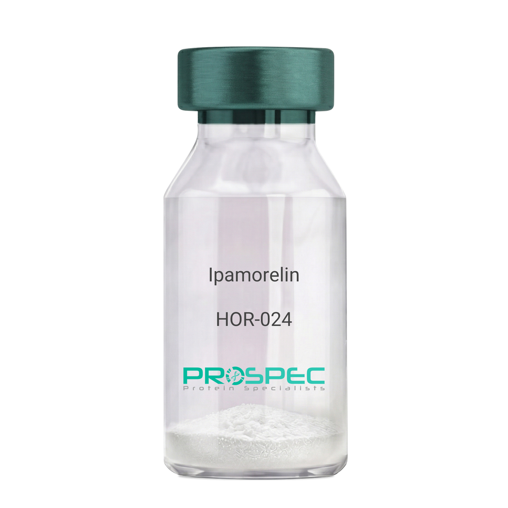
IpamorelinDescription:
Ipamorelin
Ipamorelin
Product # :
HOR-024Price :
Quantity :
Shipping Method :
Shipped at Room temp
More Info
- description
- formulation
- purity
- More Info
- Citations
Description
Ipamorelin Synthetic is a single, non-glycosylated polypeptide chain containing 4 amino acids, having a molecular mass of 711.85 Dalton and a Molecular formula of C38H49N9O5.
Formulation
The protein was lyophilized with no additives.
Purity
Greater than 97.0% as determined by analysis by RP-HPLC.
Citations
More Info
-
Introduction
Ipamorelin is a peptide selective agonist of the ghrelin/growth hormone secretagogue receptor and a growth hormone secretagogue. Ipamorelin is a pentapeptide that was derived from GHRP1. Ipamorelin significantly increases plasma growth hormone levels in both animals and humans. Like pralmorelin and GHRP-6, ipamorelin does not affect prolactin, FSH, LH or TSH levels. However, unlike GHRP2 and GHRP6, but as growth hormone-releasing hormone (GHRH), ipamorelin does not stimulate the secretion of adrenocorticotropic hormone (ACTH) or cortisol, and is highly selective for inducing the secretion only of GH.
-
Synonyms
Ipamorelin
-
Physical Appearance
Sterile Filtered White lyophilized (freeze-dried) powder.
-
Stability
Lyophilized Ipamorelin although stable at room temperature for 3 weeks, should be stored desiccated below -18°C. Upon reconstitution Ipamorelin should be stored at 4°C between 2-7 days and for future use below -18°C. For long term storage it is recommended to add a carrier protein (0.1% HSA or BSA). Please prevent freeze-thaw cycles.
-
Solubility
It is recommended to reconstitute the lyophilized Ipamorelin in sterile 18MΩ-cm H2O not less than 100 µg/ml, which can then be further diluted to other aqueous solutions.
-
Amino Acid Sequence
Aib-His-D-2-Nal-D-Phe-Lys-NH2.
ProSpec's products are furnished for LABORATORY RESEARCH USE ONLY. The product may not be used as drugs, agricultural or pesticidal products, food additives or household chemicals.
- View Data Sheet
Name :
CD44 Anti-Human, BiotinDescription:
CD44, Mouse Anti-Human Biotin
MDU2, MDU3, MIC4, CDW44, CSPG8, HCELL, HUTCH-I, Phagocytic glycoprotein I, PGP-1, Extracellular matrix receptor-III, ECMR-III, Hermes antigen, Hyaluronate receptor, Heparan sulfate proteoglycan, Epican) CDw44.
Product # :
ANT-243Price :
Quantity :
Shipping Method :
Shipped at Room temp
More Info
- formulation
- More Info
- Citations
Formulation
1mg/ml in PBS (after reconstitution).
Citations
More Info
-
Introduction
CD44 is a cell-surface glycoprotein that plays a role in cell-cell interactions, cell adhesion and migration. CD44 is a receptor for hyaluronic acid and also interacts with other ligands, such as osteopontin, collagens, and matrix metalloproteinases. CD44 participates in a wide variety of cellular functions such as lymphocyte activation, recirculation and homing, hematopoiesis, and tumor metastasis. CD44 and CD49d are putative activity markers and CD44 a potential novel therapeutic target in multiple sclerosis. Increased CD44 antigen is associated with relapses in non-small cell lung cancers.
-
Synonyms
MDU2, MDU3, MIC4, CDW44, CSPG8, HCELL, HUTCH-I, Phagocytic glycoprotein I, PGP-1, Extracellular matrix receptor-III, ECMR-III, Hermes antigen, Hyaluronate receptor, Heparan sulfate proteoglycan, Epican) CDw44.
-
Solubility
Reconstitute with of H20. Mix gently, wash the sides of the vial and wait 30-60 seconds before use.
-
Immunogen
Purified human T-Cells.
-
Ig Subclass
Mouse IgG2a.
-
Clone
hCD44.
-
Applications
Staining antibody. For staining, use 5-10µl/1,000,000 cells.
-
Available Conjugates
This antibody is also available conjugated to biotin and FITC. For staining with biotin or FITC-conjugated antibody use 5-10µl/106 cells.
-
Type
Mouse Anti Human Monoclonal.
-
Storage Procedures
Lyophilized: store at 4°C. After reconstitution, if not intended for use within a month, aliquot and store at -20°C.
-
Purification Method
Protein-A.
ProSpec's products are furnished for LABORATORY RESEARCH USE ONLY. The product may not be used as drugs, agricultural or pesticidal products, food additives or household chemicals.