Skip to content

High-quality research proteins.

  • 1-800-805-6531

  • Mon - Sat 8.00-18.00, Sun - Closed

  • info@prospecbio.com

  • PO Box 6591 East Brunswick NJ 08816

  • Products
    • Cytokines
    • Growth factors
    • Chemokines
    • Cd antigens
    • Neurotrophins
    • Hormones
    • Enzymes
    • Viral antigens
    • Recombinant proteins
    • Natural proteins
    • Monoclonal antibodies
    • Polyclonal antibodies
    • Thermal Packaging
  • About
    • Company
    • Founder
    • Contribution
  • Customers
    • Eastern Asia
    • Eastern Europe
    • North America
    • South America
    • South East Asia
    • Southern Asia
    • Western Europe
  • Ordering
    • Ordering Method
    • Ordering - Credit Card
    • Ordering - PO
    • Shipping Fees
    • FAQs
  • Services
    • DNA Cloning
    • Protein Expression
    • Monoclonal Antibody
    • Fermentation
    • Technology License
  • Contact us
Log in

Country/region

  • USD $ | Albania
  • USD $ | Andorra
  • USD $ | Argentina
  • USD $ | Armenia
  • USD $ | Australia
  • USD $ | Austria
  • USD $ | Azerbaijan
  • USD $ | Belarus
  • USD $ | Belgium
  • USD $ | Bolivia
  • USD $ | Brazil
  • USD $ | Bulgaria
  • USD $ | Cambodia
  • USD $ | Canada
  • USD $ | Chile
  • USD $ | China
  • USD $ | Colombia
  • USD $ | Costa Rica
  • USD $ | Croatia
  • USD $ | Cyprus
  • USD $ | Czechia
  • USD $ | Denmark
  • USD $ | Estonia
  • USD $ | Finland
  • USD $ | France
  • USD $ | Georgia
  • USD $ | Germany
  • USD $ | Greece
  • USD $ | Hong Kong SAR
  • USD $ | Hungary
  • USD $ | Iceland
  • USD $ | India
  • USD $ | Ireland
  • USD $ | Israel
  • USD $ | Italy
  • USD $ | Japan
  • USD $ | Kazakhstan
  • USD $ | Latvia
  • USD $ | Liechtenstein
  • USD $ | Lithuania
  • USD $ | Madagascar
  • USD $ | Malta
  • USD $ | Mexico
  • USD $ | Moldova
  • USD $ | Monaco
  • USD $ | Netherlands
  • USD $ | New Zealand
  • USD $ | North Macedonia
  • USD $ | Norway
  • USD $ | Panama
  • USD $ | Paraguay
  • USD $ | Peru
  • USD $ | Philippines
  • USD $ | Poland
  • USD $ | Portugal
  • USD $ | Romania
  • USD $ | Singapore
  • USD $ | Slovakia
  • USD $ | Slovenia
  • USD $ | South Africa
  • USD $ | South Korea
  • USD $ | Spain
  • USD $ | Sri Lanka
  • USD $ | Sweden
  • USD $ | Switzerland
  • USD $ | Taiwan
  • USD $ | Thailand
  • USD $ | Türkiye
  • USD $ | Ukraine
  • USD $ | United Kingdom
  • USD $ | United States
  • USD $ | Uruguay
  • USD $ | Vatican City
  • USD $ | Venezuela
  • USD $ | Vietnam
  • Facebook
  • Instagram
logoProSpec - Recombinant Protein Manufacturer for academic and R&D industry
Become a distributor
Account Log in
Wishlist
Cart Cart(0)
  • Products
    +
    • Cytokines
    • Growth factors
    • Chemokines
    • Cd antigens
    • Neurotrophins
    • Hormones
    • Enzymes
    • Viral antigens
    • Recombinant proteins
    • Natural proteins
    • Monoclonal antibodies
    • Polyclonal antibodies
    • Thermal Packaging
  • About
    +
    • Company
    • Founder
    • Contribution
  • Customers
    +
    • Eastern Asia
    • Eastern Europe
    • North America
    • South America
    • South East Asia
    • Southern Asia
    • Western Europe
  • Ordering
    +
    • Ordering Method
    • Ordering - Credit Card
    • Ordering - PO
    • Shipping Fees
    • FAQs
  • Services
    +
    • DNA Cloning
    • Protein Expression
    • Monoclonal Antibody
    • Fermentation
    • Technology License
  • Contact us

Item added to your cart

View cart
View All
  • Cytokines
  • Growth factors
  • Chemokines
  • Cd antigens
  • Neurotrophins
  • Hormones
  • Enzymes
  • Viral antigens
  • Recombinant proteins
  • Natural proteins
  • Monoclonal antibodies
  • Polyclonal antibodies
  • Thermal Packaging
  • Tumor Necrosis Factor

    Tumor Necrosis Factor

    View All
  • 4-1BB

    4-1BB

    View All
  • Adiponectin

    Adiponectin

    View All
  • AIF1

    AIF1

    View All
  • Other Cytokines

    Other Cytokines

    View All
  • AITRL

    AITRL

    View All
  • Angiopoietin

    Angiopoietin

    View All
  • Apolipoprotein

    Apolipoprotein

    View All
  • B-Cell Activating Factor

    B-Cell Activating Factor

    View All
  • Beta Defensin

    Beta Defensin

    View All
  • Bone Morphogenetic Protein

    Bone Morphogenetic Protein

    View All
  • B type Natriuretic Peptide

    B type Natriuretic Peptide

    View All
  • BST

    BST

    View All
  • Betacellulin

    Betacellulin

    View All
  • Cardiotrophin

    Cardiotrophin

    View All
  • Activin

    Activin

    View All
  • Fibroblast Growth Factor

    Fibroblast Growth Factor

    View All
  • Other Growth Factors

    Other Growth Factors

    View All
  • CSF

    CSF

    View All
  • CTGF

    CTGF

    View All
  • IGFBP

    IGFBP

    View All
  • VEGF Protein

    VEGF Protein

    View All
  • EGF

    EGF

    View All
  • Epigen

    Epigen

    View All
  • Erythropoietin

    Erythropoietin

    View All
  • Insulin-Like Growth Factor

    Insulin-Like Growth Factor

    View All
  • Growth Hormone

    Growth Hormone

    View All
  • HDGF

    HDGF

    View All
  • Hepatocyte Growth Factor

    Hepatocyte Growth Factor

    View All
  • Insulin

    Insulin

    View All
  • BCA-1/ BLC (CXCL13)

    BCA-1/ BLC (CXCL13)

    View All
  • BRAK (CXCL14)

    BRAK (CXCL14)

    View All
  • C-10 (CCL6)

    C-10 (CCL6)

    View All
  • Other Chemokines

    Other Chemokines

    View All
  • MEC (CCL28)

    MEC (CCL28)

    View All
  • LD78-beta (CCL3L1)

    LD78-beta (CCL3L1)

    View All
  • CTACK (CCL27)

    CTACK (CCL27)

    View All
  • CXCL16

    CXCL16

    View All
  • CXCL17

    CXCL17

    View All
  • Platelet Factor-4 (CXCL4)

    Platelet Factor-4 (CXCL4)

    View All
  • ENA-78 (CXCL5)

    ENA-78 (CXCL5)

    View All
  • Eotaxin (CCL11,24,26)

    Eotaxin (CCL11,24,26)

    View All
  • Exodus-2 (CCL21)

    Exodus-2 (CCL21)

    View All
  • Fractalkine (CX3CL1)

    Fractalkine (CX3CL1)

    View All
  • CXCL6

    CXCL6

    View All
  • Other CD Antigens

    Other CD Antigens

    View All
  • CD14

    CD14

    View All
  • CD164

    CD164

    View All
  • CD1

    CD1

    View All
  • CD2

    CD2

    View All
  • CD200

    CD200

    View All
  • CD207

    CD207

    View All
  • CD226

    CD226

    View All
  • CD23

    CD23

    View All
  • CD244

    CD244

    View All
  • CD247

    CD247

    View All
  • CD27

    CD27

    View All
  • CD274

    CD274

    View All
  • CD300

    CD300

    View All
  • CD33

    CD33

    View All
  • Other Neurotrophins

    Other Neurotrophins

    View All
  • Beta-NGF

    Beta-NGF

    View All
  • BDNF

    BDNF

    View All
  • CDNF

    CDNF

    View All
  • CNTF

    CNTF

    View All
  • GDNF

    GDNF

    View All
  • Glia Maturation Factor

    Glia Maturation Factor

    View All
  • MANF

    MANF

    View All
  • Midkine

    Midkine

    View All
  • Neuroglobin

    Neuroglobin

    View All
  • Neurotrophic factors

    Neurotrophic factors

    View All
  • Neuregulin

    Neuregulin

    View All
  • Neuropilin

    Neuropilin

    View All
  • Pigment Epithelium-Derived Factor

    Pigment Epithelium-Derived Factor

    View All
  • Pleiotrophin

    Pleiotrophin

    View All
  • Peptide Hormones

    Peptide Hormones

    View All
  • Other Hormones

    Other Hormones

    View All
  • HCG

    HCG

    View All
  • Endothelin

    Endothelin

    View All
  • Exendin

    Exendin

    View All
  • FSH

    FSH

    View All
  • Glucagon

    Glucagon

    View All
  • GHRP

    GHRP

    View All
  • GLP

    GLP

    View All
  • Thyrostimulin

    Thyrostimulin

    View All
  • Inhibin A

    Inhibin A

    View All
  • LHRH

    LHRH

    View All
  • Melanotan

    Melanotan

    View All
  • Procalcitonin

    Procalcitonin

    View All
  • PTH

    PTH

    View All
  • Peroxiredoxin

    Peroxiredoxin

    View All
  • Peptidase

    Peptidase

    View All
  • Synthetase

    Synthetase

    View All
  • Transferase

    Transferase

    View All
  • Hydrolase

    Hydrolase

    View All
  • Dehydrogenase

    Dehydrogenase

    View All
  • ACE2

    ACE2

    View All
  • Esterase

    Esterase

    View All
  • Protein Kinases

    Protein Kinases

    View All
  • Other Enzymes

    Other Enzymes

    View All
  • Phosphatase

    Phosphatase

    View All
  • Deaminase

    Deaminase

    View All
  • Oxygenase

    Oxygenase

    View All
  • Adenylate Kinase

    Adenylate Kinase

    View All
  • Lyase

    Lyase

    View All
  • Chagas

    Chagas

    View All
  • SARS

    SARS

    View All
  • Influenza

    Influenza

    View All
  • ASFV

    ASFV

    View All
  • Astrovirus

    Astrovirus

    View All
  • Borrelia

    Borrelia

    View All
  • Helicobacter Pylori

    Helicobacter Pylori

    View All
  • Chikungunya

    Chikungunya

    View All
  • Chlamydia

    Chlamydia

    View All
  • Cytomegalo

    Cytomegalo

    View All
  • Dengue

    Dengue

    View All
  • EBV

    EBV

    View All
  • Ebola

    Ebola

    View All
  • Feline Leukemia Virus

    Feline Leukemia Virus

    View All
  • Hepatitis A

    Hepatitis A

    View All
  • Other

    Other

    View All
  • Actin

    Actin

    View All
  • ADAM

    ADAM

    View All
  • Complement Component

    Complement Component

    View All
  • Ag85

    Ag85

    View All
  • Eukaryotic Translation Initiation Factor

    Eukaryotic Translation Initiation Factor

    View All
  • Anterior Gradient Protein

    Anterior Gradient Protein

    View All
  • Heat Shock Protein

    Heat Shock Protein

    View All
  • Alpha-2-HS-Glycoprotein

    Alpha-2-HS-Glycoprotein

    View All
  • Hemoglobin

    Hemoglobin

    View All
  • Allergy

    Allergy

    View All
  • Anaplasma

    Anaplasma

    View All
  • Angiogenin

    Angiogenin

    View All
  • Ankyrin Repeat Domain

    Ankyrin Repeat Domain

    View All
  • Annexin

    Annexin

    View All
  • Other Natural Proteins

    Other Natural Proteins

    View All
  • Aprotinin

    Aprotinin

    View All
  • Transferrin

    Transferrin

    View All
  • Avidin

    Avidin

    View All
  • Anti Coagulation Factors

    Anti Coagulation Factors

    View All
  • Natural Albumin

    Natural Albumin

    View All
  • Natural Coagulation Factors

    Natural Coagulation Factors

    View All
  • Fibronectin

    Fibronectin

    View All
  • Thrombin

    Thrombin

    View All
  • Anti Human Enzyme

    Anti Human Enzyme

    View All
  • Other Monoclonal Antibodies

    Other Monoclonal Antibodies

    View All
  • Anti Human Cytokine

    Anti Human Cytokine

    View All
  • Anti Human Heat Shock Protein

    Anti Human Heat Shock Protein

    View All
  • Anti Mouse Lymphocyte

    Anti Mouse Lymphocyte

    View All
  • Anti Human Chemokine

    Anti Human Chemokine

    View All
  • Anti Human Lymphocyte

    Anti Human Lymphocyte

    View All
  • Anti Viral Monoclonal

    Anti Viral Monoclonal

    View All
  • Anti Mouse Cytokine

    Anti Mouse Cytokine

    View All
  • Anti-GST Monoclonal

    Anti-GST Monoclonal

    View All
  • Other Polyclonal Antibodies

    Other Polyclonal Antibodies

    View All
  • Anti Viral

    Anti Viral

    View All
  • Anti-GST Monoclonal

    Anti-GST Monoclonal

    View All
Back

Search results

1000 results found for “albumin”

Name

Description

Product #

Price

Quantity

Shipping Method

  • View Data Sheet

    Name :

    Apolipoprotein A1 Human, His

    Description:

    Apolipoprotein A-I Human Recombinant, His Tag

    Apolipoprotein A-I, Apo-AI, ApoA-I, APOA1, MGC117399.

    Product # :

    CYT-661

    Price :

    Quantity :

    Shipping Method :

    Ice Icon

    Shipped with Ice Packs

    Add To Cart

    More Info

    • description
    • source
    • formulation
    • purity
    • More Info
    • sds-page
    • Citations

    Description

    APOA1 Human Recombinant produced in E.Coli is a single, non-glycosylated, polypeptide chain containing 264 amino acids (25-267 a.a.) and having a molecular mass of 30.3kDa. APOA1 is fused to 20 a.a. His-Tag at N-terminus and purified by proprietary chromatographic techniques.

    Source

    Escherichia Coli.

    Formulation

    The APOA1 solution containing 20mM Tris-HCl buffer (pH8.0) and 10% glycerol.

    Purity

    Greater than 95.0% as determined by SDS-PAGE.

    sds-page

    Apolipoprotein A1-sds-page - Product image 1

    Citations

    More Info

    • Introduction

      APOA1 (Apolipoprotein A-1) is a human protein with a specific role in lipid metabolism being the main protein component of HDL in the plasma. APOA1 promotes cholesterol efflux from tissues to the liver for excretion. Furthermore, APOA1 is a cofactor for LCAT, which is responsible for the formation of most plasma cholesteryl esters. In addition, APOA1 activates spermatozoa motility as part of the SPAP complex. The APOA1 gene is strongly linked with two other apolipoprotein genes on chromosome 11. Defects in the APOA1 gene are linked to HDL deficiency including Tangier disease, and with systemic non-neuropathic amyloidosis. High levels of APOA1 are linked to the manifestation of asthma and atopy.

    • Synonyms

      Apolipoprotein A-I, Apo-AI, ApoA-I, APOA1, MGC117399.

    • Physical Appearance

      Sterile filtered colorless solution.

    • Stability

      Store at 4°C if entire vial will be used within 2-4 weeks. Store, frozen at -20°C for longer periods of time. For long term storage it is recommended to add a carrier protein (0.1% HSA or BSA).Avoid multiple freeze-thaw cycles.

    • Amino Acid Sequence

      MGSSHHHHHH SSGLVPRGSH MDEPPQSPWD RVKDLATVYV DVLKDSGRDY VSQFEGSALG KQLNLKLLDN WDSVTSTFSK LREQLGPVTQ EFWDNLEKET EGLRQEMSKD LEEVKAKVQP YLDDFQKKWQ EEMELYRQKV EPLRAELQEG ARQKLHELQE KLSPLGEEMR DRARAHVDAL RTHLAPYSDE LRQRLAARLE ALKENGGARL AEYHAKATEH LSTLSEKAKP ALEDLRQGLL PVLESFKVSF LSALEEYTKK LNTQ.

    • Background

      What is the molecular weight/Mw of APOLIPOPROTEIN A1 Protein?
      APOLIPOPROTEIN A1 Protein has a total Mw of 30.3kDa.

      What is the source or expression system of APOLIPOPROTEIN A1 Protein?
      Escherichia Coli.

      What is the Purity of APOLIPOPROTEIN A1 Protein?
      APOLIPOPROTEIN A1 Protein is >95% pure as determined by SDS-PAGE.

      What is the Biological Activity of APOLIPOPROTEIN A1 Protein?
      The biological functionality of APOLIPOPROTEIN A1 Protein will be determined in the future.

      What is the amino acid sequence of APOLIPOPROTEIN A1 Protein?
      MGSSHHHHHH SSGLVPRGSH MDEPPQSPWD RVKDLATVYV DVLKDSGRDY VSQFEGSALG KQLNLKLLDN WDSVTSTFSK LREQLGPVTQ EFWDNLEKET EGLRQEMSKD LEEVKAKVQP YLDDFQKKWQ EEMELYRQKV EPLRAELQEG ARQKLHELQE KLSPLGEEMR DRARAHVDAL RTHLAPYSDE LRQRLAARLE ALKENGGARL AEYHAKATEH LSTLSEKAKP ALEDLRQGLL PVLESFKVSF LSALEEYTKK LNTQ

      What applications can APOLIPOPROTEIN A1 Protein be used in?
      APOLIPOPROTEIN A1 Protein can probably be used in western blot, ELISA and Lateral Flow.

      What is the endotoxin level for APOLIPOPROTEIN A1 Protein?
      The endotoxin level is minimal, APOLIPOPROTEIN A1 Protein was purified using conventional chromatography techniques.

    ProSpec's products are furnished for LABORATORY RESEARCH USE ONLY. The product may not be used as drugs, agricultural or pesticidal products, food additives or household chemicals.

    Apoa1 Human
  • View Data Sheet

    Name :

    Protein-L, His

    Description:

    Protein L Recombinant, His Tag

    Product # :

    PRO-1930

    Price :

    Quantity :

    Shipping Method :

    Room Temp Icon

    Shipped at Room temp

    Add To Cart

    More Info

    • description
    • source
    • formulation
    • purity
    • More Info
    • Citations

    Description

    Recombinant Protein-L produced in E.Coli is a single non-glycosylated polypeptide chain fused with a 6×His tag at N-terminus. Protein-L is comprised of 5 IgG-binding regions of protein L (B1-B2-B3-B4-B5) containing 372 amino acids in total and having a molecular mass of 41.5kDa, however, it migrates with an apparent molecular mass of 46kDa on SDS-PAGE. Cell wall binding region, cell membrane binding region and albumin binding region have been eliminated from the recombinant Protein-L to guarantee the maximum specific IgG binding.

    Source

    Escherichia Coli.

    Formulation

    Protein-L was lyophilized without any additives.

    Purity

    Greater than 97.0% as determined by:
    (a) Analysis by SEC-HPLC.
    (b) Analysis by SDS-PAGE.

    Citations

    More Info

    • Introduction

      The Recombinant Protein L is comprised of 5 kappa-binding domains. Protein L has the exceptional ability to bind through kappa light chain interactions without hindering with the antibody’s antigen-binding site. This gives Protein L the capacity to bind a broader range of Ig classes and subclasses than other antibody-binding proteins. The recombinant Protein L is perfect for purification of polyclonal or monoclonal IgG antibodies. Protein L binds to IgG from humans, mice, rats and pigs.

    • Physical Appearance

      Sterile Filtered White lyophilized (freeze-dried) powder.

    • Stability

      Lyophilized Protein-L although stable at room temperature for 3 weeks, should be stored desiccated below -18°C. Upon reconstitution Protein-L should be stored at 4°C between 2-7 days and for future use below -18°C. For long term storage it is recommended to add a carrier protein (0.1% HSA or BSA). Please prevent freeze-thaw cycles.

    • Solubility

      It is recommended to reconstitute the lyophilized Protein-L in sterile 18M-cm H2O not less than 0.1mg/ml, which can then be further diluted to other aqueous solutions.

    • Amino Acid Sequence

      MHHHHHHKEE TPETPETDSE EEVTIKANLI FANGSTQTAE FKGTFEKATS EAYAYADTLK KDNGEYTVDV ADKGYTLNIK FAGKEKTPEE PKEEVTIKAN LIYADGKTQT AEFKGTFEEA TAEAYRYADA LKKDNGEYTV DVADKGYTLN IKFAGKEKTP EEPKEEVTIK ANLIYADGKT QTAEFKGTFE EATAEAYRYA DLLAKENGKY TVDVADKGYT LNIKFAGKEK TPEEPKEEVT IKANLIYADG KTQTAEFKGT FAEATAEAYR YADLLAKENG KYTADLEDGG YTINIRFAGK KVDEKPEEKE QVTIKENIYF EDGTVQTATF KGTFAEATAE AYRYADLLSK EHGKYTADLE DGGYTINIRF AG.

    ProSpec's products are furnished for LABORATORY RESEARCH USE ONLY. The product may not be used as drugs, agricultural or pesticidal products, food additives or household chemicals.

    Protein L His
  • View Data Sheet

    Name :

    LGALS3 Human

    Description:

    Galectin-3 Human Recombinant

    Galectin-3, GAL3, MAC2, CBP35, GALB, GALIG, LGALS2, LGALS3, Galactose-specific lectin 3, Mac-2 antigen, IgE-binding protein, 35 kDa lectin, Carbohydrate-binding protein 35, CBP 35, Laminin-binding protein, Lectin L-29, L-31, Galactoside-binding protein, GALBP.

    Product # :

    CYT-606

    Price :

    Quantity :

    Shipping Method :

    Room Temp Icon

    Shipped at Room temp

    Add To Cart

    More Info

    • description
    • source
    • formulation
    • purity
    • More Info
    • Citations

    Description

    LGALS3 Human Recombinant produced in E.Coli is a single, non-glycosylated, polypeptide chain containing 250 amino acids and having a molecular mass of 26.2kDa. The LGALS3 is purified by proprietary chromatographic techniques.

    Source

    Escherichia Coli.

    Formulation

    The Galectin-3 protein was lyophilized from a sterile 0.2 micron filtered aqueous solution containing 10 mM sodium phosphate and 50mM sodium chloride, pH 7.5.

    Purity

    Greater than 95.0% as determined by analysis by SDS-PAGE.

    Citations

    More Info

    • Introduction

      Galectin-3 mediates with the alpha-3, beta-1 integrin the stimulation by cspg4 of endothelial cells migration. Galectin-3 plays an necessary part during the acquisition of vasculogenic mimicry and angiogenic properties associated with melanoma progression. LGALS3 overexpression is highly expressed in early stages of papillary carcinoma, and its expression intensity declines during tumor progression. Serum levels of LGALS3 are high in patients with thyroid malignancy but there is considerable overlap in serum LGALS3 concentrations between those with benign and malignant nodular thyroid disease. LGLAS3 takes part as an immune regulator to inhibit T-cell immune responses and promote tumor growth, as a result providing a new mechanism for tumor immune tolerance.

    • Synonyms

      Galectin-3, GAL3, MAC2, CBP35, GALB, GALIG, LGALS2, LGALS3, Galactose-specific lectin 3, Mac-2 antigen, IgE-binding protein, 35 kDa lectin, Carbohydrate-binding protein 35, CBP 35, Laminin-binding protein, Lectin L-29, L-31, Galactoside-binding protein, GALBP.

    • Physical Appearance

      Sterile Filtered White lyophilized (freeze-dried) powder.

    • Stability

      Lyophilized Galectin-3 Recombinant although stable at room temperature for 3 weeks, should be stored desiccated below -18°C. Upon reconstitution Galectin-3 should be stored at 4°C between 2-7 days and for future use below -18°C.For long term storage it is recommended to add a carrier protein (0.1% HSA or BSA).Please prevent freeze-thaw cycles.

    • Solubility

      It is recommended to reconstitute the lyophilized LGALS3 in sterile 18MΩ-cm H2O not less than 100µg/ml, which can then be further diluted to other aqueous solutions.

    • Amino Acid Sequence

      MADNFSLHDAL SGSGNPNPQG WPGAWGNQPA GAGGYPGASY PGAYPGQAPP GAYPGQAPPG AYPGAPGAYP GAPAPGVYPG PPSGPGAYPS SGQPSATGAY PATGPYGAPA GPLIVPYNLP LPGGVVPRML ITILGTVKPN ANRIALDFQR GNDVAFHFNP RFNENNRRVI VCNTKLDNNW GREERQSVFP FESGKPFKIQ VLVEPDHFKV AVNDAHLLQY NHRVKKLNEI SKLGISGDID LTSASYTMI.

    • Background

      What is the molecular weight/Mw of LGALS3 HUMAN Protein?
      LGALS3 HUMAN Protein has a total Mw of 26.2kDa.

      What is the source or expression system of LGALS3 HUMAN Protein?
      Escherichia Coli.

      What is the Purity of LGALS3 HUMAN Protein?
      LGALS3 HUMAN Protein is >95% pure as determined by SDS-PAGE.

      What is the Biological Activity of LGALS3 HUMAN Protein?
      The biological functionality of LGALS3 HUMAN Protein will be determined in the future.

      What is the amino acid sequence of LGALS3 HUMAN Protein?
      MADNFSLHDAL SGSGNPNPQG WPGAWGNQPA GAGGYPGASY PGAYPGQAPP GAYPGQAPPG AYPGAPGAYP GAPAPGVYPG PPSGPGAYPS SGQPSATGAY PATGPYGAPA GPLIVPYNLP LPGGVVPRML ITILGTVKPN ANRIALDFQR GNDVAFHFNP RFNENNRRVI VCNTKLDNNW GREERQSVFP FESGKPFKIQ VLVEPDHFKV AVNDAHLLQY NHRVKKLNEI SKLGISGDID LTSASYTMI.

      What applications can LGALS3 HUMAN Protein be used in?
      LGALS3 HUMAN Protein can probably be used in western blot, ELISA and Lateral Flow.

      What is the endotoxin level for LGALS3 HUMAN Protein?
      The endotoxin level is minimal, LGALS3 HUMAN Protein was purified using conventional chromatography techniques.


    ProSpec's products are furnished for LABORATORY RESEARCH USE ONLY. The product may not be used as drugs, agricultural or pesticidal products, food additives or household chemicals.

    Galectin 3 Human
  • View Data Sheet

    Name :

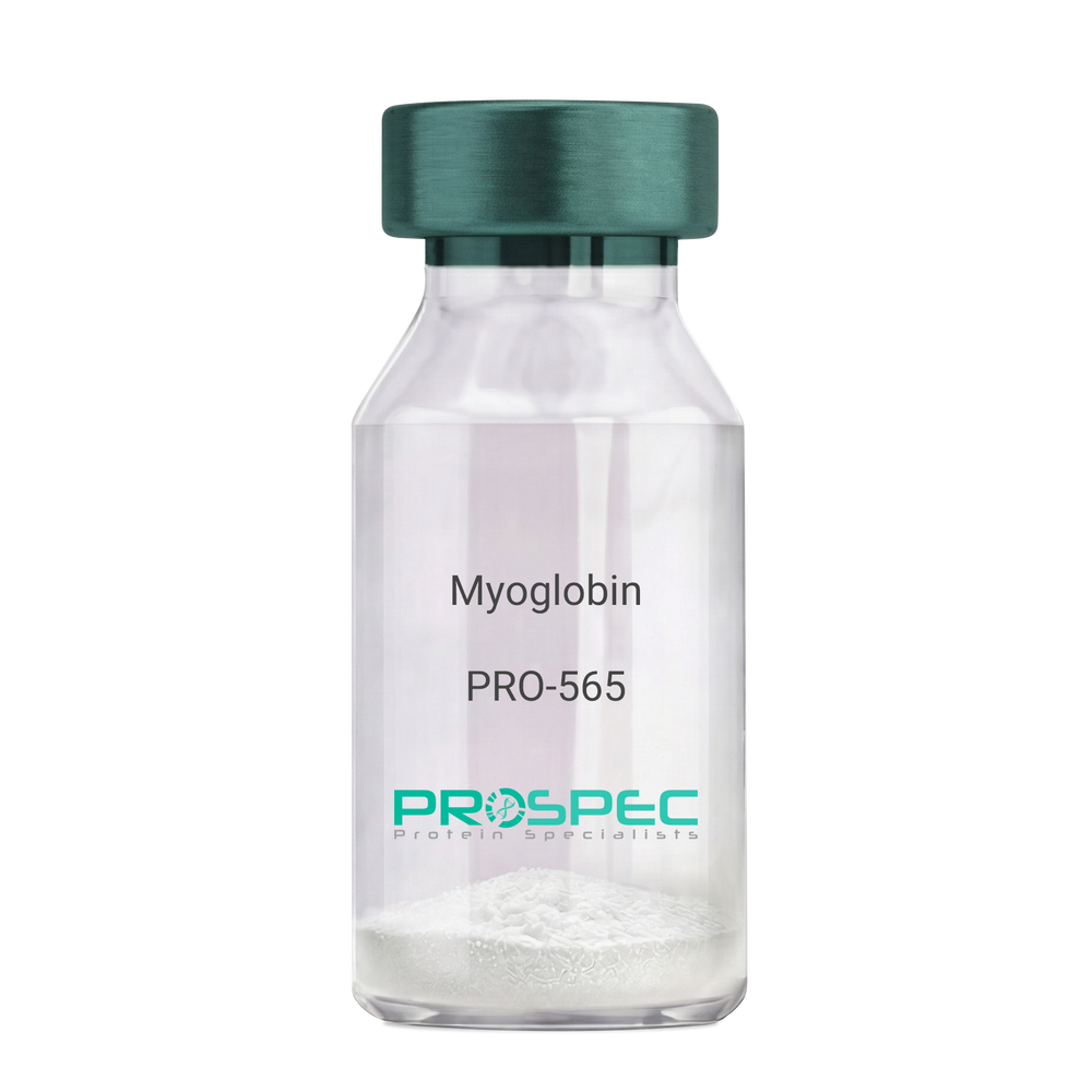
    Myoglobin

    Description:

    Myoglobin Human

    Myoglobin, MB, PVALB, MGC13548.

    Product # :

    PRO-565

    Price :

    Quantity :

    Shipping Method :

    Ice Icon

    Shipped with Ice Packs

    Add To Cart

    More Info

    • description
    • source
    • formulation
    • purity
    • More Info
    • Citations

    Description

    Human Myoglobin produced in Human Cardiac Tissues having a molecular mass of 17.5kDa. Myoglobin is released from recently injured myocardial cells within a few hours of Infarction. Peak levels are reached more quickly than CK-MB or Troponin complex.

    Source

    Human Cardiac Tissues.

    Formulation

    The protein solution is in 0.05M phosphate buffer pH 7.5 containing 0.15M NaCl and 0.09% NaN3. Filtered through a 0.2µM membrane.

    Purity

    Greater than 96.0%.

    Citations

    More Info

    • Introduction

      Myoglobin is a member of the globin superfamily and can be found in skeletal and cardiac muscles. It is a haemoprotein that contributs to intracellular oxygen storage and transcellular facilitated diffusion of oxygen. Myoglobin has a single-chain globular structure of 153 amino acids, containing a heme prosthetic group (iron-containing porphyrin) in the core around which the remaining apoprotein folds. Myoglobin has 8 alpha helices and a hydrophobic core. Myoglobin’s molecular weight is 16.7 kDa, and it is the primary oxygen-carrying pigment of muscle tissues. The binding of oxygen in myoglobin is different from the cooperative oxygen binding in hemoglobin, since positive collaboration is a property of multimeric/oligomeric proteins only. Instead, the binding of oxygen by myoglobin is uninfluenced by the oxygen pressure in the surrounding tissue. Myoglobin is frequently referred to as having an "instant binding tenacity" to oxygen given its hyperbolic oxygen dissociation curve. Different organisms are able to hold their breaths longer due to high concentrations of myoglobin in their muscle cells. Myoglobin is responsible for the pigments that make meat red. The color of the meat is partly determined by the charge of the iron atom in myoglobin and the oxygen attached to it. Myoglobin is found in Type I muscle, Type II A and Type II B, but it is mostly deemed that myoglobin is not found in smooth muscle. Myoglobin is discharged from damaged muscle tissue (rhabdomyolysis), which contains very high concentrations of myoglobin. Even though the released myoglobin is filtered by the kidneys, it is toxic to the renal tubular epithelium and thus may cause acute renal failure.

    • Synonyms

      Myoglobin, MB, PVALB, MGC13548.

    • Physical Appearance

      Sterile Filtered red solution.

    • Stability

      Human Myoglobin should be stored at 2-8°C.

    • Human Virus Test

      Starting material donor tested and certified negative for HIV I & II antibodies, Hepatitis B surface antigen, and Hepatitis C antibodies.

    ProSpec's products are furnished for LABORATORY RESEARCH USE ONLY. The product may not be used as drugs, agricultural or pesticidal products, food additives or household chemicals.

    Myoglobin Human
  • View Data Sheet

    Name :

    Ferritin Human

    Description:

    Human Liver Ferritin

    Product # :

    PRO-564

    Price :

    Quantity :

    Shipping Method :

    Ice Icon

    Shipped with Ice Packs

    Add To Cart

    More Info

    • description
    • source
    • formulation
    • purity
    • More Info
    • Citations

    Description

    Ferritin is a glycoprotein produced in Human Liver having a molecular mass of 440- 450kDa and pI of 5.5, which stores iron atoms in the ferric state. It is predominantly intracellular, where it forms an exchangeable pool of iron acting as an iron store. Ferritin level in serum is directly proportional to body iron stores and serum levels are an excellent indicator in monitoring iron status in anemia. It can be used as a marker for inflammation and also used for monitoring and prediction of future events in coronary artery disease.

    Source

    Human Liver.

    Formulation

    The protein solution is in 0.05M TRIS buffer pH 7.5 containing 1.0M NaCl and 0.09% NaN3.

    Purity

    Greater than 96.0%.

    Citations

    More Info

    • Introduction

      Ferritin is the main intracellular iron storage protein in prokaryotes and eukaryotes. Ferritin’s major functions are the storage of iron in a soluble and nontoxic state and its release in a controlled fashion. An iron-containing protein complex is found mostly in the intestinal mucosa, spleen, and liver. Ferritin is composed of 24 subunits of the heavy and light chains. Variation in ferritin subunit composition may influence the rates of iron uptake and release in different tissues. Defects in the light chain ferritin gene are linked to a number of neurodegenerative diseases and hyperferritinemia-cataract syndrome. The genes that encode the light and heavy chains are on located different chromosomes. The light chain genes are in chromosome region 19q13.3-q13.4 whilst those for the heavy chain are in chromosome region 11q12-q13. Ferritin is shaped like a hollow sphere, inside which the iron is stored in the Fe(III) oxidation state. The iron is integrated in the mineral ferrihydrite, [FeO(OH)]8[FeO(H2PO4)], which is attached to the inner wall of the sphere. To release iron once the body needs it, the iron must be altered from the Fe(III) to the Fe(II) oxidation state. Subsequently, the iron leaves through channels in the spherical structure. Therefore, the structure of ferritin is tremendously important for the protein's ability to store and release iron in a controlled mode.
      The amount of ferritin in the blood (serum ferritin level) is directly related to the amount of iron stored in the body. The body has a "buffer" against iron deficiency (if the blood has too little iron, ferritin can release more) and, to a lesser extent, iron overload (if the blood and tissues of the body have too much iron, ferritin can help store the excess iron).

    • Physical Appearance

      Sterile Filtered brownish solution.

    • Stability

      Human Ferritin should be stored at 2-8°C.

    • Human Virus Test

      Tissue sample tested and found negative for HIV-1 & 2 antibodies, Hepatatis B surface antigen, Syphilis RPR and Hepatatis C antibodies.

    ProSpec's products are furnished for LABORATORY RESEARCH USE ONLY. The product may not be used as drugs, agricultural or pesticidal products, food additives or household chemicals.

    Ferritin Human
  • View Data Sheet

    Name :

    Thyroglobulin Human

    Description:

    Thyroglobulin Human

    Thyroglobulin, TGN, AITD3, TG.

    Product # :

    PRO-443

    Price :

    Quantity :

    Shipping Method :

    Room Temp Icon

    Shipped at Room temp

    Add To Cart

    More Info

    • description
    • source
    • formulation
    • purity
    • More Info
    • sds-page
    • Citations

    Description

    Human Thyroglobulin is a glycosylated, polypeptide chain having a total molecular mass of 660 kDa (330 kDa per subunit).

    Source

    Human thyroid glands.

    Formulation

    Lyophilized from 0.02M NH4HCO3. May contain traces of buffer salts.

    Purity

    Greater than 98.0%.

    sds-page

    Thyroglobulin Human Native SDS-PAGE - Product image 1

    Citations

    More Info

    • Introduction

      Thyroglobulin (TG) represents one of the main autoantigenic targets in autoimmune thyroid disease of humans. TG is a large globular dimeric glycoprotein with a total molecular weight of 660 kDa, which occupies a key precursor role in the biosynthesis of the thyroid hormones. Approximately 75% of the total protein content of the thyroid follicle consists of TG. The thyroid gland uses the Thyroglobulin in order to produce the thyroid hormones thyroxine (T4) and triiodothyronine (T3). Thyroglobulin is produced by the thyroid epithelial cells (thyrocytes) which form spherical follicles. Thyroglobulin is subsequently secreted and stored in the follicular lumen.
      Patients with Hashimoto's thyroiditis or Graves' disease, frequently develop antibodies against Thyroglobulin. Tg-specific antibodies help in the diagnosis of the above diseases, however they also may be present in apparently healthy euthyroid individuals. Blood Thyroglobulin levels can be used as a tumor marker for certain kinds of thyroid cancer, and the may also be elevated in cases of Graves' disease.

    • Synonyms

      Thyroglobulin, TGN, AITD3, TG.

    • Physical Appearance

      Sterile Filtered Off-White lyophilized (freeze-dried) powder.

    • Stability

      Human Thyroglobulin although stable at room temperature for 3 weeks, should be stored between 2-8°C.

    • Solubility

      Use phosphate buffer, pH>7.0 containing 0.15M NaCl is recommended.

    • Human Virus Test

      Starting material tested and certified negative for HIV I & II antibodies, Hepatitis B surface antigen, Hepatitis C antibodies, Syphilis and HIV-1/HBV/HCV NAT.

    ProSpec's products are furnished for LABORATORY RESEARCH USE ONLY. The product may not be used as drugs, agricultural or pesticidal products, food additives or household chemicals.

    Thyroglobulin Human
  • View Data Sheet

    Name :

    ANGPTL2 Human

    Description:

    Angiopoietin-like Protein 2 Human Recombinant

    Angiopoietin-related protein 2, Angiopoietin-like protein 2, ANGPTL2, ARP2, HARP.

    Product # :

    CYT-765

    Price :

    Quantity :

    Shipping Method :

    Ice Icon

    Shipped with Ice Packs

    Add To Cart

    More Info

    • description
    • source
    • formulation
    • purity
    • More Info
    • sds-page
    • Citations

    Description

    ANGPTL2 Human Recombinant produced in E.Coli is a single, non-glycosylated, polypeptide chain containing 493 amino acids ( 22-493 a.a.) including a 20 a.a N-terminal His tag. The total molecular mass is 57.1kDa (calculated).

    Source

    Escherichia Coli.

    Formulation

    ANGPTL2 protein solution (0.5mg/ml) contains 20mM Tris HCL (pH7-8) and 10% glycerol.

    Purity

    Greater than 85.0% as determined by SDS-PAGE.

    sds-page

    ANGPTL2-sds-page - Product image 1

    Citations

    More Info

    • Introduction

      Angiopoietins belong to the vascular endothelial growth factor family and the only known growth factors largely specific for vascular endothelium. Angiopoietins-1, 2 and 4 partake in the formation of blood vessels. ANGPTL2 displays angiogenic effects. Angiopoietin-like Protein 2 (ANGPTL2) is an anti-diabetic factor. ANGPTL2 induces sprouting in endothelial cells through an autocrine and paracrine action. ANGPTL2 increases insulin sensitivity in adipocytes. In addition, ANGPTL2 is a mediator of chronic adipose tissue inflammation. ANGPTL2 is widely expressed in the heart, small intestine, spleen and stomach. ANGPTL2 is also found in lower levels in the colon, ovary, adrenal gland, skeletal muscle and in prostate.

    • Synonyms

      Angiopoietin-related protein 2, Angiopoietin-like protein 2, ANGPTL2, ARP2, HARP.

    • Physical Appearance

      Sterile filtered colorless solution.

    • Stability

      Store at 4°C if entire vial will be used within 2-4 weeks.Store, frozen at -20°C for longer periods of time.For long term storage it is recommended to add a carrier protein (0.1% HSA or BSA).Avoid multiple freeze-thaw cycles.

    • Amino Acid Sequence

      MGSSHHHHHH SSGLVPRGSH MGQEDGFEGT EEGSPREFIY LNRYKRAGES QDKCTYTFIV PQQRVTGAIC VNSKEPEVLL ENRVHKQELE LLNNELLKQK RQIETLQQLV EVDGGIVSEV KLLRKESRNM NSRVTQLYMQ LLHEIIRKRD NALELSQLEN RILNQTADML QLASKYKDLE HKYQHLATLA HNQSEIIAQL EEHCQRVPSA RPVPQPPPAA PPRVYQPPTY NRIINQISTN EIQSDQNLKV LPPPLPTMPT LTSLPSSTDK PSGPWRDCLQ ALEDGHDTSS IYLVKPENTN RLMQVWCDQR HDPGGWTVIQ RRLDGSVNFF RNWETYKQGF GNIDGEYWLG LENIYWLTNQ GNYKLLVTME DWSGRKVFAE YASFRLEPES EYYKLRLGRY HGNAGDSFTW HNGKQFTTLD RDHDVYTGNC AHYQKGGWWY NACAHSNLNG VWYRGGHYRS RYQDGVYWAE FRGGSYSLKK VVMMIRPNPN TFH

    • Background

      Angiopoietin-like Protein 2 Human Recombinant: A Potential Therapeutic Target for Metabolic and Cardiovascular Disorders

      Abstract:

      Angiopoietin-like protein 2 (ANGPTL2) has emerged as a crucial regulator in metabolic and cardiovascular disorders. This multifunctional protein is involved in various biological processes, including angiogenesis, adipose tissue function, and inflammation. The availability of human recombinant ANGPTL2 protein has provided researchers with a valuable tool to explore its therapeutic potential. This concise review provides an overview of the role of ANGPTL2 in metabolic and cardiovascular health and discusses the potential of ANGPTL2 human recombinant protein as a therapeutic target.

      Introduction:

      Metabolic disorders, such as obesity and type 2 diabetes, are closely associated with cardiovascular diseases and pose significant global health challenges. ANGPTL2, a member of the angiopoietin-like protein family, has gained attention for its involvement in metabolic regulation and cardiovascular homeostasis. Through interactions with various receptors and signaling pathways, ANGPTL2 influences lipid metabolism, insulin sensitivity, inflammation, and vascular integrity.

      Mechanisms of ANGPTL2 Action:

      ANGPTL2 acts through binding to integrins, toll-like receptors (TLRs), and other receptors on different cell types, including adipocytes, endothelial cells, and immune cells. By influencing angiogenesis, inflammation, and extracellular matrix remodeling, ANGPTL2 affects adipose tissue function, lipid metabolism, and insulin signaling.

      Role of ANGPTL2 in Metabolic Regulation:

      ANGPTL2 plays a critical role in metabolic regulation and the development of metabolic disorders. It promotes adipose tissue inflammation, impairs adipogenesis, and alters adipokine secretion, contributing to metabolic dysfunction. Furthermore, ANGPTL2 modulates lipid metabolism by regulating lipoprotein lipase activity, affecting triglyceride clearance, and promoting hepatic lipid accumulation.

      ANGPTL2 in Cardiovascular Health and Disease:

      Increasing evidence suggests that ANGPTL2 is implicated in cardiovascular diseases, including atherosclerosis and heart failure. ANGPTL2 promotes vascular inflammation, endothelial dysfunction, and smooth muscle cell proliferation, which contribute to atherosclerotic plaque progression and vascular remodeling. Additionally, ANGPTL2 influences cardiac remodeling and fibrosis, impacting heart failure development.

      Therapeutic Potential of ANGPTL2 Human Recombinant Protein:

      The availability of ANGPTL2 human recombinant protein opens avenues for therapeutic interventions targeting metabolic and cardiovascular disorders. Preclinical studies employing ANGPTL2 blockade or supplementation have shown promising results in improving metabolic parameters, reducing atherosclerosis, and preserving cardiac function. However, further research is needed to optimize the clinical application of ANGPTL2 human recombinant protein, including dosage, timing, and delivery methods.

      Conclusion:

      ANGPTL2 holds promise as a therapeutic target for metabolic and cardiovascular disorders. Its involvement in key biological processes makes it an attractive candidate for interventions aiming to improve metabolic health and prevent cardiovascular complications. The development of ANGPTL2 human recombinant protein provides a valuable tool for investigating its therapeutic potential further.

      What is the molecular weight/Mw of ANGPTL2 Protein?
      ANGPTL2 Protein has a total Mw of 57.1kDa.

      What is the source or expression system of ANGPTL2 Protein?
      Escherichia Coli.

      What is the Purity of ANGPTL2 Protein?
      ANGPTL2 Protein is >85% pure as determined by SDS-PAGE.

      What is the Biological Activity of ANGPTL2 Protein?
      The biological functionality of ANGPTL2 Protein will be determined in the future.

      What is the amino acid sequence of ANGPTL2 Protein?
      MGSSHHHHHH SSGLVPRGSH MGQEDGFEGT EEGSPREFIY LNRYKRAGES QDKCTYTFIV PQQRVTGAIC VNSKEPEVLL ENRVHKQELE LLNNELLKQK RQIETLQQLV EVDGGIVSEV KLLRKESRNM NSRVTQLYMQ LLHEIIRKRD NALELSQLEN RILNQTADML QLASKYKDLE HKYQHLATLA HNQSEIIAQL EEHCQRVPSA RPVPQPPPAA PPRVYQPPTY NRIINQISTN EIQSDQNLKV LPPPLPTMPT LTSLPSSTDK PSGPWRDCLQ ALEDGHDTSS IYLVKPENTN RLMQVWCDQR HDPGGWTVIQ RRLDGSVNFF RNWETYKQGF GNIDGEYWLG LENIYWLTNQ GNYKLLVTME DWSGRKVFAE YASFRLEPES EYYKLRLGRY HGNAGDSFTW HNGKQFTTLD RDHDVYTGNC AHYQKGGWWY NACAHSNLNG VWYRGGHYRS RYQDGVYWAE FRGGSYSLKK VVMMIRPNPN TFH

      What applications can ANGPTL2 Protein be used in?
      ANGPTL2 Protein can probably be used in western blot, ELISA and Lateral Flow.

      What is the endotoxin level for ANGPTL2 Protein?
      The endotoxin level is minimal, ANGPTL2 Protein was purified using conventional chromatography techniques.

    ProSpec's products are furnished for LABORATORY RESEARCH USE ONLY. The product may not be used as drugs, agricultural or pesticidal products, food additives or household chemicals.

    Angptl2 Human
  • View Data Sheet

    Name :

    GDNF Human

    Description:

    Glial-Derived Neurotrophic Factor Human Recombinant

    ATF1, ATF2, HFB1-GDNF, GDNF.

    Product # :

    CYT-305

    Price :

    Quantity :

    Shipping Method :

    Room Temp Icon

    Shipped at Room temp

    Add To Cart

    More Info

    • description
    • source
    • formulation
    • purity
    • biological activity
    • More Info
    • sds-page
    • Citations

    Description

    Glial derived Neurotrophic Factor Human Recombinant produced in E.Coli is a non-glycosylated disulfide-linked homodimer containing 2 x 135 amino acids and having a total molecular mass of approximately 30kDa. GDNF is purified by proprietary chromatographic techniques.

    Source

    Escherichia Coli.

    Formulation

    GDNF was lyophilized from a 0.2µm filtered concentrated solution in 1×PBS, pH 7.4 and 5% Trehalose.

    Purity

    Greater than 95.0% as determined by analysis by SDS-PAGE.

    Biological Activity

    The ED50 was determined by the proliferation of rat C6 cells is < 0.1 ng/ml, corresponding to a specific activity of > 1.0x10⁷ units/mg.

    sds-page

    GDNF sds-page - Product image 1

    Citations

    More Info

    • Introduction

      GDNF promotes the survival and differentiation of minergic neurons in culture, and is able to prevent apoptosis of motor neurons induced by axotomy. The encoded protein is processed to a mature secreted form that exists as a homodimer. The mature form of the protein is a ligand for the product of the RET (rearranged during transfection) protooncogene. In addition to the transcript encoding GDNF, two additional alternative transcripts encoding distinct proteins, referred to as astrocyte-derived trophic factors, have also been described. Mutations in this gene may be associated with Hirschsprung disease.
      GDNF enhances survival and morphological differentiation of minergic neurons and increases their high-affinity uptake.

    • Synonyms

      ATF1, ATF2, HFB1-GDNF, GDNF.

    • Physical Appearance

      Sterile Filtered White lyophilized (freeze-dried) powder.

    • Stability

      Lyophilized Glial-derived Neurotrophic Factor although stable at room temperature for 3 weeks, should be stored desiccated below -18°C. Upon reconstitution GDNF should be stored at 4°C between 2-7 days and for future use below -18°C.For long term storage it is recommended to add a carrier protein (0.1% HSA or BSA).Please prevent freeze-thaw cycles.

    • Solubility

      It is recommended to reconstitute the lyophilized Glial Derived Neurotrophic Factor in sterile 18MΩ-cm H2O not less than 100µg/ml, which can then be further diluted to other aqueous solutions.

    • Amino Acid Sequence

      SPDKQMAVLP RRERNRQAAA ANPENSRGKG RRGQRGKNRG CVLTAIHLNV TDLGLGYETK EELIFRYCSG SCDAAETTYD KILKNLSRNR RLVSDKVGQA CCRPIAFDDD LSFLDDNLVY HILRKHSAKR CGCI.

    • Background

      What is the molecular weight/Mw of GDNF HUMAN Protein?
      GDNF HUMAN Protein has a total Mw of 30kDa.

      What is the source or expression system of GDNF HUMAN Protein?
      Escherichia Coli.

      What is the Purity of GDNF HUMAN Protein?
      GDNF HUMAN Protein is >95% pure as determined by SDS-PAGE.

      What is the Biological Activity of GDNF HUMAN Protein?
      The ED50 was determined by the proliferation of rat C6 cells is < 0.1 ng/ml, corresponding to a specific activity of > 1.0x10⁷ units/mg.

      What is the amino acid sequence of GDNF HUMAN Protein?
      SPDKQMAVLP RRERNRQAAA ANPENSRGKG RRGQRGKNRG CVLTAIHLNV TDLGLGYETK EELIFRYCSG SCDAAETTYD KILKNLSRNR RLVSDKVGQA CCRPIAFDDD LSFLDDNLVY HILRKHSAKR CGCI.
      What applications can GDNF HUMAN Protein be used in?
      GDNF HUMAN Protein can probably be used in western blot, ELISA and Lateral Flow.

      What is the endotoxin level for GDNF HUMAN Protein?
      The endotoxin level is minimal, GDNF HUMAN Protein was purified using conventional chromatography techniques.


    ProSpec's products are furnished for LABORATORY RESEARCH USE ONLY. The product may not be used as drugs, agricultural or pesticidal products, food additives or household chemicals.

    Gdnf Human
  • View Data Sheet

    Name :

    Follistatin Human

    Description:

    Follistatin Human Recombinant

    FST, FS

    Product # :

    CYT-232

    Price :

    Quantity :

    Shipping Method :

    Room Temp Icon

    Shipped at Room temp

    Add To Cart

    More Info

    • description
    • source
    • formulation
    • purity
    • biological activity
    • More Info
    • Citations

    Description

    Follistatin Human Recombinant produced in E.Coli is a single, non-glycosylated polypeptide chain containing 288 amino acids and having a total molecular mass of 31.5kDa.The FST is purified by proprietary chromatographic techniques.

    Source

    Escherichia Coli.

    Formulation

    Lyophilized from a concentrated (1mg/ml) solution containing no additives.

    Purity

    Greater than 95.0% as determined by:
    (a) Analysis by RP-HPLC.
    (b) Analysis by SDS-PAGE.

    Biological Activity

    The activity is determined by the ability to neutralize ACTV inhibitory effect of mouse MPC-11 cells. The expected ED50 is 100-400ng/ml, corresponding to a Specific Activity of 2,500-10,000units/mg in the presence of 7.5ng/ml ACTV A.

    Citations

    More Info

    • Introduction

      Follistatin is a single-chain gonadal protein that specifically inhibits follicle-stimulating hormone release. The single FST gene encodes two isoforms, FST317 and FST344 containing 317 and 344 amino acids respectively, resulting from alternative splicing of the precursor mRNA. In a study in which 37 candidate genes were tested for linkage and association with polycystic ovary syndrome (PCOS) or hyperandrogenemia in 150 families, evidence was found for linkage between PCOS and follistatin. Follistatin functions as an ACTV antagonist. specific inhibitor of the biosynthesis and secretion of pituitary follicle stimulating hormone (fsh).

    • Synonyms

      FST, FS

    • Physical Appearance

      Sterile Filtered White lyophilized (freeze-dried) powder.

    • Stability

      Lyophilized Follistatin although stable at room temperature for 3 weeks, should be stored desiccated below -18°C. Upon reconstitution FST should be stored at 4°C between 2-7 days and for future use below -18°C.For long term storage it is recommended to add a carrier protein (0.1% HSA or BSA).Please prevent freeze-thaw cycles.

    • Solubility

      It is recommended to reconstitute the lyophilized Follistatin in sterile 18MΩ-cm H2O not less than 100µg/ml, which can then be further diluted to other aqueous solutions.

    • Amino Acid Sequence

      The sequence of the first five N-terminal amino acids was determined and was found to be Gly-Asn-Cys-Trp-Leu.

    • Background

      What is the molecular weight/Mw of FOLLISTATIN HUMAN Protein?
      FOLLISTATIN HUMAN Protein has a total Mw of 31.5kDa.

      What is the source or expression system of FOLLISTATIN HUMAN Protein?
      Escherichia Coli.

      What is the Purity of FOLLISTATIN HUMAN Protein?
      FOLLISTATIN HUMAN Protein is >95% pure as determined by SDS-PAGE.

      What is the Biological Activity of FOLLISTATIN HUMAN Protein?
      The activity is determined by the ability to neutralize ACTV inhibitory effect of mouse MPC-11 cells. The expected ED50 is 100-400ng/ml, corresponding to a Specific Activity of 2,500-10,000units/mg in the presence of 7.5ng/ml ACTV A.

      What is the amino acid sequence of FOLLISTATIN HUMAN Protein?
      The sequence of the first five N-terminal amino acids was determined and was found to be Gly-Asn-Cys-Trp-Leu.

      What applications can FOLLISTATIN HUMAN Protein be used in?
      FOLLISTATIN HUMAN Protein can probably be used in western blot, ELISA and Lateral Flow.

      What is the endotoxin level for FOLLISTATIN HUMAN Protein?
      The endotoxin level is minimal, FOLLISTATIN HUMAN Protein was purified using conventional chromatography techniques.

    ProSpec's products are furnished for LABORATORY RESEARCH USE ONLY. The product may not be used as drugs, agricultural or pesticidal products, food additives or household chemicals.

    Follistatin Human
  • View Data Sheet

    Name :

    TFRC Native

    Description:

    Transferrin Receptor Human

    Transferrin receptor protein 1, TR, TfR, TfR1, Trfr, T9, p90, CD_antigen: CD71, Transferrin receptor, serum form, sTfR, TFRC, CD71

    Product # :

    PRO-2742

    Price :

    Quantity :

    Shipping Method :

    Ice Icon

    Shipped with Ice Packs

    Add To Cart

    More Info

    • description
    • source
    • formulation
    • More Info
    • Citations

    Description

    Human Transferrin Receptor Protein produced in human serum tissue having a molecular mass of 85kDa.

    Source

    Human serum.

    Formulation

    TFRC Native solution (0.2µm filtered) contains 250mM TRIS-HCl buffer, 0.15M NaCl, 0.09% NaN3, pH 8.0.

    Citations

    More Info

    • Introduction

      Transferrin receptor protein 1 (TFRC) is required for iron delivery from transferring to cells. The TFRC protein is a transmembrane glycoprotein comprised of 2 disulfide-linked monomers joined by 2 disulfide bonds. Each monomer will bind one holo-transferrin molecule producing an iron-Tf-TfR complex which enters the cell by endocytosis.

    • Synonyms

      Transferrin receptor protein 1, TR, TfR, TfR1, Trfr, T9, p90, CD_antigen: CD71, Transferrin receptor, serum form, sTfR, TFRC, CD71

    • Physical Appearance

      Sterile Filtered brown solution.

    • Stability

      Store at 4°C if entire vial will be used within 2-4 weeks. Store, frozen at -20°C for longer periods of time. For long term storage it is recommended to add a carrier protein (0.1% HSA or BSA). Avoid multiple freeze-thaw cycles.

    • Human Virus Test

      Starting material donor has been tested and certified negative for antibodies to HIV-1, HIV-2, HCV, HBSAG, Parvovirus B19, Syphilis and HIV/HBV/HCV (PCR).

    ProSpec's products are furnished for LABORATORY RESEARCH USE ONLY. The product may not be used as drugs, agricultural or pesticidal products, food additives or household chemicals.

    Transferrin Receptor Tfrc
  • View Data Sheet

    Name :

    SELPLG Human

    Description:

    Selectin P Ligand Human Recombinant

    Cutaneous Lymphocyte-Associated Associated Antigen, Selectin P Ligand, PSGL-1, CD162 Antigen, P-Selectin Glycoprotein Ligand 1, CLA.

    Product # :

    PRO-2714

    Price :

    Quantity :

    Shipping Method :

    Ice Icon

    Shipped with Ice Packs

    Add To Cart

    More Info

    • description
    • source
    • formulation
    • purity
    • More Info
    • Citations

    Description

    SELPLG Human Recombinant produced in HEK293 Cells is a single, glycosylated polypeptide chain containing 496 amino acids (42-295 a.a) and having a molecular mass of 53.4kDa.SELPLG is fused to a 239 amino acid hIgG-His-Tag at C-terminus & purified by proprietary chromatographic techniques.

    Source

    HEK293 Cells.

    Formulation

    The SELPLG solution (1mg/1ml) contains phosphate buffered saline (pH7.4) and 10% glycerol.

    Purity

    Greater than 90.0% as determined by SDS-PAGE.

    Citations

    More Info

    • Introduction

      SELPLG glycoprotein functions as a high affinity counter-receptor for the cell adhesion selectin molecules (P, E and L) located in stimulated T lymphocytes and myeloid cells. SELPLG binds leukocytes to activated platelets or endothelia expressing selectins, a vital role in leukocyte trafficking throughout inflammation. In order to have a high-affinity binding activity SELPLG needs two post-translational modifications, tyrosine sulfation and the addition of the sialyl Lewis x tetrasaccharide (sLex) to its O-linked glycans. Polymorphisms and abnormal expression of SELPLG are linked to defects in the innate and adaptive immune response. Alternate splicing results in multiple transcript variants.

    • Synonyms

      Cutaneous Lymphocyte-Associated Associated Antigen, Selectin P Ligand, PSGL-1, CD162 Antigen, P-Selectin Glycoprotein Ligand 1, CLA.

    • Physical Appearance

      Sterile filtered colorless solution.

    • Stability

      Store at 4°C if entire vial will be used within 2-4 weeks.Store, frozen at -20°C for longer periods of time.For long term storage it is recommended to add a carrier protein (0.1% HSA or BSA).Avoid multiple freeze-thaw cycles.

    • Amino Acid Sequence

      DGSQATEYEY LDYDFLPETE PPEMLRNSTD TTPLTGPGTP ESTTVEPAAR RSTGLDAGGA VTELTTELAN MGNLSTDSAA MEIQTTQPAA TEAQTTPLAA TEAQTTRLTA TEAQTTPLAA TEAQTTPPAA TEAQTTQPTG LEAQTTAPAA MEAQTTAPAA MEAQTTPPAA MEAQTTQTTA MEAQTTAPEA TEAQTTQPTA TEAQTTPLAA MEALSTEPSA TEALSMEPTT KRGLFIPFSV SSVTHKGIPM AASNLSVLEP KSCDKTHTCP PCPAPELLGG PSVFLFPPKP KDTLMISRTP EVTCVVVDVS HEDPEVKFNW YVDGVEVHNA KTKPREEQYN STYRVVSVLT VLHQDWLNGK EYKCKVSNKA LPAPIEKTIS KAKGQPREPQ VYTLPPSRDE LTKNQVSLTC LVKGFYPSDI AVEWESNGQP ENNYKTTPPV LDSDGSFFLY SKLTVDKSRW QQGNVFSCSV MHEALHNHYT QKSLSLSPGK HHHHHH

    ProSpec's products are furnished for LABORATORY RESEARCH USE ONLY. The product may not be used as drugs, agricultural or pesticidal products, food additives or household chemicals.

    Cd162 Human
  • View Data Sheet

    Name :

    Hemopexin Human, Sf9

    Description:

    Hemopexin Human Recombinant, Sf9

    Hemopexin, Beta-1B-Glycoprotein, HX, Beta-1B-glycoprotein.

    Product # :

    PRO-2544

    Price :

    Quantity :

    Shipping Method :

    Ice Icon

    Shipped with Ice Packs

    Add To Cart

    More Info

    • description
    • source
    • formulation
    • purity
    • More Info
    • Citations

    Description

    Hemopexin produced in Sf9 Baculovirus cells is a single, glycosylated polypeptide chain containing 448 amino acids (24-462a.a.) and having a molecular mass of 50.4kDa. (Molecular size on SDS-PAGE will appear at approximately 50-70kDa).Hemopexin is expressed with a 9 amino acid His tag at C-Terminus and purified by proprietary chromatographic techniques.

    Source

    Sf9, Baculovirus cells.

    Formulation

    Hemopexin protein solution (0.5mg/ml) contains 10% glycerol & Phosphate Buffered Saline (pH 7.4).

    Purity

    Greater than 90.0% as determined by SDS-PAGE.

    Citations

    More Info

    • Introduction

      Hemopexin (or haemopexin) is a plasma protein that binds heme with the highest affinity of any known protein. Hemopexin is generally expressed in liver, and belongs to acute phase reactants, the synthesis of which is induced after inflammation. Heme is potentially very toxic because of its ability to intercalate into lipid membrane and to generate hydroxyl radicals. Hemopexin’s function of scavenging the heme released or lost by the turnover of heme proteins such as hemoglobin defends the body from the oxidative damage that free heme can cause. Additionally, hemopexin discharges its bound ligand for internalisation upon interacting with a specific receptor located on the surface of liver cells. This hemopexin function is in order to preserve the body's iron. Hemopexin’s levels in the serum are an indication of how much heme is present in the blood. Low Hemopexin levels show that there is a lot of it in the serum. For that reason, low hemopexin levels indicate that there has been consid

    • Synonyms

      Hemopexin, Beta-1B-Glycoprotein, HX, Beta-1B-glycoprotein.

    • Physical Appearance

      Sterile filtered colorless solution.

    • Stability

      Store at 4°C if entire vial will be used within 2-4 weeks.Store, frozen at -20°C for longer periods of time. For long term storage it is recommended to add a carrier protein (0.1% HSA or BSA). Avoid multiple freeze-thaw cycles.

    • Amino Acid Sequence

      ADPTPLPPTS AHGNVAEGET KPDPDVTERC SDGWSFDATT LDDNGTMLFF KGEFVWKSHK WDRELISERW KNFPSPVDAA FRQGHNSVFL IKGDKVWVYP PEKKEKGYPK LLQDEFPGIP SPLDAAVECH RGECQAEGVL FFQGDREWFW DLATGTMKER SWPAVGNCSS ALRWLGRYYC FQGNQFLRFD PVRGEVPPRY PRDVRDYFMP CPGRGHGHRN GTGHGNSTHH GPEYMRCSPH LVLSALTSDN HGATYAFSGT HYWRLDTSRD GWHSWPIAHQ WPQGPSAVDA AFSWEEKLYL VQGTQVYVFL TKGGYTLVSG YPKRLEKEVG TPHGIILDSV DAAFICPGSS RLHIMAGRRL WWLDLKSGAQ ATWTELPWPH EKVDGALCME KSLGPNSCSA NGPGLYLIHG PNLYCYSDVE KLNAAKALPQ PQNVTSLLGC THHHHHHH.

    ProSpec's products are furnished for LABORATORY RESEARCH USE ONLY. The product may not be used as drugs, agricultural or pesticidal products, food additives or household chemicals.

    Hemopexin Protein
  • View Data Sheet

    Name :

    ABO Human

    Description:

    ABO Blood Group Human Recombinant

    Histo-blood group ABO system transferase, Fucosylglycoprotein 3-alpha-galactosyltransferase, Fucosylglycoprotein alpha-N-acetylgalactosaminyltransferase, Glycoprotein-fucosylgalactoside alpha-N-acetylgalactosaminyltransferase, Glycoprotein-fucosylgalactoside alpha-galactosyltransferase, Histo-blood group A transferase, A transferase, Histo-blood group B transferase, B transferase, NAGAT, ABO, GTB, A3GALNT, A3GALT1.

    Product # :

    ENZ-167

    Price :

    Quantity :

    Shipping Method :

    Ice Icon

    Shipped with Ice Packs

    Add To Cart

    More Info

    • description
    • source
    • formulation
    • purity
    • More Info
    • Citations

    Description

    ABO Human Recombinant produced in E.Coli is a single, non-glycosylated polypeptide chain containing 322 amino acids (54-354 a.a) and having a molecular mass of 37.4kDa.ABO is fused to a 21 amino acid His-tag at N-terminus & purified by proprietary chromatographic techniques.

    Source

    Escherichia Coli.

    Formulation

    ABO protein solution (0.5mg/ml) containing 20mM Tris-HCl buffer (pH 8.0), 2mM DTT, 20% glycerol and 200mM NaCl.

    Purity

    Greater than 85.0% as determined by SDS-PAGE.

    Citations

    More Info

    • Introduction

      NAGAT (ABO) is a member of the glycosyltransferase 6 family. The ABO protein is the basis of the ABO blood group system and related to the first discovered blood group system, ABO. The allele that is present in an individual determines the blood group. The histo-blood group ABO is comprised of 3 carbohydrate antigens: A, B, and H. A, B, and AB individuals express a glycosyltransferase activity which converts the H antigen to the A antigen (by addition of UDP-GalNAc) or to the B antigen (by addition of UDP-Gal), whereas O individuals are deficient of such activity.

    • Synonyms

      Histo-blood group ABO system transferase, Fucosylglycoprotein 3-alpha-galactosyltransferase, Fucosylglycoprotein alpha-N-acetylgalactosaminyltransferase, Glycoprotein-fucosylgalactoside alpha-N-acetylgalactosaminyltransferase, Glycoprotein-fucosylgalactoside alpha-galactosyltransferase, Histo-blood group A transferase, A transferase, Histo-blood group B transferase, B transferase, NAGAT, ABO, GTB, A3GALNT, A3GALT1.

    • Physical Appearance

      Sterile Filtered colorless solution.

    • Stability

      Store at 4°C if entire vial will be used within 2-4 weeks. Store, frozen at -20°C for longer periods of time. For long term storage it is recommended to add a carrier protein (0.1% HSA or BSA).Avoid multiple freeze-thaw cycles.

    • Amino Acid Sequence

      MGSSHHHHHH SSGLVPRGSH MAVREPDHLQ RVSLPRMVYP QPKVLTPCRK DVLVVTPWLA PIVWEGTFNI DILNEQFRLQ NTTIGLTVFA IKKYVAFLKL FLETAEKHFM VGHRVHYYVF TDQPAAVPRV TLGTGRQLSV LEVRAYKRWQ DVSMRRMEMI SDFCERRFLS EVDYLVCVDV DMEFRDHVGV EILTPLFGTL HPGFYGSSRE AFTYERRPQS QAYIPKDEGD FYYLGGFFGG SVQEVQRLTR ACHQAMMVDQ ANGIEAVWHD ESHLNKYLLR HKPTKVLSPE YLWDQQLLGW PAVLRKLRFT AVPKNHQAVR NP.

    ProSpec's products are furnished for LABORATORY RESEARCH USE ONLY. The product may not be used as drugs, agricultural or pesticidal products, food additives or household chemicals.

    Abo Human
  • View Data Sheet

    Name :

    OTOR Human, His

    Description:

    Otoraplin Human Recombinant, His Tag

    Otoraplin, Melanoma Inhibitory Activity-Like Protein, Fibrocyte-Derived Protein, FDP, MIAL1, MIAL, Melanoma inhibitory activity-like protein.

    Product # :

    CYT-884

    Price :

    Quantity :

    Shipping Method :

    Ice Icon

    Shipped with Ice Packs

    Add To Cart

    More Info

    • description
    • source
    • formulation
    • purity
    • More Info
    • Citations

    Description

    OTOR Human Recombinant produced in E.Coli is a single, non-glycosylated polypeptide chain containing 128 amino acids (26-128 a.a) and having a molecular mass of 14.3kDa. OTOR is fused to a 25 amino acid His-tag at N-terminus & purified by proprietary chromatographic techniques.

    Source

    Escherichia Coli.

    Formulation

    OTOR protein solution (0.25mg/ml) containing Phosphate buffered saline (pH7.4), 30% glycerol and 1mM DTT.

    Purity

    Greater than 90% as determined by SDS-PAGE.

    Citations

    More Info

    • Introduction

      OTOR proteins is also known as fibrocyte-derived protein (Fdp) and Melanoma inhibitory activity-like (MIAL). Otoraplin is a member of the melanoma-inhibiting activity gene family. Otoraplin is a secreted 16 kDa globular protein that is expressed in the inner ear by periotic mesenchyme and developing and mature fibrocytes. OTOR is highly homologous to MIA/cartilage-derived retinoic acid-sensitive protein (CD-RAP), which is a cartilage-specific protein that is also expressed in malignant melanoma cells. The 111 amino acid mature human otoraplin contains 1 SH3 domain (46 – 107 amino acids) and a Tyr at position 50 that is reportedly sulfated. Otoraplin takes pasrt in the initiation of periotic mesenchyme chondrogenesis.
      Otoraplin is secreted through the Golgi apparatus and plays a role in cartilage development and maintenance. A frequent polymorphism in the translation start codon of OTOR can abolish translation and may be associated with forms of deafness.

    • Synonyms

      Otoraplin, Melanoma Inhibitory Activity-Like Protein, Fibrocyte-Derived Protein, FDP, MIAL1, MIAL, Melanoma inhibitory activity-like protein.

    • Physical Appearance

      Sterile Filtered clear solution.

    • Stability

      Store at 4°C if entire vial will be used within 2-4 weeks. Store, frozen at -20°C for longer periods of time. For long term storage it is recommended to add a carrier protein (0.1% HSA or BSA).Avoid multiple freeze-thaw cycles.

    • Amino Acid Sequence

      MGSSHHHHHH SSGLVPRGSH MGSHMLASKK LCADDECVYT ISLASAQEDY NAPDCRFINV KKGQQIYVYS KLVKENGAGE FWAGSVYGDG QDEMGVVGYF PRNLVKEQRV YQEATKEVPT TDIDFFCE.

    ProSpec's products are furnished for LABORATORY RESEARCH USE ONLY. The product may not be used as drugs, agricultural or pesticidal products, food additives or household chemicals.

    Otor Human His
  • View Data Sheet

    Name :

    BTLA Human

    Description:

    B And T Lymphocyte Associated Human Recombinant

    B- and T-lymphocyte attenuator, B- and T-lymphocyte-associated protein, CD272, B And T Lymphocyte Associated, B- And T-Lymphocyte-Associated Protein, B- And T, Lymphocyte Attenuator, CD272 Antigen, BTLA1.

    Product # :

    PRO-2505

    Price :

    Quantity :

    Shipping Method :

    Ice Icon

    Shipped with Ice Packs

    Add To Cart

    More Info

    • description
    • source
    • formulation
    • purity
    • More Info
    • Citations

    Description

    BTLA produced in Sf9 Insect cells is a single, glycosylated polypeptide chain containing 136 amino acids (31-157 a.a.) and having a molecular mass of 15.7kDa (Molecular size on SDS-PAGE will appear at approximately 18-28kDa).BTLA is expressed with a 9 amino acid His tag at C-Terminus and purified by proprietary chromatographic techniques.

    Source

    Sf9, Insect cells.

    Formulation

    BTLA protein solution (0.5mg/ml) contains Phosphate Buffered Saline (pH 7.4) and 10% glycerol.

    Purity

    Greater than 90.0% as determined by SDS-PAGE.

    Citations

    More Info

    • Introduction

      BTLA, also known as B And T Lymphocyte Associated, is an inhibitory molecule which is part of the Ig superfamily. BTLA is a type 1 transmembrane glycoprotein in the CD28 family of T cell costimulatory molecules. BTLA is a 3rd inhibitory receptor on T lymphocytes with resemblances to CTLA-4 & PD-1. Moreover, BTLA is a ligand for TNF (receptor) superfamily, TNFRSF14, and HVEM. BTLA-HVEM complexes negatively regulate T-cell immune responses.

    • Synonyms

      B- and T-lymphocyte attenuator, B- and T-lymphocyte-associated protein, CD272, B And T Lymphocyte Associated, B- And T-Lymphocyte-Associated Protein, B- And T, Lymphocyte Attenuator, CD272 Antigen, BTLA1.

    • Physical Appearance

      Sterile filtered colorless solution.

    • Stability

      Store at 4°C if entire vial will be used within 2-4 weeks. Store, frozen at -20°C for longer periods of time. For long term storage it is recommended to add a carrier protein (0.1% HSA or BSA).Avoid multiple freeze-thaw cycles.

    • Amino Acid Sequence

      ADPKESCDVQ LYIKRQSEHS ILAGDPFELE CPVKYCANRP HVTWCKLNGT TCVKLEDRQT SWKEEKNISF FILHFEPVLP NDNGSYRCSA NFQSNLIESH STTLYVTDVK SASERPSKDE MASRPWLLYS HHHHHH.

    ProSpec's products are furnished for LABORATORY RESEARCH USE ONLY. The product may not be used as drugs, agricultural or pesticidal products, food additives or household chemicals.

    Btla Human
  • View Data Sheet

    Name :

    S100A12 Human

    Description:

    S100 Calcium Binding Protein A12 Human Recombinant

    Extracellular newly identified RAGE-binding protein, Protein S100-A12, CGRP, Calcium-binding protein in amniotic fluid 1, CAAF1, Calgranulin-C, CAGC, EN-RAGE, Neutrophil S100 protein, S100 calcium-binding protein A12, p6, S100A12, MRP6, ENRAGE.

    Product # :

    PRO-152

    Price :

    Quantity :

    Shipping Method :

    Room Temp Icon

    Shipped at Room temp

    Add To Cart

    More Info

    • description
    • source
    • formulation
    • More Info
    • Citations

    Description

    The Recombinant Human S100A12 produced in E.coli has a molecular mass of 11.63kDa containing 101 amino acid residues of the human S100A12 and fused to a 10 a.a. His tag at N-terminus.

    Source

    Escherichia Coli.

    Formulation

    S100A12 was filtered (0.4 µm) and lyophilized in 0.5 mg/ml in 20mM Tris and 50mM NaCl, pH 7.5.

    Citations

    More Info

    • Introduction

      S100A12 belongs to the S100 family of proteins containing 2 EF-hand calcium-binding motifs. S100 family members are localized in the cytoplasm and/or nucleus of a wide range of cells, and are involved in the regulation of a number of cellular processes such as cell cycle progression and differentiation. S100A12 may be involved in specific calcium-dependent signal transduction pathways and its regulatory effect on cytoskeletal components may modulate various neutrophil activities.

    • Synonyms

      Extracellular newly identified RAGE-binding protein, Protein S100-A12, CGRP, Calcium-binding protein in amniotic fluid 1, CAAF1, Calgranulin-C, CAGC, EN-RAGE, Neutrophil S100 protein, S100 calcium-binding protein A12, p6, S100A12, MRP6, ENRAGE.

    • Stability

      Store lyophilized protein at -20°C. Aliquot the product after reconstitution to avoid repeated freezing/thawing cycles. Reconstituted protein can be stored at 4°C for a limited period of time; it does not show any change after two weeks at 4°C.

    • Solubility

      It is recommended to add deionized water to prepare a working stock solution of approximately 0.5 mg/ml and let the lyophilized pellet dissolve completely. Product is not sterile! Please filter the product by an appropriate sterile filter before using it in the cell culture.

    • Amino Acid Sequence

      MKHHHHHHAS TKLEEHLEGI VNIFHQYSVR KGHFDTLSKG ELKQLLTKEL ANTIKNIKDK AVIDEIFQGL DANQDEQVDFQEFISLVAIA LKAAHYHTHK E.

    ProSpec's products are furnished for LABORATORY RESEARCH USE ONLY. The product may not be used as drugs, agricultural or pesticidal products, food additives or household chemicals.

    S100A12 Human
  • View Data Sheet

    Name :

    Adipsin Human, Sf9

    Description:

    Complement Factor D Human Recombinant, Sf9

    Complement Factor D (Adipsin), Properdin Factor D, D Component Of Complement (Adipsin), C3 Convertase Activator, EC 3.4.21.46, ADIPSIN, PFD, AND, DF, Complement Factor D Preproprotein, Complement Factor D, EC 3.4.21, Complement factor D.

    Product # :

    PRO-2213

    Price :

    Quantity :

    Shipping Method :

    Ice Icon

    Shipped with Ice Packs

    Add To Cart

    More Info

    • description
    • source
    • formulation
    • purity
    • More Info
    • Citations

    Description

    Adipsin produced in Sf9 Baculovirus cells is a single, glycosylated polypeptide chain containing 241 amino acids (21-253a.a.) and having a molecular mass of 26.01kDa. (Molecular size on SDS-PAGE will appear at approximately 28-40kDa). Adipsin is expressed with an 8 amino acid His tag at C-Terminus and purified by proprietary chromatographic techniques.

    Source

    Sf9, Baculovirus cells.

    Formulation

    Adipsin protein solution (1mg/ml) contains phosphate buffered saline (pH7.4).

    Purity

    Greater than 95.0% as determined by SDS-PAGE.

    Citations

    More Info

    • Introduction

      Complement Factor D (Adipsin), which belongs to the trypsin family of peptidases, is involved in the alternative complement pathway of the complement system where it cleaves factor B. In the alternative complement pathway, Adipsin is best known for its role in humoral suppression of infectious agents. In addition, Adipsin is a serine protease which is secreted by adipocytes into the bloodstream. Ultimately, Adipsin has a high level of expression in fat, proposing a role for adipose tissue in immune system biology.

    • Synonyms

      Complement Factor D (Adipsin), Properdin Factor D, D Component Of Complement (Adipsin), C3 Convertase Activator, EC 3.4.21.46, ADIPSIN, PFD, AND, DF, Complement Factor D Preproprotein, Complement Factor D, EC 3.4.21, Complement factor D.

    • Physical Appearance

      Sterile Filtered colorless solution.

    • Stability

      Store at 4°C if entire vial will be used within 2-4 weeks. Store, frozen at -20°C for longer periods of time. For long term storage it is recommended to add a carrier protein (0.1% HSA or BSA).Avoid multiple freeze-thaw cycles.

    • Amino Acid Sequence

      PPRGRILGGR EAEAHARPYM ASVQLNGAHL CGGVLVAEQW VLSAAHCLED AADGKVQVLL GAHSLSQPEP SKRLYDVLRA VPHPDSQPDT IDHDLLLLQL SEKATLGPAV RPLPWQRVDR DVAPGTLCDV AGWGIVNHAG RRPDSLQHVL LPVLDRATCN RRTHHDGAIT ERLMCAESNR RDSCKGDSGG PLVCGGVLEG VVTSGSRVCG NRKKPGIYTR VASYAAWIDS VLAVEHHHHH H.

    ProSpec's products are furnished for LABORATORY RESEARCH USE ONLY. The product may not be used as drugs, agricultural or pesticidal products, food additives or household chemicals.

    Adipsin Human Sf9
  • View Data Sheet

    Name :

    DCUN1D4 Human

    Description:

    DCN1 Defective in Cullin Neddylation 1 Domain Containing 4 Human Recombinant

    DCN1 Defective In Cullin Neddylation 1 Domain Containing 4, Defective In Cullin Neddylation Protein 1-Like Protein 4, DCUN1 Domain-Containing Protein 4, KIAA0276, DCN1-Like Protein 4.

    Product # :

    PRO-1715

    Price :

    Quantity :

    Shipping Method :

    Ice Icon

    Shipped with Ice Packs

    Add To Cart

    More Info

    • description
    • source
    • formulation
    • purity
    • More Info
    • Citations

    Description

    DCUN1D4 Human Recombinant produced in E.coli is a single, non-glycosylated polypeptide chain containing 315 amino acids (1-292) and having a molecular mass of 36.5kDa.DCUN1D4 is fused to a 23 amino acid His-tag at N-terminus & purified by proprietary chromatographic techniques.

    Source

    Escherichia Coli.

    Formulation

    The DCUN1D4 solution (0.25mg/ml) contains 20mM Tris-HCl buffer (pH8.0), 0.2M NaCl, 2mM DTT, 2mM EDTA and 50% glycerol.

    Purity

    Greater than 85.0% as determined by SDS-PAGE.

    Citations

    More Info

    • Introduction

      DCUN1D4 holds 1 DCUN1 domain.

    • Synonyms

      DCN1 Defective In Cullin Neddylation 1 Domain Containing 4, Defective In Cullin Neddylation Protein 1-Like Protein 4, DCUN1 Domain-Containing Protein 4, KIAA0276, DCN1-Like Protein 4.

    • Physical Appearance

      Sterile Filtered clear solution.

    • Stability

      Store at 4°C if entire vial will be used within 2-4 weeks. Store, frozen at -20°C for longer periods of time. For long term storage it is recommended to add a carrier protein (0.1% HSA or BSA).Avoid multiple freeze-thaw cycles.

    • Amino Acid Sequence

      MGSSHHHHHH SSGLVPRGSH MGSMHSDAAA VNFQLNSHLS TLANIHKIYH TLNKLNLTED IGQDDHQTGS LRSCSSSDCF NKVMPPRKKR RPASGDDLSA KKSRHDSMYR KYDSTRIKTE EEAFSSKRCL EWFYEYAGTD DVVGPEGMEK FCEDIGVEPE NVVMLVLAWK LDAQNMGYFT LQEWLKGMTS LQCDTTEKLR NTLDYLRSFL NDSTNFKLIY RYAFDFAREK DQRSLDINTA KCMLGLLLGK IWPLFPVFHQ FLEQSKYKVI NKDQWCNVLE FSRTINLDLS NYDEDGAWPV LLDEFVEWYK DKQMS

    ProSpec's products are furnished for LABORATORY RESEARCH USE ONLY. The product may not be used as drugs, agricultural or pesticidal products, food additives or household chemicals.

    Dcun1D4 Human
  • View Data Sheet

    Name :

    ANAPC13 Human

    Description:

    Anaphase Promoting Complex Subunit 13 Human Recombinant

    Anaphase-promoting complex subunit 13, APC13, Cylosome subunit 13, ANAPC13, SWM1.

    Product # :

    PRO-1037

    Price :

    Quantity :

    Shipping Method :

    Ice Icon

    Shipped with Ice Packs

    Add To Cart

    More Info

    • description
    • source
    • formulation
    • purity
    • More Info
    • Citations

    Description

    ANAPC13 Human Recombinant produced in E.Coli is a single, non-glycosylated, polypeptide chain containing 89 amino acids (1-74) and having a molecular mass of 10kDa (Molecular weight on SDS-PAGE will appear higher).ANAPC13 protein is fused to a 15 amino acid T7-tag at N-terminus and is purified by standard chromatography.

    Source

    Escherichia Coli.

    Formulation

    The ANAPC13 solution (1mg/ml) contains 20mM Tris-HCl buffer (pH8.0), 20% glycerol, 1mM DTT and 0.1M NaCl.

    Purity

    Greater than 90.0% as determined by SDS-PAGE.

    Citations

    More Info

    • Introduction

      Anaphase-promoting complex subunit 13 (ANAPC13) is a component of the anaphase promoting complex, which is a large ubiquitin-protein ligase that controls cell cycle progression by regulating the degradation of cell cycle regulators such as B-type cyclins. The ANAPC13 protein is evolutionarily conserved and is essential for the integrity and ubiquitin ligase activity of the anaphase promoting complex.

    • Synonyms

      Anaphase-promoting complex subunit 13, APC13, Cylosome subunit 13, ANAPC13, SWM1.

    • Physical Appearance

      Sterile Filtered clear solution.

    • Stability

      Store at 4°C if entire vial will be used within 2-4 weeks. Store, frozen at -20°C for longer periods of time. For long term storage it is recommended to add a carrier protein (0.1% HSA or BSA).Avoid multiple freeze-thaw cycles.

    • Amino Acid Sequence

      MASMTGGQQM GRGSHMDSEV QRDGRILDLI DDAWREDKLP YEDVAIPLNE LPEPEQDNGG TTESVKEQEM KWTDLALQYL HENVPPIGN.

    ProSpec's products are furnished for LABORATORY RESEARCH USE ONLY. The product may not be used as drugs, agricultural or pesticidal products, food additives or household chemicals.

    Anapc13 Human
  • View Data Sheet

    Name :

    SLPI Human

    Description:

    Secretory Leukocyte Peptidase Inhibitor Human Recombinant

    Antileukoproteinase, ALK1, ALP, BLPI, HUSI, HUSI-I, MPI, WAP4, WFDC4, WAP four-disulfide core domain protein 4, Protease inhibitor WAP4, Seminal proteinase inhibitor, Mucus proteinase inhibitor.

    Product # :

    PRO-849

    Price :

    Quantity :

    Shipping Method :

    Ice Icon

    Shipped with Ice Packs

    Add To Cart

    More Info

    • description
    • source
    • formulation
    • purity
    • More Info
    • Citations

    Description

    SLPI Recombinant E.coli produced in E.Coli is a single, non-glycosylated polypeptide chain containing 128 amino acids (26-132 a.a.) and having a molecular mass of 14 kDa. The SLPI is fused to a 21 amino acid His-Tag at N-terminus and purified by proprietary chromatographic techniques.

    Source

    Escherichia Coli.

    Formulation

    SLPI Human solution containing 20mM Tris-HCl pH-8.0, 2mM DTT, 0.1M NaCl & 20% glycerol.

    Purity

    Greater than 80.0% as determined by SDS-PAGE.

    Citations

    More Info

    • Introduction

      SLPI is a secreted inhibitor which guards epithelial tissues from serine proteases. SLPI is localized in various secretions including seminal plasma, cervical mucus, and bronchial secretions, and has affinity for trypsin, leukocyte elastase, and cathepsin G. SLPI inhibitory effect contributes to the immune response by protecting epithelial surfaces from attack by endogenous proteolytic enzymes. SLPI protein has broad-spectrum anti-biotic activity. SLPI is an acid-stable proteinase inhibitor with strong affinities for trypsin, chymotrypsin, elastase, and cathepsin G. SLPI prevents elastase-mediated damage to oral and mucosal tissues.

    • Synonyms

      Antileukoproteinase, ALK1, ALP, BLPI, HUSI, HUSI-I, MPI, WAP4, WFDC4, WAP four-disulfide core domain protein 4, Protease inhibitor WAP4, Seminal proteinase inhibitor, Mucus proteinase inhibitor.

    • Physical Appearance

      Sterile filtered colorless solution.

    • Stability

      Store at 4°C if entire vial will be used within 2-4 weeks. Store, frozen at -20°C for longer periods of time. For long term storage it is recommended to add a carrier protein (0.1% HSA or BSA).Avoid multiple freeze-thaw cycles.

    • Amino Acid Sequence

      MGSSHHHHHH SSGLVPRGSH MSGKSFKAGV CPPKKSAQCL RYKKPECQSD WQCPGKKRCC PDTCGIKCLD PVDTPNPTRR KPGKCPVTYG QCLMLNPPNF CEMDGQCKRD LKCCMGMCGK SCVSPVKA.

    ProSpec's products are furnished for LABORATORY RESEARCH USE ONLY. The product may not be used as drugs, agricultural or pesticidal products, food additives or household chemicals.

    Slpi Human
  • View Data Sheet

    Name :

    OLR1 Human, HEK

    Description:

    Oxidized Low Density Lipoprotein Receptor 1 Human Recombinant, HEK

    Oxidized low density lipoprotein (lectin-like) receptor 1, CLEC8A, hLOX1, SCARE1, Lectin-type oxidized LDL receptor 1, Lectin-like oxidized LDL receptor 1, C-type lectin domain family 8 member A, LOXIN, SLOX1, ox LDL receptor 1, Oxidized low-density lipoprotein receptor 1 soluble form, scavenger receptor class E member 1, SR-E1.

    Product # :

    PRO-2293

    Price :

    Quantity :

    Shipping Method :

    Room Temp Icon

    Shipped at Room temp

    Add To Cart

    More Info

    • description
    • source
    • formulation
    • purity
    • More Info
    • Citations

    Description

    OLR1 Human Recombinant produced in HEK cells is a single, glycosylated, polypeptide chain (Ser61-Gln273) containing a total of 221 amino acids, having a calculated molecular mass of 25.3kDa and fused to a 8 aa Flag tag at N-Terminus.

    Source

    HEK 293.

    Formulation

    OLR1 was filtered (0.4µm) and lyophilized in phosphate buffered saline pH 7.5.

    Purity

    Greater than 95.0% as determined by SDS-PAGE.

    Citations

    More Info

    • Introduction

      OLR1 is a type II membrane protein which belongs to the C-type lectin family and performs as a cell-surface receptor for Ox-LDL. Ox-LDL has a part in early ather-osclerosis, which includes the transformation of monocyte-derived macro-phages to foam cells in atherosclerotic lesions. In addition, OLR1 protein triggers the activation of the NF?B signal transduction pathway.

    • Synonyms

      Oxidized low density lipoprotein (lectin-like) receptor 1, CLEC8A, hLOX1, SCARE1, Lectin-type oxidized LDL receptor 1, Lectin-like oxidized LDL receptor 1, C-type lectin domain family 8 member A, LOXIN, SLOX1, ox LDL receptor 1, Oxidized low-density lipoprotein receptor 1 soluble form, scavenger receptor class E member 1, SR-E1.

    • Physical Appearance

      Filtered White lyophilized (freeze-dried) powder.

    • Stability

      Store lyophilized protein at -20°C. Aliquot the product after reconstitution to avoid repeated freezing/thawing cycles. Reconstituted protein can be stored at 4°C for a limited period of time; it does not show any change after two weeks at 4°C.

    • Solubility

      It is recommended to add deionized water to prepare a working stock solution of approximately 0.5 mg/ml and let the lyophilized pellet dissolve completely. Filter sterilize your culture media/working solutions containing this non-sterile product before using in cell culture.

    • Amino Acid Sequence

      DYKDDDDKSQ VSDLLTQEQA NLTHQKKKLE GQISARQQAE EASQESENEL KEMIETLARK LNEKSKEQME LHHQNLNLQE TLKRVANCSA PCPQDWIWHG ENCYLFSSGS FNWEKSQEKC LSLDAKLLKI NSTADLDFIQ QAISYSSFPF WMGLSRRNPS YPWLWEDGSP LMPHLFRVRG AVSQTYPSGT CAYIQRGAVY AENCILAAFS ICQKKANLRA Q.

    ProSpec's products are furnished for LABORATORY RESEARCH USE ONLY. The product may not be used as drugs, agricultural or pesticidal products, food additives or household chemicals.

    Olr1 Human Hek
  • View Data Sheet

    Name :

    BIRC7 Human, (1-280 a.a.)

    Description:

    Baculoviral IAP Repeat-Containing 7 (1-280 a.a.) Human Recombinant

    Baculoviral IAP repeat-containing protein 7, KIAP, MLIAP, RNF50, ML-IAP, LIVIN, Kidney inhibitor of apoptosis protein, Melanoma inhibitor of apoptosis protein, RING finger protein 50, BIRC7.

    Product # :

    PRO-1042

    Price :

    Quantity :

    Shipping Method :

    Ice Icon

    Shipped with Ice Packs

    Add To Cart

    More Info

    • description
    • source
    • formulation
    • purity
    • More Info
    • Citations

    Description

    BIRC7 Human Recombinant produced in E.Coli is a single, non-glycosylated polypeptide chain containing 304 amino acids (1-280 a.a.) and having a molecular mass of 33.4kDa.BIRC7 is fused to a 24 amino acid His-tag at N-terminus & purified by proprietary chromatographic techniques.

    Source

    Escherichia Coli.

    Formulation

    BIRC7 protein solution (0.5mg/ml) containing 20mM Tris-HCl buffer (pH 8.0), 2mM DTT, 40% glycerol, 300mM NaCl and 1mM EDTA.

    Purity

    Greater than 90.0% as determined by SDS-PAGE.

    Citations

    More Info

    • Introduction

      BIRC7 is a part of the inhibitor of apoptosis protein (IAP) family and contains a single copy of a baculovirus IAP repeat (BIR) as well as a RING-type zinc finger domain. The BIR domain is necessary for inhibitory activity & cooperates with caspases, while the RING finger domain sometimes increases anti-apoptotic activity but doesn’t inhibit apoptosis by itself. 2 transcript variants encoding diverse isoforms have been found for this gene. The 2 isoforms have dissimilar antiapoptotic characteristics, with isoform a protecting cells from apoptosis induced by staurosporine and isoform b defending cells from apoptosis induced by etoposide.
      BIRC7 has direct contact with several caspases together with caspase-3, caspase-7, & caspase –9. BIRC7 inhibits the activation of caspase-9 induced by Apaf-1, cytochrome c, and dATP.

    • Synonyms

      Baculoviral IAP repeat-containing protein 7, KIAP, MLIAP, RNF50, ML-IAP, LIVIN, Kidney inhibitor of apoptosis protein, Melanoma inhibitor of apoptosis protein, RING finger protein 50, BIRC7.

    • Physical Appearance

      Sterile Filtered clear solution.

    • Stability

      Store at 4°C if entire vial will be used within 2-4 weeks. Store, frozen at -20°C for longer periods of time. For long term storage it is recommended to add a carrier protein (0.1% HSA or BSA).Avoid multiple freeze-thaw cycles.

    • Amino Acid Sequence

      MGSSHHHHHH SSGLVPRGSH MGSHMGPKDS AKCLHRGPQP SHWAAGDGPT QERCGPRSLG SPVLGLDTCR AWDHVDGQIL GQLRPLTEEE EEEGAGATLS RGPAFPGMGS EELRLASFYD WPLTAEVPPE LLAAAGFFHT GHQDKVRCFF CYGGLQSWKR GDDPWTEHAK WFPSCQFLLR SKGRDFVHSV QETHSQLLGS WDPWEEPEDA APVAPSVPAS GYPELPTPRR EVQSESAQEP GARDVEAQLR RLQEERTCKV CLDRAVSIVF VPCGHLVCAE CAPGLQLCPI CRAPVRSRVR TFLS.

    ProSpec's products are furnished for LABORATORY RESEARCH USE ONLY. The product may not be used as drugs, agricultural or pesticidal products, food additives or household chemicals.

    Birc7 Human 1 280 Aa
  • View Data Sheet

    Name :

    PFN2 Human

    Description:

    Profilin-2 Human Recombinant

    Profilin-II, PFN2, Profilin-2, PFL, D3S1319E.

    Product # :

    PRO-809

    Price :

    Quantity :

    Shipping Method :

    Ice Icon

    Shipped with Ice Packs

    Add To Cart

    More Info

    • description
    • source
    • formulation
    • purity
    • More Info
    • Citations

    Description

    PFN2 Human Recombinant produced in E.Coli is a single, non-glycosylated, polypeptide chain containing 160 amino acids (1-140 a.a.) and having a molecular mass of 17.2 kDa. PFN2 protein is fused to a 20 amino acid His-Tag at N-terminus and purified by standard chromatography.

    Source

    Escherichia Coli.

    Formulation

    PFN2 protein solution (1mg/ml) containing 20mM Tris-HCl pH-8, 1mM DTT and 10% glycerol.

    Purity

    Greater than 95% as determined by SDS-PAGE.

    Citations

    More Info

    • Introduction

      PFN2 is a ubiquitous actin monomer-binding protein which is part of the profilin family. PFN2 regulates actin polymerization in response to extra cellular signals. PFN2 binds to actin and affects the structure of the cytoskeleton. At high concentrations, profilin prevents the polymerization of actin, while it increases it at low concentrations. PFN2 binds to PIP2, it inhibits the formation of IP3 and DG.

    • Synonyms

      Profilin-II, PFN2, Profilin-2, PFL, D3S1319E.

    • Physical Appearance

      Sterile filtered colorless solution.

    • Stability

      Store at 4°C if entire vial will be used within 2-4 weeks. Store, frozen at -20°C for longer periods of time. For long term storage it is recommended to add a carrier protein (0.1% HSA or BSA).Avoid multiple freeze-thaw cycles.

    • Amino Acid Sequence

      MGSSHHHHHH SSGLVPRGSH MAGWQSYVDN LMCDGCCQEA AIVGYCDAKY VWAATAGGVF QSITPIEIDM IVGKDREGFF TNGLALGAKK CSVIRDSLYV DGDCTMDIRT KSQGGEPTYN VAVGRAGRVL VFVMGKEGVH GGGLNKKAYS MAKYLRDSGF.

    ProSpec's products are furnished for LABORATORY RESEARCH USE ONLY. The product may not be used as drugs, agricultural or pesticidal products, food additives or household chemicals.

    Pfn2 Human
  • View Data Sheet

    Name :

    TGM2 Human

    Description:

    Tissue Transglutaminase Human Recombinant

    Protein gamma-glutamyltransferase 2, EC 2.3.2.13, Tissue transglutaminase, TGase C, TGC, TG(C), Transglutaminase-2, TGase-H, TG2, TGM2.

    Product # :

    ENZ-394

    Price :

    Quantity :

    Shipping Method :

    Ice Icon

    Shipped with Ice Packs

    Add To Cart

    More Info

    • description
    • source
    • formulation
    • purity
    • More Info
    • Citations

    Description

    Tissue Transglutaminase Human Recombinant produced in E.coli is a non-glycosylated, polypeptide chain having a molecular mass of 78,018 Dalton. tTG is expressed with a -6xHis tag and purified by proprietary chromatographic techniques. By point mutation of the active center the catalytic transglutaminase activity has been eliminated, resulting in increased stability during storage and coating.

    Source

    Escherichia Coli.

    Formulation

    TGM2 is supplied in 16mM HEPES buffer pH-8.0, 400mM NaCl, and 20% glycerol.

    Purity

    Greater than 95% as determined by SDS-PAGE.

    Citations

    More Info

    • Introduction

      Celiac disease is an enteropathy that is characterized by intestinal lesions of variable severity. Tissue-type transglutaminase (tTG) is believed to be the predominant autoantigen for celiac disease and the corresponding autoantibodies show higher sensitivity and specificity than anti-gliadin antibodies. Highly pure recombinant human tTG is now available to replace the traditionally used tTG fraction from guinea pig.
      Tissue-type transglutaminase antigens have been specifically modified for improved handling: exchange of an active site amino acid eliminates the protein cross-linking activity of the enzyme, while maintaining the native three-dimensional structure and the enzyme's secondary GTPase activity. This engineering assures reproducible properties of the antigen preparations through the absence of variable and ill-defined covalent aggregates of tTG antigen and host cell proteins.

    • Synonyms

      Protein gamma-glutamyltransferase 2, EC 2.3.2.13, Tissue transglutaminase, TGase C, TGC, TG(C), Transglutaminase-2, TGase-H, TG2, TGM2.

    • Physical Appearance

      Sterile Filtered clear solution.

    • Stability

      Store at 4°C if entire vial will be used within 2-4 weeks. Store, frozen at -20°C for longer periods of time. Avoid multiple freeze-thaw cycles.

    ProSpec's products are furnished for LABORATORY RESEARCH USE ONLY. The product may not be used as drugs, agricultural or pesticidal products, food additives or household chemicals.

    Tgm2 Human
  • 1
  • …
  • 18
  • 19
  • 20
  • 21
  • 22
  • …
  • 42
Your browser does not support the video tag.

Products

  • Cytokines
  • Growth Factors
  • Chemokines
  • CD Antigens
  • Neurotrophins
  • Hormones
  • Enzymes
  • Viral Antigens
  • Recombinant Proteins
  • Natural Proteins
  • Monoclonal Antibodies
  • Polyclonal Antibodies
  • Thermal Packaging

About

  • Company
  • Founder
  • Contribution
  • Contact Us
  • My Account

Ordering

  • Ordering Method
  • Ordering - Credit Card
  • Ordering PO
  • Shipping Fees
  • FAQ's

Legal

  • Terms & Conditions
  • Privacy
  • Site Map
  • info@prospecbio.com
  • Facebook
  • Instagram
  • Linkedin Profile

© 2026 ProSpec-Tany TechnoGene Ltd. All Rights Reserved.

  • Choosing a selection results in a full page refresh.
  • Opens in a new window.