Search results
1000 results found for “ahcy”
Name
Description
Product #
Price
Quantity
Shipping Method
- View Data Sheet
Name :
OTUB2 AntibodyDescription:
Mouse Anti Human Ubiquitin Aldehyde Binding 2
Ubiquitin thioesterase OTUB2, Deubiquitinating enzyme OTUB2, OTU domain-containing ubiquitin aldehyde-binding protein 2, Otubain-2, Ubiquitin-specific-processing protease OTUB2, OTUB2, C14orf137, OTB2, OTU2.
Product # :
ANT-743Price :
Quantity :
Shipping Method :
Shipped with Ice Packs
More Info
- formulation
- More Info
Formulation
1mg/ml containing PBS, pH-7.4, 10% Glycerol and 0.02% Sodium Azide.
More Info
-
Introduction
Ubiquitin thioesterase OTUB2 (OTUB2) is a member of the peptidase C65 family. OTUB2 functions as a hydrolase which can remove conjugated ubiquitin from proteins in vitro and may thus play a key regulatory role at the level of protein turnover by preventing degradation.
-
Synonyms
Ubiquitin thioesterase OTUB2, Deubiquitinating enzyme OTUB2, OTU domain-containing ubiquitin aldehyde-binding protein 2, Otubain-2, Ubiquitin-specific-processing protease OTUB2, OTUB2, C14orf137, OTB2, OTU2.
-
Physical Appearance
Sterile filtered colorless solution.
-
Immunogen
Anti-human OTUB2 mAb, is derived from hybridization of mouse F0 myeloma cells with spleen cells from BALB/c mice immunized with a recombinant human OTUB2 protein 1-234 amino acids purified from E. coli.
-
Ig Subclass
Mouse IgG1 heavy chain and k light chain.
-
Clone
PAT1F8AT.
-
Applications
OTUB2 antibody has been tested by ELISA and Western blot analysis to assure specificity and reactivity. Since application varies, however, each investigation should be titrated by the reagent to obtain optimal results.
-
Type
Mouse Anti Human Monoclonal.
-
Storage Procedures
For periods up to 1 month store at 4°C, for longer periods of time, store at -20°C. Prevent freeze thaw cycles.
-
Purification Method
OTUB2 antibody was purified from mouse ascitic fluids by protein-A affinity chromatography.
ProSpec's products are furnished for LABORATORY RESEARCH USE ONLY. The product may not be used as drugs, agricultural or pesticidal products, food additives or household chemicals.
- View Data Sheet
Name :
EGF Long HumanDescription:
Epidermal Growth Factor Long Human Recombinant
Urogastrone, URG, EGF.
Product # :
CYT-798Price :
Quantity :
Shipping Method :
Shipped at Room temp
More Info
- description
- source
- formulation
- purity
- biological activity
- More Info
Description
Recombinant Human EGF Long produced in E.coli cells is a single non-glycosylated, polypeptide chain containing 106 amino acids and having a molecular mass of 12.3kDa. The EGF Long is purified by proprietary chromatographic techniques.
Source
Escherichia Coli.
Formulation
The EGF Long was lyophilized from a 0.2µm filtered concentrated solution in 10mM HCl.
Purity
Greater than 95.0% as determined by:
(a) Analysis by RP-HPLC.
(b) Analysis by SDS-PAGE.Biological Activity
The ED50 as determined by a cell proliferation assay using murine Balb/c 3T3 cells is less than 1.0 ng/ml, corresponding to a specific activity of > 1.0 × 10⁶ IU/mg.More Info
-
Introduction
Epidermal growth factor has a profound effect on the differentiation of specific cells in vivo and is a potent mitogenic factor for a variety of cultured cells of both ectodermal and mesodermal origin. The EGF precursor is believed to exist as a membrane-bound molecule which is proteolytically cleaved to generate the 53-amino acid peptide hormone that stimulates cells to divide. EGF stimulates the growth of various epidermal and epithelial tissues in vivo and in vitro and of some fibroblasts in cell culture. Long EGF is a recombinant analog of Human EGF developed as a replacement for use in therapeutic cell culture applications as a like-for-like supplement for Recombinant Human or native EGF. It includes the Human EGF amino acid sequence plus a 53 amino acid N-terminal extension peptide.
-
Synonyms
Urogastrone, URG, EGF.
-
Physical Appearance
Sterile Filtered White lyophilized (freeze-dried) powder.
-
Stability
Lyophilized EGF Long although stable at room temperature for 3 weeks, should be stored desiccated below -18°C. Upon reconstitution EGF Long should be stored at 4°C between 2-7 days and for future use below -18°C.For long term storage it is recommended to add a carrier protein (0.1% HSA or BSA).Please prevent freeze-thaw cycles.
-
Solubility
It is recommended to reconstitute the lyophilized EGF Long in sterile 100mM AcOH (acetic Acid) not less than 100µg/ml, which can then be further diluted to other aqueous solutions.
-
Amino Acid Sequence
MFPAMPLSSL FANAVLRAQH LHQLAADTYK EFERAYIPEG QRYSIQVNFA HYGNSDSECP LSHDGYCLHD GVCMYIEALD KYACNCVVGY IGERCQYRDL KWWELR
-
Background
What is the molecular weight/Mw of EGF Protein?
EGF Protein has a total Mw of 12.3kDa.
What is the source or expression system of EGF Protein?
Escherichia Coli.
What is the Purity of EGF Protein?
EGF Protein is >95% pure as determined by SDS-PAGE.
What is the Biological Activity of EGF Protein?
The ED50 as determined by a cell proliferation assay using murine Balb/c 3T3 cells is less than 1.0 ng/ml, corresponding to a specific activity of > 1.0 × 10⁶ IU/mg.
What is the amino acid sequence of EGF Protein?
MFPAMPLSSL FANAVLRAQH LHQLAADTYK EFERAYIPEG QRYSIQVNFA HYGNSDSECP LSHDGYCLHD GVCMYIEALD KYACNCVVGY IGERCQYRDL KWWELR
What applications can EGF Protein be used in?
EGF Protein can probably be used in western blot, ELISA and Lateral Flow.
What is the endotoxin level for EGF Protein?
The endotoxin level is minimal, EGF Protein was purified using conventional chromatography techniques.
ProSpec's products are furnished for LABORATORY RESEARCH USE ONLY. The product may not be used as drugs, agricultural or pesticidal products, food additives or household chemicals.
- View Data Sheet
Name :
L-AsparaginaseDescription:
L-Asparaginase
Product # :
ENZ-287Price :
Quantity :
Shipping Method :
Shipped at Room temp
More Info
- description
- source
- formulation
- purity
- biological activity
- More Info
Description
L-asparaginase was purified from E.coli ASI.357.
Source
Escherichia Coli.
Formulation
The enzyme was lyophilized with no additives.
Purity
Greater than 96.0% as determined by SDS-PAGE.
Biological Activity
One IU of L- Asparaginase is defined as that amount of enzyme required to generate 1 µmol of ammonia per minute at pH 7.3 and 37°C.
More Info
-
Introduction
L-Asparaginase is an enzyme that depletes L-Asparagine "an important nutrient for cancer cells" resulting in cancer/tumor cell starvation. L-asparaginase is an anti-tumor agent derived from E.coli.,which can inhibit the growth of malignant cells. It is used mainly for the induction of remission in acute lymphoblastic leukaemia. Because of the lymph node origin of malignant B cells in Multiple Myeloma, L-Asparagine is an essential amino acid for their cell metabolism, and, consequently, L-Asparaginase may be of value in managing the disease.
The rationale behind asparaginase is that it takes advantage of the fact that ALL cellsare unable to synthesize the non-essential amino acidasparaginewhereas normal cells are able to make their own asparagine. These leukemic cells depend on circulating asparagine. Asparaginase however catalyzes the conversion of L-asparagine to aspartic acidand ammonia. This deprives the leukemic cell of circulating asparagine. -
Physical Appearance
Sterile Filtered White lyophilized (freeze-dried) powder.
-
Stability
Lyophilized L-Asparaginase although stable at room temperature for 3 weeks, should be stored desiccated below -18°C. Upon reconstitution L-Asparaginase should be stored at 4°C between 2-7 days and for future use below -18°C. Please prevent freeze-thaw cycles.
-
Solubility
It is recommended to reconstitute the lyophilized L-Asparaginase in 18M-cm H2O at 1mg/ml.
-
Background
L-Asparaginase Enzyme: Unraveling Its Therapeutic Potential in Cancer Treatment
Abstract: L-Asparaginase is an enzyme that plays a crucial role in cancer therapy by depleting the levels of asparagine in the blood, thereby inhibiting the growth of cancer cells.
This research paper provides a comprehensive analysis of L-Asparaginase, including its biochemical properties, mechanisms of action, therapeutic applications, and clinical implications.
The paper explores the significance of L-Asparaginase as a key enzyme in cancer treatment and highlights its potential for improving patient outcomes.
- Introduction L-Asparaginase is an enzyme widely used in cancer therapy, particularly in the treatment of acute lymphoblastic leukemia (ALL). This section introduces L-Asparaginase and its role in cancer treatment, emphasizing its importance in medical research and clinical practice.
- Biochemical Properties of L-Asparaginase L-Asparaginase belongs to the class of enzymes known as hydrolases and catalyzes the hydrolysis of asparagine to aspartic acid and ammonia. This section discusses the biochemical properties of L-Asparaginase, including its structure, catalytic mechanism, and factors influencing its activity and stability.
- Mechanisms of Action L-Asparaginase exerts its anti-cancer effects by depleting circulating asparagine, an essential amino acid for cancer cell survival. This section delves into the mechanisms of action of L-Asparaginase, including its ability to selectively target cancer cells and induce metabolic stress, leading to cell death. The impact of asparagine deprivation on cancer cell metabolism and survival is also explored.
- Therapeutic Applications of L-Asparaginase L-Asparaginase has demonstrated therapeutic efficacy in the treatment of various malignancies, including ALL and certain solid tumors. This section provides an overview of the therapeutic applications of L-Asparaginase, highlighting its use as a first-line treatment in ALL and its potential in other cancer types. The challenges and limitations associated with L-Asparaginase therapy are also discussed.
- Clinical Implications and Future Perspectives L-Asparaginase therapy has shown promising results in improving patient outcomes, but it is not without side effects and challenges. This section discusses the clinical implications of L-Asparaginase treatment, including its impact on patient survival, toxicity profile, and the development of resistance. Additionally, the future prospects of L-Asparaginase therapy, such as the development of novel formulations and combination strategies, are explored.
- Conclusion L-Asparaginase is a vital enzyme in cancer treatment, particularly in the management of ALL. This research paper has provided a comprehensive analysis of L-Asparaginase, highlighting its biochemical properties, mechanisms of action, therapeutic applications, and clinical implications. Further research on L-Asparaginase and its optimization in cancer therapy will enhance our understanding and pave the way for improved treatments.
-
Unit Definition
One unit of enzyme catalyzes hydrolyzation of 10 nanomoles of dUTP to dUMP in one hour at 85 Centigrade.
-
Specific Activity
250IU/mg.
ProSpec's products are furnished for LABORATORY RESEARCH USE ONLY. The product may not be used as drugs, agricultural or pesticidal products, food additives or household chemicals.
- View Data Sheet
Name :
AMPD2 HumanDescription:
AMPD2 Human Recombinant
(Isoform L), EC 3.5.4.6, SPG63, AMP Deaminase Isoform L, AMP Deaminase 2, AMPD Isoform L, AMPD, PCH9, AMP deaminase 2.
Product # :
ENZ-835Price :
Quantity :
Shipping Method :
Shipped with Ice Packs
More Info
- description
- source
- formulation
- purity
- More Info
Description
AMPD2 Human Recombinant produced in E.Coli is a single, non-glycosylated polypeptide chain containing 667 amino acids (236-879 a.a) and having a molecular mass of 77.0kDa. AMPD2 is fused to a 23 amino acid His-tag at N-terminus & purified by proprietary chromatographic techniques.
Source
Escherichia Coli.
Formulation
AMPD2 protein solution (0.25mg/ml) containing Phosphate buffered saline (pH7.4) and 10% glycerol.
Purity
Greater than 85% as determined by Analysis by SDS-PAGE.
More Info
-
Introduction
AMPD2 is significant in purine metabolism by converting AMP to IMP. AMPD2 which functions as a homotetramer, is one of the three AMP deaminases shown in mammals. More than a few transcript variants encoding differentisoforms have been discovered for AMPD2.
-
Synonyms
(Isoform L), EC 3.5.4.6, SPG63, AMP Deaminase Isoform L, AMP Deaminase 2, AMPD Isoform L, AMPD, PCH9, AMP deaminase 2.
-
Physical Appearance
Sterile filtered colorless solution.
-
Stability
Store at 4°C if entire vial will be used within 2-4 weeks. Store, frozen at -20°C for longer periods of time. For long term storage it is recommended to add a carrier protein (0.1% HSA or BSA). Avoid multiple freeze-thaw cycles.
-
Amino Acid Sequence
MGSSHHHHHH SSGLVPRGSH MGSDLLDAAK SVVRALFIRE KYMALSLQSF CPTTRRYLQQ LAEKPLETRT YEQGPDTPVS ADAPVHPPAL EQHPYEHCEP STMPGDLGLG LRMVRGVVHV YTRREPDEHC SEVELPYPDL QEFVADVNVL MALIINGPIK SFCYRRLQYL SSKFQMHVLL NEMKELAAQK KVPHRDFYNI RKVDTHIHAS SCMNQKHLLR FIKRAMKRHL EEIVHVEQGR EQTLREVFES MNLTAYDLSV DTLDVHADRN TFHRFDKFNA KYNPIGESVL REIFIKTDNR VSGKYFAHII KEVMSDLEES KYQNAELRLS IYGRSRDEWD KLARWAVMHR VHSPNVRWLV QVPRLFDVYR TKGQLANFQE MLENIFLPLF EATVHPASHP ELHLFLEHVD GFDSVDDESK PENHVFNLES PLPEAWVEED NPPYAYYLYY TFANMAMLNH LRRQRGFHTF VLRPHCGEAG PIHHLVSAFM LAENISHGLL LRKAPVLQYL YYLAQIGIAM SPLSNNSLFL SYHRNPLPEY LSRGLMVSLS TDDPLQFHFT KEPLMEEYSI ATQVWKLSSC DMCELARNSV LMSGFSHKVK SHWLGPNYTK EGPEGNDIRR TNVPDIRVGY RYETLCQELA LITQAVQSEM LETIPEEAGI TMSPGPQ.
ProSpec's products are furnished for LABORATORY RESEARCH USE ONLY. The product may not be used as drugs, agricultural or pesticidal products, food additives or household chemicals.
- View Data Sheet
Name :
SERPINA1 Human, ActiveDescription:
Alpha-1 Antitrypsin, Active Human Recombinant
Alpha-1-antitrypsin, Alpha-1 protease inhibitor, Alpha-1-antiproteinase, SERPINA1, A1AT, PI, A1A, AAT, PI1, MGC9222, PRO2275, MGC23330.
Product # :
PRO-907Price :
Quantity :
Shipping Method :
Shipped at Room temp
More Info
- description
- source
- formulation
- purity
- biological activity
- More Info
Description
SERPINA1 Human Recombinant produced in rice is a single, non-glycosylated polypeptide chain containing 384 amino acids and having a molecular mass of 43.1 kDa.The SERPINA1 protein is purified by proprietary chromatographic techniques.
Source
Rice Grain (Oryza Sativa).
Formulation
SERPINA1 was lyophilized from a concentrated solution containing recombinant Albumin.
Purity
Greater than 95% as determined by SDS-PAGE.
Biological Activity
3~5 mg SERPINA1 will inhibit 1 mg PPE with an activity of 10.8 units per mg proteinMore Info
-
Introduction
SERPINA1 is secreted and is a serine protease inhibitor which its targets include elastase, plasmin, collagenase, leucocytic proteases, trypsin, chymotrypsin, and plasminogen activator. Defects in SERPINA1 gene can cause emphysema or liver disease. SERPINA1 is an endogenous inhibitor of serine proteases and inhibits the catalytic domain of human recombinant matriptase in vitro. Rise in SERPINA1 occurs as an acute phase response to tissue necrosis and inflammation. mutations in SERPINA1 and SLC11A1 genes change the balance between elastase produced by leukocytes during phagocytosis.
-
Synonyms
Alpha-1-antitrypsin, Alpha-1 protease inhibitor, Alpha-1-antiproteinase, SERPINA1, A1AT, PI, A1A, AAT, PI1, MGC9222, PRO2275, MGC23330.
-
Physical Appearance
Sterile Filtered White lyophilized (freeze-dried) powder.
-
Stability
Lyophilized SERPINA1 although stable at room temperature for 3 weeks, should be stored desiccated below -18°C. Upon reconstitution SERPINA1 should be stored at 4°C between 2-7 days and for future use below -18°C.For long term storage it is recommended to add a carrier protein (0.1% HSA or BSA).Please prevent freeze-thaw cycles.
-
Solubility
It is recommended to reconstitute the lyophilized SERPINA1 in sterile 18M-cm H2O not less than 100µg/ml, which can then be further diluted to other aqueous solutions.
ProSpec's products are furnished for LABORATORY RESEARCH USE ONLY. The product may not be used as drugs, agricultural or pesticidal products, food additives or household chemicals.
- View Data Sheet
Name :
Cys-Protein-LDescription:
Cys-Protein L Recombinant
Product # :
PRO-1933Price :
Quantity :
Shipping Method :
Shipped at Room temp
More Info
- description
- source
- formulation
- purity
- More Info
Description
Recombinant Protein-L produced in E.Coli is a single non-glycosylated polypeptide chain fused with a Cys at N-terminus. Protein-L is comprised of 5 IgG-binding regions of protein L (B1-B2-B3-B4-B5) containing 366 amino acids in total and having a molecular mass of 40.6kDa, however, it migrates with an apparent molecular mass of 46kDa on SDS-PAGE. Cell wall binding region, cell membrane binding region and albumin binding region have been eliminated from the recombinant Protein-L to guarantee the maximum specific IgG binding.
Source
Escherichia Coli.
Formulation
Protein-L was lyophilized without any additives.
Purity
Greater than 95.0% as determined by SDS-PAGE.
More Info
-
Introduction
The Recombinant Protein L is comprised of 5 kappa-binding domains. Protein L has the exceptional ability to bind through kappa light chain interactions without hindering with the antibody’s antigen-binding site. This gives Protein L the capacity to bind a broader range of Ig classes and subclasses than other antibody-binding proteins. The recombinant Protein L is perfect for purification of polyclonal or monoclonal IgG antibodies. Protein L binds to IgG from humans, mice, rats and pigs.
-
Physical Appearance
Sterile Filtered White lyophilized (freeze-dried) powder.
-
Stability
Lyophilized Protein-L although stable at room temperature for 3 weeks, should be stored desiccated below -18°C. Upon reconstitution Protein-L should be stored at 4°C between 2-7 days and for future use below -18°C. For long term storage it is recommended to add a carrier protein (0.1% HSA or BSA). Please prevent freeze-thaw cycles.
-
Solubility
It is recommended to reconstitute the lyophilized Protein-L in sterile 18M-cm H2O not less than 0.1mg/ml, which can then be further diluted to other aqueous solutions.
-
Amino Acid Sequence
CKEETPETPE TDSEEEVTIK ANLIFANGST QTAEFKGTFE KATSEAYAYA DTLKKDNGEY TVDVADKGYT LNIKFAGKEK TPEEPKEEVT IKANLIYADG KTQTAEFKGT FEEATAEAYR YADALKKDNG EYTVDVADKG YTLNIKFAGK EKTPEEPKEE VTIKANLIYA DGKTQTAEFK GTFEEATAEA YRYADLLAKE NGKYTVDVAD KGYTLNIKFA GKEKTPEEPK EEVTIKANLI YADGKTQTAE FKGTFAEATA EAYRYADLLA KENGKYTADL EDGGYTINIR FAGKKVDEKP EEKEQVTIKE NIYFEDGTVQ TATFKGTFAE ATAEAYRYAD LLSKEHGKYT ADLEDGGYTI NIRFAG.
ProSpec's products are furnished for LABORATORY RESEARCH USE ONLY. The product may not be used as drugs, agricultural or pesticidal products, food additives or household chemicals.
- View Data Sheet
Name :
GDNF HumanDescription:
Glial-Derived Neurotrophic Factor Human Recombinant
ATF1, ATF2, HFB1-GDNF, GDNF.
Product # :
CYT-305Price :
Quantity :
Shipping Method :
Shipped at Room temp
More Info
- description
- source
- formulation
- purity
- biological activity
- More Info
- sds-page
Description
Glial derived Neurotrophic Factor Human Recombinant produced in E.Coli is a non-glycosylated disulfide-linked homodimer containing 2 x 135 amino acids and having a total molecular mass of approximately 30kDa. GDNF is purified by proprietary chromatographic techniques.
Source
Escherichia Coli.
Formulation
GDNF was lyophilized from a 0.2µm filtered concentrated solution in 1×PBS, pH 7.4 and 5% Trehalose.
Purity
Greater than 95.0% as determined by analysis by SDS-PAGE.
Biological Activity
The ED50 was determined by the proliferation of rat C6 cells is < 0.1 ng/ml, corresponding to a specific activity of > 1.0x10⁷ units/mg.
sds-page
More Info
-
Introduction
GDNF promotes the survival and differentiation of minergic neurons in culture, and is able to prevent apoptosis of motor neurons induced by axotomy. The encoded protein is processed to a mature secreted form that exists as a homodimer. The mature form of the protein is a ligand for the product of the RET (rearranged during transfection) protooncogene. In addition to the transcript encoding GDNF, two additional alternative transcripts encoding distinct proteins, referred to as astrocyte-derived trophic factors, have also been described. Mutations in this gene may be associated with Hirschsprung disease.
GDNF enhances survival and morphological differentiation of minergic neurons and increases their high-affinity uptake. -
Synonyms
ATF1, ATF2, HFB1-GDNF, GDNF.
-
Physical Appearance
Sterile Filtered White lyophilized (freeze-dried) powder.
-
Stability
Lyophilized Glial-derived Neurotrophic Factor although stable at room temperature for 3 weeks, should be stored desiccated below -18°C. Upon reconstitution GDNF should be stored at 4°C between 2-7 days and for future use below -18°C.For long term storage it is recommended to add a carrier protein (0.1% HSA or BSA).Please prevent freeze-thaw cycles.
-
Solubility
It is recommended to reconstitute the lyophilized Glial Derived Neurotrophic Factor in sterile 18MΩ-cm H2O not less than 100µg/ml, which can then be further diluted to other aqueous solutions.
-
Amino Acid Sequence
SPDKQMAVLP RRERNRQAAA ANPENSRGKG RRGQRGKNRG CVLTAIHLNV TDLGLGYETK EELIFRYCSG SCDAAETTYD KILKNLSRNR RLVSDKVGQA CCRPIAFDDD LSFLDDNLVY HILRKHSAKR CGCI.
-
Background
What is the molecular weight/Mw of GDNF HUMAN Protein?
GDNF HUMAN Protein has a total Mw of 30kDa.
What is the source or expression system of GDNF HUMAN Protein?
Escherichia Coli.
What is the Purity of GDNF HUMAN Protein?
GDNF HUMAN Protein is >95% pure as determined by SDS-PAGE.
What is the Biological Activity of GDNF HUMAN Protein?
The ED50 was determined by the proliferation of rat C6 cells is < 0.1 ng/ml, corresponding to a specific activity of > 1.0x10⁷ units/mg.
What is the amino acid sequence of GDNF HUMAN Protein?
SPDKQMAVLP RRERNRQAAA ANPENSRGKG RRGQRGKNRG CVLTAIHLNV TDLGLGYETK EELIFRYCSG SCDAAETTYD KILKNLSRNR RLVSDKVGQA CCRPIAFDDD LSFLDDNLVY HILRKHSAKR CGCI.
What applications can GDNF HUMAN Protein be used in?
GDNF HUMAN Protein can probably be used in western blot, ELISA and Lateral Flow.
What is the endotoxin level for GDNF HUMAN Protein?
The endotoxin level is minimal, GDNF HUMAN Protein was purified using conventional chromatography techniques.
ProSpec's products are furnished for LABORATORY RESEARCH USE ONLY. The product may not be used as drugs, agricultural or pesticidal products, food additives or household chemicals.
- View Data Sheet
Name :
AMH (452-560) HumanDescription:
Anti-Mullerian Hormone (452-560) Human Recombinant
Anti-Muellerian hormone, AMH, Muellerian-inhibiting substance, MIS, MIF.
Product # :
PRO-2843Price :
Quantity :
Shipping Method :
Shipped at Room temp
More Info
- description
- source
- formulation
- purity
- biological activity
- More Info
Description
Anti-Mullerian Hormone Human Recombinant produced in CHO is a Homodimer, glycosylated, polypeptide chain containing 109 amino acids (Ser452-Arg560) and having a total molecular mass of 23.4Da.
Anti-Mullerian Hormone is purified by proprietary chromatographic techniques.
Source
CHO Cells.
Formulation
AMH Lyophilized from a 0.2µm filtered solution in 35% (v/v) Acetonitrile and 0.1% (v/v) TFA.
Purity
Greater than 97% as determined by SDS-PAGE and SEC-HPLC analyses.
Biological Activity
Immobilized Recombinant Human AMH at 3 µg/mL (100 µL/well) will bind Recombinant Human MIS RII Fc Chimera with a linear range of 2.0-100 ng/mL. The activity was measured by its binding ability in a functional ELISA.More Info
-
Introduction
Anti-Mullerian Hormone also known as AMH is a member of the TGF-beta family. AMH is a glycoprotein which is produced by the Sertoli cells of the testis, causes regression of the Muellerian duct. AMH inhibits the growth of tumors derived from tissues of Muellerian duct origin. Moreover, AMH participates in Leydig cell differentiation and function in addition to follicular development in adult females.
-
Synonyms
Anti-Muellerian hormone, AMH, Muellerian-inhibiting substance, MIS, MIF.
-
Physical Appearance
Filtered White lyophilized (freeze-dried) powder.
-
Stability
Store lyophilized protein at -20°C. Aliquot the product after reconstitution to avoid repeated freezing/thawing cycles. Reconstituted protein can be stored at 4°C for a limited period of time; it does not show any change after two weeks at 4°C.
-
Solubility
It is recommended to reconstitute in sterile 4mM HCl to a concentration of 100µg/ml, which can then be further diluted to other aqueous solutions.
-
Amino Acid Sequence
SAGATAADGP CALRELSVDL RAERSVLIPE TYQANNCQGV CGWPQSDRNP RYGNHVVLLL KMQVRGAALA RPPCCVPTAY AGKLLISLSE ERISAHHVPN MVATECGCR.
ProSpec's products are furnished for LABORATORY RESEARCH USE ONLY. The product may not be used as drugs, agricultural or pesticidal products, food additives or household chemicals.
- View Data Sheet
Name :
A2M HumanDescription:
Macroglobulin Alpha-2 Human
Alpha-2-macroglobulin, Alpha-2-M, A2M, CPAMD5, FWP007, S863-7, alpha 2M, DKFZp779B086.
Product # :
PRO-551Price :
Quantity :
Shipping Method :
Shipped at Room temp
More Info
- description
- source
- formulation
- purity
- More Info
Description
Human Alpha-2 Macroglobulin is a tetrameric glycoprotein, produced in Human plasma and having a molecular mass of 725 kDa.
Source
Human Plasma.
Formulation
Lyophilized from a concentrated solution containing 5mM potassium phosphate buffer, pH 6.5 and 1:1 ratio (w/w) of Glycine.
Purity
Greater than 95.0%.
More Info
-
Introduction
Alpha-2 Macroglobulin is a serine proteases inhibitor, which inhibits coagulation by inactivating thrombin and Kallikrein, it inhibits fibrinolysis by inactivating plasmin and involved in transport. Alpha-2-Macroglobulin is a large plasma protein, which is produced by the liver, it’s composed of 4 identical subunits bound together by -S-S- bonds. A2M is able to inactivate many kinds of proteinases (including serine-, cysteine-, aspartic- and metalloproteinases). A2M has a 35 amino acid "bait" region in its structure. Proteinases bind and cleave the “bait” region become bound to A2M. Macrophage receptors recognize the proteinase-A2M complex and clear it from the system. A2M binds to and removes MMP-2 and MMP-9 (active forms of the gelatinase) from the circulation using scavenger receptors on the phagocytes. The levels of Alpha-2-macroglobulin are increased in nephrotic syndrome which is a condition where the kidneys start to leak out some of the smaller blood proteins. Due to its large size, A2-macroglobulin is retained in the bloodstream. Increased production of all proteins causes A2-macroglobulin concentration to increase. Chronic renal failure might lead to amyloid by alpha-2-macroglobulin. A2M is raised in cirrhosis, pregnancy and diabetes.
-
Synonyms
Alpha-2-macroglobulin, Alpha-2-M, A2M, CPAMD5, FWP007, S863-7, alpha 2M, DKFZp779B086.
-
Physical Appearance
Sterile Filtered White lyophilized (freeze-dried) powder.
-
Stability
Lyophilized A2M protein although stable at room temperature for 3 weeks, should be stored desiccated below -18°C. Upon reconstitution and filter sterilization A2M can be stored at 4°C for up to 4 weeks. For long term storage and more diluted solutions it is recommended to add a carrier protein (0.1% HSA or BSA). Please prevent freeze-thaw cycles.
-
Solubility
It is recommended to reconstitute the lyophilized A2M in sterile 18MΩ-cm H2O.
-
Human Virus Test
Serum from each donor has been tested and found negative for antibody to HIV-1, HIV-2, HCV and HBV.
ProSpec's products are furnished for LABORATORY RESEARCH USE ONLY. The product may not be used as drugs, agricultural or pesticidal products, food additives or household chemicals.
- View Data Sheet
Name :
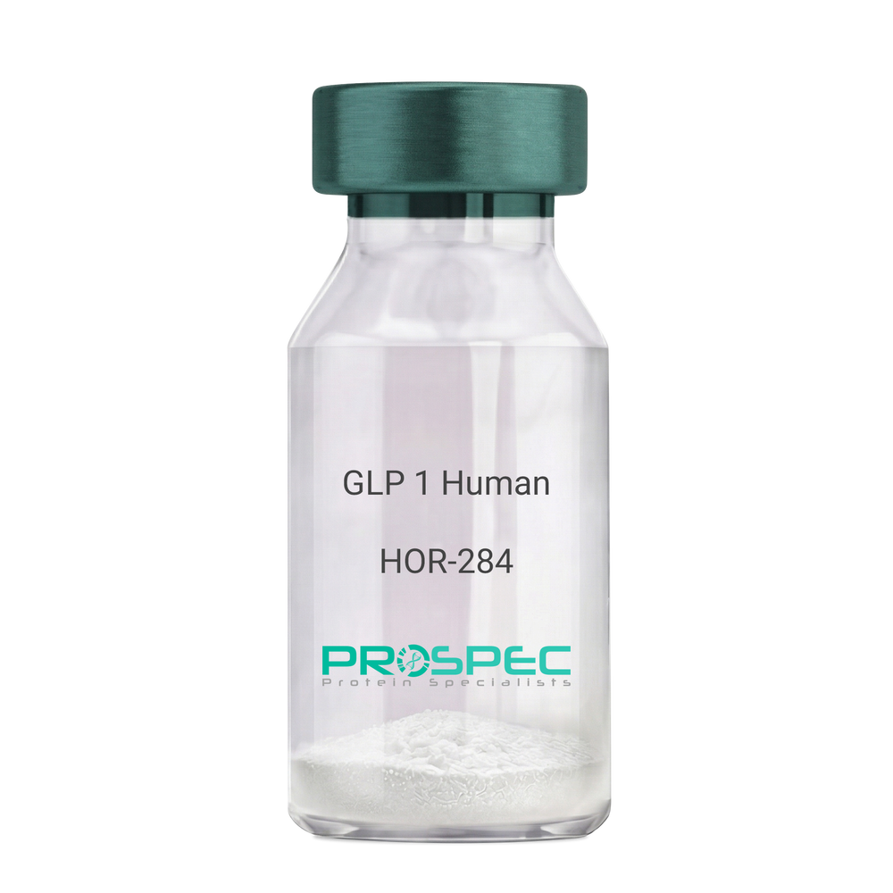
GLP 1 HumanDescription:
Human Glucagon Like Peptide-1
GLP1, Glucagon Like Peptide-1, Incretin hormone.
Product # :
HOR-284Price :
Quantity :
Shipping Method :
Shipped at Room temp
More Info
- description
- formulation
- purity
- More Info
Description
Glucagon Like Peptide-1 is a single, non-glycosylated, polypeptide chain containing 30 amino acids and having a molecular mass of 3297.7 Dalton.The GLP-1 is purified by proprietary chromatographic techniques.
Formulation
The GLP-1 peptide was lyophilized with no additives.
Purity
Greater than 96.0% as determined by RP-HPLC.
More Info
-
Introduction
Glucagon-like peptide-1 (GLP-1) is derived from the transcription product of the proglucagon gene. The major source of GLP-1 in the body is the intestinal L cell that secretes GLP-1 as a guthormone. The biologically active forms of GLP-1 are: GLP-1-(7-37) and GLP-1-(7-36)NH2.
GLP-1 secretion by L cells is dependent on the presence of nutrients in the lumen of the small intestine. The secretagogues (agents that causes or stimulates secretion) of this hormone include major nutrients like carbohydrate, proteinand lipid. Once in the circulation, GLP-1 has a half life of less than 2 minutes, due to rapid degradation by the enzyme dipeptidyl peptidase-4.
GLP-1 possesses several physiological properties that make it a subject of intensive investigation as a potential treatment of diabetes mellitus. The known physiological functions of GLP-1 include: Increases insulin secretion from the pancreas in a glucose-dependent manner, decreases glucagon secretion from the pancreas, increases beta cells mass and insulin gene expression, inhibits acid secretion and gastric emptying in the stomach, decreases food intake by increasing satiety. -
Synonyms
GLP1, Glucagon Like Peptide-1, Incretin hormone.
-
Physical Appearance
Sterile Filtered White lyophilized (freeze-dried) powder.
-
Stability
Lyophilized Glucagon Like Peptide-1 although stable at room temperature for 3 weeks, should be stored desiccated below -18°C. Upon reconstitution GLP-1 should be stored at 4°C between 2-7 days and for future use below -18°C. For long term storage it is recommended to add a carrier protein (0.1% HSA or BSA).Please prevent freeze-thaw cycles.
-
Solubility
It is recommended to reconstitute the lyophilized Glucagon Like Peptide-1 in sterile H2O at 0.5mg/ml, which can then be further diluted to other aqueous solutions.
-
Amino Acid Sequence
H-His-Ala-Glu-Gly-Thr-Phe-Thr-Ser-Asp-Val-Ser-Ser-Tyr-Leu-Glu-Gly-Gln-Ala-Ala-Lys-Glu-Phe-Ile-Ala-Trp-Leu-Val-Lys-Gly-Arg-NH2.
ProSpec's products are furnished for LABORATORY RESEARCH USE ONLY. The product may not be used as drugs, agricultural or pesticidal products, food additives or household chemicals.
- View Data Sheet
Name :
GHK-CuDescription:
GHK-Cu
Copper Tripeptide-1
Product # :
HOR-063Price :
Quantity :
Shipping Method :
Shipped at Room temp
More Info
- description
- formulation
- purity
- More Info
Description
GHK-Cu is a synthetic single, non-glycosylated polypeptide chain containing 3 amino acids, having a molecular mass of 401.91 Dalton and a Molecular formula of C14H22N6O4Cu.
Formulation
The protein was lyophilized with no additives.
Purity
Greater than 97.0% as determined by analysis by RP-HPLC.
More Info
-
Physical Appearance
Sterile Filtered White lyophilized (freeze-dried) powder.
-
Stability
Lyophilized GHK-Cu although stable at room temperature for 3 weeks, should be stored desiccated below -18°C. Upon reconstitution GHK-Cu should be stored at 4°C between 2-7 days and for future use below -18°C.
For long term storage it is recommended to add a carrier protein (0.1% HSA or BSA).
Please prevent freeze-thaw cycles.
-
Solubility
It is recommended to reconstitute the lyophilized GHK-Cu in sterile 18MΩ-cm H2O not less than 100 µg/ml, which can then be further diluted to other aqueous solutions.
-
Amino Acid Sequence
Gly-His-Lys.
-
Background
GHK-Cu is located in human plasma and has an ability to modify gene expression to a healthier state, influencing more than 4,000 human genes. GHK-Cu can change pathological gene expression to a healthy mode, mainly in chronic conditions as metastatic cancer, COPD and Ulcerative Colitis. GHK-Cu was discovered to be effective in systemic repair and neuroprotection.
ProSpec's products are furnished for LABORATORY RESEARCH USE ONLY. The product may not be used as drugs, agricultural or pesticidal products, food additives or household chemicals.
- View Data Sheet
Name :
Aprotinin ProteinDescription:
Aprotinin
Pancreatic trypsin inhibitor, Basic protease inhibitor, BPI, BPTI, Aprotinin, AP.
Product # :
PRO-285Price :
Quantity :
Shipping Method :
Shipped at Room temp
More Info
- description
- source
- formulation
- More Info
Description
Aprotinin is a natural proteinase inhibitor polypeptide consisting of fifty-eight amino acids {C284H432N84O79S7} arranged in a single polypeptide chain, cross-linked by three disulfide bridges and having a molecular mass of 6512.
Source
Bovine Lung.
Formulation
The protein (1mg/ml) was lyophilized with no additives.
More Info
-
Introduction
Aprotinin inhibits the activity of several proteolytic enzymes such as chymotrypsin, kallikrein, plasmin and trypsin. Aprotinin is present in blood and in most tissues, with a high concentration in lung. Aprotinin inhibits pro-inflammatory cytokine release and maintains glycoprotein homeostasis. In platelets, aprotinin reduces glycoprotein loss (e.g., GpIb, GpIIb/IIIa), while in granulocytes it prevents the expression of pro-inflammatory adhesive glycoproteins (e.g., CD11b).
-
Synonyms
Pancreatic trypsin inhibitor, Basic protease inhibitor, BPI, BPTI, Aprotinin, AP.
-
Physical Appearance
Sterile Filtered White lyophilized (freeze-dried) powder.
-
Stability
Lyophilized Aprotinin although stable at room temperature for 3 weeks, should be stored desiccated below -18°C. Upon reconstitution Aprotinin should be stored at 4°C between 2-7 days and for future use below -18°C.For long term storage it is recommended to add a carrier protein (0.1% HSA or BSA).Please prevent freeze-thaw cycles.
-
Solubility
It is recommended to reconstitute the lyophilized Aprotinin in sterile 18MΩ-cm H2O not less than 100µg/ml, which can then be further diluted to other aqueous solutions.
-
Specific Activity
5,940 KIU (Kallikrein Inactivator Units) per mg, 3.3 pH.Eur.U/mg.
ProSpec's products are furnished for LABORATORY RESEARCH USE ONLY. The product may not be used as drugs, agricultural or pesticidal products, food additives or household chemicals.
- View Data Sheet
Name :
Thrombin Human, HEKDescription:
Thrombin Human Recombinant, HEK
Prothrombin, EC 3.4.21.5, Coagulation factor II, F2, PT, THPH1, RPRGL2.
Product # :
PRO-1422Price :
Quantity :
Shipping Method :
Shipped with Ice Packs
More Info
- description
- source
- formulation
- biological activity
- More Info
Description
Recombinant Human Thrombin produced in HEK cells, having a total molecular weight of 36kDa. The Thrombin is purified by proprietary chromatographic techniques.
Source
HEK
Formulation
The Thrombin solution contains 20mM MES, pH6.0 and 500mM Choline Chloride.
Biological Activity
5396 NIH Units/mg.
The activity was determined in NIH units by comparing to Sigma’s human plasma thrombin. Protein concentration was measured using E(0.1%)@280nm = 1.83.More Info
-
Introduction
Thrombin enzyme (Activated Factor IIa) is an important clotting promoter that controls the transformation of soluble fibrinogen to insoluble active fibrin strands. Thrombin is a coagulation protein and a serine protease (EC 3.4.21.5) that catalyzes many coagulation-related reactions. Thrombin triggers factor-XI, factor-V, Factor-XIII and factor-VIII. Thrombin endorses platelet activation, using activation of protease-activated receptors on the platelet. As a result of its high proteolytic specificity, thrombin has become an important biochemical protein. The thrombin cleavage site (Leu-Val-Pro-Arg-Gly-Ser) is widely used in linker regions of recombinant fusion protein constructs. After the purification of the fusion protein, thrombin is used to cleave between the Arginine and Glycine residues of the cleavage site, efficiently removing the purification tag from the protein of interest with a high degree of specificity.
-
Synonyms
Prothrombin, EC 3.4.21.5, Coagulation factor II, F2, PT, THPH1, RPRGL2.
-
Physical Appearance
Sterile Filtered colorless solution.
-
Stability
Store frozen at -20°C to -80°C for long periods of time. Avoid multiple freeze-thaw cycles.
-
Assay Conditions
Thrombin (1nM) was assayed using SPECTROZYME TH as a substrate (20µM) in 5mM Tris-HCl (pH8.0), 0.1% PEG, 200mM NaCl at 25°C.
ProSpec's products are furnished for LABORATORY RESEARCH USE ONLY. The product may not be used as drugs, agricultural or pesticidal products, food additives or household chemicals.
- View Data Sheet
Name :
CTSD HumanDescription:
Cathepsin-D Human Recombinant
Cathepsin D, EC 3.4.23.5, CTSD, CPSD, CLN10, MGC2311.
Product # :
ENZ-378Price :
Quantity :
Shipping Method :
Shipped with Ice Packs
More Info
- description
- source
- formulation
- purity
- More Info
Description
CTSD produced in HEK293 cells is a single, glycosylated polypeptide chain containing 398 amino acids (21-412 a.a.) and having a molecular mass of 43.4kDa. CTSD is expressed with a 6 amino acid His tag at C-Terminus and purified by proprietary chromatographic techniques.
Source
HEK293 Cells
Formulation
CTSD at 1mg/ml in 50mM MES, pH5.5, 100mM NaCl and 20% Glycerol.
Purity
Greater than 95% as determined by SDS-PAGE
More Info
-
Introduction
Cathepsin D is synthesized as a 54kDa precursor, which is proteolytically processed to an intermediate 48kDa single chain, which matures into more stable 34kDa and 14kDa two chain form. It is an estrogen-regulated lysosomal protease that has been suggested to facilitate cancer cell migration and invasion by digesting the basement membrane, extracellular matrix, and xonnective tissue. Because of its mitogenic and proteolytic activities, it has been implicated as a prognostic marker in many tumor types. Cathepsin D is expressed in epithelial cells as well as in macrophages.
-
Synonyms
Cathepsin D, EC 3.4.23.5, CTSD, CPSD, CLN10, MGC2311.
-
Physical Appearance
Sterile Filtered clear solution.
-
Stability
Store vial at -20°C to -80°C. When stored at the recommended temperature, this protein is stable for 12 months.Please prevent freeze-thaw cycles.
-
Amino Acid Sequence
LVRIPLHKFT SIRRTMSEVG GSVEDLIAKG PVSKYSQAVP AVTEGPIPEV LKNYMDAQYY GEIGIGTPPQ CFTVVFDTGS SNLWVPSIHC KLLDIACWIH HKYNSDKSST YVKNGTSFDI HYGSGSLSGY LSQDTVSVPC QSASSASALG GVKVERQVFG EATKQPGITF IAAKFDGILG MAYPRISVNN VLPVFDNLMQ QKLVDQNIFS FYLSRDPDAQ PGGELMLGGT DSKYYKGSLS YLNVTRKAYW QVHLDQVEVA SGLTLCKEGC EAIVDTGTSL MVGPVDEVRE LQKAIGAVPL IQGEYMIPCE KVSTLPAITL KLGGKGYKLS PEDYTLKVSQ AGKTLCLSGF MGMDIPPPSG PLWILGDVFI GRYYTVFDRD NNRVGFAEAA RLHHHHHH
-
Enzymatic Activity
> 20 pmol/min/ug, defined as the amount of enzyme which cleaves 1pmol of Mca-PLGLDpa-AR-NH2/min at pH-3.5 at 25C.
ProSpec's products are furnished for LABORATORY RESEARCH USE ONLY. The product may not be used as drugs, agricultural or pesticidal products, food additives or household chemicals.
- View Data Sheet
Name :
FGF 2 Human (147 a.a.)Description:
Fibroblast Growth Factor Basic 147 a.a. Human Recombinant
Prostatropin, HBGH-2, HBGF-2, FGF-2, FGF-b.
Product # :
CYT-557Price :
Quantity :
Shipping Method :
Shipped at Room temp
More Info
- description
- source
- formulation
- purity
- biological activity
- More Info
Description
Fibroblast Growth Factor-2 Human Recombinant (FGF-2) produced in E.Coli is a single, non-glycosylated, polypeptide chain containing 147 amino acids and having a molecular mass of 16.5kDa. The FGF2 is purified by proprietary chromatographic techniques.
Source
Escherichia Coli.
Formulation
The bFGF was lyophilized from a sterile filtered solution containing 20mM Tris-HCl, pH 7.6 and 150mM NaCl.
Purity
Greater than 98.0% as determined by SDS-PAGE.
Biological Activity
The ED50, Calculated by the dose- dependent proliferation of mouse BALB/c 3T3 cells is < 0.05ng/ml corresponding to a specific activity of 2.0x10⁷ units/mg.
More Info
-
Introduction
FGF-basic is a member of the fibroblast growth factor (FGF) family. FGF family members possess broad mitogenic and cell survival activities, and are involved in a variety of biological processes, including embryonic development, cell growth, morphogenesis, tissue repair, tumor growth and invasion. This protein functions as a modifier of endothelial cell migration and proliferation, as well as an angiogenic factor. It acts as a mitogen for a variety of mesoderm- and neuroectoderm-derived cells in vitro, thus is thought to be involved in organogenesis. Three alternatively spliced variants encoding different isoforms have been described. The heparin-binding growth factors are angiogenic agents in vivo and are potent mitogens for a variety of cell types in vitro. There are differences in the tissue distribution and concentration of these 2 growth factors.
-
Synonyms
Prostatropin, HBGH-2, HBGF-2, FGF-2, FGF-b.
-
Physical Appearance
Sterile Filtered White lyophilized (freeze-dried) powder.
-
Stability
Lyophilized basic-FGF although stable at room temperature for 3 weeks, should be stored desiccated below -18°C. Upon reconstitution FGFb should be stored at 4°C between 2-7 days and for future use below -18°C.For long term storage it is recommended to add a carrier protein (0.1% HSA or BSA).Please prevent freeze-thaw cycles.
-
Solubility
It is recommended to reconstitute the lyophilized FGF-B in sterile 18MΩ-cm H2O not less than 100µg/ml, which can then be further diluted to other aqueous solutions.
-
Amino Acid Sequence
MPALPEDGGS GAFPPGHFKD PKRLYCKNGG FFLRIHPDGR VDGVREKSDP HIKLQLQAEE RGVVSIKGVC ANRYLAMKED GRLLASKCVT DECFFFERLE SNNYNTYRSR KYTSWYVALK RTGQYKLGSK TGPGQKAILF LPMSAKS.
-
Background
What is the molecular weight/Mw of FGF 2 Protein?
FGF 2 Protein has a total Mw of 16.5kDa.
What is the source or expression system of FGF 2 Protein?
Escherichia Coli.
What is the Purity of FGF 2 Protein?
FGF 2 Protein is >98% pure as determined by SDS-PAGE.
What is the Biological Activity of FGF 2 Protein?
The ED50, Calculated by the dose- dependent proliferation of mouse BALB/c 3T3 cells is < 0.05ng/ml corresponding to a specific activity of 2.0x10⁷ units/mg.
What is the amino acid sequence of FGF 2 Protein?
MPALPEDGGS GAFPPGHFKD PKRLYCKNGG FFLRIHPDGR VDGVREKSDP HIKLQLQAEE RGVVSIKGVC ANRYLAMKED GRLLASKCVT DECFFFERLE SNNYNTYRSR KYTSWYVALK RTGQYKLGSK TGPGQKAILF LPMSAKS.
What applications can FGF 2 Protein be used in?
FGF 2 Protein can probably be used in western blot, ELISA and Lateral Flow.
What is the endotoxin level for FGF 2 Protein?
The endotoxin level is minimal, FGF 2 Protein was purified using conventional chromatography techniques.
ProSpec's products are furnished for LABORATORY RESEARCH USE ONLY. The product may not be used as drugs, agricultural or pesticidal products, food additives or household chemicals.
- View Data Sheet
Name :
QPCT HumanDescription:
Glutaminyl-Peptide Cyclotransferase Human Recombinant
Glutaminyl-Peptide Cyclotransferase, Glutaminyl Cyclase, QC, Glutaminyl-TRNA Cyclotransferase, Glutamyl Cyclase, EC 2.3.2.5, SQC, EC, GCT, Glutaminyl-peptide cyclotransferase.
Product # :
ENZ-912Price :
Quantity :
Shipping Method :
Shipped with Ice Packs
More Info
- description
- source
- formulation
- purity
- More Info
Description
QPCT produced in Sf9 Baculovirus cells is a single, glycosylated polypeptide chain containing 339 amino acids (29-361a.a.) and having a molecular mass of 38.7kDa (Molecular size on SDS-PAGE will appear at approximately 28-40kDa). QPCT is expressed with a 6 amino acid His tag at C-Terminus and purified by proprietary chromatographic techniques.
Source
Sf9, Baculovirus cells.
Formulation
QPCT protein solution (0.5mg/ml) contains Phosphate Buffered Saline (pH 7.4) and 10% glycerol.
Purity
Greater than 90.0% as determined by SDS-PAGE.
More Info
-
Introduction
Glutaminyl-Peptide Cyclotransferase, also known as QPCT is a member of the glutaminyl-peptide cyclotransferase family. QPCT is responsible for the biosynthesis of pyroglutamyl peptides. Furthermore, QPCT is partial against acidic and tryptophan residues adjacent to the N-terminal glutaminyl residue and a lack of importance of chain length following the second residue. QPCT catalyzes N-terminal pyroglutamate formation, also in vitro, it catalyzes pyroglutamate formation of N-terminally truncated form of APP amyloid-beta peptides [Glu-3]-beta-amyloid.
-
Synonyms
Glutaminyl-Peptide Cyclotransferase, Glutaminyl Cyclase, QC, Glutaminyl-TRNA Cyclotransferase, Glutamyl Cyclase, EC 2.3.2.5, SQC, EC, GCT, Glutaminyl-peptide cyclotransferase.
-
Physical Appearance
Sterile Filtered colorless solution.
-
Stability
Store at 4°C if entire vial will be used within 2-4 weeks. Store, frozen at -20°C for longer periods of time. For long term storage it is recommended to add a carrier protein (0.1% HSA or BSA).Avoid multiple freeze-thaw cycles.
-
Amino Acid Sequence
VSPSASAWPE EKNYHQPAIL NSSALRQIAE GTSISEMWQN DLQPLLIERY PGSPGSYAAR QHIMQRIQRL QADWVLEIDT FLSQTPYGYR SFSNIISTLN PTAKRHLVLA CHYDSKYFSH WNNRVFVGAT DSAVPCAMML ELARALDKKL LSLKTVSDSK PDLSLQLIFF DGEEAFLHWS PQDSLYGSRH LAAKMASTPH PPGARGTSQL HGMDLLVLLD LIGAPNPTFP NFFPNSARWF ERLQAIEHEL HELGLLKDHS LEGRYFQNYS YGGVIQDDHI PFLRRGVPVL HLIPSPFPEV WHTMDDNEEN LDESTIDNLN KILQVFVLEY LHLHHHHHH.
ProSpec's products are furnished for LABORATORY RESEARCH USE ONLY. The product may not be used as drugs, agricultural or pesticidal products, food additives or household chemicals.
- View Data Sheet
Name :
CTGF Human, HEKDescription:
Connective Tissue Growth Factor Human Recombinant , HEK
CCN2, NOV2, HCS24, IGFBP8, MGC102839, CTGF.
Product # :
CYT-687Price :
Quantity :
Shipping Method :
Shipped with Ice Packs
More Info
- description
- source
- formulation
- purity
- More Info
Description
The CTGF Human Recombinant produced in HEK293 cells, is 36kDa protein containing a total of 329 amino acid residues (aa 27-349) including a C-terminal 6×His tag.
Source
HEK293 cells.
Formulation
CTGF filtered (0.2µm) solution in 0.1M Citrate buffer pH 4.7 and 20% (w/v) glycerol.
Purity
Greater than 90.0% as determined by SDS-PAGE.
More Info
-
Introduction
Connective Tissue Growth Factor belongs to the CCN family of proteins. The CCN family presently consists of six members in human also known as: Cyr61 (Cystein rich 61), CTGF (Connective Tissue Growth Factor), Nov (Nephroblastoma Overexpressed gene), WISP-1, 2 and 3 (Wnt-1 Induced Secreted Proteins). The CCN genes encode secreted proteins associated with the Extracellular Matrix (ECM) and cell membrane.
CCN proteins are matricellular proteins which are involved in the regulation of various cellular functions including: proliferation, differentiation, survival, adhesion and migration. They are expressed in derivatives of the three embryonic sheets and are implicated in the development of kidney, nervous system, muscle, bone marrow, cartilage and bone. During adulthood, they are implicated in wound healing, bone fracture repair, and pathologies such as: fibrosis, vascular ailments and tumorigenesis.
Full length secreted CCN proteins can show an antiproliferative activity, whereas truncated isoforms are likely to stimulate proliferation and behave as oncogenes.
The full length protein consists of four modules: Module I shares partial identity with the N-terminal part of the Insulin-like Growth Factor Binding Proteins (IGFBPs).
Module II includes a stretch of 70amino acid residues – which shares sequence identity with the Von Willebrand Factor Type C repeat (VWC).
Module III contains sequences sharing identity with the Thrombospondin type 1 repeat (TSP1) (WSXCSXXCG), which is thought to be implicated in the binding of sulfated glycoconjugates and to be important for cell adhesion.
Module IV, also designated CT, is encoded by exon5. It is the leasts conserved one of the four domains at the level of nucleotide sequence, but it appears to be critical for several of the biological functions attributed to the CCN proteins. Module IV resembles the CT domain of several extracellular protein including, Von Willebrand's factor and mucins. Sequence similarities to heparin-binding motifs are also found within this domain.
Proteolysis of the secreted full-length CCN proteins that has been reported in the case of CCN2 and CCN3 might result in the production of CCN-derived peptides with high affinity for ligands that full-length CNN proteins bind only poorly. Amino-truncated CCN2 isoforms were biologically active whereas no specific biological activity has been attributed to the truncated CCN3. Although the molecular processes underlying the production of these secreted isoforms is presently unknown, it is important to note that proteolysis occur at the same amino acid residues in both CCN2 and CCN3. An elevated expression of CCN2 has also been detected by Northern blotting in human invasive mammary ductal carcinomas, dermatofibromas, pyogenic granuloma, endothelial cells of angiolipomas and angioleiomyomas, and in pancreatic tumors. A study performed with chondrosarcomas representative of various histological grades established that CCN2 expression was closely correlated with increasing levels of malignancy.
In agreement with CCN2 playing a role in brain tumor angiogenesis, immunocytochemistry studies indicated that both glioblastoma tumor cells and proliferating endothelial cells stained positive for CCN2. In astrocytomas, CCN2 expression was particularly elevated in high grade tumors, with a marked effect of CCN2 on cell proliferation. Downregulation of CCN2 expression in these cells was associated with a growth arrest at the G1/S transition while over-expression of CCN2 induced a two-fold increase of the number of cells in the G1 phase. Gene profiling analysis allowed to identify a set of about 50 genes whose expression might account for the proliferative activity of CCN2 in these cells.
CCN2 was seen in a higher proportion of mononuclear cells of patients with acute lymphoblastic leukemia. -
Synonyms
CCN2, NOV2, HCS24, IGFBP8, MGC102839, CTGF.
-
Physical Appearance
Filtered colorless solution.
-
Stability
Store at 4°C if entire vial will be used within 2-4 weeks. Store, frozen at -20°C for longer periods of time. For long term storage it is recommended to add a carrier protein (0.1% HSA or BSA).Avoid multiple freeze-thaw cycles.
-
Amino Acid Sequence
QNCSGPCRCP DEPAPRCPAG VSLVLDGCGC CRVCAKQLGE LCTERDPCDP HKGLFCHFGS PANRKIGVCT AKDGAPCIFG GTVYRSGESF QSSCKYQCTC LDGAVGCMPL CSMDVRLPSP DCPFPRRVKL PGKCCEEWVC DEPKDQTVVG PALAAYRLED TFGPDPTMIR ANCLVQTTEW SACSKTCGMG ISTRVTNDNA SCRLEKQSRL CMVRPCEADL EENIKKGKKC IRTPKISKPI KFELSGCTSM KTYRAKFCGV CTDGRCCTPH RTTTLPVEFK CPDGEVMKKN MMFIKTCACH YNCPGDNDIF ESLYYRKMYG DMA HHHHHH.
-
Background
What is the molecular weight/Mw of CTGF Protein?
CTGF Protein has a total Mw of 36kDa.
What is the source or expression system of CTGF Protein?
HEK293 cells.
What is the Purity of CTGF Protein?
CTGF Protein is >90% pure as determined by SDS-PAGE.
What is the Biological Activity of CTGF Protein?
The biological functionality of CTGF Protein will be determined in the future.
What is the amino acid sequence of CTGF Protein?
QNCSGPCRCP DEPAPRCPAG VSLVLDGCGC CRVCAKQLGE LCTERDPCDP HKGLFCHFGS PANRKIGVCT AKDGAPCIFG GTVYRSGESF QSSCKYQCTC LDGAVGCMPL CSMDVRLPSP DCPFPRRVKL PGKCCEEWVC DEPKDQTVVG PALAAYRLED TFGPDPTMIR ANCLVQTTEW SACSKTCGMG ISTRVTNDNA SCRLEKQSRL CMVRPCEADL EENIKKGKKC IRTPKISKPI KFELSGCTSM KTYRAKFCGV CTDGRCCTPH RTTTLPVEFK CPDGEVMKKN MMFIKTCACH YNCPGDNDIF ESLYYRKMYG DMA HHHHHH.
What applications can CTGF Protein be used in?
CTGF Protein can probably be used in western blot, ELISA and Lateral Flow.
What is the endotoxin level for CTGF Protein?
The endotoxin level is minimal, CTGF Protein was purified using conventional chromatography techniques.
ProSpec's products are furnished for LABORATORY RESEARCH USE ONLY. The product may not be used as drugs, agricultural or pesticidal products, food additives or household chemicals.
- View Data Sheet
Name :
MIF Human His NDescription:
Macrophage Migration Inhibitory Factor Human, Recombinant His Tag N-Terminus
Phenylpyruvate tautomerase, Glycosylation-inhibiting factor, GIF, MMIF, MIF.
Product # :
CYT-431Price :
Quantity :
Shipping Method :
Shipped with Ice Packs
More Info
- description
- source
- formulation
- purity
- More Info
Description
MIF human Recombinant, fused to 40 a.a. His-tag at N-terminus, was cloned into an E. coli expression vector and was purified to apparent homogeneity by using conventional column chromatography techniques. Macrophage Inducing Factor Human Recombinant ( 1-115 a.a. ) is a single, non-glycosylated, polypeptide chain having a total amino acids of 155 and molecular mass of 17kDa.
Source
Escherichia Coli.
Formulation
1mg/ml solution containing PBS pH-7.4.
Purity
Greater than 95.0% as determined by:
(a) Analysis by RP-HPLC.
(b) Analysis by SDS-PAGE.More Info
-
Introduction
The cytokine Macrophage migration inhibitory factor (MIF) has been identified to be secreted by the pituitary gland and the monocyte/macrophage and to play an important role in endotoxic shock. MIF has the unique property of being released from macrophages and T cells in response to physiological concentrations of glucocorticoids. The secretion of MIF is tightly regulated and decreases at high, anti-inflammatory steroid concentration.
-
Synonyms
Phenylpyruvate tautomerase, Glycosylation-inhibiting factor, GIF, MMIF, MIF.
-
Physical Appearance
Sterile filtered colorless solution.
-
Stability
Liquid MIF although stable 4°C for 1 week, should be stored below -18°C. Please prevent freeze-thaw cycles.
-
Amino Acid Sequence
MRGSHHHHHHGMASMTGGQQMGRDLYDDDDKDRWGSMPMFIVNTNV PRASVPDGFL SELTQQLAQA TGKPPQYIAV HVVPDQLMAF GGSSEPCALC LHSIGKIGGA QNRSYSKLLC GLLAERLRIS PDRVYINYYD MNAANVGWNN STFA.
ProSpec's products are furnished for LABORATORY RESEARCH USE ONLY. The product may not be used as drugs, agricultural or pesticidal products, food additives or household chemicals.
- View Data Sheet
Name :
HCST HumanDescription:
Hematopoietic Cell Signal Transducer Human Recombinant
Hematopoietic Cell Signal Transducer, DNAX-Activation Protein 10, Phosphoinositide-3-Kinase Adaptor Protein, Transmembrane Adapter Protein KAP10, Kinase Assoc Pro Of ~10kDa, Membrane Protein DAP10, PIK3AP, DAP10, KAP10, Kinase Assoc Protein.
Product # :
PRO-2522Price :
Quantity :
Shipping Method :
Shipped with Ice Packs
More Info
- description
- source
- formulation
- purity
- More Info
Description
HCST produced in Sf9 Baculovirus cells is a single, glycosylated polypeptide chain containing 271 amino acids (20-48 a.a.) and having a molecular mass of 30.1kDa. (Molecular size on SDS-PAGE will appear at approximately 28-40kDa).HCST is expressed with a 242 amino acid hIgG-His-tag at C-Terminus and purified by proprietary chromatographic techniques.
Source
Sf9, Baculovirus cells.
Formulation
HCST protein solution (1mg/ml) contains 10% glycerol & Phosphate Buffered Saline (pH 7.4).
Purity
Greater than 90.0% as determined by SDS-PAGE.
More Info
-
Introduction
HCST (Hematopoietic Cell Signal Transducer) is part of the DAP10 family. HCST is capable of being a part of an immunoreceptor complex. HCST takes a vital part in inducing cytotoxicity against MHC class I chain-associated MICA & target cells expressing cell surface ligands such as UL16-binding protein (ULBP) & MICB. HCST complex participates in proliferation as well as cell survival by activating T & NK cell responses.
-
Synonyms
Hematopoietic Cell Signal Transducer, DNAX-Activation Protein 10, Phosphoinositide-3-Kinase Adaptor Protein, Transmembrane Adapter Protein KAP10, Kinase Assoc Pro Of ~10kDa, Membrane Protein DAP10, PIK3AP, DAP10, KAP10, Kinase Assoc Protein.
-
Physical Appearance
Sterile Filtered colorless solution.
-
Stability
Store at 4°C if entire vial will be used within 2-4 weeks. Store, frozen at -20°C for longer periods of time. For long term storage it is recommended to add a carrier protein (0.1% HSA or BSA).Avoid multiple freeze-thaw cycles.
-
Amino Acid Sequence
ADPTTPGERS SLPAFYPGTS GSCSGCGSLS LPLEPKSCDK THTCPPCPAP ELLGGPSVFL FPPKPKDTLM ISRTPEVTCV VVDVSHEDPE VKFNWYVDGV EVHNAKTKPR EEQYNSTYRV VSVLTVLHQD WLNGKEYKCK VSNKALPAPI EKTISKAKGQ PREPQVYTLP PSRDELTKNQ VSLTCLVKGF YPSDIAVEWE SNGQPENNYK TTPPVLDSDG SFFLYSKLTV DKSRWQQGNV FSCSVMHEAL HNHYTQKSLS LSPGKHHHHH H.
ProSpec's products are furnished for LABORATORY RESEARCH USE ONLY. The product may not be used as drugs, agricultural or pesticidal products, food additives or household chemicals.
- View Data Sheet
Name :
UBE2R2 HumanDescription:
Ubiquitin Conjugating Enzyme E2R 2 Human Recombinant
CDC34B, E2-CDC34B, UBC3B, Ubiquitin carrier protein R2, Ubiquitin-conjugating enzyme E2-CDC34B, Ubiquitin-protein ligase R2, Ubiquitin-conjugating enzyme E2 R2, EC 6.3.2.19.
Product # :
ENZ-511Price :
Quantity :
Shipping Method :
Shipped with Ice Packs
More Info
- description
- source
- formulation
- purity
- More Info
Description
UBE2R2 Human Recombinant produced in E.Coli is a single, non-glycosylated polypeptide chain containing 261 amino acids (1-238 a.a) and having a molecular mass of 29.6kDa (Molecular size on SDS-PAGE will appear higher).UBE2R2 is fused to a 23 amino acid His-tag at N-terminus & purified by proprietary chromatographic techniques.
Source
Escherichia Coli.
Formulation
UBE2R2 protein solution (0.25mg/ml) containing 20mM Tris-HCl buffer (pH 8.0), 0.15M NaCl, 20% glycerol and 1mM DTT.
Purity
Greater than 85.0% as determined by SDS-PAGE.
More Info
-
Introduction
Ubiquitin-conjugating enzyme E2 R2 (UBE2R2) is a member of the ubiquitin-conjugating enzyme family. Protein kinase CK2 is a ubiquitous and pleiotropic Ser/Thr protein kinase implicated in cell growth andtransformation. This protein is a protein similar to the E2 ubiquitin conjugating enzyme UBC3/CDC34. Studies propose that CK2-dependent phosphorylation of this ubiquitin-conjugating enzyme functions by regulating beta-TrCP substrate recognition and induces its interaction with beta-TrCP, enhancing beta-catenin degradation. UBE2R2 receives ubiquitin from the E1 complex and catalyzes its covalent attachment to other proteins. In vitro catalyzes monoubiquitination and 'Lys-48'-linked polyubiquitination. UBE2R2 may be implicated in degradation of katenin. Among the diseases associated with UBE2R2 are cblc, and herpes simplex.
-
Synonyms
CDC34B, E2-CDC34B, UBC3B, Ubiquitin carrier protein R2, Ubiquitin-conjugating enzyme E2-CDC34B, Ubiquitin-protein ligase R2, Ubiquitin-conjugating enzyme E2 R2, EC 6.3.2.19.
-
Physical Appearance
Sterile Filtered clear solution.
-
Stability
Store at 4°C if entire vial will be used within 2-4 weeks. Store, frozen at -20°C for longer periods of time. For long term storage it is recommended to add a carrier protein (0.1% HSA or BSA).Avoid multiple freeze-thaw cycles.
-
Amino Acid Sequence
MGSSHHHHHH SSGLVPRGSH MGSMAQQQMT SSQKALMLEL KSLQEEPVEG FRITLVDESD LYNWEVAIFG PPNTLYEGGY FKAHIKFPID YPYSPPTFRF LTKMWHPNIY ENGDVCISIL HPPVDDPQSG ELPSERWNPT QNVRTILLSV ISLLNEPNTF SPANVDASVM FRKWRDSKGK DKEYAEIIRK QVSATKAEAE KDGVKVPTTL AEYCIKTKVP SNDNSSDLLY DDLYDDDIDD EDEEEEDADC YDDDDSGNEE S
ProSpec's products are furnished for LABORATORY RESEARCH USE ONLY. The product may not be used as drugs, agricultural or pesticidal products, food additives or household chemicals.
- View Data Sheet
Name :
SAA1 MonkeyDescription:
Serum Amyloid A (APO-SAA1) Rhesus Macaque Recombinant
Serum amyloid A protein, SAA, Amyloid protein A, Amyloid fibril protein AA, SAA1, SAA2, PIG4, TP53I4, MGC111216.
Product # :
CYT-719Price :
Quantity :
Shipping Method :
Shipped at Room temp
More Info
- description
- source
- formulation
- purity
- biological activity
- More Info
Description
SAA1 monkey Recombinant produced in E.Coli is a single, non-glycosylated, polypeptide chain containing 104 amino acids and having a total molecular mass of 11.8 kDa. SAA1 is purified by proprietary chromatographic techniques.
Source
Escherichia Coli.
Formulation
The protein was lyophilized with 1x PBS pH-7.4
Purity
Greater than 97.0% as determined by: (a) Analysis by RP-HPLC. (b) Analysis by SDS-PAGE.
Biological Activity
Determined by its ability to chemoattract human monocytes using a concentration range of 1.0-10.0 ng/ml.More Info
-
Introduction
SAA1 protein is an acute phase apolipoprotein reactant which is produced mostly by hepatocytes and under regulation of inflammatory cytokines. SAA1 (Serum amyloid A1) protein is produced mainly in the liver and circulates in low levels in the blood. The SAA1 seems to have a role in the immune system. SAA1 protein levels increase in the blood and other tissues under conditions of inflammation. SAA1 may facilitate the repair of injured tissues; it also acts as an antibacterial agent, and signals the migration of germ-fighting cells to sites of infection. SAA1 also functions as an apolipoprotein of the HDL complex.
Elevated levels of SAA1 ultimately affect secondary amyloidosis, extracellular amassing of amyloid fibrils, resulting from a circulating precursor, in a variety of tissues and organs. The most widespread type of amyloidosis appears secondary to chronic inflammatory disease, mainly rheumatoid arthritis. The SAA1 cleavage product a designated amyloid protein A is deposited systemically as amyloid in vital organs such as the liver, spleen, and kidneys in chronic inflammatory diseases patients. These deposits are extremely insoluble and resistant to proteolysis; they disrupt tissue structure and compromise performance. -
Synonyms
Serum amyloid A protein, SAA, Amyloid protein A, Amyloid fibril protein AA, SAA1, SAA2, PIG4, TP53I4, MGC111216.
-
Physical Appearance
Sterile Filtered White lyophilized (freeze-dried) powder.
-
Stability
Lyophilized SAA1 although stable at room temperature for 3 weeks, should be stored desiccated below -18°C. Upon reconstitution SAA1 should be stored at 4°C between 2-7 days and for future use below -18°C. For long term storage it is recommended to add a carrier protein (0.1% HSA or BSA). Please prevent freeze-thaw cycles.
-
Solubility
It is recommended to reconstitute the lyophilized SAA1 in sterile 18MΩ-cm H2O not less than 100µg/ml, which can then be further diluted to other aqueous solutions.
-
Amino Acid Sequence
RSWFSFLGEA YDGARDMWRA YSDMKEANYK NSDKYFHARG NYDAAQRGPG GVWAAEVISD ARENIQKLLG RGAEDTLADQ AANEWGRSGK DPNHFRPAGL PEKY.
ProSpec's products are furnished for LABORATORY RESEARCH USE ONLY. The product may not be used as drugs, agricultural or pesticidal products, food additives or household chemicals.
- View Data Sheet
Name :
CCBL1 HumanDescription:
Cysteine Conjugate-Beta Lyase Cytoplasmic Human Recombinant
Cysteine Conjugate-Beta Lyase, Cytoplasmic, Glutamine Transaminase K, Cysteine Conjugate-Beta Lyase; Cytoplasmic (Glutamine Transaminase K, Kyneurenine Aminotransferase), Kynurenine--Oxoglutarate Transaminase I, Glutamine--Phenylpyruvate Transaminase, Cysteine-S-Conjugate Beta-Lyase, Kynurenine Aminotransferase I, Kyneurenine Aminotransferase, KATI, GTK, Glutamine-Phenylpyruvate Aminotransferase, Kynurenine--Oxoglutarate Transaminase 1, Beta-Lysase, Kidney, EC 4.4.1.13, EC 2.6.1.64, EC 2.6.1.7, KAT1, Kynurenine--oxoglutarate transaminase 1.
Product # :
ENZ-878Price :
Quantity :
Shipping Method :
Shipped with Ice Packs
More Info
- description
- source
- formulation
- purity
- More Info
Description
CCBL1 Human Recombinant produced in E.Coli is a single, non-glycosylated polypeptide chain containing 445 amino acids (1-422 a.a) and having a molecular mass of 50.3kDa. CCBL1 is fused to a 23 amino acid His-tag at N-terminus & purified by proprietary chromatographic techniques.
Source
Escherichia Coli.
Formulation
CCBL1 protein solution (1mg/ml) containing Phosphate buffered saline (pH7.4), 10% glycerol and 1mM DTT.
Purity
Greater than 90.0% as determined by SDS-PAGE.
More Info
-
Introduction
Cysteine Conjugate-Beta Lyase Cytoplasmic also known as CCBL1 is a member of the class-I pyridoxal-phosphate-dependent aminotransferase family. CCBL1 catalyzes the irreversible transamination of the L-tryptophan metabolite L-kynurenine to form kynurenic acid (KA) it also metabolizes the cysteine conjugates of certain halogenated alkenes and alkanes to form reactive metabolites. Furthermore, CCBL1 catalyzes the beta-elimination of S-conjugates and Se-conjugates of L-(seleno) cysteine, resulting in the cleavage of the C-S or C-Se bond.
-
Synonyms
Cysteine Conjugate-Beta Lyase, Cytoplasmic, Glutamine Transaminase K, Cysteine Conjugate-Beta Lyase; Cytoplasmic (Glutamine Transaminase K, Kyneurenine Aminotransferase), Kynurenine--Oxoglutarate Transaminase I, Glutamine--Phenylpyruvate Transaminase, Cysteine-S-Conjugate Beta-Lyase, Kynurenine Aminotransferase I, Kyneurenine Aminotransferase, KATI, GTK, Glutamine-Phenylpyruvate Aminotransferase, Kynurenine--Oxoglutarate Transaminase 1, Beta-Lysase, Kidney, EC 4.4.1.13, EC 2.6.1.64, EC 2.6.1.7, KAT1, Kynurenine--oxoglutarate transaminase 1.
-
Physical Appearance
Sterile Filtered colorless solution.
-
Stability
Store at 4°C if entire vial will be used within 2-4 weeks. Store, frozen at -20°C for longer periods of time. For long term storage it is recommended to add a carrier protein (0.1% HSA or BSA).Avoid multiple freeze-thaw cycles.
-
Amino Acid Sequence
MGSSHHHHHH SSGLVPRGSH MGSMAKQLQA RRLDGIDYNP WVEFVKLASE HDVVNLGQGF PDFPPPDFAV EAFQHAVSGD FMLNQYTKTF GYPPLTKILA SFFGELLGQE IDPLRNVLVT VGGYGALFTA FQALVDEGDE VIIIEPFFDC YEPMTMMAGG RPVFVSLKPG PIQNGELGSS SNWQLDPMEL AGKFTSRTKA LVLNTPNNPL GKVFSREELE LVASLCQQHD VVCITDEVYQ WMVYDGHQHI SIASLPGMWE RTLTIGSAGK TFSATGWKVG WVLGPDHIMK HLRTVHQNSV FHCPTQSQAA VAESFEREQL LFRQPSSYFV QFPQAMQRCR DHMIRSLQSV GLKPIIPQGS YFLITDISDF KRKMPDLPGA VDEPYDRRFV KWMIKNKGLV AIPVSIFYSV PHQKHFDHYI RFCFVKDEAT LQAMDEKLRK WKVEL.
ProSpec's products are furnished for LABORATORY RESEARCH USE ONLY. The product may not be used as drugs, agricultural or pesticidal products, food additives or household chemicals.
- View Data Sheet
Name :
GH Zebrafish MutantDescription:
Growth Hormone Mutant Zebrafish Recombinant
GH1, GH, GHN, GH-N, hGH-N, Pituitary growth hormone, Growth hormone 1, Somatotropin.
Product # :
CYT-709Price :
Quantity :
Shipping Method :
Shipped at Room temp
More Info
- description
- source
- formulation
- purity
- More Info
Description
Somatotropin Zebrafish Mutant G113R Recombinant produced in E.Coli is a single, non-glycosylated polypeptide chain containing 185 amino acids with an additional Ala at the N-terminus and having a molecular mass of 21.18 kDa. The Zebrafish Mutant G113R Recombinant is purified by proprietary chromatographic techniques.
Source
Escherichia Coli.
Formulation
The protein was lyophilized from a concentrated (1mg/ml) solution with 0.5% NaHCO3 pH-8.
Purity
Greater than 95.0% as determined by:
(a) Analysis by RP-HPLC.
(b) Analysis by SDS-PAGE.More Info
-
Introduction
GH is a member of the somatotropin/prolactin family of hormones which play an important role in growth control. The gene, along with four other related genes, is located at the growth hormone locus on chromosome 17 where they are interspersed in the same transcriptional orientation; an arrangement which is thought to have evolved by a series of gene duplications. The five genes share a remarkably high degree of sequence identity. Alternative splicing generates additional isoforms of each of the five growth hormones, leading to further diversity and potential for specialization. This particular family member is expressed in the pituitary but not in placental tissue as is the case for the other four genes in the growth hormone locus. Mutations in or deletions of the gene lead to growth hormone deficiency and short stature.
-
Synonyms
GH1, GH, GHN, GH-N, hGH-N, Pituitary growth hormone, Growth hormone 1, Somatotropin.
-
Physical Appearance
Sterile Filtered White lyophilized (freeze-dried) powder.
-
Stability
Lyophilized Growth-Hormone although stable at room temperature for at least two weeks, should be stored desiccated below -18°C. Upon reconstitution and filter sterilization GH can be stored at 4°C, pH 9 for up to 4 weeks. For long term storage and more diluted solutions it is recommended to add a carrier protein (0.1% HSA or BSA). Please prevent freeze-thaw cycles.
-
Solubility
It is recommended to reconstitute the lyophilized Zebrafish Mutant G113R in 0.4% NaHCO3 or water adjusted to pH-9, not less than 100µg/ml, which can then be further diluted to other aqueous solutions, preferably in a presence of a carrier protein such as BSA or similar.
-
Amino Acid Sequence
The sequence of the first six N-terminal amino acids was determined and was found to be Ala-Gln-Arg-Leu-Phe-Asn.
ProSpec's products are furnished for LABORATORY RESEARCH USE ONLY. The product may not be used as drugs, agricultural or pesticidal products, food additives or household chemicals.
- View Data Sheet
Name :
HAVCR2 HumanDescription:
Hepatitis A Virus Cellular Receptor 2 Human Recombinant
Hepatitis A virus cellular receptor 2, HAVcr-2, T-cell immunoglobulin and mucin domain-containing protein 3, TIMD-3, T-cell membrane protein 3, TIM-3, HAVCR2, TIM3, TIMD3, TIM3, KIM-3.
Product # :
HAV-224Price :
Quantity :
Shipping Method :
Shipped with Ice Packs
More Info
- description
- source
- formulation
- purity
- More Info
Description
Recombinant Human HAVCR2 produced in E. coli is a single polypeptide chain containing 206 amino acids (aa 22-202) and having a molecular mass of 22.7kDa.HAVCR2 is fused to a 25 amino acid His-tag at N-terminus & purified by proprietary chromatographic techniques.
Source
Escherichia Coli.
Formulation
The HAVCR2 solution (1mg/ml) contains 20mM Tris-HCl buffer (pH 8.0), 0.4M urea and 10% glycerol.
Purity
Greater than 90% as determined by SDS-PAGE.
More Info
-
Introduction
Hepatitis A Virus Cellular Receptor 2 (HAVCR2) is a member of the immunoglobulin superfamily. HACVR2 controls macrophage activation. HACVR2 inhibits T-helper type 1 lymphocyte (Th1)-mediated auto- and alloimmune responses and stimulates immunological tolerance.
-
Synonyms
Hepatitis A virus cellular receptor 2, HAVcr-2, T-cell immunoglobulin and mucin domain-containing protein 3, TIMD-3, T-cell membrane protein 3, TIM-3, HAVCR2, TIM3, TIMD3, TIM3, KIM-3.
-
Physical Appearance
Sterile filtered colorless solution.
-
Stability
Store at 4°C if entire vial will be used within 2-4 weeks. Store, frozen at -20°C for longer periods of time. For long term storage it is recommended to add a carrier protein (0.1% HSA or BSA).Avoid multiple freeze-thaw cycles.
-
Amino Acid Sequence
MGSSHHHHHH SSGLVPRGSH MGSHMSEVEY RAEVGQNAYL PCFYTPAAPG NLVPVCWGKG ACPVFECGNV VLRTDERDVN YWTSRYWLNG DFRKGDVSLT IENVTLADSG IYCCRIQIPG IMNDEKFNLK LVIKPAKVTP APTLQRDFTA AFPRMLTTRG HGPAETQTLG SLPDINLTQI STLANELRDS RLANDLRDSG ATIRIG.
ProSpec's products are furnished for LABORATORY RESEARCH USE ONLY. The product may not be used as drugs, agricultural or pesticidal products, food additives or household chemicals.