Search results
1000 results found for “Papillomavirus”
Name
Description
Product #
Price
Quantity
Shipping Method
- View Data Sheet
Name :
HCV Core 16.8kDaDescription:
Hepatitis C Virus Nucleocapsid (core) 16.8kDa Recombinant
Product # :
HCV-011Price :
Quantity :
Shipping Method :
Shipped with Ice Packs
More Info
- description
- source
- formulation
- purity
- More Info
Description
Recombinant HCV Core produced in E.coli is a non-glycosylated polypeptide chain having a molecular mass of 16.8 kDa and fused to a His tag at N-terminus.
Source
Escherichia Coli.
Formulation
HCV Core containing PBS, pH-7.4.
Purity
Greater than 95.0% as determined by SDS-PAGE.
More Info
-
Introduction
HCV is a small 50nm, enveloped, single-stranded, positive sense RNAvirus in the family Flaviviridae.
HCV has a high rate of replication with approximately one trillion particles produced each day in an infected individual. Due to lack of proofreading by the HCV RNA polymerase, the HCV has an exceptionally high mutation rate, a factor that may help it elude the host's immune response. Hepatitis C virus is classified into six genotypes(1-6) with several subtypes within each genotype. The preponderance and distribution of HCV genotypes varies globally. Genotype is clinically important in determining potential response to interferon-based therapy and the required duration of such therapy. Genotypes 1 and 4 are less responsive to interferon-based treatment than are the other genotypes (2, 3, 5 and 6). -
Physical Appearance
Sterile filtered colorless solution.
-
Stability
HCV Core although stable at room temperature for 4 weeks, should be stored below -18°C. Please prevent freeze thaw cycles.
ProSpec's products are furnished for LABORATORY RESEARCH USE ONLY. The product may not be used as drugs, agricultural or pesticidal products, food additives or household chemicals.
- View Data Sheet
Name :
HCV NS5B (2634-2752 a.a)Description:
Hepatitis C Virus NS5B (2634-2752 a.a) Recombinant
Product # :
HCV-003Price :
Quantity :
Shipping Method :
Shipped with Ice Packs
More Info
- description
- source
- formulation
- purity
- More Info
Description
Recombinant Hepatitis C Virus NS5B produced in E. coli is a single polypeptide chain containing 155 amino acids (aa 2634-2752) and having a molecular mass of 17kDa (NCBI Accession # NP_671491).Recombinant HCV NS5B is fused to a 36 amino acid His-tag at N-terminus & purified by proprietary chromatographic techniques.
Source
Escherichia Coli.
Formulation
The HCV NS5B protein solution (0.25mg/ml) contains 20mM Tris-HCl buffer (pH 8.0), 0.15M NaCl, 10% glycerol and 1mM DTT.
Purity
Greater than 90% as determined by SDS-PAGE.
More Info
-
Introduction
HCV is a small 50nm, enveloped, single-stranded, positive sense RNAvirus in the family Flaviviridae. HCV has a high rate of replication with approximately one trillion particles produced each day in an infected individual. Due to lack of proofreading by the HCV RNA polymerase, the HCV has an exceptionally high mutation rate, a factor that may help it elude the host's immune response. Hepatitis C virus is classified into six genotypes(1-6) with several subtypes within each genotype. The preponderance and distribution of HCV genotypes varies globally. Genotype is clinically important in determining potential response to interferon-based therapy and the required duration of such therapy. Genotypes 1 and 4 are less responsive to interferon-based treatment than are the other genotypes (2, 3, 5 and 6).
-
Physical Appearance
Sterile filtered colorless solution.
-
Stability
Store at 4°C if entire vial will be used within 2-4 weeks. Store, frozen at -20°C for longer periods of time. For long term storage it is recommended to add a carrier protein (0.1% HSA or BSA).Avoid multiple freeze-thaw cycles.
-
Amino Acid Sequence
MRGSHHHHHH GMASMTGGQQ MGRDLYDDDD KDRWGSPMGF SYDTRCFDST VTESDIRTEE AIYQCCDLDP QARVAIKSLT ERLYVGGPLT NSRGENCGYR RCRASGVLTT SCGNTLTCYI KARAACRAAG LQDCTMLVCG DDLVVICESA GVQED.
ProSpec's products are furnished for LABORATORY RESEARCH USE ONLY. The product may not be used as drugs, agricultural or pesticidal products, food additives or household chemicals.
- View Data Sheet
Name :
POLR2J2 HumanDescription:
Polymerase II Polypeptide J2 Human Recombinant
HRPB11B, RPB11b1, POLR2J2, DNA-directed RNA polymerase II subunit RPB11-b1.
Product # :
ENZ-689Price :
Quantity :
Shipping Method :
Shipped with Ice Packs
More Info
- description
- source
- formulation
- purity
- More Info
Description
POLR2J2 Human Recombinant produced in E.coli is a single, non-glycosylated polypeptide chain containing 138 amino acids (1-115) and having a molecular mass of 15.5kDa. POLR2J2 is fused to a 23 amino acid His-tag at N-terminus & purified by proprietary chromatographic techniques.
Source
E.coli.
Formulation
The POLR2J2 solution (0.25mg/ml) contains 20mM Tris-HCl buffer (pH 8.0), 0.15M NaCl, 10% glycerol, 1mM DTT and 250mM Imidazole.
Purity
Greater than 95% as determined by SDS-PAGE.
More Info
-
Introduction
POLR2J2 belongs to the RNA polymerase II subunit 11 gene family, that includes 3 genes in a cluster on chromosome 7q22.1 and a pseudogene on chromosome 7p13. DNA directed RNA polymerase II polypeptide J family encodes a subunit of RNA polymerase II, the polymerase which is responsible for synthesizing messenger RNA in eukaryotes. This locus produces multiple, otherwise spliced transcripts which express isoforms with distinct C-termini compared to DNA directed RNA polymerase II polypeptide J. Most or all variants are spliced to include additional non-coding exons at the 3' end that makes them candidates for nonsense-mediated decay (NMD). Therefore, it is unknown if this locus expresses a protein or proteins in vivo.
-
Synonyms
HRPB11B, RPB11b1, POLR2J2, DNA-directed RNA polymerase II subunit RPB11-b1.
-
Physical Appearance
Sterile Filtered colorless solution.
-
Stability
Store at 4°C if entire vial will be used within 2-4 weeks. Store, frozen at -20°C for longer periods of time. For long term storage it is recommended to add a carrier protein (0.1% HSA or BSA).Avoid multiple freeze-thaw cycles.
-
Amino Acid Sequence
MGSSHHHHHH SSGLVPRGSH MGSMNAPPAF ESFLLFEGEK ITINKDTKVP KACLFTINKE DHTLGNIIKS QLLKDPQVLF AGYKVPHPLE HKIIIRVQTT PDYSPQEAFT NAITDLISEL SLLEERFRTC LLPLRLLP.
ProSpec's products are furnished for LABORATORY RESEARCH USE ONLY. The product may not be used as drugs, agricultural or pesticidal products, food additives or household chemicals.
- View Data Sheet
Name :
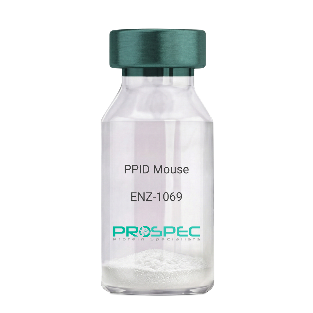
PPID MouseDescription:
Peptidylprolyl Isomerase D Mouse Recombinant
Peptidyl-prolyl cis-trans isomerase D, PPIase D, 40 kDa peptidyl-prolyl cis-trans isomerase, Cyclophilin-40, CYP-40, Cyclophilin-related protein, CYP40, CYPD, PPID, Peptidylprolyl Isomerase D.
Product # :
ENZ-1069Price :
Quantity :
Shipping Method :
Shipped with Ice Packs
More Info
- description
- source
- formulation
- purity
- biological activity
- More Info
Description
PPID Mouse Recombinant produced in E.Coli is a single, non-glycosylated polypeptide chain containing 395 amino acids (1-370a.a.) and having a molecular mass of 43.4kDa. PPID is fused to a 25 amino acid His-tag at N-terminus & purified by proprietary chromatographic techniques.
Source
Escherichia Coli.
Formulation
PPID protein solution (1mg/ml) containing 20mM Tris-Hcl buffer (pH8.0), 10% glycerol and 1mM DTT.
Purity
Greater than 90.0% as determined by SDS-PAGE.
Biological Activity
Specific activity is > 700nmol/min/mg, and is defined as the amount of enzyme that cleaves 1umole of suc-AAFP-PNA per minute at 37°C in Tris–HCl pH 8.0 using chymotrypsin.
More Info
-
Introduction
Cyclophilin-D is a member of the peptidyl-prolyl cis-trans isomerase (PPIase) family. PPIases catalyze the cis-trans isomerization of proline imidic peptide bonds in oligopeptides and speeds up the protein folding. Cyclophilin-D possess PPIase activity and binds to the immunosuppressant cyclosporin-A. Cyclophilin-D is very well known that its overexpression suppresses the apoptosis in cancer cell. Cyclophilin-D suppresses apoptotic cell death by the use of mitochondrial hexokinase-2 dependent mechanism in cancer cells.
-
Synonyms
Peptidyl-prolyl cis-trans isomerase D, PPIase D, 40 kDa peptidyl-prolyl cis-trans isomerase, Cyclophilin-40, CYP-40, Cyclophilin-related protein, CYP40, CYPD, PPID, Peptidylprolyl Isomerase D.
-
Physical Appearance
Sterile Filtered colorless solution.
-
Stability
Store at 4°C if entire vial will be used within 2-4 weeks. Store, frozen at -20°C for longer periods of time. For long term storage it is recommended to add a carrier protein (0.1% HSA or BSA). Please prevent freeze-thaw cycles.
-
Amino Acid Sequence
MGSSHHHHHH SSGLVPRGSH MGSEFMSHAS PAAKPSNSKN PRVFFDVDIG GERVGRIVLE LFADIVPKTA ENFRALCTGE KGTGSTTGKP LHFKGCPFHR IIKKFMIQGG DFSNQNGTGG ESIYGEKFED ENFHYKHDRE GLLSMANAGP NTNGSQFFIT TVPTPHLDGK HVVFGQVIKG LGVARTLENV EVNGEKPAKL CVIAECGELK EGDDWGIFPK DGSGDSHPDF PEDADIDLKD VDKILLISED LKNIGNTFFK SQNWEMAIKK YAKVLRYVDS SKAVIEKADR SRLQPIALSC VLNIGACKLK MSNWQGAIDS CLEALEMDPS NTKALYRKAQ GWQGLKEYDQ ALADLKKAQE IAPGDKAIQA ELLKVKQMIK AQKDKEKAVY AKMFA.
ProSpec's products are furnished for LABORATORY RESEARCH USE ONLY. The product may not be used as drugs, agricultural or pesticidal products, food additives or household chemicals.
- View Data Sheet
Name :
PGAM1 MouseDescription:
Phosphoglycerate Mutase 1 Mouse Recombinant
Phosphoglycerate mutase 1, BPG-dependent PGAM 1, Phosphoglycerate mutase isozyme B, PGAM-B, Pgam1, Pgam-1, 2310050F24Rik.
Product # :
ENZ-627Price :
Quantity :
Shipping Method :
Shipped with Ice Packs
More Info
- description
- source
- formulation
- purity
- More Info
Description
PGAM1 Mouse Recombinant produced in E.coli is a single, non-glycosylated polypeptide chain containing 278 amino acids (1-254) and having a molecular mass of 31.4kDa.PGAM1 is fused to a 24 amino acid His-tag at N-terminus & purified by proprietary chromatographic techniques.
Source
Escherichia Coli.
Formulation
The PGAM1 solution (1mg/ml) contains 20mM Tris-HCl buffer (pH8.0), 20% glycerol, 0.1M NaCl and 1mM DTT.
Purity
Greater than 95% as determined by SDS-PAGE.
More Info
-
Introduction
PGAM1 is part of the phosphoglycerate mutase family. PGAM1 is an essential component of glucose and 2,3-BPGA (2,3-bisphosphoglycerate) metabolism and catalyzes the reversible reaction of 3-phosphoglycerate (3-PGA) to 2-phosphoglycerate (2-PGA) in the glycolytic pathway. PGAM1 is a dimeric enzyme containing, in different tissues, different proportions of a slow-migrating muscle (MM) isozyme, a fast-migrating brain (BB) isozyme, and a hybrid form (MB). PGAM1 mutations lead to muscle phosphoglycerate mutase deficiency, a.k.a. glycogen storage disease X.
-
Synonyms
Phosphoglycerate mutase 1, BPG-dependent PGAM 1, Phosphoglycerate mutase isozyme B, PGAM-B, Pgam1, Pgam-1, 2310050F24Rik.
-
Physical Appearance
Sterile Filtered clear colorless solution.
-
Stability
Store at 4°C if entire vial will be used within 2-4 weeks. Store, frozen at -20°C for longer periods of time. For long term storage it is recommended to add a carrier protein (0.1% HSA or BSA).Avoid multiple freeze-thaw cycles.
-
Amino Acid Sequence
MGSSHHHHHH SSGLVPRGSH MGSHMAAYKL VLIRHGESAW NLENRFSGWY DADLSPAGHE EAKRGGQALR DAGYEFDICF TSVQKRAIRT LWTVLDAIDQ MWLPVVRTWR LNERHYGGLT GLNKAETAAK HGEAQVKIWR RSYDVPPPPM EPDHPFYSNI SKDRRYADLT EDQLPSCESL KDTIARALPF WNEEIVPQIK EGKRVLIAAH GNSLRGIVKH LEGLSEEAIM ELNLPTGIPI VYELDKNLKP IKPMQFLGDE ETVRKAMEAV AAQGKVKK.
ProSpec's products are furnished for LABORATORY RESEARCH USE ONLY. The product may not be used as drugs, agricultural or pesticidal products, food additives or household chemicals.
- View Data Sheet
Name :
KGF 2 RatDescription:
Keratinocyte Growth Factor-2 Rat Recombinant
FGFA, FGF10, FGF-10, KGF-2, Fibroblast growth factor 10.
Product # :
CYT-127Price :
Quantity :
Shipping Method :
Shipped at Room temp
More Info
- description
- source
- formulation
- purity
- biological activity
- More Info
- sds-page
Description
KGF 2 Rat Recombinant produced in E.Coli is a single, non-glycosylated polypeptide chain containing 179 amino acids and having a molecular mass of 20.0kDa. The KGF 2 is purified by proprietary chromatographic techniques.
Source
Escherichia Coli.
Formulation
Lyophilized from a 0.2µm filtered concentrated solution in 1×PBS, pH 7.4 containing 5% trehalose.
Purity
Greater than 97.0% as determined by:
(a) Analysis by RP-HPLC.
(b) Analysis by SDS-PAGE.Biological Activity
Fully biologically active when compared to standard. The ED50 as determined by the dose-dependent stimulation of thymidine uptake by BaF3 cells expressing FGF receptors is <0.5ng/ml.sds-page
More Info
-
Introduction
KGF-2 is a member of the fibroblast growth factor (FGF) family. FGF family members possess broad mitogenic and cell survival activities, and are involved in a variety of biological processes, including embryonic development, cell growth, morphogenesis, tissue repair, tumor growth and invasion. FGF-10 exhibits mitogenic activity for keratinizing epidermal cells, but essentially no activity for fibroblasts, which is similar to the biological activity of FGF7. Studies of the mouse homolog of suggested that this gene is required for embryonic epidermal morphogenesis including brain development, lung morphogenesis, and initiation of lim bud formation. This gene is also implicated to be a primary factor in the process of wound healing.
-
Synonyms
FGFA, FGF10, FGF-10, KGF-2, Fibroblast growth factor 10.
-
Physical Appearance
Sterile Filtered White lyophilized (freeze-dried) powder.
-
Stability
Lyophilized KGF 2 although stable at room temperature for 3 weeks, should be stored desiccated below -18°C. Upon reconstitution KGF 2 should be stored at 4°C between 2-7 days and for future use below -18°C. For long term storage it is recommended to add a carrier protein (0.1% HSA or BSA).Please prevent freeze-thaw cycles.
-
Solubility
It is recommended to reconstitute the lyophilized KGF 2 Rat Recombinant in sterile 18M-cm H2O not less than 100µg/ml, which can then be further diluted to other aqueous solutions.
-
Amino Acid Sequence
QALGQDMVSP EATNSSSSSS SSSSSSSFSS PSSAGRHVRS YNHLQGDVRW RKLFSFTKYF LKIEKNGKVS GTKKENCPYS ILEITSVEIG VVAVKAINSN YYLAMNKKGK LYGSKEFNND CKLKERIEEN GYNTYASFNW QHNGRQMYVA LNGKGAPRRG QKTRRKNTSA HFLPMVVHS
ProSpec's products are furnished for LABORATORY RESEARCH USE ONLY. The product may not be used as drugs, agricultural or pesticidal products, food additives or household chemicals.
- View Data Sheet
Name :
Dengue NS1 ST4, InsectDescription:
Dengue Virus NS1 Subtype 4 Recombinant, Insect Cells
Product # :
DEN-032Price :
Quantity :
Shipping Method :
Shipped with Ice Packs
More Info
- description
- source
- formulation
- purity
- More Info
Description
Recombinant Dengue Virus NS1 Subtype 4 produced in Insect Cells is a polypeptide chain containing amino acids 776-1130 and having a molecular weight of approximately 50kDa. Dengue NS1 ST4 is purified by proprietary chromatographic technique.
Source
Insect cells.
Formulation
Dengue NS1 ST4 protein solution in 1xD-PBS, pH7.4, 0.1% Thimerosal, 5mM EDTA, 1?g/ml of Leupeptin, Aprotinin and Pepstatin A.
Purity
Protein is >95% pure as determined by 12.5% SDS-PAGE.
More Info
-
Introduction
Caused by one of four closely related virus serotypes of the genus Flavivirus, family Flaviviridae, each serotype is sufficiently different that there is no cross-protection and epidemics caused by multiple serotypes (hyperendemicity) can occur. In cell culture experiments and mice Morpholino antisense oligos have shown specific activity against Dengue virus.
-
Physical Appearance
Sterile filtered colorless solution.
-
Stability
Store at 4°C if entire vial will be used within 2-4 weeks. Store, frozen at -20°C for longer periods of time. For long term storage it is recommended to add a carrier protein (0.1% HSA or BSA).Avoid multiple freeze-thaw cycles.
ProSpec's products are furnished for LABORATORY RESEARCH USE ONLY. The product may not be used as drugs, agricultural or pesticidal products, food additives or household chemicals.
- View Data Sheet
Name :
CoV Spike PorcineDescription:
Mouse Anti Porcine Coronavirus Spike Monoclonal
Product # :
ANT-756Price :
Quantity :
Shipping Method :
Shipped with Ice Packs
More Info
- formulation
- More Info
Formulation
1mg/ml in 1X PBS pH-7.2 & 0.01% NaN3.
More Info
-
Introduction
SARS Coronavirus is an enveloped virus containing three outer structural proteins, namely the membrane (M), envelope (E), and spike (S) proteins. Spike (S)-glycoprotein of the virus interacts with a cellular receptor and mediates membrane fusion to allow viral entry into susceptible target cells. Accordingly, S-protein plays an important role in virus infection cycle and is the primary target of neutralizing antibodies.
It has recently been shown that SARS (severe acute respiratory syndrome) is caused by a human coronavirus. Human coronaviruses are the major cause of upper respiratory tract illness, such as the common cold, in humans. Coronaviruses are positive-stranded RNA viruses, featuring the largest viral RNA genomes known to date (27-31 kb). The first step in coronavirus infection is binding of the viral spike protein, a 139-kDa protein, to certain receptors on host cells. The spike protein is the main surface antigen of the coronavirus. The glycosilated spike protein (as well as the nucleocapsid protein) can be detected in infected cell culture supernatants with antisera from SARS patients. -
Stability
Store at 4C, stable for 2 weeks. For long-term storage, store at -20C.
-
Immunogen
The antibody was developed using purified porcine coronavirus. (eptopoe not mapped)
-
Applications
This poricne Coronavirus Spike monoclonal antibody can be used in WB and ELISA
-
Isotype
IgG2a
-
Type
Mouse antibody Monoclonal.
ProSpec's products are furnished for LABORATORY RESEARCH USE ONLY. The product may not be used as drugs, agricultural or pesticidal products, food additives or household chemicals.
- View Data Sheet
Name :
PARK7 AntibodyDescription:
Parkinson Disease Protein 7, Mouse Anti Human
Protein DJ-1, Oncogene DJ1, Parkinson disease protein 7, PARK7, DJ1, DJ-1, FLJ27376.
Product # :
ANT-317Price :
Quantity :
Shipping Method :
Shipped with Ice Packs
More Info
- formulation
- More Info
Formulation
1mg/ml containing PBS, pH-7.4, 0.02% Sodium Azide and 10% Glycerol.
More Info
-
Introduction
The PARK7 is a ubiquitously expressed protein involved in various cellular processes including spermatogenesis and fertilization, cancer, RNA-binding, androgen-receptor signaling and oxidative stress. Mutations in the PARK7 are the cause of autosomal recessive early-onset Parkinson’s disease 7 (Park7).
-
Synonyms
Protein DJ-1, Oncogene DJ1, Parkinson disease protein 7, PARK7, DJ1, DJ-1, FLJ27376.
-
Immunogen
Anti-human PARK7 mAb is derived from hybridization of mouse SP2/O myeloma cells with spleen cells from BALB/c mice immunized with recombinant human PARK7 amino acids 1-189 purified from E. coli.
-
Ig Subclass
Mouse IgG1 heavy chain and κ light chain.
-
Clone
P1B11AT.
-
Applications
PARK7 antibody has been tested by ELISA and Western blot analysis to assure specificity and reactivity. Since application varies, however, each investigation should be titrated by the reagent to obtain optimal results. Recommended dilution range for Western blot analysis is 1:500 ~ 2,000. Recommended starting dilution is 1:1,000.
-
Type
Mouse Anti Human Monoclonal.
-
Storage Procedures
For periods up to 1 month store at 4°C, for longer periods of time, store at -20°C. Prevent freeze thaw cycles.
-
Purification Method
PARK7 antibody was purified from mouse ascitic fluids by protein-G affinity chromatography.
ProSpec's products are furnished for LABORATORY RESEARCH USE ONLY. The product may not be used as drugs, agricultural or pesticidal products, food additives or household chemicals.
- View Data Sheet
Name :
PARP2 AntibodyDescription:
Poly (ADP-Ribose) Polymerase 2, Mouse Anti Human
ADPRT2, ADPRTL2, ADPRTL3, ARTD2, pADPRT-2, PARP-2, Poly [ADP-ribose] polymerase 2, hPARP-2, ADP-ribosyltransferase diphtheria toxin-like 2, NAD (+) ADP-ribosyltransferase 2, Poly [ADP-ribose] synthase 2.
Product # :
ANT-491Price :
Quantity :
Shipping Method :
Shipped with Ice Packs
More Info
- formulation
- More Info
Formulation
1mg/ml containing PBS, pH-7.4, 10% Glycerol and 0.02% Sodium Azide.
More Info
-
Introduction
Poly (ADP-Ribose) Polymerase 2 (PARP2) contains a catalytic domain which is homologous to that of poly (ADP-ribosyl) transferase but lacks an N-terminal DNA binding domain which activates the C-terminal catalytic domain of poly (ADP-ribosyl) transferase. PARP2 is capable of catalyzing a poly (ADP-ribosyl)ation reaction. The basic residues within the N-terminal area of this protein can bear potential DNA-binding properties, and can be engaged in the nuclear and/or nucleolar targeting of the protein. There has been found two alternatively spliced transcript variants encoding distinct isoforms.
-
Synonyms
ADPRT2, ADPRTL2, ADPRTL3, ARTD2, pADPRT-2, PARP-2, Poly [ADP-ribose] polymerase 2, hPARP-2, ADP-ribosyltransferase diphtheria toxin-like 2, NAD (+) ADP-ribosyltransferase 2, Poly [ADP-ribose] synthase 2.
-
Physical Appearance
Sterile filtered colorless solution.
-
Immunogen
Anti-human PARP2 mAb, clone PAT29G4A, is derived from hybridization of mouse F0 myeloma cells with spleen cells from BALB/c mice immunized with a recombinant human PARP2 protein 233-583 amino acids purified from E. coli.
-
Ig Subclass
Mouse IgG2a heavy chain and Kappa light chain.
-
Clone
PAT29G4A.
-
Applications
The antibody has been tested by ELISA, Western blot analysis to assure specificity and reactivity. Since application varies, however, each investigation should be titrated by the reagent to obtain optimal results. Recommended dilution range for Western blot analysis is 1:500 ~ 1:5000. Recommended starting dilution is 1:500.
-
Type
Mouse Anti Human Monoclonal.
-
Storage Procedures
For periods up to 1 month store at 4°C, for longer periods of time, store at -20°C. Prevent freeze thaw cycles.
-
Purification Method
PARP2 antibody was purified from mouse ascitic fluids by protein-A affinity chromatography.
ProSpec's products are furnished for LABORATORY RESEARCH USE ONLY. The product may not be used as drugs, agricultural or pesticidal products, food additives or household chemicals.
- View Data Sheet
Name :
VAMP2 AntibodyDescription:
Synaptobrevin-2, Mouse Anti Human
Vesicle-associated membrane protein 2, SYB2, VAMP-2, Synaptobrevin-2, VAMP2, FLJ11460.
Product # :
ANT-334Price :
Quantity :
Shipping Method :
Shipped with Ice Packs
More Info
- formulation
- More Info
Formulation
1mg/ml containing PBS, pH-7.4, & 0.1% Sodium Azide.
More Info
-
Introduction
Synaptobrevin 2 which is an 18 kDa integral membrane protein localized to the cytoplasmic surface of synaptic vesicle, consists of a proline-rich N-terminal region, a highly conserved hydrophilic domain, followed by a transmembrane anchor and a C-terminal. Synaptobrevin 2 is predominantly expressed in Langerhans islets and glomerular cells. The N-terminal domain of the protein (residues 1-89) forms a specific SNARE complex with the target membrane-associated t- or Q-SNAREs syntaxin 1 and SNAP-25.
-
Synonyms
Vesicle-associated membrane protein 2, SYB2, VAMP-2, Synaptobrevin-2, VAMP2, FLJ11460.
-
Immunogen
Anti-human VAMP2 mAb is derived from hybridization of mouse SP2/O myeloma cells with spleen cells from BALB/c mice immunized with recombinant human VAMP2 amino acids 1-89 purified from E. coli.
-
Ig Subclass
Mouse IgG1 heavy chain and κ light chain.
-
Clone
P3E5AT.
-
Applications
VAMP2 antibody has been tested by ELISA and Western blot analysis to assure specificity and reactivity. Since application varies, however, each investigation should be titrated by the reagent to obtain optimal results. Recommended dilution range for Western blot analysis is 1:1,000 ~ 3,000. Recommended starting dilution is 1:2,000.
-
Type
Mouse Anti Human Monoclonal.
-
Storage Procedures
For periods up to 1 month store at 4°C, for longer periods of time, store at -20°C. Prevent freeze thaw cycles.
-
Purification Method
VAMP2 antibody was purified from mouse ascitic fluids by protein-G affinity chromatography.
ProSpec's products are furnished for LABORATORY RESEARCH USE ONLY. The product may not be used as drugs, agricultural or pesticidal products, food additives or household chemicals.
- View Data Sheet
Name :
CoV-2-Spike (1-260)Description:
Coronavirus 2019 Spike (1-260 a.a.), Recombinant
Product # :
SARS-022Price :
Quantity :
Shipping Method :
Shipped with Ice Packs
More Info
- description
- source
- formulation
- purity
- More Info
Description
The HEK293 derived recombinant protein contains the Coronavirus 2019-Spike N-Terminal Domain, Wuhan-Hu-1 strain, amino acids 1-260 fused to Fc tag at C-terminal.
Source
HEK293
Formulation
CoV-2 spike protein solution is supplied in DPBS.
Purity
Protein is >95% pure as determined SDS-PAGE.
More Info
-
Introduction
A human infecting coronavirus (viral pneumonia) called 2019 novel coronavirus, 2019-nCoV was found in the fish market at the city of Wuhan, Hubei province of China on December 2019.
The 2019-nCoV shares an 87% identity to the 2 bat-derived severe acute respiratory syndrome 2018 SARS-CoV-2 located in Zhoushan of eastern China. 2019-nCoV has an analogous receptor-BD-structure to that of 2018 SARS-CoV, even though there is a.a. diversity so thus the 2019-nCoV might bind to ACE2 receptor protein (angiotensin-converting enzyme 2) in humans.
While bats are possibly the host of 2019-nCoV, researchers suspect that animal from the ocean sold at the seafood market was an intermediate host. RSCU analysis proposes that the 2019-nCoV is a recombinant within the viral spike glycoprotein between the bat coronavirus and an unknown coronavirus.
-
Physical Appearance
Sterile Filtered clear solution.
-
Stability
CoV-2 Spike Protein is shipped on ice packs. Upon arrival, Store at -20°C.
-
Purification Method
Purified by Protein-G chromatographic technique.
ProSpec's products are furnished for LABORATORY RESEARCH USE ONLY. The product may not be used as drugs, agricultural or pesticidal products, food additives or household chemicals.
- View Data Sheet
Name :
p53 HumanDescription:
p53 Protein Human Recombinant
Cellular tumor antigen p53, Tumor suppressor p53, Phosphoprotein p53, Antigen NY-CO-13, TP53, P53, LFS1, TRP53, FLJ92943.
Product # :
PRO-742Price :
Quantity :
Shipping Method :
Shipped with Ice Packs
More Info
- description
- source
- formulation
- More Info
Description
p53 Human Recombinant full length produced in E.Coli is a non-glycosylated, polypeptide chain having a total Mw of 81kDa. p53 Human Recombinant is fused to GST tag and purified by proprietary chromatographic techniques.
Source
Escherichia Coli.
Formulation
Purified human p53 in 50mM Tris-HCl, pH-7.5 and 10mM L-glutathione (reduced).
More Info
-
Introduction
Tumor protein p53 responds to various cellular stresses by regulating target genes that induce cell cycle arrest, apoptosis, senescence, DNA repair, or changes in metabolism. p53 is a tumor suppressor gene expressed in a wide variety of tissue types and is involved in regulating cell growth, replication, and apoptosis. p53 is a DNA-binding protein containing transcription activation, DNA-binding & oligomerization domains.
p53 binds to mdm2, SV40 T antigen and human papilloma virus E6 protein p53 senses DNA damage and possibly facilitating repair. p53 protein is a transcription factor which is encoded in humans by the TP53 gene. Alterations of TP53 occur not only as somatic mutations in human malignancies, but also as germline mutations in some cancer-prone families with Li-Fraumeni syndrome. p53 mutants that often occur in many different human cancers fail to bind the consensus DNA binding site, and hence cause the loss of tumor suppressor activity. Mutation involving p53 is found in a wide variety of malignant tumors, including breast, ovarian, bladder, colon, lung, and melanoma. The p53 expression in normal cells is low and in an assortment of transformed cell lines is high, which may contribute to transformation and malignancy. Multiple p53 variants encode distinct isoforms, which can regulate p53 transcriptional activity. p53’s significance in multicellular organisms is in cell cycle regulation therefore it functions as a tumor suppressor that is involved in preventing cancer. p53’s role in conserving stability by preventing genome mutation has earned it descriptions such as "the guardian of the genome," "the guardian angel gene," and the "master watchman.” The name p53 refers to its evident molecular mass: it migrates as a 53kDa protein on SDS-PAGE. However, based on calculations from its amino acid residues, p53's mass is in fact only 43.7kDa. This difference is attributed to the high number of proline residues in the protein which slow its migration on SDS-PAGE, consequently making it appear larger than it actually is. -
Synonyms
Cellular tumor antigen p53, Tumor suppressor p53, Phosphoprotein p53, Antigen NY-CO-13, TP53, P53, LFS1, TRP53, FLJ92943.
-
Physical Appearance
Sterile Filtered clear solution.
-
Stability
For long term storage store at -20°C. Avoid freeze/thaw cycles.
ProSpec's products are furnished for LABORATORY RESEARCH USE ONLY. The product may not be used as drugs, agricultural or pesticidal products, food additives or household chemicals.
- View Data Sheet
Name :
GARS AntibodyDescription:
Glycyl-TRNA Synthetase, Mouse Anti Human
Glycine--tRNA ligase, Diadenosine tetraphosphate synthetase, AP-4-A synthetase, Glycyl-tRNA synthetase, GlyRS, GARS, CMT2D, DSMAV, HMN5, SMAD1.
Product # :
ANT-544Price :
Quantity :
Shipping Method :
Shipped with Ice Packs
More Info
- formulation
- More Info
Formulation
1mg/ml containing PBS, pH-7.4, 10% Glycerol and 0.02% Sodium Azide.
More Info
-
Introduction
GARS is an (alpha)2 dimer which is a member of the class II family of tRNA synthetases. GARS is a glycyl-tRNA synthetase, one of the aminoacyl-tRNA synthetases which charge tRNAs with their cognate amino acids. GARS catalyzes the attachment of glycine to tRNA(Gly). In addition, GARS is able to produce diadenosine tetraphosphate (Ap4A), which is a universal pleiotropic signaling molecule required for cell regulation pathways, by direct condensation of two ATPs. GARS has been demonstrated to be a target of autoantibodies in the human autoimmune diseases, polymyositis or dermatomyositis.
-
Synonyms
Glycine--tRNA ligase, Diadenosine tetraphosphate synthetase, AP-4-A synthetase, Glycyl-tRNA synthetase, GlyRS, GARS, CMT2D, DSMAV, HMN5, SMAD1.
-
Physical Appearance
Sterile filtered colorless solution.
-
Immunogen
Anti-human GARS mAb, is derived from hybridization of mouse F0 myeloma cells with spleen cells from BALB/c mice immunized with recombinant human GARS amino acids 43-289 purified from E. coli.
-
Ig Subclass
Mouse IgG1 heavy chain and k light chain.
-
Clone
PAT4E10AT.
-
Applications
GARS antibody has been tested by ELISA, Western blot analysis to assure specificity and reactivity. Since application varies, however, each investigation should be titrated by the reagent to obtain optimal results. Recommended starting dilution is 1:500.
-
Type
Mouse Anti Human Monoclonal.
-
Storage Procedures
For periods up to 1 month store at 4°C, for longer periods of time, store at -20°C. Prevent freeze thaw cycles.
-
Purification Method
GARS antibody was purified from mouse ascitic fluids by protein-A affinity chromatography.
ProSpec's products are furnished for LABORATORY RESEARCH USE ONLY. The product may not be used as drugs, agricultural or pesticidal products, food additives or household chemicals.
- View Data Sheet
Name :
Rabbit SARS CoV-2 IgG S1Description:
Recombinant Anti Rabbit SARS CoV-2 IgG Spike S1
Product # :
ANT-762Price :
Quantity :
Shipping Method :
Shipped with Ice Packs
More Info
- description
- source
- formulation
- More Info
Description
Recombinant Anti Rabbit SARS CoV-2 IgG Kappa Spike S1 is a recombinant monoclonal antibody derived from HEK293 cells which recognizes the SARS-CoV and SARS-CoV-2 Spike glycoprotein. CoV-2 IgG S1 Rabbit Antibody binds to both SARS-CoV and SARS-CoV-2 with high affinity at amino acids 318-510 (RBD, Receptor Binding Domain) in the S1 subunit of the Spike protein. This chimeric rabbit antibody was made using the variable domain sequences of the original Human IgG1 format.
Source
HEK293 Cells.
Formulation
1mg/ml in PBS and 0.02% Proclin 300.
More Info
-
Introduction
SARS Coronavirus is an enveloped virus containing three outer structural proteins, namely the membrane (M), envelope (E), and spike (S) proteins. Spike (S)-glycoprotein of the virus interacts with a cellular receptor and mediates membrane fusion to allow viral entry into susceptible target cells. Accordingly, S-protein plays an important role in virus infection cycle and is the primary target of neutralizing antibodies.It has recently been shown that SARS is caused by a human coronavirus. Human coronaviruses are the major cause of upper respiratory tract illness in humans, such as the common cold. Coronaviruses are positive-stranded RNA viruses, featuring the largest viral RNA genomes known to date (27-31 kb). The first step in coronavirus infection is binding of the viral spike protein, a 139-kDa protein, to certain receptors on host cells. The spike protein is the main surface antigen of the coronavirus. The most prominent protein in the culture supernatants infected with SARS virus is a 46 kDa nucleocapsid protein. This suggests that the nucleocapsid protein is a major immunogen that may be useful for early diagnostics.
-
Physical Appearance
Sterile Filtered liquid formulation.
-
Stability
Store at 4°C, stable for 2 weeks. For long-term storage, store at -20°C.
-
Immunogen
The native monoclonal antibody was generated by sequencing peripheral blood lymphocytes of a patient exposed to the SARS-CoV
-
Applications
ELISA.
-
Type
Mouse antibody Monoclonal.
-
Purification Method
Protein A affinity purified.
ProSpec's products are furnished for LABORATORY RESEARCH USE ONLY. The product may not be used as drugs, agricultural or pesticidal products, food additives or household chemicals.
- View Data Sheet
Name :
Dengue 1 NS1 AntibodyDescription:
Polyclonal Rabbit Anti Dengue 1 NS1
Product # :
ANT-083Price :
Quantity :
Shipping Method :
Shipped with Ice Packs
More Info
- description
- source
- formulation
- purity
- More Info
Description
Polyclonal antibody to dengue-1 NS1 IgG was derived from the rabbit immunized with full length recombinant dengue NS1, reactive to all dengue serotype NS1.
Source
Rabbit.
Formulation
0.2M glycine pH 7.5 and 0.02% NaN3.
Purity
Protein is >95% pure as determined by 12% PAGE (coomassie staining). Purified by protein A chromatography.
More Info
-
Stability
Should be stored below -18°C. Please prevent freeze thaw cycles.
-
Applications
Immunoassay.
-
Isotype
Purified IgG.
ProSpec's products are furnished for LABORATORY RESEARCH USE ONLY. The product may not be used as drugs, agricultural or pesticidal products, food additives or household chemicals.
- View Data Sheet
Name :
HCV Mosaic-BDescription:
Hepatitis C Virus Mosaic Antigen-B Recombinant
Product # :
HCV-002Price :
Quantity :
Shipping Method :
Shipped with Ice Packs
More Info
- description
- source
- formulation
- purity
- More Info
Description
HCV Mosaic-B protein contains a long core peptide residues 1-120a.a., 1192-1415a.a. of NS3, three epitopes from NS4 and two epitopes from NS5 and the genotype of all sequences are 1b. A specific peptide was identified from each region; the four peptides from each region were linked together and expressed in E .coli. The recombinant protein migrates at 65kDa.
Source
Escherichia Coli.
Formulation
HCV Mosaic-B protein solution containing 50mM arginine in PBS.
Purity
Protein is >95% pure as determined by 12% PAGE (coomassie staining).
More Info
-
Introduction
Hepatitis C is one type of viral hepatitis - a liver disease - caused by the hepatitis C virus (HCV) which is usually passed through contact with infected blood but can also pass through sex with an infected person and from mother to baby during childbirth.
HCV core protein, NS3, NS4, and NS5 proteins of hepatitis C virus are all essential for detection of antibodies against HCV. -
Physical Appearance
Sterile Filtered clear solution.
-
Stability
Store at 4°C if entire vial will be used within 2-4 weeks. Store, frozen at -20°C for longer periods of time. For long term storage it is recommended to add a carrier protein (0.1% HSA or BSA).Avoid multiple freeze-thaw cycles.
-
Applications
ELISA, gold conjugation.
ProSpec's products are furnished for LABORATORY RESEARCH USE ONLY. The product may not be used as drugs, agricultural or pesticidal products, food additives or household chemicals.
- View Data Sheet
Name :
Omp PyloriDescription:
Helicobacter Pylori Outer Membrane Protein Recombinant
Product # :
PRO-369Price :
Quantity :
Shipping Method :
Shipped with Ice Packs
More Info
- description
- source
- formulation
- purity
- More Info
Description
Omp Pylori recombinant antigen is produced in E. coli expressing the H. pylori outer membrane protein having the Mw of 23 kDa. Omp Pylori recombinant antigen is well recognized by specific IgG and IgM from H. pylori infected patients.
Source
E.Coli
Formulation
The Omp Pylori recombinant protein is formulated in 1xPBS pH 7.4.
Purity
Greater than 95% pure as determined by 12% PAGE (Coomassie staining).
More Info
-
Introduction
Helicobacter pylori, a Gram-negative, microaerophilic bacterium that populates in the stomach, predominantly at the antrum. Helicobacter pylori causes a chronic inflammation of the mucoid lining of stomach and is highly related to the growth of duodenal and gastric ulcers and stomach cancer. Over 50% of the world's population harbor H. pylori in their upper gastrointestinal tract, infection is more prevalent in developing countries. Thus far, no ideal target H. pylori antigen has been developed for the diagnostic purpose. 23 kDa H. pylori outer membrane protein was identified with a good sensitivity and coverage in the diagnosis of H. pylori infection.
-
Physical Appearance
Sterile filtered liquid formulation.
-
Stability
Omp Pylori although stable at 4°C for 1 week, should be stored below -18°C. Please prevent freeze thaw cycles.
-
Amino Acid Sequence
MLVTKLAPDFKAPAVLGNNEVDEHFELSKNLGKNGAILFFWP
KDFTFVCPTEIIAFDKRVKDFQEKGFNVIGVSIDSEQVHFAWK
NTPVEKGGIGQVTFPMVADITKSISRDYDVLFEEAIALRGAFLI
DKNMKVRHAVINDLPLGRNADEMLRMVDALLHFEEHGEVCP
AGWRKGDKGMKATHQGVAEYLKENSIKL. -
Purification Method
Purified by proprietary chromatographic technique.
ProSpec's products are furnished for LABORATORY RESEARCH USE ONLY. The product may not be used as drugs, agricultural or pesticidal products, food additives or household chemicals.
- View Data Sheet
Name :
SARS MERSDescription:
SARS MERS Spike S1 Recombinant
Product # :
SARS-002Price :
Quantity :
Shipping Method :
Shipped with Ice Packs
More Info
- description
- source
- formulation
- purity
- More Info
Description
Recombinant SARS MERS Spike S1 is a peptide from amino acids 367-606 of spike protein S1 produced in E. coli and fused to a 6xHis tag at its C-terminus (UniProt accession #AHC74088).SARS MERS is purified by a proprietary chromatographic technique.
Source
Escherichia Coli.
Formulation
SARS MERS protein solution is supplied in PBS, 25mM arginine and 0.05% sodium azide.
Purity
Protein is >95% pure as determined by 10% PAGE (coomassie staining).
More Info
-
Introduction
Since April 2012, cases of the Middle East Respiratory Syndrome Coronavirus (MERS-CoV) have been identified in the following countries: Saudi Arabia, Qatar, Jordan, the United Arab Emirates, Oman, Kuwait, Yemen, Lebanon, Iran, Algeria, the United Kingdom, France, Italy, Greece, Germany, the Netherlands, Austria, Tunisia, Egypt, Malaysia, Turkey and the United States of America. Coronaviruses are the cause of the common cold, SARS (severe acute respiratory syndrome) and other severe illnesses with high mortality rates, all are classified into coronavirus family. MERS-CoV is a new type of SARS found in the coronavirus family causing severe pneumonia with sudden and serious respiratory illness with high mortality rates as well. Since January 27th 2015, the WHO has reported 956 human cases, including 351 deaths. More cases of the new coronavirus strain are expected. Like in other coronaviruses, large surface spike glycoprotein is a central structural protein of this virus; it is located above the virion surface to bind and enter into the target cell. Spike protein has 2 domains- S1 and S2. The S1 domain is responsible for cellular tropism and interaction with target cell, while the S2 domain is responsible for membrane fusion. The C-terminal of S1 domain contains a receptor binding domain, and is also a potential target for vaccine development and an antigen for diagnosis.
-
Physical Appearance
Sterile filtered clear solution.
-
Stability
Store at 4°C if entire vial will be used within 2-4 weeks. Store, frozen at -20°C for longer periods of time. For long term storage it is recommended to add a carrier protein (0.1% HSA or BSA).Avoid multiple freeze-thaw cycles.
-
Amino Acid Sequence
EAKPSGSVVEQAEGVECDFSPLLSGTPPQVYNFKRLVFTNCNYNLTKLLSLFSVND
FTCSQISPAAIASNCYSSLILDYFSYPLSMKSDLSVSSAGPISQFNYKQSFSNPTC
LILATVPHNLTTITKPLKYSYINKCSRLLSDDRTEVPQLVNANQYSPCVSIVPSTV
WEDGDYYRKQLSPLEGGGWLVASGSTVAMTEQLQMGFGITVQYGTDTNSVCPKLEF
ANDTKIASQLGNCVEYHHHHHH. -
Applications
Immunoassay.
ProSpec's products are furnished for LABORATORY RESEARCH USE ONLY. The product may not be used as drugs, agricultural or pesticidal products, food additives or household chemicals.
- View Data Sheet
Name :
HTATIP2 AntibodyDescription:
HIV-1 Tat Interactive Protein 2, Mouse Anti Human
TIP30, CC3, SDR44U1, HTATIP2, EC=1.1.1.-, HIV-1 TAT-interactive protein 2, FLJ26963.
Product # :
ANT-522Price :
Quantity :
Shipping Method :
Shipped with Ice Packs
More Info
- formulation
- More Info
Formulation
1mg/ml containing PBS, pH-7.4, 10% Glycerol and 0.02% Sodium Azide.
More Info
-
Introduction
HTATIP2 is part of the short-chain dehydrogenases/reductases (SDR) family which acts as a tumor suppressor in metabolic suppression, inhibition of angiogenesis and induces the expression of apoptosis related genes Bad and Siva. HTATIP2 cooperates with the activation domain of HIV-1 TAT and enhances its transcription by phosphorylating RNA polymerase II (Pol II). Defects in HTATIP2 are related with hepatocellular carcinomas and apoptotic resistant tumor cells, implicating a probable use for HTATIP2 in antitumor therapy.
-
Synonyms
TIP30, CC3, SDR44U1, HTATIP2, EC=1.1.1.-, HIV-1 TAT-interactive protein 2, FLJ26963.
-
Physical Appearance
Sterile filtered colorless solution.
-
Immunogen
Anti-human HTATIP2 mAb, is derived from hybridization of mouse F0 myeloma cells with spleen cells from BALB/c mice immunized with recombinant human HTATIP2 amino acids 1-242 purified from E. coli.
-
Ig Subclass
Mouse IgG2a heavy chain and ? light chain.
-
Clone
PAT5D6AT.
-
Applications
HTATIP2 antibody has been tested by ELISA, Western blot analysis, Flow cytometry and ICC/IF to assure specificity and reactivity. Since application varies, however, each investigation should be titrated by the reagent to obtain optimal results.
-
Type
Mouse Anti Human Monoclonal.
-
Storage Procedures
For periods up to 1 month store at 4°C, for longer periods of time, store at -20°C. Prevent freeze thaw cycles.
-
Purification Method
HTATIP2 antibody was purified from mouse ascitic fluids by protein-A affinity chromatography.
ProSpec's products are furnished for LABORATORY RESEARCH USE ONLY. The product may not be used as drugs, agricultural or pesticidal products, food additives or household chemicals.
- View Data Sheet
Name :
NKp44 AntibodyDescription:
Natural killer cell p44-related protein, Mouse Anti Human
Natural cytotoxicity triggering receptor 2, Natural killer cell p44-related protein, NK cell-activating receptor, Lymphocyte antigen 95 homolog, NK-p44, NKp44, CD336, NCR2, LY95, dJ149M18.1.
Product # :
ANT-548Price :
Quantity :
Shipping Method :
Shipped with Ice Packs
More Info
- formulation
- More Info
Formulation
1mg/ml containing PBS, pH-7.4, 10% Glycerol and 0.02% Sodium Azide.
More Info
-
Introduction
NCR2 (NKp44) belongs to the natural cytotoxicity receptor (NCR) family which is expressed on activated human NK cells. NKp44 exhibits a single extracellular Ig-like V domain and a transmembrane portion containing the charged residue (Lysine), possibly involved in the association with KARAP/DAP12 molecules. The gene coding for NKp44 maps on human chromosome 6p21.1.
-
Synonyms
Natural cytotoxicity triggering receptor 2, Natural killer cell p44-related protein, NK cell-activating receptor, Lymphocyte antigen 95 homolog, NK-p44, NKp44, CD336, NCR2, LY95, dJ149M18.1.
-
Physical Appearance
Sterile filtered colorless solution.
-
Immunogen
Anti-human NKp44 mAb, is derived from hybridization of mouse F0 myeloma cells with spleen cells from BALB/c mice immunized with a recombinant human NKp44 protein 19-130 amino acids purified from E. coli.
-
Ig Subclass
Mouse IgG2a heavy chain and k light chain.
-
Clone
PAT1G6AT.
-
Applications
NKp44 antibody has been tested by ELISA, Western blot analysis to assure specificity and reactivity. Since application varies, however, each investigation should be titrated by the reagent to obtain optimal results. Recommended starting dilution is 1:1000.
-
Type
Mouse Anti Human Monoclonal.
-
Storage Procedures
For periods up to 1 month store at 4°C, for longer periods of time, store at -20°C. Prevent freeze thaw cycles.
-
Purification Method
NKp44 antibody was purified from mouse ascitic fluids by protein-A affinity chromatography.
ProSpec's products are furnished for LABORATORY RESEARCH USE ONLY. The product may not be used as drugs, agricultural or pesticidal products, food additives or household chemicals.
- View Data Sheet
Name :
CFLAR AntibodyDescription:
CASP8 and FADD-like apoptosis regulator, Mouse Anti Human
CASP8 and FADD-like apoptosis regulator, Cellular FLICE-like inhibitory protein, Caspase-eight-related protein, Caspase-like apoptosis regulatory protein, MACH-related inducer of toxicity, Caspase homolog, Inhibitor of FLICE, FADD-like antiapoptotic molecule 1, Usurpin, c-FLIP, Casper, CLARP, MRIT, CASH, I-FLICE, FLAME-1, CFLAR, CASP8AP1.
Product # :
ANT-433Price :
Quantity :
Shipping Method :
Shipped with Ice Packs
More Info
- formulation
- More Info
Formulation
1mg/ml containing PBS, pH-7.4, & 0.1% Sodium Azide.
More Info
-
Introduction
CFLAR contains two death effector domains (DEDs) and a caspase-like domain. CFLAR may play a critical role between cell survival and cell death pathway in mammalian cells. CFLAR also interacts with adapter protein FADD and caspase-8 and -10, and potently inhibits apoptosis induced by all known death receptors DR3 (death receptor 3), TRAIL-R (TNF-related apoptosis-inducing ligand receptor) and TNFR1 (tumor necrosis factor receptor 1).
-
Synonyms
CASP8 and FADD-like apoptosis regulator, Cellular FLICE-like inhibitory protein, Caspase-eight-related protein, Caspase-like apoptosis regulatory protein, MACH-related inducer of toxicity, Caspase homolog, Inhibitor of FLICE, FADD-like antiapoptotic molecule 1, Usurpin, c-FLIP, Casper, CLARP, MRIT, CASH, I-FLICE, FLAME-1, CFLAR, CASP8AP1.
-
Immunogen
Anti-human CFLAR mAb is derived from hybridization of mouse F0 myeloma cells with spleen cells from BALB/c mice immunized with recombinant human CFLAR amino acids 1-376 purified from E. coli.
-
Ig Subclass
Mouse IgG3 heavy chain and κ light chain.
-
Clone
P5D8AT.
-
Applications
CFLAR antibody has been tested by ELISA and Western blot analysis to assure specificity and reactivity. Since application varies, however, each investigation should be titrated by the reagent to obtain optimal results. Recommended dilution range for Western blot analysis is 1:500 ~ 1,000. Recommended starting dilution is 1:500.
-
Type
Mouse Anti Human Monoclonal.
-
Storage Procedures
For periods up to 1 month store at 4°C, for longer periods of time, store at -20°C. Prevent freeze thaw cycles.
-
Purification Method
CFLAR antibody was purified from mouse ascitic fluids by protein-G affinity chromatography.
ProSpec's products are furnished for LABORATORY RESEARCH USE ONLY. The product may not be used as drugs, agricultural or pesticidal products, food additives or household chemicals.
- View Data Sheet
Name :
Borrelia Afzelii OspADescription:
Borrelia Afzelii Outer Surface Protein A Recombinant
Product # :
BOR-011Price :
Quantity :
Shipping Method :
Shipped with Ice Packs
More Info
- description
- source
- formulation
- purity
- More Info
Description
Recombinant Borrelia Afzelii Outer Surface Protein A produced in E.coli is a non-glycosylated, polypeptide chain having a calculated molecular mass of 30kDa. Borrelia Afzelii OspA is expressed with a -6x His tag at N-terminus and purified by proprietary chromatographic techniques.
Source
Escherichia Coli.
Formulation
Borrelia Afzelii OspA is supplied in 20mM HEPES buffer pH-8.0 and 20% glycerol.
Purity
Greater than 95.0% as determined by SDS-PAGE.
More Info
-
Introduction
Borrelia belongs to a genus of bacteria of the spirochete phylum. Borrelia causes borreliosis, which is a zoonotic, vector-borne disease transmitted mainly by ticks and some by lice, depending on the species. Of the 36 known species of Borrelia, 12 are distinguished to cause Lyme disease or borreliosis and are transmitted by ticks. The main Borrelia species causing Lyme disease are Borrelia burgdorferi, Borrelia afzelii, and Borrelia garinii. The Borrelia genus members have a linear chromosome which is about 900 kbp in length as well as an excess of both linear and circular plasmids in the 5-220 kbp size range. The plasmids are atypical, as compared to most bacterial plasmids, since they contain many paralogous sequences, a large number of pseudogenes and, in some cases, essential genes. Moreover, a number of the plasmids have features suggesting that they are prophages.
-
Physical Appearance
Sterile Filtered clear solution.
-
Stability
Store at 4°C if entire vial will be used within 2-4 weeks. Store, frozen at -20°C for longer periods of time. Avoid multiple freeze-thaw cycles.
-
Immunological Functions
1. Binds IgG- and IgM-type human antibodies.2. Immunodot test with Lyme disease positive/negative plasma; suitable for LTT (lymphocyte transformation test).
ProSpec's products are furnished for LABORATORY RESEARCH USE ONLY. The product may not be used as drugs, agricultural or pesticidal products, food additives or household chemicals.
- View Data Sheet
Name :
PGAM1 Mouse, ActiveDescription:
Phosphoglycerate Mutase 1 Mouse Recombinant, Active
Phosphoglycerate mutase 1, BPG-dependent PGAM 1, Phosphoglycerate mutase isozyme B, PGAM-B, Pgam1, Pgam-1, 2310050F24Rik.
Product # :
ENZ-980Price :
Quantity :
Shipping Method :
Shipped with Ice Packs
More Info
- description
- source
- formulation
- purity
- biological activity
- More Info
Description
PGAM1 Mouse Recombinant produced in E.coli is a single, non-glycosylated polypeptide chain containing 278 amino acids (1-254) and having a molecular mass of 31.4kDa.PGAM1 is fused to a 24 amino acid His-tag at N-terminus & purified by proprietary chromatographic techniques.
Source
Escherichia Coli.
Formulation
The PGAM1 solution (1mg/ml) contains 20mM Tris-HCl buffer (pH8.0), 20% glycerol, 0.1M NaCl and 1mM DTT.
Purity
Greater than 95% as determined by SDS-PAGE.
Biological Activity
Specific activity is >150units/mg, in which One unit will convert 1.0 umole of 3-phosphoglycerate to 2-phosphoglcerate per minute at pH 7.6 at 37C.More Info
-
Introduction
PGAM1 is part of the phosphoglycerate mutase family. PGAM1 is an essential component of glucose and 2,3-BPGA (2,3-bisphosphoglycerate) metabolism and catalyzes the reversible reaction of 3-phosphoglycerate (3-PGA) to 2-phosphoglycerate (2-PGA) in the glycolytic pathway. PGAM1 is a dimeric enzyme containing, in different tissues, different proportions of a slow-migrating muscle (MM) isozyme, a fast-migrating brain (BB) isozyme, and a hybrid form (MB). PGAM1 mutations lead to muscle phosphoglycerate mutase deficiency, a.k.a. glycogen storage disease X.
-
Synonyms
Phosphoglycerate mutase 1, BPG-dependent PGAM 1, Phosphoglycerate mutase isozyme B, PGAM-B, Pgam1, Pgam-1, 2310050F24Rik.
-
Physical Appearance
Sterile Filtered clear colorless solution.
-
Stability
Store at 4°C if entire vial will be used within 2-4 weeks. Store, frozen at -20°C for longer periods of time. For long term storage it is recommended to add a carrier protein (0.1% HSA or BSA).Avoid multiple freeze-thaw cycles.
-
Amino Acid Sequence
MGSSHHHHHH SSGLVPRGSH MGSHMAAYKL VLIRHGESAW NLENRFSGWY DADLSPAGHE EAKRGGQALR DAGYEFDICF TSVQKRAIRT LWTVLDAIDQ MWLPVVRTWR LNERHYGGLT GLNKAETAAK HGEAQVKIWR RSYDVPPPPM EPDHPFYSNI SKDRRYADLT EDQLPSCESL KDTIARALPF WNEEIVPQIK EGKRVLIAAH GNSLRGIVKH LEGLSEEAIM ELNLPTGIPI VYELDKNLKP IKPMQFLGDE ETVRKAMEAV AAQGKVKK.
ProSpec's products are furnished for LABORATORY RESEARCH USE ONLY. The product may not be used as drugs, agricultural or pesticidal products, food additives or household chemicals.