Skip to content

High-quality research proteins.

  • 1-800-805-6531

  • Mon - Sat 8.00-18.00, Sun - Closed

  • info@prospecbio.com

  • PO Box 6591 East Brunswick NJ 08816

  • Products
    • Cytokines
    • Growth factors
    • Chemokines
    • Cd antigens
    • Neurotrophins
    • Hormones
    • Enzymes
    • Viral antigens
    • Recombinant proteins
    • Natural proteins
    • Monoclonal antibodies
    • Polyclonal antibodies
    • Thermal Packaging
  • About
    • Company
    • Founder
    • Contribution
  • Customers
    • Eastern Asia
    • Eastern Europe
    • North America
    • South America
    • South East Asia
    • Southern Asia
    • Western Europe
  • Ordering
    • Ordering Method
    • Ordering - Credit Card
    • Ordering - PO
    • Shipping Fees
    • FAQs
  • Services
    • DNA Cloning
    • Protein Expression
    • Monoclonal Antibody
    • Fermentation
    • Technology License
  • Contact us
Log in

Country/region

  • USD $ | Albania
  • USD $ | Andorra
  • USD $ | Argentina
  • USD $ | Armenia
  • USD $ | Australia
  • USD $ | Austria
  • USD $ | Azerbaijan
  • USD $ | Belarus
  • USD $ | Belgium
  • USD $ | Bolivia
  • USD $ | Brazil
  • USD $ | Bulgaria
  • USD $ | Cambodia
  • USD $ | Canada
  • USD $ | Chile
  • USD $ | China
  • USD $ | Colombia
  • USD $ | Costa Rica
  • USD $ | Croatia
  • USD $ | Cyprus
  • USD $ | Czechia
  • USD $ | Denmark
  • USD $ | Estonia
  • USD $ | Finland
  • USD $ | France
  • USD $ | Georgia
  • USD $ | Germany
  • USD $ | Greece
  • USD $ | Hong Kong SAR
  • USD $ | Hungary
  • USD $ | Iceland
  • USD $ | India
  • USD $ | Ireland
  • USD $ | Israel
  • USD $ | Italy
  • USD $ | Japan
  • USD $ | Kazakhstan
  • USD $ | Latvia
  • USD $ | Liechtenstein
  • USD $ | Lithuania
  • USD $ | Madagascar
  • USD $ | Malta
  • USD $ | Mexico
  • USD $ | Moldova
  • USD $ | Monaco
  • USD $ | Netherlands
  • USD $ | New Zealand
  • USD $ | North Macedonia
  • USD $ | Norway
  • USD $ | Panama
  • USD $ | Paraguay
  • USD $ | Peru
  • USD $ | Philippines
  • USD $ | Poland
  • USD $ | Portugal
  • USD $ | Romania
  • USD $ | Singapore
  • USD $ | Slovakia
  • USD $ | Slovenia
  • USD $ | South Africa
  • USD $ | South Korea
  • USD $ | Spain
  • USD $ | Sri Lanka
  • USD $ | Sweden
  • USD $ | Switzerland
  • USD $ | Taiwan
  • USD $ | Thailand
  • USD $ | Türkiye
  • USD $ | Ukraine
  • USD $ | United Kingdom
  • USD $ | United States
  • USD $ | Uruguay
  • USD $ | Vatican City
  • USD $ | Venezuela
  • USD $ | Vietnam
  • Facebook
  • Instagram
logoProSpec - Recombinant Protein Manufacturer for academic and R&D industry
Become a distributor
Account Log in
Wishlist
Cart Cart(0)
  • Products
    +
    • Cytokines
    • Growth factors
    • Chemokines
    • Cd antigens
    • Neurotrophins
    • Hormones
    • Enzymes
    • Viral antigens
    • Recombinant proteins
    • Natural proteins
    • Monoclonal antibodies
    • Polyclonal antibodies
    • Thermal Packaging
  • About
    +
    • Company
    • Founder
    • Contribution
  • Customers
    +
    • Eastern Asia
    • Eastern Europe
    • North America
    • South America
    • South East Asia
    • Southern Asia
    • Western Europe
  • Ordering
    +
    • Ordering Method
    • Ordering - Credit Card
    • Ordering - PO
    • Shipping Fees
    • FAQs
  • Services
    +
    • DNA Cloning
    • Protein Expression
    • Monoclonal Antibody
    • Fermentation
    • Technology License
  • Contact us

Item added to your cart

View cart
View All
  • Cytokines
  • Growth factors
  • Chemokines
  • Cd antigens
  • Neurotrophins
  • Hormones
  • Enzymes
  • Viral antigens
  • Recombinant proteins
  • Natural proteins
  • Monoclonal antibodies
  • Polyclonal antibodies
  • Thermal Packaging
  • Tumor Necrosis Factor

    Tumor Necrosis Factor

    View All
  • 4-1BB

    4-1BB

    View All
  • Adiponectin

    Adiponectin

    View All
  • AIF1

    AIF1

    View All
  • Other Cytokines

    Other Cytokines

    View All
  • AITRL

    AITRL

    View All
  • Angiopoietin

    Angiopoietin

    View All
  • Apolipoprotein

    Apolipoprotein

    View All
  • B-Cell Activating Factor

    B-Cell Activating Factor

    View All
  • Beta Defensin

    Beta Defensin

    View All
  • Bone Morphogenetic Protein

    Bone Morphogenetic Protein

    View All
  • B type Natriuretic Peptide

    B type Natriuretic Peptide

    View All
  • BST

    BST

    View All
  • Betacellulin

    Betacellulin

    View All
  • Cardiotrophin

    Cardiotrophin

    View All
  • Activin

    Activin

    View All
  • Fibroblast Growth Factor

    Fibroblast Growth Factor

    View All
  • Other Growth Factors

    Other Growth Factors

    View All
  • CSF

    CSF

    View All
  • CTGF

    CTGF

    View All
  • IGFBP

    IGFBP

    View All
  • VEGF Protein

    VEGF Protein

    View All
  • EGF

    EGF

    View All
  • Epigen

    Epigen

    View All
  • Erythropoietin

    Erythropoietin

    View All
  • Insulin-Like Growth Factor

    Insulin-Like Growth Factor

    View All
  • Growth Hormone

    Growth Hormone

    View All
  • HDGF

    HDGF

    View All
  • Hepatocyte Growth Factor

    Hepatocyte Growth Factor

    View All
  • Insulin

    Insulin

    View All
  • BCA-1/ BLC (CXCL13)

    BCA-1/ BLC (CXCL13)

    View All
  • BRAK (CXCL14)

    BRAK (CXCL14)

    View All
  • C-10 (CCL6)

    C-10 (CCL6)

    View All
  • Other Chemokines

    Other Chemokines

    View All
  • MEC (CCL28)

    MEC (CCL28)

    View All
  • LD78-beta (CCL3L1)

    LD78-beta (CCL3L1)

    View All
  • CTACK (CCL27)

    CTACK (CCL27)

    View All
  • CXCL16

    CXCL16

    View All
  • CXCL17

    CXCL17

    View All
  • Platelet Factor-4 (CXCL4)

    Platelet Factor-4 (CXCL4)

    View All
  • ENA-78 (CXCL5)

    ENA-78 (CXCL5)

    View All
  • Eotaxin (CCL11,24,26)

    Eotaxin (CCL11,24,26)

    View All
  • Exodus-2 (CCL21)

    Exodus-2 (CCL21)

    View All
  • Fractalkine (CX3CL1)

    Fractalkine (CX3CL1)

    View All
  • CXCL6

    CXCL6

    View All
  • Other CD Antigens

    Other CD Antigens

    View All
  • CD14

    CD14

    View All
  • CD164

    CD164

    View All
  • CD1

    CD1

    View All
  • CD2

    CD2

    View All
  • CD200

    CD200

    View All
  • CD207

    CD207

    View All
  • CD226

    CD226

    View All
  • CD23

    CD23

    View All
  • CD244

    CD244

    View All
  • CD247

    CD247

    View All
  • CD27

    CD27

    View All
  • CD274

    CD274

    View All
  • CD300

    CD300

    View All
  • CD33

    CD33

    View All
  • Other Neurotrophins

    Other Neurotrophins

    View All
  • Beta-NGF

    Beta-NGF

    View All
  • BDNF

    BDNF

    View All
  • CDNF

    CDNF

    View All
  • CNTF

    CNTF

    View All
  • GDNF

    GDNF

    View All
  • Glia Maturation Factor

    Glia Maturation Factor

    View All
  • MANF

    MANF

    View All
  • Midkine

    Midkine

    View All
  • Neuroglobin

    Neuroglobin

    View All
  • Neurotrophic factors

    Neurotrophic factors

    View All
  • Neuregulin

    Neuregulin

    View All
  • Neuropilin

    Neuropilin

    View All
  • Pigment Epithelium-Derived Factor

    Pigment Epithelium-Derived Factor

    View All
  • Pleiotrophin

    Pleiotrophin

    View All
  • Peptide Hormones

    Peptide Hormones

    View All
  • Other Hormones

    Other Hormones

    View All
  • HCG

    HCG

    View All
  • Endothelin

    Endothelin

    View All
  • Exendin

    Exendin

    View All
  • FSH

    FSH

    View All
  • Glucagon

    Glucagon

    View All
  • GHRP

    GHRP

    View All
  • GLP

    GLP

    View All
  • Thyrostimulin

    Thyrostimulin

    View All
  • Inhibin A

    Inhibin A

    View All
  • LHRH

    LHRH

    View All
  • Melanotan

    Melanotan

    View All
  • Procalcitonin

    Procalcitonin

    View All
  • PTH

    PTH

    View All
  • Peroxiredoxin

    Peroxiredoxin

    View All
  • Peptidase

    Peptidase

    View All
  • Synthetase

    Synthetase

    View All
  • Transferase

    Transferase

    View All
  • Hydrolase

    Hydrolase

    View All
  • Dehydrogenase

    Dehydrogenase

    View All
  • ACE2

    ACE2

    View All
  • Esterase

    Esterase

    View All
  • Protein Kinases

    Protein Kinases

    View All
  • Other Enzymes

    Other Enzymes

    View All
  • Phosphatase

    Phosphatase

    View All
  • Deaminase

    Deaminase

    View All
  • Oxygenase

    Oxygenase

    View All
  • Adenylate Kinase

    Adenylate Kinase

    View All
  • Lyase

    Lyase

    View All
  • Chagas

    Chagas

    View All
  • SARS

    SARS

    View All
  • Influenza

    Influenza

    View All
  • ASFV

    ASFV

    View All
  • Astrovirus

    Astrovirus

    View All
  • Borrelia

    Borrelia

    View All
  • Helicobacter Pylori

    Helicobacter Pylori

    View All
  • Chikungunya

    Chikungunya

    View All
  • Chlamydia

    Chlamydia

    View All
  • Cytomegalo

    Cytomegalo

    View All
  • Dengue

    Dengue

    View All
  • EBV

    EBV

    View All
  • Ebola

    Ebola

    View All
  • Feline Leukemia Virus

    Feline Leukemia Virus

    View All
  • Hepatitis A

    Hepatitis A

    View All
  • Other

    Other

    View All
  • Actin

    Actin

    View All
  • ADAM

    ADAM

    View All
  • Complement Component

    Complement Component

    View All
  • Ag85

    Ag85

    View All
  • Eukaryotic Translation Initiation Factor

    Eukaryotic Translation Initiation Factor

    View All
  • Anterior Gradient Protein

    Anterior Gradient Protein

    View All
  • Heat Shock Protein

    Heat Shock Protein

    View All
  • Alpha-2-HS-Glycoprotein

    Alpha-2-HS-Glycoprotein

    View All
  • Hemoglobin

    Hemoglobin

    View All
  • Allergy

    Allergy

    View All
  • Anaplasma

    Anaplasma

    View All
  • Angiogenin

    Angiogenin

    View All
  • Ankyrin Repeat Domain

    Ankyrin Repeat Domain

    View All
  • Annexin

    Annexin

    View All
  • Other Natural Proteins

    Other Natural Proteins

    View All
  • Aprotinin

    Aprotinin

    View All
  • Transferrin

    Transferrin

    View All
  • Avidin

    Avidin

    View All
  • Anti Coagulation Factors

    Anti Coagulation Factors

    View All
  • Natural Albumin

    Natural Albumin

    View All
  • Natural Coagulation Factors

    Natural Coagulation Factors

    View All
  • Fibronectin

    Fibronectin

    View All
  • Thrombin

    Thrombin

    View All
  • Anti Human Enzyme

    Anti Human Enzyme

    View All
  • Other Monoclonal Antibodies

    Other Monoclonal Antibodies

    View All
  • Anti Human Cytokine

    Anti Human Cytokine

    View All
  • Anti Human Heat Shock Protein

    Anti Human Heat Shock Protein

    View All
  • Anti Mouse Lymphocyte

    Anti Mouse Lymphocyte

    View All
  • Anti Human Chemokine

    Anti Human Chemokine

    View All
  • Anti Human Lymphocyte

    Anti Human Lymphocyte

    View All
  • Anti Viral Monoclonal

    Anti Viral Monoclonal

    View All
  • Anti Mouse Cytokine

    Anti Mouse Cytokine

    View All
  • Anti-GST Monoclonal

    Anti-GST Monoclonal

    View All
  • Other Polyclonal Antibodies

    Other Polyclonal Antibodies

    View All
  • Anti Viral

    Anti Viral

    View All
  • Anti-GST Monoclonal

    Anti-GST Monoclonal

    View All
Back

Search results

1000 results found for “Haptoglobin”

Name

Description

Product #

Price

Quantity

Shipping Method

  • View Data Sheet

    Name :

    IL 22 Mouse, PEG

    Description:

    Interleukin-22 Mouse Recombinant, Pegylated

    IL-TIF, TIFa, IL-10-related T-cell-derived-inducible factor, IL-22, ILTIF, IL-D110, zcyto18, MGC79382, MGC79384, TIFIL-23.

    Product # :

    CYT-701

    Price :

    Quantity :

    Shipping Method :

    Room Temp Icon

    Shipped at Room temp

    Add To Cart

    More Info

    • description
    • source
    • formulation
    • purity
    • biological activity
    • More Info

    Description

    Pegylated Interleukin-22 Mouse Recombinant produced in E.Coli is a single, non-glycosylated homodimeric polypeptide chain containing 147 amino acids and an aditional Ala amino acid at N-terminus having a molecular mass of 36 kDa as determioned by mass spectometry. However due to enlarged hydrodymanic volume it runs on the SDS-PAGE as a 50 kDa protein and in gel-filtration on Superdex 200 as over 200 kDa protein. The Murine IL-22 is Mono-pegylated (with 20 kDa PEG) purified by proprietary chromatographic techniques.

    Source

    Escherichia Coli.

    Formulation

    Lyophilized from a concentrated solution at 0.65mg/ml containing 0.003mM NaHCO3.

    Purity

    Greater than 98.0% as determined by:
    (a) Analysis by Gel-Filtration.
    (b) Analysis by SDS-PAGE.

    Biological Activity

    The ED50 as determined by STAT3 phosphorylation assay in HepG cells. The activity in vitro was found to be ~ 10% compared to the non-pegylated mouse IL22.

    More Info

    • Introduction

      Interleukin-22 (IL-22), also known as IL-10-related T cell-derived inducible factor (IL-TIF) was initially identified as a gene induced by IL-9 in mouse T cells and mast cells. Mouse IL-22 cDNA encodes a 179 amino acid residue protein with a putative 33 amino acids signal peptide that is cleaved to generate a 147 aa mature protein that shares approximately 79% and 22% aa sequence identity with human IL-22 and IL-10, respectively. IL-22 has been shown to activate STAT-1 and STAT-3 in several hepatoma cell lines and upregulate the production of acute phase proteins. IL-22 is produced by normal mouse T cells upon Con A activation. Mouse IL-22 expression is also induced in various organs upon lipopolysaccharide injection, suggesting that IL-22 may be involved in inflammatory responses. The functional IL-22 receptor complex consists of two receptor subunits, IL-22R (previously an orphan receptor named CRF2-9) and IL-10Rβ (previously known as CRF2-4), belonging to the class II cytokine recep

    • Synonyms

      IL-TIF, TIFa, IL-10-related T-cell-derived-inducible factor, IL-22, ILTIF, IL-D110, zcyto18, MGC79382, MGC79384, TIFIL-23.

    • Physical Appearance

      Sterile Filtered White lyophilized (freeze-dried) powder.

    • Stability

      Lyophilized pegylated murine IL22 although stable at room temperature for several days, should be stored desiccated below -20°C. Upon reconstitution at 0.1mg/ml pegylated mouse IL22 and up to 2mg/ml, filter and sterilized, the protein can be stored at 4 degrees Celsius for several weeks making it suitable for long term infusion studies using osmotic pumps. At lower concentration addition of a carrier protein (0.1% HSA or BSA) is suggested. Please prevent freeze-thaw cycles.

    • Solubility

      It is recommended to reconstitute the lyophilized pegylated mouse Interleukin -22 in sterile 0.4% NaHCO3 adjusted to pH 8-9 not less than 100µg/ml, which can then be further diluted to other aqueous solutions.

    ProSpec's products are furnished for LABORATORY RESEARCH USE ONLY. The product may not be used as drugs, agricultural or pesticidal products, food additives or household chemicals.

    Il 22 Mouse Pegylated
  • View Data Sheet

    Name :

    LGALS4 Human

    Description:

    Galectin-4 Human Recombinant

    Galectin-4, Gal-4, Lactose-binding lectin 4, L-36 lactose-binding protein, L36LBP, Antigen NY-CO-27, LGALS4, lectin galactoside-binding soluble 4, GAL4.

    Product # :

    CYT-686

    Price :

    Quantity :

    Shipping Method :

    Ice Icon

    Shipped with Ice Packs

    Add To Cart

    More Info

    • description
    • source
    • formulation
    • purity
    • More Info
    • sds-page

    Description

    Galectin-4 produced in E.Coli is a single, non-glycosylated polypeptide chain containing 343 amino acids (1-323 a.a.) and having a molecular mass of 38.1kDa.Galectin-4 is fused to 20 amino acid His-Tag at N-terminus and purified by proprietary chromatographic techniques.

    Source

    Escherichia Coli.

    Formulation

    The LGALS4 protein solution contains 20mM Tris-HCl, pH-8, 1mM DTT and 10% glycerol.

    Purity

    Greater than 95.0% as determined by SDS-PAGE.

    sds-page

    LGALS4 Human-SDS-PAGE - Product image 1

    More Info

    • Introduction

      Galectin-4 is a member of the subfamily of galectins composed of two carbohydrate recognition domains having similar peptide chains. The galectins are a family of beta-galactoside-binding proteins having a role in modulating cell-cell and cell-matrix interactions, which inhibits chronic inflammations, GVHD, and allergic responses. LGALS4 expression is limited to small intestine, colon, and rectum, and it is underexpressed in colorectal cancer. LGALS4 binds as an endogenous ligand to glycosphingolipids having 3-O-sulfated Gal residues and bind as well to cholesterol-3-sulfate. LGALS4 takes part in cell adhesion. LGALS4 plays a role in crosslinking the lateral cell membranes of the surface-lining epithelial cells, thus supporting epithelial integrity against mechanical stress exerted by the bowel lume. LGALS4 is in charge of intestinal inflammation via selective regulation of peripheral and mucosal T-cell cell cycle, in addition to cell death by apoptosis of T-cells by a pathway independent of the activation of caspases. LGALS4 blockade decreases TNF-alpha inhibitor induced T-cell death. LGALS4 decreases pro-inflammatory cytokine secretion including IL-6 & IL-17.

    • Synonyms

      Galectin-4, Gal-4, Lactose-binding lectin 4, L-36 lactose-binding protein, L36LBP, Antigen NY-CO-27, LGALS4, lectin galactoside-binding soluble 4, GAL4.

    • Physical Appearance

      Sterile filtered colorless solution.

    • Stability

      Store at 4°C if entire vial will be used within 2-4 weeks. Store, frozen at -20°C for longer periods of time. For long term storage it is recommended to add a carrier protein (0.1% HSA or BSA).Avoid multiple freeze-thaw cycles.

    • Amino Acid Sequence

      MGSSHHHHHH SSGLVPRGSH MAYVPAPGYQ PTYNPTLPYY QPIPGGLNVG MSVYIQGVAS EHMKRFFVNF VVGQDPGSDV AFHFNPRFDG WDKVVFNTLQ GGKWGSEERK RSMPFKKGAA FELVFIVLAE HYKVVVNGNP FYEYGHRLPL QMVTHLQVDG DLQLQSINFI GGQPLRPQGP PMMPPYPGPG HCHQQLNSLP TMEGPPTFNP PVPYFGRLQG GLTARRTIII KGYVPPTGKS FAINFKVGSS GDIALHINPR MGNGTVVRNS LLNGSWGSEE KKITHNPFGP GQFFDLSIRC GLDRFKVYAN GQHLFDFAHR LSAFQRVDTL EIQGDVTLSY VQI.

    • Background

      What is the molecular weight/Mw of LGALS4 HUMAN Protein?
      LGALS4 HUMAN Protein has a total Mw of 38.1kDa.

      What is the source or expression system of LGALS4 HUMAN Protein?
      Escherichia Coli.

      What is the Purity of LGALS4 HUMAN Protein?
      LGALS4 HUMAN Protein is >95% pure as determined by SDS-PAGE.

      What is the Biological Activity of LGALS4 HUMAN Protein?
      The biological functionality of LGALS4 HUMAN Protein will be determined in the future.

      What is the amino acid sequence of LGALS4 HUMAN Protein?
      MGSSHHHHHH SSGLVPRGSH MAYVPAPGYQ PTYNPTLPYY QPIPGGLNVG MSVYIQGVAS EHMKRFFVNF VVGQDPGSDV AFHFNPRFDG WDKVVFNTLQ GGKWGSEERK RSMPFKKGAA FELVFIVLAE HYKVVVNGNP FYEYGHRLPL QMVTHLQVDG DLQLQSINFI GGQPLRPQGP PMMPPYPGPG HCHQQLNSLP TMEGPPTFNP PVPYFGRLQG GLTARRTIII KGYVPPTGKS FAINFKVGSS GDIALHINPR MGNGTVVRNS LLNGSWGSEE KKITHNPFGP GQFFDLSIRC GLDRFKVYAN GQHLFDFAHR LSAFQRVDTL EIQGDVTLSY VQI.

      What applications can LGALS4 HUMAN Protein be used in?
      LGALS4 HUMAN Protein can probably be used in western blot, ELISA and Lateral Flow.

      What is the endotoxin level for LGALS4 HUMAN Protein?
      The endotoxin level is minimal, LGALS4 HUMAN Protein was purified using conventional chromatography techniques.


    ProSpec's products are furnished for LABORATORY RESEARCH USE ONLY. The product may not be used as drugs, agricultural or pesticidal products, food additives or household chemicals.

    Lgals4 Human
  • View Data Sheet

    Name :

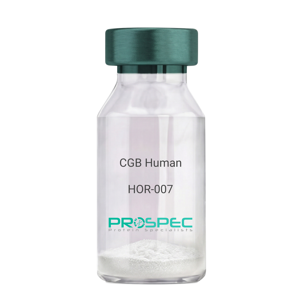
    CGB Human

    Description:

    CGB Human Recombinant

    CGB3, CGB5, CGB7, CGB8, hCGB,  CG-beta, CGB.

    Product # :

    HOR-007

    Price :

    Quantity :

    Shipping Method :

    Ice Icon

    Shipped with Ice Packs

    Add To Cart

    More Info

    • description
    • source
    • formulation
    • purity
    • More Info

    Description

    CGB Human Recombinant produced in E.Coli is a single, non-glycosylated polypeptide chain containing 168 amino acids (21-165 a.a.) and having a molecular mass of 17.9kDa. CGB is fused to a 23 amino acid His-tag at N-terminus & purified by proprietary chromatographic techniques.

    Source

    Escherichia Coli.

    Formulation

    CGB protein solution (0.5mg/ml) containing 20mM Tris-HCl buffer (pH 8.0), 0.4M Urea and 10% glycerol.

    Purity

    Greater than 85.0% as determined by SDS-PAGE.

    More Info

    • Introduction

      Glycoprotein hormones are heterodimers consisting of a common alpha subunit and a unique beta subunit which confers biological specificity. CGB is a member of the glycoprotein hormone beta chain family and is the beta 3 subunit of (CG). CG is produced by the trophoblastic cells of the placenta and stimulates the ovaries. The beta subunit of CG is encoded by 6 genes which are arranged in tandem and inverted pairs on chromosome 19q13.3 and contiguous with the luteinizing hormone beta subunit gene.

    • Synonyms

      CGB3, CGB5, CGB7, CGB8, hCGB, CG-beta, CGB.

    • Physical Appearance

      Sterile filtered colorless solution.

    • Stability

      Store at 4°C if entire vial will be used within 2-4 weeks. Store, frozen at -20°C for longer periods of time. For long term storage it is recommended to add a carrier protein (0.1% HSA or BSA).Avoid multiple freeze-thaw cycles.

    • Amino Acid Sequence

      MGSSHHHHHH SSGLVPRGSH MGSSKEPLRP RCRPINATLA VEKEGCPVCI TVNTTICAGY CPTMTRVLQG VLPALPQVVC NYRDVRFESI RLPGCPRGVN PVVSYAVALS CQCALCRRST TDCGGPKDHP LTCDDPRFQD SSSSKAPPPS LPSPSRLPGP SDTPILPQ.

    ProSpec's products are furnished for LABORATORY RESEARCH USE ONLY. The product may not be used as drugs, agricultural or pesticidal products, food additives or household chemicals.

    Cgb Human
  • View Data Sheet

    Name :

    HTLV-1 mosaic

    Description:

    HTLV-1 Mosaic Recombinant

    Product # :

    HIV-144

    Price :

    Quantity :

    Shipping Method :

    Ice Icon

    Shipped with Ice Packs

    Add To Cart

    More Info

    • description
    • formulation
    • purity
    • More Info

    Description

    The E.Coli derived recombinant mosiac protein contains the gp21 and gp46 immunodominant regions, 374-400 amino acids and 190-201 amino acids. The protein is fused with GST at N-terminus.

    Formulation

    20mM Phosphate buffer PH 7.5.

    Purity

    Protein is >95% pure as determined by SDS-PAGE.

    More Info

    • Introduction

      Human T-lymphotropic virus (HTLV) is a human, single-stranded RNA retrovirus that causes T-cell leukemia and T-cell lymphoma. The virus activates a subset of T-helper cellscalled Th1cells. The result is a proliferation of Th1 cells and overproduction of Th1 related cytokines (mainly IFN-gamma and TNF-alpha). Feedback mechanisms of these cytokines cause a suppression of the Th2 lymphocytes and a reduction of Th2 cytokine production (mainly IL-4, IL-5, IL-10 and IL-13). The end result is a reduction in the ability of the infected host to mount an adequate immune response to invading organisms that require a predominantly Th2 dependant response (these include parasitic infections and production of mucosal and humoral antibodies).

    • Stability

      HTLV-1 Mosaic although stable at 4°C for 1 week, should be stored below -18°C. Please prevent freeze thaw cycles.

    • Applications

      HTLV-1 Mosaic can be used as an antigen in ELISA and Western Blots. Excellent reagent for correct detection of HTLV infections, with minimal specificity problems.

    • Specificity

      Immunoreactive with sera of HTLV-I infected individuals

    • Purification Method

      HTLV-1 Mosaic was purified by proprietary chromatographic technique.

    ProSpec's products are furnished for LABORATORY RESEARCH USE ONLY. The product may not be used as drugs, agricultural or pesticidal products, food additives or household chemicals.

    Htlv 1 Mosaic
  • View Data Sheet

    Name :

    Omp Pylori

    Description:

    Helicobacter Pylori Outer Membrane Protein Recombinant

    Product # :

    PRO-369

    Price :

    Quantity :

    Shipping Method :

    Ice Icon

    Shipped with Ice Packs

    Add To Cart

    More Info

    • description
    • source
    • formulation
    • purity
    • More Info

    Description

    Omp Pylori recombinant antigen is produced in E. coli expressing the H. pylori outer membrane protein having the Mw of 23 kDa. Omp Pylori recombinant antigen is well recognized by specific IgG and IgM from H. pylori infected patients.

    Source

    E.Coli

    Formulation

    The Omp Pylori recombinant protein is formulated in 1xPBS pH 7.4.

    Purity

    Greater than 95% pure as determined by 12% PAGE (Coomassie staining).

    More Info

    • Introduction

      Helicobacter pylori, a Gram-negative, microaerophilic bacterium that populates in the stomach, predominantly at the antrum. Helicobacter pylori causes a chronic inflammation of the mucoid lining of stomach and is highly related to the growth of duodenal and gastric ulcers and stomach cancer. Over 50% of the world's population harbor H. pylori in their upper gastrointestinal tract, infection is more prevalent in developing countries. Thus far, no ideal target H. pylori antigen has been developed for the diagnostic purpose. 23 kDa H. pylori outer membrane protein was identified with a good sensitivity and coverage in the diagnosis of H. pylori infection.

    • Physical Appearance

      Sterile filtered liquid formulation.

    • Stability

      Omp Pylori although stable at 4°C for 1 week, should be stored below -18°C. Please prevent freeze thaw cycles.

    • Amino Acid Sequence

      MLVTKLAPDFKAPAVLGNNEVDEHFELSKNLGKNGAILFFWP
      KDFTFVCPTEIIAFDKRVKDFQEKGFNVIGVSIDSEQVHFAWK
      NTPVEKGGIGQVTFPMVADITKSISRDYDVLFEEAIALRGAFLI
      DKNMKVRHAVINDLPLGRNADEMLRMVDALLHFEEHGEVCP
      AGWRKGDKGMKATHQGVAEYLKENSIKL.

    • Purification Method

      Purified by proprietary chromatographic technique.

    ProSpec's products are furnished for LABORATORY RESEARCH USE ONLY. The product may not be used as drugs, agricultural or pesticidal products, food additives or household chemicals.

    Omp Pylori
  • View Data Sheet

    Name :

    C5a Protein

    Description:

    Complement C5a Human

    Complement Component 5, C3 and PZP-Like Alpha-2-Macroglobulin Domain-Containing Protein 4, C5a Anaphylatoxin, Prepro-C5, CPAMD4, Anaphylatoxin C5a Analog, ECLZB, C5A, C5D, C5b, C5.

    Product # :

    PRO-2692

    Price :

    Quantity :

    Shipping Method :

    Ice Icon

    Shipped with Ice Packs

    Add To Cart

    More Info

    • description
    • source
    • formulation
    • purity
    • More Info

    Description

    Human Complement C5a produced in Human plasma having a molecular mass of 10.4 kDa.

    Source

    Human Plasma.

    Formulation

    C5a protein solution contains 120 mM NaCl and 10mM HEPES, pH 7.2.

    Purity

    Greater than 95.0% as determined by SDS-PAGE.

    More Info

    • Introduction

      Complement Component C5a (C5a) is involved in the complement system and it is encoded by the C5 gene in human. Complement C5 is cleaved into C5a and C5b. C5a has an imperative role in chemotaxis and C5b forms the 1st part of the complement membrane attack complex. Complement C5 is the 5th component of complement, which plays a central role in inflammatory and cell killing processes. C5a protein is composed of alpha and beta polypeptide chains, which are linked by a disulfide bridge. An activation peptide, C5a, which is an anaphylatoxin, which has potent spasmogenic and chemotactic activity, is derivative from the alpha polypeptide via cleavage with a convertase. The C5b macromolecular cleavage product forms a complex with the C6 complement component, and this complex is the basis for creation of the membrane attack complex, which includes supplementary complement components.

    • Synonyms

      Complement Component 5, C3 and PZP-Like Alpha-2-Macroglobulin Domain-Containing Protein 4, C5a Anaphylatoxin, Prepro-C5, CPAMD4, Anaphylatoxin C5a Analog, ECLZB, C5A, C5D, C5b, C5.

    • Physical Appearance

      Sterile filtered solution.

    • Stability

      C5a Human is stable at 4°C if entire vial will be used within 2-4 weeks.Store, frozen below -20°C for longer periods of time.For long term storage it is recommended to add a carrier protein (0.1% HSA or BSA).Avoid multiple freeze-thaw cycles.

    • Human Virus Test

      Plasma from each donor has been tested and found negative for antibody to HIV-1, HIV-2, HCV, HTLV-I &II, STS and HBSAG.

    ProSpec's products are furnished for LABORATORY RESEARCH USE ONLY. The product may not be used as drugs, agricultural or pesticidal products, food additives or household chemicals.

    Complement C5A
  • View Data Sheet

    Name :

    BETV4

    Description:

    Polcalcin Bet v 4 Recombinant

    Polcalcin Bet v 4, Calcium-binding pollen allergen Bet v 4, Bet v 4, BETV4.

    Product # :

    ALR-017

    Price :

    Quantity :

    Shipping Method :

    Ice Icon

    Shipped with Ice Packs

    Add To Cart

    More Info

    • description
    • source
    • formulation
    • purity
    • More Info

    Description

    Recombinant BETV4 produced in SF9 is a glycosylated, polypeptide chain having a calculated molecular mass of 10,473 Dalton. BETV4 is expressed with a 6xHis tag at N-terminus and purified by proprietary chromatographic techniques.

    Source

    Sf9 insect cells.

    Formulation

    BETV4 is supplied in 20mM HEPES buffer pH-7.9 and 6M Urea.

    Purity

    Greater than 80.0% as determined by SDS-PAGE.

    More Info

    • Introduction

      BETV4 is primarily expressed in mature birch (Betula verrucosa) pollen and Causes an allergic reaction in human. BETV4 is a calcium-binding protein of 2-EF-hand type, which is exists in pollen of various plant species. This cross-reactivity can serve as a marker allergen for plant polysensitization.

    • Synonyms

      Polcalcin Bet v 4, Calcium-binding pollen allergen Bet v 4, Bet v 4, BETV4.

    • Physical Appearance

      Sterile Filtered clear solution.

    • Stability

      Store at 4°C if entire vial will be used within 2-4 weeks. Store, frozen at -20°C for longer periods of time. Avoid multiple freeze-thaw cycles.

    • Immunological Functions

      1. Binds IgE type human antibodies. 2. Immunodot test with positive/negative sera panels.

    ProSpec's products are furnished for LABORATORY RESEARCH USE ONLY. The product may not be used as drugs, agricultural or pesticidal products, food additives or household chemicals.

    Betv4
  • View Data Sheet

    Name :

    Der P1

    Description:

    Der P1 Protein Recombinant

    Peptidase 1, Major mite fecal allergen Der p 1, Allergen Der p I, Der p 1, DERP1, Der-P1.

    Product # :

    ALR-003

    Price :

    Quantity :

    Shipping Method :

    Ice Icon

    Shipped with Ice Packs

    Add To Cart

    More Info

    • description
    • source
    • formulation
    • purity
    • More Info

    Description

    The E.Coli derived recombinant protein contains the Dermatophagoides pteronyssinus Dust Mite Der P1 protein (a.a. 20-320) and fused to a 6 His Tag at C-terminus, having a total Mw of 34.5kDa, pI 5.6.

    Source

    Escherichia Coli.

    Formulation

    60mM NaCl and 50mM Tris-HCl pH 8.0.

    Purity

    Protein is >95% pure as determined by 10% SDS-PAGE (coomassie staining).

    More Info

    • Introduction

      DERP1 is a thiol protease, with a preference for substrates with a large hydrophobic side chain in the P2 position, or with basic residues. DERP1 is a C1 peptidase family member. DERP1 has extensive endopeptidase specificity. DERP1 is N-glycosylated. N-glycanase treatment does not completely remove carbohydrates, suggesting that the protein contains additional glycosylation sites. DERP1 causes an allergic reaction in humans. Common symptoms of mite allergy are bronchial asthma, allergic rhinitis and conjunctivitis. DERP1 binds to IgE in 80% of patients with house dust allergy.

    • Synonyms

      Peptidase 1, Major mite fecal allergen Der p 1, Allergen Der p I, Der p 1, DERP1, Der-P1.

    • Physical Appearance

      Sterile filtered colorless solution.

    • Stability

      Der-P1 although stable at 4°C for 1 week, should be stored below -18°C. Please prevent freeze thaw cycles.

    • Amino Acid Sequence

      MSIKTFEEYKKAFNKSYATFEDEEAARKNFLESVKYVQSNGGAINHLSDLSLDEFKNRFLMSAEAFEHLKTQFDLNAETNACSINGNAPAEIDLRQMRTVTPIRMQGGCGSAWAFSGVAATESAYLAYRNQSLDLAEQELVDCASQHGCHGDTIPRGIEYIQHNGVVQESYYRYVAREQSCRRPNAQRFGISNYCQIYPPNVNKIREALAQTHSAIAVIIGIKDLDAFRHYDGRTIIQRDNGYQPNYHAVNIVGYSNAQGVDYWIVRNSWDTNWGDNGYGYFAANIDLMMIEEYPYVVILHHHHHH.

    • Purification Method

      Purified by proprietary chromatographic technique.

    ProSpec's products are furnished for LABORATORY RESEARCH USE ONLY. The product may not be used as drugs, agricultural or pesticidal products, food additives or household chemicals.

    Der P1
  • View Data Sheet

    Name :

    HSP27 Antibody

    Description:

    Heat Shock Protein 27 , Mouse Anti Human

    HSPB1, CMT2F, HMN2B, HSP27, HSP28, HSP25, Heat shock protein beta-1, Heat shock 27 kDa protein, Stress-responsive protein 27, SRP27, HS.76067, DKFZp586P1322.

    Product # :

    ANT-307

    Price :

    Quantity :

    Shipping Method :

    Ice Icon

    Shipped with Ice Packs

    Add To Cart

    More Info

    • formulation
    • More Info

    Formulation

    1mg/ml containing PBS, pH-7.4, & 0.1% Sodium Azide.

    More Info

    • Introduction

      HSP27 (also known as the Estrogen-Regulated 24K protein, and hsp 28) is a member of the mammalian small heat shock protein family. Hsp27 is expressed constitutively in many tissues and its expression is increased to high levels after various types of stress including elevated temperatures, toxic metals, drugs and oxidants. Also, Hsp27 is phosphorylated in vivo on three phosphorylation sites (Ser15, Ser78 and Ser82) by protein kinases including MAPKAP kinase 2 and the stress-activated protein kinase SAPK2 (p38).

    • Synonyms

      HSPB1, CMT2F, HMN2B, HSP27, HSP28, HSP25, Heat shock protein beta-1, Heat shock 27 kDa protein, Stress-responsive protein 27, SRP27, HS.76067, DKFZp586P1322.

    • Physical Appearance

      Sterile Filtered colorless solution.

    • Immunogen

      Anti-human HSP27 mAb, is derived from hybridization of mouse SP2/0 myeloma cells with spleen cells from BALB/c mice immunized with recombinant human HSP27 amino acids 1-205 purified from E. coli.

    • Ig Subclass

      Mouse IgG1 heavy chain and κ light chain.

    • Clone

      P2A5AT.

    • Applications

      HSP27 antibody has been tested by ELISA, Western blot and immunohistochemistry analysis to assure specificity and reactivity. Since application varies, however, each investigation should be titrated by the reagent to obtain optimal results. Recommended dilution range for Western blot is 1:1,000~1:2,000 and immunohistochemistry analysis is 1:50~100. Recommended starting dilution for Western blot is 1:1,000 and Immunohistochemistry is 1:50.

    • Type

      Mouse Anti Human Monoclonal.

    • Storage Procedures

      For periods up to 1 month store at 4°C, for longer periods of time, store at -20°C. Prevent freeze thaw cycles.

    • Purification Method

      HSP27 antibody was purified from mouse ascitic fluids by protein-G affinity chromatography.

    ProSpec's products are furnished for LABORATORY RESEARCH USE ONLY. The product may not be used as drugs, agricultural or pesticidal products, food additives or household chemicals.

    Hsp27 Antibody
  • View Data Sheet

    Name :

    HSP90 Alpha Antibody

    Description:

    Heat shock protein HSP 90-alpha, Mouse Anti Human

    HSPN, LAP2, HSP86, HSPC1, HSPCA, Hsp89, Hsp90, HSP90A, HSP90N, HSPCAL1, HSPCAL4, FLJ31884, Heat shock protein HSP 90-alpha, Renal carcinoma antigen NY-REN-38, HSP 86, HSP90AA1.

    Product # :

    ANT-398

    Price :

    Quantity :

    Shipping Method :

    Ice Icon

    Shipped with Ice Packs

    Add To Cart

    More Info

    • formulation
    • More Info

    Formulation

    1mg/ml containing PBS, pH-7.4, 0.02% Sodium Azide and 10% Glycerol.

    More Info

    • Introduction

      HSP90 has been identified in the cytosol, nucleus and endoplasmic reticulum, and is reported to exist in many tissue types. In several tissues, including smooth muscle, HSP90 comprises up to 2% of total cellular protein. HSP90 functions as a dimer, assembled as part of heterocomplex. It possesses ATP-binding site and low ATPase activity. HSP90 is able to associate with actin filaments in certain conditions. Another important property of HSP90 is the binding of unoccupied steroid hormone receptors. HSP90 has been characterized as a molecular chaperone able to keep the target protein in a folding-competent state. It has an enhanced chaperone activity in oligomeric form at high temperatures. HSP90 function is sensitive to bivalent cations concentration.

    • Synonyms

      HSPN, LAP2, HSP86, HSPC1, HSPCA, Hsp89, Hsp90, HSP90A, HSP90N, HSPCAL1, HSPCAL4, FLJ31884, Heat shock protein HSP 90-alpha, Renal carcinoma antigen NY-REN-38, HSP 86, HSP90AA1.

    • Physical Appearance

      Sterile Filtered clear solution.

    • Immunogen

      Anti-human HSP90A mAb, is derived from hybridization of mouse SP2/0 myeloma cells with spleen cells from BALB/c mice immunized with recombinant human HSP90A amino acids 1-732 purified from E. coli.

    • Ig Subclass

      Mouse IgG2b heavy chain and κ light chain.

    • Clone

      P4F10AT.

    • Applications

      HSP90A antibody has been tested by ELISA, Western blot and immunohistochemistry analysis to assure specificity and reactivity. Since application varies, however, each investigation should be titrated by the reagent to obtain optimal results. Recommended dilution range for Western blot is 1:500 ~ 1:2,000 and immunohistochemistry analysis is 1:100~200. Recommended starting dilution for Western blot is 1:1,000 and Immunohistochemistry is 1:100.

    • Type

      Mouse Anti Human Monoclonal.

    • Storage Procedures

      For periods up to 1 month store at 4°C, for longer periods of time, store at -20°C. Prevent freeze thaw cycles.

    • Purification Method

      HSP90A antibody was purified from mouse ascitic fluids by protein-G affinity chromatography.

    ProSpec's products are furnished for LABORATORY RESEARCH USE ONLY. The product may not be used as drugs, agricultural or pesticidal products, food additives or household chemicals.

    Hsp90 Alpha Antibody
  • View Data Sheet

    Name :

    BAFFR Human, HEK

    Description:

    BAFF (BLyS) Receptor Human Recombinant, HEK

    TNFRSF13C, CD268, BAFF-R, MGC138235, B cell-activating factor receptor.

    Product # :

    CYT-1224

    Price :

    Quantity :

    Shipping Method :

    Ice Icon

    Shipped with Ice Packs

    Add To Cart

    More Info

    • description
    • source
    • formulation
    • purity
    • biological activity
    • More Info

    Description

    BAFFR Human Recombinant is a single, glycosylated, polypeptide chain (1-78 a.a) containing a total of 314 amino acids and having a molecular mass of 34.4 kDa. BAFFR is fused to 233 a.a hIgG-Tag at C-terminus and is purified by proprietary chromatographic techniques.

    Source

    HEK293 Cells.

    Formulation

    The BAFFR solution (1mg/ml) contains 10% Glycerol and Phosphate-Buffered Saline (pH 7.4).

    Purity

    Greater than 95.0% as determined by SDS-PAGE.

    Biological Activity

    The ED50 is ≤0.7 ug/ml, measured by its ability in a functional ELISA with BAFF Human.

    More Info

    • Synonyms

      TNFRSF13C, CD268, BAFF-R, MGC138235, B cell-activating factor receptor.

    • Physical Appearance

      Sterile Filtered colorless solution.

    • Stability

      Store at 4°C if entire vial will be used within 2-4 weeks. Store, frozen at -20°C for longer periods of time. For long term storage it is recommended to add a carrier protein (0.1% HSA or BSA).Avoid multiple freeze-thaw cycles.

    • Amino Acid Sequence

      DGSMRRGPRS LRGRDAPAPT PCVPAECFDL LVRHCVACGL LRTPRPKPAG ASSPAPRTAL QPQESVGAGA GEAALPLPGL LLEPKSCDKT HTCPPCPAPE LLGGPSVFLF PPKPKDTLMI SRTPEVTCVV VDVSHEDPEV KFNWYVDGVE VHNAKTKPRE EQYNSTYRVV SVLTVLHQDW LNGKEYKCKV SNKALPAPIE KTISKAKGQP REPQVYTLPP SRDELTKNQV SLTCLVKGFY PSDIAVEWES NGQPENNYKT TPPVLDSDGS FFLYSKLTVD KSRWQQGNVF SCSVMHEALH NHYTQKSLSL SPGK.

    • Background

      B-cell Activating Factor (BAFF) and its corresponding receptor, BAFF-R, are integral components of the immune system, orchestrating crucial processes in B-cell survival, maturation, and differentiation. As we delve into the intricate world of immunology, the study of BAFF and its receptor has unveiled essential pathways that govern the immune responses in health and disease. This research investigates the multifaceted role of BAFF Receptor Protein, shedding light on its structural complexities, signaling mechanisms, and its pivotal contributions to immune regulation. By exploring the interactions between BAFF and its receptor, scientists aim to decipher the delicate balance that underlies immune homeostasis and explore potential therapeutic avenues.

      Structural Architecture of BAFF Receptor Protein:

      BAFF Receptor, a transmembrane protein predominantly expressed on B cells, belongs to the tumor necrosis factor receptor (TNFR) superfamily. Its intricate structure involves various domains, each playing a unique role in ligand binding, receptor activation, and downstream signaling. Understanding the structural intricacies of BAFF Receptor is paramount to unraveling the molecular events that govern B-cell fate decisions and immune responses.

      Physiological Significance in B-Cell Biology:

      BAFF Receptor, upon binding with its ligand BAFF, initiates a cascade of events critical for B-cell survival and function. This interaction promotes B-cell maturation, prevents premature apoptosis, and influences the formation of immune synapses. Additionally, BAFF Receptor signaling is tightly regulated to prevent excessive B-cell activation, ensuring immune tolerance and preventing autoimmune responses. Disruptions in these pathways can lead to autoimmune disorders, underscoring the crucial role of BAFF Receptor in maintaining immune equilibrium.

      Regulation of Immune Responses:

      BAFF Receptor signaling not only affects B-cell development but also has broader implications for immune responses. By modulating antibody production, B-cell activation, and immune memory, BAFF Receptor plays a vital role in shaping adaptive immunity. Its dysregulation has been implicated in various autoimmune conditions, making it an attractive target for therapeutic interventions aimed at restoring immune balance.

      BAFF Receptor as a Therapeutic Target:

      The intricate involvement of BAFF Receptor in autoimmune diseases, such as rheumatoid arthritis and systemic lupus erythematosus, has positioned it as a promising therapeutic target. Researchers are exploring monoclonal antibodies and other targeted therapies that aim to modulate BAFF Receptor signaling, providing a new frontier in autoimmune disease management. Additionally, understanding the BAFF-BAFF Receptor axis offers potential insights into the development of vaccines and immunotherapies, fostering innovative approaches in the fight against infectious diseases and malignancies.

      BAFF Receptor Protein, as a key player in immune regulation, embodies the complexities of immunology. Its interactions with BAFF orchestrate fundamental processes in B-cell biology and adaptive immunity. As scientists unravel the intricate signaling pathways and structural nuances of BAFF Receptor, they pave the way for novel therapeutic strategies and innovative treatments for autoimmune disorders and beyond. This research not only deepens our understanding of immune regulation but also holds the promise of transformative advancements in immunotherapy, ultimately shaping the future of immune-related healthcare.

      What is the molecular weight/Mw of BAFF-R Protein?
      BAFF-R Protein has a total Mw of 34.4kDa.

      What is the source or expression system of BAFF-R Protein?
      HEK293 Cells.

      What is the Purity of BAFF-R Protein?
      BAFF-R Protein is >95% pure as determined by SDS-PAGE.

      What is the Biological Activity of BAFF-R Protein?
      The ED50 is ≤0.7 ug/ml, measured by its ability in a functional ELISA with BAFF Human.

      What is the amino acid sequence of BAFF-R Protein?
      DGSMRRGPRS LRGRDAPAPT PCVPAECFDL LVRHCVACGL LRTPRPKPAG ASSPAPRTAL QPQESVGAGA GEAALPLPGL LLEPKSCDKT HTCPPCPAPE LLGGPSVFLF PPKPKDTLMI SRTPEVTCVV VDVSHEDPEV KFNWYVDGVE VHNAKTKPRE EQYNSTYRVV SVLTVLHQDW LNGKEYKCKV SNKALPAPIE KTISKAKGQP REPQVYTLPP SRDELTKNQV SLTCLVKGFY PSDIAVEWES NGQPENNYKT TPPVLDSDGS FFLYSKLTVD KSRWQQGNVF SCSVMHEALH NHYTQKSLSL SPGK.

      What applications can BAFF-R Protein be used in?
      BAFF-R Protein can probably be used in western blot, ELISA and Lateral Flow.

      What is the endotoxin level for BAFF-R Protein?
      The endotoxin level is minimal, BAFF-R Protein was purified using conventional chromatography techniques.

    ProSpec's products are furnished for LABORATORY RESEARCH USE ONLY. The product may not be used as drugs, agricultural or pesticidal products, food additives or household chemicals.

    Baff Receptor Human
  • View Data Sheet

    Name :

    ESM1 Human, SF9

    Description:

    Endothelial Cell-Specific Molecule 1 Human Recombinant, Sf9

    Endothelial Cell Specific Molecule 1, Endothelial Cell-Specific Molecule 1, ESM-1, Endocan.

    Product # :

    PRO-2588

    Price :

    Quantity :

    Shipping Method :

    Ice Icon

    Shipped with Ice Packs

    Add To Cart

    More Info

    • description
    • source
    • formulation
    • purity
    • More Info

    Description

    ESM1 produced in Sf9 Baculovirus cells is a single, glycosylated polypeptide chain containing 174 amino acids (20-184a.a.) and having a molecular mass of 19.2kDa. (Molecular size on SDS-PAGE will appear at approximately 18-28kDa). ESM1 is expressed with a 9 amino acid His tag at C-Terminus and purified by proprietary chromatographic techniques.

    Source

    Sf9, Baculovirus cells.

    Formulation

    ESM1 protein solution (0.25mg/ml) contains Phosphate Buffered Saline (pH 7.4) and 10% glycerol.

    Purity

    Greater than 95.0% as determined by SDS-PAGE.

    More Info

    • Introduction

      ESM1, also knows as, Endothelial cell-specific molecule 1 is a protein encoded by a gene called ESM1 in humans. ESM1 is a secreted protein that is highly expressed in human kidney and lung tissues (in the endothelial cells). There are suggestions regarding the protein’s involvement in endothelium-dependent pathological diseases, due to his regulation that is performed by cytokines.

    • Synonyms

      Endothelial Cell Specific Molecule 1, Endothelial Cell-Specific Molecule 1, ESM-1, Endocan.

    • Physical Appearance

      Sterile Filtered clear solution.

    • Stability

      Store at 4°C if entire vial will be used within 2-4 weeks. Store, frozen at -20°C for longer periods of time. For long term storage it is recommended to add a carrier protein (0.1% HSA or BSA).Avoid multiple freeze-thaw cycles.

    • Amino Acid Sequence

      ADLWSNNYAV DCPQHCDSSE CKSSPRCKRT VLDDCGCCRV CAAGRGETCY RTVSGMDGMK CGPGLRCQPS NGEDPFGEEF GICKDCPYGT FGMDCRETCN CQSGICDRGT GKCLKFPFFQ YSVTKSSNRF VSLTEHDMAS GDGNIVREEV VKENAAGSPV MRKWLNPRHH HHHH.

    ProSpec's products are furnished for LABORATORY RESEARCH USE ONLY. The product may not be used as drugs, agricultural or pesticidal products, food additives or household chemicals.

    Esm1 Antigen
  • View Data Sheet

    Name :

    Protein-A/G/L

    Description:

    Protein A/G/L Recombinant

    Product # :

    PRO-1936

    Price :

    Quantity :

    Shipping Method :

    Room Temp Icon

    Shipped at Room temp

    Add To Cart

    More Info

    • description
    • source
    • formulation
    • purity
    • More Info

    Description

    Recombinant Protein-A/G/L produced in E.Coli is a single non-glycosylated polypeptide chain. Protein- A/G/L is comprised of 5 IgG-binding regions of Protein A (E-D-A-B-C), 2 of protein G (C1-C3) and 5 of Protein L (B1-B2-B3-B4-B5) containing 805 amino acids in total and having a molecular mass of 89.2kDa. Cell wall binding region, cell membrane binding region and albumin binding region have been eliminated from the recombinant Protein- A/G/L to guarantee the maximum specific IgG binding.

    Source

    Escherichia Coli.

    Formulation

    Protein- A/G/L was lyophilized without any additives.

    Purity

    Greater than 95.0% as determined by:
    (a) Analysis by SEC-HPLC.
    (b) Analysis by SDS-PAGE.

    More Info

    • Introduction

      The recombinant Protein A/G/L is a genetically engineered protein which combines the IgG binding profiles of all Protein A, Protein G and Protein L. Protein A/G/L is a gene fusion product. Recombinant fusion protein A/G/L is comprised of 5 Ig-binding regions of protein L (B1-B2-B3-B4-B5), 5 IgG binding domains from Protein A (E-D-A-B-C) and 2 Ig-binding region of protein G (C1-C3). The recombinant Protein A/G/L is ideal for purification of monoclonal or polyclonal IgG antibodies. Protein A/G/L binds to IgG from humans, mice, rats, cows, goats, sheep, rabbits, guinea pigs, pigs, dogs and cats.

    • Physical Appearance

      Sterile Filtered White lyophilized (freeze-dried) powder.

    • Stability

      Lyophilized Protein-A/G/L although stable at room temperature for 3 weeks, should be stored desiccated below -18°C. Upon reconstitution Protein-A/G/L should be stored at 4°C between 2-7 days and for future use below -18°C. For long term storage it is recommended to add a carrier protein (0.1% HSA or BSA). Please prevent freeze-thaw cycles.

    • Solubility

      It is recommended to reconstitute the lyophilized Protein-A/G/L in sterile 18M-cm H2O not less than 0.1mg/ml, which can then be further diluted to other aqueous solutions.

    • Amino Acid Sequence

      NAAQHDEAQQ NAFYQVLNMP NLNADQRNGF IQSLKDDPSQ SANVLGEAQK LNDSQAPKAD AQQNNFNKDQ QSAFYEILNM PNLNEAQRNG FIQSLKDDPS QSTNVLGEAK KLNESQAPKA DNNFNKEQQN AFYEILNMPN LNEEQRNGFI QSLKDDPSQS ANLLSEAKKL NESQAPKADN KFNKEQQNAF YEILHLPNLN EEQRNGFIQS LKDDPSQSAN LLAEAKKLND AQAPKADNKF NKEQQNAFYE ILHLPNLTEE QRNGFIQSLK DDPSVSKEIL AEAKKLNDAQ APKEEDSLEG SGSGTYKLIL NGKTLKGETT TEAVDAATAE KVFKQYANDN GVDGEWTYDD ATKTFTVTEK PEVIDASELT PAVTTYKLVI NGKTLKGETT TKAVDAETAE KAFKQYANDN GVDGVWTYDD ATKTFTVTEE PRARPGSGSG KEETPETPET DSEEEVTIKA NLIFANGSTQ TAEFKGTFEK ATSEAYAYAD TLKKDNGEYT VDVADKGYTL NIKFAGKEKT PEEPKEEVTI KANLIYADGK TQTAEFKGTF EEATAEAYRY ADALKKDNGE YTVDVADKGY TLNIKFAGKE KTPEEPKEEV TIKANLIYAD GKTQTAEFKG TFEEATAEAY RYADLLAKEN GKYTVDVADK GYTLNIKFAG KEKTPEEPKE EVTIKANLIY ADGKTQTAEF KGTFAEATAE AYRYADLLAK ENGKYTADLE DGGYTINIRF AGKKVDEKPE EKEQVTIKEN IYFEDGTVQT ATFKGTFAEA TAEAYRYADL LSKEHGKYTA DLEDGGYTIN IRFAG.

    ProSpec's products are furnished for LABORATORY RESEARCH USE ONLY. The product may not be used as drugs, agricultural or pesticidal products, food additives or household chemicals.

    Protein A G L
  • View Data Sheet

    Name :

    HBcAg (1-149)

    Description:

    Hepatitis B Virus Core (1-149 a.a) Recombinant

    Product # :

    HBV-234

    Price :

    Quantity :

    Shipping Method :

    Ice Icon

    Shipped with Ice Packs

    Add To Cart

    More Info

    • description
    • source
    • formulation
    • purity
    • More Info

    Description

    HBcAg subtype adw2 produced in E.Coli, is a single, non-glycosylated, polypeptide chain containing 1-149 amino acids and having a molecular weight of approximately 20kDa. HBcAg adw2 1-149 protamine-like domain was truncated in order to enhance the differentiation of anti-HBe in ELISA test. HBcAg is fused to a his tag and purified by proprietary chromatographic techniques.

    Source

    Escherichia Coli.

    Formulation

    Tris base and 10mM K2CO3.

    Purity

    Greater than 90% as determined by SDS-PAGE.

    More Info

    • Introduction

      Hepatitis B is one of a few known non-retroviralviruses which employ reverse transcriptionas a part of its replication process. (HIV, a completely unrelated virus, also uses reverse transcription, but it is a retrovirus.) HBV invades the cell by binding to surface receptor and become internalized. The viral core particles then migrate to the hepatocyte nucleus and the partially double-stranded, relaxed circular genomes (RC-DNA) are repaired to form a covalently closed circular DNA (cccDNA), which is the template for viral genomic and sub-genomic RNAs by cellular RNA polymerase II. Of these, the pregenomic RNA(pgRNA is selectively packaged into progeny capsids and is then reverse-transcribed into new RC-DNA. The core can either bud into the endoplasmic reticulum to be enveloped or exported from the cell or recycled back into the genome for conversion to cccDNA.

    • Physical Appearance

      Sterile filtered colorless solution.

    • Stability

      Store at 4°C if entire vial will be used within 2-4 weeks. Store, frozen at -20°C for longer periods of time.For long term storage it is recommended to add a carrier protein (0.1% HSA or BSA). Avoid multiple freeze-thaw cycles.

    • Amino Acid Sequence

      MDIDPYKEFG ATVELLSFLP SDFFPSVRDL LDTASALYRE ALESPEHCSP HHTALRQAIL CWGELMTLAT WVGNNLEDPA SRDLVVNYVN TNVGLKIRQL LWFHISCLTF GRETVLEYLV SFGVWIRTPP AYRPPNAPIL

    ProSpec's products are furnished for LABORATORY RESEARCH USE ONLY. The product may not be used as drugs, agricultural or pesticidal products, food additives or household chemicals.

    Hbcag 1 149 Aa
  • View Data Sheet

    Name :

    PhI p 6

    Description:

    Pollen allergen Phl p 6 Recombinant

    Pollen allergen Phl p 6, Allergen Phl p VI, Phl p 6, PHLPVI.

    Product # :

    PRO-2308

    Price :

    Quantity :

    Shipping Method :

    Ice Icon

    Shipped with Ice Packs

    Add To Cart

    More Info

    • description
    • source
    • formulation
    • purity
    • More Info

    Description

    Recombinant Pollen allergen Phl p 6 produced in SF9 is a glycosylated, polypeptide chain having a calculated molecular mass of 13,162 Dalton. PhI p 6 is expressed with a 10xHis tag at N-terminus and purified by proprietary chromatographic techniques.

    Source

    Sf9 insect cells.

    Formulation

    PhI p 6 is supplied in 20mM HEPES buffer pH-7.9 and 6M Urea.

    Purity

    Greater than 80.0% as determined by SDS-PAGE.

    More Info

    • Introduction

      Pollen allergen Phl p 6 (PhI p 6) causes an allergic reaction in humans.

    • Synonyms

      Pollen allergen Phl p 6, Allergen Phl p VI, Phl p 6, PHLPVI.

    • Physical Appearance

      Sterile Filtered clear solution.

    • Stability

      Store at 4°C if entire vial will be used within 2-4 weeks. Store, frozen at -20°C for longer periods of time. Avoid multiple freeze-thaw cycles.

    • Immunological Functions

      1. Binds IgE type human antibodies. 2. Immunodot test with positive/negative sera panels.

    • Molar extinction coefficient

      5960; A280(1mg/ml)=0.453

    ProSpec's products are furnished for LABORATORY RESEARCH USE ONLY. The product may not be used as drugs, agricultural or pesticidal products, food additives or household chemicals.

    Phi P 6
  • View Data Sheet

    Name :

    S100G Human

    Description:

    S100 Calcium Binding Protein G Human Recombinant

    Protein S100-G, Calbindin-D9k, S100 calcium-binding protein G, Vitamin D-dependent calcium-binding protein intestinal, CABP, S100G, CABP9K, CALB3, S100D, CABP1, MGC138379.

    Product # :

    PRO-156

    Price :

    Quantity :

    Shipping Method :

    Room Temp Icon

    Shipped at Room temp

    Add To Cart

    More Info

    • description
    • source
    • formulation
    • More Info

    Description

    The Recombinant Human S100G produced in E.coli has a molecular mass of 10.04kDa containing 87 amino acid residues of the human S100G and fused to a 9 a.a. His tag at N-terminus.

    Source

    Escherichia Coli.

    Formulation

    S100G was filtered (0.4 µm) and lyophilized in 0.5 mg/ml in 20mM Tris and 50mM NaCl, pH 7.5.

    More Info

    • Introduction

      S100G (calbindin D9K) is a vitamin D-dependent calcium-binding protein. S100G, which is a cytosolic protein, is a member of a family of calcium-binding proteins that includes calmodulin, parvalbumin, troponin C, and S100 protein. In the intestine, S100G is vitamin D-dependent and its expression correlates with calcium transport activity. S100G may increase Ca2+ absorption by buffering Ca2+ in the cytoplasm and increase ATP-dependent Ca2+ transport in duodenal basolateral membrane vesicles.

    • Synonyms

      Protein S100-G, Calbindin-D9k, S100 calcium-binding protein G, Vitamin D-dependent calcium-binding protein intestinal, CABP, S100G, CABP9K, CALB3, S100D, CABP1, MGC138379.

    • Stability

      Store lyophilized protein at -20°C. Aliquot the product after reconstitution to avoid repeated freezing/thawing cycles. Reconstituted protein can be stored at 4°C for a limited period of time; it does not show any change after two weeks at 4°C.

    • Solubility

      It is recommended to add deionized water to prepare a working stock solution of approximately 0.5 mg/ml and let the lyophilized pellet dissolve completely. Product is not sterile! Please filter the product by an appropriate sterile filter before using it in the cell culture.

    • Amino Acid Sequence

      MKHHHHHHAS TKKSPEELKRS TKKSPEELKR IFEKYAAKEG DPDQLSKDEL KLLIQAEFPS LLKGPNTLDD LFQELDKNGD GEVSFEEFQV LVKKISQ.

    • Applications

      Western blotting.

    ProSpec's products are furnished for LABORATORY RESEARCH USE ONLY. The product may not be used as drugs, agricultural or pesticidal products, food additives or household chemicals.

    S100G Human
  • View Data Sheet

    Name :

    C10ORF54 Human

    Description:

    Chromosome 10 Open Reading Frame 54 Human Recombinant

    C10orf54, Chromosome 10 Open Reading Frame 54, V-Domain Ig Suppressor Of T Cell Activation, Stress-Induced Secreted Protein-1, Sisp-1, SISP1, Stress Induced Secreted Protein 1, Death Domain1alpha, DD1alpha, PP2135, B7-H5, VISTA, B7H5, GI24.

    Product # :

    PRO-2259

    Price :

    Quantity :

    Shipping Method :

    Ice Icon

    Shipped with Ice Packs

    Add To Cart

    More Info

    • description
    • source
    • formulation
    • purity
    • More Info

    Description

    C10ORF54 Human Recombinant produced in Sf9 Baculovirus cells is a single, glycosylated polypeptide chain containing 170 amino acids (33-194 a.a) and having a molecular mass of 19.1kDa. (Migrates at 28-40kDa on SDS-PAGE under reducing conditions). C10ORF54 is fused to an 8 amino acid His-tag at C-terminus & purified by proprietary chromatographic techniques.

    Source

    Sf9, Baculovirus cells.

    Formulation

    C10ORF54 protein solution (0.5mg/ml) containing Phosphate Buffered Saline (pH 7.4) and 10% glycerol.

    Purity

    Greater than 95.0% as determined by SDS-PAGE.

    More Info

    • Introduction

      Chromosome 10 Open Reading Frame 54, also known as C10ORF54 is part of the immunoglobulin superfamily. Like a transmembrane molecule, C10ORF54 is expressed in the bone on embryonic stem cells (ESCs), and on tumor cell surface as well. Accordingly, C10ORF54 supports the differentiation of ESC and ehhances BMP4 induced signaling in ESC, however C10ORF54 is also down regulated following to BMP4 exposure.

    • Synonyms

      C10orf54, Chromosome 10 Open Reading Frame 54, V-Domain Ig Suppressor Of T Cell Activation, Stress-Induced Secreted Protein-1, Sisp-1, SISP1, Stress Induced Secreted Protein 1, Death Domain1alpha, DD1alpha, PP2135, B7-H5, VISTA, B7H5, GI24.

    • Physical Appearance

      Sterile Filtered colorless solution.

    • Stability

      Store at 4°C if entire vial will be used within 2-4 weeks. Store, frozen at -20°C for longer periods of time. For long term storage it is recommended to add a carrier protein (0.1% HSA or BSA).Avoid multiple freeze-thaw cycles.

    • Amino Acid Sequence

      FKVATPYSLY VCPEGQNVTL TCRLLGPVDK GHDVTFYKTW YRSSRGEVQT CSERRPIRNL TFQDLHLHHG GHQAANTSHD LAQRHGLESA SDHHGNFSIT MRNLTLLDSG LYCCLVVEIR HHHSEHRVHG AMELQVQTGK DAPSNCVVYP SSSQDSENIT AAVEHHHHHH.

    ProSpec's products are furnished for LABORATORY RESEARCH USE ONLY. The product may not be used as drugs, agricultural or pesticidal products, food additives or household chemicals.

    C10Orf54 Human
  • View Data Sheet

    Name :

    LTF Human

    Description:

    Lactoferrin Human (Breast Milk)

    Lactotransferrin, Lactoferrin, Growth-inhibiting protein 12, Talalactoferrin, LTF, GIG12, LF, HLF2, Neutrophil Lactoferrin.

    Product # :

    PRO-1590

    Price :

    Quantity :

    Shipping Method :

    Room Temp Icon

    Shipped at Room temp

    Add To Cart

    More Info

    • description
    • source
    • formulation
    • purity
    • More Info

    Description

    The Human Lactoferrin produced from Human breast milk has a molecular mass of 76.165kDa (calculated without glycosylation) containing 691 amino acid residues.

    Source

    Human breast milk.

    Formulation

    LTF protein filtered (0.4µm) and lyophilized in 0.5 mg/ml in 0.05M phosphate buffer and 0.075M NaCl, pH 7.4.

    Purity

    Greater than 95.0% as determined by SDS-PAGE.

    More Info

    • Introduction

      Lactoferrin is a glycoprotein that belongs to the transferrin family of iron binding proteins. It is found in human breast milk as well as most epithelial surface secretions including tears, nasogastric, saliva, and bronchial. Lactoferrin binds 2 molecules of iron with very high affinity. Lactoferrin inhibits bacterial growth by withholding iron, its N-terminal region is an antimicrobial peptide. Lactotransferrin acts synergistically with lysozyme to potentiate the activity of both proteins. The multifunctional protein lactoferrin has many physiological possible roles. It is often referred to as an innate defense protein and frequently serves as the first line of defense in protection against pathogens. It has been shown to have the ability to bind iron, it is a natural anti-bacterial, anti-fungal and anti-viral, it is an antioxidant and it also has immunomodulatory properties. It has many beneficial properties, which make it a good candidate for a number of product applications. Considerable research is currently going on to explain the various suggested biological functions of lactoferrin.

    • Synonyms

      Lactotransferrin, Lactoferrin, Growth-inhibiting protein 12, Talalactoferrin, LTF, GIG12, LF, HLF2, Neutrophil Lactoferrin.

    • Physical Appearance

      Filtered White lyophilized (freeze-dried) powder.

    • Stability

      Store lyophilized protein at -20°C. Aliquot the product after reconstitution to avoid repeated freezing/thawing cycles. Reconstituted protein can be stored at 4°C for a limited period of time; it does not show any change after two weeks at 4°C.

    • Solubility

      It is recommended to add deionized water to prepare a working stock solution of approximately 0.5 mg/ml and let the lyophilized pellet dissolve completely. Product is not sterile! Please filter the product by an appropriate sterile filter before using it in the cell culture.

    • Amino Acid Sequence

      GRRRSVQWCA VSQPEATKCF QWQRNMRKVR GPPVSCIKRD SPIQCIQAIA ENRADAVTLD GGFIYEAGLA PYKLRPVAAE VYGTERQPRT HYYAVAVVKK GGSFQLNELQ GLKSCHTGLR RTAGWNVPIG TLRPFLNWTG PPEPIEAAVA RFFSASCVPG ADKGQFPNLC RLCAGTGENK CAFSSQEPYF SYSGAFKCLR DGAGDVAFIR ESTVFEDLSD EAERDEYELL CPDNTRKPVD KFKDCHLARV PSHAVVARSV NGKEDAIWNL LRQAQEKFGK DKSPKFQLFG SPSGQKDLLF KDSAIGFSRV PPRIDSGLYL GSGYFTAIQN LRKSEEEVAA RRARVVWCAV GEQELRKCNQ WSGLSEGSVT CSSASTTEDC IALVLKGEAD AMSLDGGYVY TAGKCGLVPV LAENYKSQQS SDPDPNCVDR PVEGYLAVAV VRRSDTSLTW NSVKGKKSCH TAVDRTAGWN IPMGLLFNQT GSCKFDEYFS QSCAPGSDPR SNLCALCIGD EQGENKCVPN SNERYYGYTG AFRCLAENAG DVAFVKDVTV LQNTDGNNNE AWAKDLKLAD FALLCLDGKR KPVTEARSCH LAMAPNHAVV SRMDKVERLK QVLLHQQAKF GRNGSDCPDK FCLFQSETKN LLFNDNTECL ARLHGKTTYE KYLGPQYVAG ITNLKKCSTS PLLEACEFLR K.

    ProSpec's products are furnished for LABORATORY RESEARCH USE ONLY. The product may not be used as drugs, agricultural or pesticidal products, food additives or household chemicals.

    Ltf Human
  • View Data Sheet

    Name :

    HARS Human, Sf9

    Description:

    Histidyl-tRNA Synthetase Human Recombinant, Sf9

    Histidyl-tRNA synthetase, EC 6.1.1.21, Histidine-tRNA ligase, HisRS, HRS, FLJ20491, JO-1.

    Product # :

    ENZ-335

    Price :

    Quantity :

    Shipping Method :

    Ice Icon

    Shipped with Ice Packs

    Add To Cart

    More Info

    • description
    • source
    • formulation
    • purity
    • More Info

    Description

    Histidyl-tRNA Synthetase Human Recombinant produced in baculovirus is a single, glycosylated, polypeptide chain having a molecular mass of 58.3 kDa.The Histidyl-tRNA Synthetase is fused to 6x His Tag and purified by proprietary chromatographic techniques.

    Source

    Sf9 Insect Cells.

    Formulation

    The protein solution contains 20mM HEPES, 250mM sodium chloride 0.1% and 20% Glycerol, (pH 7.5).

    Purity

    Greater than 90.0% as determined by SDS-PAGE.

    More Info

    • Introduction

      Aminoacyl-tRNA synthetases are a class of enzymes that charge tRNAs with their cognate amino acids. The protein encoded by this gene is a cytoplasmic enzyme which belongs to the class II family of aminoacyl-tRNA synthetases. The enzyme is responsible for the synthesis of histidyl-transfer RNA, which is essential for the incorporation of histidine into proteins. The gene is located in a head-to-head orientation with HARSL on chromosome five, where the homologous genes share a bidirectional promoter. The gene product is a frequent target of autoantibodies in the human autoimmune disease polymyositis/dermatomyositis.

    • Synonyms

      Histidyl-tRNA synthetase, EC 6.1.1.21, Histidine-tRNA ligase, HisRS, HRS, FLJ20491, JO-1.

    • Physical Appearance

      Sterile Filtered clear solution.

    • Stability

      Histidyl-tRNA Synthetase although stable at 4°C for 3 weeks, should be stored below -18°C.Please prevent freeze-thaw cycles.

    • Applications

      Western Blot: Strongly reactive with human anti Histidyl-tRNA Synthetase antisera.

    • Protein content

      Protein quantitation was carried out by using 0.25 - 2.0 mg/ml Bradford assay vs. BSA.

    ProSpec's products are furnished for LABORATORY RESEARCH USE ONLY. The product may not be used as drugs, agricultural or pesticidal products, food additives or household chemicals.

    Jo 1 Human Sf9
  • View Data Sheet

    Name :

    CCL20 Human, His

    Description:

    Macrophage Inflammatory protein-3 alpha (CCL20) Human Recombinant, His Tag

    S Small inducible cytokine A20 precursor, CCL20, Macrophage inflammatory protein 3 alpha, MIP-3-alpha, Liver and activation- regulated chemokine, CC chemokine LARC, Beta chemokine exodus-1, CKb4, LARC, ST38, MIP3A, MIP-3a, SCYA20.

    Product # :

    CHM-252

    Price :

    Quantity :

    Shipping Method :

    Ice Icon

    Shipped with Ice Packs

    Add To Cart

    More Info

    • description
    • source
    • formulation
    • purity
    • More Info

    Description

    MIP 3a Human Recombinant fused with a 21 amino acid His tag at N-terminus produced in E.Coli is a single, non-glycosylated, polypeptide chain containing 91 amino acids (27-96 a.a.) and having a molecular mass of 10.3kDa. The MIP 3a is purified by proprietary chromatographic techniques.

    Source

    Escherichia Coli.

    Formulation

    The MIP 3a solution (0.25 mg/ml) contains Phosphate Buffered Saline pH7.4 and 10% glycerol.

    Purity

    Greater than 85.0% as determined by SDS-PAGE.

    More Info

    • Introduction

      CCL-20 is a chemotactic factor that draws lymphocytes & neutrophils, rathar than monocytes. MIP-3 alpha inhibits proliferation of myeloid progenitors in colony formation assays. MIP3A plays a role in the formation and function of the mucosal lymphoid tissues by attracting lymphocytes and dendritic cells towards epithelial cells. C-terminal processed forms have been shown to be equally chemotactically active for leukocytes. CCL-20 holdes antibacterial activity e.coli atcc 25922 and s.aureus atcc 29213. CCL-20 gene transcription is activated by H. pylori, which activates NF-kappaB through intracellular signal pathway which involves IkappaB kinase and NF-kappaB-inducing kinase. MIP-3 alpha is invloved in chemokine-mediated lymphocyte trafficking during gastric inflammation in Helicobacter infection. CCL-20 expression is involved in the recruitment of CD45R0-positive T cell subsets into the intestinal lamina propria. MIP-3A is in charge of the advancement of pulpal inflammation through the recruitment of C-C motif Receptor 6-expressing lymphocytes Vaginal epithelial cells respond to factors present in semen by secreting MIP-3 alpha, which increases langerhans cells recruitment during HIV transmission.

    • Synonyms

      S Small inducible cytokine A20 precursor, CCL20, Macrophage inflammatory protein 3 alpha, MIP-3-alpha, Liver and activation- regulated chemokine, CC chemokine LARC, Beta chemokine exodus-1, CKb4, LARC, ST38, MIP3A, MIP-3a, SCYA20.

    • Physical Appearance

      Sterile Filtered colorless solution.

    • Stability

      Store at 4°C if entire vial will be used within 2-4 weeks. Store, frozen at -20°C for longer periods of time.For long term storage it is recommended to add a carrier protein (0.1% HSA or BSA).Avoid multiple freeze-thaw cycles.

    • Amino Acid Sequence

      MGSSHHHHHH SSGLVPRGSH MASNFDCCLG YTDRILHPKF IVGFTRQLAN EGCDINAIIF HTKKKLSVCA NPKQTWVKYI VRLLSKKVKN M.

    • Background

      What is the molecular weight/Mw of CCL20 HUMAN, HIS Protein?
      CCL20 HUMAN, HIS Protein has a total Mw of 10.3kDa.

      What is the source or expression system of CCL20 HUMAN, HIS Protein?
      Escherichia Coli.

      What is the Purity of CCL20 HUMAN, HIS Protein?
      CCL20 HUMAN, HIS Protein is >85% pure as determined by SDS-PAGE.

      What is the Biological Activity of CCL20 HUMAN, HIS Protein?
      The biological functionality of CCL20 HUMAN, HIS Protein will be determined in the future.

      What is the amino acid sequence of CCL20 HUMAN, HIS Protein?
      MGSSHHHHHH SSGLVPRGSH MASNFDCCLG YTDRILHPKF IVGFTRQLAN EGCDINAIIF HTKKKLSVCA NPKQTWVKYI VRLLSKKVKN M.

      What applications can CCL20 HUMAN, HIS Protein be used in?
      CCL20 HUMAN, HIS Protein can probably be used in western blot, ELISA and Lateral Flow.

      What is the endotoxin level for CCL20 HUMAN, HIS Protein?
      The endotoxin level is minimal, CCL20 HUMAN, HIS Protein was purified using conventional chromatography techniques.


    ProSpec's products are furnished for LABORATORY RESEARCH USE ONLY. The product may not be used as drugs, agricultural or pesticidal products, food additives or household chemicals.

    Mip 3A Human His
  • View Data Sheet

    Name :

    Borrelia Spielmanii OspC

    Description:

    Borrelia Spielmanii Outer Surface Protein C Recombinant

    Product # :

    BOR-009

    Price :

    Quantity :

    Shipping Method :

    Ice Icon

    Shipped with Ice Packs

    Add To Cart

    More Info

    • description
    • source
    • formulation
    • purity
    • More Info

    Description

    Recombinant Borrelia Spielmanii Outer Surface Protein C produced in E.coli is a non-glycosylated, polypeptide chain having a calculated molecular mass of 24kDa. Borrelia Spielmanii OspC is expressed with a -6x His tag at N-terminus and purified by proprietary chromatographic techniques.

    Source

    Escherichia Coli.

    Formulation

    Borrelia Spielmanii OspC is supplied in 20mM HEPES buffer pH-8, 200mM NaCl and 20% glycerol.

    Purity

    Greater than 95.0% as determined by SDS-PAGE.

    More Info

    • Introduction

      Borrelia belongs to a genus of bacteria of the spirochete phylum. Borrelia causes borreliosis, which is a zoonotic, vector-borne disease transmitted mainly by ticks and some by lice, depending on the species. Of the 36 known species of Borrelia, 12 are distinguished to cause Lyme disease or borreliosis and are transmitted by ticks. The main Borrelia species causing Lyme disease are Borrelia burgdorferi, Borrelia afzelii, and Borrelia garinii. The Borrelia genus members have a linear chromosome which is about 900 kbp in length as well as an excess of both linear and circular plasmids in the 5-220 kbp size range. The plasmids are atypical, as compared to most bacterial plasmids, since they contain many paralogous sequences, a large number of pseudogenes and, in some cases, essential genes. Moreover, a number of the plasmids have features suggesting that they are prophages.

    • Physical Appearance

      Sterile Filtered clear solution.

    • Stability

      Store at 4°C if entire vial will be used within 2-4 weeks. Store, frozen at -20°C for longer periods of time. Avoid multiple freeze-thaw cycles.

    • Immunological Functions

      1. Binds IgG- and IgM-type human antibodies.2. Immunodot test with Lyme disease positive/negative plasma; suitable for LTT (lymphocyte transformation test).

    • Applications

      Western blot with patient sample.

    ProSpec's products are furnished for LABORATORY RESEARCH USE ONLY. The product may not be used as drugs, agricultural or pesticidal products, food additives or household chemicals.

    Borrelia Spielmanii Ospc
  • View Data Sheet

    Name :

    LYVE1 Mouse Sf9

    Description:

    Lymphatic Vessel Endothelial Hyaluronic Acid Receptor 1 Mouse Recombinant, Sf9

    Lymphatic vessel endothelial hyaluronic acid receptor 1 precursor, LYVE-1, Cell surface retention sequence-binding protein 1, CRSBP-1, Hyaluronic acid receptor, Extracellular link domain-containing protein.

    Product # :

    PKA-251

    Price :

    Quantity :

    Shipping Method :

    Room Temp Icon

    Shipped at Room temp

    Add To Cart

    More Info

    • description
    • source
    • formulation
    • purity
    • More Info

    Description

    Soluble LYVE1 Mouse Recombinant fused to a C-terminal His-tag (6xHis) produced in baculovirus is a monomeric, glycosylated, polypeptide containing 228 amino acids (Met-1 to Gly 228) and having a molecular mass of 25 kDa but as a result of glycosilation the Mw is 40 kDa. The LYVE-1 is purified by proprietary chromatographic techniques.

    Source

    Insect Cells.

    Formulation

    LYVE1 was lyophilized from a concentrated (1mg/ml) sterile solution containing no additives.

    Purity

    Greater than 95.0% as determined by:
    (a)Analysis by RP-HPLC.
    (b)Analysis by SDS-PAGE.

    More Info

    • Introduction

      LYVE-1 has been identified as a major receptor for HA (extracellular matrix glycosaminoglycan hyaluronan) on the lymph vessel wall. The deduced amino acid sequence of LYVE-1 predicts a 322-residue type I integral membrane polypeptide 41% similar to the CD44 HA receptor with a 212-residue extracellular domain containing a single Link module the prototypic HA binding domain of the Link protein superfamily. Like CD44, the LYVE-1 molecule binds both soluble and immobilized HA. However, unlike CD44, the LYVE-1 molecule colocalizes with HA on the luminal face of the lymph vessel wall and is completely absent from blood vessels. Hence, LYVE-1 is the first lymph-specific HA receptor to be characterized and is a uniquely powerful marker for lymph vessels themselves.

    • Synonyms

      Lymphatic vessel endothelial hyaluronic acid receptor 1 precursor, LYVE-1, Cell surface retention sequence-binding protein 1, CRSBP-1, Hyaluronic acid receptor, Extracellular link domain-containing protein.

    • Physical Appearance

      Sterile Filtered White lyophilized (freeze-dried) powder.

    • Stability

      Lyophilized sLYVE-1 although stable at room temperature for 3 weeks, should be stored desiccated below -18°C. Upon reconstitution sLYVE-1 should be stored at 4°C between 2-7 days and for future use below -18°C.For long term storage it is recommended to add a carrier protein (0.1% HSA or BSA).Please prevent freeze-thaw cycles.

    • Solubility

      It is recommended to reconstitute the lyophilized LYVE1 in sterile water not less than 100µg/ml, which can then be further diluted to other aqueous solutions.

    ProSpec's products are furnished for LABORATORY RESEARCH USE ONLY. The product may not be used as drugs, agricultural or pesticidal products, food additives or household chemicals.

    Lyve1 Mouse Sf9
  • View Data Sheet

    Name :

    HMGN3 Human

    Description:

    High Mobility Group Nucleosomal Binding Domain 3 Human Recombinant

    High Mobility Group Nucleosomal Binding Domain 3, TRIP7, TR-Interacting Protein 7, High Mobility Group Nucleosome-Binding Domain-Containing Protein 3, Thyroid Hormone Receptor Interacting Protein 7, Thyroid Receptor-Interacting Protein 7, Thyroid Hormone Receptor Interactor 7, PNAS-24, PNAS-25, TRIP-7, High mobility group nucleosome-binding domain-containing protein 3.

    Product # :

    PRO-2068

    Price :

    Quantity :

    Shipping Method :

    Ice Icon

    Shipped with Ice Packs

    Add To Cart

    More Info

    • description
    • source
    • formulation
    • purity
    • More Info

    Description

    HMGN3 Human Recombinant produced in E.Coli is a single, non-glycosylated polypeptide chain containing 100 amino acids (1-77 a.a) and having a molecular mass of 10.8 kDa. HMGN3 is fused to a 23 amino acid His-tag at N-terminus & purified by proprietary chromatographic techniques.

    Source

    Escherichia Coli.

    Formulation

    HMGN3 protein solution (0.5mg/ml) containing 20mM Tris-HCl buffer (pH7.0) and 10% glycerol.

    Purity

    Greater than 95% as determined by SDS-PAGE.

    More Info

    • Introduction

      High Mobility Group Nucleosomal Binding Domain 3 (HMGN3), binds thyroid hormone receptor beta, however it occurs only in the presence of thyroid hormone. Thyroid hormone receptors are hormone-dependent transcription factors which regulate expression of a variety of particular target genes. HMGN3 is considered to reduce the compactness of the chromatin fiber in nucleosomes, in that way enhancing transcription from chromatin templates.

    • Synonyms

      High Mobility Group Nucleosomal Binding Domain 3, TRIP7, TR-Interacting Protein 7, High Mobility Group Nucleosome-Binding Domain-Containing Protein 3, Thyroid Hormone Receptor Interacting Protein 7, Thyroid Receptor-Interacting Protein 7, Thyroid Hormone Receptor Interactor 7, PNAS-24, PNAS-25, TRIP-7, High mobility group nucleosome-binding domain-containing protein 3.

    • Physical Appearance

      Sterile filtered colorless solution.

    • Stability

      Store at 4°C if entire vial will be used within 2-4 weeks. Store, frozen at -20°C for longer periods of time. For long term storage it is recommended to add a carrier protein (0.1% HSA or BSA).Avoid multiple freeze-thaw cycles.

    • Amino Acid Sequence

      MGSSHHHHHH SSGLVPRGSH MGSMPKRKSP ENTEGKDGSK VTKQEPTRRS ARLSAKPAPP KPEPKPRKTS AKKEPGAKIS RGAKGKKEEK QEAGKEGTEN.

    ProSpec's products are furnished for LABORATORY RESEARCH USE ONLY. The product may not be used as drugs, agricultural or pesticidal products, food additives or household chemicals.

    Hmgn3 Human
  • View Data Sheet

    Name :

    MYOZ1 Human

    Description:

    Myozenin 1 Human Recombinant

    CS-2, FATZ, MYOZ, Calsarcin-2, Filamin-, actinin- and telethonin-binding protein,Protein FATZ, MYOZ1.

    Product # :

    PRO-1652

    Price :

    Quantity :

    Shipping Method :

    Ice Icon

    Shipped with Ice Packs

    Add To Cart

    More Info

    • description
    • source
    • formulation
    • purity
    • More Info

    Description

    MYOZ1 Human Recombinant produced in E.Coli is a single, non-glycosylated polypeptide chain containing 322 amino acids (1-299 a.a.) and having a molecular mass of 34.1kDa.MYOZ1 is fused to a 23 amino acid His-tag at N-terminus & purified by proprietary chromatographic techniques.

    Source

    Escherichia Coli.

    Formulation

    MYOZ1 protein solution (1mg/ml) contains 20mM Tris-HCl buffer, (pH 8.0), 0.4M UREA and 10% glycerol.

    Purity

    Greater than 90.0% as determined by SDS-PAGE.

    More Info

    • Introduction

      Myozenin 1 (MYOZ1) is a member of the myozenin family. MYOZ1 is mostly expressed in the skeletal muscle. Members of the myozenin family act as calcineurin-interacting proteins which helps tether calcineurin to the sarcomere of cardiac and skeletal muscle. The myozenin family plays a significant role in modulation of calcineurin signaling.

    • Synonyms

      CS-2, FATZ, MYOZ, Calsarcin-2, Filamin-, actinin- and telethonin-binding protein,Protein FATZ, MYOZ1.

    • Physical Appearance

      Sterile Filtered clear solution.

    • Stability

      Store at 4°C if entire vial will be used within 2-4 weeks. Store, frozen at -20°C for longer periods of time. For long term storage it is recommended to add a carrier protein (0.1% HSA or BSA).Avoid multiple freeze-thaw cycles.

    • Amino Acid Sequence

      MGSSHHHHHH SSGLVPRGSH MGSMPLSGTP APNKKRKSSK LIMELTGGGQ ESSGLNLGKK ISVPRDVMLE ELSLLTNRGS KMFKLRQMRV EKFIYENHPD VFSDSSMDHF QKFLPTVGGQ LGTAGQGFSY SKSNGRGGSQ AGGSGSAGQY GSDQQHHLGS GSGAGGTGGP AGQAGRGGAA GTAGVGETGS GDQAGGEGKH ITVFKTYISP WERAMGVDPQ QKMELGIDLL AYGAKAELPK YKSFNRTAMP YGGYEKASKR MTFQMPKFDL GPLLSEPLVL YNQNLSNRPS FNRTPIPWLS SGEPVDYNVD IGIPLDGETE EL.

    ProSpec's products are furnished for LABORATORY RESEARCH USE ONLY. The product may not be used as drugs, agricultural or pesticidal products, food additives or household chemicals.

    Myoz1 Human
  • 1
  • …
  • 18
  • 19
  • 20
  • 21
  • 22
  • …
  • 42
Your browser does not support the video tag.

Products

  • Cytokines
  • Growth Factors
  • Chemokines
  • CD Antigens
  • Neurotrophins
  • Hormones
  • Enzymes
  • Viral Antigens
  • Recombinant Proteins
  • Natural Proteins
  • Monoclonal Antibodies
  • Polyclonal Antibodies
  • Thermal Packaging

About

  • Company
  • Founder
  • Contribution
  • Contact Us
  • My Account

Ordering

  • Ordering Method
  • Ordering - Credit Card
  • Ordering PO
  • Shipping Fees
  • FAQ's

Legal

  • Terms & Conditions
  • Privacy
  • Site Map
  • info@prospecbio.com
  • Facebook
  • Instagram
  • Linkedin Profile

© 2026 ProSpec-Tany TechnoGene Ltd. All Rights Reserved.

  • Choosing a selection results in a full page refresh.
  • Opens in a new window.