Search results
1000 results found for “Serpin”
Name
Description
Product #
Price
Quantity
Shipping Method
- View Data Sheet
Name :
XPNPEP1 HumanDescription:
X-Prolyl Aminopeptidase-1 Human Recombinant
X-Prolyl Aminopeptidase (Aminopeptidase P) 1, Soluble, XPNPEPL, SAMP, X-Prolyl Aminopeptidase 1, Soluble, Aminoacylproline Aminopeptidase, Cytosolic Aminopeptidase P, Soluble Aminopeptidase P, X-Pro Aminopeptidase 1, EC 3.4.11.9, XPNPEPL1, X-Prolyl Aminopeptidase (Aminopeptidase P)-Like, Aminopeptidase P, Cytosolic, Xaa-Pro Aminopeptidase 1, XPNPEP, APP1, Xaa-Pro aminopeptidase 1.
Product # :
ENZ-880Price :
Quantity :
Shipping Method :
Shipped with Ice Packs
More Info
- description
- source
- formulation
- purity
- More Info
Description
XPNPEP1 Human Recombinant produced in E.Coli is a single, non-glycosylated polypeptide chain containing 655 amino acids (1-623 a.a) and having a molecular mass of 73.4kDa. XPNPEP1 is fused to a 32 amino acid His-tag at N-terminus & purified by proprietary chromatographic techniques.
Source
Escherichia Coli.
Formulation
XPNPEP1 protein solution (1mg/ml) containing Phosphate buffered saline (pH7.4) and 20% glycerol.
Purity
Greater than 85.0% as determined by SDS-PAGE.
More Info
-
Introduction
X-Prolyl Aminopeptidase-1, also known as XPNPEP1 is a member of the peptidase M24B family. XPNPEP1 encodes the cytosolic form of a metalloaminopeptidase which catalyzes the cleavage of the N-terminal amino acid adjacent to a proline residue. Furthermore, XPNPEP1 plays a role in degradation as well as maturation of tachykinins, neuropeptides and peptide hormones.
-
Synonyms
X-Prolyl Aminopeptidase (Aminopeptidase P) 1, Soluble, XPNPEPL, SAMP, X-Prolyl Aminopeptidase 1, Soluble, Aminoacylproline Aminopeptidase, Cytosolic Aminopeptidase P, Soluble Aminopeptidase P, X-Pro Aminopeptidase 1, EC 3.4.11.9, XPNPEPL1, X-Prolyl Aminopeptidase (Aminopeptidase P)-Like, Aminopeptidase P, Cytosolic, Xaa-Pro Aminopeptidase 1, XPNPEP, APP1, Xaa-Pro aminopeptidase 1.
-
Physical Appearance
Sterile filtered colorless solution.
-
Stability
Store at 4°C if entire vial will be used within 2-4 weeks. Store, frozen at -20°C for longer periods of time. For long term storage it is recommended to add a carrier protein (0.1% HSA or BSA).Avoid multiple freeze-thaw cycles.
-
Amino Acid Sequence
MGSSHHHHHH SSGLVPRGSH MGSEFELRRQ ASMPPKVTSE LLRQLRQAMR NSEYVTEPIQ AYIIPSGDAH QSEYIAPCDC RRAFVSGFDG SAGTAIITEE HAAMWTDGRY FLQAAKQMDS NWTLMKMGLK DTPTQEDWLV SVLPEGSRVG VDPLIIPTDY WKKMAKVLRS AGHHLIPVKE NLVDKIWTDR PERPCKPLLT LGLDYTGISW KDKVADLRLK MAERNVMWFV VTALDEIAWL FNLRGSDVEH NPVFFSYAII GLETIMLFID GDRIDAPSVK EHLLLDLGLE AEYRIQVHPY KSILSELKAL CADLSPREKV WVSDKASYAV SETIPKDHRC CMPYTPICIA KAVKNSAESE GMRRAHIKDA VALCELFNWL EKEVPKGGVT EISAADKAEE FRRQQADFVD LSFPTISSTG PNGAIIHYAP VPETNRTLSL DEVYLIDSGA QYKDGTTDVT RTMHFGTPTA YEKECFTYVL KGHIAVSAAV FPTGTKGHLL DSFARSALWD SGLDYLHGTG HGVGSFLNVH EGPCGISYKT FSDEPLEAGM IVTDEPGYYE DGAFGIRIEN VVLVVPVKTK YNFNNRGSLT FEPLTLVPIQ TKMIDVDSLT DKECDWLNNY HLTCRDVIGK ELQKQGRQEA LEWLIRETQP ISKQH.
ProSpec's products are furnished for LABORATORY RESEARCH USE ONLY. The product may not be used as drugs, agricultural or pesticidal products, food additives or household chemicals.
- View Data Sheet
Name :
CRHBP (22-322), HumanDescription:
Corticotropin Releasing Hormone Binding Protein (22-322 a.a.) Human Recombinant
Corticotropin releasing hormone binding protein, CRF-BP, CRH-BP, CRF-binding protein.
Product # :
HOR-025Price :
Quantity :
Shipping Method :
Shipped with Ice Packs
More Info
- description
- source
- formulation
- purity
- More Info
Description
CRHBP Human Recombinant produced in E.Coli is a single, non-glycosylated polypeptide chain containing 324 amino acids (22-322 a.a) and having a molecular mass of 36.1kDa.CRHBP is fused to a 23 amino acid His-tag at N-terminus & purified by proprietary chromatographic techniques.
Source
Escherichia Coli.
Formulation
CRHBP protein (1mg/ml) contains 10% glycerol, 0.4M urea and 20mM Tris-HCl buffer (pH 8.0).
Purity
Greater than 85.0% as determined by SDS-PAGE.
More Info
-
Introduction
Corticotropin Releasing Hormone Binding Protein or CRHBP is a hormone that stimulates the preopiomelanocortin-derived peptides secretion & synthesis. In general, the levels of CRHBP in the human plasma are relatively low. There are times when we can find high levels of the hormone, mainly in pregnancy, after parturition the levels drop back, the high levels are thought to be derived from the placenta. CRH-binding protein can be found in the plasma, this protein inactivates CRH, in order to avoid inappropriate pituitary-adrenal stimulation during gestation.
-
Synonyms
Corticotropin releasing hormone binding protein, CRF-BP, CRH-BP, CRF-binding protein.
-
Physical Appearance
Sterile Filtered colorless solution.
-
Stability
Store at 4°C if entire vial will be used within 2-4 weeks. Store, frozen at -20°C for longer periods of time. For long term storage it is recommended to add a carrier protein (0.1% HSA or BSA).Avoid multiple freeze-thaw cycles.
-
Amino Acid Sequence
MGSSHHHHHH SSGLVPRGSH MGSESRYLEL REAADYDPFL LFSANLKREL AGEQPYRRAL RCLDMLSLQG QFTFTADRPQ LHCAAFFISE PEEFITIHYD QVSIDCQGGD FLKVFDGWIL KGEKFPSSQD HPLPSAERYI DFCESGLSRR SIRSSQNVAM IFFRVHEPGN GFTLTIKTDP NLFPCNVISQ TPNGKFTLVV PHQHRNCSFS IIYPVVIKIS DLTLGHVNGL QLKKSSAGCE GIGDFVELLG GTGLDPSKMT PLADLCYPFH GPAQMKVGCD NTVVRMVSSG KHVNRVTFEY RQLEPYELEN PNGNSIGEFC LSGL
ProSpec's products are furnished for LABORATORY RESEARCH USE ONLY. The product may not be used as drugs, agricultural or pesticidal products, food additives or household chemicals.
- View Data Sheet
Name :
VZV ORF9Description:
Varicella Zoster Virus ORF9 Recombinant
Product # :
VZV-232Price :
Quantity :
Shipping Method :
Shipped with Ice Packs
More Info
- description
- formulation
- purity
- More Info
Description
The E.Coli derived recombinant protein contains the VZV ORF9 immunodominant regions, amino acids 6-28, 76-100.
Formulation
25mM Tris-Hcl pH 8.0, 1mM EDTA and 50% glycerol.
Purity
Varicella protein is >95% pure as determined by 10% PAGE (coomassie staining).
More Info
-
Introduction
VZV is closely related to the herpes simplex viruses(HSV), sharing much genomehomology. The known envelope glycoproteins (gB, gC, gE, gH, gI, gK, gL) correspond with those in HSV, however there is no equivalent of HSV gD. VZV virons are spherical and 150-200 nm in diameter. Their lipidenvelope encloses the nucleocapsid of 162 capsomeres arranged in a hexagonalform. Its DNAis a single, linear, double-stranded molecule, 125,000 nt long. The virus is very susceptible to disinfectants, notably sodium hypochlorite. Within the body it can be treated by a number of drugs and therapeutic agents including aciclovir, zoster-immune globulin(ZIG), and vidarabine.
-
Stability
Varicella protein although stable at 4°C for 1 week, should be stored below -18°C. Please prevent freeze thaw cycles.
-
Specificity
Immunoreactive with sera of VZV-infected individuals.
-
Purification Method
Varicella was purified by proprietary chromatographic technique.
ProSpec's products are furnished for LABORATORY RESEARCH USE ONLY. The product may not be used as drugs, agricultural or pesticidal products, food additives or household chemicals.
- View Data Sheet
Name :
IFN tau OvineDescription:
IFN-Tau Ovine Recombinant
IFN-tau1, Trophoblast protein 1, TP-1, Trophoblastin, Antiluteolysin, Trophoblast antiluteolytic protein, IFN-tau, IFN tau-1.
Product # :
CYT-377Price :
Quantity :
Shipping Method :
Shipped at Room temp
More Info
- description
- source
- formulation
- purity
- biological activity
- More Info
Description
IFN-Tau Ovine Recombinant produced in E.Coli is a single, non-glycosylated, polypeptide chain containing 172 amino acids and having a molecular mass of 19914.7 Dalton.The IFN-Tau is purified by proprietary chromatographic techniques.
Source
Escherichia Coli.
Formulation
Lyophilized from (1mg/ml) solution containing PBS pH-7.4.
Purity
Greater than 95.0% as determined by both:
(a) Analysis by RP-HPLC.
(b) Analysis by SDS-PAGE.Biological Activity
The specific activity as determined in a viral resistance assay using bovine kidney MDBK cells was found to be 10,000,000IU/mg.More Info
-
Introduction
IFN-tau is also known as TP-1 (trophoblast protein-1) is a new class of type I IFN that is secreted by the trophoblast and is the signal for maternal recognition of pregnancy in sheep. IFN- tau has potent immunosuppressive and antiviral activities similar to other type I IFN but is less cytotoxic than IFN-alpha and IFN-beta. The current investigation concerns the effect of recombinant ovine IFN- tau (rOIFN- tau) on the modulation of MHC class I and II expression on cloned mouse cerebrovascular endothelial (CVE) cells.
IFN-tau induced tyrosine phosphorylation of Stat1 and upregulated the expression of MHC class I on CVE. One proposed action by which type I IFN reduces the relapse rate in MS is via interference with IFN-?-induced MHC class II expression. IFN- tau was shown to downregulate IFN-?-induced MHC class II expression on CVE and, hence, may be of potential therapeutic value in downregulating inflammation in the central nervous system (CNS). IFN- tau did not upregulate the expression of MHC class II on CVE. IFN- tau also inhibited the replication of Theiler's virus in CVE. -
Synonyms
IFN-tau1, Trophoblast protein 1, TP-1, Trophoblastin, Antiluteolysin, Trophoblast antiluteolytic protein, IFN-tau, IFN tau-1.
-
Physical Appearance
Sterile Filtered White lyophilized (freeze-dried) powder.
-
Stability
Lyophilized IFN-Tau although stable at room temperature for 3 weeks, should be stored desiccated below -18°C. Upon reconstitution IFN-Tau should be stored at 4°C between 2-7 days and for future use below -18°C.For long term storage it is recommended to add a carrier protein (0.1% HSA or BSA).Please prevent freeze-thaw cycles.
-
Solubility
It is recommended to reconstitute the lyophilized IFN Tau in sterile 18MΩ-cm H2O not less than 100µg/ml, which can then be further diluted to other aqueous solutions.
-
Amino Acid Sequence
The sequence of the first five N-terminal amino acids was determined and was found to be Cys-Tyr-Leu-Ser-Arg.
-
Background
What is the molecular weight/Mw of IFN TAU OVINE Protein?
IFN TAU OVINE Protein has a total Mw of 19.9kDa.
What is the source or expression system of IFN TAU OVINE Protein?
Escherichia Coli.
What is the Purity of IFN TAU OVINE Protein?
IFN TAU OVINE Protein is >95% pure as determined by SDS-PAGE.
What is the Biological Activity of IFN TAU OVINE Protein?
The specific activity as determined in a viral resistance assay using bovine kidney MDBK cells was found to be 10,000,000IU/mg.
What is the amino acid sequence of IFN TAU OVINE Protein?
The sequence of the first five N-terminal amino acids was determined and was found to be Cys-Tyr-Leu-Ser-Arg.
What applications can IFN TAU OVINE Protein be used in?
IFN TAU OVINE Protein can probably be used in western blot, ELISA and Lateral Flow.
What is the endotoxin level for IFN TAU OVINE Protein?
The endotoxin level is minimal, IFN TAU OVINE Protein was purified using conventional chromatography techniques.
ProSpec's products are furnished for LABORATORY RESEARCH USE ONLY. The product may not be used as drugs, agricultural or pesticidal products, food additives or household chemicals.
- View Data Sheet
Name :
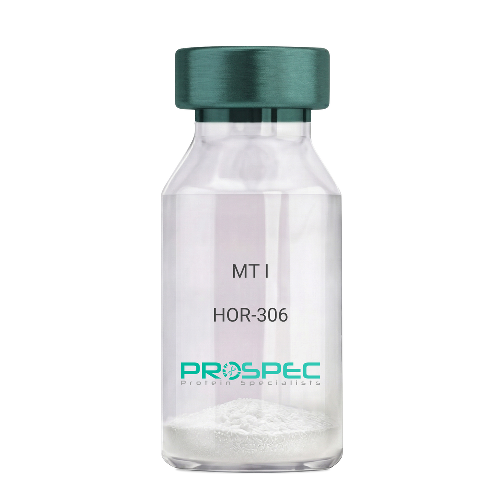
MT IDescription:
Melanotan-I
Melanotan-I, MT-I, Melanotan-1.
Product # :
HOR-306Price :
Quantity :
Shipping Method :
Shipped at Room temp
More Info
- description
- formulation
- purity
- More Info
Description
Melanotan-I has the amino acid sequence of Ser-Tyr-Ser-Nle-Glu-His-D-Phe-Arg-Trp-Gly-Lys-Pro-Val and a molecular weight of 1647.4 Dalton.
Formulation
The protein (1mg/ml) was lyophilized with no additives.
Purity
Greater than 98.0% as determined by RP-HPLC.
More Info
-
Synonyms
Melanotan-I, MT-I, Melanotan-1.
-
Physical Appearance
Sterile Filtered off-White lyophilized (freeze-dried) powder.
-
Stability
Lyophilized Melanotan-I although stable at room temperature for 3 weeks, should be stored desiccated below -18°C. Upon reconstitution MT-I should be stored at 4°C between 2-7 days and for future use below -18°C.For long term storage it is recommended to add a carrier protein (0.1% HSA or BSA).Please prevent freeze-thaw cycles.
-
Solubility
It is recommended to reconstitute the lyophilized Melanotan-I in sterile 1% acetic acid not less than 100 µg/ml, which can then be further diluted to other aqueous solutions.
ProSpec's products are furnished for LABORATORY RESEARCH USE ONLY. The product may not be used as drugs, agricultural or pesticidal products, food additives or household chemicals.
- View Data Sheet
Name :
PSIP1 HumanDescription:
PC4 And SFRS1 Interacting Protein 1 Human Recombinant
PC4 and SFRS1 Interacting Protein 1, Lens Epithelium-Derived Growth Factor, Transcriptional Coactivator P75/P52, Dense Fine Speckles 70 KDa Protein, CLL-Associated Antigen KW-7, DFS 70, PSIP2, DFS70, LEDGF, PC4 And SFRS1 Interacting Protein 2, Transcriptional Coactivator P52/P75, PAIP, P52, P75, PSIP1.
Product # :
PRO-2267Price :
Quantity :
Shipping Method :
Shipped with Ice Packs
More Info
- description
- source
- formulation
- purity
- More Info
Description
Recombinant Human PSIP1 produced in SF9 is a glycosylated, polypeptide chain having a calculated molecular mass of 71 kDa. PSIP1 is expressed with a 10xHis tag at N-terminus and purified by proprietary chromatographic techniques.
Source
Sf9 insect cells.
Formulation
PSIP1 is supplied in 20mM HEPES buffer pH-8.0, 200mM NaCl and 20% glycerol.
Purity
Greater than 84.0% as determined by SDS-PAGE.
More Info
-
Introduction
PC4 and SFRS1 Interacting Protein 1 (PSIP1) is a transcriptional coactivator involved in neuroepithelial stem cell differentiation and neurogenesis. PSIP1 is also involved particularly in lens epithelial cell gene regulation and stress responses. PSIP1 has a significant role in lens epithelial to fiber cell terminal differentiation. In addition, PSIP1 plays a protective role during stress-induced apoptosis. Furthermore, PSIP1 is a cellular cofactor for lentiviral integration.
-
Synonyms
PC4 and SFRS1 Interacting Protein 1, Lens Epithelium-Derived Growth Factor, Transcriptional Coactivator P75/P52, Dense Fine Speckles 70 KDa Protein, CLL-Associated Antigen KW-7, DFS 70, PSIP2, DFS70, LEDGF, PC4 And SFRS1 Interacting Protein 2, Transcriptional Coactivator P52/P75, PAIP, P52, P75, PSIP1.
-
Physical Appearance
Sterile Filtered clear solution.
-
Stability
Store at 4°C if entire vial will be used within 2-4 weeks. Store, frozen at -20°C for longer periods of time. Avoid multiple freeze-thaw cycles.
-
Immunological Functions
1. Binds IgG type human auto-antibodies.2. Immunodot test with positive/negative samples.
ProSpec's products are furnished for LABORATORY RESEARCH USE ONLY. The product may not be used as drugs, agricultural or pesticidal products, food additives or household chemicals.
- View Data Sheet
Name :
SurvodutideDescription:
Survodutide
Product # :
HOR-061Price :
Quantity :
Shipping Method :
Shipped at Room temp
More Info
- description
- formulation
- purity
- More Info
Description
Survodutide is a synthetic single, non-glycosylated polypeptide chain containing 29 amino acids, having a molecular mass of 4231.62 Dalton and a Molecular formula of C192H289N47O61.
Formulation
The protein was lyophilized with no additives.
Purity
Greater than 97.0% as determined by analysis by RP-HPLC.
More Info
-
Physical Appearance
Sterile Filtered White lyophilized (freeze-dried) powder.
-
Stability
Lyophilized Survodutide although stable at room temperature for 3 weeks, should be stored desiccated below -18°C. Upon reconstitution Survodutide should be stored at 4°C between 2-7 days and for future use below -18°C.
For long term storage it is recommended to add a carrier protein (0.1% HSA or BSA).
Please prevent freeze-thaw cycles.
-
Solubility
It is recommended to reconstitute the lyophilized Survodutide in sterile 18MΩ-cm H2O not less than 100 µg/ml, which can then be further diluted to other aqueous solutions.
-
Amino Acid Sequence
H-His-1-Aminocyclobutanecarboxylic acid-Gln-Gly-Thr-Phe-Thr-Ser-Asp-Tyr-Ser-Lys-Tyr-Leu-Asp-Glu-ArgAla-Ala-Lys-Asp-Phe-Ile-Lys-(Gly-Gly-Ser-Gly-Ser-Gly-γ-Gludiacid18)-Trp-Leu-Glu-Ser-Ala-NH2.
-
Background
Survodutide is a dual agonist targeting both the GLP-1 and Glucagon receptors. It has a glucagon component, which effects directly on the liver to increase energy expenditure and clear fat. Survodutide takes part in treatment of liver disease, mainly Metabolic Dysfunction-Associated Steatohepatitis, while simultaneously advancing through Phase 3 trials for obesity.
ProSpec's products are furnished for LABORATORY RESEARCH USE ONLY. The product may not be used as drugs, agricultural or pesticidal products, food additives or household chemicals.
- View Data Sheet
Name :
Hepsin HumanDescription:
Hepsin Human Recombinant
HPN, TMPRSS1, Serine protease hepsin, Transmembrane protease serine 1.
Product # :
PRO-2846Price :
Quantity :
Shipping Method :
Shipped at Room temp
More Info
- description
- source
- formulation
- purity
- biological activity
- More Info
Description
Hepsin Human Recombinant produced in Cho cells is a covalently-linked heterodimer having a total molecular mass of 43.0kDa. Hepsin is fused to a 6 amino acid His-tag at C-terminus & purified by proprietary chromatographic techniques.
Source
CHO cells.
Formulation
The Hepsin protein was Lyophilized from a 0.2µm filtered concentrated solution in 20mM Tris and 150mM NaCl, pH 8.0.
Purity
Greater than 95.0% as determined by SDS-PAGE.
Biological Activity
The specific activity is >20,000 pmol/min/μg and was measured by its ability to cleave tert-butoxycarbonyl-Gln-Arg-Arg-7-amino-4-methylcoumarin (Boc-QRR-AMC).More Info
-
Synonyms
HPN, TMPRSS1, Serine protease hepsin, Transmembrane protease serine 1.
-
Physical Appearance
Filtered White lyophilized (freeze-dried) powder.
-
Stability
Lyophilized Hepsin Active although stable at room temperature for 3 weeks, should be stored desiccated below -18°C. Upon reconstitution Hepsin Active should be stored at 4°C between 2-7 days and for future use below -18°C.
Please prevent freeze-thaw cycles. -
Solubility
It is recommended to reconstitute the lyophilized Hepsin in sterile 18MΩ-cm H2O not less than 100µg/ml, which can then be further diluted to other aqueous solutions.
-
Amino Acid Sequence
Light Chain (Non-catalytic Chain)
RSDQEPLYPV QVSSADARLM VFDKTEGTWR LLCSSRSNAR VAGLSCEEMG FLRALTHSEL DVRTAGANGT SGFFCVDEGR LPHTQRLLE VISVCDCPRGR FLAAICQDCG RRKLPVDR.
Heavy Chain (Catalytic Chain)
IVGGRDTSLG RWPWQVSLRY DGAHLCGGSL LSGDWVLTAA HCFPERNRVL SRWRVFAGAV AQASPHGLQL GVQAVVYHGG YLPFRDPNSE ENSNDIALVH LSSPLPLTEY IQPVCLPAAG QALVDGKICT VTGWGNTQYY GQQAGVLQEA RVPIISNDVC NGADFYGNQI KPKMFCAGYP EGGIDACQGD SGGPFVCEDS ISRTPRWRLC GIVSWGTGCA LAQKPGVYTK VSDFREWIFQ AIKTHSEASG MVTQLHHHHH H.
-
Background
Hepsin is a type II transmembrane serine protease expressed primarily on epithelial cells, it takes part in extracellular proteolysis by activating precursor proteins such as pro-HGF and contributes to tissue remodeling, cell signaling, and normal epithelial function. Recombinant Hepsin is used to study prostate cancer, extracellular matrix remodelling, protease signalling pathways, tumor invasion, metastasis, HGF/MET signaling, and for screening inhibitors that target serine proteases
What is the molecular weight / Mw of Hepsin Protein?
Hepsin Protein has a total Mw of 43kDa.
What is the source or expression system of Hepsin Protein?
CHO Cells
What is the Purity of Hepsin Protein?
Hepsin Protein is >95% pure as determined by SDS-PAGE.
What is the Biological Activity of Hepsin Protein?
The enzymatic activity was measured by its ability to cleave tert-butoxycarbonyl-Gln-Arg-Arg-7-amino-4-methylcoumarin (Boc-QRR-AMC). The specific activity is >20,000 pmol/min/μg.
What is the amino acid sequence of Hepsin Protein?
Light Chain (Non-catalytic Chain)
RSDQEPLYPV QVSSADARLM VFDKTEGTWR LLCSSRSNAR VAGLSCEEMG FLRALTHSEL DVRTAGANGT SGFFCVDEGR LPHTQRLLE VISVCDCPRGR FLAAICQDCG RRKLPVDR
Heavy Chain (Catalytic Chain)
IVGGRDTSLG RWPWQVSLRY DGAHLCGGSL LSGDWVLTAA HCFPERNRVL SRWRVFAGAV AQASPHGLQL GVQAVVYHGG YLPFRDPNSE ENSNDIALVH LSSPLPLTEY IQPVCLPAAG QALVDGKICT VTGWGNTQYY GQQAGVLQEA RVPIISNDVC NGADFYGNQI KPKMFCAGYP EGGIDACQGD SGGPFVCEDS ISRTPRWRLC GIVSWGTGCA LAQKPGVYTK VSDFREWIFQ AIKTHSEASG MVTQLHHHHH H
What applications can Hepsin Protein be used in?
Hepsin Protein can probably be used in western blot, ELISA and Lateral Flow.
What is the endotoxin level for Hepsin Protein?
The endotoxin level is minimal, Hepsin Protein was purified using conventional chromatography techniques.
ProSpec's products are furnished for LABORATORY RESEARCH USE ONLY. The product may not be used as drugs, agricultural or pesticidal products, food additives or household chemicals.
- View Data Sheet
Name :
LASP1 HumanDescription:
LIM and SH3 Protein 1 Human Recombinant
LIM and SH3 protein 1, MLN50, Lasp-1, Metastatic lymph node gene 50 protein.
Product # :
PRO-1031Price :
Quantity :
Shipping Method :
Shipped with Ice Packs
More Info
- description
- source
- formulation
- purity
- More Info
Description
LASP1 Human Recombinant produced in E.coli is a single, non-glycosylated polypeptide chain containing 285 amino acids (1-261) and having a molecular mass of 32.3kDa.LASP1 is fused to a 24 amino acid His-tag at N-terminus & purified by proprietary chromatographic techniques.
Source
E.coli.
Formulation
The LASP1 solution (0.25mg/ml) contains 20mM Tris-HCl buffer (pH 8.0), 1mM DTT and 20% glycerol.
Purity
Greater than 80% as determined by SDS-PAGE.
More Info
-
Introduction
LASP1 has a vital part in the regulation of dynamic actin-based, cytoskeletal activities. Agonist-dependent changes in LASP1 phosphorylation can additionally assist in regulation of actin-associated ion transport activities in the parietal cell and in several other F-actin-rich secretory epithelial cell types.
-
Synonyms
LIM and SH3 protein 1, MLN50, Lasp-1, Metastatic lymph node gene 50 protein.
-
Physical Appearance
Sterile Filtered colorless solution.
-
Stability
Store at 4°C if entire vial will be used within 2-4 weeks. Store, frozen at -20°C for longer periods of time. For long term storage it is recommended to add a carrier protein (0.1% HSA or BSA).Avoid multiple freeze-thaw cycles.
-
Amino Acid Sequence
MGSSHHHHHH SSGLVPRGSH MGSHMNPNCA RCGKIVYPTE KVNCLDKFWH KACFHCETCK MTLNMKNYKG YEKKPYCNAH YPKQSFTMVA DTPENLRLKQ QSELQSQVRY KEEFEKNKGK GFSVVADTPE LQRIKKTQDQ ISNIKYHEEF EKSRMGPSGG EGMEPERRDS QDGSSYRRPL EQQQPHHIPT SAPVYQQPQQ QPVAQSYGGY KEPAAPVSIQ RSAPGGGGKR YRAVYDYSAA DEDEVSFQDG DTIVNVQQID DGWMYGTVER TGDTGMLPAN YVEAI
ProSpec's products are furnished for LABORATORY RESEARCH USE ONLY. The product may not be used as drugs, agricultural or pesticidal products, food additives or household chemicals.
- View Data Sheet
Name :
Prelamin-ADescription:
Prelamin-A Recombinant
Prelamin-A/C, LMNA, LMN1, FPL, IDC, LFP, CDDC, EMD2, FPLD, HGPS, LDP1, LMNC, PRO1, CDCD1, CMD1A, FPLD2, LMNL1, CMT2B1, LGMD1B.
Product # :
PRO-689Price :
Quantity :
Shipping Method :
Shipped with Ice Packs
More Info
- description
- source
- formulation
- purity
- More Info
Description
Recombinant Prelamin-A is a 74kDa precursor of the nuclear lamin A protein. Prelamin-A is a structural component of the nuclear lamina and it is encoded by lamin A/C gene (LMNA). Due to the presence of a CAAX box sequence at carboxyl terminus, Prelamin-A in vivo goes through a serial of post-translational modifications, resulting in the farnesylation of the cysteine thiol, removal of the AAX tripeptide, carboxyl-methylation of the cysteinyl carboxy group and proteolysis of 18 C-terminal amino acids residues that lead to mature lamin A. Diverse mutations in the lamin A/C gene are associated with different deseases that are collectively called laminophaties, including Emery-Dreifuss muscular dystrophy, familial partial lipodystrophy, limb girdle muscular dystrophy, dilated cardiomyopathy, Charcot-Marie-Tooth disease, and Hutchinson-Gilford progeria syndrome. Recombinant human prelamin A is fused to a 6 Histidine tag at the N-terminus.
Source
Escherichia Coli.
Formulation
The Prelamin-A solution (0.1mg/ml) contains 10% Glycerol.
Purity
Greater than 90% as determined by SDS-PAGE.
More Info
-
Synonyms
Prelamin-A/C, LMNA, LMN1, FPL, IDC, LFP, CDDC, EMD2, FPLD, HGPS, LDP1, LMNC, PRO1, CDCD1, CMD1A, FPLD2, LMNL1, CMT2B1, LGMD1B.
-
Physical Appearance
Sterile filtered colorless solution.
-
Stability
Store at 4°C if entire vial will be used within 2-4 weeks. Store, frozen at -20°C for longer periods of time. For long term storage it is recommended to add a carrier protein (0.1% HSA or BSA).Avoid multiple freeze-thaw cycles.
-
Amino Acid Sequence
HHHHHH-METPSQRRATRSGAQASSTPLSPTRITRLQEKEDLQELNDRLAVYIDRVHSLETENAGLRLRITES
EEVVSREVSGIKAAYEAELGDARKTLDSVAKERARLQLELSKVREEFKELKARNTKKEGDLIAAQA
RLKDLEALLNSKEAALSTALSEKRTLEGELHDLRGQVAKLEAALGEAKKQLQDEMLRRVDAENRL
QTMKEELDFQKNIYSEELRETKRRHETRLVEIDNGKQREFESRLADALQELRAQHEDQVEQYKKE
LEKTYSAKLDNARQSAERNSNLVGAAHEELQQSRIRIDSLSAQLSQLQKQLAAKEAKLRDLEDSLA
RERDTSRRLLAEKEREMAEMRARMQQQLDEYQELLDIKLALDMEIHAYRKLLEGEEERLRLSPSP
TSQRSRGRASSHSSQTQGGGSVTKKRKLESTESRSSFSQHARTSGRVAVEEVDEEGKFVRLRN
KSNEDQSMGNWQIKRQNGDDPLLTYRFPPKFTLKAGQVVTIWAAGAGATHSPPTDLVWKAQNT
WGCGNSLRTALINSTGEEVAMRKLVRSVTVVEDDEDEDGDDLLHHHHGSHCSSSGDPAEYNLRS
RTVLCGTCGQPADKASASGSGAQVGGPISSGSSASSVTVTRSYRSVGGSGGGSFGDNLVTRSYL
LGNSSPRTQSPQNCSIM
ProSpec's products are furnished for LABORATORY RESEARCH USE ONLY. The product may not be used as drugs, agricultural or pesticidal products, food additives or household chemicals.
- View Data Sheet
Name :
Streptavidin ProteinDescription:
Streptavidin
Product # :
PRO-283Price :
Quantity :
Shipping Method :
Shipped at Room temp
More Info
- description
- source
- formulation
- More Info
Description
Streptavidin is a protein produced by Streptomyces avidinii and isolated by purification from fermentation broth. The pure, homogeneous protein shows predominantly one single band in SDS PAGE. Streptavidin consists of 4 identical subunits, each bearing an active binding site for biotin. Streptavidin has a molecular weight of 55kDa.
Source
Bacterium Streptomyces avidinii.
Formulation
The Streptavidin was lyophilized from a 25mg/ml solution in 10 mM potassium phosphate buffer pH 6.5
More Info
-
Introduction
Streptavidin is a tetrameric protein secreted by Streptomyces avidinii which binds firmly to biotin. Streptavidin is wilyde used in molecular biology through its unique high affinity for the vitamin biotin. The dissociation constant (Kd) of the biotin-streptavidin complex is about ~10-15 mol/L. The strong affinity recognition of biotin and biotinylated molecules has made streptavidin one of the most important components in diagnostics and laboratory kits. The streptavidin/biotin system has one of the biggest free energies of association of yet observed for noncovalent binding of a protein and small ligand in aqueous solution (K_assoc = 10**14). The complexes are also extremely stable over a wide range of temperature and pH.
-
Physical Appearance
Sterile Filtered lyophilized powder.
-
Stability
Streptavidin although stable at 4°C for 3 weeks, should be stored desiccated below -18°C. For longer storage in dissolved form add 1mM EDTA and/or 0.02 % NaN3 or pass the solution through a sterile filter.Please prevent freeze-thaw cycles.
-
Solubility
Gives a clear solution at 10mg/ml in 4.0 mM potassium phosphate pH 6.5
-
Specific Activity
The biological activity is 16.8 U/mg, 1 unit binds 1µg biotin.
ProSpec's products are furnished for LABORATORY RESEARCH USE ONLY. The product may not be used as drugs, agricultural or pesticidal products, food additives or household chemicals.
- View Data Sheet
Name :
ANXA1 HumanDescription:
Annexin A1 Human Recombinant
ANX1, LPC1, ANXA1, Lipocortin I, Calpactin II, Chrombindin-9, p35, Annexin-1, Phospholipase A2 inhibitory protein, Annexin I, Annexin A1.
Product # :
PRO-679Price :
Quantity :
Shipping Method :
Shipped with Ice Packs
More Info
- description
- source
- formulation
- purity
- More Info
Description
ANXA1 produced in E.Coli is a single, non-glycosylated polypeptide chain containing 346 amino acids (1-346 a.a.) and having a molecular mass of 38.7 kDa.ANXA1 is purified by proprietary chromatographic techniques.
Source
Escherichia Coli.
Formulation
The ANXA1 protein solution contains 20mM Tris-HCl, pH-8, 100mM NaCl, 1mM DTT and 10% glycerol.
Purity
Greater than 90.0% as determined by SDS-PAGE.
More Info
-
Introduction
ANXA1 is part of the family of Ca(2+)-dependent phospholipid binding proteins which have a Mw between 35kDa-40kDa and are situated on the cytosolic face of the plasma membrane. ANXA1 protein has a Mw of 40kDa, with phospholipase A2 inhibitory activity to bind from two to four calcium ions with high affinity. Since phospholipase A2 is necessary for the biosynthesis of the potent mediators of inflammation, prostaglandins and leukotrienes, ANXA1 might have potential anti-inflammatory activity. ANXA1 promotes membrane fusion and iplays a role in exocytosis. The recognition of ANXA1 protein by immunocytochemical leads a simple, highly sensitive and specific assay for diagnosis of hairy cell leukemia.
-
Synonyms
ANX1, LPC1, ANXA1, Lipocortin I, Calpactin II, Chrombindin-9, p35, Annexin-1, Phospholipase A2 inhibitory protein, Annexin I, Annexin A1.
-
Physical Appearance
Sterile filtered colorless solution.
-
Stability
Store at 4°C if entire vial will be used within 2-4 weeks. Store, frozen at -20°C for longer periods of time. For long term storage it is recommended to add a carrier protein (0.1% HSA or BSA).Avoid multiple freeze-thaw cycles.
-
Amino Acid Sequence
MAMVSEFLKQ AWFIENEEQE YVQTVKSSKG GPGSAVSPYP TFNPSSDVAA LHKAIMVKGV DEATIIDILT KRNNAQRQQI KAAYLQETGK
PLDETLKKAL TGHLEEVVLA LLKTPAQFDA DELRAAMKGL GTDEDTLIEI LASRTNKEIR DINRVYREEL KRDLAKDITS DTSGDFRNAL
LSLAKGDRSE DFGVNEDLAD SDARALYEAG ERRKGTDVNV FNTILTTRSY PQLRRVFQKY TKYSKHDMNK VLDLELKGDI EKCLTAIVKCATSKPAFFAE KLHQAMKGVG TRHKALIRIM VSRSEIDMND IKAFYQKMYG ISLCQAILDE TKGDYEKILV ALCGGN.
ProSpec's products are furnished for LABORATORY RESEARCH USE ONLY. The product may not be used as drugs, agricultural or pesticidal products, food additives or household chemicals.
- View Data Sheet
Name :
Adiponectin Mouse, TrimericDescription:
Adiponectin Mouse Recombinant, Trimeric form
Acrp30, AdipoQ, GBP-28, APM-1, ACDC.
Product # :
CYT-247Price :
Quantity :
Shipping Method :
Shipped at Room temp
More Info
- description
- source
- formulation
- purity
- More Info
Description
Trimeric form of Acrp30 Mouse was expressed in HEK293 cells.The cysteine 39 was replaced with alanine (C39A) 9. mAd-C39A can only form trimer, but not hexamer or HMW form.
Source
HEK293 (Human embryonic kidney cell line).
Formulation
Mouse Acrp30 filtered (0.4µm) and lyophilized from 0.5 mg/ml in 0.05M phosphate buffer, 0.05M NaCl, pH 7.2.
Purity
Acrp30 Mouse purity is greater than 95% as determined by SDS-PAGE.
More Info
-
Introduction
Adiponectin is a hormone exclusively expressed from adipose tissue. Many studies demonstrate that adiponectin has direct anti-diabetic, anti-atherogenic and anti-inflammatory functions. APM-1 can increase insulin sensitivity of skeletal muscle, attenuate hepatic lipogenesis and luconeogenesis, regulate NO production in endothelial cells, inhibit proliferation of smooth muscle cells and prevent lipid accumulation of macrophage cells.
In the circulation, adiponectin is present as three different oligomeric complexes, including the high molecular weight (HMW), the middle molecular weight (MMW, also called hexamer) and low molecular weigh (MMW, also called trimer) forms 8. Different oligomeric complex of adiponectin activates different signaling pathways and exerts distinct functions. -
Synonyms
Acrp30, AdipoQ, GBP-28, APM-1, ACDC.
-
Physical Appearance
Filtered white lyophilized (freeze-dried) powder.
-
Stability
Store lyophilized Acrp30 Mouse at -20°C. Aliquot the product after reconstitution to avoid repeated freezing/thawing cycles. Reconstituted Acrp30 Mouse can be stored at 4°C for a limited period of time; it does not show any change after two weeks at 4°C.
-
Solubility
Add deionized water to prepare a working stock solution of approximately 0.5 mg/mL and let the lyophilized pellet dissolve completely. Product is not sterile! Please filter the product by an appropriate sterile filter before using it in the cell culture.
-
Background
What is the molecular weight/Mw of ADIPONECTIN Protein?
ADIPONECTIN Protein has a total Mw of 26kDa.
What is the source or expression system of ADIPONECTIN Protein?
HEK293
What is the Purity of ADIPONECTIN Protein?
ADIPONECTIN Protein is >95% pure as determined by SDS-PAGE.
What is the Biological Activity of ADIPONECTIN Protein?
The biological functionality of ADIPONECTIN Protein will be determined in the future.
What applications can ADIPONECTIN Protein be used in?
ADIPONECTIN Protein can probably be used in western blot, ELISA and Lateral Flow.
What is the endotoxin level for ADIPONECTIN Protein?
The endotoxin level is minimal, ADIPONECTIN Protein was purified using conventional chromatography techniques.
ProSpec's products are furnished for LABORATORY RESEARCH USE ONLY. The product may not be used as drugs, agricultural or pesticidal products, food additives or household chemicals.
- View Data Sheet
Name :
ATXN3 HumanDescription:
Ataxin-3 Human Recombinant
Ataxin-3, Machado-Joseph disease protein 1, Spinocerebellar ataxia type 3 protein, ATXN3, ATX3, MJD, MJD1, SCA3, AT3, JOS.
Product # :
PRO-708Price :
Quantity :
Shipping Method :
Shipped with Ice Packs
More Info
- description
- source
- formulation
- purity
- More Info
Description
ATXN3 produced in E.Coli is a single, non-glycosylated polypeptide chain containing 370 amino acids (1-370 a.a.) and having a molecular mass of 42.4kDa.ATXN3 is purified by proprietary chromatographic techniques.
Source
Escherichia Coli.
Formulation
The ATXN3 protein solution contains 20mM Tris-HCl buffer (pH 7.5), 2mM DTT, 50mM NaCl and 10% glycerol.
Purity
Greater than 90.0% as determined by SDS-PAGE.
More Info
-
Introduction
Ataxin 3 is otherwise known as Machado-Joseph disease protein 1. Machado–Joseph disease is a hereditary autosomal dominant neurodegenerative disorder. ATXN3 contains trinucleotide CAG repeats in the coding region, and the expansion of these repeats from the normal 13-36 to 68-79 causes the Machado-Joseph disease. ATXN3 is a poly-ubiquitin-binding protein whose cellular turnover is regulated by its catalytic activity.
In addition, ATXN3 is a proteasome-associated factor which mediates the degradation of ubiquitinated proteins. ATXN3 folds reversibly using a single intermediate; partial destabilization of ATXN3 by chemical denaturation causes the formation of fibrillar aggregates by the non-pathological variant.
Ataxin-3 interacts with the major histone acetyltransferases cAMP-response-element binding protein (CREB)-binding protein, p300, and p300/CREB-binding protein-associated factor and hinders transcription by these coactivators. -
Synonyms
Ataxin-3, Machado-Joseph disease protein 1, Spinocerebellar ataxia type 3 protein, ATXN3, ATX3, MJD, MJD1, SCA3, AT3, JOS.
-
Physical Appearance
Sterile filtered colorless solution.
-
Stability
Store at 4°C if entire vial will be used within 2-4 weeks.Store, frozen at -20°C for longer periods of time. For long term storage it is recommended to add a carrier protein (0.1% HSA or BSA).Avoid multiple freeze-thaw cycles.
-
Amino Acid Sequence
MESIFHEKQE GSLCAQHCLN NLLQGEYFSP VELSSIAHQL DEEERMRMAE GGVTSEDYRT FLQQPSGNMD DSGFFSIQVI SNALKVWGLELILFNSPEYQ RLRIDPINER SFICNYKEHW FTVRKLGKQW FNLNSLLTGP ELISDTYLAL FLAQLQQEGY SIFVVKGDLP DCEADQLLQM IRVQQMHRPK LIGEELAQLK EQRVHKTDLE RVLEANDGSG MLDEDEEDLQ RALALSRQEI DMEDEEADLR RAIQLSMQGS SRNISQDMTQ TSGTNLTSEE LRKRREAYFE KQQQKQQQQQ QQQQQQQQQQ QQQQGDLSGQ SSHPCERPAT SSGALGSDLG DAMSEEDMLQ AAVTMSLETV RNDLKTEGKK.
ProSpec's products are furnished for LABORATORY RESEARCH USE ONLY. The product may not be used as drugs, agricultural or pesticidal products, food additives or household chemicals.
- View Data Sheet
Name :
HSV-1 gD (84-175)Description:
Herpes Simplex Virus-1 gD (84-175 a.a.) Recombinant
Product # :
HSV-231Price :
Quantity :
Shipping Method :
Shipped with Ice Packs
More Info
- description
- source
- formulation
- purity
- More Info
Description
The E.Coli derived recombinant protein contains the HSV-1 gD (84-175) immunodominant region.
Source
Escherichia Coli.
Formulation
0.1 % SDS, 50% glycerol and 100 mM NaCl.
Purity
Protein is >95% pure as determined by SDS-PAGE
More Info
-
Introduction
Receptors across the cell membrane interact with some viral glycoproteins therefor enabling HSV to enter the host cell. The HSV enter through pores created by the binding of particular receptors across the cell membrane with the virus's coating envelope, following by a fusion of the HSV and the host cell. HSV enters the host cell through the same mechanism and stages as other viruses do. Initially, matching receptors across the virus's envelope and host cell's membrane interacts and bring the two together. During the transitional stage, begins fusion between the host cell and virus (hemifusion state). The closing stage happens when a steady pore was made; through these pores the virus's particles enter the cell.
-
Physical Appearance
Sterile Filtered clear solution.
-
Stability
HSV-1 gD although stable at 4°C for 1 week, should be stored below -18°C. Please prevent freeze thaw cycles.
ProSpec's products are furnished for LABORATORY RESEARCH USE ONLY. The product may not be used as drugs, agricultural or pesticidal products, food additives or household chemicals.
- View Data Sheet
Name :
SQSTM1 HumanDescription:
Sequestosome 1 Human Recombinant
A170, OSIL, p60, p62, p62B, PDB3, ZIP3, EBIAP, ORCA, OSIL, SQSTM1.
Product # :
PRO-806Price :
Quantity :
Shipping Method :
Shipped with Ice Packs
More Info
- description
- source
- formulation
- purity
- More Info
Description
SQSTM1 Human Recombinant produced in E.Coli is a single, non-glycosylated, polypeptide chain containing 364 amino acids (1-356 a.a.) and having a molecular mass of 39.7 kDa. SQSTM1 protein is fused to an 8 amino acid His-Tag at C-terminus and purified by standard chromatography.
Source
Escherichia Coli.
Formulation
SQSTM1 Human 0.5mg/ml solution containing 20mM Tris-HCl pH-8, 1mM DTT and 10% glycerol.
Purity
Greater than 85% as determined by SDS-PAGE.
More Info
-
Introduction
SQSTM1 is an adapter protein which binds ubiquitin and mediates the activation of NFKB1 by TNF-a, NGF and IL-1. SQSTM1 is involved in titin/TTN downstream signaling in muscle cells. SQSTM1 controls signaling cascades through ubiquitination. SQSTM1 participates in cell differentiation, apoptosis, immune response and regulation of K(+) channels. Mutations in UBA domain of the SQSTM1 protein cause Paget’s disease because the UBA is essential for aggregate sequestration and cell survival.
-
Synonyms
A170, OSIL, p60, p62, p62B, PDB3, ZIP3, EBIAP, ORCA, OSIL, SQSTM1.
-
Physical Appearance
Sterile filtered colorless solution.
-
Stability
Store at 4°C if entire vial will be used within 2-4 weeks. Store, frozen at -20°C for longer periods of time. For long term storage it is recommended to add a carrier protein (0.1% HSA or BSA).Avoid multiple freeze-thaw cycles.
-
Amino Acid Sequence
MAMSYVKDDI FRIYIKEKKE CRRDHRPPCA QEAPRNMVHP NVICDGCNGP VVGTRYKCSV CPDYDLCSVC EGKGLHRGHT KLAFPSPFGH LSEGFSHSRW LRKVKHGHFG WPGWEMGPPG NWSPRPPRAG EARPGPTAES ASGPSEDPSV NFLKNVGESV AAALSPLGIE VDIDVEHGGK RSRLTPVSPE SSSTEEKSSS QPSSCCSDPS KPGGNVEGAT QSLAEQMRKI ALESEGRPEE QMESDNCSGG DDDWTHLSSK EVDPSTGELQ SLQMPESEGP SSLDPSQEGP TGLKEAALYP HLPPEADPRL IESLSQMLSM GFSDEGGWLT RLLQTKNYDI GAALDTIQYS KHPPPLLEHH HHHH.
ProSpec's products are furnished for LABORATORY RESEARCH USE ONLY. The product may not be used as drugs, agricultural or pesticidal products, food additives or household chemicals.
- View Data Sheet
Name :
Bartonella SucBDescription:
Bartonella Henselae SucB Recombinant
Product # :
PRO-2570Price :
Quantity :
Shipping Method :
Shipped with Ice Packs
More Info
- description
- source
- formulation
- purity
- More Info
Description
Recombinant Bartonella Henselae SucB produced in E.Coli is a single, non-glycosylated polypeptide chain having a molecular mass of 48kDa. Bartonella SucB is expressed with a -10x His tag at N-terminus and purified by proprietary chromatographic techniques.
Source
Escherichia Coli.
Formulation
Bartonella SucB is supplied at a 20mM HEPES buffer pH-8.0, 200mM NaCl and 20% Glycerol.
Purity
Greater than 95% as determined by SDS-PAGE.
More Info
-
Introduction
Numerous forms of Bartonellosis are caused by Bartonella henselae as well as Cat Scratch Disease & Bacillary Angiomatosis. It has been found that up to 95% of patients with Cat Scratch Disease show antibodies against Bartonella henselae antigens. Highly immunoreactive proteins which are produced by the Bartonella henselae are the most important antigens used for the diagnosis of Cat Scratch Disease. The immunogenic protein dihydrolipoamide-succinyltransferase also named SucB is an enzyme of the alpha-ketoglutarate dehydrogenase complex, which catalyzes the transport of a succinyl group from the S-succinyldihydrolipoyl moiety to coenzyme A.
-
Physical Appearance
Sterile Filtered solution.
-
Stability
Store at 4°C if entire vial will be used within 2-4 weeks. Store, frozen at -20°C for longer periods of time. Avoid multiple freeze-thaw cycles.
ProSpec's products are furnished for LABORATORY RESEARCH USE ONLY. The product may not be used as drugs, agricultural or pesticidal products, food additives or household chemicals.
- View Data Sheet
Description:
Mouse Anti SARS Nucleocapsid Biotinylated
Product # :
ANT-764Price :
Quantity :
Shipping Method :
Shipped with Ice Packs
More Info
- description
- formulation
- purity
- More Info
Description
SARS Nucleoprotein Biotinylated antibody clone PT3851 is specific for the nucleocapsid SARS-CoV protein and recognizes the nucleoprotein of SARS-CoV-2 by ELISA.
Formulation
1mg/ml in DPBS, 1% BSA and 0.1% Proclin 950.
Purity
Greater than 90% as determined by SDS-PAGE gel.
More Info
-
Introduction
SARS Coronavirus is an enveloped virus containing three outer structural proteins, namely the membrane (M), envelope (E), and spike (S) proteins. Spike (S)-glycoprotein of the virus interacts with a cellular receptor and mediates membrane fusion to allow viral entry into susceptible target cells. Accordingly, S-protein plays an important role in virus infection cycle and is the primary target of neutralizing antibodies.It has recently been shown that SARS is caused by a human coronavirus. Human coronaviruses are the major cause of upper respiratory tract illness in humans, such as the common cold. Coronaviruses are positive-stranded RNA viruses, featuring the largest viral RNA genomes known to date (27-31 kb). The first step in coronavirus infection is binding of the viral spike protein, a 139-kDa protein, to certain receptors on host cells. The spike protein is the main surface antigen of the coronavirus. The most prominent protein in the culture supernatants infected with SARS virus is a 46 kDa nucleocapsid protein. This suggests that the nucleocapsid protein is a major immunogen that may be useful for early diagnostics.
-
Physical Appearance
Sterile Filtered liquid formulation.
-
Stability
Store at 4°C, stable for 2 weeks. For long-term storage, store at -20°C.
-
Immunogen
Recombinant protein fragment 1-49 a.a. of the SARS nucleoprotein.
-
Clone
PT3851
-
Applications
ELISA.
-
Isotype
Mouse IgG2b.
-
Type
Mouse antibody Monoclonal.
-
Purification Method
Protein A affinity purified.
ProSpec's products are furnished for LABORATORY RESEARCH USE ONLY. The product may not be used as drugs, agricultural or pesticidal products, food additives or household chemicals.
- View Data Sheet
Name :
SPP1 Human, HEKDescription:
Osteopontin Human Recombinant, HEK
Secreted Phosphoprotein-1, OPN, BNSP, BSPI, ETA-1, MGC110940, SPP-1, Osteopontin, Bone sialoprotein 1, Urinary stone protein, Nephropontin, Uropontin, SPP1.
Product # :
CYT-047Price :
Quantity :
Shipping Method :
Shipped at Room temp
More Info
- description
- source
- formulation
- purity
- More Info
Description
Osteopontin Human Recombinant is a single, glycosylated, polypeptide chain produced in HEK293 cells, is a full length protein (amino acids 17-314) fused with a polyhistidine tag at the C-terminus, having a total calculated molecular mass of 34.5kDa (The actual molecular mass may be approximately 60-65kDa in SDS-PAGE under reducing conditions due to glycosylation).Osteopontin is purified by proprietary chromatographic techniques.
Source
HEK293 cells.
Formulation
Osteopontin was lyophilized from a 0.2µM filtered solution of 20mM PBS and 150mM NaCl, pH 7.2.
Purity
Greater than 95.0% as determined by SDS-PAGE.
More Info
-
Introduction
Osteopontin is a glycoprotein that was first identified in osteoblasts and is involved in bone remodeling, immune functions in fibroblasts, macrophages, and lymphocytes during inflammation and wound healing. SPP1 binds tightly to hydroxyapatite. SPP1 forms an integral part of the mineralized matrix. SPP1 is vital to cell-matrix interaction.
Secreted Phosphoprotein-1 protects against cardiac ischemia-reperfusion injury via late preconditioning. Expression of both Ostepontin and CD44 in hepatocellular carcinoma is linked with advanced tumor stage and contributes to prognosis information. SPP1 is the most over-expressed gene in intrahepatic cholangiocarcinoma. Secreted Phosphoprotein-1 overexpression is related with interstitial lung diseases. -
Synonyms
Secreted Phosphoprotein-1, OPN, BNSP, BSPI, ETA-1, MGC110940, SPP-1, Osteopontin, Bone sialoprotein 1, Urinary stone protein, Nephropontin, Uropontin, SPP1.
-
Physical Appearance
Sterile Filtered White lyophilized (freeze-dried) powder.
-
Stability
Store at 4°C if entire vial will be used within 2-4 weeks. Store, frozen at -20°C for longer periods of time.Please avoid freeze thaw cycles.
-
Solubility
It is recommended to reconstitute the lyophilized SPP1 in 1xPBS to a concentration no less than 100 µg/ml, which can then be further diluted to other aqueous solutions.
-
Amino Acid Sequence
IPVKQADSGSSEEKQLYNKYPDAVATWLNPDPSQKQNLLAPQNAVSSEETNDFKQETL
PSKSNESHDHMDDMDDEDDDDHVDSQDSIDSNDSDDVDDTDDSHQSDESHHSDESDEL
VTDFPTDLPATEVFTPVVPTVDTYDGRGDSVVYGLRSKSKKFRRPDIQYPDATDEDIT
SHMESEELNGAYKAIPVAQDLNAPSDWDSRGKDSYETSQLDDQSAETHSHKQSRLYKRK
ANDESNEHSDVIDSQELSKVSREFHSHEFHSHEDMLVVDPKSKEEDKHLKFRISHELDS
ASSEVNVDHHHHHH.
ProSpec's products are furnished for LABORATORY RESEARCH USE ONLY. The product may not be used as drugs, agricultural or pesticidal products, food additives or household chemicals.
- View Data Sheet
Name :
p38a/SAPK2 HumanDescription:
p38a/SAPK2 Human Recombinant
Mitogen-activated protein kinase 14, EC 2.7.11.24, Mitogen-activated protein kinase p38 alpha, MAP kinase p38 alpha, Cytokine suppressive anti-inflammatory drug-binding protein, CSAID-binding protein, CSBP, MAX-interacting protein 2, MAP kinase MXI2, SAPK2A, RK, p38, EXIP, Mxi2, CSBP1, CSBP2, CSPB1, PRKM14, PRKM15, p38ALPHA.
Product # :
PKA-217Price :
Quantity :
Shipping Method :
Shipped with Ice Packs
More Info
- description
- source
- formulation
- purity
- More Info
Description
p38/SAPK2 is a non-glycosilated polypeptide produced by phosphorylation of the purified p38 alpha with MKK6 having a molecular mass of 42.7 kDa.
Source
Escherichia Coli.
Formulation
p38/SAPK2 is supplied in 25mM Tris-HCl, 150mM NaCl, 1mM DTT, 50% glycerol, pH 8.5.
Purity
Greater than 95% as determined by SDS-PAGE.
More Info
-
Introduction
p38a/SAPK2 is a member of the MAP kinase family. MAP kinases act as an integration point for multiple biochemical signals, and are involved in a wide variety of cellular processes such as proliferation, differentiation, transcription regulation and development. This kinase is activated by various environmental stresses and proinflammatory cytokines. The activation requires its phosphorylation by MAP kinase kinases (MKKs), or its autophosphorylation triggered by the interaction of MAP3K7IP1/TAB1 protein with this kinase. The substrates of this kinase include transcription regulator ATF2, MEF2C, and MAX, cell cycle regulator CDC25B, and tumor suppressor p53, which suggest the roles of this kinase in stress related transcription and cell cycle regulation, as well as in genotoxic stress response. Four alternatively spliced transcript variants of this gene encoding distinct isoforms have been reported.
-
Synonyms
Mitogen-activated protein kinase 14, EC 2.7.11.24, Mitogen-activated protein kinase p38 alpha, MAP kinase p38 alpha, Cytokine suppressive anti-inflammatory drug-binding protein, CSAID-binding protein, CSBP, MAX-interacting protein 2, MAP kinase MXI2, SAPK2A, RK, p38, EXIP, Mxi2, CSBP1, CSBP2, CSPB1, PRKM14, PRKM15, p38ALPHA.
-
Physical Appearance
Sterile Filtered clear solution.
-
Stability
Store at 4°C if entire vial will be used within 2-4 weeks. Store, frozen at -20°C for longer periods of time. Avoid multiple freeze-thaw cycles.
-
Unit Definition
No protease activity detectable, specific activity is 2.357.900 U*/mg (*1 U = 1 pmol/min transferred to myelin basic protein at 30°C).
ProSpec's products are furnished for LABORATORY RESEARCH USE ONLY. The product may not be used as drugs, agricultural or pesticidal products, food additives or household chemicals.
- View Data Sheet
Name :
TriptorelinDescription:
Triptorelin Acetate
Product # :
HOR-238Price :
Quantity :
Shipping Method :
Shipped at Room temp
More Info
- description
- formulation
- purity
- More Info
Description
Triptorelin C64H82N18O13, Pyr-His-Trp-Ser-Tyr-D-Trp-Leu-Arg-Pro-Gly-NH2 is a synthetic analogue of gonadorelin (GnRH). As a result of the substitution of the 6th amino acid residue in the native molecule, the agonistic effect is more pronounced and the plasma half-life prolonged.
Formulation
The protein (1 mg/ml) was lyophilized with no additives.
Purity
Greater than 98.0% as determined by:
(a) Analysis by RP-HPLC.
(b) Analysis by SDS-PAGE.More Info
-
Physical Appearance
Sterile Filtered White lyophilized (freeze-dried) powder.
-
Stability
Lyophilized Trp although stable at room temperature for 3 weeks, should be stored desiccated below -18°C. Upon reconstitution Trp should be stored at 4°C between 2-7 days and for future use below -18°C.For long term storage it is recommended to add a carrier protein (0.1% HSA or BSA).Please prevent freeze-thaw cycles.
-
Solubility
It is recommended to reconstitute the lyophilized Trp in sterile 18MΩ-cm H2O not less than 100 µg/ml, which can then be further diluted to other aqueous solutions.
-
Background
What is the Purity of TRIPTORELIN Protein?
TRIPTORELIN Protein is >98% pure as determined by SDS-PAGE.
What is the Biological Activity of TRIPTORELIN Protein?
The biological functionality of TRIPTORELIN Protein will be determined in the future.
What applications can TRIPTORELIN Protein be used in?
TRIPTORELIN Protein can probably be used in western blot, ELISA and Lateral Flow.
What is the endotoxin level for TRIPTORELIN Protein?
The endotoxin level is minimal, TRIPTORELIN Protein was purified using conventional chromatography techniques.
ProSpec's products are furnished for LABORATORY RESEARCH USE ONLY. The product may not be used as drugs, agricultural or pesticidal products, food additives or household chemicals.
- View Data Sheet
Name :
SARS MERS RBD, ActiveDescription:
SARS MERS Spike Receptor Binding Domain Recombinant, Active
Middle East respiratory syndrome coronavirus, Human betacoronavirus 2c EMC/2012, MERS-CoV, MERS, MERSCoV RBD, MERS RBD, receptor binding domain, RBD, Spike RBD protein, Spike glycoprotein, S glycoprotein, E2, Peplomer protein
Product # :
SARS-060Price :
Quantity :
Shipping Method :
Shipped with Ice Packs
More Info
- description
- source
- formulation
- purity
- biological activity
- More Info
Description
SARS MERS RBD Recombinant produced in Sf9 Baculovirus cells is a single, glycosylated polypeptide chain containing 258 amino acids (358-606 aa) and having a molecular mass of 28.2kDa. SARS MERS RBD is fused to a 6 amino acid His-tag at C-terminus & purified by proprietary chromatographic techniques.
Source
Sf9, Baculovirus cells.
Formulation
The SARS MERS RBD solution (0.5mg/ml) contains 10% glycerol and Phosphate-Buffered Saline (pH 7.4).
Purity
Greater than 90.0% as determined by SDS-PAGE.
Biological Activity
Measured by its binding ability in a functional ELISA with Human DPPIV/CD26 (CAT# enz-1187).
More Info
-
Synonyms
Middle East respiratory syndrome coronavirus, Human betacoronavirus 2c EMC/2012, MERS-CoV, MERS, MERSCoV RBD, MERS RBD, receptor binding domain, RBD, Spike RBD protein, Spike glycoprotein, S glycoprotein, E2, Peplomer protein
-
Physical Appearance
Sterile filtered colorless solution.
-
Stability
Store at 4°C if entire vial will be used within 2-4 weeks.Store, frozen at -20°C for longer periods of time.For long term storage it is recommended to add a carrier protein (0.1% HSA or BSA).Avoid multiple freeze-thaw cycles.
-
Amino Acid Sequence
ADPSGVYSVS SFEAKPSGSV VEQAEGVECD FSPLLSGTPP QVYNFKRLVF TNCNYNLTKL LSLFSVNDFT CSQISPAAIA SNCYSSLILD YFSYPLSMKS DLSVSSAGPI SQFNYKQSFS NPTCLILATV PHNLTTITKP LKYSYINKCS RLLSDDRTEV PQLVNANQYS PCVSIVPSTV WEDGDYYRKQ LSPLEGGGWL VASGSTVAMT EQLQMGFGIT VQYGTDTNSV CPKLEFANDT KIASQLGNCV EYHHHHHH.
-
Background
The severe acute respiratory syndrome coronavirus (SARS-CoV) and Middle East respiratory syndrome coronavirus (MERS-CoV) have posed significant global health threats in recent years. Central to their pathogenesis is the interaction between the viral spike proteins and host cell receptors. This research aims to investigate the receptor binding domain (RBD) of SARS and MERS spike proteins and its implications for viral entry and the development of therapeutic interventions. Understanding the molecular mechanisms underlying viral-host interactions can pave the way for targeted therapeutic strategies against these deadly coronaviruses.
Structure and Function of SARS and MERS Spike RBD:
The spike proteins of SARS-CoV and MERS-CoV are critical for viral entry into host cells. These proteins consist of two subunits: S1, responsible for receptor binding, and S2, involved in membrane fusion. The receptor binding domain (RBD) within the S1 subunit specifically interacts with host cell receptors, enabling viral attachment and entry. The RBDs of SARS and MERS spike proteins exhibit unique structural features and binding affinities for their respective receptors.
Interaction with ACE2 and DPP4 Receptors:
The SARS-CoV spike protein RBD interacts with the angiotensin-converting enzyme 2 (ACE2) receptor, which is abundantly expressed in the respiratory tract. The binding of SARS-CoV RBD to ACE2 facilitates viral entry into host cells. On the other hand, the MERS-CoV spike protein RBD interacts with the dipeptidyl peptidase 4 (DPP4) receptor, predominantly expressed in the lungs and other tissues. The ACE2 and DPP4 receptors play crucial roles in determining the host range and tissue tropism of SARS and MERS coronaviruses.
Implications for Viral Pathogenesis:
The binding of SARS and MERS spike RBDs to their respective receptors triggers conformational changes in the spike protein, leading to membrane fusion and subsequent viral entry. This process is crucial for viral replication and the spread of infection within the host. The specificity and affinity of the RBD-receptor interaction influence viral tropism, tissue damage, and disease severity. Understanding the determinants of RBD-receptor binding can provide insights into viral pathogenesis and potential therapeutic targets.
Development of Therapeutic Interventions:
The RBD of SARS and MERS spike proteins represents a promising target for the development of antiviral therapeutics. Several strategies have been explored, including monoclonal antibodies and small molecule inhibitors, to disrupt the RBD-receptor interaction and inhibit viral entry. These approaches aim to block the binding interface between the spike RBD and the host receptor, thereby preventing viral attachment and entry. Additionally, vaccine development efforts have focused on generating neutralizing antibodies against the RBD to elicit protective immune responses.
Conclusion:
The investigation of the receptor binding domain (RBD) of SARS and MERS spike proteins sheds light on the molecular mechanisms underlying viral entry and pathogenesis. The specific interactions between the spike RBD and host cell receptors play a crucial role in determining viral tropism and tissue damage. Targeting the RBD-receptor interaction holds promise for the development of effective therapeutics against SARS-CoV, MERS-CoV, and potentially other related coronaviruses. Further research and development efforts are needed to exploit the potential of the spike RBD as a therapeutic target and to combat future coronavirus outbreaks.
ProSpec's products are furnished for LABORATORY RESEARCH USE ONLY. The product may not be used as drugs, agricultural or pesticidal products, food additives or household chemicals.
- View Data Sheet
Name :
IFNG MouseDescription:
IFN-Gamma Mouse Recombinant
Immune IFN, type II IFN, T cell IFN, MAF, IFNG, IFG, IFI, IFN-gamma.
Product # :
CYT-358Price :
Quantity :
Shipping Method :
Shipped at Room temp
More Info
- description
- source
- formulation
- purity
- biological activity
- More Info
Description
IFN-gamma Mouse Recombinant produced in E.Coli is a single, non-glycosylated, polypeptide chain containing 134 amino acids and having a molecular mass of 15.6kDa.The IFN-gamma is purified by proprietary chromatographic techniques.
Source
Escherichia Coli.
Formulation
Lyophilized from a 0.2µm filtered concentrated (1mg/ml) solution in PBS, pH 7.4 and 5% trehalose.
Purity
Greater than 95.0% as determined by:
(a) Analysis by RP-HPLC.
(b) Analysis by SDS-PAGE.Biological Activity
The specific activity as determined in a viral resistance assay is < 0.1 ng/ml, corresponding to a specific activity of 10,000,000 IU/mg
More Info
-
Introduction
IFN-gamma produced by lymphocytes activated by specific antigens or mitogens.
IFN-gamma, in addition to having antiviral activity, has important immunoregulatory functions, it is a potent activator of macrophages, and has antiproliferative effects on transformed cells and it can potentiate the antiviral and antitumor effects of the type I IFNs. -
Synonyms
Immune IFN, type II IFN, T cell IFN, MAF, IFNG, IFG, IFI, IFN-gamma.
-
Physical Appearance
Sterile Filtered White lyophilized (freeze-dried) powder.
-
Stability
Lyophilized IFN-gamma although stable at room temperature for 3 weeks, should be stored desiccated below -18°C. Upon reconstitution IFN-gamma should be stored at 4°C between 2-7 days and for future use below -18°C.For long term storage it is recommended to add a carrier protein (0.1% HSA or BSA).Please prevent freeze-thaw cycles.
-
Solubility
It is recommended to reconstitute the lyophilized IFN-gamma in sterile distilled water or 20mM AcOH at concentrations ranging between 0.1mg-0.5mg/ml, which can then be further diluted to other aqueous solutions.
-
Amino Acid Sequence
MHGTVIESLE SLNNYFNSSG IDVEEKSLFL DIWRNWQKDG DMKILQSQII SFYLRLFEVL KDNQAISNNI SVIESHLITT FFSNSKAKKD AFMSIAKFEV NNPQVQRQAF NELIRVVHQL LPESSLRKRK RSRC.
-
Background
What is the molecular weight/Mw of IFNG MOUSE Protein?
IFNG MOUSE Protein has a total Mw of 15.6kDa.
What is the source or expression system of IFNG MOUSE Protein?
Escherichia Coli.
What is the Purity of IFNG MOUSE Protein?
IFNG MOUSE Protein is >95% pure as determined by SDS-PAGE.
What is the Biological Activity of IFNG MOUSE Protein?
The specific activity as determined in a viral resistance assay is < 0.1 ng/ml, corresponding to a specific activity of 10,000,000 IU/mg
What is the amino acid sequence of IFNG MOUSE Protein?
MHGTVIESLE SLNNYFNSSG IDVEEKSLFL DIWRNWQKDG DMKILQSQII SFYLRLFEVL KDNQAISNNI SVIESHLITT FFSNSKAKKD AFMSIAKFEV NNPQVQRQAF NELIRVVHQL LPESSLRKRK RSRC.
What applications can IFNG MOUSE Protein be used in?
IFNG MOUSE Protein can probably be used in western blot, ELISA and Lateral Flow.
What is the endotoxin level for IFNG MOUSE Protein?
The endotoxin level is minimal, IFNG MOUSE Protein was purified using conventional chromatography techniques.
ProSpec's products are furnished for LABORATORY RESEARCH USE ONLY. The product may not be used as drugs, agricultural or pesticidal products, food additives or household chemicals.
- View Data Sheet
Name :
SARS Mosaic S(N)Description:
SARS-Associated Coronavirus Spike Mosaic S (N) Recombinant
Product # :
SARS-258Price :
Quantity :
Shipping Method :
Shipped with Ice Packs
More Info
- description
- formulation
- purity
- More Info
Description
The E.Coli derived 38 kDa mosaic protein contains the N-terminal section of the Spike protein 12-53, 90-115, 171-203 amino acids immunodominant regions.
Formulation
25mM Tris-HCl, 0.4% sarcosyl, 0.25% Triton –100 and 50% glycerol.
Purity
SARS Mosaic Protein is >95% pure as determined by 10% PAGE (coomassie staining).
More Info
-
Introduction
SARS Coronavirus is an enveloped virus containing three outer structural proteins, namely the membrane (M), envelope (E), and spike (S) proteins. Spike (S)-glycoprotein of the virus interacts with a cellular receptor and mediates membrane fusion to allow viral entry into susceptible target cells. Accordingly, S-protein plays an important role in virus infection cycle and is the primary target of neutralizing antibodies.
-
Stability
SARS Mosaic protein is shipped at ambient temperature. Upon arrival, store at -20C.
-
Applications
SARS Mosaic antigen is suitable for ELISA and Western blots, excellent antigen for detection of SARS with minimal specificity problems.
-
Specificity
SARS Mosaic protein is Immunoreactive with sera of SARS-infected individuals.
-
Purification Method
SARS Mosaic protein was purified by proprietary chromatographic technique.
ProSpec's products are furnished for LABORATORY RESEARCH USE ONLY. The product may not be used as drugs, agricultural or pesticidal products, food additives or household chemicals.